Next Issue
Volume 11, April
Previous Issue
Volume 11, February


J. Cardiovasc. Dev. Dis., Volume 11, Issue 3 (March 2024) – 26 articles

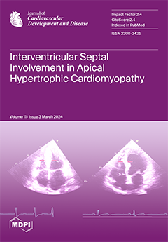
Cover Story (view full-size image): We hypothesized that interventricular septal involvement in apical hypertrophic cardiomyopathy (ApHCM-Mixed) adversely impacts ventricular structure and function when compared with isolated apical hypertrophy (ApHCM-Pure). ApHCM-Mixed had larger left ventricular (LV) mass index, worse LVt and right ventricular (RV) global longitudinal strain, impaired RV free wall strain, and lower LV myocardial work indices. At the 3.9-year follow-up, five deaths were observed, all occurring in the ApHCM-Mixed group and with four being cardiac-related. This subgroup had preserved LV ejection fraction with significant subclinical bi-ventricular dysfunction. ApHCM-Mixed represents a distinct morphology in hypertrophic cardiomyopathy with more impaired ventricular function and mechanics when compared with ApHCM-Pure. View this paper
  • Issues are regarded as officially published after their release is announced to the table of contents alert mailing list.
  • You may sign up for e-mail alerts to receive table of contents of newly released issues.
  • PDF is the official format for papers published in both, html and pdf forms. To view the papers in pdf format, click on the "PDF Full-text" link, and use the free Adobe Reader to open them.
Order results
Result details
Section
Select all
Export citation of selected articles as:
22 pages, 5446 KiB  
Review
Cardiac Allograft Vasculopathy: Challenges and Advances in Invasive and Non-Invasive Diagnostic Modalities
by Moaz A. Kamel, Isabel G. Scalia, Amro T. Badr, Nima Baba Ali, Juan M. Farina, Milagros Pereyra, Mohammed Tiseer Abbas, Ahmed K. Mahmoud, Robert L. Scott, David E. Steidley, Julie L. Rosenthal, Lisa M. Lemond, Kristen A. Sell-Dottin, Brian W. Hardaway, Timothy Barry, Ming Yang, Chieh-Ju Chao, Clinton E. Jokerst, Chadi Ayoub and Reza Arsanjani
J. Cardiovasc. Dev. Dis. 2024, 11(3), 95; https://doi.org/10.3390/jcdd11030095 - 21 Mar 2024
Viewed by 791
Abstract
Cardiac allograft vasculopathy (CAV) is a distinct form of coronary artery disease that represents a major cause of death beyond the first year after heart transplantation. The pathophysiology of CAV is still not completely elucidated; it involves progressive circumferential wall thickening of both [...] Read more.
Cardiac allograft vasculopathy (CAV) is a distinct form of coronary artery disease that represents a major cause of death beyond the first year after heart transplantation. The pathophysiology of CAV is still not completely elucidated; it involves progressive circumferential wall thickening of both the epicardial and intramyocardial coronary arteries. Coronary angiography is still considered the gold-standard test for the diagnosis of CAV, and intravascular ultrasound (IVUS) can detect early intimal thickening with improved sensitivity. However, these tests are invasive and are unable to visualize and evaluate coronary microcirculation. Increasing evidence for non-invasive surveillance techniques assessing both epicardial and microvascular components of CAV may help improve early detection. These include computed tomography coronary angiography (CTCA), single-photon emission computed tomography (SPECT), positron emission tomography (PET), and vasodilator stress myocardial contrast echocardiography perfusion imaging. This review summarizes the current state of diagnostic modalities and their utility and prognostic value for CAV and also evaluates emerging tools that may improve the early detection of this complex disease. Full article
Show Figures

Figure 1

13 pages, 751 KiB  
Article
Mortality Time-Trends of Different Cardiovascular Diseases in a Practically Extinct Cohort of Italian Middle-Aged Men Followed-Up for 61 Years: A Possible Etiological Explanation?
by Paolo Emilio Puddu, Paolo Piras and Alessandro Menotti
J. Cardiovasc. Dev. Dis. 2024, 11(3), 94; https://doi.org/10.3390/jcdd11030094 - 21 Mar 2024
Viewed by 608
Abstract
Purpose. To study a male Italian cohort (initially aged 40–59, n = 1712) during 61 years and the natural history of major CVD mortality categories including coronary heart disease (CHD), stroke and other heart diseases of uncertain etiology (HDUE), including congestive heart failure) [...] Read more.
Purpose. To study a male Italian cohort (initially aged 40–59, n = 1712) during 61 years and the natural history of major CVD mortality categories including coronary heart disease (CHD), stroke and other heart diseases of uncertain etiology (HDUE), including congestive heart failure) along with their risk factor relationships. Methods and Results. Cox models were run with 12 covariates as possible predictors measured at entry to the study. About 93% of all CVD deaths were covered by the three major groups selected here (N = 751): 37.4% of them were diagnosed as CHD, 30.6% as stroke and 28.5% as HDUE. CHD declined in the last 20 years of follow-up, while a sharp increase in HDUE mortality was seen. Baseline mean levels of serum cholesterol were 209.6, 204.2 and 198.0 mg/dL, respectively, for CHD, stroke and HDUE deaths: the multivariable coefficients of serum cholesterol were positive and significant for CHD (p < 0.0001), and stroke (p = 0.0203) and not significant for HDUE (p = 0.3467). In Fine–Gray models, the algebraic signs of cholesterol coefficients were opposite for CHD versus the other mortality categories (t = 3.13). The predictive performances of remaining risk factors were varied whereas that of Cox models was not very good, probably due to the attrition phenomenon and possible competing risks. Conclusion. Large differences in natural history and risk factors were found comparing the three CVD conditions, potentially indicating different etiologies and pointing to the need of not mixing them up in a grouped CVD category. Full article
(This article belongs to the Section Epidemiology, Lifestyle, and Cardiovascular Health)
Show Figures

Figure 1

13 pages, 597 KiB  
Article
Short-Term Mortality in Hospitalized Patients with Congestive Heart Failure: Markers of Thrombo-Inflammation Are Independent Risk Factors and Only Weakly Associated with Renal Insufficiency and Co-Morbidity Burden
by Jose Iglesias, Nelson Okoh, Song Peng Ang, Cristina A. Rodriguez, Jia Ee Chia and Jerrold S. Levine
J. Cardiovasc. Dev. Dis. 2024, 11(3), 93; https://doi.org/10.3390/jcdd11030093 - 20 Mar 2024
Viewed by 813
Abstract
Congestive heart failure (CHF) is associated with significant morbidity and mortality. There has been renewed interest in using thrombo-inflammatory markers as prognostic tools in patients with CHF. To determine if thrombo-inflammatory markers are independent risk factors for 28-day mortality in hospitalized CHF patients, [...] Read more.
Congestive heart failure (CHF) is associated with significant morbidity and mortality. There has been renewed interest in using thrombo-inflammatory markers as prognostic tools in patients with CHF. To determine if thrombo-inflammatory markers are independent risk factors for 28-day mortality in hospitalized CHF patients, we retrospectively analyzed admission data extracted from 2008 consecutive patients admitted with a diagnosis of CHF to Zigong Fourth People’s Hospital. Multivariate Cox proportional hazards analysis demonstrated that the thrombo-inflammatory markers thrombin time, platelet/lymphocyte ratio (PLR), and D-dimer level were independent predictors of mortality. In addition, variables reflecting the severity of CHF (New York Heart Association class > 2), impaired renal function (elevated serum creatinine [SCr]), impaired organ perfusion (elevated BUN), and chronic liver disease were also independent predictors of mortality. Thrombo-inflammatory biomarkers were only weakly associated with SCr and the burden of co-morbidity, suggesting that thrombo-inflammation may in large part be attributable to CHF itself and that, moreover, its presence may confer an increased risk of mortality. Further large-scale prospective studies are needed to determine the existence and the consequences of a thrombo-inflammatory phenotype among patients with CHF. Full article
Show Figures

Figure 1

24 pages, 1462 KiB  
Review
Implantable Cardioverter Defibrillator Tachycardia Therapies: Past, Present and Future Directions
by Andrew M. Leong, Ahran D. Arnold and Zachary I. Whinnett
J. Cardiovasc. Dev. Dis. 2024, 11(3), 92; https://doi.org/10.3390/jcdd11030092 - 20 Mar 2024
Viewed by 794
Abstract
Implantable cardioverter defibrillators (ICDs) have a long history and have progressed significantly since the 1980s. They have become an essential part of the prevention of sudden cardiac death, with a proven survival benefit in selected patient groups. However, with more recent trials and [...] Read more.
Implantable cardioverter defibrillators (ICDs) have a long history and have progressed significantly since the 1980s. They have become an essential part of the prevention of sudden cardiac death, with a proven survival benefit in selected patient groups. However, with more recent trials and with the introduction of contemporary heart failure therapy, there is a renewed interest and new questions regarding the role of a primary prevention ICD, especially in patients with heart failure of non-ischaemic aetiology. This review looks at the history and evolution of ICDs, appraises the traditional evidence for ICDs and looks at issues relating to patient selection, risk stratification, competing risk, future directions and a proposed contemporary ICD decision framework. Full article
(This article belongs to the Special Issue Implantable Cardioverter Defibrillator (ICD) in Cardiac Disease)
Show Figures

Figure 1

13 pages, 1217 KiB  
Article
Early Atherosclerosis in Familial Hypercholesterolemia Patients: Significance of Vascular Markers for Risk Stratification
by Urte Aliosaitiene, Zaneta Petrulioniene, Egidija Rinkuniene, Antanas Mainelis, Jurate Barysiene, Urte Smailyte, Vaida Sileikiene and Aleksandras Laucevicius
J. Cardiovasc. Dev. Dis. 2024, 11(3), 91; https://doi.org/10.3390/jcdd11030091 - 13 Mar 2024
Viewed by 917
Abstract
BACKGROUND: Familial hypercholesterolemia (FH) is a genetic disorder that manifests as impaired low-density lipoprotein cholesterol (LDL-C) metabolism, resulting in lifelong exposure to high cholesterol levels and increased risk of cardiovascular disease (CVD). There is heterogeneity in cardiovascular risk for FH patients, so risk [...] Read more.
BACKGROUND: Familial hypercholesterolemia (FH) is a genetic disorder that manifests as impaired low-density lipoprotein cholesterol (LDL-C) metabolism, resulting in lifelong exposure to high cholesterol levels and increased risk of cardiovascular disease (CVD). There is heterogeneity in cardiovascular risk for FH patients, so risk stratification is of utmost importance. The aim of this study was to evaluate the impact of increases in LDL-C and the impact of other CVD risk factors on vascular markers in the FH patient population. METHODS: A total of 428 patients were included in this study and divided into two groups according to age: ≤40 years in the first group and ≥41 years in the second group. Vascular markers of atherosclerosis included the common carotid artery (CCA) intima–media thickness (IMT), pulse wave velocity (PWV), flow-mediated dilation (FMD), ankle–brachial index (ABI), and cardio-vascular index (CAVI). The influence of traditional CVD risk factors on atherosclerotic changes in vascular markers was analyzed. RESULTS: A statistically significant difference in IMT was detected between the same sex and different age groups (p < 0.001), whereas no significant difference was detected between the sexes within each age group. In the ≤40-year-old group, the mean IMT among males was 612.5 μm (±88.2) and that among females was 580.6 μm (±77.7) (p > 0.05); in the ≥41-year-old group, the mean IMT was 697.4 μm (±138.4) for males and 700.3 μm (±114.4) for females (p > 0.05). Higher LDL-C was associated with greater IMT (r = 0.405; p = 0.009) in the younger age group (≤40 years); however, in the older age group (≥41 years), this correlation was not evident (r = −0.07; p = 0.596). Carotid plaque formation was more common among males (OR = 2.2; 95% CI: 1.2–4.0) and hypertensive patients (OR = 2.7; 95% CI: 1.6–4.7). Age was a mildly significant risk factor for increased ABI (β = 0.13, p < 0.05). FMD was found to be impaired for all patients, and no risk factors were shown to have further influence. Age was a significant risk factor for increased arterial stiffness, as measured by both the CAVI and PWV. Conclusions: Although vascular markers of atherosclerosis may provide a unique and valuable way to evaluate cardiovascular risk, the results of this study show that only increased IM thickness could be beneficial for risk stratification in young FH patients, whereas other vascular markers of atherosclerosis would be excessive, as they do not provide merit in risk evaluation in this population. Full article
(This article belongs to the Special Issue Epidemiology and Risk of Cardiovascular Disease)
Show Figures

Figure 1

15 pages, 2075 KiB  
Article
Incremental Value of Biventricular Strain in Patients with Severe Aortic Stenosis
by Camille Sarrazyn, Xavier Galloo, Maria Chiara Meucci, Steele C. Butcher, Kensuke Hirsawa, Rinchyenkhand Myagmardorj, Frank van der Kley, Tine De Backer, Jeroen J. Bax and Nina Ajmone Marsan
J. Cardiovasc. Dev. Dis. 2024, 11(3), 90; https://doi.org/10.3390/jcdd11030090 - 13 Mar 2024
Viewed by 773
Abstract
(1) Background: Left ventricular global longitudinal (LVGLS) and right ventricular free wall strain (RVFWS) demonstrated separate prognostic values in patients with severe aortic stenosis (AS). However, studies evaluating the combined assessment of LVGLS and RVFWS have shown contradictory results. This study explored the [...] Read more.
(1) Background: Left ventricular global longitudinal (LVGLS) and right ventricular free wall strain (RVFWS) demonstrated separate prognostic values in patients with severe aortic stenosis (AS). However, studies evaluating the combined assessment of LVGLS and RVFWS have shown contradictory results. This study explored the prognostic value of combining LVGLS and RVFWS in a large group of severe AS patients referred for transcatheter aortic valve implantation. (2) Methods: Patients were classified into three groups: preserved (LVGLS ≥ 15% AND RVFWS > 20%), single-ventricle impaired (LVGLS < 15% OR RVFWS ≤ 20%), or biventricular-impaired strain group (LVGLS < 15% AND RVFWS ≤ 20%). The cut-off values were based on previously published data and spline analyses. The endpoint was all-cause mortality. (3) Results: Of the 712 patients included (age 80 ± 7 years, 53% men), 248 (35%) died. The single-ventricle impaired and biventricular-impaired (vs. preserved) strain groups showed significantly lower 5-year survival rates (68% and 55% vs. 77%, respectively, p < 0.001). Through multivariable analysis, single-ventricle impaired (HR 1.762; 95% CI: 1.114–2.788; p = 0.015) and biventricular-impaired strain groups (HR 1.920; 95% CI: 1.134–3.250; p = 0.015) were independently associated with all-cause mortality. These findings were confirmed with a sensitivity analysis in patients with preserved LV ejection fraction. (4) Conclusions: In patients with severe AS, biventricular strain allows better risk stratification, even if LV ejection fraction is preserved. Full article
(This article belongs to the Special Issue Feature Review Papers in Imaging)
Show Figures

Figure 1

12 pages, 912 KiB  
Article
Pulmonary Vasodilator Therapy Is Associated with Decreased Mortality in Patients with Chronic Lung Disease and Severe Pulmonary Hypertension
by Olivia Schanz, Gerard J. Criner, Parth Rali and Shameek Gayen
J. Cardiovasc. Dev. Dis. 2024, 11(3), 89; https://doi.org/10.3390/jcdd11030089 - 08 Mar 2024
Viewed by 1039
Abstract
The mortality benefit of PAH-specific therapy for patients with pulmonary hypertension (PH) associated with lung disease is not clear. Our aim was to determine whether pulmonary arterial hypertension (PAH)-specific therapy is associated with reduced mortality among all patients with PH associated with lung [...] Read more.
The mortality benefit of PAH-specific therapy for patients with pulmonary hypertension (PH) associated with lung disease is not clear. Our aim was to determine whether pulmonary arterial hypertension (PAH)-specific therapy is associated with reduced mortality among all patients with PH associated with lung disease and in patients with chronic lung disease and severe PH. This was a retrospective cohort study of patients at our institution with chronic lung disease and PH. Survival analysis was performed by comparing patients who received PAH-specific therapy with patients who did not receive pulmonary vasodilators in the entire cohort and in a subgroup of patients with severe PH defined as PVR > 5 WU. We identified 783 patients with chronic lung disease and PH; 246 patients met the new criteria for severe PH. In the entire cohort, a similar survival probability was seen between the treated and untreated PH groups (logrank p = 0.67). In the severe PH subgroup, patients treated with PAH-specific therapy had increased survival probability (logrank p = 0.03). PAH-specific therapy was independently and significantly associated with decreased mortality in severe PH (HR 0.31, 95% CI 0.11–0.88, p = 0.03). PAH-specific therapy may confer a mortality benefit in patients with chronic lung disease and severe PH, which is now defined as PVR > 5 WU, similarly to those with pulmonary arterial hypertension. Full article
(This article belongs to the Special Issue Pulmonary Hypertension: Current Status of Diagnosis and Treatment)
Show Figures

Figure 1

14 pages, 844 KiB  
Review
RNA-Binding Proteins in Cardiomyopathies
by De-Li Shi
J. Cardiovasc. Dev. Dis. 2024, 11(3), 88; https://doi.org/10.3390/jcdd11030088 - 05 Mar 2024
Viewed by 1547
Abstract
The post-transcriptional regulation of gene expression plays an important role in heart development and disease. Cardiac-specific alternative splicing, mediated by RNA-binding proteins, orchestrates the isoform switching of proteins that are essential for cardiomyocyte organization and contraction. Dysfunctions of RNA-binding proteins impair heart development [...] Read more.
The post-transcriptional regulation of gene expression plays an important role in heart development and disease. Cardiac-specific alternative splicing, mediated by RNA-binding proteins, orchestrates the isoform switching of proteins that are essential for cardiomyocyte organization and contraction. Dysfunctions of RNA-binding proteins impair heart development and cause the main types of cardiomyopathies, which represent a heterogenous group of abnormalities that severely affect heart structure and function. In particular, mutations of RBM20 and RBFOX2 are associated with dilated cardiomyopathy, hypertrophic cardiomyopathy, or hypoplastic left heart syndrome. Functional analyses in different animal models also suggest possible roles for other RNA-binding proteins in cardiomyopathies because of their involvement in organizing cardiac gene programming. Recent studies have provided significant insights into the causal relationship between RNA-binding proteins and cardiovascular diseases. They also show the potential of correcting pathogenic mutations in RNA-binding proteins to rescue cardiomyopathy or promote cardiac regeneration. Therefore, RNA-binding proteins have emerged as promising targets for therapeutic interventions for cardiovascular dysfunction. The challenge remains to decipher how they coordinately regulate the temporal and spatial expression of target genes to ensure heart function and homeostasis. This review discusses recent advances in understanding the implications of several well-characterized RNA-binding proteins in cardiomyopathies, with the aim of identifying research gaps to promote further investigation in this field. Full article
Show Figures

Figure 1

10 pages, 620 KiB  
Article
Left Ventricular Diastolic Dysfunction Is Associated with Poor Functional Outcomes after Endovascular Thrombectomy
by Tony Y. W. Li, Emma M. S. Toh, Ying Ying Koh, Aloysius S. T. Leow, Bernard P. L. Chan, Hock-Luen Teoh, Raymond C. S. Seet, Anil Gopinathan, Cunli Yang, Vijay K. Sharma, Leonard L. L. Yeo, Mark Y. Chan, William K. F. Kong, Kian-Keong Poh, Benjamin Y. Q. Tan and Ching-Hui Sia
J. Cardiovasc. Dev. Dis. 2024, 11(3), 87; https://doi.org/10.3390/jcdd11030087 - 05 Mar 2024
Viewed by 874
Abstract
Introduction: With the advent of endovascular thrombectomy (ET), patients with acute ischaemic strokes (AIS) with large vessel occlusion (LVO) have seen vast improvements in treatment outcomes. Left ventricular diastolic dysfunction (LVDD) has been shown to herald poorer prognosis in conditions such as myocardial [...] Read more.
Introduction: With the advent of endovascular thrombectomy (ET), patients with acute ischaemic strokes (AIS) with large vessel occlusion (LVO) have seen vast improvements in treatment outcomes. Left ventricular diastolic dysfunction (LVDD) has been shown to herald poorer prognosis in conditions such as myocardial infarction. However, whether LVDD is related to functional recovery and outcomes in ischaemic stroke remains unclear. We studied LVDD for possible relation with clinical outcomes in patients with LVO AIS who underwent ET. Methods: We studied a retrospective cohort of 261 LVO AIS patients who had undergone ET at a single comprehensive stroke centre and correlated LVDD to short-term mortality (in-hospital death) as well as good functional recovery defined as modified Rankin Scale of 0–2 at 3 months. Results: The study population had a mean age of 65-years-old and were predominantly male (54.8%). All of the patients underwent ET with 206 (78.9%) achieving successful reperfusion. Despite this, 25 (9.6%) patients demised during the hospital admission and 149 (57.1%) did not have good function recovery at 3 months. LVDD was present in 82 (31.4%) patients and this finding indicated poorer outcomes in terms of functional recovery at 3 months (OR 2.18, 95% CI 1.04–4.54, p = 0.038) but was not associated with increased in-hospital mortality (OR 2.18, 95% CI 0.60–7.99, p = 0.240) after adjusting for various confounders. Conclusion: In addition to conventional echocardiographic indices such as left ventricular ejection fraction, LVDD may portend poorer outcomes after ET, and this relationship should be investigated further. Full article
(This article belongs to the Special Issue Stroke: Risk Factors, Mechanisms, Outcomes and Ethnicity)
Show Figures

Figure 1

8 pages, 7314 KiB  
Case Report
Atypical Progeria Primarily Manifesting as Premature Cardiac Valvular Disease Segregates with LMNA-Gene Variants
by Hoi W. Wu, Ivo P. Van de Peppel, Julie W. Rutten, J. Wouter Jukema, Emmelien Aten, Ingrid M. Jazet, Tamara T. Koopmann, Daniela Q. C. M. Barge-Schaapveld and Nina Ajmone Marsan
J. Cardiovasc. Dev. Dis. 2024, 11(3), 86; https://doi.org/10.3390/jcdd11030086 - 05 Mar 2024
Viewed by 919
Abstract
Mutations in the LMNA-gene can cause a variety of ‘laminopathies’. These laminopathies are associated with a range of phenotypes, including disorders affecting the adipose tissue, peripheral nerves, the heart, such as dilated cardiomyopathy and conduction system abnormalities, and less commonly, progeroid disorders. [...] Read more.
Mutations in the LMNA-gene can cause a variety of ‘laminopathies’. These laminopathies are associated with a range of phenotypes, including disorders affecting the adipose tissue, peripheral nerves, the heart, such as dilated cardiomyopathy and conduction system abnormalities, and less commonly, progeroid disorders. This case series describes two families in which two novel LMNA-gene variants were identified, and who presented with an atypical progeroid phenotype with primarily premature aortic and mitral valve stenosis. Interestingly, these families exhibited no clear evidence of multisystem involvement, illustrating the complex role of lamins A/C. Full article
Show Figures

Figure 1

10 pages, 245 KiB  
Article
Electrocardiography in Children Hospitalized for COVID-19 and Not Suffering from Multisystem Inflammatory Syndrome in Children (MIS-C): An Observational Study
by Cristian Locci, Pier Paolo Bassareo, Chiara Fanelli, Ivana Maida, Laura Saderi, Mariangela V. Puci, Giovanni Sotgiu, Maria Chiara Culeddu, Stefania Piga, Antonella Oppo and Roberto Antonucci
J. Cardiovasc. Dev. Dis. 2024, 11(3), 85; https://doi.org/10.3390/jcdd11030085 - 04 Mar 2024
Viewed by 966
Abstract
The risk of cardiac involvement with electrophysiological abnormalities during COVID-19 infection has been reported in adults but remains poorly studied in children. Our aim was to determine the frequency of cardiac involvement and the necessity of routine cardiac evaluation in children hospitalized for [...] Read more.
The risk of cardiac involvement with electrophysiological abnormalities during COVID-19 infection has been reported in adults but remains poorly studied in children. Our aim was to determine the frequency of cardiac involvement and the necessity of routine cardiac evaluation in children hospitalized for COVID-19. This observational study included 127 children, with a median (IQR) age of 2 (0.83–6.0) years, who were hospitalized for COVID-19 between 1 January 2021 and 31 August 2022, 62 (48.8%) of whom were males. Each patient underwent an ECG on admission and discharge as well as a laboratory assessment. A comparison between patients with COVID-19 and healthy controls showed significantly higher HR (p < 0.0001) and lower PR values (p = 0.02) in the first group. No arrhythmias or other electrocardiographic abnormalities were detected during hospitalization. The median levels of troponin, NT-proBNP, ferritin, and D-dimer were significantly higher in children aged <2 years, but they fell within the normal range for their age. Our results indicate that a detectable cardiac involvement is very rare in children hospitalized for COVID-19 and not suffering from Multisystem Inflammatory Syndrome in Children (MIS-C) and suggest that routine electrocardiographic assessment is not mandatory in these patients in the absence of cardiac symptoms/signs. Full article
11 pages, 1759 KiB  
Article
Objective Quantification of Bilateral Bubble Contrast Echocardiography Correlates with Systemic Oxygenation in Patients with Single Ventricle Circulation
by Ashley Phimister, Chana Bushee, Monica Merbach, Sai Alekha Challa, Amy Y. Pan and Andrew D. Spearman
J. Cardiovasc. Dev. Dis. 2024, 11(3), 84; https://doi.org/10.3390/jcdd11030084 - 01 Mar 2024
Viewed by 846
Abstract
Bubble contrast echocardiography is commonly used to diagnose pulmonary arteriovenous malformations (PAVMs) in single ventricle congenital heart disease (CHD), yet previous studies inconsistently report a correlation between bubble echoes and oxygenation. In this study, we sought to re-evaluate the correlation between bubble echoes [...] Read more.
Bubble contrast echocardiography is commonly used to diagnose pulmonary arteriovenous malformations (PAVMs) in single ventricle congenital heart disease (CHD), yet previous studies inconsistently report a correlation between bubble echoes and oxygenation. In this study, we sought to re-evaluate the correlation between bubble echoes and oxygenation by assessing total bilateral shunting and unilateral shunting. We conducted a single-center, retrospective study of patients with single ventricle CHD and previous Glenn palliation who underwent a cardiac catheterization and bubble echocardiogram during the same procedure from 2011 to 2020. Spearman’s rank correlation was performed to examine the relationship between total bilateral shunting and measures of systemic oxygenation, as well as unilateral shunting and ipsilateral pulmonary vein oxygenation. For all patients (n = 72), total bilateral shunting moderately correlated with peripheral oxygen saturation (SpO2) (rs = −0.44, p < 0.0001). For patients with Glenn/Kawashima circulation (n = 49), total bilateral shunting was moderately correlated (SpO2: rs = −0.38, p < 0.01). In contrast, unilateral shunting did not correlate with ipsilateral pulmonary vein oxygenation for any vein measured (p = 0.16–p > 0.99). In conclusion, the total burden of bilateral bubble shunting correlated with systemic oxygenation and may better reflect the total PAVM burden from all lung segments. Unilateral correlation may be adversely influenced by non-standardized approaches to pulmonary vein sampling. Full article
(This article belongs to the Special Issue Echocardiography in Pediatric Heart Disease)
Show Figures

Figure 1

10 pages, 1460 KiB  
Article
Radiation Exposure and Contrast Agent Use during Endovascular Aortic Repair Using Mobile Versus Fixed Angiography Systems
by Amir Arnautovic, Waseem Garabet, Reinhold Thomas Ziegler, Joscha Mulorz, Sönke Maximilian Braß, Alexander Oberhuber, Hubert Schelzig, Markus Udo Wagenhäuser and Philip Dueppers
J. Cardiovasc. Dev. Dis. 2024, 11(3), 83; https://doi.org/10.3390/jcdd11030083 - 29 Feb 2024
Viewed by 853
Abstract
Background: For (thoracic) endovascular aortic repair ((T)EVAR) procedures, both mobile (standard operating room (SOR)) and fixed C-arm (hybrid operating room (HOR)) systems are available. This study evaluated differences in key procedural parameters, and procedural success for (T)EVAR in the SOR versus the HOR. [...] Read more.
Background: For (thoracic) endovascular aortic repair ((T)EVAR) procedures, both mobile (standard operating room (SOR)) and fixed C-arm (hybrid operating room (HOR)) systems are available. This study evaluated differences in key procedural parameters, and procedural success for (T)EVAR in the SOR versus the HOR. Methods: All patients who underwent standard elective (T)EVAR at the Clinic for Vascular and Endovascular Surgery at the University Hospital Duesseldorf, Germany, between 1 January 2012 and 1 January 2019 were included. Data were retrieved from archived medical records. Endpoints were analyzed for SOR versus HOR during (T)EVAR. Results: A total of 93 patients, including 50 EVAR (SOR (n = 20); HOR (n = 30)) and 43 TEVAR (SOR (n = 22); HOR (n= 21)) were included. The dose area product (DAP) for EVAR and TEVAR was lower in the SOR than in the HOR (EVAR, SOR: 1635 ± 1088 cGy·cm2; EVAR, HOR: 7819 ± 8928 cGy·cm2; TEVAR, SOR: 8963 ± 34,458 cGy·cm2; TEVAR, HOR: 14,591 ± 11,584 cGy·cm2 (p < 0.05)). Procedural fluoroscopy time was shorter in the SOR than in the HOR for EVAR and TEVAR (EVAR, SOR: 7 ± 4 min; EVAR, HOR: 18.8 ± 11.3 min; TEVAR, SOR: 6.6 ± 9.6 min; TEVAR, HOR: 13.9 ± 11.8 min (p < 0.05)). Higher volumes of contrast agent were applied during EVAR and TEVAR in the SOR than in the HOR (EVAR, SOR: 57.5 ± 20 mL; EVAR: HOR: 33.3 ± 5 mL (p < 0.05); TEVAR; SOR: 71.5 ± 53.4 mL, TEVAR, HOR: 48.2 ± 27.5 mL (p ≥ 0.05). Conclusion: The use of a fixed C-arm angiography system in the HOR results in higher radiation exposure and longer fluoroscopy times but lower contrast agent volumes when compared with mobile C-arm systems in the SOR. Because stochastic radiation sequelae are more likely to be tolerated in an older patient population and, in addition, there is a higher incidence of CKD in this patient population, allocation of patients to the HOR for standard (T)EVAR seems particularly advisable based on our results. Full article
(This article belongs to the Section Imaging)
Show Figures

Figure 1

13 pages, 2428 KiB  
Article
Echocardiographic-Fluoroscopic Fusion Imaging Improves Interventionalists’ Learning Curve for Percutaneous Left Atrial Appendage Closure—Initial, Single-Center, Retrospective Observations
by Dominika Kanschik, Houtan Heidari, Kathrin Klein, Amin Polzin, Verena Veulemans, Jürgen Leick, Malte Kelm, Christian Jung, Tobias Zeus and Shazia Afzal
J. Cardiovasc. Dev. Dis. 2024, 11(3), 82; https://doi.org/10.3390/jcdd11030082 - 29 Feb 2024
Viewed by 851
Abstract
Due to the complex and variable anatomy of the left atrial appendage, percutaneous left atrial appendage closure (LAAC) can be challenging. In this study, we investigated the impact of fusion imaging (FI) on the LAAC learning curve of two interventionalists. The first interventionalist [...] Read more.
Due to the complex and variable anatomy of the left atrial appendage, percutaneous left atrial appendage closure (LAAC) can be challenging. In this study, we investigated the impact of fusion imaging (FI) on the LAAC learning curve of two interventionalists. The first interventionalist (IC 1) was initially trained without FI and continued his training with FI. The second interventionalist (IC 2) performed all procedures with FI. We compared the first 36 procedures without FI of IC 1 (group 1) with his next 36 interventions with FI (group 2). Furthermore, group 1 was compared to 36 procedures of IC 2 who directly started his training with FI (group 3). Group 1 demonstrated that the learning curve without FI has a flat course with weak correlations for fluoroscopy time, contrast volume, and procedure time, but not for dose area product. Group 2 with FI showed improvement with a steep course and strong correlations for all four parameters. In group 3, we also saw a steep progression with strong correlations. Furthermore, the mean measurements of the parameters in the groups with FI decreased significantly as an indicator of procedural efficacy. We demonstrated that FI may improve the learning curve of experienced and non-experienced ICs. Full article
(This article belongs to the Section Cardiac Surgery)
Show Figures

Figure 1

13 pages, 2195 KiB  
Article
Transferring Surgical Expertise: Analyzing the Learning Curve of Robotic Cardiac Surgery Operative Time Reduction When Surgeon Moves from One Experienced Center to Another
by Sherif M. Khairallah, Mohamed Rahouma and Stephanie L. Mick
J. Cardiovasc. Dev. Dis. 2024, 11(3), 81; https://doi.org/10.3390/jcdd11030081 - 29 Feb 2024
Viewed by 959
Abstract
Background: Robotically assisted cardiac surgery is performed in a team setting and is well known to be associated with learning curves. Surgeon and operative team learning curves are distinct entities, with total operative time representing the entire operative team (surgery, anesthesia, nursing, and [...] Read more.
Background: Robotically assisted cardiac surgery is performed in a team setting and is well known to be associated with learning curves. Surgeon and operative team learning curves are distinct entities, with total operative time representing the entire operative team (surgery, anesthesia, nursing, and perfusion) and cross-clamp time representing mainly the surgical team. Little is known about how a team learning curve evolves when an experienced surgeon transitions from one surgical center to another. This study investigates the dynamics of the team learning curve expressed as total operative time in the case of a surgeon with previous experience transitioning to a new team. Methods: A retrospective analysis was conducted on robotic cardiac surgeries performed by a surgeon who transitioned from one experienced surgical center to another. Operative time data were collected and categorized to assess the evolution of the learning curve. Statistical analysis, including learning curve modeling and linear regression analysis, was used to evaluate changes in total time in the operating room per case. Results: 103 cases were included in Weill Cornell Medicine (2019–2023). The median patient age was 63 years, 68% were males, 90.3% of cases were repaired for degenerative mitral valve disease, and the median body mass index was 23.87. Operative time (ORT) decreased from a median of 5.00 h [95%CI: 4.76, 6.00] in the first 30 cases to 4.83 [95%CI: 4.10, 5.27] thereafter, with the apparent curve plateauing indicative of the adaptation period to the new surgical environment (p = 0.01). Subgroup analysis among mitral cases (n = 93) showed a decrease in ORT from 5.00 [95%CI: 4.71, 5.98] in the first 26 cases to 4.83 [95%CI: 4.14, 5.30] (p = 0.045). There was no difference between the initial 30 cases and subsequent cases regarding cardiopulmonary bypass time, myocardial ischemia time, reoperation for bleeding, prolonged ventilation, reintubation, renal failure, need for an intra-aortic balloon pump, readmission to the ICU, reoperation for valvular dysfunction within 30 days, pneumonia, and deep venous thrombosis. Multivariate significant predictors of longer operative time were the first 30 cases, resection-based repairs, and MAZE as a concomitant procedure. Conclusions: Total operative time can be expected to decrease after about 30 cases when an experienced robotic surgeon moves between centers. Complications and cross-clamp times are less susceptible to a learning curve phenomenon in such a circumstance, as these depend primarily on the operating surgeon’s level of experience. Understanding these dynamics can inform the planning and management of surgical transitions, ensuring optimal patient care and continued improvement in surgical outcomes. Full article
Show Figures

Graphical abstract

11 pages, 3517 KiB  
Article
Impact of Intracranial Volume and Brain Volume on the Prognostic Value of Computed Tomography Perfusion Core Volume in Acute Ischemic Stroke
by Jan W. Hoving, Praneeta R. Konduri, Manon L. Tolhuisen, Miou S. Koopman, Henk van Voorst, Laura M. Van Poppel, Jasper D. Daems, Adriaan C. G. M. van Es, Marianne A. A. van Walderveen, Hester F. Lingsma, Diederik W. J. Dippel, Wim H. Van Zwam, Henk A. Marquering, Charles B. L. M. Majoie and Bart J. Emmer
J. Cardiovasc. Dev. Dis. 2024, 11(3), 80; https://doi.org/10.3390/jcdd11030080 - 28 Feb 2024
Viewed by 880
Abstract
Background: Computed tomography perfusion (CTP)-estimated core volume is associated with functional outcomes in acute ischemic stroke. This relationship might differ among patients, depending on brain volume. Materials and Methods: We retrospectively included patients from the MR CLEAN Registry. Cerebrospinal fluid (CSF) and intracranial [...] Read more.
Background: Computed tomography perfusion (CTP)-estimated core volume is associated with functional outcomes in acute ischemic stroke. This relationship might differ among patients, depending on brain volume. Materials and Methods: We retrospectively included patients from the MR CLEAN Registry. Cerebrospinal fluid (CSF) and intracranial volume (ICV) were automatically segmented on NCCT. We defined the proportion of the ICV and total brain volume (TBV) affected by the ischemic core as ICVcore and TBVcore. Associations between the core volume, ICVcore, TBVcore, and functional outcome are reported per interquartile range (IQR). We calculated the area under the curve (AUC) to assess diagnostic accuracy. Results: In 200 patients, the median core volume was 13 (5–41) mL. Median ICV and TBV were 1377 (1283–1456) mL and 1108 (1020–1197) mL. Median ICVcore and TBVcore were 0.9 (0.4–2.8)% and 1.7 (0.5–3.6)%. Core volume (acOR per IQR 0.48 [95%CI 0.33–0.69]), ICVcore (acOR per IQR 0.50 [95%CI 0.35–0.69]), and TBVcore (acOR per IQR 0.41 95%CI 0.33–0.67]) showed a lower likelihood of achieving improved functional outcomes after 90 days. The AUC was 0.80 for the prediction of functional independence at 90 days for the CTP-estimated core volume, the ICVcore, and the TBVcore. Conclusion: Correcting the CTP-estimated core volume for the intracranial or total brain volume did not improve the association with functional outcomes in patients who underwent EVT. Full article
Show Figures

Figure 1

13 pages, 1134 KiB  
Article
Prophylactic Pulmonary Artery Banding in Pediatric Dilated Cardiomyopathy: An Additional Therapeutic Option
by Elena Panaioli, Diala Khraiche, Margaux Pontailler, Flavie Ader, Olivier Raisky, Regis Gaudin and Damien Bonnet
J. Cardiovasc. Dev. Dis. 2024, 11(3), 79; https://doi.org/10.3390/jcdd11030079 - 27 Feb 2024
Viewed by 1041
Abstract
Dilated cardiomyopathy (DCM) is the most common childhood cardiomyopathy and is associated with considerable early mortality. Heart transplantation is often the only viable life-saving option. Pulmonary artery banding (PAB) has been recently proposed as a bridge or alternative to transplantation for DCM. In [...] Read more.
Dilated cardiomyopathy (DCM) is the most common childhood cardiomyopathy and is associated with considerable early mortality. Heart transplantation is often the only viable life-saving option. Pulmonary artery banding (PAB) has been recently proposed as a bridge or alternative to transplantation for DCM. In our cohort, PAB was selectively addressed to heritable DCM or DCM with congenital left ventricle aneurysm (CLVA). This study aimed to describe the clinical evolution and left ventricle reverse remodeling (LVRR) over time (6 months and 1 year after surgery). Ten patients with severe DCM received PAB between 2016 and 2021 and underwent clinical and postoperative echocardiography follow-ups. The median age at PAB was <1 year. The in-hospital mortality was zero. Two patients died two months after PAB of end-stage heart failure. The modified Ross class was improved in the eight survivors with DCM and remained stable in the two patients with CLVA. We observed a positive LVRR (LV end-diastolic diameter Z-score: 8.4 ± 3.7 vs. 2.8 ± 3; p < 0.05; LV ejection fraction: 23.8 ± 5.8 to 44.5 ± 13.1 (p < 0.05)). PAB might be useful as part of the armamentarium available in infants and toddlers with severe DCM not sufficiently responding to medical treatment with limited probability of spontaneous recovery. Full article
Show Figures

Figure 1

12 pages, 618 KiB  
Review
Updated Clinical Classification and Hemodynamic Definitions of Pulmonary Hypertension and Its Clinical Implications
by Mithum Kularatne, Christian Gerges, Mitja Jevnikar, Marc Humbert and David Montani
J. Cardiovasc. Dev. Dis. 2024, 11(3), 78; https://doi.org/10.3390/jcdd11030078 - 27 Feb 2024
Viewed by 1340
Abstract
Pulmonary hypertension (PH) refers to a pathologic elevation of the mean pulmonary artery pressure (mPAP) and is associated with increased morbidity and mortality in a wide range of medical conditions. These conditions are classified according to similarities in pathophysiology and management in addition [...] Read more.
Pulmonary hypertension (PH) refers to a pathologic elevation of the mean pulmonary artery pressure (mPAP) and is associated with increased morbidity and mortality in a wide range of medical conditions. These conditions are classified according to similarities in pathophysiology and management in addition to their invasive hemodynamic profiles. The 2022 ESC/ERS guidelines for the diagnosis and treatment of pulmonary hypertension present the newest clinical classification system and includes significant updates to the hemodynamic definitions. Pulmonary hypertension is now hemodynamically defined as an mPAP > 20 mmHg, reduced from the previous threshold of ≥25 mmHg, due to important insights from both normative and prognostic data. Pulmonary vascular resistance has been extended into the definition of pre-capillary pulmonary hypertension, with an updated threshold of >2 Wood Units (WU), to help differentiate pulmonary vascular disease from other causes of increased mPAP. Exercise pulmonary hypertension has been reintroduced into the hemodynamic definitions and is defined by an mPAP/cardiac output slope of >3 mmHg/L/min between rest and exercise. While these new hemodynamic thresholds will have a significant impact on the diagnosis of pulmonary hypertension, no evidence-based treatments are available for patients with mPAP between 21–24 mmHg and/or PVR between 2–3 WU or with exercise PH. This review highlights the evidence underlying these major changes and their implications on the diagnosis and management of patients with pulmonary hypertension. Full article
(This article belongs to the Special Issue New Perspectives on Pulmonary Hypertension (PH))
Show Figures

Figure 1

13 pages, 1847 KiB  
Article
Metabolic Dysfunction Associated Liver Disease in Patients Undergoing Coronary Computed Tomography Angiography
by Rares Ilie Orzan, Rares Ioan Gligor, Renata Agoston, Carmen Cionca, Alexandru Zlibut, Raluca Pais, Andrada Seicean and Lucia Agoston-Coldea
J. Cardiovasc. Dev. Dis. 2024, 11(3), 77; https://doi.org/10.3390/jcdd11030077 - 26 Feb 2024
Viewed by 946
Abstract
In this single-center cross-sectional study on patients undergoing coronary computed tomography angiography (CCTA), we assessed the prognostic significance of metabolic dysfunction associated steatotic liver disease (MASLD), metabolic syndrome (MetS), and CCTA-derived parameters for predicting major adverse cardiovascular events (MACE). Over a mean follow-up [...] Read more.
In this single-center cross-sectional study on patients undergoing coronary computed tomography angiography (CCTA), we assessed the prognostic significance of metabolic dysfunction associated steatotic liver disease (MASLD), metabolic syndrome (MetS), and CCTA-derived parameters for predicting major adverse cardiovascular events (MACE). Over a mean follow-up of 26.9 months, 2038 patients were analyzed, with 361 (17.7%) experiencing MACE. MASLD was associated with a higher MACE incidence (25.90% vs. 14.71% without MASLD, p < 0.001). Cox regression revealed significant associations between MASLD, coronary calcium score (CCS), number of plaques (NoP), epicardial fat volume (EFV), and MACE, with hazard ratios of 1.843, 1.001, 1.097, and 1.035, respectively (p < 0.001 for all). A composite risk score integrating CCS, NoP, EFV, and MASLD demonstrated superior predictive value for MACE (AUC = 0.948) compared to individual variables (p < 0.0001 for all). In conclusion, MASLD is linked to an elevated risk of MACE, and a comprehensive risk-scoring system incorporating imaging and clinical factors enhances MACE prediction accuracy. Full article
(This article belongs to the Section Imaging)
Show Figures

Figure 1

14 pages, 3390 KiB  
Review
Ultra-High-Frequency ECG in Cardiac Pacing and Cardiac Resynchronization Therapy: From Technical Concept to Clinical Application
by Uyên Châu Nguyên, Jesse H. J. Rijks, Filip Plesinger, Leonard M. Rademakers, Justin Luermans, Karin C. Smits, Antonius M. W. van Stipdonk, Frits W. Prinzen, Kevin Vernooy, Josef Halamek, Karol Curila and Pavel Jurak
J. Cardiovasc. Dev. Dis. 2024, 11(3), 76; https://doi.org/10.3390/jcdd11030076 - 23 Feb 2024
Viewed by 1439
Abstract
Identifying electrical dyssynchrony is crucial for cardiac pacing and cardiac resynchronization therapy (CRT). The ultra-high-frequency electrocardiography (UHF-ECG) technique allows instantaneous dyssynchrony analyses with real-time visualization. This review explores the physiological background of higher frequencies in ventricular conduction and the translational evolution of UHF-ECG [...] Read more.
Identifying electrical dyssynchrony is crucial for cardiac pacing and cardiac resynchronization therapy (CRT). The ultra-high-frequency electrocardiography (UHF-ECG) technique allows instantaneous dyssynchrony analyses with real-time visualization. This review explores the physiological background of higher frequencies in ventricular conduction and the translational evolution of UHF-ECG in cardiac pacing and CRT. Although high-frequency components were studied half a century ago, their exploration in the dyssynchrony context is rare. UHF-ECG records ECG signals from eight precordial leads over multiple beats in time. After initial conceptual studies, the implementation of an instant visualization of ventricular activation led to clinical implementation with minimal patient burden. UHF-ECG aids patient selection in biventricular CRT and evaluates ventricular activation during various forms of conduction system pacing (CSP). UHF-ECG ventricular electrical dyssynchrony has been associated with clinical outcomes in a large retrospective CRT cohort and has been used to study the electrophysiological differences between CSP methods, including His bundle pacing, left bundle branch (area) pacing, left ventricular septal pacing and conventional biventricular pacing. UHF-ECG can potentially be used to determine a tailored resynchronization approach (CRT through biventricular pacing or CSP) based on the electrical substrate (true LBBB vs. non-specified intraventricular conduction delay with more distal left ventricular conduction disease), for the optimization of CRT and holds promise beyond CRT for the risk stratification of ventricular arrhythmias. Full article
Show Figures

Figure 1

19 pages, 320 KiB  
Review
Sonothrombolysis for Ischemic Stroke
by Narayanaswamy Venketasubramanian, Leonard L. L. Yeo, Benjamin Tan and Bernard P. L. Chan
J. Cardiovasc. Dev. Dis. 2024, 11(3), 75; https://doi.org/10.3390/jcdd11030075 - 22 Feb 2024
Viewed by 1212
Abstract
Stroke is a major cause of death and disability globally, with ischemic stroke being the predominant mechanism. While spontaneous recanalization may occur, significant neuronal injury would have occurred in the interim. Intravenous thrombolysis administered within the first 4.5 h after stroke onset and [...] Read more.
Stroke is a major cause of death and disability globally, with ischemic stroke being the predominant mechanism. While spontaneous recanalization may occur, significant neuronal injury would have occurred in the interim. Intravenous thrombolysis administered within the first 4.5 h after stroke onset and endovascular thrombectomy within 24 h in patients with a salvageable penumbra improves functional independence. Ultrasound has been shown in both in vivo and in vitro models to enhance clot lysis, even more-so in the presence of thrombolytic agents. The use of transcranial Doppler and transcranial color-coded Doppler ultrasound in acute IS has been reported in case series, case-controlled studies, and clinical trials. While ultrasound at a frequency of 300 kHz increases the risk of intracranial hemorrhage, the 2 MHz range ultrasound aids thrombolysis and improves recanalization without significantly increasing the risk of symptomatic intracranial hemorrhage. Despite this, functional independence was not increased in clinical trials, nor was a benefit shown with the adjunctive use of microbubbles or microspheres. Nonetheless, newer technologies such as endovascular ultrasound, endovascular delivery of microbubbles, and thrombolytic-filled microbubbles await clinical trials. More evidence is needed before sonothrombolysis can be routinely used in the hyperacute management of ischemic stroke. Full article
(This article belongs to the Section Stroke and Cerebrovascular Disease)
13 pages, 2947 KiB  
Article
Interventricular Septal Involvement Is Associated with More Impaired Ventricular Function and Mechanics in Apical Hypertrophic Cardiomyopathy
by Christos G. Mihos, Tarec K. Elajami, Deepika Misra, Pranav Venkataraman, Nicholas Gosdenovich and Rafle Fernandez
J. Cardiovasc. Dev. Dis. 2024, 11(3), 74; https://doi.org/10.3390/jcdd11030074 - 21 Feb 2024
Viewed by 1008
Abstract
Background: The interventricular septum has an important role in bi-ventricular performance. We hypothesized that septal involvement in apical hypertrophic cardiomyopathy (ApHCM-Mixed) adversely impacts ventricular structure and function when compared with isolated apical hypertrophy (ApHCM-Pure). Methods: A total of 72 patients (ApHCM-Mixed = 36, [...] Read more.
Background: The interventricular septum has an important role in bi-ventricular performance. We hypothesized that septal involvement in apical hypertrophic cardiomyopathy (ApHCM-Mixed) adversely impacts ventricular structure and function when compared with isolated apical hypertrophy (ApHCM-Pure). Methods: A total of 72 patients (ApHCM-Mixed = 36, ApHCM-Pure = 36) with serial 2D and speckle-tracking echocardiographic analyses were identified. Ventricular function and mechanics were characterized by left (LV) and right (RV) ventricular global longitudinal strain (GLS), RV free wall strain, and LV myocardial work indices, and clinical events were adjudicated. Results: Clinical characteristics were similar between groups (mean age, 66 ± 15 years; 49% female; LV ejection fraction, 68 ± 11%). The ApHCM-Mixed group had larger LV mass indexes (141 ± 39 vs. 111 ± 30 g/m2, p < 0.001), worse LV (−9.6 ± 3.1 vs. −14.4 ± 3.4%, p < 0.001) and RV GLS (−14.3 ± 6.7 vs. −19.2 ± 5.2%, p = 0.001), impaired RV free wall strain (−18.5 ± 7.4 vs. −22.4 ± 6.3%, p = 0.02), and lower LV myocardial work indices including global work index (938 ± 306 vs. 1272 ± 339 mmHg%, p < 0.001), when compared with the ApHCM-Pure group. At a mean follow-up of 3.9 years, these differences all persisted. Five deaths were observed, all occurring in the ApHCM-Mixed group (14% vs. 0, p = 0.05), and with four being cardiac-related. This subgroup had a mean LV ejection fraction of 63%, LV GLS of −8.7%, an LV global work index of 875 mmHg%, and RV free wall strain of −15.9%, indicating significant subclinical bi-ventricular dysfunction. Conclusions: ApHCM-Mixed represents a distinct morphology in hypertrophic cardiomyopathy associated with more impaired ventricular function and mechanics when compared with ApHCM-Pure. Full article
Show Figures

Figure 1

13 pages, 754 KiB  
Article
Exploring the Correlation between Systemic Inflammatory Markers and Carotid Atherosclerosis Indices in Middle-Aged Adults: A Cross-Sectional Study
by Ji-Eun Song, Ji-In Hwang, Hae-Jin Ko, Ji-Yeon Park, Hee-Eun Hong and A-Sol Kim
J. Cardiovasc. Dev. Dis. 2024, 11(3), 73; https://doi.org/10.3390/jcdd11030073 - 21 Feb 2024
Viewed by 1019
Abstract
Background: This study investigated the association between atherosclerosis and systemic inflammation markers, specifically the C-reactive protein (CRP), erythrocyte sedimentation rate (ESR), neutrophil-to-lymphocyte ratio (NLR), and platelet-to-lymphocyte ratio (PLR), in healthy middle-aged adults. Methods: A retrospective cross-sectional study was conducted on a total of [...] Read more.
Background: This study investigated the association between atherosclerosis and systemic inflammation markers, specifically the C-reactive protein (CRP), erythrocyte sedimentation rate (ESR), neutrophil-to-lymphocyte ratio (NLR), and platelet-to-lymphocyte ratio (PLR), in healthy middle-aged adults. Methods: A retrospective cross-sectional study was conducted on a total of 1264 Korean adults aged 40–65. We assessed these inflammatory markers and carotid metrics, such as carotid intima–media thickness (cIMT), plaque number (PN), plaque stenosis score (PSS), and plaque score (PS), using linear regression, logistic regression, and receiver operating characteristic analysis. Results: In males, the ESR and CRP were significantly correlated with the PN (p < 0.001 and p = 0.048, respectively). The ESR was correlated with the PN in females (p = 0.004). The NLR and PLR both correlated with the PS in males (p < 0.001 and p = 0.015, respectively) and females (p = 0.015 and p = 0.023, respectively). The odds ratio for the NLR as a risk factor for increased cIMT was 1.15 (95% confidence interval [CI], 1.03–2.15) for males and 1.05 (95% CI, 1.01–1.29) for females. The AUC for the NLR and PLR as a predictor for the PS showed significance in both men and women. Conclusions: Inflammatory markers, particularly the NLR and PLR, demonstrate a correlation with carotid atherosclerosis. Both the NLR and PLR hold potential as valuable surrogate markers for carotid atherosclerosis. To further substantiate their predictive efficacy, further prospective studies are needed. Full article
Show Figures

Figure 1

14 pages, 619 KiB  
Review
Neutrophil Extracellular Traps (NETs) and Atherosclerosis: Does Hypolipidemic Treatment Have an Effect?
by Petros Spyridonas Adamidis, Despoina Pantazi, Iraklis C. Moschonas, Evangelos Liberopoulos and Alexandros D. Tselepis
J. Cardiovasc. Dev. Dis. 2024, 11(3), 72; https://doi.org/10.3390/jcdd11030072 - 21 Feb 2024
Viewed by 1166
Abstract
Neutrophil extracellular traps (NETs) have attracted much attention recently, beyond elemental host immunity, due to their fundamental implication in a variety of pathologic conditions and widespread impactful diseases. Atherosclerotic cardiovascular disease (ASCVD) is one of them, and a major cause of mortality and [...] Read more.
Neutrophil extracellular traps (NETs) have attracted much attention recently, beyond elemental host immunity, due to their fundamental implication in a variety of pathologic conditions and widespread impactful diseases. Atherosclerotic cardiovascular disease (ASCVD) is one of them, and a major cause of mortality and disability worldwide. Consequently, years of basic and clinical research were dedicated to shedding light on every possible pathophysiologic mechanism that could be used as an effective prevention and treatment tool to ameliorate its burden. This led to the development of complex and prevention protocols and regimens that are now widely used, with lipid-lowering treatment being the current cornerstone; however, this is not adequate to alleviate the residual cardiovascular risk, which remains prominent. Despite the demonstrated pathogenic role of NETs in the progression and complications of ASCVD, little is known about their potential as a therapeutic target and the effects hypolipidemics exert on them. Full article
Show Figures

Figure 1

11 pages, 1069 KiB  
Article
Mitral Valve Surgery for Mitral Regurgitation Results in Reduced Left Ventricular Ejection Fraction in Barlow’s Disease as Compared with Fibro-Elastic Deficiency
by Lobke L. Pype, Philippe B. Bertrand, Philippe Debonnaire, Sebastiaan Dhont, Boukje Hoekman, Bernard P. Paelinck, Dina De Bock, Hein Heidbuchel, Emeline M. Van Craenenbroeck and Caroline M. Van De Heyning
J. Cardiovasc. Dev. Dis. 2024, 11(3), 71; https://doi.org/10.3390/jcdd11030071 - 21 Feb 2024
Viewed by 1009
Abstract
Surgical correction of severe mitral regurgitation (MR) can reverse left ventricular (LV) remodeling in patients with mitral valve prolapse (MVP). However, whether this process is similar to the case in Barlow’s Disease (BD) and Fibro-elastic Deficiency (FED) is currently unknown. The aim of [...] Read more.
Surgical correction of severe mitral regurgitation (MR) can reverse left ventricular (LV) remodeling in patients with mitral valve prolapse (MVP). However, whether this process is similar to the case in Barlow’s Disease (BD) and Fibro-elastic Deficiency (FED) is currently unknown. The aim of this study is to evaluate post-operative LV reverse remodeling and function in patients with BD versus FED. In this study, 100 MVP patients (BD = 37 and FED = 63) with severe MR who underwent mitral valve surgery at three Belgian centers were retrospectively included. Transthoracic echocardiography was used to assess MR severity, LV volumes and function before surgery and 6 months thereafter. Baseline MR severity, LV ejection fraction (LVEF), indexed LV end-diastolic (LVEDVi) and end-systolic volumes (LVESVi) were not different between the groups. After a median follow-up of 278 days, there was a similar decrease in LVEDVi, but a trend towards a smaller decrease in LVESVi in BD compared to FED (−3.0 ± 11.2 mL/m2 vs. −5.3 ± 9.0 mL/m2; p = 0.154). This resulted in a significantly larger decrease in LVEF in BD (−8.3 ± 9.6%) versus FED (−3.9 ± 6.9%) after adjusting for baseline LVEF (p < 0.001) and type of surgical intervention (p = 0.01). These findings suggest that LV (reverse) remodeling in BD could be affected by other mechanisms beyond volume overload, potentially involving concomitant cardiomyopathy. Full article
Show Figures

Figure 1

18 pages, 1293 KiB  
Review
Cardiopulmonary Exercise Testing in Heart Failure
by Michel Juarez, Cristian Castillo-Rodriguez, Dina Soliman, Gaspar Del Rio-Pertuz and Kenneth Nugent
J. Cardiovasc. Dev. Dis. 2024, 11(3), 70; https://doi.org/10.3390/jcdd11030070 - 20 Feb 2024
Viewed by 1376
Abstract
Cardiopulmonary exercise testing (CPET) provides important information for the assessment and management of patients with heart failure. This testing measures the respiratory and cardiac responses to exercise and allows measurement of the oxygen uptake (V˙O2) max and the [...] Read more.
Cardiopulmonary exercise testing (CPET) provides important information for the assessment and management of patients with heart failure. This testing measures the respiratory and cardiac responses to exercise and allows measurement of the oxygen uptake (V˙O2) max and the relationship between minute ventilation (V˙E) and carbon dioxide excretion (V˙CO2). These two parameters help classify patients into categories that help predict prognosis, and patients with a V˙O2 < 14 mL/kg/min and V˙E/V˙CO2 slope >35 have a poor prognosis. This testing has been used in drug trials to determine complex physiologic responses to medications, such as angiotensin-converting enzyme inhibitors. For example, a study with enalapril demonstrated that the peak V˙O2 was 14.6 ± 1.6 mL/kg/min on placebo and 15.8 ± 2.0 mL/kg/min on enalapril after 15 days of treatment. The V˙E/V˙CO2 slopes were 43 ± 8 on placebo and 39 ± 7 on enalapril. Chronic heart failure and reduced physical activity measured by cardiopulmonary exercise testing are associated with increases in BNP, and several studies have demonstrated that cardiac rehabilitation is associated with reductions in BNP and increases in V˙O2. Therefore, BNP measurements can help determine the benefits of cardiac rehabilitation and provide indirect estimates of changes in V˙O2. In addition, measurement of microRNAs can determine the status of skeletal muscle used during physical activity and the changes associated with rehabilitation. However, CPET requires complicated technology, and simpler methods to measure physical activity could help clinicians to manage their patients. Recent advances in technology have led to the development of portable cardiopulmonary exercise testing equipment, which can be used in various routine physical activities, such as walking upstairs, sweeping the floor, and making the bed, to provide patients and clinicians a better understanding of the patient’s current symptoms. Finally, current smart watches can provide important information about the cardiorespiratory system, identify unexpected clinical problems, and help monitor the response to treatment. The organized use of these devices could contribute to the management of certain aspects of these patients’ care, such as monitoring the treatment of atrial fibrillation. This review article provides a comprehensive overview of the current use of CPET in heart failure patients and discusses exercise principles, methods, clinical applications, and prognostic implications. Full article
(This article belongs to the Special Issue Current Challenges in Heart Failure and Cardiac Transplantation)
Show Figures

Figure 1

Previous Issue
Next Issue
Back to TopTop