Article

Synthesis and Structural Characterization of an Amorphous and Photoluminescent Mixed Eu/Zr Coordination Compound, a Potential Marker for Gunshot Residues

by
Ayla Roberta Borges Serra
1,
Thiago Rui Casagrande
2,
Juliana Fonseca de Lima
3,
Marcelo Firmino de Oliveira
2,
Severino Alves Júnior
1,
Marcos de Oliveira Junior
4,* and
Osvaldo Antonio Serra
2,*
1
Fundamental Department of Chemistry, Federal University of Pernambuco, Recife 50670-901, PE, Brazil
2
Chemistry Department, Faculty of Philosophy, Sciences and Letters at Ribeirão Preto, University of São Paulo, Ribeirão Preto 14040-901, SP, Brazil
3
Institute of Chemistry, Rio de Janeiro State University, Rio de Janeiro 20550-900, RJ, Brazil
4
São Carlos Institute of Physics, University of São Paulo, P.O. Box 369, São Carlos 13566-590, SP, Brazil
*
Authors to whom correspondence should be addressed.
Submission received: 27 July 2022 / Revised: 7 November 2022 / Accepted: 9 November 2022 / Published: 11 November 2022
(This article belongs to the Special Issue Feature Papers—Multidisciplinary Sciences 2022)

Abstract

:
Hydrogels based on mixed zirconium/europium ions and benzene tricarboxylic acid were synthesized by hydrothermal reaction. A solid glass-like material is formed upon drying, showing strong reddish luminescence. The system was characterized by solid-state nuclear magnetic resonance, thermal analyses, and infrared spectroscopy. The results reveal the amorphous character of the structure and the presence of at least four types of binding modes between the metal oxide clusters and benzene tricarboxylic acid. On the other hand, thermogravimetric analysis (TGA) showed high thermal stability, with the material decomposing at temperatures higher than 500 °C. The combination of intense Eu3+ luminescence with large thermal stability makes this material a strong candidate for application as a luminescent red marker for gunshot residue (GSR). As proof of concept, we show the feasibility of this application by performing shooting tests using our compound as a GSR marker. After the shots, the residual luminescent particles could be visualized in the triggered cartridge, inner the muzzle of the firearm, and a lower amount on the hands of the shooter, using a UV lamp (λ = 254 nm). Remarkably, our results also show that the Eu3+ emission for the GSR is very similar to that observed for the original solid material. These characteristics are of huge importance since they provide a chance to use smaller amounts of the marker in the ammunition, lowering the costs of potential industrial manufacturing processes.

1. Introduction

Gunshot residue (GSR) is a verification of substantial significance in firearm-related crimes. Materials incorporating lanthanoids may be potential photoluminescent markers considering their unique spectroscopic properties. Lanthanoid materials can be used in several applications, from lasers [1,2] to catalysis [3,4,5].
Although the excited state lifetimes of Ln3+ complexes are usually long, the f-f transitions are forbidden, yielding weak luminescence due to low molar absorptivity [6]. To overcome this issue, the commonly accepted method in luminescent lanthanoid compounds is called the antenna effect, which consists in sensitizing Ln3+ emission by the addition of a powerfully absorbing molecule. This process can be elucidation in three steps: (1) light absorption by chromophore molecule reaching an excited state; (2) energy transfer to Ln3+ ions, populating the metal ion excited state and (3) Ln3+ ion light emission [7]. Among many applications, we highlight the potential of these compounds as photoluminescent markers. More specifically, luminescent coordination compounds with high thermal stability have great potential for application as luminescent markers for Gunshot Residue (GSR) [8,9]. The luminescent markers make the GSR particles easily visible, allowing the visualization in loco, employing only a portable UV lamp. This aspect makes possible a more effective crime scene investigation, the collection of luminescent GSR (LGSR), and finally, laboratory study. According to previous works [10,11], the coordination compound, to act as a good marker, must present high photoluminescence and high thermal stability. The first is important to detect LGSR easily, and the second is to avoid decomposition during the firing.
The energy transfer process from the triplet state of organic linkers (T1) to Ln3+ ions depends on multiple mechanisms. We highlight that the luminescence efficiency is sensitive to the energy gap (ΔE) between the T1 state of the organic linker and the resonance level of the Eu3+ ion. Luminescence occurs only when the energy levels of the organic linker are higher than the resonance level of the Eu3+ ion [12]. Tricarboxylate ligands act as good sensitizers for Eu3+ ions, considering they have shown appropriate triplet states with an energy gap of around 25,000 cm−1 [8]. In this sense, we have chosen a tricarboxylate anion from benzenetricarboxylic acid (H3btc) as a ligand [13].
Considering the difference from the previous work [8,9,10,11], herein, we propose the synthesis of a mixed Eu-Zr coordination compound because Zr-organometallic compounds have extraordinary thermal and chemical stability. It is explained due the interaction of Zr(IV) and carboxylate oxygen atoms as they act as hard acid and base, respectively.
The compound with amorphous structure and composition is defined by TGA analysis and EDS mapping as [(Eu2Zr)(btc)3(Hbtc)0.5·6H2O)], where btc and Hbtc are 1,3,5-benzenetricarboxylates, was obtained from hydrothermal synthesis. Due to the amorphous character (Figure S1), the sample was characterized by techniques sensitive to local range ordering, such as Fourier-Transformed Infra-Red (FTIR) and solid-state Nuclear Magnetic Resonance (NMR) spectroscopy. The combination of FTIR with 13C and 1H NMR provides a powerful approach for the identification of structural units and the determination of binding modes between the organic linkers and the metallic center [14,15,16].
Herein, we have also made a careful spectroscopic study applying LUMPAC software, version 1.4.1 [17], and important optical spectroscopic parameters were determined to demonstrate the applicability of the Eu/Zr coordination compound as LGSRs [17].

2. Materials and Methods

2.1. Synthesis of the [(Eu2Zr)(btc)3(Hbtc)0.5·6H2O)]

All chemicals were of AR grade, and the Eu2O3 (Sigma-Aldrich, purity > 99.99%) was calcined before use. The synthesis route is schematically represented in Figure 1a. Benzenetricarboxylic acid (1.00 mmol) was dissolved in water/ethanol (8/8 mL) in a Teflon-lined stainless-steel reactor. Then, the aqueous EuCl3 (6 mL, 0.100 mol·L−1, 0.60 mmol) and ZrCl4 (0.400 mol, 0.0932 g) solutions were added to the reaction mixture. The reactor was sealed and kept at 120 °C for 24 h. The reactor was then allowed to cool down at ambient conditions, reaching room temperature after 6 h. A gel compound was isolated. Subsequently, the translucid precipitate was collected by centrifugation (10,000 rpm, 10 min) and washed with H2O/EtOH (20 mL) three times. The material was dried for 24 h at 80 °C under vacuum and, after that, for another 24 h at 150 °C. The final material is a solid powder with a glass-like aspect, Figure 1b. Yield: 65%. Anal. Calc. for [(Eu2Zr)(btc)3(Hbtc)0.5·6 H2O)]: %C: 30.8, %H: 1.90 Found: %C:30.75, %H: 1.86.

2.2. Physical Characterization

The Fourier-transformed infrared (FTIR) spectra were recorded on a Shimadzu IRprestige-21 spectrophotometer in the range 4000–400 cm−1. Before the measurements, the samples were dispersed in KBr pellets. Photoluminescence excitation and the emission spectra were measured on a HORIBA SPEX Fluorolog 3 fluorimeter at room temperature. Thermogravimetric analyses were carried out on a TA instruments Q-600 thermal analyzer under airflow, using a heating rate of 10 °C/min in the range from 25 °C to 1000 °C. Composition and morphology were examined by scanning (FEG-SEM) (Mira 3, Tescan, Czech Republic) microscope. The elemental analyses for C and H were performed using a Fisons—EA1108 CHN analyzer.

2.3. Solid State Nuclear Magnetic Resonance (NMR)

13C and 1H solid-state NMR spectra were measured on a Bruker Avance Neo spectrometer operating at 14.1 T (600 MHz for 1H Larmor frequency) equipped with a Bruker HX 1.3 mm probe. Before the NMR measurements, the samples were ground and dried at 160 °C for 20 h in an ambient atmosphere. 1H MAS spectra were obtained with the EASY (Elimination of Artifacts in NMR Spectroscopy) pulse sequence [18], which eliminates fast relaxing probe background signal. The spinning speed for MAS was 60 kHz, and the π/2 pulse length of 1.0 µs. The recycle delays for the first and second blocks in the sequence were set to 2 s and 0.5 ms, and 32 scans were acquired. 13C{1H} cross-polarization magic angle spinning (CPMAS) spectra were recorded at a 30 kHz spinning rate using contact times ranging from 50 µs to 1.0 ms and recycle delay of 2 s. Direct polarization 13C spectra were recorded using the DEPTH pulse sequence to suppress probe background signal [19], with π/2 pulse duration of 2.75 μs, recycle delay of 60 s and MAS at 30 kHz. All 13C-observed spectra were acquired with 15°/−15° TPPM [20] proton decoupling in the 1H channel during the data acquisition. 1H-13C-1H double cross-polarization (double-CP) experiments were performed at 60 kHz MAS, using the pulse sequence described by Baccile et al. [21], where polarization is first transferred from 1H to 13C spin followed by a block where 13C magnetization is stored on the z-axis while 1H magnetization is saturated by a train of ten (10) π/2 pulses [21]. Finally, 13C magnetization is converted back to xy-plane and polarization is transferred from 13C to nearby 1H and detected on the 1H channel. The first contact pulse was fixed at 2 ms, while the second pulse was fixed at 500 µs to selectively excite the 1H species in the local environment of 13C species. Typical π/2 pulse lengths were adjusted to 1.15 and 2 µs for 1H and 13C, respectively and up to 8192 scans were accumulated. Adiabatic tangentially ramped contact pulses [22] on the 1H channel (νrf = 80 ± 20 kHz) and squared pulses on the X channel (νrf = 140 kHz) were used to achieve 1H → 13C and 13C → 1H polarization transfer. No 13C decoupling was employed during the 1H acquisition. Indirectly detected 2D 1H-13C heteronuclear correlation (HETCOR) spectra were acquired with a modification of the double-CP sequence described above. After the first polarization transfer, the 13C coherences are allowed to evolve during time t1, while 1H decoupling was achieved by a single 2 µs π pulse applied in the middle of the t1 period, as described by Wiench et al. [23]. The first CP block was set to 1 ms in order to maximize 1H-13C polarization transfer, and the second CP was set to 500 µs. 2D 1H-1H double-quantum—single-quantum (DQ-SQ) correlation NMR experiments were performed at 60.8 kHz MAS, using R 14 4 5 symmetry-based homonuclear recoupling scheme and no homonuclear decoupling during DQ evolution [24]. Excitation and reconversion times were 65.8 µs (four rotor cycles), and the increment interval in the indirect dimension was set to 16.4 μs (rotor period); 256 t1 increments were acquired, with 16 scans of accumulations for each t1 increment and 2 s recycle delay. 13C and 1H chemical shifts are reported relative to tetramethylsilane (TMS) using α-glycine as a secondary reference, 13C-δ(C=O) = 176.5 ppm and 1H-δ(NH3) = 8.5 ppm [25,26].

2.4. Gunshot Residue (GSR)

GSR tests were conducted following similar conditions reported by Marques, L. F. et al. (2020). Eu/Zr coordination compounds were added to the gunpowder in a percentage of 5 and 10% (15 and 30 mg). A 0.380 pistol (Taurus, 638 Pro) equipped with cartridges (EXPO Gold Hex GR) containing the modified gunpowder was employed. The shots were fired against a wood target, and the shooter’s position was fixed 2 m from the target. Gunshot residues that remained inside the firearm and cartridges and those scattered on the floor were detected by UV light (254 nm). Images were collected by a cellphone camera (Apple iPhone 12, camera 12MP).

3. Results

3.1. Vibrational Spectroscopy (Infrared) and Thermal Analysis

Figure 2 displays the infrared spectra of the synthesized [(Eu2Zr)(btc)3(Hbtc)0.5·6H2O)] material and pure ligand H3btc. Typically, the absorption bands of 1613–1560 cm−1 and 1453/1399 cm−1 were correlated to asymmetric and symmetric stretching vibrations of COO−, respectively. The difference between symmetric and asymmetric vibration frequencies is larger than 160 cm−1, which does not let us rule out the existence of both monodentate and bidentate carboxylate species [13,27]. These observations indicate the coordination of lanthanoid ions by carboxylate groups from the ligand. The bands at 1111 and 935 cm−1 were assigned to the in-plane bending δ(CCH) and out-of-plane bending γ(CCH) of the aromatic ring in the btc linker. The three peaks between 650–780 cm−1 were compatible with the C-H out-of-plane of the benzene ring. The wide band around 1750–3000 cm−1 is related to the stretching modes of the hydroxyl species of the COOH group and aromatic-CH in the structure of H3btc. The band localized at 1713 cm−1 suggests that the H3btc ligand was not completely deprotonated, as also confirmed by the NMR results described below.
Figure 3 exhibits the TGA curves of [(Eu2Zr)(btc)3(Hbtc)0.5·6H2O)] after dehydration at 160 °C for 24h in comparison with the curve for the H3btc precursor. A weight loss of 10% between 25 and 200 °C indicated the presence of water molecules (found at 9%, calculated at 8.8%). The main loss occurs between 450–597 °C, with a maximum at 547 °C, which is much higher than the decomposition temperature of the H3btc precursor (373 °C), indicating the higher thermal stability of this new compound (see Figure 3). The observed residue, at 1000 °C, agrees with a possible compound described as [(Eu2Zr)(btc)3(Hbtc)0.5·6H2O)] (found 38.0%, calculated 39%). We consider the residue to be Eu2O3 + ZrO2. The EDS analysis (Figure S2) confirmed the expected compound formula proposed by TGA.
Comparing the TGA curves of the material containing the Eu/Zr mixture with the material containing only Eu (Figure S3), we observed a difference of more than 100 °C in the decomposition temperature. Considering that thermal stability is one of the main characteristics of the material to be applied in LSGR, the mixture of metallic ions is a good indicator for this application.

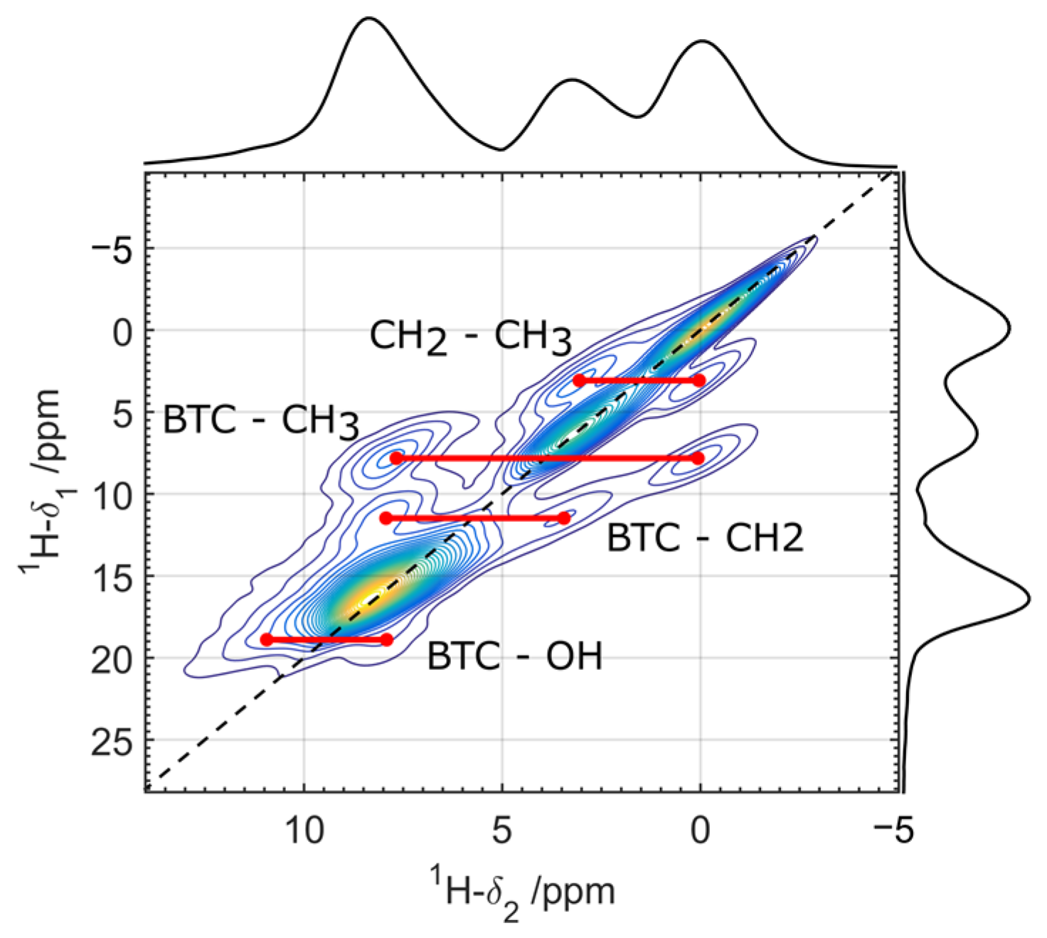
3.2. Solid-State NMR Characterization

Additional details about the structure can be obtained by solid-state 13C and 1H NMR. More specifically, in complement with FTIR results, NMR can give information about the coordination modes of the carboxylate species from the btc linker. Figure 4 shows 13C MAS NMR spectra for [(Eu2Zr)(btc)3(Hbtc)0.5·6H2O)] obtained with 13C{1H} CP with varying contact time (50, 100, 500 and 1000 µs) and 13C direct polarization. Only the region where peaks from btc are observed is displayed. The complete 13C spectrum is provided as supplemental material, showing additional resonances attributed to ethanol molecules adsorbed on the sample (see Figure S4 in the Supporting Information). The schemes for the expected coordination modes of btc with the metallic cluster are also shown in Figure 4 [14,15,16]. Due to the amorphous character of the sample, the 13C NMR lines are poorly resolved. Deconvolution of the spectra into Gaussian lines was performed by observing the lineshape variations as a function of the contact time in the CP experiment. The best-fit deconvolutions are displayed in Figure 4, and the parameters for the direct polarization experiment are given in Table 1. The position and line widths for each Gaussian component were fixed among the various spectra. The deconvolutions show lines centered at 132, 135, 164, 167, 171, 174 and 185 ppm. The lines centered at 129, and 133 ppm are, respectively, attributed to the aromatic carbons C2 and to the quaternary carbons C1 in btc (see Scheme in Figure 4). These attributions are supported by the relative intensity variations of both components as a function of the CP contact time. The protonated species, C2, shows relatively high intensities for low contact times, while the non-protonated C1 species needs more time for magnetization build-up. The line at 185 ppm may be attributed to carboxylate groups with chelating coordination with the metallic center (V in the scheme of Figure 4), as a downfield shift of 5 to 13 ppm is expected for these species when compared to bidentate or monodentate bridging ones [14,15,16]. The line at 164 ppm corresponds to non-coordinated (I), while the line at 167 ppm corresponds to bidentate bridged carboxylate groups performing H-bonds with MOH species (IV), as confirmed by the double resonance experiments described below. According to FTIR results, the presence of monodentate coordination cannot be discarded. From our results, we could not distinguish between monodentate (II) and bidentate species (III), being the attribution of the lines around 171 and 174 ppm is still not resolved [15].
Finally, the 13C spectrum obtained by direct polarization (Figure 4, top) provides quantitative information. The parameters for this spectrum are displayed in Table 1. An intensity ratio of approximately 1:1:1 is observed for the three types of C in btc, validating the quantitative character of the experiment. A considerable fraction of carboxylate groups is in the non-coordinated configuration, about 14%, corresponding to an average of 0.4 non-coordinated COOH groups for each btc molecule. These species can be understood as structural defects, and their relatively high concentration is in agreement with the amorphous character of the sample and the absence of pores (as observed in nitrogen adsorption experiments, not shown).
Figure 5 shows the solid state 1H spectrum (top) measured under 60 kHz MAS. Even at high rotation speeds, the 1H spectrum still presents a complex structure, due to the presence of many H species, besides those from btc, such as molecules or agglomerates of water adsorbed on the material. To simplify the 1H spectrum, a 1H{13C} Double-CP experiment was also performed (Figure 5, bottom spectrum) [22]. In this experiment, only those 1H dipolarly coupled to 13C are observed. This spectrum shows 1H lines at three chemical shift regions: 8.3 ppm, corresponding to unresolved 1H signals from the aromatic ring and OH in free carboxylate groups, and 3.4, 0.3 ppm, corresponding respectively to CH2 and CH3 in ethanol molecules adsorbed on the structure [28]. By comparing both spectra in Figure 5, the 1H NMR signal for proton species not coupled to 13C can be identified. A broad shoulder around 10 ppm is observed for the 1H MAS spectrum, which is not present for the 1H{13C} Double-CP one. This resonance can be attributed to OH species or water molecules H-bonded to the metal oxide [14].
In order to confirm the 13C attributions for the different coordination modes of btc, a 1H{13C} Double-CP HETCOR experiment was performed. The 2D spectrum is displayed in Figure 6. Relatively long CP contact times (500 µs) were used in the experiment in order to observe correlations for the non-protonated carbon species (C1 and I to V in the scheme of Figure 4). This figure also displays the 1H dimension projections corresponding to the 13C shifts at 185, 174, 171, 167, 164, 135 and 130 ppm (obtained from the integration over a range of ±2 ppm). The protons bound to the C2 carbon of the btc present a resonance centered at 8.1 ppm, as shown by the 1H projection for this site. As shown by the projections, the main source of polarization for carbon species resonating at 167 to 185 ppm comes from the aromatic protons, indicating that these species correspond to carboxylate groups coordinated with the metallic oxide species. From the 13C chemical shift, the line at 185 ppm is attributed to the chelating coordination of the btc with on metallic atom (V, see scheme in Figure 4) [13,14,15]. The 1H projection for the 13C species resonating at 167 ppm shows a shoulder at around 10.7 ppm, which may be attributed to OH species from the metallic cluster, letting us attribute the 13C line at 167 ppm to bidentate bridged btc sharing one proton with the metallic species via H-bond. As discussed before, unfortunately, the lines at 174 and 171 ppm cannot be unequivocally attributed [15]. Both lines correspond to bidentate (III) and monodentate (II) bridging modes. The existence of both modes is supported by FTIR results. 13C resonating at 164 ppm, on the other hand, correlates with protons resonating at 7.3 ppm. This chemical shift can be associated with COOH groups from non-coordinated carboxylate species in btc. A correlation peak with 1H shift at 3.5 ppm, attributed to CH2 groups from ethanol, is also observed for this species, indicating special proximity between COOH groups in btc and ethanol. Finally, the 1H projection for carbon C2 presents a broad line centered at 7.6 ppm, which can be attributed to the mutual interaction with 1H in C3 and COOH.
Figure 7 shows the 2D DQ-SQ 1H-1H correlation spectrum for [(Eu2Zr)(btc)3(Hbtc)0.5·6H2O)]. The spectrum reveals dipolar coupling between 1H in CH2 and CH3 groups in ethanol and between btc protons with CH3, CH2 and OH groups from the metal oxide cluster, revealing spatial proximity between these groups. Most probably, ethanol OH groups coordinate with btc COO ones by hydrogen bound. The same conclusions are drawn from 1H-1H exchange spectroscopy (see Figure S5 in the Supporting Information). The interaction of ethanol with btc species prevents the coordination with the metallic species, blocking the growth of the structure. Finally, the presence of self-correlation peaks on the diagonal of the DQ-SQ indicates intermolecular proximity between btc moieties, which is an indication of a collapsed poreless structure.

3.3. Photoluminescence Studies

The photophysical properties exhibited by the compound [(Eu2Zr)(btc)3(Hbtc)0.5·6H2O)] has been studied. The 5D07F2 transition of Eu3+ ion centered at 614 nm is hypersensitive and its accompaniment, at 300 K, gives the excitation spectrum depicted in Figure 8a. The spectrum exhibits intense broad-band emission between 200 and 310 nm, maximum at 280 nm, related to LCCT from ligand btc to Zr/Eu metal and the usual sharp lines determined by f–f transitions of Eu3+ ions. The UV-VIS spectra, Figure S6, also showed the strongest adsorption in this range, not being possible to observe the absorption bands related to 7F07L6 and 7F05D2 transitions.
Besides the sharp excitation assigned to 7F05D2 (465 nm) intra-configurational 4f6 transition of Eu3+, the strong broad-band excitation at 280 nm can be attributed to the ligand absorption band, which indicates that the sensitization occurs by the btc ligands (antenna effect).
The emission spectrum, Figure 8b, was obtained at 300 K, exciting the complex at 300 nm (singlet state of btc). Emission bands coming from 5D0 levels were observed in 590 nm for 5D07F1; 613 nm for 5D07F2; 648 nm for 5D07F3; and 695 nm for 5D07F4. The 5D07F0 has a width at half height of 120 cm−1, as the agreement of several slightly different conformations, as in polymeric structures [29,30]. The 5D07F2 transition is mostly influenced by the dynamic coupling since the 7F2 sublevels are in the energy range of important vibrations. The 5D07F1 transition is not perturbed since the 7F1 sublevels lie in an energy range in which there are no intense IR or Raman bands. It is observed that the electric dipole prevailed compared to a magnetic dipole since the 5D07F2 transition shows higher values of intensity than the 5D07F1 transition. To determine the symmetry site of the Eu3+ ion, the ratio of the intensities between 5D07F2 and 5D07F1 transitions is studied [31,32]. The ratio calculated from Figure 8b is about 3.95, suggesting a low symmetry site (non-centrosymmetric environment) [31,32,33] for Eu3+ ion in [(Eu2Zr)(btc)3(Hbtc)0.5·6H2O)]. According to the 5D07F0 transition, it is possible to define the micro-symmetry, as in the Cnv, Cn, or Cs point groups, since this transition is observed only for low micro-symmetry [34]. Only one peak is verified for 5D07F0 transition (Figure 8b), cantered at 576.3 nm (~1735.21 cm−1); this observation suggests that all Eu3+ ions emitting sites are equivalent. The lifetime of the 5D0 excited state is τ = 0.22 ± 0.01 ms (Figure S6), which can be due to high deactivation rates by the oscillators O-H from water or ethanol molecules coordinated with the ion. Generally, OH species can bind to the Eu3+ ions, as demonstrated by solid-state NMR since the ligand does not occupy the entire coordination sphere. The low luminescence quantum yields, and lifetimes can be explained by the deactivation process that occurs from the vibronic coupling between Eu and OH oscillators. The large nonradiative rates also prove this supposition.
Table 2 present the values obtained from the experimental luminescence study, such as the radiative (Arad) and nonradiative (Arad) emission rates, intensity parameters (Ω2 and Ω4) and quantum efficiency (η) for [(Eu2Zr)(btc)3(Hbtc)0.5·6H2O)] compound. The water molecules coordinated with Eu3+ ions favor the nonradiative decay due to the vibronic coupling of the O-H oscillators. Based on that, it is possibly related to the experimental radiative rate (Arad = 354.1 s−1), nonradiative rate (Anrad = 4191.4 s−1), and the short lifetime τ of 0.22 ms with the relaxation process. Whereas the described compound showed a low Ω4 value (see Table 2), we can assume a considerable rigidity related to the structure [34].

3.4. Experimental test of [(Eu2Zr)(btc)3(Hbtc)0.5·6H2O)] Compound as a Red Luminescent Marker for Ammunition

The experimental results obtained for the Eu/Zr compound indicated a strong emission of light in the red region when submitted to the application of ultraviolet light (λ = 254 nm). TGA and DSC studies also indicated considerable thermal stability for this compound, which was in accordance with previous studies reported in the literature for similar Eu/Zr compounds [9]. These characteristics allowed the proposed compound to be used as an alternative chemical marker in GSR analysis. After the shots, the luminescent residue inside the cartridges of the 0.380 pistol gun was identified using a commercial 254 nm ultraviolet light lamp. It was possible to produce several shots with the marked 0.380 ammunition using 15 to 30 mg of the chemical marker. It is important to mention that the total mass of commercial gunpowder ammunition consisted of 345mg per capsule. It was possible to obtain multiple points of luminescent detection in different regions of the pistol and also in the cartridges, even when minimum amounts of 15 mg of the compound were used. In Figure 9a–f, it is clearly possible to observe the images obtained under UV light excitation of the luminescent residues, in the ejected capsule, on target and inside the gun barrel. It was also possible to analyze by luminescence the LGSR recovered from the gun (Figure 8c), where the spectra maintain similar spectra from the original one. In fact, although the spectra show more noise due to the low amount of material recovered, the spectral profile is maintained, and the intensity ratio of the 5D07F1/5D07F2 in both cases was near 0.261, which confirms no modification in the compound after the shot. Since the luminescent properties of the compound remained unaltered, it is indicative that this material can be effectively used as tagging material for ammunition.

4. Conclusions

The hydrothermal method was employed successfully to obtain a glass-like aspect mixed Zr/Eu coordination compound with benzene tricarboxylic acid (H3btc) as marker of gunshot residues (GSRs). Powder XRD results show that the compound has an amorphous structure. Solid-state NMR results reveal the connectivity between the btc organic linker and the Zr/Eu metal oxide clusters. The results show two biding modes, chelating, and bridging. Quantitative 13C NMR results also reveal the presence of an expressive amount of non-coordinated COOH groups. On average, we estimate that there are 1.7 (out of three) non-bridging COOH per btc molecule, in total agreement with elemental analysis. This high degree of non-bridging sites explains the amorphous character of the sample and the absence of pores, contrary to the observed for other btc-based metal-organic compounds. [(Eu2Zr)(btc)3(Hbtc)0.5·6H2O)] compound structure was elucidated, and its photoluminescence study was carefully conducted. The ligand presence allowed an efficient intramolecular energy transfer resulting in an intense red emission by Eu3+ ions. Thermogravimetric analysis showed the high thermal stability of our Eu/Zr compound. Considering the thermal stability and luminescence data, [(Eu2Zr)(btc)3(Hbtc)0.5·6H2O)] was added to gunpowder and gunshots were evaluated, the presence of Eu/Zr coordination compound enabled visual detection of the particles on the floor, in the firearm and cartridges. Although other lanthanoid compounds have been reported as ammunition markers, there is no photoluminescence information regarding the Eu/Zr coordination compound derived from benzene tricarboxylic acid. Mixed Eu/Zr luminescent particles with unique characteristics can support criminal investigations involving firearms.

Supplementary Materials

The following supporting information can be downloaded at: https://www.mdpi.com/article/10.3390/sci4040043/s1, Figure S1: XRD pattern of luminescent compound [(Eu2Zr)(btc)3(Hbtc)0.5·6H2O)]; Figure S2: Chemical composition (EDS) analysis of [(Eu2Zr)(btc)3(Hbtc)0.5·6H2O)]; Figure S3: TGA/DSC analysis of Eu-btc; Figure S4: 13C{1H} CPMAS spectrum of the compound [(Eu2Zr)(btc)3(Hbtc)0.5·6H2O)]; Figure S5: 2D 1H-1H EXSY spectra for [(Eu2Zr)(btc)3(Hbtc)0.5·6H2O)] measured at two mixing times, 1 ms and 10 ms, under 60 kHz MAS. The dashed lines indicate the presence of 1H-1H correlations for the longer mixing time. For short mixing times (1 ms), the spectrum reveals no exchange between distinct 1H species. For longer mixing times, cross-peaks revealing exchange between 1H in CH2 and CH3 groups in ethanol become visible, while the same is true for the correlation between btc protons with CH2 and OH groups from the metal oxide cluster, revealing spatial proximity between these groups. Most probably, ethanol OH groups coordinate with btc COO ones by hydrogen bound. The interaction of ethanol with btc species prevents the coordination with the metallic species, blocking the growth of the structure. Experimental details: experiments were performed at 60 kHz using a 1.4 s recycle delay and mixing times of 1 and 10 ms. A total of 512 points were collected in the indirect dimension with a time increment of 16.67 μs, corresponding to a spectral width of 60 kHz; Figure S6: UV-VIS spectra of the luminescent compound [(Eu2Zr)(btc)3(Hbtc)0.5·6H2O)]; Figure S7: Exponential fitting of the decay curve of luminescent compound [(Eu2Zr)(btc)3(Hbtc)0.5∙6H2O)].

Author Contributions

Conceptualization, A.R.B.S., M.d.O.J. and O.A.S.; Data curation, A.R.B.S., T.R.C., J.F.d.L., M.F.d.O. and S.A.J.; Formal analysis, A.R.B.S. and M.d.O.J.; Funding acquisition, O.A.S.; Investigation, M.d.O.J.; Methodology, A.R.B.S. and M.d.O.J.; Project administration, O.A.S.; Resources, O.A.S.; Supervision, O.A.S.; Writing—original draft, A.R.B.S. and M.d.O.J.; Writing—review and editing, A.R.B.S., J.F.d.L., M.F.d.O., S.A.J. and M.d.O.J. All authors have read and agreed to the published version of the manuscript.

Funding

The authors acknowledge the financial support from CNPq (426467/2018-3, O.A.S.), CAPES, FAPESP (2018/07514-3, O.A.S.) and FACEPE (BFP-0202-1.06/20 A.R.B.S.). M.d.O.J. is grateful for a CNPq grant (nº 311069/2020-7).

Informed Consent Statement

Not applicable.

Data Availability Statement

Solid state NMR data available at: de Oliveira Junior, Marcos. (2022). Solid state NMR data of amorphous MOF [(Eu2Zr)(btc)3(Hbtc)0.5·6H2O)] [Data set]. Zenodo. https://doi.org/10.5281/zenodo.7311799.

Acknowledgments

We would like to thank Cynthia Maria de Campos Prado Manso for fruitful discussions.

Conflicts of Interest

The authors declare no conflict of interest.

References

  1. Eliseeva, S.V.; Bünzli, J.-C.G. Rare earths: Jewels for functional materials of the future. New J. Chem. 2011, 35, 1165. [Google Scholar] [CrossRef] [Green Version]
  2. Filho, P.C.S.; Larquet, E.; Dragoe, D.; Serra, O.A.; Gacoin, T. Lanthanoid-doped phosphate/vanadate mixed hollow particles as ratiometric luminescent sensors. ACS Appl. Mater. Interfaces 2017, 9, 1635. [Google Scholar] [CrossRef]
  3. Nascimento, L.F.; Lima, J.F.; de Filho, P.C.; Serra, O.A. Control of diesel particulate emission based on Ag/CeOx/FeOy catalysts supported on cordierite. Chem. Eng. J. 2016, 290, 454. [Google Scholar] [CrossRef]
  4. Huang, X.; Han, S.; Huang, W.; Liu, X. Enhancing solar cell efficiency: The search for luminescent materials as spectral converters. Chem. Soc. Rev. 2013, 42, 173. [Google Scholar] [CrossRef] [Green Version]
  5. Nascimento, L.F.; Serra, O.A. Washcoating of cordierite honeycomb with ceria-copper mixed oxides for catalytic diesel soot combustion. Process Saf. Environ. Prot. 2016, 101, 134. [Google Scholar] [CrossRef]
  6. Cotton, S. Lanthanoid and Actinide Chemistry; John Wiley & Sons, Ltd.: Chichester, UK, 2006. [Google Scholar]
  7. Bünzli, J.-C.G.; Moret, E.; Foiret, V.; Schen, K.J.; Mingzhao, W.; Linpei, J. Structural and photophysical properties of europium(III) mixed complexes with β-diketonates and o-phenanthroline. J. Alloys Compd. 1994, 208, 107. [Google Scholar] [CrossRef]
  8. Filho, E.V.; de Filho, P.C.; Serra, O.A.; Weber, I.T.; Lucena, M.A.M.; Luz, P.P. New luminescent lanthanoid-based coordination compounds: Synthesis, studies of optical properties and application as marker for gunshot residues. J. Lumin. 2018, 202, 89. [Google Scholar] [CrossRef]
  9. Júnior, J.C.A.; Santos, G.L.; Colaço, M.V.; Barroso, R.C.; Ferreira, F.F.; Santos, M.V.; de Campos, N.R.; Marinho, M.V.; Jesus, L.T.; Freire, R.O.; et al. New EuIII pyromellitic Metal–Organic Framework of intense red-orange luminescence and high thermal stability for marking in gunshot residues. J. Phys. Chem. C 2020, 124, 9996. [Google Scholar] [CrossRef]
  10. Talhari, A.L.R.; Lucena, M.A.M.; Mauricio, F.G.M.; Oliveira, M.F.L.; Veiga-Souza, F.H.; Alves, S., Jr.; Weber, I.T. Luminescent marker for GSR: Evaluation of the acute oral and inhalation toxicity of the MOF [Eu(DPA)(HDPA)]. ACS Appl. Bio Mater. 2020, 3, 3049. [Google Scholar] [CrossRef]
  11. Weber, I.T.; Melo, A.J.; Lucena, M.A.; Consoli, E.F.; Rodrigues, M.O.; de Sá, G.F.; Maldaner, A.O.; Talhavini, M.; Alves, S., Jr. Use of luminescent gunshot residues markers in forensic contex. Forensic Sci. Int. 2014, 244, 276. [Google Scholar] [CrossRef]
  12. Lima, P.P.; Nobre, S.S.; Freire, R.O.; Júnior, S.A.; Ferreira, R.A.S.; Pischel, U.; Malta, O.L.; Carlos, L.D. Energy transfer mechanisms in organic−inorganic hybrids incorporating europium(iii):  A quantitative assessment by light emission spectroscopy. J. Phys. Chem. C 2007, 111, 17627–17634. [Google Scholar] [CrossRef]
  13. Liu, K.; Jia, G.; Zheng, Y.H.; Song, Y.H.; Yang, M.; Huang, Y.J.; Zhang, L.H.; You, H.P. Room-temperature synthesis and luminescence properties of Eu3+/Tb3+-doped La(1,3,5-btc)(H2O)(6). Inorg. Chem. Comm. 2009, 12, 1246. [Google Scholar] [CrossRef]
  14. de Roo, J.; Baquero, E.A.; Coppel, Y.; de Keukeleere, K.; van Driessche, I.; Nayral, C.; Hens, Z.; Delpech, F. Insights into the Ligand Shell, Coordination Mode, and Reactivity of Carboxylic Acid Capped Metal Oxide Nanocrystals. ChemPlusChem 2016, 81, 1216. [Google Scholar] [CrossRef] [Green Version]
  15. Ye, B.H.; Li, X.Y.; Williams, I.D.; Chen, X.M. Synthesis and structural characterization of di- and tetranuclear zinc complexes with phenolate and carboxylate bridges. Correlations between 13C NMR chemical shifts and carboxylate binding modes. Inorg. Chem. 2002, 41, 6426. [Google Scholar] [CrossRef] [PubMed]
  16. Faccini, F.; Fric, H.; Schubert, U.; Wendel, E.; Tsetsgee, O.; Müller, K.; Bertagnolli, H.; Venzo, A.; Gross, S. ω-Mercapto-functionalized hafnium- and zirconium-oxoclusters as nanosized building blocks for inorganic-organic hybrid materials: Synthesis, characterization and photothiol-ene polymerization. J. Mater. Chem. 2007, 17, 3297. [Google Scholar] [CrossRef]
  17. Dutra, J.D.L.; Bispo, T.D.; Freire, R.O. LUMPAC lanthanoid luminescence software: Efficient and user friendly. J. Comput. Chem. 2014, 35, 772. [Google Scholar] [CrossRef]
  18. Jaeger, C.; Hemmann, F. EASY: A simple tool for simultaneously removing background, deadtime and acoustic ringing in quantitative NMR spectroscopy—Part I: Basic principle and applications. Solid State Nucl. Magn. Reson. 2014, 57, 22. [Google Scholar] [CrossRef]
  19. Cory, D.G.; Ritchey, W.M. Suppression of signals from the probe in Bloch decay spectra. J. Magn. Reson. 1988, 80, 128. [Google Scholar] [CrossRef]
  20. Bennett, A.E.; Rienstra, C.M.; Auger, M.; Lakshmi, K.V.; Griffin, R.G. Heteronuclear decoupling in rotating solids. J. Chem. Phys. 1995, 103, 6951. [Google Scholar] [CrossRef]
  21. Baccile, N.; Laurent, G.; Bonhomme, C.; Innocenzi, P.; Babonneau, F. Solid-state NMR characterization of the surfactant-silica interface in templated silicas: Acidic versus basic conditions. Chem. Mater. 2007, 19, 1343. [Google Scholar] [CrossRef]
  22. Christiansen, S.C.; Hedin, N.; Epping, J.D.; Janicke, M.T.; del Amo, Y.; Demarest, M.; Brzezinski, M.; Chmelka, B.F. Sensitivity considerations in polarization transfer and filtering using dipole-dipole couplings: Implications for biomineral systems. Solid State Nucl. Magn. Reson. 2006, 29, 170. [Google Scholar] [CrossRef] [PubMed]
  23. Wiench, J.W.; Bronnimann, C.E.; Lin, V.S.-Y.; Pruski, M. Chemical shift correlation NMR spectroscopy with indirect detection in fast rotating solids: Studies of organically functionalized mesoporous silicas. J. Am. Chem. Soc. 2007, 129, 12076. [Google Scholar] [CrossRef] [PubMed]
  24. Mafra, L.; Siegel, R.; Fernandez, C.; Schneider, D.; Aussenac, F.; Rocha, J. High-resolution 1H homonuclear dipolar recoupling NMR spectra of biological solids at MAS rates up to 67 KHz. J. Magn. Reson. 2009, 199, 111–114. [Google Scholar] [CrossRef]
  25. Kimura, H.; Nakamura, K.; Eguchi, A.; Sugisawa, H.; Deguchi, K.; Ebisawa, K.; Suzuki, E.I.; Shoji, A. Structural study of α-amino-acid crystals by 1H CRAMPS NMR spectroscopy. J. Mol. Struct. 1998, 447, 247. [Google Scholar] [CrossRef]
  26. Potrzebowski, M.J.; Tekely, P.; Dusausoy, Y. Comment to 13C-NMR studies of α and γ polymorphs of glycine. Solid State Nucl. Magn. Reson. 1998, 11, 253. [Google Scholar] [CrossRef]
  27. Nakamoto, K. Infrared and Raman Spectra of Inorganic and Coordination Compounds, 6th ed.; John Wiley & Sons, Inc.: New York, NY, USA, 2006. [Google Scholar]
  28. Babij, N.R.; McCusker, E.O.; Whiteker, G.T.; Canturk, B.; Choy, N.; Creemer, L.C.; Amicis, C.V.D.; Hewlett, N.M.; Johnson, P.L.; Knobelsdorf, J.A.; et al. NMR chemical shifts of trace impurities: Industrially preferred solvents used in process and green chemistry. Org. Process Res. Dev. 2016, 20, 661. [Google Scholar] [CrossRef]
  29. Cai, Z.; Wei, C.; Sun, B.; Wei, H.; Liu, Z.; Bian, Z.; Huang, C. Luminescent europium(III) complexes based on tridentate isoquinoline ligands with extremely high quantum yield. Inorg. Chem. Front. 2021, 8, 41. [Google Scholar] [CrossRef]
  30. Barbosa, C.D.; Da Luz, L.L.; Paz, F.A.; Malta, O.L.; Rodrigues, M.; Júnior, S.A.; Ferreira, R.A.; Carlos, L.D. Site-selective Eu(iii) spectroscopy of highly efficient luminescent mixed-metal Pb(ii)/Eu(iii) coordination polymers. RSC Adv. 2017, 7, 6093. [Google Scholar] [CrossRef] [Green Version]
  31. Capobianco, J.A.; Proulx, P.P.; Bettinelli, M.; Negrisolo, F. Absorption and emission spectroscopy of Eu3+ in metaphosphate glasses. Phys. Rev. B Condens. Matter. 1990, 42, 5936. [Google Scholar] [CrossRef]
  32. Nogami, M.; Umehara, N.; Hayakawa, T. Effect of hydroxyl bonds on persistent spectral hole burning in Eu3+-doped BaO−P2O5 glasses. Phys. Rev. B 1998, 58, 6166. [Google Scholar] [CrossRef]
  33. de Oliveira, M., Jr.; Gonçalves, T.S.; Ferrari, C.; Magon, C.J.; Pizani, P.S.; de Camargo, A.S.S.; Eckert, H. Structure–Property Relations in Fluorophosphate Glasses: An Integrated Spectroscopic Strategy. J. Phys. Chem. C 2017, 121, 2968. [Google Scholar] [CrossRef]
  34. Galaço, A.R.B.S.; Freire, R.O.; de Jesus, L.T.; Serra, O.A. Experimental and Theoretical study of isoreticular lanthanoid organic framework (LOF): Structure and luminescence. J. Lumin. 2020, 223, 179. [Google Scholar]
Figure 1. (a) Schematical illustration of the synthesis procedure; (b) The digital images of the amorphous [(Eu2Zr)(btc)3(Hbtc)0.5·6H2O)] coordination compound.
Sci 04 00043 g001
Figure 2. Infrared Spectra for [(Eu2Zr)(btc)3(Hbtc)0.5·6H2O)] compound and for the benzenetricarboxylic acid (H3btc) precursor.
Sci 04 00043 g002
Figure 3. TGA curves (solid lines) for [(Eu2Zr)(btc)3(Hbtc)0.5·6H2O)] and H3btc represent the successive mass losses. The derivatives of the TGA curves are shown as dashed lines.
Sci 04 00043 g003
Figure 4. (Left): 13C MAS NMR spectra (black curves) obtained by 13C{1H} cross-polarization (CP) and direct polarization on 13C with background suppression and 1H decoupling (Direct). Colored curves are spectral deconvolutions into Gaussian functions. Assignments for the observed peaks are given as roman numerals, corresponding to the scheme on the right. Assignments for species II and III could not be unequivocally provided, both lies in the region indicated by an horizontal line in the figure. (Right): Possible coordination modes for the carboxylate groups in btc with the metallic species. R’ is a carboxylate group in one of the coordination modes I to V.
Sci 04 00043 g004
Figure 5. 1H MAS (top) and 1H{13C} Double-CP (bottom) spectra for the [(Eu5Zr3)(btc)9(H2O)18] compound. Both spectra were obtained under a 60 kHz MAS spinning rate.
Sci 04 00043 g005
Figure 6. Two-dimensional 1H{13C} Double-CP HETCOR [(Eu2Zr)(btc)3(Hbtc)0.5·6H2O)] (left) and traces taken from the vertical dimension, showing partial 1H spectra (normalized by intensity) corresponding to several shifts of 13C (right). The experiment was carried out under MAS conditions at 60 kHz with 1H detection. The numbers indicated in the two-dimensional spectrum correspond to the carbon labeling in Figure 4.
Sci 04 00043 g006
Figure 7. 2D 1H DQ-SQ correlation spectrum for [(Eu2Zr)(btc)3(Hbtc)0.5·6H2O)] measured under 60 kHz MAS using R 14 4 5 symmetry-based homonuclear recoupling and no homonuclear decoupling during DQ evolution. The red lines indicate the presence of 1H-1H correlations.
Sci 04 00043 g007
Figure 8. (a) Excitation (λem = 615 nm), (b) emission (λexc = 300 nm) spectra of [(Eu2Zr)(btc)3(Hbtc)0.5·6H2O)] and (c) emission (λexc = 300 nm) spectra of [(Eu2Zr)(btc)3(Hbtc)0.5·6H2O)] in GSR, recorded in the solid state.
Sci 04 00043 g008
Figure 9. Red residual luminescent in GSR: (a) in the ejected capsule; (b) in the triggered cartridge; (c) inside the bolt of the firearm; (d) inside the slide of the firearm; (e) over the magazine and (f) under the magazine. UV light irradiation, λ = 254 nm.
Sci 04 00043 g009
Table 1. Spectral parameters were obtained from the deconvolution of the 13C NMR spectrum measured with direct polarization. δiso is the isotropic chemical shift, FWHM is the full-width at half-maximum, and I is the relative area for each spectral component.
Attributionδiso (±1 ppm)FWHM (±0.5 ppm)I (±2%)
C11325.631
C21355.833
I1643.65
II, III a167/1713.9/3.615/8
IV1747.17
V1853.11
a Attributions to species II and III could not be unequivocally given.
Table 2. Experimental luminescence parameters. Experimental Nonradiative (Anrad), Radiative (Arad) Decay Rates; Intensity Parameters Ω2 and Ω4; Quantum Efficiency (η) and lifetime (τ).
AradAnradΩ2 (10−20 s)Ω4 (10−20 s)ητ (ms)
354.084191.377.05.737.790.22
Publisher’s Note: MDPI stays neutral with regard to jurisdictional claims in published maps and institutional affiliations.

