Article

Optical and Spectroscopic Properties of Ho:Lu2O3 Transparent Ceramics Elaborated by Spark Plasma Sintering

1
Faculty of Science and Technology, University of Limoges, CNRS, IRCER, UMR 7315, F-87068 Limoges, France
2
Bordeaux INP, University of Bordeaux, CNRS, ICMCB, UMR 5026, F-33600 Pessac, France
3
Institut d’Optique Graduate School, University of Bordeaux, CNRS, LP2N, UMR 5298, F-33400 Talence, France
*
Author to whom correspondence should be addressed.
Ceramics 2024, 7(1), 208-221; https://doi.org/10.3390/ceramics7010013
Submission received: 9 December 2023 / Revised: 31 January 2024 / Accepted: 6 February 2024 / Published: 8 February 2024
(This article belongs to the Special Issue Transparent Ceramics—a Theme Issue in Honor of Dr. Adrian Goldstein)

Abstract

:
In this work, transparent ceramics were manufactured from nanopowders synthesized by aqueous coprecipitation followed by Spark Plasma Sintering (SPS) to ensure rapid and full densification. The photoluminescence of Ho:Lu2O3 transparent ceramics was studied in the Visible and IR domains as a function of Ho3+ dopant level from 0.5 at.% to 10 at.%. A cross-relaxation mechanism was identified and favors the 2 μm emission. All of the obtained results indicate that the optical properties are very similar between Lu2−xHoxO3 transparent ceramics and single crystals. Thus, the SPS technique appears to be a very promising method to manufacture such ceramics, which could be used as amplifier media for high-energy solid-state lasers.

1. Introduction

In recent years, transparent ceramics have appeared as a very interesting new class of material for high-power solid-state lasers, leading to the development of multi-kilowatt laser systems [1,2,3,4]. Among the variety of the targeted laser characteristics, lasers emitting at 2 µm have been mainly studied for their great potential in the medical field because of the absorption bands of water around this wavelength [5,6,7,8]. Compared to other 2 µm emitting ions, Ho3+ has the advantage of presenting the largest emission cross-section [9]. Due to their high mechanical resistance, good chemical stability and high thermal conductivity, rare-earth sesquioxides (Sc2O3, Y2O3 and Lu2O3) and garnet structures like YAG (Yttrium Aluminum Garnet) are well adapted and the most used host matrix for such ions [10,11,12,13]. Among these materials, Lu2O3 is of primary interest because it maintains a high thermal conductivity close to 12 W·m−1·K−1 even for high concentrations of dopant [14].
A high concentration of active ion has great potential for use in high-power laser applications, especially in sesquioxide ceramic matrices where the dopant is inserted in a solid solution [15,16]. Indeed, it can be supposed that a higher doping rate could induce a higher luminescence intensity [17,18]. However, a high dopant volume density can also generate undesired energy transfers, called non-radiative transitions, such as cross-relaxation (CR) or excited-state absorption (ESA), causing quenching of the photoluminescence signal. For the Ho3+ ion, the most common transfers are CRs between two ions, called the donor and acceptor, which are performed by up-conversion or down-conversion depending on the initial state of the acceptor and the final state of the donor [19].
The main results concerning the previous developments of sesquioxide ceramics for laser applications were recently summarized in the review of Liu et al. [20]. According to their conclusions, Lu2O3 sesquioxide ceramics seem to be the most promising host materials for high-power laser operation. However, they highlight the long road ahead to obtain transparent sesquioxide ceramics of large size and high optical quality. Especially, they pointed out the crucial role played by the synthesis step of nanopowders, as well as the production of powder compacts with ideal microstructures. According to the works cited in the literature, the elaboration of transparent ceramics requires control of each process step, especially that of sintering. In fact, full densification and total elimination of porosity is mandatory to ensure high optical properties and high laser performances. Pressureless sintering under vacuum, pressure-assisted densification or a combination of these techniques is an efficient way to remove all pores, and non-conventional sintering techniques like Hot Pressing (HP), Hot Isostatic Pressing (HIP) or Spark Plasma Sintering (SPS also called Field Assisted Sintering Technique—FAST) have been developed and extensively studied for a wide variety of ceramics. Some recent studies were lead, for example, on Y2O3 by HP + HIP [21], Yb:Lu2O3 and Tm:Lu2O3 by vacuum sintering + HIP [22,23] or Er:(ScxY1−x)2O3 by vacuum sintering [24]. Among other techniques, Spark Plasma Sintering relies on the densification of a powder inside an electrically conductive matrix, heated by the Joule effect thanks to the application of a pulsed current with a high intensity, and submitted to a uniaxial pressure generally in the range of 50–150 MPa. SPS is increasingly used for fast and efficient densification of numerous ceramics, including nitrides, carbides and oxides [25]. However, the fast shrinkage and physical phenomena involved during sintering lead to a lack of knowledge on sintering mechanisms, more precisely on the densification step and the subsequently formed microstructures. Moreover, carbon contamination from the common use of a graphite matrix is a main issue often encountered for the sintering of oxides and could strongly affect their optical and spectroscopic properties [26]. Concerning the elaboration of transparent ceramics, few studies have compared the microstructure optical properties’ relationships to other non-conventional sintering techniques or have highlighted the specificities of SPS on this family of materials. As an example, some studies were interested in Nd:Lu2O3 [27,28] or Yb:Sc2O3 [29] ceramics elaborated by either SPS or HIP techniques. Their main conclusions highlighted that such ceramics in the rare-earth oxide family have similar optical properties to single crystals. In the case of the Ho3+ ion, no study has been reported on the implication of the fabrication process parameters on the luminescence properties of as-obtained materials, more especially on the CR mechanisms.
In this work, transparent ceramics of Lu2O3 with various Ho3+ doping rates were manufactured by a process combining nanopowder synthesis and direct densification by Spark Plasma Sintering. Chemical synthesis by the co-precipitation route has the advantage of producing well-crystallized Ho-doped Lu2O3 nanopowders with nanometric grain size and great chemical homogeneity [30]. The microstructure (i.e., grain size), luminescence properties and optical transparency of as-obtained transparent ceramics were studied as a function of the holmium doping content. The results were compared to ones reported in the literature for similar single crystals or transparent ceramics manufactured by HIP in order to highlight the specificities induced by the SPS process.

2. Materials and Methods

2.1. Synthesis of Ho:Lu2O3 Nanopowders

Ho-doped Lu2O3 nanopowders were synthesized by inverse co-precipitation. Rare-earth nitrates were used as precursor and ammonium bicarbonate as precipitant. Commercial oxides of Lu2O3 and Ho2O3 (AUER REMY, Lehvoss group, Hamburg, Germany) were first dissolved in hot nitric acid (HNO3, 65 vol.%, Thermo Fisher Scientific, Waltham, MA, USA). The nitrate solution was poured drop by drop at a rate of 3 mL·min−1 into the solution of ammonium bicarbonate (NH4HCO3, 99wt.%, Acros Organics, Thermo Fisher Scientific, Waltham, MA, USA). The co-precipitation was operated at room temperature with vortex stirring according to the set-up described in Figure 1.
Ammonia solution (NH4OH, 35 vol.%, Thermo Fisher Scientific, Waltham, MA, USA) was added to maintain the pH at 9. Then, the solution was aged for 24 h under stirring at room temperature. The obtained precipitate was washed three times with distilled water by centrifugation and three times with ethanol and was dried at 70 °C. The dried precipitate was milled in an agate mortar and calcined at 1000 °C for 2 h under air to form the final crystallized Ho:Lu2O3 nanopowder. Powder morphology, structure and chemical homogeneity were obtained by TEM (Transmission Electronic Microscopy) in classical or scanning (STEM) modes and high-resolution TEM coupled with SAED (Selected Area Electron Diffraction) (2100 F, JEOL, Tokyo, Japan). Crystalline phases were identified by X-ray diffraction analyses (D8, Bruker, Karmsruhe, Germany) using CuKα radiation. CuKα2 radiation contribution was removed and obtained diagrams were indexed with DIFFRACplus EVA™ software V5 and the PDFmain™ database.
The TEM observations reported in Figure 2a show a slightly edged shape and narrow size distribution of particles with a mean diameter of about 63 nm. No holmium segregation in the form of isolated Ho2O3 particles was detected, as illustrated by the STEM micrograph in Figure 2b. That means there is no differential precipitation of Ho and Lu cations during the synthesis process. The particles also appeared well crystallized in the bixbyite structure (Ia-3, cubic C-type), as revealed by diffraction pattern (Figure 2c). As a result, the Ho:Lu2O3 nanopowder can be considered as a Lu(2−x)HoxO3 homogeneous solid-solution (x = 0.02 for 1 at.% holmium doping content) with a cubic crystalline structure. The diffraction patterns registered as a function of the calcination temperature in Figure 2d also prove that the powder is single phased in the bixbyite structure and well crystallized for temperatures higher than 600 °C. A calcination temperature of 1000 °C was chosen regarding the higher crystallinity of the powder particles at this temperature.

2.2. Densification by Spark Plasma Sintering

A commercial device (825 series 8000 A, FUJI-SPS, Saitama, Japan) was used for SPS experiments. The powder samples were put in a 13 mm diameter graphite die with Papyex® foil (MERSEN, La Défense, France). Sintering experiments were led under primary vacuum (P < 20 Pa) with a pulse sequence of 12:2. A digital IR pyrometer was used for thermal regulation on the surface of the graphite die, which started detecting at a minimum temperature of 573 °C. For all treatments used to elaborate fully dense ceramics, the heating rate was fixed to 100 °C·min−1 until 1200 °C. Then, the heating rate was 10 °C·min−1 until 1400 °C, where the temperature was maintained for 15 min under uniaxial pressure of 130 MPa. The cooling rate was 100 °C·min−1. Then, ceramics were annealed under air at 985 °C for at least 50 h to re-oxidize them and eliminate the carbon contamination caused by the contact with graphite.

2.3. Characterization of Sintered Samples

Prior to optical characterizations, the samples were ground to obtain flat and parallel surfaces and then mirror polished with 30 nm colloidal silica after pre-polishing with SiC papers and diamond pastes. A Cary 5000 spectrophotometer was used to record the transmission curves of transparent samples (Agilent, Santa Clara, CA, USA). The spectra resolution was 1 nm with an integration time equal to 0.1 s.
The photoluminescence spectra of various doped ceramics were obtained from 450 nm to 2200 nm into 3 sub-intervals: Visible [450–800 nm], Near-IR [1000–1300 nm] and Mid-IR [1800–2200 nm]. For all ranges, a photoluminescence spectrometer was used (Fluorolog3, Horiba, Kyoto, Japan) using a 450 W Xe lamp for excitation in Visible and 450 nm laser diode (50 mW) for Near-IR and Mid-IR. For detection, a photo multiplicator detector (PMT-Hamamatsu R298, Shizuoka, Japan) was used in Visible, an InGaAs detector for Near-IR and a PbS photoconductor for Mid-IR. For Visible acquisitions, the spectral resolution was 2 nm/mm of opening slits (for both entry and exit ones). Some corrections were applied to the raw spectra to account for variations in the intensity of the excitation source, set-up geometry and detector sensitivity. For measurements in IR range, the spectral resolution was 12 nm/mm of opening slits and the integration time was 0.5 s.
Lifetime measurements were performed with a 10 nm step-by for each wavelength using an Optical Parametric Oscillator (OPO) for source pumping. Infrared lifetime measurements were performed with an Edinburgh FLS1000 spectrometer coupled with a digital Tektronix oscilloscope to record the time-dependent decay. All the measurements were taken at room temperature.

3. Results

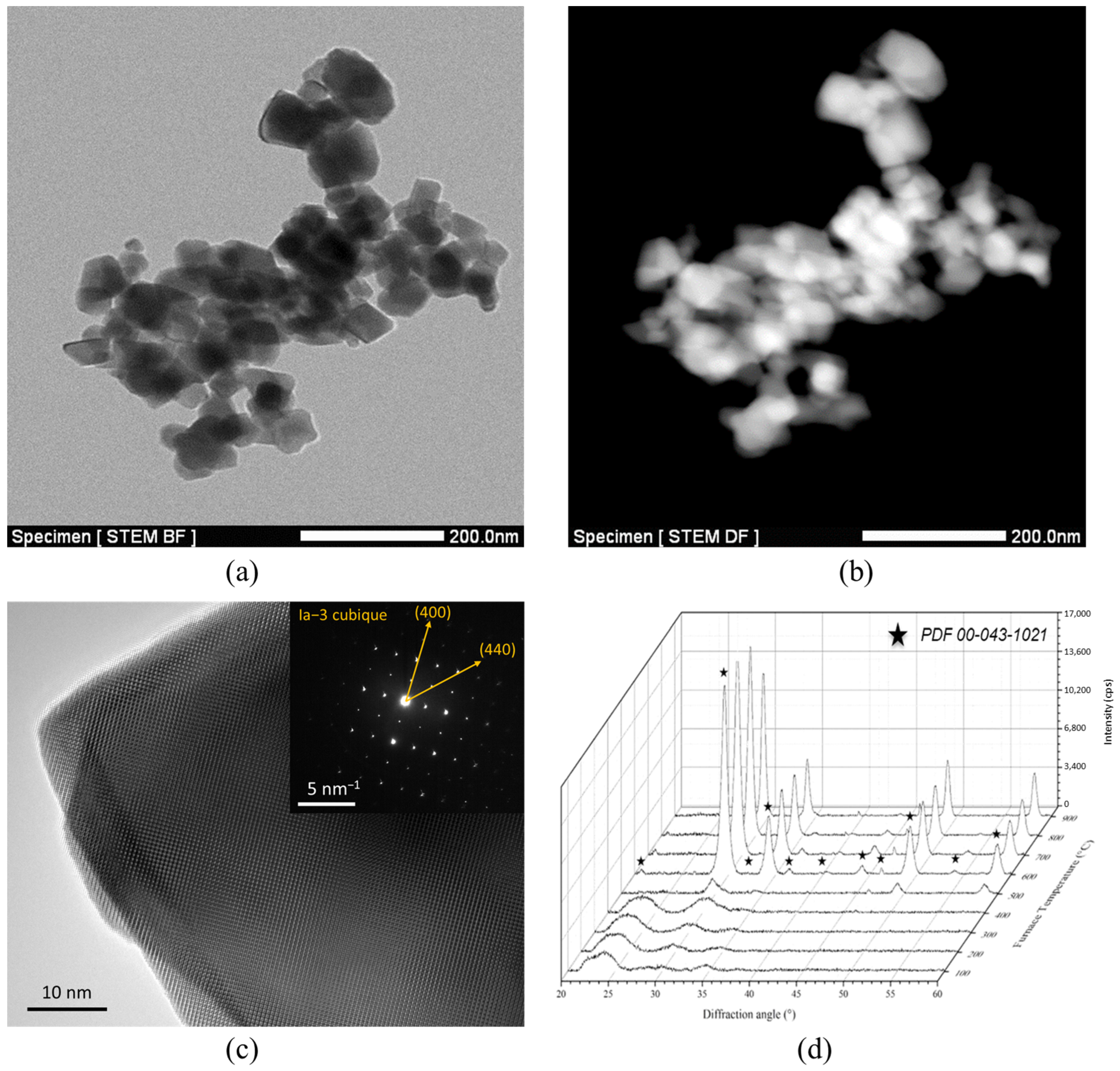
3.1. Sample Microstructure and Optical Transmittance

After sintering at 1400 °C for 15 min, annealing and polishing, Lu2O3 ceramics with doping rates from 0.5 at.% to 10 at.% Ho were obtained (Figure 3). All specimens were 2 mm thick and presented good transparency.
The SEM micrographs taken for the 1 at.%Ho:Lu2O3 sample (Figure 4a) showed a dense microstructure with a small and homogeneous grain size in the order of 200–500 nm. Nevertheless, few intergranular and/or intragranular residual pores were also found, especially by the TEM observations (Figure 4b). Their diameter did not exceed 50 nm and their concentration was estimated to be 90 pores·µm−3. These defects could be related to the presence of microstructural inhomogeneities in powder compacts before sintering and/or rapid grain growth [31]. In fact, it has been highlighted in a previous paper that grain growth starts to be significant for temperatures higher than 1300 °C in such ceramics during sintering by SPS, independently of the applied pressure [32].
The transmission curves of the samples are presented on Figure 5. Data have been obtained for wavelengths from 400 nm to 2500 nm. All of the ceramics present the same optical absorption peaks corresponding to 4f electronic transitions of Ho3+ ion with corresponding intensity generally increasing with the doping rate. The most intense absorption peaks between 1800 nm and 2200 nm correspond to the 5I85I7 transition level. The optical transmittance baseline of all samples presents a continuous decrease from wavelengths of 1200 nm to 300 nm. The obtained curves were compared to the maximum theoretical transmittance of the Lu2O3 samples. To determine these values, one has to consider the general law giving the transmittance of a transparent material (Equation (1)):
T ( λ ) = T m a x exp ( α x )
where Tmax is the maximal transmittance that could be obtained for a perfectly polished sample with planar and parallel surfaces, α is the attenuation coefficient and x is the sample thickness. By considering multiple reflections at the sample’s surface (no antireflective coatings were applied in our case), the maximal transmittance at normal incidence is given by Equation (2):
T m a x ( λ ) = 2 n ( λ ) ( 1 + n ( λ ) 2 )
where n(λ) is the refractive index of cubic Lu2O3 given by Equation (3) [33]:
n ( λ ) 2 = 3.6196 + 0.04131 λ 2 0.0238 0.00856 · λ 2
As a result, one is able to calculate the maximum transmittance value for a perfectly transparent Lu2O3 sample. The values are reported in Figure 5.
According to Figure 5a, a strong difference appears between the maximum transmittance and the transmittance baseline of Lu2O3 samples. This phenomenon is well-known for transparent ceramics and is generally induced by light scattering coming from residual porosity [34]. However, despite the presence of such optical losses, all Ho-doped Lu2O3 ceramics have a transmittance higher than 75%, i.e., more than 90% of the theoretical limit, for wavelengths > 2 µm. On the absorption curves (Figure 5b), the effect of the doping rate is clearly highlighted. Indeed, the intensity of absorption transition 5I85I7 increases gradually with the Ho3+ concentration.
The absorption cross-sections of the Ho:Lu2O3 ceramics σabs were then calculated for all dopant contents from linear absorption coefficient αλ and by using Equations (4) and (5):
αλ = (Aλ/x)·ln(10)
σabs = αλ/CHo
where Aλ is the absorbance for a specific wavelength λ, x is the thickness of sample and CHo is the volume density of Ho3+. The values of volumetric density of Ho3+ (Table 1) were determined thanks to Equation (3) below:
CHo = (ρ·%atHo·NA)/(MLu2O3:Ho)
where ρ is the theoretical volumetric mass of Lu2O3 with Ia-3 cubic crystalline structure (obtained by XRD measurements), %atHo is the doping rate, NA is the Avogadro constant and MLu2O3:Ho is the molar mass of Lu2−xHoxO3 compound at a specific doping rate (0.01 < x < 0.2).
The data reported in Table 1 were used to plot the evolution of the absorption cross-section in the wavelength range 1900–1960 nm for all samples (Figure 6). It was shown that the curves seemed to follow the same evolution of σabs whatever the doping rate. Moreover, they overlapped despite a slight offset for 0.5% and 5%, which was obviously caused by a lower transmittance baseline (i.e., higher scattering by residual porosity) obtained for both samples. However, for the samples with low residual scattering, the calculated value of σabs was approximately 1 × 10⁻²⁰ cm2 at 1927 nm, which corresponded to the measured data for some Ho-doped systems, for example Ho:YAG [35].

3.2. Photoluminescence and Fluorescence Decay Time

To highlight the different energy transfers and associated light emissions and to attempt to locate some specificities of the elaboration process by SPS, photoluminescence spectroscopy was carried out on all samples in three interesting wavelength domains to highlight potential CR transitions: Visible (from 450 to 800 nm), Near-IR (from 1000 to 1300 nm) and Mid-IR (from 1800 to 2200 nm). The as-obtained results are summarized in Figure 7.
In the Visible range, the emission bands symbolized by red arrows presented a strong decrease when the doping rate increased, with an extinction at 10 at.%Ho (Figure 7a). The identified transitions were 5F3 + 5F2 + 3K85I8 at 480 nm, 5F4 + 5S25I8 at 550 nm and 5S25I7 at 750 nm. However, the transition at 650 nm corresponding to 5F55I7 was the only one to increase when the Ho3+ content increased. This change in the relative lines intensity was also observed in the Near-IR range, where the transition 5F4 + 5S25I6 at 1020 nm decreased, whereas the one at 1200 nm, associated with 5I65I8, increased until Holmium doping reached 5%. A maximum of the 5I75I8 transition in Mid-IR at 1.8–2.1 µm was observed for the same Ho3+ content (Figure 7b,c).
These variations in the intensities of the emission bands are generally representative of non-radiative transitions occurring in the system when the doping rate increases [19,36]. Indeed, when the dopant volume density increases, the average distance between the two Ho3+ ions is smaller, which facilitates undesired interactions and the generation of a concentration quenching phenomenon. In addition to this loss of emission intensity due to energy transfer on defects, it is also possible to observe some possible mechanisms corresponding to Cross-Relaxation (CR) energy transfers described in Figure 7d [37], depending on the Ho3+ content:
(1)
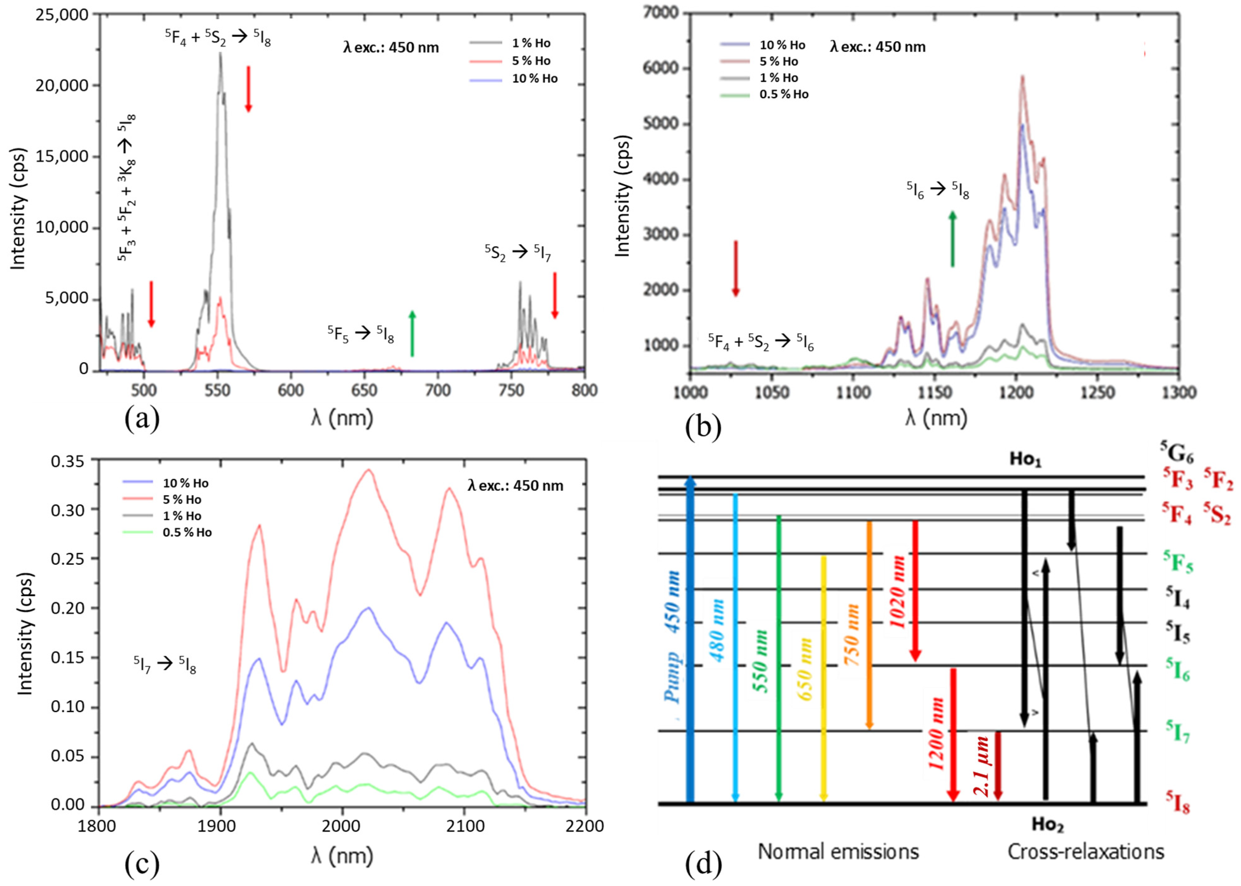
At a low doping rate (<1%), and after excitation according to the 450 nm transition 5I85G6, a cascade of transitions occurs. First, 5F3 + 5F2 + 3K85I8 radiative transition at 480 nm happens, as well as a non-radiative 5G65F4 + 5S2 transition. From this electronic level, two optical emissions occur at 550 nm and 750 nm corresponding, respectively, to the 5F4 + 5S25I8 and 5F4 + 5S25I7 transitions. In a second step, the transition 5I75I8 at 2100 nm occurs. From level 5F4, 5S2, a second transition 5F4 + 5S25I6 at 1020 nm, as well as a non-radiative transition 5F4 + 5S25F5, is observed. Then, from the 5F5 electronic level, an emission is present at 650 nm, linked to the 5F55I8 transition.
(2)
When the doping increases, due to the increase in the volume concentration of Ho3+ ions in the matrix, Ho1-Ho2 non-radiative Cross-Relaxation interactions are increasingly favored. These interactions, which induce modification of electronic level populations, lead to the overcrowding of the 5I7 and 5F5 levels according to the 5I85I7 or 5F3 + 5F25I7 transitions of Ho1 and 5F3 + 5F25F5 or 5I85F5 of Ho2. Thus, the intensity of the 5I75I8 transition in the Mid-IR at 2100 nm increases at the expense of Visible transitions up to 5% in Ho3+ ions (Figure 7a,c). Finally, a last relaxation takes place and induces the overcrowding of level 5I6 according to the transitions 5F4 + 5S25I6 and 5I85I6. Hence, the transition in the Near-IR 5I65I8 at 1200 μm is favored (Figure 7b).
(3)
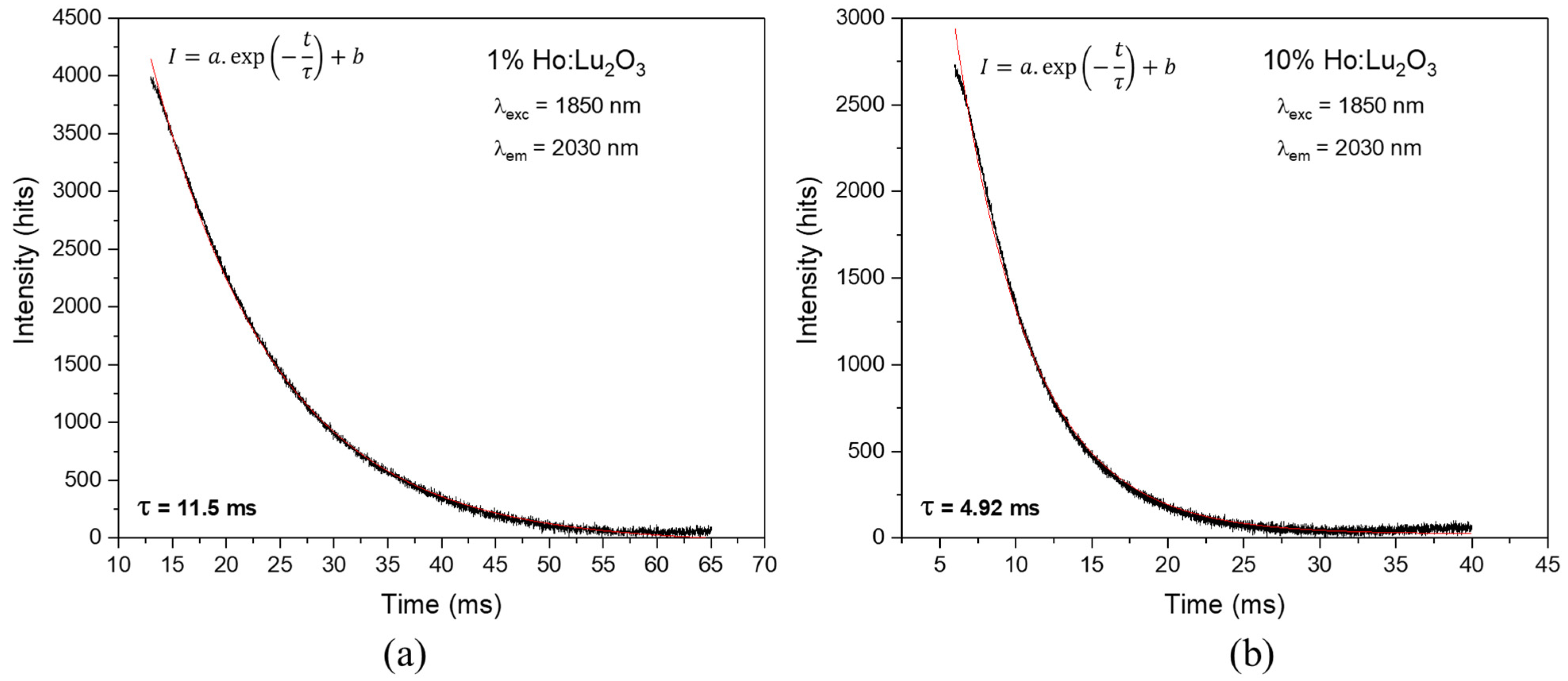
Above 5%, the phenomenon of fluorescence quenching by concentration is observed for all transitions [38]. For the highest concentration tested, namely 10% (2.84 × 10²¹ ions·cm−3), the fluorescence intensity in the Near-IR and Mid-IR is reduced by 40% compared to 5% (1.42 × 10²¹ ions·cm−3) (Figure 8b,c).
Figure 9 shows the fluorescence lifetime measured on the two extreme doping rates (i.e., 1% and 10%) in order to see the influence of Ho3+ concentration on the fluorescence decay of 5I75I8 laser transition at 2100 nm. Data were obtained for an excitation at 1850 nm and an emission at 2030 nm, corresponding to the maximum intensity observed in Figure 7c. The fluorescence lifetime was obtained by curve fitting with an exponential function. For 1%, a lifetime of 11.5 ms was obtained for 5I7 level state. Then, this lifetime decreased from 11.5 ms to 4.92 ms when the doping rate increased from 1% to 10%, which is in accordance with the literature on Ho-doped sesquioxides [39].
This phenomenon confirms the concentration quenching of the 2.1 µm emission caused by the high active ion density inside material, as mentioned in Section 3.1.

4. Discussion

In this work, transparent Lu2−xHoxO3 ceramics with fine microstructures (G < 1 µm) were obtained thanks to the SPS sintering technique. It increases the list of Lu2O3-based transparent ceramics obtained by SPS, previously doped with Nd [27,28], Tm [13] or Yb [40], proving the capability and versatility of this technique. Some of them have proven sufficient optical quality to observe a laser oscillation. One can cite also the works of Toci et al., who first demonstrated a laser effect with a 1 at.% Nd:Lu2O3 transparent ceramic obtained by SPS [41]. A few years later, Xu et al. achieved a laser slope efficiency of 38% with a maximum output of 1.25 W [42]. Many other available sintering techniques have been reported in the literature, such as air, vacuum or hot pressing (HP) pre-sintering followed by high-temperature post-HIP for a long time. The main drawback in this case relies on the large-sized grains (G > 10 µm), which are detrimental to the thermal and mechanical properties of ceramics. As an example, Kim et al. obtained HP + HIP Ho3+-doped Lu2O3 ceramics with an average grain size around 40–50 µm [39]. This difference is a consequence of the thermomechanical cycle used during the SPS treatment, performed at lower maximum temperatures and faster heating rates, which drastically limit grain growth during sintering [32]. As a result, compared to HIP, some differences in the optical properties, originating from the difference in the microstructure or carbon contamination of the samples after SPS treatment, could be expected.
First, concerning the effect of grain size, it seems to have no significant effect for Ho-doped Lu2O3. Indeed, by comparing data to 2% Ho-doped Lu2O3 ceramic obtained by HIP [39], a similar fluorescence lifetime in the order of 10 ms is obtained at a wavelength of 2140 nm. This value is also of the same order of magnitude as that obtained for single crystals of similar composition. The mean grain diameter obtained in this work is around 50 times lower than that obtained by HIP. That means that the volume of grains is about (G/G’)3 ~105 lower in our samples and the grain boundaries volume fraction is about (G’/G)~50 times higher. Therefore, the environment of Ho at grain boundaries should be similar than that observed in the bulk, and very limited Ho segregation is expected to occur in such material. This shows an advantage of this system for laser applications compared to other ones where dopant segregation at grain boundaries is observed, like in rare-earth-doped fluoride ceramics [43].
Second, concerning the use of SPS treatment, it seems to have no observable effect on the photoluminescence properties or lifetime decay according to results reported in Table 2. For example, on a 1%Ho:Lu2O3 single crystal, Dong et al. obtained a similar value of 9.8 ms for the same optical transition at a wavelength of 2030 nm [44]. A similar trend was observed for a lower Ho-doping content [45]. In addition, no additional optical transition, which could be associated with the SPS process, was observed in the absorption or luminescence spectra reported in Figure 5 and Figure 7. Moreover, Capobianco et al. obtained a very similar evolution of Visible transition intensities for Ho:Y2O3 samples sintered under air [37]. As a result, the Ho:Lu2O3 ceramics elaborated in this study present very similar optical properties compared to ceramics sintered by other techniques, as well as those of single crystals. Nanopowder chemical synthesis coupled with SPS sintering technology appears to be a very promising elaboration process to obtain laser ceramics. However, the optical quality should be still improved, i.e., the residual porosity seen in Figure 4 should be totally removed. Also, the samples are directly in contact with the graphite die during sintering and often present a grey/dark color after sintering. This was assigned by many studies to a reduction in the material linked to carbon contamination, and it can be easily removed by a reoxidation treatment under air at a moderate temperature (i.e., 900–120 °C). Nevertheless, the time required for such a treatment evolves as a function of e2, where e is the thickness of samples, because the rate of the reoxidation process is controlled by solid-state diffusion. As a result, a very long time is required for the reoxidation of samples with centimetric thickness, constituting a strong limitation of the efficiency of the whole process. Further investigations will be carried out on these two points to improve the optical quality of the ceramics and the scalability of the process.

5. Conclusions

Lu2−xHoxO3 transparent ceramics with a small grain size (<1 µm) were successfully manufactured by Spark Plasma Sintering of homemade nanopowders. The photoluminescence study performed in the Visible and IR domains has given similar results to those obtained for single crystals of similar compositions. Especially, cross-relaxation mechanisms have been identified and favor the 2 μm emission. Moreover, fluorescence lifetime measurements have highlighted concentration quenching, especially for high doping of 10% Ho. All of the obtained results indicate that the optical properties are very similar between Lu2−xHoxO3 transparent ceramics and single crystals. Thus, the distribution of Ho3+ ions should be similar in both materials, i.e., no clustering of Ho3+ ions in ceramics should take place, whatever the process parameters or the doping level. Finally, SPS appears to be a very promising method to produce transparent polycrystalline ceramics of the rare-earth oxide family. Among them, Lu2-xHoxO3 ceramics could substitute single crystals of similar composition and are very promising materials for high-energy solid-state lasers.

Author Contributions

Conceptualization, R.B. and L.V.; methodology, R.B. and L.V.; validation, R.B. and A.M.; formal analysis, L.V.; investigation, L.V. and S.G.-G.; data curation, L.V.; writing—original draft preparation, L.V.; writing—review and editing, R.B., V.J., G.D.-B., É.C. and A.M.; supervision, R.B. and A.M.; project administration, R.B.; funding acquisition, R.B. All authors have read and agreed to the published version of the manuscript.

Funding

This research was funded by the Region Nouvelle-Aquitaine (MISTRAL program n° 2017-1R50311 and HICEMIR program n° 2017-1R50311) and was also supported by an institutional grant from the National Research Agency under the investments for the future program with the reference ANR-10-LABX-0074-01 Sigma-LIM.

Institutional Review Board Statement

Not applicable.

Informed Consent Statement

Not applicable.

Data Availability Statement

The data presented in this study are available on request from the corresponding author. The data are not publicly available due to the data are part of an ongoing study.

Acknowledgments

The authors are grateful to Pierre Carles (IRCER, Limoges, France) for the TEM and STEM observations and Victor Anthony Garcia Rivera (COPL, University Laval) for the lifetime measurements performed in the frame of the International Associated Laboratory (LIA) LuMAQ.

Conflicts of Interest

The authors declare no conflicts of interest. The funders had no role in the design of the study; in the collection, analyses or interpretation of data; in the writing of the manuscript or in the decision to publish the results.

References

  1. Ikesue, A.; Aung, Y.L. Ceramic laser materials. Nat. Photon. 2008, 2, 721–727. [Google Scholar] [CrossRef]
  2. Sanghera, J.; Bayya, S.; Villalobos, G.; Kim, W.; Frantz, J.; Shaw, B.; Sadowski, B.; Miklos, R.; Baker, C.; Hunt, M.; et al. Transparent ceramics for high-energy laser systems. Opt. Mater. 2011, 33, 511–518. [Google Scholar] [CrossRef]
  3. Kim, W.; Villalobos, G.; Baker, C.; Frantz, J.; Shaw, B.; Bayya, S.; Bowman, S.; Sadowski, B.; Hunt, M.; Rock, B.; et al. Overview of transparent optical ceramics for high-energy lasers at NRL. Appl. Opt. 2015, 54, F210–F221. [Google Scholar] [CrossRef]
  4. Zhu, D.; Chen, Z.-Z.; Song, Y.-J.; Yuan, L.; Lin, Y.-Y.; Zhang, L.-N.; Cui, D.-F.; Bo, Y.; Peng, Q.-J.; Xu, Z.-Y.; et al. High Power (~10 kW) Yb:YAG Ceramic Slab Laser Operating at 1030 nm. IEEE Photon. Technol. Lett. 2023, 35, 789–792. [Google Scholar] [CrossRef]
  5. Zhang, W.X.; Zhou, J.; Liu, W.B.; Li, J.; Wang, L.; Jiang, B.X.; Pan, Y.B.; Cheng, X.J.; Xu, J.Q. Fabrication, properties and laser per-formance of Ho:YAG transparent ceramic. J. Alloys Compd. 2010, 506, 745–748. [Google Scholar] [CrossRef]
  6. Zhang, W.X.; Pan, Y.B.; Zhou, J.; Liu, W.B.; Li, J.; Jiang, B.X.; Cheng, X.J.; Xu, J.Q. Diode-Pumped Tm:YAG Ceramic Laser. J. Am. Ceram. Soc. 2009, 92, 2434–2437. [Google Scholar] [CrossRef]
  7. Scholle, K.; Lamrini, S.; Koopmann, P.; Fuhrberg, P. 2 µm Laser Sources and Their Possible Applications. In Frontiers in Guided Wave Optics and Optoelectronics; Pal, B., Ed.; IntechOpen: London, UK, 2010. [Google Scholar]
  8. Yang, H.; Zhang, J.; Qin, X.P.; Luo, D.W.; Ma, J.; Tang, D.Y.; Zhang, Q.T. Fabrication and Properties of High Quality Transparent Ho:YAG Ceramics. Solid State Phenom. 2012, 185, 51–54. [Google Scholar] [CrossRef]
  9. Payne, S.A.; Chase, L.L.; Smith, L.K.; Kway, W.L.; Krupke, W.F. Infrared cross section measurements for crystals doped with Er3+, Tm3+, and Ho3+. IEEE J. Quantum Electron. 1992, 28, 2619–2630. [Google Scholar] [CrossRef]
  10. Wang, Y.; Lan, R.; Mateos, X.; Li, J.; Hu, C.; Li, C.; Suomalainen, S.; Härkönen, A.; Guina, M.; Petrov, V.; et al. Broadly tunable mode-locked Ho:YAG ceramic laser around 21 µm. Opt. Express 2016, 24, 18003–18012. [Google Scholar] [CrossRef] [PubMed]
  11. Chen, H.; Shen, D.; Zhang, J.; Yang, H.; Tang, D.; Zhao, T.; Yang, X. In-band pumped highly efficient Ho:YAG ceramic laser with 21 W output power at 2097 nm. Opt. Lett. 2011, 36, 1575–1577. [Google Scholar] [CrossRef]
  12. Cheng, X.; Xu, J.; Wang, M.; Jiang, B.; Zhang, W.; Pan, Y. Ho:YAG ceramic laser pumped by Tm:YLF lasers at room temperature. Laser Phys. Lett. 2010, 7, 351–354. [Google Scholar] [CrossRef]
  13. Antipov, O.L.; Novikov, A.A.; Zakharov, N.G.; Zinoviev, A.P. Optical properties and efficient laser oscillation at 2066 nm of novel Tm:Lu2O3 ceramics. Opt. Mater. Express 2012, 2, 183–189. [Google Scholar] [CrossRef]
  14. Ikesue, A.; Aung, Y.L.; Lupei, V. Ceramic Lasers; Cambridge University Press: Cambridge, UK, 2013; p. 165. [Google Scholar]
  15. Salek, G.; Devoti, A.; Garcia, A.; Gaudon, M.; Jubera, V.; Demourgues, A. Tuning the composition of rare earth sesquioxides Gd2-xLaxO3:Eu3+ to control phase transitions at a high temperature to design new highly sensitive luminescence-based thermal sensors. R. Soc. Chem. 2016, 6, 298–306. [Google Scholar] [CrossRef]
  16. Permin, D.; Kurashkin, S.; Novikova, A.; Savikin, A.; Gavrishchuk, E.; Balabanov, S.; Khamaletdinova, N. Synthesis and luminescence properties of Yb-doped Y2O3, Sc2O3 and Lu2O3 solid solutions nanopowders. Opt. Mater. 2018, 77, 240–245. [Google Scholar] [CrossRef]
  17. Tang, F.; Huang, J.; Guo, W.; Wang, W.; Fei, B.; Cao, Y. Photoluminescence and laser behavior of Yb:YAG ceramic. Opt. Mater. 2012, 34, 757–760. [Google Scholar] [CrossRef]
  18. Unal, F.; Kaya, F. Kazmanli, KEffects of dopant rate and calcination parameters on photoluminescence emission of Y2O3:Eu3+ phosphors: A statistical approach. Ceram. Int. 2019, 45, 17818–17825. [Google Scholar] [CrossRef]
  19. Malinowski, M.; Kaczkan, M.; Wnuk, A.; Szufliñska, M. Emission from the high lying excited states of Ho3+ ions in YAP and YAG crystals. J. Lumin. 2004, 106, 269–279. [Google Scholar] [CrossRef]
  20. Liu, Z.; Ikesue, A.; Li, J. Research progress and prospects of rare-earth doped sesquioxide laser ceramics. J. Eur. Ceram. Soc. 2021, 41, 3895–3910. [Google Scholar] [CrossRef]
  21. Fu, Z.; Wu, N.; Long, H.; Hou, Z. Fabrication of fine-grained and high thermal properties Y2O3 transparent ceramics without sintering aids. Opt. Mater. 2024, 147, 114618. [Google Scholar] [CrossRef]
  22. Liu, Z.; Feng, Y.; Toci, G.; Pirri, A.; Patrizi, B.; Vannini, M.; Li, J. Influence of annealing on microstructures and properties of Yb:Lu2O3 transparent ceramics. J. Am. Ceram. Soc. 2023, 107, 1974–1984. [Google Scholar] [CrossRef]
  23. Liu, Z.; Toci, G.; Pirri, A.; Patrizi, B.; Feng, Y.; Hu, D.; Chen, H.; Hreniak, D.; Vannini, M.; Li, J. Fabrication and characterizations of Tm:Lu2O3 transparent ceramics for 2 μm laser applications. Opt. Mater. 2022, 131, 112705. [Google Scholar] [CrossRef]
  24. Maksimov, R.; Shitov, V.; Osipov, V.; Samatov, O.; Vakalov, D.; Malyavin, F.; Basyrova, L.; Loiko, P.; Camy, P. Fabrication, microstructure and mid-infrared luminescence of Er:(ScxY1-x)2O3 transparent ceramics. Opt. Mater. 2023, 137, 113542. [Google Scholar] [CrossRef]
  25. Chaim, R.; Levin, M.; Shlayer, A.; Estournes, C. Sintering and densification of nanocrystalline ceramic oxide powders: A review. Adv. Appl. Ceram. 2008, 107, 159–169. [Google Scholar] [CrossRef]
  26. Morita, K.; Kim, B.-N.; Yoshida, H.; Hiraga, K.; Sakka, Y. Distribution of carbon contamination in oxide ceramics occurring during spark-plasma-sintering (SPS) processing: II Effect of SPS and loading temperatures. J. Eur. Ceram. Soc. 2018, 38, 2596–2604. [Google Scholar] [CrossRef]
  27. Alombert-Goget, G.; Guyot, Y.; Guzik, M.; Boulon, G.; Ito, A.; Goto, T.; Yoshikawa, A.; Kikuchi, M. Nd3+-doped Lu2O3 transparent sesquioxide ceramics elaborated by the Spark Plasma Sintering (SPS) method. Part 1: Structural, thermal conductivity and spectroscopic characterization. Opt. Mater. 2015, 41, 3–11. [Google Scholar] [CrossRef]
  28. Boulesteix, R.; Epherre, R.; Noyau, S.; Vandenhende, M.; Maître, A.; Sallé, C.; Alombert-Goget, G.; Guyot, Y.; Brenier, A. Highly transparent Nd:Lu2O3 ceramics obtained by coupling slip-casting and spark plasma sintering. Scr. Mater. 2014, 75, 54–57. [Google Scholar] [CrossRef]
  29. Lupei, V.; Lupei, A.; Boulon, G.; Jouini, A.; Ikesue, A. Assessment of the distribution of the Yb3+ ions in Sc2O3 ceramics from cooperative absorption and emission. J. Alloys Compd. 2008, 451, 179–181. [Google Scholar] [CrossRef]
  30. Liu, Z.; Toci, G.; Pirri, A.; Patrizi, B.; Li, J.; Hu, Z.; Wei, J.; Pan, H.; Xie, T.; Vannini, M.; et al. Fabrication and laser operation of Yb:Lu2O3 transparent ceramics from co-precipitated nano-powders. J. Am. Ceram. Soc. 2019, 102, 7491–7499. [Google Scholar] [CrossRef]
  31. Thoř, T.; Rubešová, K.; Jakeš, V.; Mikolášová, D.; Cajzl, J.; Havlíček, J.; Nádherný, L.; Průša, F.; Kučerková, R.; Nikl, M. Dense ceramics of lanthanide-doped Lu2O3 prepared by spark plasma sintering. J. Eur. Ceram. Soc. 2021, 41, 741–751. [Google Scholar] [CrossRef]
  32. Viers, L.; Delaunay, F.; Boulesteix, R.; Vandenhende, M.; Antou, G.; Maître, A. Study of densification mechanisms during Spark Plasma Sintering of co-precipitated Ho:Lu2O3 nanopowders: Application to transparent ceramics for lasers. J. Eur. Ceram. Soc. 2021, 41, 7199–7207. [Google Scholar] [CrossRef]
  33. Kaminskii, A.A.; Akchurin, M.S.; Becker, P.; Ueda, K.; Bohatý, L.; Shirakawa, A.; Tokurakawa, M.; Takaichi, K.; Yagi, H.; Dong, J. Mechanical and optical properties of Lu2O3 host-ceramics for Ln3+ lasants. Laser Phys. Lett. 2008, 5, 300–303. [Google Scholar] [CrossRef]
  34. Boulesteix, R.; Maître, A.; Baumard, J.-F.; Rabinovitch, Y.; Reynaud, F. Reynaud, Light scattering by pores in transparent Nd:YAG ceramics for lasers: Correlations between microstructure and optical properties. Opt. Express 2010, 18, 14992–15002. [Google Scholar] [CrossRef]
  35. Brown, D.C.; Envid, V.; Zembek, J. Ho:YAG absorption cross sections from 1700 to 2200 nm at 83, 175, and 295 K. Appl. Opt. 2012, 51, 8147–8158. [Google Scholar] [CrossRef]
  36. Silver, J.; Barrett, E.; Marsh, P.J.; Withnall, R. Yttrium Oxide Upconverting Phosphors. 5. Upconversion Luminescent Emission from Holmium-Doped Yttrium Oxide under 632.8 nm Light Excitation. J. Phys. Chem. B 2003, 107, 9236–9242. [Google Scholar] [CrossRef]
  37. Capobianco, J.A.; Boyer, J.C.; Vetrone, F.; Speghini, A.; Bettinelli, M. Optical Spectroscopy and Upconversion Studies of Ho3+-Doped Bulk and Nanocrystalline Y2O3. Chem. Mater. 2002, 14, 2915–2921. [Google Scholar] [CrossRef]
  38. Auzel, F.; Baldacchini, G.; Laversenne, L.; Boulon, G. Radiation trapping and self-quenching analysis in Yb3+, Er3+, and Ho3+ doped Y2O3. Opt. Mater. 2003, 24, 103–109. [Google Scholar] [CrossRef]
  39. Kim, W.; Baker, C.; Bowman, S.; Florea, C.; Villalobos, G.; Shaw, B.; Sadowski, B.; Hunt, M.; Aggarwal, I.; Sanghera, J. Laser oscillation from Ho3+ doped Lu2O3 ceramics. Opt. Mater. Express 2013, 3, 913–919. [Google Scholar] [CrossRef]
  40. Kijko, V.; Maksimov, R.N.; Shitov, V.; Demakov, S. Yurovskikh, ASintering of transparent Yb-doped Lu2O3 ceramics using nanopowder produced by laser ablation method. J. Alloys Compd. 2015, 643, 207–211. [Google Scholar] [CrossRef]
  41. Toci, G.; Vannini, M.; Ciofini, M.; Lapucci, A.; Pirri, A.; Ito, A.; Goto, T.; Yoshikawa, A.; Ikesue, A.; Alombert-Goget, G.; et al. Nd3+-doped Lu2O3 transparent sesquioxide ceramics elaborated by the Spark Plasma Sintering (SPS) method. Part 2: First laser output results and comparison with Nd3+-doped Lu2O3 and Nd3+-Y2O3 ceramics elaborated by a conventional method. Opt. Mater. 2014, 41, 12–16. [Google Scholar] [CrossRef]
  42. Xu, C.; Yang, C.; Zhang, H.; Duan, Y.; Zhu, H.; Tang, D.; Huang, H.; Zhang, J. Efficient laser operation based on transparent Nd:Lu2O3 ceramic fabricated by Spark Plasma Sintering. Opt. Express 2016, 24, 20571–20579. [Google Scholar] [CrossRef] [PubMed]
  43. Boulon, G.; Epicier, T.; Zhao, W.; Guzik, M.; Pan, Y.; Jiang, B. Is There Segregation of Rare Earth Ions in Garnet Optical Ceramics? Bartolo, B.D., Collins, J., Baldassare, D.B., John, C., Eds.; Nano-Optics for enhancing light-matter interactions on a molecular scale. NATO Science for Space and Security Series B: Physics and Biophysics; Springer: Dordrecht, The Netherlands, 2013. [Google Scholar] [CrossRef]
  44. Dong, J.; Wang, W.; Xue, Y.; Hou, W.; Cao, X.; Xu, X.; Wu, F.; Luo, P.; Wang, Q.; Li, D.; et al. Crystal growth and spectroscopic analysis of Ho:Lu2O3 crystal for mid-infrared emission. J. Lumin. 2022, 251, 119192. [Google Scholar] [CrossRef]
  45. Koopmann, P.; Lamrini, S.; Scholle, K.; Schäfer, M.; Fuhrberg, P.; Huber, G. Multi-watt laser operation and laser parameters of Ho-doped Lu2O3 at 2.12 μm. Opt. Mater. Express 2011, 1, 1447–1456. [Google Scholar] [CrossRef]
Figure 1. Set-up used for Ho:Lu2O3 precipitation.
Ceramics 07 00013 g001
Figure 2. STEM micrographs of the coprecipitated 1 at.%Ho:Lu2O3 powder calcined at 1000 °C in bright field (a) or in dark field (b), and high-resolution TEM micrograph with corresponding diffraction pattern (c). XRD patterns registered as a function of the calcination temperature of 1 at.%Ho:Lu2O3 powder (d).
Ceramics 07 00013 g002
Figure 3. Transparent Ho:Lu2O3 ceramics with various Ho3+ doping rates (from the left to the right: 0.5 at.%, 1 at.%, 5 at.% and 10 at.%) sintered by SPS at 1400 °C for 15 min under 130 MPa of uniaxial pressure.
Ceramics 07 00013 g003
Figure 4. SEM (a) and TEM (b) micrographs of transparent 1 at.%Ho:Lu2O3 ceramics sintered by SPS at 1400 °C for 15 min under 130 MPa of uniaxial pressure. Residual porosity was identified by TEM (white arrows).
Ceramics 07 00013 g004
Figure 5. Optical transmission (a) and absorption (b) as a function of wavelength of Ho:Lu2O3 transparent ceramics with various Ho3+ doping rates.
Ceramics 07 00013 g005
Figure 6. Evolution of absorption cross-section of Ho:Lu2O3 transparent ceramics with various Ho3+ doping rates.
Ceramics 07 00013 g006
Figure 7. Emission spectra (excitation at 450 nm) of Ho:Lu2O3 transparent ceramics in the Visible range (a), Near-IR range (b) and Mid-IR (c) and the associated energy levels diagram built from experimentation (d).
Ceramics 07 00013 g007
Figure 8. Evolution of integrated emission intensities of Ho:Lu2O3 transparent ceramics in the Visible (400–800 nm) range, (a) Near-IR range (1000–1300 nm) (b) and Mid-IR range (1800–2200 nm) (c).
Ceramics 07 00013 g008
Figure 9. Fluorescence lifetime of 1% (a) and 10% (b) transparent ceramics. Fitted curves are represented in red lines with associated exponential decay function.
Ceramics 07 00013 g009
Table 1. Spectroscopic characteristics of Lu2-xHoxO3 samples as a function of Ho3+ concentration.
%at. Ho0.5%1%5%10%
CHo (cm−3)1.43 × 10202.85 × 10201.42 × 10212.84 × 1021
Absorption cross section at 1927 nm (cm2)1.26 × 10−201.03 × 10−201.26 × 10−201.03 × 10⁻²⁰
Fluorescence lifetime at 2030 nm (ms)-11.5-4.92
Table 2. Spectroscopic characteristics of Lu2-xHoxO3 samples as a function of process parameters.
ReferenceCompositionMaterialSinteringGmean (µm)T% at 2 µmλabs (nm)σabs (cm2)λem (nm)σem (cm2)τrad (ms)
This work1%Ho:Lu2O3CeramicSPS + air annealing<1 µm80%19271 × 10−202030 11.5
Kim et al. [39]2%Ho:Lu2O3CeramicPost-HIP + air annealing50 µm>80%1942-2140-10
Dong et al. [44]1%Ho:Lu2O3Single-crystal-->80%19291.1 × 10−2020273.3 × 10−219.5
Koopmann et al. [45]0.3%Ho:Lu2O3Single-crystal-->80%19281.2 × 10−2021244.5 × 10−2110
Disclaimer/Publisher’s Note: The statements, opinions and data contained in all publications are solely those of the individual author(s) and contributor(s) and not of MDPI and/or the editor(s). MDPI and/or the editor(s) disclaim responsibility for any injury to people or property resulting from any ideas, methods, instructions or products referred to in the content.

