
Macrocyclic Receptors for Identification and Selective Binding of Substrates of Different Nature


Abstract
:1. Introduction
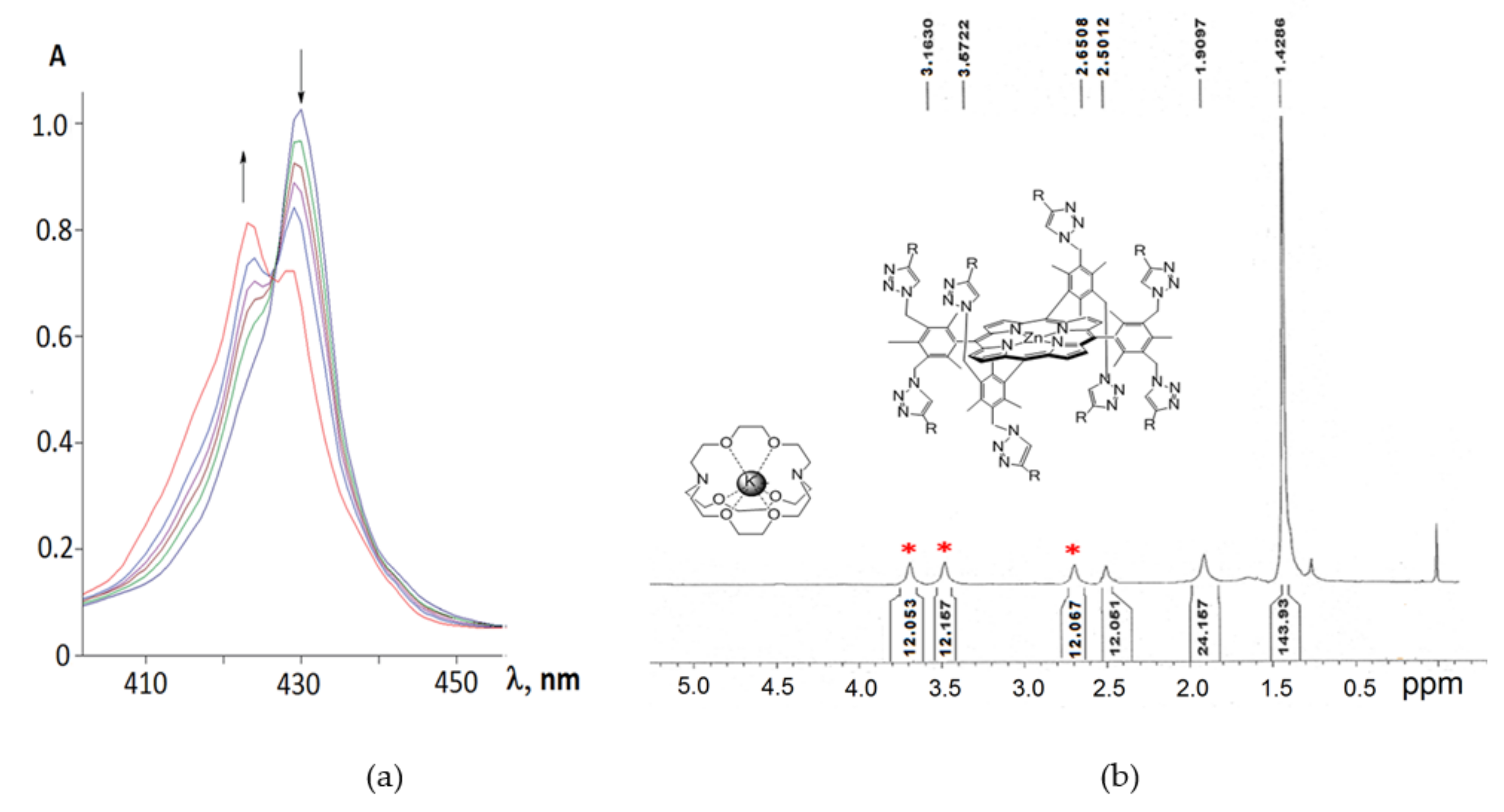
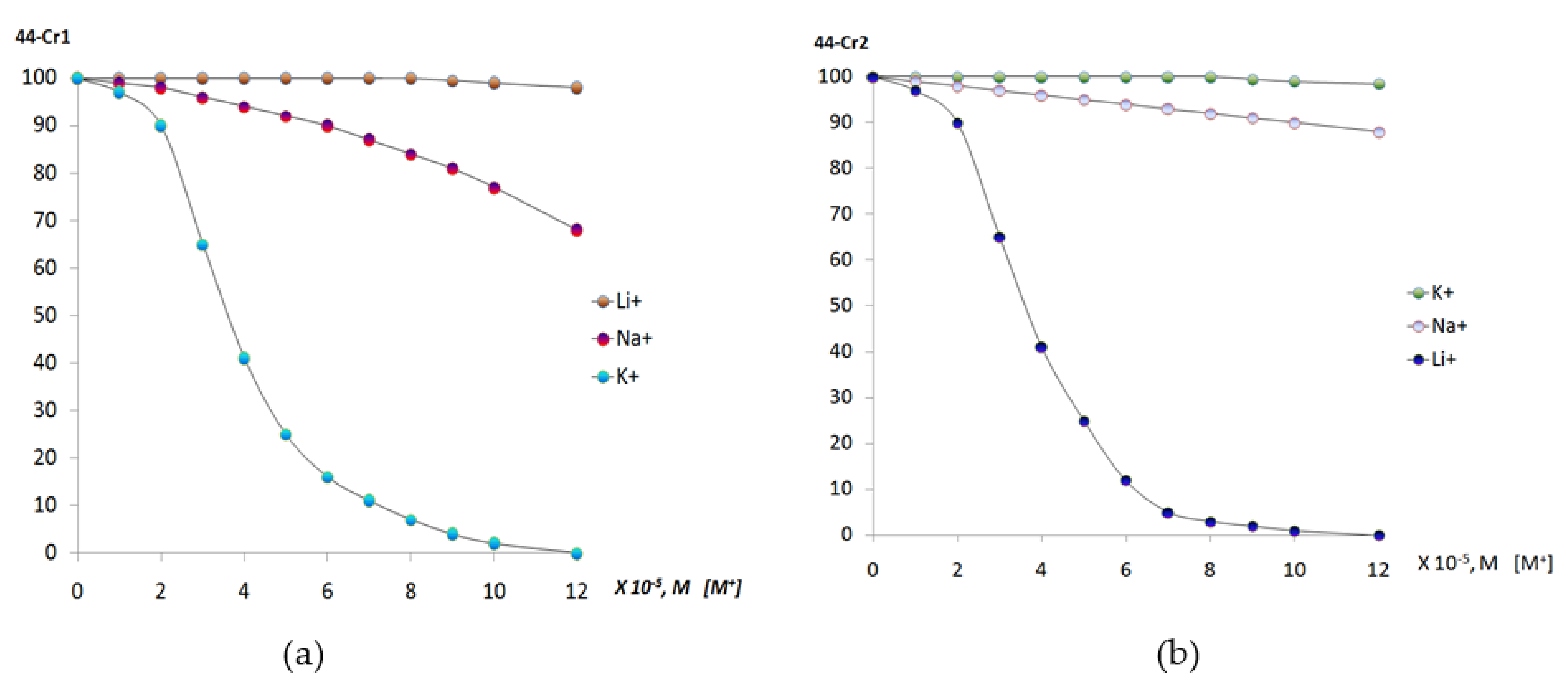
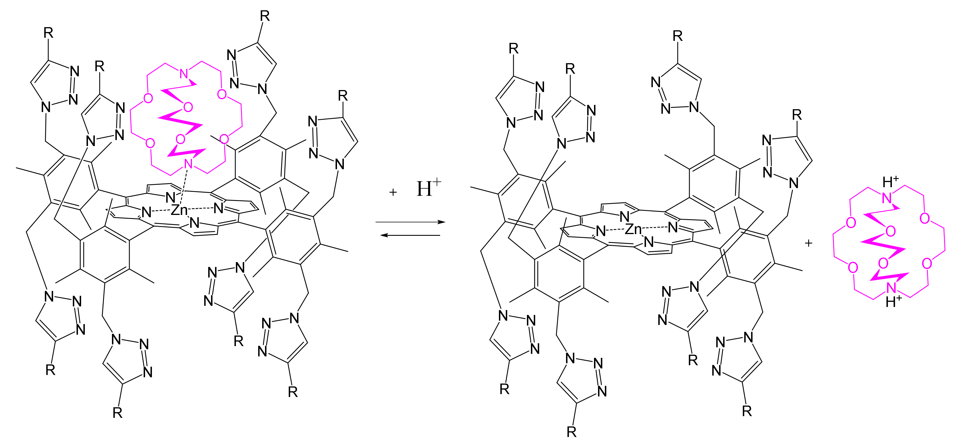
2. Molecular Receptors Based on Porphyrin Conjugates with Macrocyclic Compounds of Different Natures
3. Molecular Receptors Based on Porphyrins Modified at the Macrocycle Periphery with Bulky Substituents
4. Conclusions
Author Contributions
Funding
Institutional Review Board Statement
Informed Consent Statement
Data Availability Statement
Acknowledgments
Conflicts of Interest
References
- Beletskaya, I.; Tyurin, V.S.; Tsivadze, A.Y.; Gulard, R.; Stern, C. Supramolecular Chemistry of Metalloporphyrins. Chem. Rev. 2009, 109, 1659–1713. [Google Scholar] [CrossRef]
- Guchhait, T.; Sasmal, S.; Khan, F.S.T.; Rath, S.P. Oxo- and hydroxo-bridged diiron(III) porphyrin dimers: Inorganic and bio-inorganic perspectives and effects of intermacrocyclic interactions. Coord. Chem. Rev. 2017, 337, 112–144. [Google Scholar] [CrossRef]
- Mamardashvili, G.M.; Kaigorodova, E.Y.; Dmitrieva, O.; Mamardashvili, N.Z.; Koifman, O.I. Molecular recognition of imidazole derivatives by Co(III)-porphyrins in phosphate buffer (pH = 7.4) and cetylpyridinium chloride containing solutions. Molecules 2021, 26, 868. [Google Scholar] [CrossRef]
- Mamardashvili, N.Z.; Koifman, M.O. Synthesis of Monohydroxy-Substituted Diarylporphyrins and Their Binding Ability towards Aminobenzoic Acids. Macroheterocycles 2011, 4, 30–33. [Google Scholar] [CrossRef] [Green Version]
- Gil-Ramirez, G.; Karlen, S.D.; Shundo, A.; Porfyrakis, K.; Ito, Y.; Briggs, G.A.D.; Morton, J.J.; Anderson, H.L. A Cyclic Porphyrin Trimer as a Receptor for Fullerenes. Org. Lett. 2010, 12, 3544–3547. [Google Scholar] [CrossRef]
- Hu, J.; Liu, S. Engineering Responsive Polymer Building Blocks with Host–Guest Molecular Recognition for Functional Applications. Acc. Chem. Res. 2014, 47, 2084–2095. [Google Scholar] [CrossRef]
- Chandramouli, N.; Ferrand, Y.; Lautrette, G.; Kauffmann, B.; Mackereth, C.D.; Laguerre, M.; Dubreuil, D.; Huc, I. Iterative design of a helically folded aromatic oligoamide sequence for the selective encapsulation of fructose. Nat. Chem. 2015, 7, 334–341. [Google Scholar] [CrossRef] [PubMed]
- Margetić, D. Host-guest Studies of Bis-porphyrins. Curr. Org. Chem. 2012, 16, 829–851. [Google Scholar] [CrossRef]
- Davis, C.M.; Ohkubo, K.; Lammer, A.D.; Kim, D.S.; Kawashima, Y.; Sessler, J.L.; Fukuzumi, S. Photoinduced electron transfer in a supramolecular triad produced by porphyrin anion-induced electron transfer from tetrathiafulvalene calix[4]pyrrole to Li+@C60. Chem. Commun. 2015, 51, 9789–9792. [Google Scholar] [CrossRef] [Green Version]
- Liu, Q.; Dong, J.; Sun, Q.; Zhao, S.; Chen, Y.; Jiang, J. A novel calix[4]arene-modified porphyrin-baseddual-mode sensor for the specific detection ofdopamine with excellent performance. New J. Chem. 2019, 43, 10376–10381. [Google Scholar] [CrossRef]
- Nguyen, N.T.; Hofkens, J.; Scheblykin, I.G.; Kruk, M.; Dehaen, W. Click Reaction Synthesis and Photophysical Studies of Dendritic Metalloporphyrins. Eur. J. Org. Chem. 2014, 2014, 1766–1777. [Google Scholar] [CrossRef]
- Merkas, S.; Bouatra, S.; Rein, R.; Piantanida, I.; Zinic, M.; Solladié, N. Pre-organized dinucleosides with pendant porphyrins for the formation of sandwich type complexes with DABCO with high association constants. J. Porphyr. Phthalocyanines 2015, 19, 535–546. [Google Scholar] [CrossRef]
- Higashino, T.; Fujimori, Y.; Sugiura, K.; Tsuji, Y.; Ito, S.; Imahori, H. Synthesis of push–pull porphyrin with two electron-donating and two electron-withdrawing groups and its application to dye-sensitized solar cell. J. Porphyr. Phthalocyanines 2015, 19, 140–149. [Google Scholar] [CrossRef]
- Sharma, G.D.; Zervaki, G.E.; Ladomenou, L.; Koukaras, E.N.; Panagiotis, P.; Angaridis, P.P.; Coutsolelos, A.G. Donor-π-acceptor, triazine-linked porphyrin dyads as sensitizers for dye-sensitized solar cells. J. Porphyr. Phthalocyanines 2015, 19, 175–191. [Google Scholar] [CrossRef]
- Dhamija, A.; Mondal, P.; Saha, B.; Rath, S.P. Induction, control, and rationalization of supramolecular chirogenesis using metalloporphyrin tweezers: A structure-function correlation. Dalton Trans. 2020, 49, 10679–10700. [Google Scholar] [CrossRef]
- Milanesio, M.E.; Alvarez, M.G.; Durantini, E.N. Methoxyphenyl Porphyrin Derivatives as Phototherapeutic Agents. Curr. Bioact. Compd. 2010, 6, 97–105. [Google Scholar] [CrossRef]
- De Visser, S.P.; Valentine, J.S.; Nam, W. A Biomimetic Ferric Hydroperoxo Porphyrin Intermediate. Angew. Chem. Int. Ed. Engl. 2010, 49, 2099–2101. [Google Scholar] [CrossRef]
- Gonsalves, A.M.A.R.; Serra, A.C.; Pineiro, M. The small stones of Coimbra in the huge tetrapyrrolic chemistry building. J. Porphyr. Phthalocyanines 2009, 13, 429–445. [Google Scholar] [CrossRef]
- Maeda, C.; Kamada, T.; Aratani, N.; Osuka, A. Chiral self-discriminative self-assembling of meso-meso linked diporphyrins. Coord. Chem. Rev. 2007, 251, 2743–2752. [Google Scholar] [CrossRef]
- Hori, T.; Nakamura, Y.; Aratani, N.; Osuka, A. Exploration of electronically interactive cyclic porphyrin arrays. J. Organomet. Chem. 2007, 692, 148–155. [Google Scholar] [CrossRef]
- Mamardasvili, G.M.; Mamardashvili, N.Z.; Koifman, O.I. Self-assembling systems based on porphirins. Russ. Chem. Rev. 2008, 77, 765–782. [Google Scholar] [CrossRef]
- Mamardashvili, G.M.; Mamardashvili, N.Z.; Koifman, O.I. Synthesis and receptor properties of calix[4]pyrroles. Russ. Chem. Rev. 2015, 84, 275–287. [Google Scholar] [CrossRef]
- Balaban, T.S. Tailoring Porphyrins and Chlorins for Self-Assembly in Biomimetic Artificial Antenna Systems. Acc. Chem. Res. 2005, 38, 612–623. [Google Scholar] [CrossRef]
- Carofiglio, T.; Lubian, E.; Varotto, A. Synthesis, heterogenization and sensing properties of melamine-bridged Bis-porphyrin dimers. J. Porphyr. Phthalocyanines 2010, 14, 701–707. [Google Scholar] [CrossRef]
- Samaroo, D.; Vinodu, M.; Chen, X.; Drain, C.M. meso-Tetra(pentafluorophenyl)porphyrin as an Efficient Platform for Combinatorial Synthesis and the Selection of New Photodynamic Therapeutics using a Cancer Cell Line. J. Comb. Chem. 2007, 9, 998–1011. [Google Scholar] [CrossRef] [PubMed] [Green Version]
- Tsuda, A. Design of Porphyrin Nanoclusters toward Discovery of Novel Properties and Functions Bull. Chem. Soc. Jpn. 2009, 82, 11–28. [Google Scholar] [CrossRef] [Green Version]
- Goldberg, I. Crystal engineering of nanoporous architectures and chiralporphyrinassemblies. CrystEngComm 2008, 10, 637–645. [Google Scholar] [CrossRef]
- Boyd, P.D.W.; Reed, C.A. Fullerene−Porphyrin Constructs. Acc. Chem. Res. 2005, 38, 235–242. [Google Scholar] [CrossRef] [Green Version]
- Jurow, M.; Schuckman, A.E.; Batteas, J.D.; Drain, C.M. Porphyrins as molecular electronic components of functional devices. Coord. Chem. Rev. 2010, 254, 2297–2310. [Google Scholar] [CrossRef] [Green Version]
- Yua, G.; Zhub, B.; Shaoc, L.; Zhouc, J.; Sahad, M.L.; Shid, B.; Zhang, Z.; Hong, T.; Lib, S.; Chena, X.; et al. Host−guest complexation-mediated codelivery of anticancer drug and photosensitizer for cancer photo-chemotherapy. Proc. Natl. Acad. Sci. USA 2019, 116, 6618–6623. [Google Scholar] [CrossRef] [Green Version]
- Vinodh, M.; Alipour, F.H.; Abdirahman, A.M.; Talal, F. Al-Azemi Molecular Assemblies of Porphyrins and Macrocyclic Receptors: Recent Developments in Their Synthesis and Applications. Molecules 2012, 17, 11763–11799. [Google Scholar] [CrossRef] [Green Version]
- Blom, M.; Norrehed, S.; Andersson, C.-H.; Huang, H.; Light, M.E.; Bergquist, J.; Grennberg, H.; Gogoll, A. Synthesis and Properties of Bis-Porphyrin Molecular Tweezers: Effects of Spacer Flexibility on Binding and Supramolecular Chirogenesis. Molecules 2016, 21, 16. [Google Scholar] [CrossRef] [PubMed] [Green Version]
- Liang, X.; Honglin Zhang, H.; Luo, H.; Qin, M.; Li, M.; Zhu, W. Bio-Inspired Ni(II) Porphyrin Dimers with a Bridging Diphenyl Moiety: Facile Synthesis and Molecular Inherent Chirality. Macroheterocycles 2019, 12, 35–39. [Google Scholar] [CrossRef]
- Huang, X.; Borhan, B.; Rickman, B.H.; Nakanishi, K.; Berova, N. Binding of Cationic Bis-porphyrins Linked with p- or m- Xylylenediamine and Their Zinc(II) Complexes to Duplex DNA. Molecules 2008, 13, 3117–3128. [Google Scholar] [CrossRef] [Green Version]
- Ballester, P.; Costa, A.; Castilla, A.M.; Dey, P.M.; Frontera, A.; Gomila, R.M.; Hunter, C.A. DABCO-Directed Self-Assembly of Bisporphyrins (DABCO=1,4-Diazabicyclo[2.2.2]octane). Chem. Eur. J. 2005, 11, 2196. [Google Scholar] [CrossRef]
- Dudic, M.; Lhoták, P.; Proškova, P.; Stibor, I.; Lang, K.; Sýkora, J. Calixarene-based metalloporphyrins: Molecular tweezers for complexation of DABCO. Tetrahedron 2003, 59, 2409–2415. [Google Scholar] [CrossRef]
- Káš, M.; Lang, K.; Stibor, I.; Lhoták, P. Novel fullerene receptors based on calixarene-porphyrin conjugates. Tetrahedron Lett. 2007, 48, 477–481. [Google Scholar] [CrossRef]
- Hosseini, A.; Taylor, S.; Accorsi, G.; Armaroli, N.; Reed, C.A.; Boyd, P.D. Calix[4]arene-Linked Bisporphyrin Hosts for Fullerenes: Binding Strength, Solvation Effects, and Porphyrin−Fullerene Charge Transfer Bands. J. Am. Chem. Soc. 2006, 128, 15903–15913. [Google Scholar] [CrossRef] [Green Version]
- Jokic, D.; Zouhair Asfari, Z.; Weiss, J. The First Versatile Synthetic Approach to Cofacial Bis-Porphyrins with Calixarene Spacers. Org. Lett. 2002, 4, 2129–2132. [Google Scholar] [CrossRef]
- Mamardashvili, G.M.; Shinkar’, I.A.; Mamardashvili, N.Z.; Koifman, O.I. Calix[4]arene-Porphyrin Molecular Receptors for Selective Binding of Ethylenediamines. Russ. J. Coord. Chem. 2007, 33, 774–778. [Google Scholar] [CrossRef]
- Mamardashvili, G.M.; Mamardashvili, N.Z.; Koifman, O.I. Cation-Dependent Binding of Zinc Diethoxycarbonylcalix[4]arene-Bis(porphyrinate) Triethylenediamine. Russ. J. Coord. Chem. 2011, 37, 195–201. [Google Scholar] [CrossRef]
- Dudic, M.; Lhoták, P.; Stibor, I.; Dvoráková, H. Lang Kamil Synthesis and spectroscopic properties of porphyrin-(thia)calix[4]arene conjugates. Tetrahedron 2002, 58, 5475–5482. [Google Scholar] [CrossRef]
- Kundrat, O.; Kas, M.; Tkadlecova, M.; Lang, K.; Cvacka, J.; Stibor, I.; Lhotak, P. Thiacalix[4]arene–porphyrin conjugates with high selectivity towards fullerene C70. Tetrahedron Lett. 2007, 48, 6620–6623. [Google Scholar] [CrossRef]
- Rossom, W.V.; Kundrát, O.; Ngo, T.H.; Lhoták, P.; Dehaen, W.; Maes, W. An oxacalix[2]arene[2]pyrimidine-Bis(Zn-porphyrin) tweezer as a selective receptor towards fullerene C70. Tetrahedron Lett. 2010, 51, 2423–2426. [Google Scholar] [CrossRef]
- Dudić, M.; Lhoták, P.; Stibor, I.; Lang, K.; Proškova, P. Calix[4]arene-porphyrin Conjugates as Versatile Molecular Receptors for Anions. Org. Lett. 2003, 5, 149–152. [Google Scholar] [CrossRef]
- Yakushev, A.A.; Averin, A.D.; Sakovich, M.V.; Vatsouro, I.M.; Kovalev, V.V.; Syrbu, S.A.; Koifman, O.I.; Beletskaya, I.P. Synthesisoftheporphyrin-calix[4]areneconjugates via Pd-catalyzedaminationandtheirevaluationasfluorescentchemosensors. J. Porphyr. 2019, 23, 1551–1562. [Google Scholar] [CrossRef]
- Iwamoto, H.; Nishi, S.; Haino, T. Highly shape-selective guest encapsulation in the precisely defined cavity of a calix[4]arene-capped metalloporphyrin. Chem. Commun. 2011, 47, 12670–12672. [Google Scholar] [CrossRef]
- Nagasaki, T.; Fujishima, H.; Shinkai, S. Calix[4]arene-Capped Tetraphenylporphyrin. Synthetic Approach to a Chiral Capped Porphyrin with Regular C4 Symmetry. Chem. Lett. 1994, 23, 989–992. [Google Scholar] [CrossRef]
- Iwamoto, H.; Yukimasa, Y.; Yoshimasa Fukazawa, Y. Synthesis and binding behavior of a Zn(II)-porphyrin having calix[5]arene cap. Tetrahedron Lett. 2002, 43, 8191–8194. [Google Scholar] [CrossRef]
- Mibradt, R.; Weiss, J. Efficient Combination of Calix[4]arenes and meso-Diphenylporphyrins. Tetrahedron Lett. 1995, 36, 2999–3002. [Google Scholar] [CrossRef]
- Pognon, G.; Boudon, C.; Schenk, K.J.; Bonin, M.; Bach, B.; Weiss, J. Electrochemically Triggered Open and Closed Pacman Bis-metalloporphyrins. J. Am. Chem. Soc. 2006, 128, 3488–3489. [Google Scholar] [CrossRef] [PubMed]
- Holler, M.; Schmitt, M.; Nierengarten, J.-F. Synthesis and conformational analysis of porphyrin derivatives substituted with calix[4]arene subunits. J. Porphyr. Phthalocyanines 2011, 15, 1183–1188. [Google Scholar] [CrossRef]
- Mamardashvili, N.Z.; Mamardashvili, G.M.; Weiss, J. Complexation of Zinc Porphyrintes with 1,4-Diazabicyclo[2,2,2]octane. Russ. J. Inorg. Chem. 2005, 50, 218–224. [Google Scholar]
- Mamardashvili, N.Z.; Koifman, O.I. Cation and anion assisted binding of triethylenediamine by zinc Bis-porphyrinates. Russ. Chem. Bull. 2013, 62, 123–132. [Google Scholar] [CrossRef]
- Puglisi, A.; Purrello, R.; Rizzarelli, E.; Sortino, S.; Vecchio, G. Spectroscopic and self-association behavior of a porphyrin-β-cyclodextrinconjugate. New J. Chem. 2007, 31, 1499–1506. [Google Scholar] [CrossRef]
- Hosokawa, K.; Miura, Y.; Kiba, T.; Kakuchi, T.; Sato, S. Fluorescence Resonance Energy Transfer in Host–Guest Inclusion Complexes of Cyclodextrin–Porphyrin Composite in Aqueous Solution. Chem. Lett. 2008, 37, 60–61. [Google Scholar] [CrossRef]
- Fathalla, M.; Li, S.; Diebold, U.; Alb, A.; Jayawickramarajah, J. Water-soluble nanorods self-assembledviapristine C60 and porphyrin moieties. Chem. Commun. 2009, 28, 4209–4211. [Google Scholar] [CrossRef]
- Kralova, J.; Kejik, Z.; Briza, T.; Pouckova, P.; Kral, A.; Martasek, P.; Kral, V. Porphyrin−Cyclodextrin Conjugates as a Nanosystem for Versatile Drug Delivery and Multimodal Cancer Therapy. J. Med. Chem. 2010, 53, 128–138. [Google Scholar] [CrossRef]
- Guo, Y.; Zhang, P.; Chao, J.; Shuang, S.; Dong, C. Study on the supramolecular system of 5-(p-hydroxyphenyl)-10,15,20-tris-(4-chlorophenyl)porphyrin with cyclodextrins and its analytical characteristics. Spectrochim. Acta A 2008, 71A, 946–950. [Google Scholar] [CrossRef]
- Fathalla, M.; Neuberger, A.; Li, S.-C.; Schmehl, R.; Diebold, U.; Jayawickramarajah, J. Straightforward Self-Assembly of Porphyrin Nanowires in Water: Harnessing Adamantane/β-Cyclodextrin Interactions. J. Am. Chem. Soc. 2010, 132, 9966–9967. [Google Scholar] [CrossRef] [PubMed]
- Guo, Y.-J.; Chao, J.-B.; Pan, J.-H. Study on the interaction of 5-pyridine-10,15,20-tris-(p-chlorophenyl)porphyrin with cyclodextrins and DNA by spectroscopy. Spectrochim. Acta A 2007, 68A, 231–236. [Google Scholar] [CrossRef]
- Kiba, T.; Suzuki, H.; Hosokawa, K.; Kobayashi, H.; Baba, S.; Kakuchi, T.; Sato, S. SupramolecularJ-Aggregate Assembly of a Covalently Linked Zinc Porphyrin−β-cyclodextrin Conjugate in a Water/Ethanol Binary Mixture. J. Phys. Chem. B 2009, 113, 11560–11563. [Google Scholar] [CrossRef] [PubMed]
- Ermilov, E.A.; Menting, R.; Lau, J.T.F.; Leng, X.; Roeder, B.; Ng, D.K.P. Switching the photoinduced processes in host–guest complexes of β-cyclodextrin-substituted silicon(iv) phthalocyanines and a tetrasulfonated porphyrin. Phys. Chem. Chem. Phys. 2011, 13, 17633–17641. [Google Scholar] [CrossRef]
- Xu, H.; Ermilov, E.A.; Roeder, B.; Ng, D.K.P. Formation and energy transfer property of a subphthalocyanine–porphyrin complex held by host–guest interactions. Phys. Chem. Chem. Phys. 2010, 12, 7366–7370. [Google Scholar] [CrossRef]
- Khavasi, H.R.; Sasan, K.; Safari, N. Inclusion complex of ironmeso-tetrakis(p-sulfonatophenyl)porphyrin and 2-hydroxypropyl-β-cyclodextrin as a functional model of cytochrome P-450: Study on a supramolecular formation and its application in aqueous oxidation of styrene. J. Porphyr. Phthalocyanines 2007, 11, 874–882. [Google Scholar] [CrossRef]
- Liu, Y.; Ke, C.-F.; Zhang, H.-Y.; Cui, J.; Ding, F. Complexation-Induced Transition of Nanorod to Network Aggregates: Alternate Porphyrin and Cyclodextrin Arrays. J. Am. Chem. Soc. 2008, 130, 600–605. [Google Scholar] [CrossRef]
- Mamardashvili, G.M.; Mamardashvili, N.Z.; Koifman, O.I. Anion-Dependent Binding of Zinc Calix[4]pyrrole-Bisporphyrinate Triethylenediamine. Russ. J. Coord. Chem. 2011, 37, 872–877. [Google Scholar] [CrossRef]
- Donato Monti, D.; Monica, L.L.; Anita Scipioni, A.; Mancini, G. Effect of the inclusion of sodium cations on the binding properties of a switchable diporphyrin receptor. New J. Chem. 2001, 25, 780–782. [Google Scholar] [CrossRef]
- Iengo, E.; Zangrando, E.; Alessio, E.; Chambron, J.-C.; Heitz, V.; Flamigni, L.; Sauvage, J.-P. A Functionalized Noncovalent Macrocyclic Multiporphyrin Assembly from a Dizinc(II) Bis-Porphyrin Receptor and a Free-Base Dipyridylporphyrin. Chem. Eur. J. 2003, 9, 5879. [Google Scholar] [CrossRef]
- Flamigni, L.; Talarico, A.M.; Ventura, B.; Rein, R.; Solladié, N. A Versatile Bis-Porphyrin Tweezer Host for the Assembly of Noncovalent Photoactive Architectures: A Photophysical Characterization of the Tweezers and Their Association with Porphyrins and Other Guests. Chem. Eur. J. 2006, 12, 701. [Google Scholar] [CrossRef]
- Guldi, D.M.; Luo, C.; Swartz, A.; Scheloske, M.; Hirsch, A. Selforganisation in photoactive fullerene porphyrin based donor–acceptor ensembles. Chem. Commun. 2001, 1066–1067. [Google Scholar] [CrossRef]
- El-Khouly, M.E.; Hasegawa, J.; Momotake, A.; Sasaki, M.; Araki, Y.; Ito, O.; Arai, T. Intramolecular photoinduced processes of newly synthesized dual zinc porphyrin-fullerene triad with flexible linkers. J. Porph. Phthaloc. 2006, 10, 1380–1391. [Google Scholar] [CrossRef]
- Tashiro, K.; Aida, T. Metalloporphyrin hosts for supramolecular chemistry of fullerens. Chem. Soc. Rev. 2007, 36, 189–197. [Google Scholar] [CrossRef] [PubMed]
- D’Souza, F.; Gadde, S.; Zandler, M.E.; Itou, M.; Araki, Y.; Ito, O. Supramolecular complex composed of a covalently linked zinc porphyrin dimer and fulleropyrrolidine bearing two axially coordinating pyridine entities. Chem. Commun. 2004, 2276–2277. [Google Scholar] [CrossRef]
- Kubo, Y.; Ohno, T.; Yamanaka, J.; Tokita, S.; Iida, T.; Ishimaru, Y. Chirality-Transfer Control Using a Heterotopic Zinc(II) Porphyrin Dimer. J. Am. Chem. Soc. 2001, 123, 12700. [Google Scholar] [CrossRef]
- Brahma, S.; Ikbal, A.S.; Dhamija, A.; Rath, S.P. Highly Enhanced Bisignate Circular Dichroism of Ferrocene-Bridged Zn(II) Bisporphyrin Tweezer with Extended Chiral Substrates due to Well-Matched Host–Guest System. Inorg. Chem. 2014, 53, 2381–2395. [Google Scholar] [CrossRef]
- Nakazawa, J.; Mizuki, M.; Shimazaki, Y.; Tani, F.; Naruta, Y. Encapsulation of Small Molecules by a Cavitand Porphyrin Self-Assembled via Quadruple Hydrogen Bonds. Org. Lett. 2006, 8, 4275–4278. [Google Scholar] [CrossRef]
- Churakhina, Y.I.; Ivanova, Y.B.; Maltseva, O.V.; Mamardashvili, N.Z. Basic properties of porphyrins with polyethylenoxide spacers of various length. Russ. J. Gen. Chem. 2009, 79, 2435–2439. [Google Scholar] [CrossRef]
- Ho, K.-H.L.; Hijazi, I.; Rivier, L.; Gautier, C.; Jousselme, B.; de Miguel, G.; Romero-Nieto, C.; Guldi, D.M.; Heinrich, B.; Donnio, B.; et al. Host–Guest Complexation of [60]Fullerenes and Porphyrins Enabled by “Click Chemistry”. Chem. Eur. J. 2013, 19, 11374–11381. [Google Scholar] [CrossRef]
- Al-Azemi, T.F.; Vinodh, M. Synthesis of porphyrin conjugates based on conformationally rigid and flexible resorcin[4]arene frameworks. Tetrahedron 2011, 67, 2585–2590. [Google Scholar] [CrossRef]
- Shinoda, S.; Ohashi, M.; Tsukube, H. “Pocket Dendrimers” as Nanoscale Receptors for Bimolecular Guest Accommodation. Chem. Eur. J. 2007, 13, 81–89. [Google Scholar] [CrossRef] [PubMed]
- Shinoda, S. Nanoscale substrate recognition by porphyrin dendrimers with patched structures. J. Incl. Phenom. Macrocycl. Chem. 2007, 59, 1–9. [Google Scholar] [CrossRef]
- Huang, X.; Zhu, C.; Zhang, S.; Li, W.; Guo, Y.; Zhan, X.; Liu, Y.; Bo, Z. Porphyrin−Dithienothiophene π-Conjugated Copolymers: Synthesis and Their Applications in Field-Effect Transistors and Solar Cells. Macromolecules 2008, 41, 6895–6902. [Google Scholar] [CrossRef]
- Lee, C.Y.; Hupp, J.T. Dye Sensitized Solar Cells: TiO2 Sensitization with a Bodipy-Porphyrin Antenna System. Langmuir 2010, 26, 3760–3765. [Google Scholar] [CrossRef]
- Kruk, M.M.; Starukhin, A.S.; Mamardashvili, N.Z.; Mamardashvili, G.M.; Ivanova, Y.B.; Maltseva, O.V. Tetrapyrrolic compounds as hosts for binding of halides and alkali metal cations. J. Porphyr. Phthalocyanines 2009, 13, 1148–1158. [Google Scholar] [CrossRef]
- Li, L.; Huang, Y.; Peng, J.; Cao, Y.; Peng, X. Enhanced performance of solution-processed solar cells based onporphyrinsmall molecules with a diketopyrrolopyrrole acceptor unit and apyridineadditive. J. Mater. Chem. A 2013, 1, 2144–2150. [Google Scholar] [CrossRef]
- Favereau, L.; Warnan, J.; Anne, F.B.; Pellegrin, Y.; Blart, E.; Jacquemin, D.; Odobel, F. Diketopyrrolopyrrole-zinc porphyrin, a tuned panchromatic association fordye-sensitized solar cells. J. Mater. Chem. A 2013, 1, 7572–7575. [Google Scholar] [CrossRef]
- Maufroy, A.; Favereau, L.; Anne, F.B.; Pellegrin, Y.; Blart, E.; Hissler, M.; Jacquemin, D.; Odobel, F. Synthesis and properties of push–pull porphyrins as sensitizers for NiO based dye-sensitized solar cells. J. Mater. Chem. A 2015, 3, 3908–3917. [Google Scholar] [CrossRef] [Green Version]
- Higashino, T.; Imahori, H. Porphyrins as excellent dyes for dye-sensitized solar cells: Recent developments and insights. Dalton Trans. 2015, 44, 448–463. [Google Scholar] [CrossRef] [PubMed]
- Ballester, P.; Claudel, M.; Durot, S.; Kocher, L.; Choepffl, S.; Heitz, V. A Porphyrin Coordination Cage Assembled from Four Silver(I) Triazolyl-Pyridine Complexes. Chem. Eur. J. 2015, 21, 15339–15348. [Google Scholar] [CrossRef]
- Durot, S.; Taesch, J.; Heitz, V. Multiporphyrinic Cages: Architectures and Functions. Chem. Rev. 2014, 114, 8542–8578. [Google Scholar] [CrossRef] [PubMed]
- Qiu, W.-G.; Li, Z.-F.; Bai, G.-M.; Meng, S.-N.; Dai, H.-X.; He, H. Study on the inclusion behavior betweenmeso-tetrakis[4-(3-pyridiniumpropoxy)phenyl]prophyrin tetrakisbromide and β-cyclodextrin derivatives in aqueous solution. Spectrochim. Acta A 2007, 66A, 1189–1193. [Google Scholar] [CrossRef]
- Guo, Y.-J.; Guo, L.; Pan, J.-H. Study on the supramolecular system of tetrakis(2-hydroxy-5-sulfonatophenyl)porphyrin with cyclodextrins by spectroscopy. Phys. Chem. Liq. 2007, 45, 261–269. [Google Scholar] [CrossRef]
- Tsuchiya, Y.; Yamano, A.; Shiraki, T.; Sada, K.; Shinkai, S. Single-crystal Structure of Porphyrin Bicapped with Trimethyl-β-cyclodextrins: A Novel Dye-oriented Material. Chem. Lett. 2011, 40, 99–101. [Google Scholar] [CrossRef]
- Callari, F.L.; Mazzaglia, A.; Scolaro, L.M.; Valli, L.; Sortino, S. Biocompatible nanoparticles of amphiphilic cyclodextrins entangling porphyrins as suitable vessels for light-induced energy andelectron transfer. J. Mater. Chem. 2008, 18, 802–805. [Google Scholar] [CrossRef]
- Yu, M.; Chen, Y.; Zhang, N.; Liu, Y. Construction and transmembrane dissociation behavior of supramolecular assembly of quinolinocyclodextrin with porphyrin. Org. Biomol. Chem. 2010, 8, 4148–4154. [Google Scholar] [CrossRef]
- Ferro, S.; Jori, G.; Sortino, S.; Stancanelli, R.; Nikolov, P.; Tognon, G.; Ricchelli, F.; Mazzaglia, A. Inclusion of 5-[4-(1-Dodecanoylpyridinium)]-10,15,20-triphenylporphine in Supramolecular Aggregates of Cationic Amphiphilic Cyclodextrins: Physicochemical Characterization of the Complexes and Strengthening of the Antimicrobial Photosensitizing Activity. Biomacromolecules 2009, 10, 2592–2600. [Google Scholar] [CrossRef]
- Mora, S.J.; Cormick, M.P.; Milanesio, M.E.; Durantini, E.N. The photodynamic activity of a novel porphyrin derivative bearing a fluconazole structure in different media and against Candida albicans. Dye. Pigm. 2010, 87, 234–240. [Google Scholar] [CrossRef]
- Zhang, Y.-M.; Chen, Y.; Zhuang, R.-J.; Liu, Y. Supramolecular architecture of tetrathiafulvalene-bridged Bis(β-cyclodextrin) with porphyrin and its electron transfer behaviors. Photochem. Photobiol. Sci. 2011, 10, 1393–1398. [Google Scholar] [CrossRef]
- Zhang, Y.-M.; Chen, Y.; Yang, Y.; Liu, P.; Liu, Y. Supramolecular Architectures by Fullerene-Bridged Bis(permethyl-β-cyclodextrin)s with Porphyrins. Chem. Eur. J. 2009, 15, 11333–11340. [Google Scholar] [CrossRef]
- Valli, L.; Giancane, G.; Mazzaglia, A.; Scolaro, L.M.; Conoci, S.; Sortino, S. Photoresponsive multilayer films by assembling cationic amphiphiliccyclodextrinsandanionicporphyrinsat the air/waterinterface. J. Mater. Chem. 2007, 17, 1660–1663. [Google Scholar] [CrossRef]
- Wang, K.-R.; Guo, D.-S.; Jiang, B.-P.; Liu, Y. Excitonic coupling interactions in the self-assembly of perylene-bridged Bis-(β-cyclodextrin)s and porphyrin. Chem. Commun. 2012, 48, 3644–3646. [Google Scholar] [CrossRef] [PubMed]
- Maltceva, O.; Mamardashvili, G.; Khodov, I.; Lazovskiy, D.; Krest’yaninov, M.; Mamardashvili, N.; Dehaen, W. Molecular recognition of nitrogen—Containing bases by Zn[5,15-Bis-(2,6-dodecyloxyphenyl)]porphyrin. Supramol. Chem. 2017, 29, 360–369. [Google Scholar] [CrossRef]
- Long, L.; Jin, J.Y.; Zhang, Y.; Yang, R.; Wang, K. I sensing approach for albumin nteractionsbetweenproteinand porphyrin-containingcyclodextrinsupramolecular system: A fluorescent. Analyst 2008, 133, 1201–1208. [Google Scholar] [CrossRef]
- Oliveri, V.; Puglisi, A.; Vecchio, G. New conjugates of β-cyclodextrin with manganese(iii) salophen and porphyrin complexes as antioxidant systems. Dalton Trans. 2011, 40, 2913–2919. [Google Scholar] [CrossRef]
- Kano, K. Porphyrin-cyclodextrin supramolecular complexes as myoglobin model in water. Colloid Polym. Sci. 2008, 286, 79–84. [Google Scholar] [CrossRef]
- Watanabe, K.; Kitagishi, H.; Kano, K. Supramolecular Ferric Porphyrins as Cyanide Receptors in Aqueous Solution. ACS Med. Chem. Lett. 2011, 2, 943–947. [Google Scholar] [CrossRef] [Green Version]
- Kano, K.; Kitagishi, H.; Dagallier, C.; Kodera, M.; Matsuo, T.; Hayashi, T.; Hisaeda, Y.; Hirota, S. Iron Porphyrin−Cyclodextrin Supramolecular Complex as a Functional Model of Myoglobin in Aqueous Solution. Inorg. Chem. 2006, 45, 4448–4460. [Google Scholar] [CrossRef]
- Nguyen, N.T.; Mamardashvili, G.M.; Kulikova, O.M.; Scheblykin, I.G.; Mamardashvili, N.Z.; Dehaen, W. Binding ability of first and second generation/ carbazolylphenyl dendrimers with Zn(II) tetraphenylporphyrin core towards small heterocyclic substrates. RSC Adv. 2014, 4, 19703–19709. [Google Scholar] [CrossRef]
- Mamardashvili, G.M.; Maltceva, O.V.; Mamardashvili, N.Z.; Nguyen, N.T.; Dehaen, W. Cation assisted complexation of octacarbazolylphenyl substituted Zn(II)-tetraphenylporphyrin with [2,2,2]cryptand. RSC Adv. 2015, 5, 44557–44562. [Google Scholar] [CrossRef]
- Bukhzam, A.; Bade, N. Crown Ethers: Their Complexes and Analytical Applications. J. Applic. Chem. 2014, 3, 237–244. [Google Scholar]
- Albrecht, K.; Kasai, Y.; Kimoto, A.; Yamamoto, K. The Synthesis and Properties of Carbazole−Phenylazomethine Double Layer-Type Dendrimers. Macromolecules 2008, 41, 3793–3800. [Google Scholar] [CrossRef]
- Albrecht, K.; Kasai, Y.; Kuramoto, Y.; Yamamoto, K. Dynamic control of dendrimer–fullereneassociation by axial coordination to the core. Chem. Com. 2013, 46, 6861–6863. [Google Scholar] [CrossRef]


| “Molecular Tweezers” | Substrates | Ref. | “Molecular Tweezers” | Substrates | Ref. | ||
|---|---|---|---|---|---|---|---|
| MPorph. | Linked | MPorph. | Linked | ||||
| H2Porph.1 ZnPorph.2 |  |  | [36] [37] | H2Porph.1 |  |  | [38] |
| H2Porph.1 |  |  | [38] | H2Porph.1 H2Porph.3 H2Porph.4 |  |  | [38] |
| ZnPorph.6 |  |  | [39] | ZnPorph.7 |  |  | [40] |
| ZnPorph.7 |  |  | [41] | H2Porph.8 | 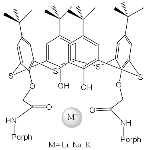 | Regulation of the interporphyrin distance by the introduction of metal cations | [42] |
| ZnPorph.3 |  |  | [43] | ZnPorph.3 | 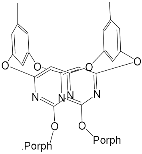 |  | [44] |
| H2Porph.9 |  | Anions Cl−, Br−, I−, NO3− | [45] | H2Porph.10 ZnPorph.10 |  | Fluorescent detection of Cu(II), Al(III), Cr(III), Zn(II), Cd(II) | [46] |
| ZnPorph.11 |  |  | [67] | ZnPorph.9 |  |  | [68] |
| ZnPorph.3 |  | H2Porph.12 | [71] | ZnPorph.3 | 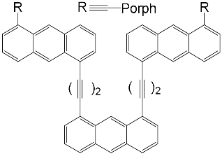 | H2Porph.12 | [70] |
| ZnPorph.6 |  | DABCO | [71] | ZnPorph.9 |  | DABCO | [72] |
| ZnPorph.13 |  |  | [73] | ZnPorph.14 |  | 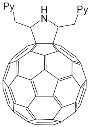 | [74] |
| Bis-porphyrin “molecular tweezers” for recognition of chiral guest molecules. | |||||||
| ZnPorph.9 | 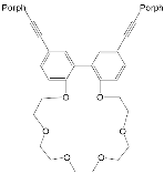 | 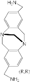 | [75] | ZnPorph.9 | 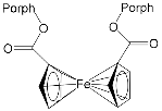 |  | [76] |
| ZnPorph.15 MgPorph.15 |  |  | [15] | ZnPorph.15 MgPorph.15 |  |  | [15] |
| ZnPorph.14 |  |  | [15] | ZnPorph.9 |  |  | [15] |
| MPorph. | R5, R10,R15 | R20 | R2,3,7,8,12,13,17,18 | MPorph. | R5, R10,R15 | R20 | R2,3,7,8,12,13,17,18 | 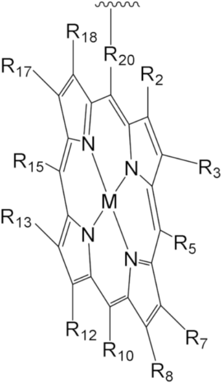 |
| 1 | 4-Me-Ph | Ph | H | |||||
| 2 | 4-Me-Ph | – | 9 | Ph | H | |||
| 3 | 3,5-But-Ph | Ph | 10 | H | – | 2,3,7,13,17,18-H, 8,12-n-C5H14 | ||
| 4 | (2,3,4,5,6-F-Ph) | 11 | Et | |||||
| 5 | 2,4,6-Me-Ph | 13 | 4-COOH-Ph | Ph | H | |||
| 6 | 5,15-H;10-(3,5But-Ph) | 2,8,12,18-Me, 3,7,13,17-Bu | 12 | 5,10-Py, 15-Ph | ||||
| 7 | 2,15-H, 10-Ph | – | 2,8,12,18-Me,3,7,13,17-Et | 14 | Ph | – | ||
| 8 | H | 2,3,17,18-H, 7,8,12,13-Et | 15 | H | 2,8,12,18-Me, 3,7,13,17-Et | |||
| Substrate | Ka | −ΔG |
|---|---|---|
| Pyridine (Py) | 53,400 ± 800 | 27.0 |
| 4-methylpyridine (4-MePy) | 20 ± 1 | 7.4 |
| Imidazole (Im) | 1,920,000 ± 40,000 | 35.8 |
| Toluene | Benzene | ||
|---|---|---|---|
| 13-M60 | 2.0 × 10³ | 13-M60 | 2.1 × 10³ |
| 13-M70 | 3.6 × 10⁴ | 13-M70 | 2.7× 10⁴ |
| Toluene-d8 | Benzene-d6 | |
|---|---|---|
| 58-M70 | 1300 ± 500 | 1100 ± 200 |
| 60-M70 | 1000 ± 200 | 4500 ± 600 |
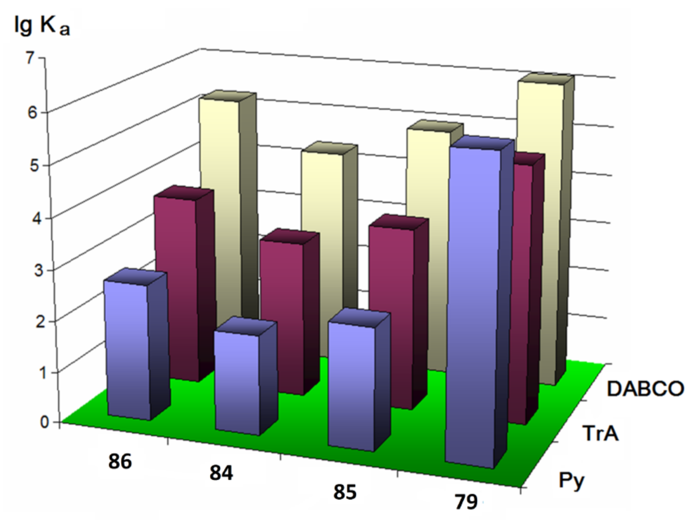
| Py | TrA | |
|---|---|---|
| 86 | 480 | 5800 |
| 84 | 90 | 1200 |
| 85 | 240 | 3900 |
| 79 | 110,000 | 970,000 |
| Metal Cation | Li+ | Na+ | K+ |
|---|---|---|---|
| Ionic radius, Ǻ | 1.36 | 1.90 | 2.66 |
| Binding constant | 1.80 | 7.21 | 9.75 |
| 82-Cr1 (Toluene) | 82-Cr1 (Toluene–Methanol) | 79-Cr1 (Toluene) | 79-Cr1 (Toluene–Methanol) |
|---|---|---|---|
| 2.88 | 2.76 | 5.96 | 5.87 |
Publisher’s Note: MDPI stays neutral with regard to jurisdictional claims in published maps and institutional affiliations. |
© 2021 by the authors. Licensee MDPI, Basel, Switzerland. This article is an open access article distributed under the terms and conditions of the Creative Commons Attribution (CC BY) license (https://creativecommons.org/licenses/by/4.0/).
Share and Cite
Mamardashvili, G.; Mamardashvili, N.; Koifman, O. Macrocyclic Receptors for Identification and Selective Binding of Substrates of Different Nature. Molecules 2021, 26, 5292. https://doi.org/10.3390/molecules26175292
Mamardashvili G, Mamardashvili N, Koifman O. Macrocyclic Receptors for Identification and Selective Binding of Substrates of Different Nature. Molecules. 2021; 26(17):5292. https://doi.org/10.3390/molecules26175292
Chicago/Turabian StyleMamardashvili, Galina, Nugzar Mamardashvili, and Oscar Koifman. 2021. "Macrocyclic Receptors for Identification and Selective Binding of Substrates of Different Nature" Molecules 26, no. 17: 5292. https://doi.org/10.3390/molecules26175292
APA StyleMamardashvili, G., Mamardashvili, N., & Koifman, O. (2021). Macrocyclic Receptors for Identification and Selective Binding of Substrates of Different Nature. Molecules, 26(17), 5292. https://doi.org/10.3390/molecules26175292