Communication

Synthesis of Temperature Sensing Nitrogen-Doped Carbon Dots and Their Application in Fluorescent Ink

1
Inner Mongolia Key Laboratory of Environmental Chemistry, College of Chemistry and Environmental Science, Inner Mongolia Normal University, 81 Zhaowudalu, Hohhot 010022, China
2
College of Pharmacy, Inner Mongolia Medical University, Jinchuankaifaqu, Hohhot 010110, China
3
College of Geographical Science, Inner Mongolia Normal University, 81 Zhaowudalu, Hohhot 010022, China
*
Authors to whom correspondence should be addressed.
Molecules 2023, 28(18), 6607; https://doi.org/10.3390/molecules28186607
Submission received: 27 July 2023 / Revised: 31 August 2023 / Accepted: 1 September 2023 / Published: 14 September 2023

Abstract

:
With the discovery of research, many properties of carbon dots are getting better and better. People have taken advantage of this and utilized them interspersed in various fields. In the present study, water-soluble nitrogen-doped carbon dots (N-CDs) with excellent optical and fluorescence thermal properties were prepared by the hydrothermal method using 4-dimethylaminopyridine and N,N′-methylenebisacrylamide as precursors. Co2+ has a selective bursting effect on the fluorescence of N-CDs. The fluorescence of N-CDs is selectively burst by Co2+, and the high sensitivity is good in the range of 0–12 μM with a detection limit of 74 nM. In addition, the good temperature response (reversible and recoverable fluorescence in the temperature range of 20~90 °C) and excellent optical properties of the N-CDs also make them new potentials in the field of fluorescent inks and temperature sensing.

1. Introduction

Cobalt exerts its good physicochemical properties in many fields, such as batteries, electrocatalysis, and photocatalysis [1,2,3,4]. And in biology, cobalt is an important micronutrient, and as one of the metal ions necessary for the formation of vitamin B complexes in the body, it is involved in the regulation of various body functions. However, if Co2+ accumulates in excess in the body, it can lead to a variety of diseases [5,6,7].
The current detection methods for Co2+ are surface-enhanced Raman scattering [8], electrochemical methods [9], and colorimetric methods [10]. These techniques require detection conditions (equipment, cumbersome procedures, or pre-treatment) that are difficult to meet in many areas. This severely limits the real-time determination of Co2+ and its content in practical applications.
With unique optical properties, low toxicity, and biocompatibility, carbon dots have many applications in optics, energy, and biomedicine [11,12,13]. As a small (typically less than 10 nm) carbon-based nanomaterial, it is expected to replace other conventional fluorescent nanomaterials in the development of a high-performance probe. Recently, Huang and his team [14] used a polymer encapsulated with carbon dots to monitor Cu2+ in the endoplasmic reticulum (ER), while the carbon dots prepared by Yang et al. targeted the ER and significantly eliminated superoxide and hydroxyl radicals [15]. In addition, the carbon dots can selectively detect a variety of other metal ions (e.g., Zn2+, Mg2+, Ag+, and Fe3+) [16,17,18,19]. The high specificity for the target substance during the experiment, simplicity, and speed of analysis make them ideal for Co2+ detection [20,21,22].
Temperature is a fundamental thermodynamic variable, and its accurate determination is very important. The reason for this is that the temperature of diseased cells in the organism is higher than that of normal cells. As a result, there is now an increasing demand for accuracy in the measurement of temperature, which is very important for the detection and treatment of diseases. Carbon dots are slowly coming into the limelight because of their good sense of temperature [23,24,25,26].
Various studies have shown that doping with N atoms can effectively modulate and change the intrinsic properties of carbon dots [27]. 4-dimethylaminopyridine is a novel and efficient catalyst, and N,N′-methylenebisacrylamide is an indispensable cross-linker and condenser in the polymer field. In this paper, 4-Dimethylaminopyridine and N,N′-methylenebisacrylamide were used for the first time as precursors for the preparation of N-CDs. We directly used two N-containing substances as precursors to increase the nitrogen content. The prepared N-CDs have better photostability and FL performance without further modification (Scheme 1). Experimental data on Co2+ recovery in real samples indicate that the method proposed in this paper is reliable and feasible for Co2+ determination in water samples. The bright blue fluorescence of N-CDs remains stable after prolonged UV light exposure or high salt concentrations. The fluorescence intensity of N-CDs decreased with increasing temperature in the range of 20~90 °C, with a good linear relationship. The fluorescence intensity was maintained at 91.76% after multiple heating and cooling steps. This reversibility and recoverability make it possible to enter the field of temperature sensing. Our study not only enriches research on heteroatom-doped carbon dots but also explores new reaction precursors for subsequent research on nitrogen-doped carbon dots.

2. Results and Discussion

2.1. Preparation of N-CDs

Using 4-dimethylaminopyridine and N,N′-methylenebisacrylamide as raw materials, N-CDs were prepared by a hydrothermal method. The reaction raw materials were optimized based on fluorescence intensity (supporting information Figure S1a,b). The fluorescence intensity is highest when the mass ratio of 4-dimethylaminopyridine to N,N′-methylenebisacrylamide is 0.4 g:0.3 g (reaction at 220 °C for 16 h).

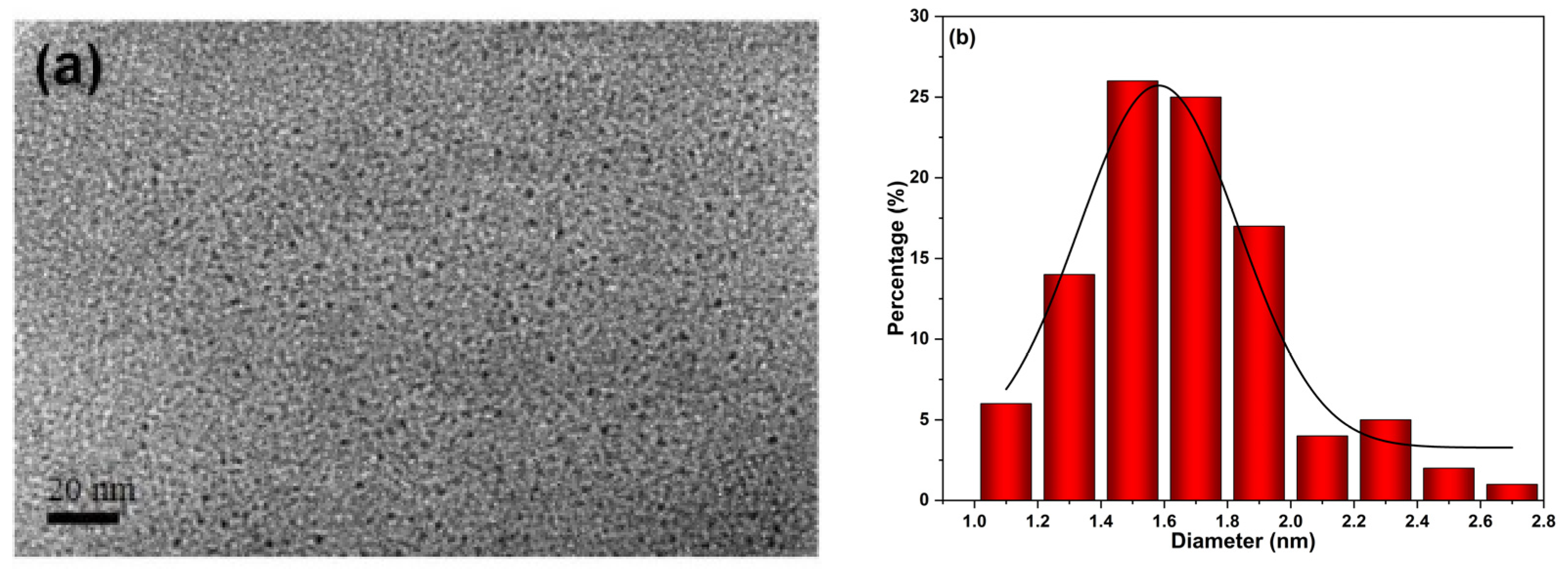
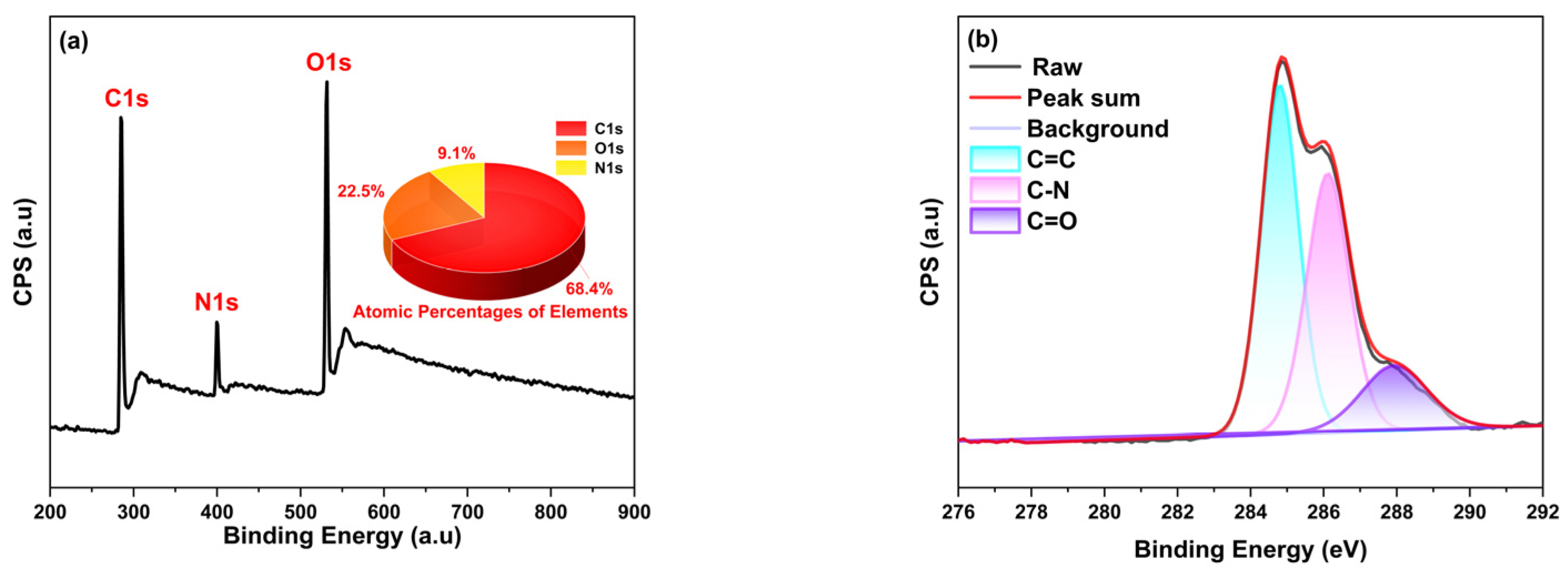
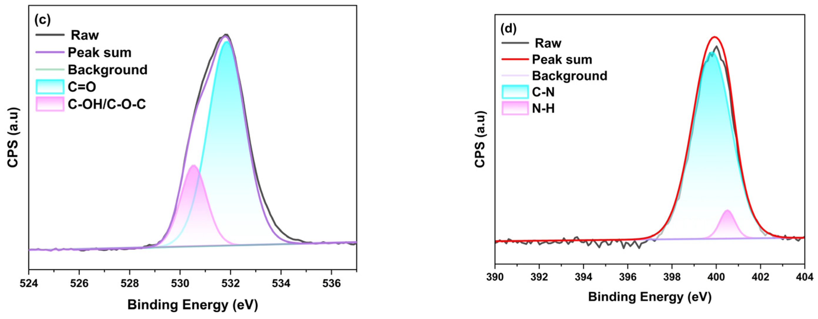
2.2. Characterization

N-CDs were spherical and well dispersed, with a particle size distribution ranging from 1.07 to 2.76 nm and a mean particle size of 1.66 nm, as shown by transmission electron microscopy (TEM) images (Figure 1a). The broad peak around 20.8° in the XRD spectrum (Figure S2a) is showing the amorphous carbon structure of N-CDs [28].
The Fourier transform infrared spectroscopy (FT-IR) plot (Figure S2b) shows that the peak tensile vibration of the O–H/N–H bond is located at 3432 cm−1 [18]. And the peak at 2932 cm−1 originates from the =C–H stretching vibration [29]. The presence of C=O, C=C, and C–N is confirmed by three peaks at 1642 cm−1, 1450 cm−1, and 1374 cm−1 [28,30]. In addition, it may also contain C–O–C (1108 cm−1). The presence of these groups allows us to prepare N-CDs with good hydrophilicity and stability in water.
From the XPS spectra, it can be seen that the N-CDs mainly contain three elements: carbon (284.7 eV, 68.36%), nitrogen (399.7 eV, 9.14%), and nitrogen (531.7 eV, 22.50%) (Figure 2a). In the high-resolution XPS spectra, the three peaks of C1s (284.8 eV, 286.1 eV, and 287.9 eV) represented the presence of C=C, C–N, and C=O, respectively (Figure 2b). The N1s spectra indicated the presence of N–H (399.8 eV) and C–N (400.5 eV) (Figure 2c). The two peaks in the O1s spectrum confirm the presence of C=O and C–OH/C–O–C (Figure 2d) [31].

2.3. Optical Properties

The results of UV-Vis absorption spectroscopy tests performed on it (Figure S3a) showed that an absorption peak near 215 nm could be attributed to a π-π* leap in the C=C bond [32,33,34]. In contrast, the absorption near 345 belongs to the n-π* leap of C=O/C=N [35,36]. In addition, Figure S3b shows the strong emission (λem = 487 nm) under optimal excitation (λex = 414 nm). The prepared N-CDs emit bright cyan fluorescence under 365 nm UV light. Figure S3c shows that the emission wavelength of N-CDs changes its position and intensity (the position is red-shifted and the intensity increases and then decreases) with the increase of the excitation wavelength (Figure S3d), indicating that the position of the emission peaks and the fluorescence intensity are both related to the change of the position of the excitation wavelength. This phenomenon is caused by surface defects, i.e., uneven particle size of N-CDs or uneven distribution of different functional groups on the surface of N-CDs [37].

2.4. The Stability of the N-CDs

The strong photobleaching resistance of N-CDs was demonstrated by the fact that their N-CDs fluorescence intensity remained essentially unchanged under UV light (365 nm) for 80 min (Figure S4a,b). The fluorescence intensity also remained essentially unchanged between 0.1 and 0.5 M NaCl concentrations, indicating that N-CDs are stable at high salt concentrations (Figure S4c). From Figure S4d, we can see that 4 min is the optimal reaction time. This is because after the addition of N-CDs to the Co2+ solution, although the burst effect can be achieved at 0.5 min (a significant reduction from the highest intensity of 1 to 0.38), it basically stabilizes after 4 min.

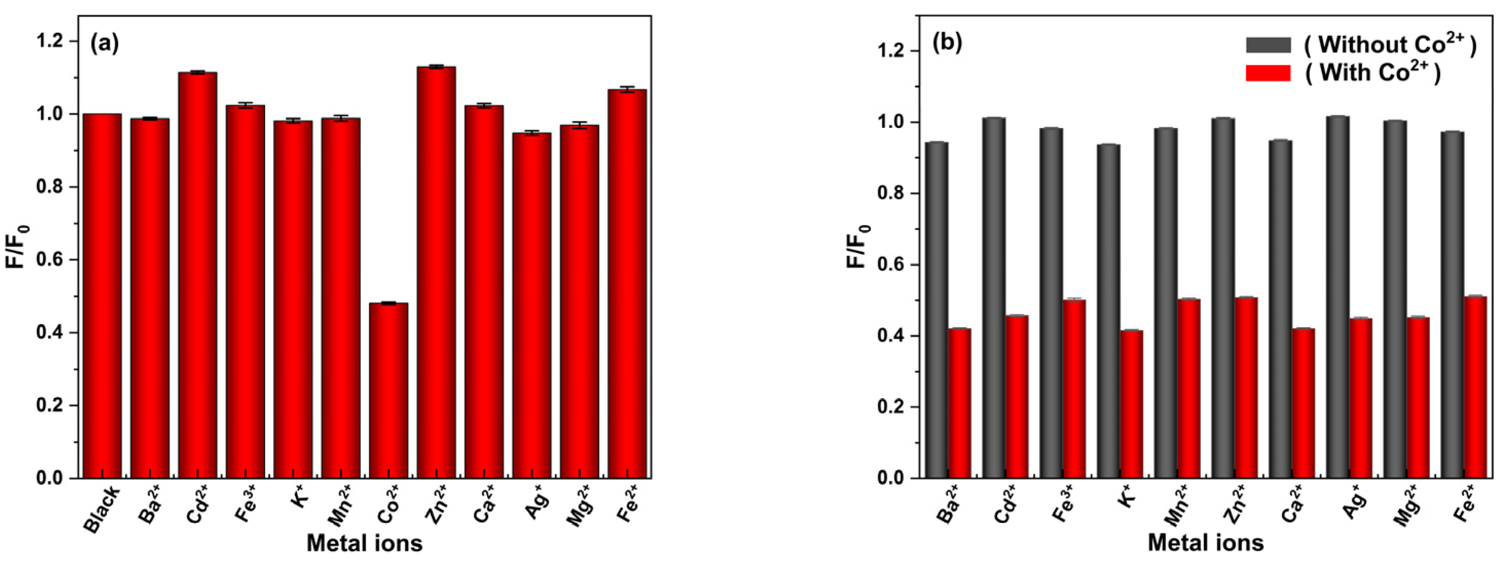
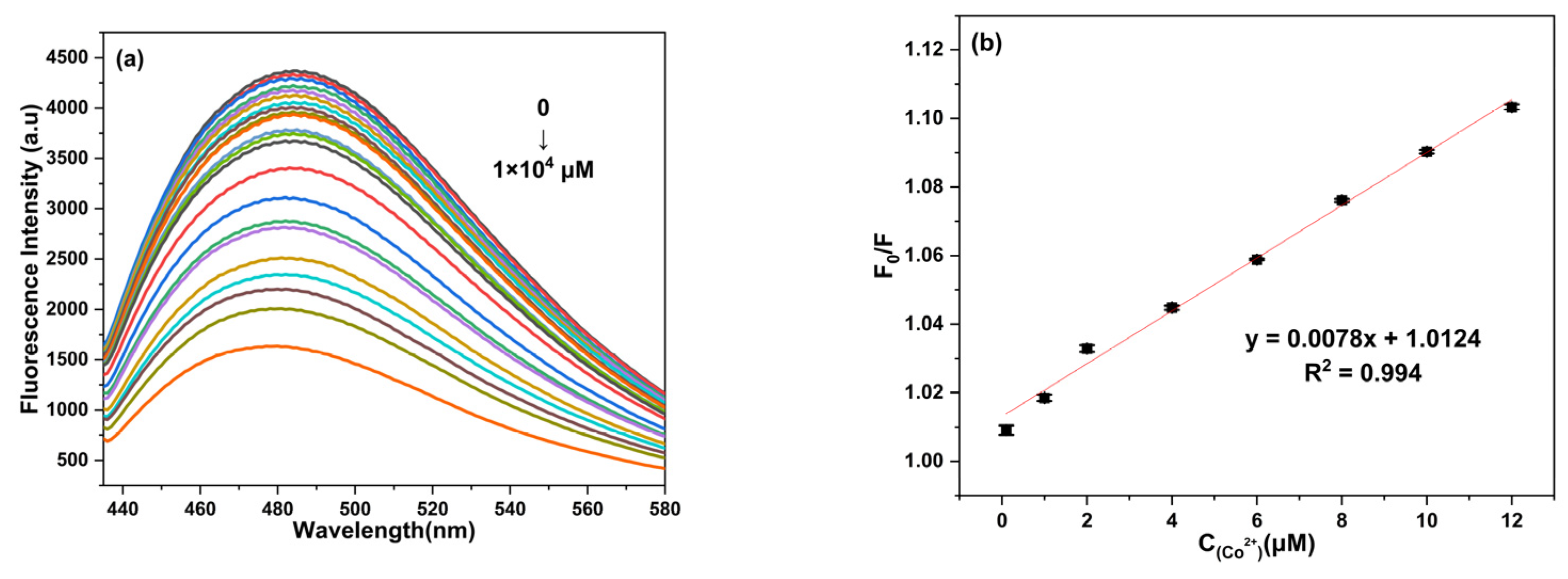
2.5. Detection of Co2+ and Method Selectivity

The high selectivity for Co2+ is essential for N-CDs. In order to test its practicality in real samples, experiments were conducted to determine the selectivity and interference immunity of this sensor. The response of N-CDs to different metal ions (Ba2+, Cd2+, Fe3+, K+, Mn2+, Zn2+, Ca2+, Ag+, Mg2+, and Fe2+) at a concentration of 0.01 M was investigated. In Figure 3a, only Co2+ induces a marked reduction in the fluorescence intensity of N-CDs at 487 nm. This indicates that N-CDs have a high specificity for Co2+. This phenomenon may be related to the fact that the surface functional groups (containing nitrogen or oxygen) of the prepared N-CDs are negatively charged and have a high affinity for Co2+ ions [36]. In addition, the addition of Co2+ to a system in which N-CDs coexist with other metal ions has a very significant effect on the coexisting system. The fluorescence intensity of N-CDs only decreased significantly when Co2+ coexisted with other metal ions, which suggests that the detection of Co2+ by N-CDs is not affected by the presence of other metal ions (Figure 3b). In addition, we found that some metal ions can cause insignificant fluorescence enhancement in N-CDs. This may be due to the chelation of functional groups on the surface of N-CDs with metal ions to form complexes, which improve photoinduced electron transfer and lead to enhanced emission [38].

2.6. Detection Limit of Co2+

As seen in Figure 4a, the fluorescence intensity of N-CDs near 487 nm decreases regularly (in the range of 0.1–1 × 10⁴ μM) with a gradual increase in the concentration of added Co2+. Figure 4b shows a good linear relationship (R2 = 0.994) between the concentration of Co2+ solution (0–12 μM) and the fluorescence intensity. The calculated result of LOD is 74 nM (3σ/s) (the calculation process of LOD is provided in ESM).
As an important parameter for assessing the analytical performance of Co2+ monitoring methods, a lower limit of detection (LOD) for Co2+ represents better analytical performance and higher sensitivity for the substance. Compared with many currently reported carbon-spot-based probes for the determination of Co2+ (Table S1), the LOD values of the N-CDs prepared in this thesis were superior to those reported for the detection of Co2+, indicating that the carbon-spots prepared here successfully achieved high sensitivity for the determination of Co2+.

2.7. Possible Detection Mechanisms

In order to investigate the mechanism by which Co2+ quenches the fluorescence of N-CDs, the possible fluorescence quenching mechanisms were analyzed in this paper using the standard Stern–Volmer equation, fluorescence lifetime curves, UV-vis absorption spectra, and fluorescence spectra. In Figure S5, Co2+ ions (0–12 μM) showed a good linear relationship with F/F0. A Ksv value of 0.007 μM−1 can be derived from the standard Stern–Volmer equation. The addition or absence of Co2+ had little effect on the average fluorescence lifetime of N-CDs (the lifetime decayed from 5.03 ns to 5.01 ns) (Figure 5a). This suggests that the bursting mechanism is unlikely to be dynamic quenching (DQE). In addition, the presence of the internal filtering effect (IFE) was demonstrated by the overlap of the emission spectra of N-CDs with the absorption peak of Co2+ in the 428 to 600 nm range (Figure 5b). The formation of a non-fluorescent complex between Co2+ and N-CDs resulted in a new absorption peak near 550 nm (Figure 5c). And this is proof of the existence of the static burst effect (SQE). The final analysis showed that Co2+ could quench the fluorescence of the N-CDs prepared in this paper based on the SQE and IFE mechanisms. This is in agreement with the literature reports [37,39].

2.8. Detection of Co2+ in Real Samples

Therefore, the results of Co2+ content determined from actual water samples (supermarket pure water) using the standard additive recovery method were 96.32–100.17% recovery of Co2+ with a relative standard deviation (RSD) of 2.65–4.42% (<5%). This implies that N-CDs are one of the ideal methods for the determination of low levels of Co2+ in a quantitative assay environment (Table S2).

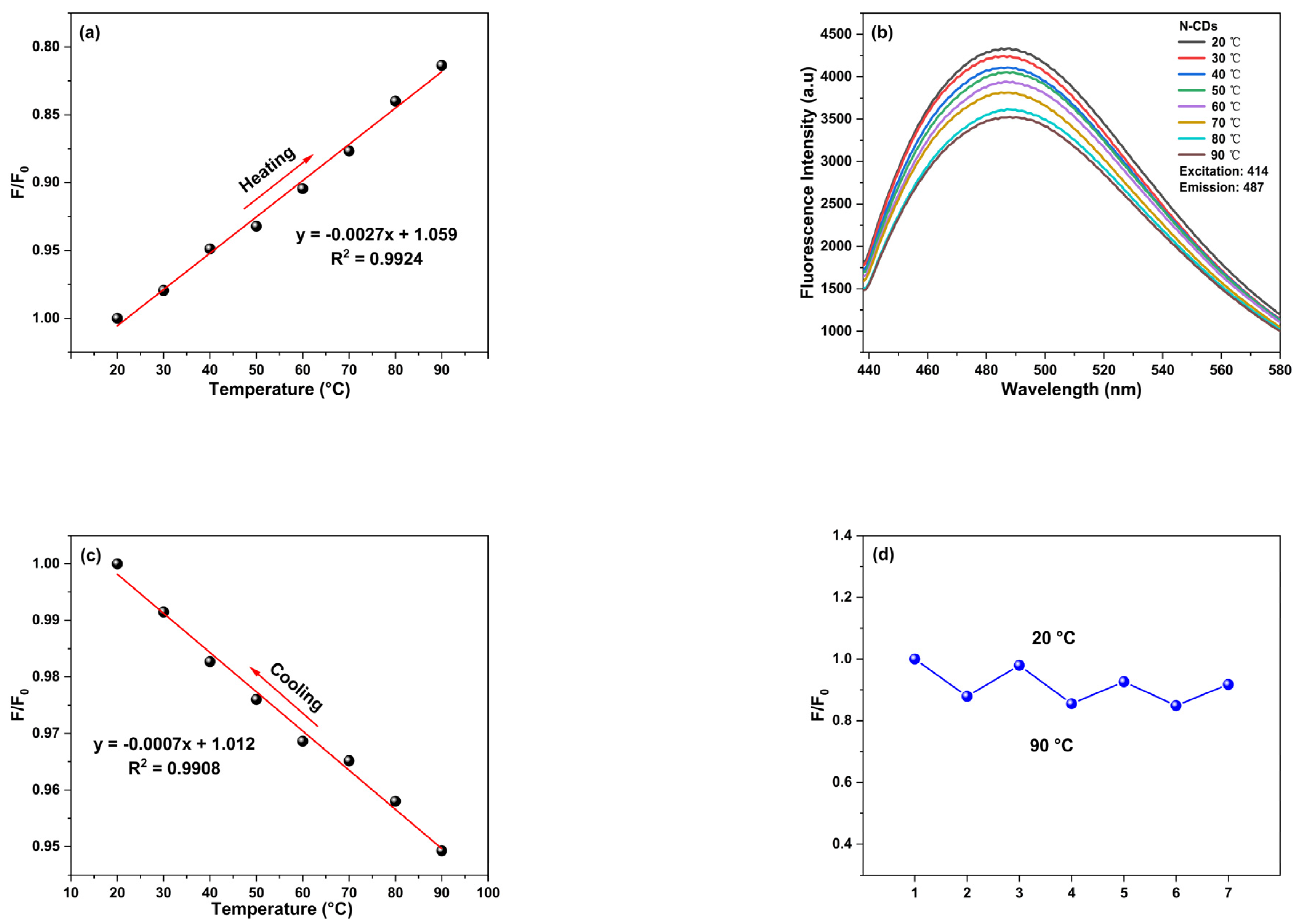
2.9. Research on Temperature Sensing Performance of N-CDs

N-CDs are temperature-sensitive by measuring their fluorescence intensity over a temperature range of 20~90 °C. The temperature from 20 °C to 90 °C was measured in 10 °C temperature gradients. The decrease in fluorescence intensity with increasing temperature can be seen in Figure 6b. The increase in temperature causes the non-radiative channels to be activated, causing more excited electrons to return to the ground state via non-radiative jumps, resulting in a decrease in fluorescence intensity [40,41,42]. It was also found that the value of (F/F0) decreases with increasing temperature (R2 = 0.9924) and increases with decreasing temperature (R2 = 0.9908) (Figure 6a,c). Moreover, in this temperature range (20~90 °C), no migration of the emission peak positions of N-CDs with temperature was observed to occur (Figure 6b). The reusability was tested by recording the fluorescence intensity of N-CDs in three cycles of heating (90 °C) and cooling (20 °C) (Figure 6d). Finally, its maximum fluorescence intensity was still maintained at 91.76%, a result that indicates that its fluorescence center was not destroyed, and the good linear properties and reusability of the fluorescence of N-CDs ensure its potential application in the field of temperature sensing.

2.10. Application of N-CDs in the Field of Fluorescent Ink

In addition to fluorescence sensing by Co2+ and temperature sensing, the N-CDs proposed in this paper have been shown to emit bright cyan fluorescence and are therefore applicable to the field of fluorescent inks. We found that fluorescent inks have excellent optical properties and can be used for information storage. Some letters and drawings were written on filter paper, as shown in Figure 7. Figure 7a–f were taken in daylight. Figure 7g–l were taken under ultraviolet light (365 nm). The UV irradiation showed a bright cyan fluorescence. The fluorescence properties of these filter papers were not affected after several months of storage at room temperature, indicating the excellent fluorescence stability of our N-CDs.

3. Experimental Instruments

3.1. Materials and Apparatus

The materials and instruments used in this work are listed in the Electronic Support Materials (ESM).

3.2. Preparation of N-CDs

The detailed procedure for preparation is given in the supporting information.

3.3. Detection of Co2+ and Interference Experiments

Before recording the fluorescence emission spectra (excitation wavelength 414 nm), a series of solutions of known Co2+ content were added to the aqueous solution of N-CDs and allowed to react for 4 min.
Similarly, in selective experiments, other common metal ions (K+, Fe2+, Fe3+, Hg2+, Ba2+, Ni2+, Cu2+, Zn2+, Mn2+, Ca2+) were added to the testing system instead of Co2+, and the changes in F0/F were recorded while other experimental conditions remained unchanged (λex/λem = 414 nm/487 nm).

4. Conclusions

In conclusion, in this paper, N-CDs were synthesized using 4-dimethylaminopyridine and N,N′-methylenebisacrylamide as precursors. The detection limits and linear ranges of N-CDs as fluorescent probes for Co2+ by fluorescence quenching were 74 nM and 0–12 μM, respectively. Compared with the existing studies for the detection of Co2+, this study not only provides a new raw material for the preparation of N-CDs but also shows that the method is suitable for the detection of Co2+ in real samples. In addition, the N-CDs have potential applications in fluorescent inks in addition to their reversible and recoverable fluorescence properties at temperature, which expands their application range.

Supplementary Materials

The following supporting information can be downloaded at: https://www.mdpi.com/article/10.3390/molecules28186607/s1 [43,44,45,46,47]. Figure S1. (a) Fluorescence emission spectra of N-CDs prepared from reaction materials with different mass ratios. (b) Changes in fluorescence intensity with different mass ratios of reaction materials; Figure S2. (a) X-ray diffraction and (b) FT-IR of N-CDs; Figure S3. N-CDs (a) UV visible absorption spectra. (b) fluorescence spectra (Illustration: Image of N-CDs under natural light (left) and ultraviolet light (365 nm) (right) irradiation). (c) fluorescence spectra at different excitation wavelengths. (d) Normalized spectra of fluorescence spectra at different excitation wavelengths; Figure S4. (a) Fluorescence spectra of N-CDs under 80 min ultraviolet lamp irradiation and (b) Line chart. (c) The effect of NaCl solution concentration on N-CDs. (d) The intensity of N-CDs varies with reaction time after the addition of Co2+ (F0 and F represent the fluorescence intensity of N-CDs at 487 nm in the presence and absence of Co2+, respectively); Figure. S5. Stern Volmer relationship between (F/F0) and Co2+ concentration; Table S1: Comparison of the performance of synthesized N-CDs with other N-CDs in Co2+ detection; Table S2: Determination of Co2+ in purified water samples.

Author Contributions

Conceptualization, J.A. and L.G.; methodology, P.L.; software, P.L.; validation, J.A., L.G. and Y.W.; formal analysis, P.L.; investigation, P.L.; resources, J.A.; data curation, P.L.; writing—original draft preparation, P.L.; writing—review and editing, P.L.; visualization, L.G.; supervision, Y.W.; project administration, J.A.; funding acquisition, J.A. All authors have read and agreed to the published version of the manuscript.

Funding

This work was supported by the National Natural Science Foundation of China (Grant No. 21864020, 82160652), the Natural Science Foundation of Inner Mongolia (Grant No. 2019MS02014, 2018MS02012), the “Young Science and Technology Talents Program” (Leading Person) in Inner Mongolia Autonomous Region Colleges and Universities (Grant No. NJYT-19-A04), the Fundamental Research Funds for the Inner Mongolia Normal University, China, (Grant No. 2022JBZD013), the Key Project of Natural Science Foundation of Inner Mongolia Autonomous Region (No. 2023ZD21), and the Science and Technology Planning Project of Inner Mongolia Autonomous Region (No. 2021GG0367).

Data Availability Statement

Not applicable.

Conflicts of Interest

The authors declare no conflict of interest.

Sample Availability

Not available.

References

  1. Lee, J.; Jang, J.; Yu, T. A facile aqueous-phase synthesis of Co-based nanostructures composed of cobalt hydroxide and cobalt oxide for enhanced photocatalytic activity of dye-sensitized H2 production. J. Alloys Compd. 2023, 942, 169078. [Google Scholar] [CrossRef]
  2. Li, S.; Zheng, X.; Jin, H.; Qian, L.; Wang, K.; Shen, Y.; Zhao, M.; Liu, R. Multivalent cobalt species supported on graphene aerogel for degradation of sulfamethoxazole via high-valent cobalt-oxo species. Chem. Eng. J. 2023, 463, 142367. [Google Scholar] [CrossRef]
  3. Shang, W.; Wang, H.; Yu, W.; He, Y.; Ma, Y.; Wu, Z.; Tan, P. Transforming the Electrochemical Behaviors of Cobalt Oxide from “Supercapacitator” to “Battery” by Atomic-Level Structure Engineering for Inspiring the Advance of Co-Based Batteries. Small 2023, 19, e2300647. [Google Scholar] [CrossRef]
  4. Zakrzewska, B.; Jabłońska, A.; Adamczyk, L.; Dembińska, B.; Kostuch, A.; Strawski, M.; Rutkowska, I.A.; Kulesza, P.J.; Marcinek, M.; Cox, J.A.; et al. Pyrolyzed cobalt hexacyanocobaltate dispersed on reduced-graphene-oxide as an electrocatalyst of the oxygen reduction reaction in an alkaline medium. J. Mater. Chem. A 2023, 11, 7286–7298. [Google Scholar] [CrossRef]
  5. Awual, M.R.; Ismael, M.; Yaita, T. Efficient detection and extraction of cobalt(II) from lithium ion batteries and wastewater by novel composite adsorbent. Sens. Actuators B 2014, 191, 9–18. [Google Scholar] [CrossRef]
  6. Liao, S.; Zhu, F.; Zhao, X.; Yang, H.; Chen, X. A reusable P, N-doped carbon quantum dot fluorescent sensor for cobalt ion. Sens. Actuators B 2018, 260, 156–164. [Google Scholar] [CrossRef]
  7. Cheng, S.; Zhang, J.; Liu, Y.; Wang, Y.; Xiao, Y.; Zhang, Y. One-pot synthesis of nitrogen-doped carbon dots for sensing of Co2+ and tetracycline antibiotics, biological imaging, and fluorescent inks. J. Nanoparticle Res. 2022, 24, 44. [Google Scholar] [CrossRef]
  8. Tsoutsi, D.; Guerrini, L.; Hermida-Ramon, J.M.; Giannini, V.; Liz-Marzán, L.M.; Wei, A.; Alvarez-Puebla, R.A. Simultaneous SERS detection of copper and cobalt at ultratrace levels. Nanoscale 2013, 5, 5841–5846. [Google Scholar] [CrossRef]
  9. Zamhari, M.; Numnuam, A.; Limbut, W.; Kanatharana, P.; Thavarungkul, P. Simultaneous Electrochemical Detection of Co(II) and Cu(II) by 1-Diazo-2-Naphthol-4-Sulfonic Acid/MWCNTs Modified Electrode. Electroanalysis 2017, 29, 2348–2357. [Google Scholar] [CrossRef]
  10. Sharma, S.; Jaiswal, A.; Uttam, K.N. Synthesis of Sensitive and Robust Lignin Capped Silver Nanoparticles for the Determination of Cobalt(II), Chromium(III), and Manganese(II) Ions by Colorimetry and Manganese(II) Ions by Surface-Enhanced Raman Scattering (SERS) in Aqueous Media. Anal. Lett. 2021, 54, 2051–2069. [Google Scholar] [CrossRef]
  11. Kumar, A.; Asu, S.; Mukherjee, P.; Singh, P.; Kumari, A.; Sahu, S.K. Single-step synthesis of N-doped carbon dots and applied for dopamine sensing, in vitro multicolor cellular imaging as well as fluorescent ink. J. Photochem. Photobiol. A Chem. 2021, 406, 113019. [Google Scholar] [CrossRef]
  12. Ren, G.; Hou, X.; Kang, Y.; Zhang, R.; Zhang, M.; Liu, W.; Li, L.; Wei, S.; Wang, H.; Wang, B.; et al. Efficient preparation of nitrogen-doped fluorescent carbon dots for highly sensitive detection of metronidazole and live cell imaging. Spectrochim. Acta Part A Mol. Biomol. Spectrosc. 2020, 234, 118251. [Google Scholar] [CrossRef] [PubMed]
  13. Liu, J.; Li, R.; Yang, B. Carbon Dots: A New Type of Carbon-Based Nanomaterial with Wide Applications. ACS Cent. Sci. 2020, 6, 2179–2195. [Google Scholar] [CrossRef] [PubMed]
  14. Huang, H.; Li, S.; Chen, B.; Wang, Y.; Shen, Z.; Qiu, M.; Pan, H.; Wang, W.; Wang, Y.; Li, X. Endoplasmic reticulum-targeted polymer dots encapsulated with ultrasonic synthesized near-infrared carbon nanodots and their application for in vivo monitoring of Cu2+. J. Colloid Interface Sci. 2022, 627, 705–715. [Google Scholar] [CrossRef]
  15. Yang, L.; Huang, H.; Wang, T.; Zhou, D.; Chen, Q.; Li, D.; Chen, S.; Lin, P. Endoplasmic reticulum-targetable selenium-doped carbon nanodots with redox-responsive fluorescence for in situ free-radical scavenging in cells and mice. Arab. J. Chem. 2023, 16, 105036. [Google Scholar] [CrossRef]
  16. Li, J.; Zuo, G.; Pan, X.; Wei, W.; Qi, X.; Su, T.; Dong, W. Nitrogen-doped carbon dots as a fluorescent probe for the highly sensitive detection of Ag+ and cell imaging. Luminescence 2018, 33, 243–248. [Google Scholar] [CrossRef]
  17. Hashemi, N.; Mousazadeh, M.H. Preparation of fluorescent nitrogen-doped carbon dots for highly selective on-off detection of Fe3+ ions in real samples. Opt. Mater. 2021, 121, 111515. [Google Scholar] [CrossRef]
  18. Sadhanala, H.K.; Aryal, S.; Sharma, K.; Orpaz, Z.; Michaeli, S.; Gedanken, A. Nitrogen-doped carbon dots as a highly selective and sensitive fluorescent probe for sensing Mg2+ ions in aqueous solution, and their application in the detection and imaging of intracellular Mg2+ ions. Sens. Actuators B 2022, 366, 131958. [Google Scholar] [CrossRef]
  19. Xu, H.; You, X.; Lu, Y.; Liang, P.; Luo, Z.; Wang, Y.; Zeng, S.; Zeng, H. Analysis of Mn2+ and Zn2+ Ions in Macroalgae with Heteroelement-Doped Carbon-Based Fluorescent Probe. Biosensors 2022, 12, 359. [Google Scholar] [CrossRef]
  20. Zi, L.; Huang, Y.; Yan, Z.; Liao, S. Thioglycolic acid-capped CuInS2/ZnS quantum dots as fluorescent probe for cobalt ion detection. J. Lumin. 2014, 148, 359–363. [Google Scholar] [CrossRef]
  21. Chen, H.; Yuan, F.; Xu, J.; Zhang, Y.; Wu, Y.; Wang, L. Simple and sensitive detection method for Cobalt(II) in water using CePO4:Tb3+ nanocrystals as fluorescent probes. Spectrochim. Acta Part A Mol. Biomol. Spectrosc. 2013, 107, 151–155. [Google Scholar] [CrossRef]
  22. Bian, W.; Ma, J.; Liu, Q.; Wei, Y.; Li, Y.; Dong, C.; Shuang, S. A novel phosphorescence sensor for Co2+ ion based on Mn-doped ZnS quantum dots. Luminescence 2014, 29, 151–157. [Google Scholar] [CrossRef] [PubMed]
  23. Zeng, Y.; Feng, W.; Qi, X.; Li, J.; Chen, J.; Lu, L.; Deng, P.; Zeng, J.; Li, F. Differential knee skin temperature following total knee arthroplasty and its relationship with serum indices and outcome: A prospective study. J. Int. Med. Res. 2016, 44, 1023–1033. [Google Scholar] [CrossRef]
  24. Yang, J.; Yin, P.; Zhou, M.; Ou, C.Q.; Guo, Y.; Gasparrini, A.; Liu, Y.; Yue, Y.; Gu, S.; Sang, S.; et al. Cardiovascular mortality risk attributable to ambient temperature in China. Heart 2015, 101, 1966–1972. [Google Scholar] [CrossRef] [PubMed]
  25. Huang, K.; Wang, Z.; Gu, Y.; Hu, Y.; Ji, Z.; Wang, S.; Lin, Z.; Li, X.; Xie, Z.; Pan, S. Glibenclamide Is Comparable to Target Temperature Management in Improving Survival and Neurological Outcome After Asphyxial Cardiac Arrest in Rats. J. Am. Hear. Assoc. 2016, 5, e003465. [Google Scholar] [CrossRef] [PubMed]
  26. Fu, F.; Xin, S.X.; Chen, W. Temperature- and frequency-dependent dielectric properties of biological tissues within the temperature and frequency ranges typically used for magnetic resonance imaging-guided focused ultrasound surgery. Int. J. Hyperth 2014, 30, 56–65. [Google Scholar] [CrossRef]
  27. Pundi, A.; Chang, C.J. Recent Advances in Synthesis, Modification, Characterization, and Applications of Carbon Dots. Polymers 2022, 14, 2153. [Google Scholar] [CrossRef]
  28. Patir, K.; Barman, B.; Basumatary, S. One Pot Synthesis of Multicolor Emissive Nitrogen Doped Carbon Dots and its Application as Acetone and Picric Acid Sensor. Iran. J. Sci. Technol. Trans. A Sci. 2021, 45, 1301–1310. [Google Scholar] [CrossRef]
  29. Li, L.; Shi, L.; Jia, J.; Chang, D.; Dong, C.; Shuang, S. Fe3+ detection, bioimaging, and patterning based on bright blue-fluorescent N-doped carbon dots. Analyst 2020, 145, 5450–5457. [Google Scholar] [CrossRef]
  30. Wang, C.; Li, L.; Wu, Y.; Gu, J.; Ma, C.; Zhu, C.; Gao, H.; Zhang, Y.; Shang, Y.; Yang, Z.; et al. Highly Photoluminescent Carbon Dots with pH-Dependent Switchable Fluorescence and Sensitivity to Tetracycline. Nano 2021, 16, 2150036. [Google Scholar] [CrossRef]
  31. Liu, B.; Wei, S.; Liu, E.; Zhang, H.; Lu, P.; Wang, J.; Sun, G. Nitrogen-doped carbon dots as a fluorescent probe for folic acid detection and live cell imaging. Spectrochim. Acta Part A Mol. Biomol. Spectrosc. 2022, 268, 120661. [Google Scholar] [CrossRef]
  32. Amjadi, M.; Hallaj, T.; Mayan, M.A. Green synthesis of nitrogen-doped carbon dots from lentil and its application for colorimetric determination of thioridazine hydrochloride. RSC Adv. 2016, 6, 104467–104473. [Google Scholar] [CrossRef]
  33. Atchudan, R.; Gangadaran, P.; Perumal, S.; Edison, T.N.J.I.; Sundramoorthy, A.K.; Rajendran, R.L.; Ahn, B.-C.; Lee, Y.R. Green Synthesis of Multicolor Emissive Nitrogen-Doped Carbon Dots for Bioimaging of Human Cancer Cells. J. Clust. Sci. 2023, 34, 1583–1594. [Google Scholar] [CrossRef]
  34. Cheng, S.; Wang, X.; Yan, X.; Xiao, Y.; Zhang, Y. Simple synthesis of green luminescent N-doped carbon dots for malachite green determination. Anal. Methods 2022, 14, 2616–2622. [Google Scholar] [CrossRef]
  35. Issa, M.A.; Zentou, H.; Jabbar, Z.H.; Abidin, Z.Z.; Harun, H.; Halim, N.A.A.; Alkhabet, M.M.; Pudza, M.Y. Ecofriendly adsorption and sensitive detection of Hg (II) by biomass-derived nitrogen-doped carbon dots: Process modelling using central composite design. Environ. Sci. Pollut. Res. 2022, 29, 86859–86872. [Google Scholar] [CrossRef]
  36. Cui, J.; Zhu, X.; Liu, Y.; Liang, L.; Peng, Y.; Wu, S.; Zhao, Y. N-Doped Carbon Dots as Fluorescent “Turn-Off” Nanosensors for Ascorbic Acid and Fe3+ Detection. ACS Appl. Nano Mater. 2022, 5, 7268–7277. [Google Scholar] [CrossRef]
  37. Xu, D.; Fu, N.; Xie, Y.; Wang, Y.; Xie, R.; Yang, H.; Sun, W.; Liu, X.; Han, A. Easy formation of nitrogen-doped carbon dots towards Hg2+ fluorescent measurement and multicolor intracellular imaging. Mater. Chem. Phys. 2021, 266, 124547. [Google Scholar] [CrossRef]
  38. Lin, L.; Wang, Y.; Xiao, Y.; Liu, W. Hydrothermal synthesis of carbon dots codoped with nitrogen and phosphorus as a turn-on fluorescent probe for cadmium(II). Microchim. Acta 2019, 186, 147. [Google Scholar] [CrossRef]
  39. Tian, M.; Zhang, J.; Liu, Y.; Wang, Y.; Zhang, Y. One-pot synthesis of nitrogen-doped carbon dots for highly sensitive determination of cobalt ions and biological imaging. Spectrochim. Acta Part A Mol. Biomol. Spectrosc. 2021, 252, 119541. [Google Scholar] [CrossRef]
  40. Tammina, S.K.; Yang, D.; Koppala, S.; Cheng, C.; Yang, Y. Highly photoluminescent N, P doped carbon quantum dots as a fluorescent sensor for the detection of dopamine and temperature. J. Photochem. Photobiol. B Biol. 2019, 194, 61–70. [Google Scholar] [CrossRef] [PubMed]
  41. Liao, X.; Chen, C.; Wang, P.; Zhou, R.; Zhao, X.; Fan, H.; Huang, Z. Carbon dots derived from cellobiose for temperature and phosalone detection. Mater. Res. Bull. 2022, 151, 111790. [Google Scholar] [CrossRef]
  42. Guo, Z.; Luo, J.; Zhu, Z.; Sun, Z.; Zhang, X.; Wu, Z.-c.; Mo, F.; Guan, A. A facile synthesis of high-efficient N,S co-doped carbon dots for temperature sensing application. Dye. Pigment. 2020, 173, 107952. [Google Scholar] [CrossRef]
  43. Liu, S.; Liu, R.; Xing, X.; Yang, C.; Xu, Y.; Wu, D. Highly photoluminescent nitrogen-rich carbon dots from melamine and citric acid for the selective detection of iron(iii) ion. RSC Adv. 2016, 6, 31884–31888. [Google Scholar] [CrossRef]
  44. Jing, N.; Tian, M.; Wang, Y.; Zhang, Y. Nitrogen-doped carbon dots synthesized from acrylic acid and ethylenediamine for simple and selective determination of cobalt ions in aqueous media. J. Lumin. 2019, 206, 169–175. [Google Scholar] [CrossRef]
  45. Boonta, W.; Talodthaisong, C.; Sattayaporn, S.; Chaicham, C.; Chaicham, A.; Sahasithiwat, S.; Kangkaew, L.; Kulchat, S. The synthesis of nitrogen and sulfur co-doped graphene quantum dots for fluorescence detection of cobalt(ii) ions in water. Mater. Chem. Front. 2020, 4, 507–516. [Google Scholar] [CrossRef]
  46. Kong, D.; Yan, F.; Han, Z.; Xu, J.; Guo, X.; Chen, L. Cobalt(ii) ions detection using carbon dots as an sensitive and selective fluorescent probe. RSC Adv. 2016, 6, 67481–67487. [Google Scholar] [CrossRef]
  47. Du, F.; Cheng, Z.; Kremer, M.; Liu, Y.; Wang, X.; Shuang, S.; Dong, C. A label-free multifunctional nanosensor based on N-doped carbon nanodots for vitamin B12 and Co2+ detection, and bioimaging in living cells and zebrafish. J. Mater. Chem. B 2020, 8, 5089–5095. [Google Scholar] [CrossRef]
Scheme 1. Method for synthesizing N-CDs and sensors for detecting Co2+.
Molecules 28 06607 sch001
Figure 1. (a) TEM images and (b) size distribution of N-CDs.
Molecules 28 06607 g001
Figure 2. (a) XPS spectra of N-CDs. (b) C1s. (c) O1s. (d) N1s spectrum of the N-CDs.
Molecules 28 06607 g002aMolecules 28 06607 g002b
Figure 3. Fluorescence response of N-CDs to (a) different metal ions. (b) Fluorescence response when Co2+ and other metal ions coexist.
Molecules 28 06607 g003
Figure 4. (a) Emission spectra of N-CDs after the addition of different amounts of Co2+. (b) Relationship between (F0/F) and Co2+ concentration (F0 and F represent the fluorescence intensity of N-CDs without and with the addition of Co2+, respectively).
Molecules 28 06607 g004
Figure 5. (a) Comparison of fluorescence lifetimes. (b) Overlay of N-CDs emission spectra and Co2+ UV-Vis absorption spectra. (c) UV-Vis absorption spectra of Co2+ and N-CDs (with and without added Co2+).
Molecules 28 06607 g005
Figure 6. (a) Changes in fluorescence intensity of N-CDs during heating (20~90 °C). (b) Emission spectra of N-CDs at different temperatures. (c) Changes in fluorescence intensity of N-CDs during cooling (20~90 °C). (d) Reversible temperature fluorescence of N-CDs.
Molecules 28 06607 g006
Figure 7. Photographs of N-CDs drawn with fluorescent ink taken in daylight (af) and (gl) under UV irradiation.
Molecules 28 06607 g007
Disclaimer/Publisher’s Note: The statements, opinions and data contained in all publications are solely those of the individual author(s) and contributor(s) and not of MDPI and/or the editor(s). MDPI and/or the editor(s) disclaim responsibility for any injury to people or property resulting from any ideas, methods, instructions or products referred to in the content.

