Liver Graft Proteomics Reveals Potential Incipient Mechanisms behind Early Renal Dysfunction after Liver Transplantation
Abstract
1. Introduction
2. Results
2.1. Clinical Outcomes
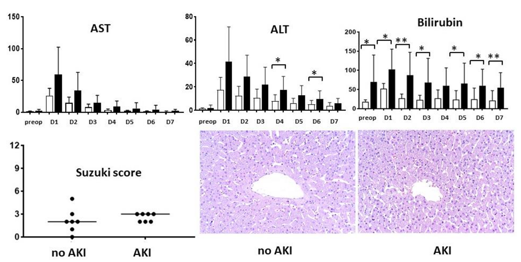
2.2. Liver Injury
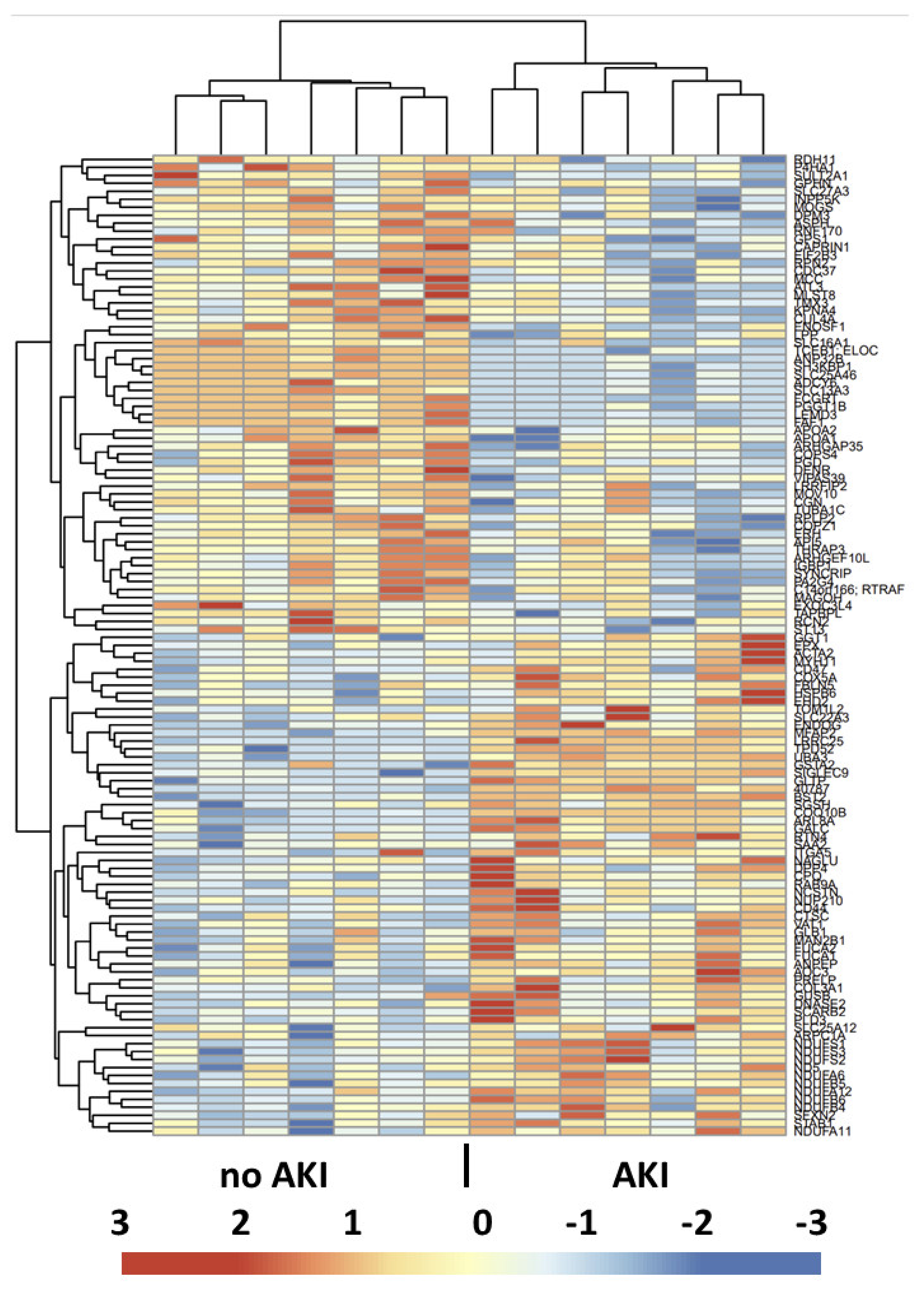
2.3. Proteomic Analysis
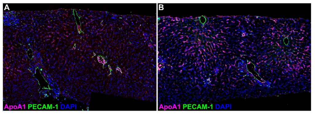
2.4. Validation Study—Immunofluorescence
2.5. Circulating Cytokines and SAA2
3. Discussion
4. Materials and Methods
4.1. Patients and Study Design
4.2. Assessment of Organ Dysfunction
4.3. Histology
4.4. Global Protein Quantification
4.5. Cytokine and Serum Amyloid A2
4.6. Statistical Analysis
Author Contributions
Funding
Institutional Review Board Statement
Informed Consent Statement
Data Availability Statement
Conflicts of Interest
References
- Adam, R.; Karam, V.; Cailliez, V.; OGrady, J.G.; Mirza, D.; Cherqui, D.; Klempnauer, J.; Salizzoni, M.; Pratschke, J.; Jamieson, N.; et al. 2018 Annual Report of the European Liver Transplant Registry (ELTR)—50-year evolution of liver transplantation. Improvement in early posttransplant patient survival has increased the importance of understanding the causes and risk factors for late posttransplant mortality. Transpl. Int. 2018, 31, 1293–1317. [Google Scholar] [PubMed]
- Kwong, A.J.; Ebel, N.H.; Kim, W.R.; Lake, J.R.; Smith, J.M.; Schladt, D.P.; Skeans, M.A.; Foutz, J.; Gauntt, K.; Cafarella, M.; et al. OPTN/SRTR 2020 Annual Data Report: Liver. Am. J. Transplant. 2022, 22 (Suppl. 2), 204–309. [Google Scholar] [CrossRef] [PubMed]
- Lindenger, C.; Castedal, M.; Schult, A.; Åberg, F. Long-term survival and predictors of relapse and survival after liver transplantation for alcoholic liver disease. Scand. J. Gastroenterol. 2018, 53, 1553–1561. [Google Scholar] [CrossRef] [PubMed]
- Watt, K.D.; Pedersen, R.A.; Kremers, W.K.; Heimbach, J.K.; Charlton, M.R. Evolution of causes and risk factors for mortality post-liver transplant: Results of the NIDDK long-term follow-up study. Am. J. Transplant. 2010, 10, 1420–1427. [Google Scholar] [CrossRef] [PubMed]
- Ojo, A.O.; Held, P.J.; Port, F.K.; Wolfe, R.A.; Leichtman, A.B.; Young, E.W.; Arndorfer, J.; Christensen, L.; Merion, R.M. Chronic renal failure after transplantation of a nonrenal organ. N. Engl. J. Med. 2003, 349, 931–940. [Google Scholar] [CrossRef] [PubMed]
- Barri, Y.M.; Sanchez, E.Q.; Jennings, L.W.; Melton, L.B.; Hays, S.; Levy, M.F.; Klintmalm, G.B. Acute kidney injury following liver transplantation: Definition and outcome. Liver Transpl. 2009, 15, 475–483. [Google Scholar] [CrossRef]
- Leithead, J.A.; Rajoriya, N.; Gunson, B.K.; Muiesan, P.; Ferguson, J.W. The evolving use of higher risk grafts is associated with an increased incidence of acute kidney injury after liver transplantation. J. Hepatol. 2014, 60, 1180–1186. [Google Scholar] [CrossRef]
- Umbro, I.; Tinti, F.; Scalera, I.; Evison, F.; Gunson, B.; Sharif, A.; Ferguson, J.; Muiesan, P.; Mitterhofer, A.P. Acute kidney injury and post-reperfusion syndrome in liver transplantation. World J. Gastroenterol. 2016, 22, 9314–9323. [Google Scholar] [CrossRef]
- Skytte Larsson, J.; Bragadottir, G.; Redfors, B.; Ricksten, S.E. Renal function and oxygenation are impaired early after liver transplantation despite hyperdynamic systemic circulation. Crit. Care 2017, 21, 87. [Google Scholar] [CrossRef]
- Jochmans, I.; Meurisse, N.; Neyrinck, A.; Verhaegen, M.; Monbaliu, D.; Pirenne, J. Hepatic ischemia/reperfusion injury associates with acute kidney injury in liver transplantation: Prospective cohort study. Liver Transpl. 2017, 23, 634–644. [Google Scholar] [CrossRef]
- Oltean, M.; Bagge, J.; Dindelegan, G.; Kenny, D.; Molinaro, A.; Hellström, M.; Nilsson, O.; Sihlbom, C.; Casselbrant, A.; Davila, M.; et al. The Proteomic Signature of Intestinal Acute Rejection in the Mouse. Metabolites 2021, 12, 23. [Google Scholar] [CrossRef] [PubMed]
- Crowl, R.M.; Stoller, T.J.; Conroy, R.R.; Stoner, C.R. Induction of phospholipase A2 gene expression in human hepatoma cells by mediators of the acute phase response. J. Biol. Chem. 1991, 266, 2647–2651. [Google Scholar] [CrossRef]
- Moshage, H. Cytokines and the hepatic acute phase response. J. Pathol. 1997, 181, 257–266. [Google Scholar] [CrossRef]
- Pullerits, R.; Oltean, S.; Flodén, A.; Oltean, M. Circulating resistin levels are early and significantly increased in deceased brain dead organ donors, correlate with inflammatory cytokine response and remain unaffected by steroid treatment. J. Transl. Med. 2015, 13, 201. [Google Scholar] [CrossRef]
- Chu, M.J.; Dare, A.J.; Phillips, A.R.; Bartlett, A.S. Donor Hepatic Steatosis and Outcome After Liver Transplantation: A Systematic Review. J. Gastrointest. Surg. 2015, 19, 1713–1724. [Google Scholar] [CrossRef]
- Seifalian, A.M.; Chidambaram, V.; Rolles, K.; Davidson, B.R. In Vivo demonstration of impaired microcirculation in steatotic human liver grafts. Liver Transpl. Surg. 1998, 4, 71–77. [Google Scholar] [CrossRef]
- Gehrau, R.C.; Mas, V.R.; Dumur, C.I.; Suh, J.L.; Sharma, A.K.; Cathro, H.P.; Maluf, D.G. Donor Hepatic Steatosis Induce Exacerbated Ischemia-Reperfusion Injury Through Activation of Innate Immune Response Molecular Pathways. Transplantation 2015, 99, 2523–2533. [Google Scholar] [CrossRef]
- Adibhatla, R.M.; Hatcher, J.F.; Dempsey, R.J. Cytidine-5′-diphosphocholine affects CTP-phosphocholine cytidylyltransferase and lyso-phosphatidylcholine after transient brain ischemia. J. Neurosci. Res. 2004, 76, 390–396. [Google Scholar] [CrossRef]
- Ogata, K.; Jin, M.B.; Taniguchi, M.; Suzuki, T.; Shimamura, T.; Kitagawa, N.; Magata, S.; Fukai, M.; Ishikawa, H.; Ono, T.; et al. Attenuation of ischemia and reperfusion injury of canine livers by inhibition of type II phospholipase A2 with LY329722. Transplantation 2001, 71, 1040–1046. [Google Scholar] [CrossRef]
- Uhlar, C.M.; Whitehead, A.S. The kinetics and magnitude of the synergistic activation of the serum amyloid A promoter by IL-1 beta and IL-6 is determined by the order of cytokine addition. Scand. J. Immunol. 1999, 49, 399–404. [Google Scholar] [CrossRef]
- De Buck, M.; Gouwy, M.; Wang, J.M.; Van Snick, J.; Proost, P.; Struyf, S.; Van Damme, J. The cytokine-serum amyloid A-chemokine network. Cytokine Growth Factor Rev. 2016, 30, 55–69. [Google Scholar] [CrossRef] [PubMed]
- Ruiz-Blázquez, P.; Pistorio, V.; Fernández-Fernández, M.; Moles, A. The multifaceted role of cathepsins in liver disease. J. Hepatol. 2021, 75, 1192–1202. [Google Scholar] [CrossRef] [PubMed]
- Nakamura, K.; Kageyama, S.; Kupiec-Weglinski, J.W. The Evolving Role of Neutrophils in Liver Transplant Ischemia-Reperfusion Injury. Curr. Transplant. Rep. 2019, 6, 78–89. [Google Scholar] [CrossRef]
- Hamon, Y.; Legowska, M.; Hervé, V.; Dallet-Choisy, S.; Marchand-Adam, S.; Vanderlynden, L.; Demonte, M.; Williams, R.; Scott, C.J.; Si-Tahar, M.; et al. Neutrophilic Cathepsin C Is Maturated by a Multistep Proteolytic Process and Secreted by Activated Cells during Inflammatory Lung Diseases. J. Biol. Chem. 2016, 291, 8486–8499. [Google Scholar] [CrossRef]
- Lee, H.T.; Park, S.W.; Kim, M.; D’Agati, V.D. Acute kidney injury after hepatic ischemia and reperfusion injury in mice. Lab Investig. 2009, 89, 196–208. [Google Scholar] [CrossRef] [PubMed]
- Sosa, R.A.; Zarrinpar, A.; Rossetti, M.; Lassman, C.R.; Naini, B.V.; Datta, N.; Rao, P.; Harre, N.; Zheng, Y.; Spreafico, R.; et al. Early cytokine signatures of ischemia/reperfusion injury in human orthotopic liver transplantation. JCI Insight. 2016, 1, e89679. [Google Scholar] [CrossRef]
- López-López, V.; Pérez-Sánz, F.; de Torre-Minguela, C.; Marco-Abenza, J.; Robles-Campos, R.; Sánchez-Bueno, F.; Pons, J.A.; Ramírez, P.; Baroja-Mazo, A. Proteomics in Liver Transplantation: A Systematic Review. Front. Immunol. 2021, 12, 672829. [Google Scholar] [CrossRef]
- Vascotto, C.; Cesaratto, L.; D’Ambrosio, C.; Scaloni, A.; Avellini, C.; Paron, I.; Baccarani, U.; Adani, G.L.; Tiribelli, C.; Quadrifoglio, F.; et al. Proteomic analysis of liver tissues subjected to early ischemia/reperfusion injury during human orthotopic liver transplantation. Proteomics 2006, 6, 3455–3465. [Google Scholar] [CrossRef]
- Cai, H.; Qi, S.; Yan, Q.; Ling, J.; Du, J.; Chen, L. Global proteome profiling of human livers upon ischemia/reperfusion treatment. Clin. Proteom. 2021, 18, 3. [Google Scholar] [CrossRef]
- Garcia-Criado, F.J.; Palma-Vargas, J.M.; Valdunciel-Garcia, J.J.; Toledo, A.H.; Misawa, K.; Gomez-Alonso, A.; Toledo-Pereyra, L.H. Tacrolimus (FK506) down-regulates free radical tissue levels, serum cytokines, and neutrophil infiltration after severe liver ischemia. Transplantation 1997, 64, 594–598. [Google Scholar] [CrossRef]
- Oltean, M.; Pullerits, R.; Zhu, C.; Blomgren, K.; Hallberg, E.C.; Olausson, M. Donor pretreatment with FK506 reduces reperfusion injury and accelerates intestinal graft recovery in rats. Surgery 2007, 141, 667–677. [Google Scholar] [CrossRef] [PubMed]
- Romagnoli, S.; Ricci, Z.; Ronco, C. Perioperative Acute Kidney Injury: Prevention, Early Recognition, and Supportive Measures. Nephron 2018, 140, 105–110. [Google Scholar] [CrossRef] [PubMed]
- Jassem, W.; Fuggle, S.; Thompson, R.; Arno, M.; Taylor, J.; Byrne, J.; Heaton, N.; Rela, M. Effect of ischemic preconditioning on the genomic response to reperfusion injury in deceased donor liver transplantation. Liver Transpl. 2009, 15, 1750–1765. [Google Scholar] [CrossRef] [PubMed]
- Emadali, A.; Muscatelli-Groux, B.; Delom, F.; Jenna, S.; Boismenu, D.; Sacks, D.B.; Metrakos, P.P.; Chevet, E. Proteomic analysis of ischemia-reperfusion injury upon human liver transplantation reveals the protective role of IQGAP1. Mol. Cell. Proteom. 2006, 5, 1300–1313. [Google Scholar] [CrossRef] [PubMed]
- Norén, Å.; Åberg, F.; Mölne, J.; Bennet, W.; Friman, S.; Herlenius, G. Perioperative kidney injury in liver transplantation: A prospective study with renal histology and measured glomerular filtration rates. Scand. J. Gastroenterol. 2022, 57, 595–602. [Google Scholar] [CrossRef] [PubMed]
- Khwaja, A. KDIGO clinical practice guidelines for acute kidney injury. Nephron Clin. Pract. 2012, 120, c179–c184. [Google Scholar] [CrossRef]
- Suzuki, S.; Nakamura, S.; Koizumi, T.; Sakaguchi, S.; Baba, S.; Muro, H.; Fujise, Y. The beneficial effect of a prostaglandin I2 analog on ischemic rat liver. Transplantation 1991, 52, 979–983. [Google Scholar] [CrossRef]
- Crescitelli, R.; Lässer, C.; Jang, S.C.; Cvjetkovic, A.; Malmhäll, C.; Karimi, N.; Höög, J.L.; Johansson, I.; Fuchs, J.; Thorsell, A.; et al. Subpopulations of extracellular vesicles from human metastatic melanoma tissue identified by quantitative proteomics after optimized isolation. J. Extracell. Vesicles 2020, 9, 1722433. [Google Scholar] [CrossRef]


| All (n = 14) | AKI 0 (n = 7) | AKI 2 + 3 (n = 7) | p Value | |
|---|---|---|---|---|
| Donor | ||||
| Age, years | 62 (33–67) | 45 (25–71) | 62 (59–64) | 0.97 |
| Gender: female/male | 4/10 | 2/5 | 2/5 | 1 |
| BMI | 25 (23–29) | 23 (18–28) | 29 (24–30) | 0.04 |
| Cause of death | ||||
| Cerebrovascular accident | 5 | 1 | 4 | 0.27 |
| Trauma | 6 | 5 | 1 | 0.10 |
| Other | 3 | 1 | 2 | 1 |
| ICU stay, hours | 36 (25–58) | 36 (16–65) | 36 (28–57) | 1 |
| Preservation solution: Custodiol/UW/IGL-1 | 9/3/2 | 6/1/0 | 3/2/2 | 0.34 |
| DRI | 1.8 (1.3–1.9) | 1.4 (1.3–1.8) | 1.9 (1.4–2.0) | 0.02 |
| Cold ischemia time, min | 431 (374–625) | 437 (375–637) | 399 (372–621) | 0.81 |
| Recipient | ||||
| Age, years | 44 (34–56) | 41 (24–57) | 49 (28–56) | 1 |
| Gender: female/male | 2/12 | 1/6 | 1/6 | 1 |
| BMI | 25 (20–29) | 20 (19–27) | 29 (21–32) | 0.03 |
| Diabetes Mellitus | 2 | 0 | 2 | 0.46 |
| MELD score | 12 (7–15) | 8 (7–12) | 15 (12–20) | 0.03 |
| Ascites | 6 | 3 | 3 | 1 |
| Etiology of liver disease * | ||||
| Primary sclerosing cholangitis | 6 | 3 | 3 | 1 |
| Alcohol | 2 | 2 | 0 | 0.46 |
| Hepatitis B virus | 1 | 1 | 0 | 1 |
| Hepatitis C virus | 3 | 0 | 3 | 0.19 |
| Hepatocellular carcinoma | 4 | 1 | 3 | 0.56 |
| Other ** | 2 | 1 | 1 | |
| Duration of surgery, hours | 6 (6–8) | 6 (5.5–7.5) | 8 (6–9.5) | 0.27 |
| Intraoperative bleeding, mL | 1150 (575–2425) | 650 (500–2500) | 1800 (1000–2400) | 0.33 |
| Serum creatinine at admission, mg/dL | 0.78 (0.64–1.0) | 0.8 (0.71–1.0) | 0.76 (0.62–1.03) | 0.60 |
| mGFR, ml/min/1.73 m² | 102 (98–109) | 99 (84–110) | 102 (101–103) | 0.52 |
| Post-reperfusion syndrome | 0 | |||
| ICU stay, hours | 22 (15–50) | 24 (16–54) | 20 (14–49) | 0.90 |
| RRT | 0 |
| Accession | Gene Symbol | Description | Fold Changes (Average Value) | p Value | Biological Process | Biological Function |
|---|---|---|---|---|---|---|
| P0DJI9 | SAA2 | Serum amyloid A-2 protein | 12.87 | 0.03 | Inflammation | Acute-phase protein |
| P11678 | EPX | Eosinophil peroxidase | 3.53 | 0.03 | Inflammation | Neutrophil degranulation |
| P02461 | COL3A1 | Collagen alpha-1(III) chain | 2.60 | <0.01 | Cell structure | Assembly of collagen |
| P14555 | PLA2G2A | Phospholipase A2, membrane associated | 2.52 | 0.04 | Inflammation | Antimicrobial peptide |
| P62736 | ACTA2 | Actin | 2.51 | 0.03 | Cell structure | Vascular smooth muscle contraction |
| Q14108 | SCARB2 | Lysosome membrane protein 2 | 2.41 | 0.02 | Transport | Lysosome structure |
| P09210 | GSTA2 | Glutathione S-transferase A2 | 2.24 | <0.01 | Cell differentiation | Glutathione conjugation |
| Q9UBX1 | CTSF | Cathepsin F | 2.22 | 0.05 | Inflammation | MHC-II antigen presentation |
| P41219 | PRPH | Peripherin | 2.15 | 0.05 | Cell structure | Axonal regeneration after injury |
| P35749 | MYH11 | Myosin-11 | 2.11 | 0.03 | Structural protein | Smooth muscle contraction |
| P21810 | BGN | Biglycan | 2.10 | 0.04 | Cell structure | Articular cartilage development |
| P55327 | TPD52 | Tumor protein D52 | 1.92 | 0.01 | Cell differentiation | Golgi associated vesicle biosynthesis |
| P51888 | PRELP | Prolargin | 1.83 | 0.02 | Cell aging | Anchoring collagen I-II |
| Q9UBR2 | CTSZ | Cathepsin Z | 1.83 | 0.04 | Inflammation | Proteolysis, metabolism of angiotensinogen to angiotensins, neutrophil degranulation |
| Q9Y646 | CPQ | Carboxypeptidase Q | 1.79 | 0.01 | Metabolic process | Post-translation protein modification |
| O76070 | SNCG | Gamma-synuclein | 1.75 | 0.04 | Cell-cell interaction | Neurofilament network integrity |
| Q8IV08 | PLD3 | 5’-3’ exonuclease PLD3 | 1.74 | 0.01 | Inflammation | Phagocytosis, synthesis of phosphatidylglycerol |
| Q9NVA2 | SEPTIN11 | Septin-11 | 1.73 | 0.02 | Cell division | Bacterial invasion of epithelial cells |
| P27487 | DPP4 | Dipeptidyl peptidase 4 | 1.64 | 0.04 | Metabolic process | Protein digestion and absorption GLP1 |
| Q9UBX5 | FBLN5 | Fibulin-5 | 1.64 | 0.02 | Cell structure | Elastic fibers associated protein |
| P54803 | GALC | Galactocerebrosidase | 1.63 | 0.02 | Metabolic process | Glycosphingolipid metabolism |
| O00115 | DNASE2 | Deoxyribonuclease-2-alpha | 1.60 | 0.04 | Cell death | Lysosome components |
| Q9BTY2 | FUCA2 | Plasma alpha-L-fucosidase | 1.60 | 0.04 | Metabolic process | Regulation of insulin-like growth factor transport and uptake |
| P55001 | MFAP2 | Microfibrillar-associated protein 2 | 1.57 | 0.02 | Cell structure | Elastic fibers associated protein |
| P15144 | ANPEP | Aminopeptidase N | 1.56 | 0.01 | Angiogenesis | Metabolism of angiotensinogen to angiotensins, neutrophil degranulation |
| P16070 | CD44 | CD44 antigen | 1.53 | 0.03 | Cell-cell interaction. Inflammation. | Hyaluronan collagen interaction protein |
| P53634 | CTSC | Dipeptidyl peptidase 1 | 1.53 | 0.05 | Immune response | Chaperon binding |
| P19440 | GGT1 | Glutathione hydrolase 1 proenzyme | 1.52 | 0.03 | Metabolic process | Glutathione synthesis and recycling |
| P51809 | VAMP7 | Vesicle-associated membrane protein 7 | 1.52 | 0.04 | Transport | Cargo recognition for clathrin-mediated endocytosis, Golgi associated vesicle biogenesis |
| Q10589 | BST2 | Bone marrow stromal antigen 2 | 1.49 | 0.01 | Inflammation | Interferon alpha/beta signaling, neutrophil degranulation |
| O14558 | HSPB6 | Heat shock protein beta-6 | 1.48 | 0.03 | Metabolic process | Smooth muscle vasorelaxation and cardiac myocyte contractility |
| Q99536 | VAT1 | Synaptic vesicle membrane protein VAT-1 | 1.47 | 0.01 | Metabolic process | Neutrophil degranulation |
| O00754 | MAN2B1 | Lysosomal alpha-mannosidase | 1.46 | 0.04 | Metabolic process | Lysosomal oligosaccharide catabolism, neutrophil degranulation |
| P08236 | GUSB | Beta-glucuronidase | 1.45 | 0.04 | Metabolic process | Degradation of dermatan and keratan sulphate |
| P51688 | SGSH | N-sulphoglucosamine sulphohydrolase | 1.45 | 0.01 | Metabolic process | Heparan sulfate degradation |
| Q9H2V7 | SPNS1 | Protein spinster homolog 1 | 1.45 | 0.05 | Transport | Sphingolipid transporter |
| Q16853 | AOC3 | Membrane primary amine oxidase | 1.43 | <0.01 | Inflammation | Phase I functionalization of compounds |
| Q96BM9 | ARL8A | ADP-ribosylation factor-like protein 8A | 1.42 | 0.03 | Cell division | Lysosome motility |
| P10619 | CTSA | Lysosomal protective protein | 1.41 | 0.05 | Inflammation | Glycosphingolipid metabolism. Neutrophil degranulation |
| P16284 | PECAM1 | Platelet endothelial cell adhesion molecule | 1.41 | 0.04 | Inflammation | Leukocyte trans-endothelial migration |
| P54802 | NAGLU | Alpha-N-acetylglucosaminidase | 1.41 | 0.04 | Metabolic process | Degradation of heparan sulphate |
| Q14249 | ENDOG | Endonuclease G | 1.39 | <0.01 | Cell aging | Apoptosis |
| O75751 | SLC22A3 | Solute carrier family 22 member 3 | 1.38 | 0.03 | Transport | Abacavir transmembrane transport |
| P04066 | FUCA1 | Tissue alpha-L-fucosidase | 1.36 | 0.03 | Inflammation | Neutrophil degranulation |
| Q08722 | CD47 | Leukocyte surface antigen CD47 | 1.35 | 0.02 | Cell-cell interaction. Inflammation | Modulation of integrins |
| O75746 | SLC25A12 | Calcium-binding mitochondrial carrier protein Aralar1 | 1.34 | 0.05 | Transport | Epileptic encephalopathy |
| P16278 | GLB1 | Beta-galactosidase | 1.32 | 0.05 | Metabolic process | Galactose metabolism |
| Q86Y39 | NDUFA11 | NADH dehydrogenase [ubiquinone] 1 alpha subcomplex subunit 11 | 1.32 | 0.01 | Metabolic process | Complex I biogenesis |
| Q8N386 | LRRC25 | Leucine-rich repeat-containing protein 25 | 1.32 | <0.01 | Inflammation | Interferon signaling pathway |
| P07203 | GPX1 | Glutathione peroxidase 1 | 1.31 | 0.04 | Cell death | Detoxification of reactive oxygen species |
| Q92542 | NCSTN | Nicastrin | 1.31 | 0.01 | Cell proliferation | Alzheimer’s disease, NOTCH signaling pathway |
| Q96NB2 | SFXN2 | Sideroflexin-2 | 1.31 | 0.03 | Transport | Transport of serine into mitochondria |
| Q9NQC3 | RTN4 | Reticulon-4 | 1.31 | 0.03 | Cell structure | Formation and stabilization of ER tubules |
| Q9NZD2 | GLTP | Glycolipid transfer protein | 1.31 | 0.02 | Transport | Transfer of various glycosphingolipids |
| Q9Y336 | SIGLEC9 | Sialic acid-binding Ig-like lectin 9 | 1.31 | 0.01 | Metabolic process | Sialic-acid dependent binding to cells |
| P08648 | ITGA5 | Integrin alpha-5 | 1.30 | 0.04 | Cell differentiation | Interaction with fibronectin and fibrinogen |
| P20674 | COX5A | Cytochrome c oxidase subunit 5A | 1.29 | 0.01 | Metabolic process | Oxidative phosphorylation |
| Q9NY15 | STAB1 | Stabilin-1 | 1.28 | 0.03 | Cell-cell interaction | Scavenger receptor for acetylated low-density lipoprotein |
| P56556 | NDUFA6 | NADH dehydrogenase [ubiquinone] 1 alpha subcomplex subunit 6 | 1.27 | <0.01 | Metabolic process | NADH to the respiratory chain |
| Q8TBC4 | UBA3 | NEDD8-activating enzyme E1 catalytic subunit | 1.27 | 0.01 | Metabolic process | Antigen processing: ubiquitination and proteasome degradation |
| Q9H8M1 | COQ10B | Coenzyme Q-binding protein COQ10 homolog B | 1.27 | 0.01 | Metabolic process | Respiratory electron transport |
| O43674 | NDUFB5 | NADH dehydrogenase [ubiquinone] 1 beta subcomplex subunit 5 | 1.25 | 0.03 | Metabolic process | Mitochondrial membrane respiratory chain NADH dehydrogenase |
| O75306 | NDUFS2 | NADH dehydrogenase [ubiquinone] iron-sulfur protein 2 | 1.25 | 0.01 | Metabolic process | NADH to the respiratory chain |
| P03915 | ND5 | NADH-ubiquinone oxidoreductase chain 5 | 1.25 | 0.02 | Transport | Complex I biogenesis |
| Q9NZN4 | EHD2 | EH domain-containing protein 2 | 1.25 | 0.03 | Transport | Endocytosis, internalization of GLUT4 |
| P28331 | NDUFS1 | NADH-ubiquinone oxidoreductase 75 kDa subunit | 1.24 | 0.01 | Metabolic process | Mitochondrial membrane respiratory chain |
| P36969 | GPX4 | Phospholipid hydroperoxide glutathione peroxidase | 1.24 | 0.05 | Metabolic process | Glutathione metabolism |
| P51178 | PLCD1 | 1-phosphatidylinositol 4,5-bisphosphate phosphodiesterase delta-1 | 1.24 | 0.04 | Metabolic process | The production of the second messenger molecules |
| Q9UI09 | NDUFA12 | NADH dehydrogenase [ubiquinone] 1 alpha subcomplex subunit 12 | 1.24 | 0.02 | Metabolic process | NADH to the respiratory chain |
| O75489 | NDUFS3 | NADH dehydrogenase [ubiquinone] iron-sulfur protein 3 | 1.23 | 0.01 | Metabolic process | NADH to the respiratory chain |
| O95139 | NDUFB6 | NADH dehydrogenase [ubiquinone] 1 beta subcomplex subunit 6 | 1.23 | 0.01 | Metabolic process | Mitochondrial membrane respiratory chain NADH dehydrogenase |
| O95168 | NDUFB4 | NADH dehydrogenase [ubiquinone] 1 beta subcomplex subunit 4 | 1.22 | 0.04 | Transport | Mitochondrial membrane respiratory chain NADH dehydrogenase |
| Q6ZVM7 | TOM1L2 | TOM1-like protein 2 | 1.22 | 0.03 | Transport | Protein transport, mitogenic signaling |
| P51151 | RAB9A | Ras-related protein Rab-9A | 1.21 | 0.04 | Metabolic process | Trafficking of melanogenic enzymes |
| Q8TEM1 | NUP210 | Nuclear pore membrane glycoprotein 210 | 1.21 | 0.04 | Metabolic process | RNA transport |
| Q92747 | ARPC1A | Actin-related protein 2/3 complex subunit 1A | 1.21 | 0.05 | Cell structure | Mediates the formation of branched actin networks |
| O15162 | PLSCR1 | Phospholipid scramblase 1 | 1.20 | 0.02 | Cell death | Lipid scrambling, lipid flip-flop |
| O43678 | NDUFA2 | NADH dehydrogenase [ubiquinone] 1 alpha subcomplex subunit 2 | 1.20 | <0.01 | Metabolic process | NADH to the respiratory chain |
| Q9HD45 | TM9SF3 | Transmembrane 9 superfamily member 3 | 1.20 | 0.03 | Metabolic process | Unknown |
| P09669 | COX6C | Cytochrome c oxidase subunit 6C | 1.19 | 0.02 | Metabolic process | Respiratory electron transport |
| Q96K19 | RNF170 | E3 ubiquitin-protein ligase RNF170 | 0.80 | 0.04 | Metabolic process | Stimulus-induced inositol 1,4,5-trisphosphate receptor type 1 (ITPR1) ubiquitination and degradation via the endoplasmic reticulum-associated degradation (ERAD) |
| Q9BZZ5 | API5 | Apoptosis inhibitor 5 | 0.80 | 0.04 | Cell death | Protein assembly |
| Q9P2X0 | DPM3 | Dolichol-phosphate mannosyltransferase subunit 3 | 0.80 | 0.03 | Metabolic process | Stabilizer subunit of the dolichol-phosphate mannose (DPM) synthase complex |
| Q9UQ80 | PA2G4 | Proliferation-associated protein 2G4 | 0.80 | 0.04 | Cell proliferation | Erbb3-regulated signal transduction pathway |
| P63167 | DYNLL1 | Dynein light chain 1 | 0.79 | 0.04 | Transport | Cargos protein |
| P78318 | IGBP1 | Immunoglobulin-binding protein 1 | 0.79 | 0.05 | Inflammation | Signal transduction. |
| Q13724 | MOGS | Mannosyl-oligosaccharide glucosidase | 0.79 | 0.02 | Metabolic process | N-glycan biosynthesis |
| Q5K4L6 | SLC27A3 | Solute carrier family 27 member 3 | 0.79 | 0.05 | Metabolic process | Acyl-CoA ligase activity for long-chain and very-long-chain fatty acids |
| Q9Y285 | FARSA | Phenylalanine--tRNA ligase alpha subunit | 0.79 | 0.05 | Metabolic process | Aminoacyl-TRNα biosynthesis |
| P53609 | PGGT1B | Geranylgeranyl transferase type-1 subunit beta | 0.78 | 0.03 | Metabolic process | Transfer of a geranyl-geranyl moiety |
| Q13098 | GPS1 | COP9 signalosome complex subunit 1 | 0.78 | 0.04 | Cell differentiation | Cop9 signalosome complex |
| Q6DD88 | ATL3 | Atlastin-3 | 0.78 | 0.03 | Transport | Fusion of endoplasmic reticulum membrane |
| Q92688 | ANP32B | Acidic leucine-rich nuclear phosphoprotein 32 family member B | 0.78 | 0.01 | Cell differentiation | Cell proliferation, apoptosis, cell cycle |
| Q96AT9 | RPE | Ribulose-phosphate 3-epimerase | 0.78 | 0.03 | Metabolic process | Biosynthesis of amino acids |
| Q9NRY4 | ARHGAP35 | Rho GTPase-activating protein 35 | 0.78 | 0.02 | Cell communication | Rho gap activity |
| Q9Y608 | LRRFIP2 | Leucine-rich repeat flightless-interacting protein 2 | 0.78 | 0.02 | Metabolic process | Unknown |
| O60506 | SYNCRIP | Heterogeneous nuclear ribonucleoprotein Q | 0.77 | 0.03 | Cell differentiation | mRNA processing mechanisms |
| Q13619 | CUL4A | Cullin-4A | 0.77 | 0.01 | Metabolic process | Nucleotide excision repair |
| Q96JJ7 | TMX3 | Protein disulfide-isomerase TMX3 | 0.77 | 0.03 | Metabolic process | Folding of proteins containing disulfide bonds |
| Q14444 | CAPRIN1 | Caprin-1 | 0.76 | 0.02 | Cell communication | Synaptic plasticity in neurons and cell proliferation |
| Q9HCE1 | MOV10 | Helicase MOV-10 | 0.76 | 0.02 | Metabolic process | mRNA target degradation |
| Q9NQX3 | GPHN | Gephyrin | 0.76 | 0.02 | Cell structure | Membrane protein-cytoskeleton interactions |
| P05387 | RPLP2 | 60S acidic ribosomal protein P2 | 0.75 | 0.01 | Cell structure | Elongation step of protein synthesis. |
| Q14257 | RCN2 | Reticulocalbin-2 | 0.75 | 0.03 | Metabolic process | Type 4 Bardet–Biedl syndrome |
| Q9BT40 | INPP5K | Inositol polyphosphate 5-phosphatase K | 0.75 | 0.04 | Metabolic process | Insulin-dependent glucose uptake |
| Q9BV40 | VAMP8 | Vesicle-associated membrane protein 8 | 0.75 | 0.05 | Inflammation | Platelet activation |
| Q9HCE6 | ARHGEF10L | Rho guanine nucleotide exchange factor 10-like protein | 0.75 | 0.04 | Cell communication | Guanine nucleotide exchange factor |
| Q9P2M7 | CGN | Cingulin | 0.75 | 0.03 | Cell-cell interaction | Tight junction |
| P23508 | MCC | Colorectal mutant cancer protein | 0.74 | 0.01 | Cell death | Suppresses cell proliferation and the WNT/β-catenin pathway |
| Q9NR50 | EIF2B3 | Translation initiation factor eIF-2B subunit gamma | 0.74 | 0.03 | Metabolic process | RNA transport |
| Q06520 | SULT2A1 | Bile salt sulfotransferase | 0.71 | <0.01 | Metabolic process | Bile secretion |
| P84090 | ERH | Enhancer of rudimentary homolog | 0.69 | 0.01 | Metabolic process | Cell cycle |
| Q96B97 | SH3KBP1 | SH3 domain-containing kinase-binding protein 1 | 0.69 | 0.01 | Cell-cell interaction | Endocytosis |
| O43306 | ADCY6 | Adenylate cyclase type 6 | 0.68 | 0.04 | Metabolic process | Formation of the signaling molecule camp downstream of G protein-coupled receptors |
| P33176 | KIF5B | Kinesin-1 heavy chain | 0.68 | 0.04 | Cell communication | Dopaminergic synapse, endocytosis |
| Q9NZ32 | ACTR10 | Actin-related protein 10 | 0.68 | 0.02 | Cell structure | Microtubule-based movement |
| Q7L5Y1 | ENOSF1 | Mitochondrial enolase superfamily member 1 | 0.67 | <0.01 | Metabolic process | Fructose and mannose metabolism |
| Q9BQE3 | TUBA1C | Tubulin alpha-1C chain | 0.66 | 0.03 | Cell structure | Constituent of microtubules |
| Q16611 | BAK1 | Bcl-2 homologous antagonist/killer | 0.65 | 0.04 | Cell death | Apoptosis |
| Q17RC7 | EXOC3L4 | Exocyst complex component 3-like protein 4 | 0.65 | 0.03 | Transport | Unknown |
| Q9UNN5 | FAF1 | FAS-associated factor 1 | 0.65 | 0.03 | Cell death | Ubiquitin-binding protein |
| Q9Y2W1 | THRAP3 | Thyroid hormone receptor-associated protein 3 | 0.65 | 0.02 | Metabolic process | Pre-mRNA splicing |
| Q9H9C1 | VIPAS39 | Spermatogenesis-defective protein 39 homolog | 0.64 | 0.03 | Cell structure | Maintenance of the apical-basolateral polarity |
| Q96A49 | SYAP1 | Synapse-associated protein 1 | 0.62 | 0.03 | Metabolic process | mTOTC2-mediated phosphorylation of AKT1 |
| Q9BX59 | TAPBPL | Tapasin-related protein | 0.62 | 0.02 | Inflammation | Antigen processing and presentation pathway, |
| P24386 | CHM | Rab proteins geranylgeranyltransferase component A 1 | 0.58 | 0.04 | Cell structure | Substrate-binding subunit of the Rab geranylgeranyltransferase complex |
| Q92539 | LPIN2 | Phosphatidate phosphatase LPIN2 | 0.58 | 0.02 | Metabolic process | Metabolism of fatty acids |
| Q96AG3 | SLC25A46 | Solute carrier family 25 member 46 | 0.58 | 0.01 | Cell structure | Mitochondrial organization |
| P02652 | APOA2 | Apolipoprotein A-II | 0.57 | 0.02 | Metabolic process | PPAR signaling pathway, stabilize HDL |
| Q9Y2U8 | LEMD3 | Inner nuclear membrane protein Man1 | 0.57 | 0.01 | Cell communication | Repressor of TGF-β, activin, and BMP signaling |
| P02647 | APOA1 | Apolipoprotein A-I | 0.55 | 0.02 | Metabolic process | Reverse transport of cholesterol |
| P32456 | GBP2 | Guanylate-binding protein 2 | 0.50 | 0.05 | Inflammation | NOD-like receptor signaling pathway |
| P13674 | P4HA1 | Prolyl 4-hydroxylase subunit alpha-1 | 0.47 | 0.03 | Cell structure | Arginine and proline metabolism |
| Q9UKK3 | PARP4 | Protein mono-ADP-ribosyltransferase PARP4 | 0.47 | 0.03 | Cell death | Apoptosis |
| Q8WWT9 | SLC13A3 | Solute carrier family 13 member 3 | 0.42 | 0.02 | Transport | Sodium-coupled sulphate, di- and tri-carboxylate transporters |
| Grade | Congestion | Vacuolization | Necrosis |
|---|---|---|---|
| 0 | None | None | None |
| 1 | Minimal (10%) | Minimal (10%) | Singe-cell necrosis |
| 2 | Mild (<30%) | Mild (<30%) | Mild (<30%) |
| 3 | Moderate (30–60%) | Moderate (30–60%) | Moderate (30–60%) |
| 4 | Severe (>60%) | Severe (>60%) | Severe (>60%) |
Publisher’s Note: MDPI stays neutral with regard to jurisdictional claims in published maps and institutional affiliations. |
© 2022 by the authors. Licensee MDPI, Basel, Switzerland. This article is an open access article distributed under the terms and conditions of the Creative Commons Attribution (CC BY) license (https://creativecommons.org/licenses/by/4.0/).
Share and Cite
Norén, Å.; Oltean, M.; Friman, S.; Molinaro, A.; Mölne, J.; Sihlbom, C.; Herlenius, G.; Thorsell, A. Liver Graft Proteomics Reveals Potential Incipient Mechanisms behind Early Renal Dysfunction after Liver Transplantation. Int. J. Mol. Sci. 2022, 23, 11929. https://doi.org/10.3390/ijms231911929
Norén Å, Oltean M, Friman S, Molinaro A, Mölne J, Sihlbom C, Herlenius G, Thorsell A. Liver Graft Proteomics Reveals Potential Incipient Mechanisms behind Early Renal Dysfunction after Liver Transplantation. International Journal of Molecular Sciences. 2022; 23(19):11929. https://doi.org/10.3390/ijms231911929
Chicago/Turabian StyleNorén, Åsa, Mihai Oltean, Styrbjörn Friman, Antonio Molinaro, Johan Mölne, Carina Sihlbom, Gustaf Herlenius, and Annika Thorsell. 2022. "Liver Graft Proteomics Reveals Potential Incipient Mechanisms behind Early Renal Dysfunction after Liver Transplantation" International Journal of Molecular Sciences 23, no. 19: 11929. https://doi.org/10.3390/ijms231911929
APA StyleNorén, Å., Oltean, M., Friman, S., Molinaro, A., Mölne, J., Sihlbom, C., Herlenius, G., & Thorsell, A. (2022). Liver Graft Proteomics Reveals Potential Incipient Mechanisms behind Early Renal Dysfunction after Liver Transplantation. International Journal of Molecular Sciences, 23(19), 11929. https://doi.org/10.3390/ijms231911929


