15 pages, 9307 KiB  
Review
Potential Role of Fenestrated Septa in Axonal Transport of Golgi Cisternae and Gap Junction Formation/Function
by Camillo Peracchia
Int. J. Mol. Sci. 2023, 24(6), 5385; https://doi.org/10.3390/ijms24065385 - 11 Mar 2023
Cited by 2 | Viewed by 1616
Abstract
Crayfish axons contain a system of parallel membranous cisternae spaced by ~2 μm and oriented perpendicularly to the axon’s long axis. Each cisterna is composed of two roughly parallel membranes, separated by a 150–400 Å wide space. The cisternae are interrupted by 500–600 [...] Read more.
Crayfish axons contain a system of parallel membranous cisternae spaced by ~2 μm and oriented perpendicularly to the axon’s long axis. Each cisterna is composed of two roughly parallel membranes, separated by a 150–400 Å wide space. The cisternae are interrupted by 500–600 Å pores, each occupied by a microtubule. Significantly, filaments, likely made of kinesin, often bridge the gap between the microtubule and the edge of the pore. Neighboring cisternae are linked by longitudinal membranous tubules. In small axons, the cisternae seem to be continuous across the axon, while in large axons they are intact only at the axon’s periphery. Due to the presence of pores, we have named these structures “Fenestrated Septa” (FS). Similar structures are also present in vertebrates, including mammals, proving that they are widely expressed in the animal kingdom. We propose that FS are components of the “anterograde transport” mechanism that moves cisternae of the Golgi apparatus (GA) toward the nerve ending by means of motor proteins, likely to be kinesins. In crayfish lateral giant axons, we believe that vesicles that bud off FS at the nerve ending contain gap junction hemichannels (innexons) for gap junction channel and hemichannel formation and function. Full article
(This article belongs to the Special Issue Connexin Hemichannels: Structure, Function, and Dysfunction)
Show Figures

Figure 1

15 pages, 2005 KiB  
Article
The Endothelial Glycocalyx as a Target of Excess Soluble Fms-like Tyrosine Kinase-1
by Annika Schulz, Carolin C. Drost, Bettina Hesse, Katrin Beul, Göran R. Boeckel, Alexander Lukasz, Hermann Pavenstädt, Marcus Brand and Giovana S. Di Marco
Int. J. Mol. Sci. 2023, 24(6), 5380; https://doi.org/10.3390/ijms24065380 - 11 Mar 2023
Cited by 2 | Viewed by 2335
Abstract
Soluble fms-like tyrosine kinase-1 (sFlt-1) is a secreted protein that binds heparan sulfate expressed on the endothelial glycocalyx (eGC). In this paper we analyze how excess sFlt-1 causes conformational changes in the eGC, leading to monocyte adhesion, a key event triggering vascular dysfunction. [...] Read more.
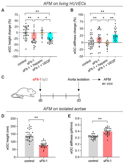
Soluble fms-like tyrosine kinase-1 (sFlt-1) is a secreted protein that binds heparan sulfate expressed on the endothelial glycocalyx (eGC). In this paper we analyze how excess sFlt-1 causes conformational changes in the eGC, leading to monocyte adhesion, a key event triggering vascular dysfunction. In vitro exposure of primary human umbilical vein endothelial cells to excess sFlt-1 decreased eGC height and increased stiffness as determined by atomic force microscopy (AFM). Yet, structural loss of the eGC components was not observed, as indicated by Ulex europaeus agglutinin I and wheat germ agglutinin staining. Moreover, the conformation observed under excess sFlt-1, a collapsed eGC, is flat and stiff with unchanged coverage and sustained content. Functionally, this conformation increased the endothelial adhesiveness to THP-1 monocytes by about 35%. Heparin blocked all these effects, but the vascular endothelial growth factor did not. In vivo administration of sFlt-1 in mice also resulted in the collapse of the eGC in isolated aorta analyzed ex vivo by AFM. Our findings show that excess sFlt-1 causes the collapse of the eGC and favors leukocyte adhesion. This study provides an additional mechanism of action by which sFlt-1 may cause endothelial dysfunction and injury. Full article
(This article belongs to the Special Issue New Insights into Endothelial Injury)
Show Figures

Figure 1

18 pages, 1497 KiB  
Review
Chronic Pain-Associated Cardiovascular Disease: The Role of Sympathetic Nerve Activity
by Christian A. Reynolds and Zeljka Minic
Int. J. Mol. Sci. 2023, 24(6), 5378; https://doi.org/10.3390/ijms24065378 - 11 Mar 2023
Cited by 24 | Viewed by 7330
Abstract
Chronic pain affects many people world-wide, and this number is continuously increasing. There is a clear link between chronic pain and the development of cardiovascular disease through activation of the sympathetic nervous system. The purpose of this review is to provide evidence from [...] Read more.
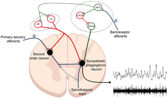
Chronic pain affects many people world-wide, and this number is continuously increasing. There is a clear link between chronic pain and the development of cardiovascular disease through activation of the sympathetic nervous system. The purpose of this review is to provide evidence from the literature that highlights the direct relationship between sympathetic nervous system dysfunction and chronic pain. We hypothesize that maladaptive changes within a common neural network regulating the sympathetic nervous system and pain perception contribute to sympathetic overactivation and cardiovascular disease in the setting of chronic pain. We review clinical evidence and highlight the basic neurocircuitry linking the sympathetic and nociceptive networks and the overlap between the neural networks controlling the two. Full article
(This article belongs to the Special Issue Sympathetic Nerves and Cardiovascular Diseases)
Show Figures

Figure 1

11 pages, 1502 KiB  
Article
Pro-Arrhythmic Potential of Accumulated Uremic Toxins Is Mediated via Vulnerability of Action Potential Repolarization
by Willem B. van Ham, Carlijn M. Cornelissen, Elizaveta Polyakova, Stephanie M. van der Voorn, Merel L. Ligtermoet, Jantine Monshouwer-Kloots, Marc A. Vos, Alexandre Bossu, Eva van Rooij, Marcel A. G. van der Heyden and Toon A. B. van Veen
Int. J. Mol. Sci. 2023, 24(6), 5373; https://doi.org/10.3390/ijms24065373 - 11 Mar 2023
Cited by 3 | Viewed by 1872
Abstract
Chronic kidney disease (CKD) is represented by a diminished filtration capacity of the kidneys. End-stage renal disease patients need dialysis treatment to remove waste and toxins from the circulation. However, endogenously produced uremic toxins (UTs) cannot always be filtered during dialysis. UTs are [...] Read more.
Chronic kidney disease (CKD) is represented by a diminished filtration capacity of the kidneys. End-stage renal disease patients need dialysis treatment to remove waste and toxins from the circulation. However, endogenously produced uremic toxins (UTs) cannot always be filtered during dialysis. UTs are among the CKD-related factors that have been linked to maladaptive and pathophysiological remodeling of the heart. Importantly, 50% of the deaths in dialysis patients are cardiovascular related, with sudden cardiac death predominating. However, the mechanisms responsible remain poorly understood. The current study aimed to assess the vulnerability of action potential repolarization caused by exposure to pre-identified UTs at clinically relevant concentrations. We exposed human induced pluripotent stem cell-derived cardiomyocytes (hiPSC-CMs) and HEK293 chronically (48 h) to the UTs indoxyl sulfate, kynurenine, or kynurenic acid. We used optical and manual electrophysiological techniques to assess action potential duration (APD) in the hiPSC-CMs and recorded IKr currents in stably transfected HEK293 cells (HEK-hERG). Molecular analysis of KV11.1, the ion channel responsible for IKr, was performed to further understand the potential mechanism underlying the effects of the UTs. Chronic exposure to the UTs resulted in significant APD prolongation. Subsequent assessment of the repolarization current IKr, often most sensitive and responsible for APD alterations, showed decreased current densities after chronic exposure to the UTs. This outcome was supported by lowered protein levels of KV11.1. Finally, treatment with an activator of the IKr current, LUF7244, could reverse the APD prolongation, indicating the potential modulation of electrophysiological effects caused by these UTs. This study highlights the pro-arrhythmogenic potential of UTs and reveals a mode of action by which they affect cardiac repolarization. Full article
Show Figures

Figure 1

14 pages, 2988 KiB  
Review
Molecular and Cellular Regulations in the Development of the Choroidal Circulation System
by Satoshi Imanishi, Yohei Tomita, Kazuno Negishi, Kazuo Tsubota and Toshihide Kurihara
Int. J. Mol. Sci. 2023, 24(6), 5371; https://doi.org/10.3390/ijms24065371 - 11 Mar 2023
Cited by 2 | Viewed by 3336
Abstract
Disorders in the development and regulation of blood vessels are involved in various ocular disorders, such as persistent hyperplastic primary vitreous, familial exudative vitreoretinopathy, and choroidal dystrophy. Thus, the appropriate regulation of vascular development is essential for healthy ocular functions. However, regulation of [...] Read more.
Disorders in the development and regulation of blood vessels are involved in various ocular disorders, such as persistent hyperplastic primary vitreous, familial exudative vitreoretinopathy, and choroidal dystrophy. Thus, the appropriate regulation of vascular development is essential for healthy ocular functions. However, regulation of the developing choroidal circulation system has not been well studied compared with vascular regulation in the vitreous and the retina. The choroid is a vascular-rich and uniquely structured tissue supplying oxygen and nutrients to the retina, and hypoplasia and the degeneration of the choroid are involved in many ocular disorders. Therefore, understanding the developing choroidal circulation system expands our knowledge of ocular development and supports our understanding of ocular disorders. In this review, we examine studies on regulating the developing choroidal circulation system at the cellular and molecular levels and discuss the relevance to human diseases. Full article
(This article belongs to the Special Issue Changes in Retinal Disease in Association with Choroid)
Show Figures

Figure 1

12 pages, 1070 KiB  
Article
Action of Platelet-Rich Plasma on In Vitro Cellular Bioactivity: More than Platelets
by Maider Beitia, Diego Delgado, Jon Mercader, Pello Sánchez, Leonor López de Dicastillo and Mikel Sánchez
Int. J. Mol. Sci. 2023, 24(6), 5367; https://doi.org/10.3390/ijms24065367 - 10 Mar 2023
Cited by 24 | Viewed by 2899
Abstract
Platelet-rich plasma (PRP) is a biological therapy in which one of the mechanisms of action is the stimulation of biological processes such as cell proliferation. The size of PRP’s effect depends on multiple factors, one of the most important being the composition of [...] Read more.
Platelet-rich plasma (PRP) is a biological therapy in which one of the mechanisms of action is the stimulation of biological processes such as cell proliferation. The size of PRP’s effect depends on multiple factors, one of the most important being the composition of PRP. The aim of this study was to analyze the relationship between cell proliferation and the levels of certain growth factors (IGF-1, HGF, PDGF, TGF-β and VEG) in PRP. First, the composition and effect on cell proliferation of PRP versus platelet-poor plasma (PPP) were compared. Subsequently, the correlation between each growth factor of PRP and cell proliferation was evaluated. Cell proliferation was higher in cells incubated with lysates derived from PRP compared to those cultured with lysates derived from PPP. In terms of composition, the levels of PDGF, TGF-β, and VEGF were significantly higher in PRP. When analyzing the PRP growth factors, IGF-1 was the only factor that correlated significantly with cell proliferation. Of those analyzed, the level of IGF-1 was the only one that did not correlate with platelet levels. The magnitude of PRP’s effect depends not only on platelet count but also on other platelet-independent molecules. Full article
Show Figures

Figure 1

14 pages, 759 KiB  
Review
Models of Congenital Adrenal Hyperplasia for Gene Therapies Testing
by Olga Glazova, Asya Bastrich, Andrei Deviatkin, Nikita Onyanov, Samira Kaziakhmedova, Liudmila Shevkova, Nawar Sakr, Daria Petrova, Maria V. Vorontsova and Pavel Volchkov
Int. J. Mol. Sci. 2023, 24(6), 5365; https://doi.org/10.3390/ijms24065365 - 10 Mar 2023
Cited by 4 | Viewed by 3524
Abstract
The adrenal glands are important endocrine organs that play a major role in the stress response. Some adrenal glands abnormalities are treated with hormone replacement therapy, which does not address physiological requirements. Modern technologies make it possible to develop gene therapy drugs that [...] Read more.
The adrenal glands are important endocrine organs that play a major role in the stress response. Some adrenal glands abnormalities are treated with hormone replacement therapy, which does not address physiological requirements. Modern technologies make it possible to develop gene therapy drugs that can completely cure diseases caused by mutations in specific genes. Congenital adrenal hyperplasia (CAH) is an example of such a potentially treatable monogenic disease. CAH is an autosomal recessive inherited disease with an overall incidence of 1:9500–1:20,000 newborns. To date, there are several promising drugs for CAH gene therapy. At the same time, it remains unclear how new approaches can be tested, as there are no models for this disease. The present review focuses on modern models for inherited adrenal gland insufficiency and their detailed characterization. In addition, the advantages and disadvantages of various pathological models are discussed, and ways of further development are suggested. Full article
Show Figures

Figure 1

22 pages, 1238 KiB  
Review
The Role of Regulated Programmed Cell Death in Osteoarthritis: From Pathogenesis to Therapy
by Suqing Liu, Yurong Pan, Ting Li, Mi Zou, Wenji Liu, Qingqing Li, Huan Wan, Jie Peng and Liang Hao
Int. J. Mol. Sci. 2023, 24(6), 5364; https://doi.org/10.3390/ijms24065364 - 10 Mar 2023
Cited by 38 | Viewed by 6092
Abstract
Osteoarthritis (OA) is a worldwide chronic disease that can cause severe inflammation to damage the surrounding tissue and cartilage. There are many different factors that can lead to osteoarthritis, but abnormally progressed programmed cell death is one of the most important risk factors [...] Read more.
Osteoarthritis (OA) is a worldwide chronic disease that can cause severe inflammation to damage the surrounding tissue and cartilage. There are many different factors that can lead to osteoarthritis, but abnormally progressed programmed cell death is one of the most important risk factors that can induce osteoarthritis. Prior studies have demonstrated that programmed cell death, including apoptosis, pyroptosis, necroptosis, ferroptosis, autophagy, and cuproptosis, has a great connection with osteoarthritis. In this paper, we review the role of different types of programmed cell death in the generation and development of OA and how the different signal pathways modulate the different cell death to regulate the development of OA. Additionally, this review provides new insights into the radical treatment of osteoarthritis rather than conservative treatment, such as anti-inflammation drugs or surgical operation. Full article
(This article belongs to the Special Issue Focus on Cartilage Biology)
Show Figures

Figure 1

15 pages, 991 KiB  
Review
Xerostomia and Its Cellular Targets
by Yoon-Jung Kim
Int. J. Mol. Sci. 2023, 24(6), 5358; https://doi.org/10.3390/ijms24065358 - 10 Mar 2023
Cited by 13 | Viewed by 6662
Abstract
Xerostomia, the subjective feeling of a dry mouth associated with dysfunction of the salivary glands, is mainly caused by radiation and chemotherapy, various systemic and autoimmune diseases, and drugs. As saliva plays numerous essential roles in oral and systemic health, xerostomia significantly reduces [...] Read more.
Xerostomia, the subjective feeling of a dry mouth associated with dysfunction of the salivary glands, is mainly caused by radiation and chemotherapy, various systemic and autoimmune diseases, and drugs. As saliva plays numerous essential roles in oral and systemic health, xerostomia significantly reduces quality of life, but its prevalence is increasing. Salivation mainly depends on parasympathetic and sympathetic nerves, and the salivary glands responsible for this secretion move fluid unidirectionally through structural features such as the polarity of acinar cells. Saliva secretion is initiated by the binding of released neurotransmitters from nerves to specific G-protein-coupled receptors (GPCRs) on acinar cells. This signal induces two intracellular calcium (Ca2+) pathways (Ca2+ release from the endoplasmic reticulum and Ca2+ influx across the plasma membrane), and this increased intracellular Ca2+ concentration ([Ca2+]i) causes the translocation of the water channel aquaporin 5 (AQP5) to the apical membrane. Consequently, the GPCR-mediated increased [Ca2+]i in acinar cells promotes saliva secretion, and this saliva moves into the oral cavity through the ducts. In this review, we seek to elucidate the potential of GPCRs, the inositol 1,4,5-trisphosphate receptor (IP3R), store-operated Ca2+ entry (SOCE), and AQP5, which are essential for salivation, as cellular targets in the etiology of xerostomia. Full article
(This article belongs to the Special Issue Recent Advances in Salivary Gland and Their Function 2.0)
Show Figures

Figure 1

19 pages, 3564 KiB  
Article
Combinatorial Network of Transcriptional and miRNA Regulation in Colorectal Cancer
by Rupesh Kumar, Maged Mostafa Mahmoud, Hanaa M. Tashkandi, Shafiul Haque, Steve Harakeh, Kalaiarasan Ponnusamy and Shazia Haider
Int. J. Mol. Sci. 2023, 24(6), 5356; https://doi.org/10.3390/ijms24065356 - 10 Mar 2023
Cited by 2 | Viewed by 2626
Abstract
Colorectal cancer is one of the leading causes of cancer-associated mortality across the worldwide. One of the major challenges in colorectal cancer is the understanding of the regulatory mechanisms of biological molecules. In this study, we aimed to identify novel key molecules in [...] Read more.
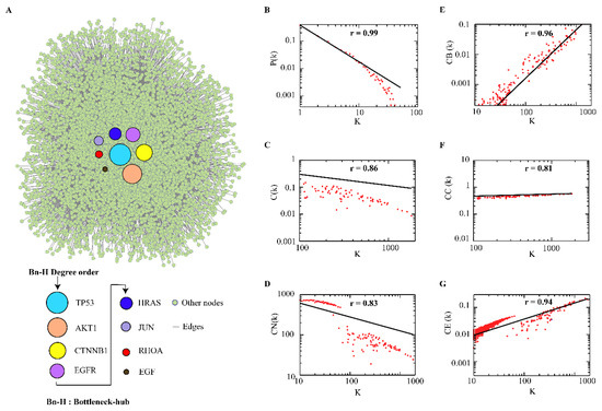
Colorectal cancer is one of the leading causes of cancer-associated mortality across the worldwide. One of the major challenges in colorectal cancer is the understanding of the regulatory mechanisms of biological molecules. In this study, we aimed to identify novel key molecules in colorectal cancer by using a computational systems biology approach. We constructed the colorectal protein–protein interaction network which followed hierarchical scale-free nature. We identified TP53, CTNBB1, AKT1, EGFR, HRAS, JUN, RHOA, and EGF as bottleneck-hubs. The HRAS showed the largest interacting strength with functional subnetworks, having strong correlation with protein phosphorylation, kinase activity, signal transduction, and apoptotic processes. Furthermore, we constructed the bottleneck-hubs’ regulatory networks with their transcriptional (transcription factor) and post-transcriptional (miRNAs) regulators, which exhibited the important key regulators. We observed miR-429, miR-622, and miR-133b and transcription factors (EZH2, HDAC1, HDAC4, AR, NFKB1, and KLF4) regulates four bottleneck-hubs (TP53, JUN, AKT1 and EGFR) at the motif level. In future, biochemical investigation of the observed key regulators could provide further understanding about their role in the pathophysiology of colorectal cancer. Full article
(This article belongs to the Special Issue Molecular Advances in Colorectal Cancer)
Show Figures

Figure 1

17 pages, 4573 KiB  
Article
Hepatocyte-Specific Triggering of Hepatic Stellate Cell Profibrotic Activation by Apoptotic Bodies: The Role of Hepatoma-Derived Growth Factor, HIV, and Ethanol
by Moses New-Aaron, Siva Sankar Koganti, Murali Ganesan, Sharma Kanika, Vikas Kumar, Weimin Wang, Edward Makarov, Kusum K. Kharbanda, Larisa Y. Poluektova and Natalia A. Osna
Int. J. Mol. Sci. 2023, 24(6), 5346; https://doi.org/10.3390/ijms24065346 - 10 Mar 2023
Cited by 6 | Viewed by 2410
Abstract
Liver disease is one of the leading comorbidities in HIV infection. The risk of liver fibrosis development is potentiated by alcohol abuse. In our previous studies, we reported that hepatocytes exposed to HIV and acetaldehyde undergo significant apoptosis, and the engulfment of apoptotic [...] Read more.
Liver disease is one of the leading comorbidities in HIV infection. The risk of liver fibrosis development is potentiated by alcohol abuse. In our previous studies, we reported that hepatocytes exposed to HIV and acetaldehyde undergo significant apoptosis, and the engulfment of apoptotic bodies (ABs) by hepatic stellate cells (HSC) potentiates their pro-fibrotic activation. However, in addition to hepatocytes, under the same conditions, ABs can be generated from liver-infiltrating immune cells. The goal of this study is to explore whether lymphocyte-derived ABs trigger HSC profibrotic activation as strongly as hepatocyte-derived ABs. ABs were generated from Huh7.5-CYP2E1 (RLW) cells and Jurkat cells treated with HIV+acetaldehyde and co-culture with HSC to induce their pro-fibrotic activation. ABs cargo was analyzed by proteomics. ABs generated from RLW, but not from Jurkat cells activated fibrogenic genes in HSC. This was driven by the expression of hepatocyte-specific proteins in ABs cargo. One of these proteins is Hepatocyte-Derived Growth Factor, for which suppression attenuates pro-fibrotic activation of HSC. In mice humanized with only immune cells but not human hepatocytes, infected with HIV and fed ethanol, liver fibrosis was not observed. We conclude that HIV+ABs of hepatocyte origin promote HSC activation, which potentially may lead to liver fibrosis progression. Full article
Show Figures

Figure 1

18 pages, 4149 KiB  
Article
Inter- and Intramolecular RNA–RNA Interactions Modulate the Regulation of Translation Mediated by the 3′ UTR in West Nile Virus
by Cristina Romero-López, Margarita Roda-Herreros, Beatriz Berzal-Herranz, Sara Esther Ramos-Lorente and Alfredo Berzal-Herranz
Int. J. Mol. Sci. 2023, 24(6), 5337; https://doi.org/10.3390/ijms24065337 - 10 Mar 2023
Cited by 3 | Viewed by 2136
Abstract
RNA viruses rely on genomic structural elements to accomplish the functions necessary to complete the viral cycle. These elements participate in a dynamic network of RNA–RNA interactions that determine the overall folding of the RNA genome and may be responsible for the fine [...] Read more.
RNA viruses rely on genomic structural elements to accomplish the functions necessary to complete the viral cycle. These elements participate in a dynamic network of RNA–RNA interactions that determine the overall folding of the RNA genome and may be responsible for the fine regulation of viral replication and translation as well as the transition between them. The genomes of members of the genus Flavivirus are characterized by a complexly folded 3′ UTR with a number of RNA structural elements that are conserved across isolates of each species. The present work provides evidence of intra- and intermolecular RNA–RNA interactions involving RNA structural elements in the 3′ UTR of the West Nile virus genome. The intermolecular interactions can be visualized in vitro by the formation of molecular dimers involving the participation of at least the SLI and 3′DB elements. Certainly, the 3′ UTR of dengue virus, which lacks the SLI element, forms molecular dimers in lower quantities via a single interaction site, probably 3′DB. The functional analysis of sequence or deletion mutants revealed an inverse relationship between 3′ UTR dimerization and viral translation efficiency in cell cultures. A network of RNA–RNA interactions involving 3′ UTR structural elements might therefore exist, helping to regulate viral translation. Full article
(This article belongs to the Special Issue RNA Regulatory Networks at the Crossroad of Human Diseases 3.0)
Show Figures

Figure 1

24 pages, 6297 KiB  
Article
KDM5D Histone Demethylase Identifies Platinum-Tolerant Head and Neck Cancer Cells Vulnerable to Mitotic Catastrophe
by Tsung-Ming Chen, Chih-Ming Huang, Syahru Agung Setiawan, Ming-Shou Hsieh, Chih-Chi Sheen and Chi-Tai Yeh
Int. J. Mol. Sci. 2023, 24(6), 5310; https://doi.org/10.3390/ijms24065310 - 10 Mar 2023
Cited by 4 | Viewed by 3043
Abstract
Head and neck squamous cell carcinoma (HNSCC) is a major contributor to cancer incidence globally and is currently managed by surgical resection followed by adjuvant chemoradiotherapy. However, local recurrence is the major cause of mortality, indicating the emergence of drug-tolerant persister cells. A [...] Read more.
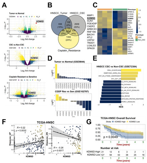
Head and neck squamous cell carcinoma (HNSCC) is a major contributor to cancer incidence globally and is currently managed by surgical resection followed by adjuvant chemoradiotherapy. However, local recurrence is the major cause of mortality, indicating the emergence of drug-tolerant persister cells. A specific histone demethylase, namely lysine-specific demethylase 5D (KDM5D), is overexpressed in diverse types of cancers and involved in cancer cell cycle regulation. However, the role of KDM5D in the development of cisplatin-tolerant persister cells remains unexplored. Here, we demonstrated that KDM5D contributes to the development of persister cells. Aurora Kinase B (AURKB) disruption affected the vulnerability of persister cells in a mitotic catastrophe–dependent manner. Comprehensive in silico, in vitro, and in vivo experiments were performed. KDM5D expression was upregulated in HNSCC tumor cells, cancer stem cells, and cisplatin-resistant cells with biologically distinct signaling alterations. In an HNSCC cohort, high KDM5D expression was associated with a poor response to platinum treatment and early disease recurrence. KDM5D knockdown reduced the tolerance of persister cells to platinum agents and caused marked cell cycle deregulation, including the loss of DNA damage prevention, and abnormal mitosis-enhanced cell cycle arrest. By modulating mRNA levels of AURKB, KDM5D promoted the generation of platinum-tolerant persister cells in vitro, leading to the identification of the KDM5D/AURKB axis, which regulates cancer stemness and drug tolerance of HNSCC. Treatment with an AURKB inhibitor, namely barasertib, resulted in a lethal consequence of mitotic catastrophe in HNSCC persister cells. The cotreatment of cisplatin and barasertib suppressed tumor growth in the tumor mouse model. Thus, KDM5D might be involved in the development of persister cells, and AURKB disruption can overcome tolerance to platinum treatment in HNSCC. Full article
(This article belongs to the Special Issue Molecular Mechanisms and Pathophysiology of Head and Neck Diseases)
Show Figures

Figure 1

20 pages, 716 KiB  
Review
microRNAs as Biomarkers of Endothelial Dysfunction and Therapeutic Target in the Pathogenesis of Atrial Fibrillation
by Vanessa Desantis, Maria Assunta Potenza, Luca Sgarra, Carmela Nacci, Antonietta Scaringella, Sebastiano Cicco, Antonio Giovanni Solimando, Angelo Vacca and Monica Montagnani
Int. J. Mol. Sci. 2023, 24(6), 5307; https://doi.org/10.3390/ijms24065307 - 10 Mar 2023
Cited by 9 | Viewed by 3016
Abstract
The pathophysiology of atrial fibrillation (AF) may involve atrial fibrosis/remodeling and dysfunctional endothelial activities. Despite the currently available treatment approaches, the progression of AF, its recurrence rate, and the high mortality risk of related complications underlay the need for more advanced prognostic and [...] Read more.
The pathophysiology of atrial fibrillation (AF) may involve atrial fibrosis/remodeling and dysfunctional endothelial activities. Despite the currently available treatment approaches, the progression of AF, its recurrence rate, and the high mortality risk of related complications underlay the need for more advanced prognostic and therapeutic strategies. There is increasing attention on the molecular mechanisms controlling AF onset and progression points to the complex cell to cell interplay that triggers fibroblasts, immune cells and myofibroblasts, enhancing atrial fibrosis. In this scenario, endothelial cell dysfunction (ED) might play an unexpected but significant role. microRNAs (miRNAs) regulate gene expression at the post-transcriptional level. In the cardiovascular compartment, both free circulating and exosomal miRNAs entail the control of plaque formation, lipid metabolism, inflammation and angiogenesis, cardiomyocyte growth and contractility, and even the maintenance of cardiac rhythm. Abnormal miRNAs levels may indicate the activation state of circulating cells, and thus represent a specific read-out of cardiac tissue changes. Although several unresolved questions still limit their clinical use, the ease of accessibility in biofluids and their prognostic and diagnostic properties make them novel and attractive biomarker candidates in AF. This article summarizes the most recent features of AF associated with miRNAs and relates them to potentially underlying mechanisms. Full article
Show Figures

Figure 1

16 pages, 3017 KiB  
Article
Spermidine Rescues Bioenergetic and Mitophagy Deficits Induced by Disease-Associated Tau Protein
by Lauren H. Fairley, Imane Lejri, Amandine Grimm and Anne Eckert
Int. J. Mol. Sci. 2023, 24(6), 5297; https://doi.org/10.3390/ijms24065297 - 10 Mar 2023
Cited by 17 | Viewed by 3816
Abstract
Abnormal tau build-up is a hallmark of Alzheimer’s disease (AD) and more than 20 other serious neurodegenerative diseases. Mitochondria are paramount organelles playing a predominant role in cellular bioenergetics, namely by providing the main source of cellular energy via adenosine triphosphate generation. Abnormal [...] Read more.
Abnormal tau build-up is a hallmark of Alzheimer’s disease (AD) and more than 20 other serious neurodegenerative diseases. Mitochondria are paramount organelles playing a predominant role in cellular bioenergetics, namely by providing the main source of cellular energy via adenosine triphosphate generation. Abnormal tau impairs almost every aspect of mitochondrial function, from mitochondrial respiration to mitophagy. The aim of our study was to investigate the effects of spermidine, a polyamine which exerts neuroprotective effects, on mitochondrial function in a cellular model of tauopathy. Recent evidence identified autophagy as the main mechanism of action of spermidine on life-span prolongation and neuroprotection, but the effects of spermidine on abnormal tau-induced mitochondrial dysfunction have not yet been investigated. We used SH-SY5Y cells stably expressing a mutant form of human tau protein (P301L tau mutation) or cells expressing the empty vector (control cells). We showed that spermidine improved mitochondrial respiration, mitochondrial membrane potential as well as adenosine triphosphate (ATP) production in both control and P301L tau-expressing cells. We also showed that spermidine decreased the level of free radicals, increased autophagy and restored P301L tau-induced impairments in mitophagy. Overall, our findings suggest that spermidine supplementation might represent an attractive therapeutic approach to prevent/counteract tau-related mitochondrial impairments. Full article
Show Figures

Figure 1