Article

A Genome-Wide Analysis of the Jasmonic Acid Biosynthesis Gene Families in Peanut Reveals Their Crucial Roles in Growth and Abiotic Stresses

Peanut Research Institute, College of Agronomy, Shenyang Agricultural University, Shenyang 110161, China
*
Authors to whom correspondence should be addressed.
Int. J. Mol. Sci. 2024, 25(13), 7054; https://doi.org/10.3390/ijms25137054
Submission received: 11 May 2024 / Revised: 20 June 2024 / Accepted: 25 June 2024 / Published: 27 June 2024
(This article belongs to the Special Issue Plant Defense-Related Genes and Their Networks)

Abstract

:
Abiotic stress is a limiting factor in peanut production. Peanut is an important oil crop and cash crop in China. Peanut yield is vulnerable to abiotic stress due to its seeds grown underground. Jasmonic acid (JA) is essential for plant growth and defense against adversity stresses. However, the regulation and mechanism of the jasmonic acid biosynthesis pathway on peanut defense against abiotic stresses are still limitedly understood. In this study, a total of 64 genes encoding key enzymes of JA biosynthesis were identified and classified into lipoxygenases (AhLOXs), alleno oxide synthases (AhAOSs), allene oxide cyclases (AhAOCs), and 12-oxo-phytodienoic acid reductases (AhOPRs) according to gene structure, conserved motif, and phylogenetic feature. A cis-regulatory element analysis indicated that some of the genes contained stress responsive and hormone responsive elements. In addition to proteins involved in JA biosynthesis and signaling, they also interacted with proteins involved in lipid biosynthesis and stress response. Sixteen putative Ah-miRNAs were identified from four families targeting 35 key genes of JA biosynthesis. A tissue expression pattern analysis revealed that AhLOX2 was the highest expressed in leaf tissues, and AhLOX32 was the highest expressed in shoot, root, and nodule tissues. AhLOX16, AhOPR1, and AhOPR3 were up-regulated under drought stress. AhLOX16, AhAOS3, AhOPR1, and AhAOC4 had elevated transcript levels in response to cold stress. AhLOX5, AhLOX16, AhAOC3, AhOPR1, and AhOPR3 were up-regulated for expression under salt stress. Our study could provide a reference for the study of the abiotic stress resistance mechanism in peanut.

1. Introduction

Plant hormones play important roles in the regulation of plant responses to various stimuli and stresses [1]. Jasmonic acid (JA) is an oxidized fatty acid derivative, which together with its derivatives and active precursor 12-oxo-phytodienoic acid (12-OPDA) are collectively known as jasmonates (JAs) [2]. Jasmonic acid can inhibit root growth, accelerate fruit ripening, and regulate flowering plant development [3,4]. As a signal transduction molecule, it is closely related to plant resistance to biological (injury, insect bite, etc.) and abiotic stresses (drought, low temperature, salinity, and wounding), and may participate in the crosstalk between various hormones [5,6]. The biosynthesis of JA is performed in the cytoplasm, chloroplast, and peroxisome [4]. Firstly, α-linolenic acid (α-LeA) is catalyzed in the chloroplast and transported to peroxisomes as a synthetic feedstock. Subsequently, cis-(+)-12-oxo-phytodienoic acid (12-OPDA) is generated via lipoxygenase (LOX; EC1.13.11.12), alleno oxide synthase (AOS; EC4.2.1.92) and allene oxide cyclase (AOC; EC5.3.99.6). Then, with the help of 12-oxo-phytodienoic acid reductase (OPDA reductase, OPR; EC1.3.1.42), 12-OPDA is reduced to OPC-8:0, followed by three rounds of β-oxidation [3,7]. The final product of β-oxidation is (3R,7S)-JA (OPC-2:0), which can form the more stable isomer (3R,7R)-JA [8,9]. These two isomers of JA coexist in plants. Eventually, JA diffuses into the cytoplasm, and various derivatives are formed through different modifications [10,11,12].
Lipoxygenase (LOX; EC1.13.11.12), a non-heme iron-containing oxygenase, can catalyze the oxygenation of polyunsaturated fatty acids to produce hydroperoxides [13]. The lipoxygenase domain and PLAT_LH2 domain are the core components of LOXs. According to the binding site of its substrate during oxygenation, LOXs in plants can be divided into 9-LOXs and 13-LOXs, of which 13-LOXs can be further divided into type I and type II 13-LOXs [14]. Alleno oxide synthase (AOS; EC4.2.1.92) belongs to the CYP74 subfamily within the cytochrome P450 superfamily. Alleno oxide synthases have been verified to play a crucial role in the biosynthesis of oxylipins, bioactive compounds involved in signal and defense reactions in higher plants, mammals, and algae [15]. Alleno oxide synthases do not require molecular oxygen, nor do they require cytochrome P450 reductases that rely on NAD(P)H as co-factors, but instead use their hydrogen peroxide substrates as a source and oxygen donor for reducing equivalents [16,17]. Allene oxide cyclase (AOC; EC5.3.99.6) is a dimeric enzyme located in chloroplasts [18]. Since first cloned in tomato by Ziegler [19], AOC genes have been isolated and identified in many plant species such as Arabidopsis, rice, wheat, soybean, and cannabis [19,20,21,22,23,24]. 12-Oxo-phytodienoic acid reductase (OPDA reductase, OPR; EC1.3.1.42) belongs to the old yellow enzyme (OYE) family and is classified as FMN-dependent oxidant oxidase [25,26]. In 1997, OPRs were first identified in the Arabidopsis genome and divided into OPRI and OPRII [27]. In rice, the classification of OPRs into five classes was based on the three-dimensional structural differences and deep replication nodes of the proteins, which determine the substrate specificity and catalytic activity of the different isomeric OPR proteins [28].
It has been shown that key enzymes catalyzing JA biosynthesis play an important role in plant adaptation to adversity stress. In mung bean (Vigna radiata), LOXs’ activity was significantly elevated after an external application of 20% PEG-6000 [29]. Both 9-LOXs and 13-LOXs have been reported to be closely associated with plant responses to external stimuli. The 13-LOX subfamily plays a role in wounding and other defense responses [30]. At the 190 s time point, the combined lox3B-lox4A mutation reduced JA concentration damaged leaf 8 by approximately 20% relative to the wild type in Arabidopsis. The over-expression of LOX10 enhances tolerance to salinity stress in rice seedlings [31]. The 9-LOX subfamily, on the other hand, has been linked to pathogen defense [32]. An OPDA-like 9-LOX-derived compound, 10-OPEA (10-oxo-11-phytoenoic acid), plays an important role in maize fungal defense [33]. Alleno oxide synthases have been reported to limit JA levels in injured plants and to affect reproductive fertility. A functional mutation in AOSs results in injured Arabidopsis that shows no increase in endogenous JA levels. The wild type shows a 100-fold increase in endogenous JA levels in 1 h. Allene oxide cyclase is an important member in the biosynthesis of JA, and transgenic tobacco plants overexpressing GmAOC1 have leaves that remain green and possess higher fresh weights and normal root growth after treatment with 150 mM NaCl, whereas the wild type turns markedly yellow and has severely retarded root growth, suggesting that the over-expression of GmAOC1 improves the plant’s tolerance to salt stress [22].
Peanut (Arachis hypogaea L.) is an important oil crop with high nutritional and commercial value in China [34]. China is one of the world’s largest peanut producers, with more than 4.6 million hm2 sown area and more than 17 million tons of pod production, accounting for 40% of the global total [35]. China has gradually formed the oil crop planting structure with peanut and rape as the main elements in the development of agricultural production and increased agricultural supply has become increasingly prominent. During the growth period, peanuts suffer from a variety of natural disasters and weather, which have a greater impact on the growth and development of peanuts, often causing a decline in photosynthesis, slow growth, and serious production reduction or even extinction [36]. Under abiotic stress, photosynthesis, nutrient absorption, metabolism, growth, and final crop yield of peanut will all undergo adverse effects [37]. Therefore, clarifying the molecular mechanism of abiotic adaptation in peanut and genetic improvement for stress tolerance was crucial to safeguard national edible oil and food security. However, there are fewer reports on the regulation and mechanism of action of the JA biosynthetic pathway on peanut stress tolerance. In the present study, we identified key enzyme proteins of JA biosynthesis in allotetraploid peanut and analyzed their phylogenetic relationship, gene structure, conserved domains and motifs, chromosomal distribution, phylogenetic features, cis-regulatory elements, and expression profiles in response to drought, cold, and salt treatments. Our results provide a bioinformatics analysis and candidate genes that may be potentially utilized in improving crop resistance to abiotic stress.

2. Results

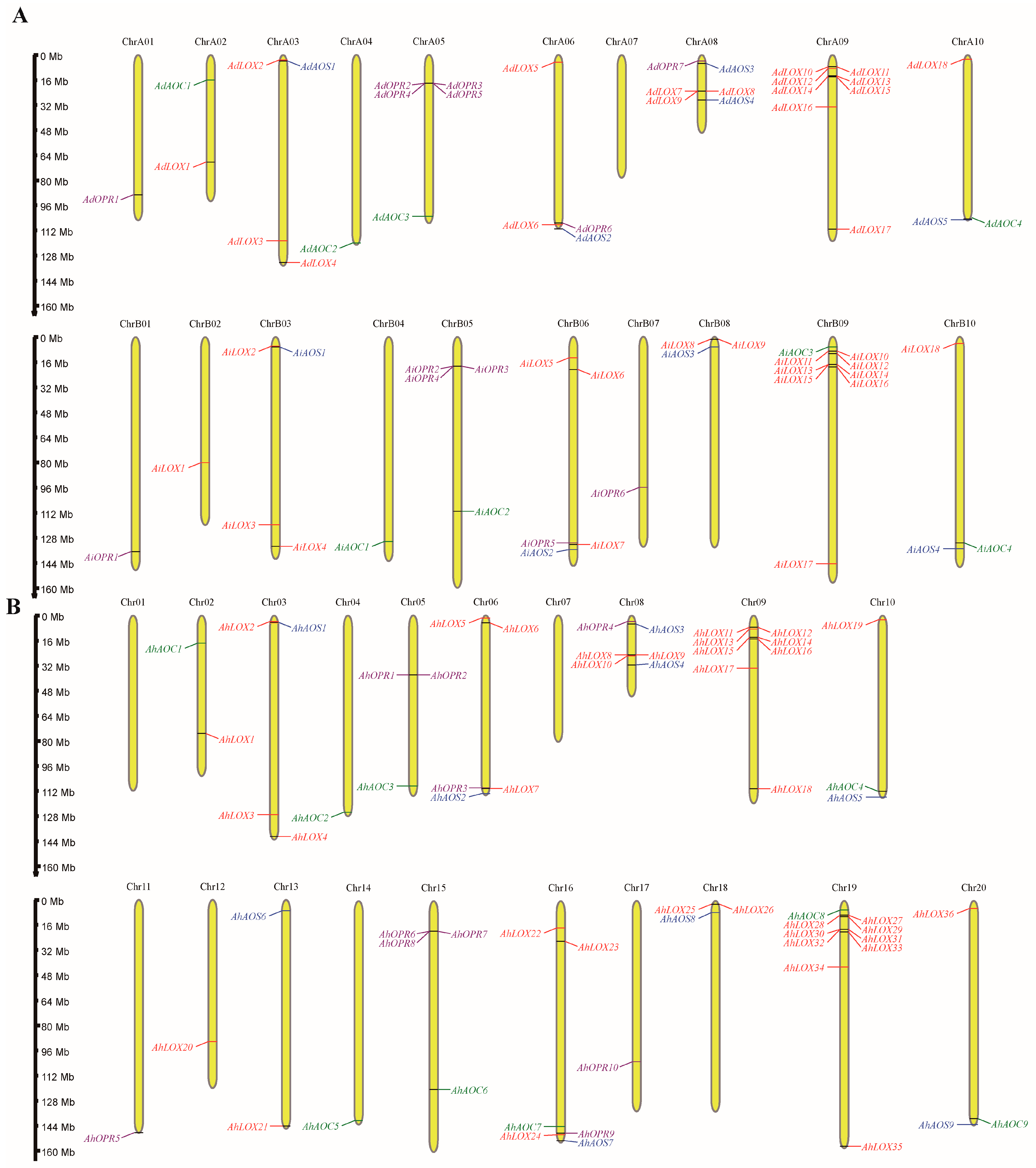
2.1. Identification and Characterization of JA Biosynthesis Gene Families in Arachis

A total of 130 genes encoding key enzyme proteins of JA biosynthesis were identified in A. duranensis, A. ipaensis, and A. hypogaea genomes by HMMER [38] (Tables S1–S4). These genes corresponded to four gene families (lipoxygenase, LOX; allene oxide synthase, AOS; allene oxide cyclase, AOC; and 12-oxophytodienoate reductase, OPR). The remaining sequences were checked for the existence of complete domains utilizing Batch Web CD-Search Tool, Pfam, and SMART websites. A total of 72 LOXs, 18 AOSs, 17 AOCs, and 23 OPRs were finally confirmed as key enzyme proteins of JA biosynthesis and renamed based on their chromosomal locations (Figure 1). In this study, LOX, AOS, AOC, and OPR genes were unevenly distributed in nineteen of twenty chromosomes of A. duranensis (A genome) and A. ipaensis (B genome). Chromosome 19 contained the most genes and no JA biosynthesis gene was discovered in chromosome A07, 01, and 17. Many homologous genes in allotetraploid peanut were mapped to key enzyme genes of JA biosynthesis in A and B genomes. Multiple sequence alignment results demonstrated that the homologous gene pairs did have high sequence similarity (Table S5).

2.2. Physicochemical Properties of JA Biosynthesis Genes

In order to explore the sequence characteristics of 72 LOXs, 18 AOSs, 17 AOCs, and 23 OPRs in Arachis, the opening reading frame (ORF) lengths, theoretical isoelectric point (pI), and other basic information were analyzed. The length of LOX proteins varied from 509 to 477 amino acids (AAs). The molecular weights (MWs) ranged from 58.35 kDa to 53.18 kDa. The predicted isoelectric point value of LOX proteins was between 4.98 and 9.15. The instability index of LOX proteins was between 33.31 and 52.05, and the aliphatic index was between 78.96 and 93.88. All predicted LOX proteins showed a grand average of hydropathicity (GRAVY) value below zero, meaning hydrophilic protein (Table S1). The peanut AOS proteins have a maximum length of 590 AAs and a minimum of 336 AAs, with pI ranging from 5.79 to 8.91. The MW ranged from 42.8 kDa to 66.5 kDa. The instability index of AOS proteins was between 33.39 and 49.44, and the aliphatic index was between 79.29 and 94.64. All predicted AOS proteins’ GRAVY value below zero means that they were hydrophilic protein (Table S2). The AOC protein lengths ranged from 178 to 303 amino acids. The MW of AOC proteins varied from 19.78 kDa to 33.64 kDa, and the pI values ranged from 6.90 to 9.83. The instability index of AOC proteins was between 27.78 and 55.52, and the aliphatic index was between 74.78 and 98.74. All predicted AOC proteins showed a GRAVY value below zero (Table S3). The protein lengths of 23 OPR genes ranged from 339 AAs to 573 AAs. The MW ranged from 37.56 kDa to 64.44 kDa and pI of these proteins ranged from 5.52 to 6.84. The instability index of OPR proteins was between 29.86 and 43.17, and the aliphatic index was between 74.01 and 84.06. All predicted OPR proteins’ GRAVY value below zero means that they were hydrophilic protein (Table S4). Subcellular localization results demonstrated that all LOX protein was located in the cytoplasm, 12 AOSs worked in the endoplasmic reticulum, and 5 AOS and 17 AOC proteins were located in the chloroplast. All OPR proteins were located in the cytoplasm apart from AdOPR1, AiOPR1, and AhOPR5 (Tables S1–S4).

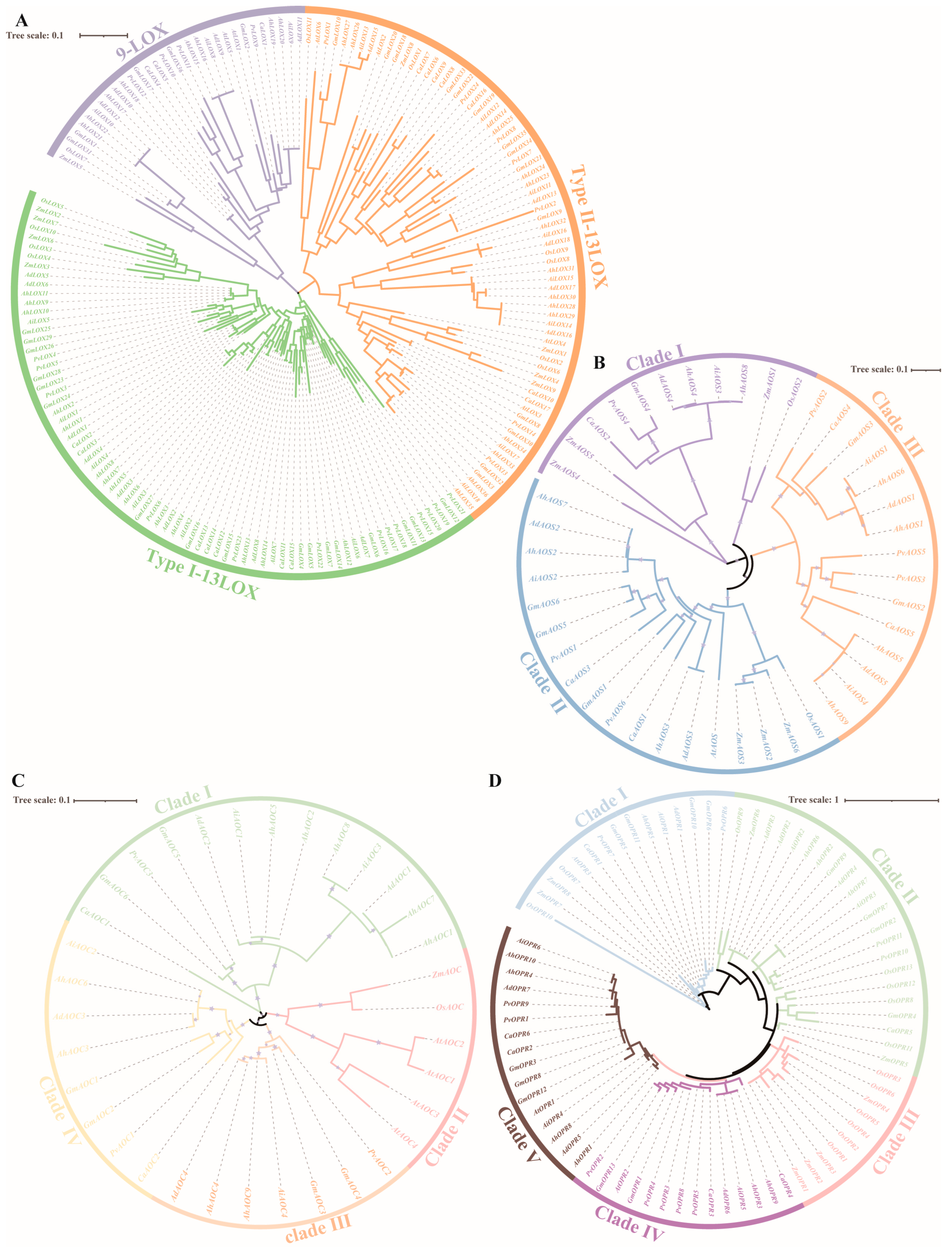
2.3. Phylogenetic Analysis of JA Biosynthesis Gene Families

To analyze the classification and evolutionary relationships among the protein members of JA biosynthesis gene families in different species, four phylogenetic trees were constructed utilizing the HMM search tool based on the protein sequences in Arabidopsis thaliana, Oryza sativa, Zea mays, Glycine max, Phaseolus vulgaris, Cicer arietinum, A. duranensis, A. ipaensis, and A. hypogaea (Figure 2). A multiple-sequence alignment was established among 18 AdLOXs, 18 AiLOXs, 36 AhLOXs, 6 AtLOXs, 11 OsLOXs, 9ZmLOXs, 36 GmLOXs, 24 PvLOXs, and 17 CaLOXs using clustalW software (Version 2.1).
According to previous studies in Arabidopsis, LOX proteins could be classified into three distinct subfamilies: 9-LOX, type I 13-LOX, and type II 13-LOX (Figure 2A). AhLOX had 8, 14, and 14 members in three subfamilies, respectively. In the 9-LOX subfamily, AdLOX9-12, AiLOX8-10, and AhLOX15-22 were closely related to GmLOX1-2, GmLOX17, GmLOX31, GmLOX36, PvLOX9-12, CaLOX1, and CaLOX4-5. AdLOX1-8, AiLOX1-7, and AhLOX1-14 had the highest relationship with GmLOX4-7, GmLOX11-16, GmLOX23-29, PvLOX3-6, PvLOX15-23, CaLOX2, and CaLOX11-15 in type-I 13-LOX. In the same trend as in the type I 13-LOX family, AdLOX, AiLOX, and AhLOX were closely related to GmLOXs, PvLOXs, and CaLOXs, and the largest number of AtLOXs, OsLOXs, and ZmLOXs were gathered in the type II 13-LOX family. In general, peanut LOX is closely related to soybean, chickpeas, and common beans, which belong to Fabaceae.
The nine AhAOS members were non-randomly distributed in clade I, II, and III (Figure 2B). The phylogenetic relationship analysis revealed that AdAOS4, AiAOS3, AhAOS4, and AhAOS8 were tightly related to OsAOS2, ZmAOS1, ZmAOS4-5, GmAOS4, PvAOS4, and CaAOS2. There were three ZmAOSs in clade I and II, respectively. There were the largest number of AhAOSs in clade III. It is noteworthy that the only AtAOS appeared in clade II and AtAOSs, OsAOSs, and ZmAOSs were not found in clade III.
A total of 34 AhAOC proteins were identified and classified into four clades: clade I, II, III, and IV (Figure 2C). AdAOC1-2, AiAOC1, AiAOC3, AhAOC1-2, AhAOC5, and AhAOC7-8 were in close proximity to GmAOC5-6, PvAOC3, and CaAOC1. Four AtAOCs, one OsAOC, and one ZmAOC were all clustered, and no members of other species were discovered in clade II. Clade III and IV contained the same amount of Arachis AOC members. Only AOCs of Fabaceae existed in clade I, III, and IV; this phenomenon suggested that Arachis AOC members had high homology with AOC in Glycine max, Phaseolus vulgaris, and Cicer arietinum.
According to the branches of the phylogenetic tree and classification of OPR gene family members in Arabidopsis thaliana and Oryza sativa [39,40], five, four, nine, one, two, six, six, six, and five members of the OPR family were identified and divided into five clades in A. duranensis, A. ipaensis, A. hypogaea, Arabidopsis thaliana, Oryza sativa, Zea mays, Glycine max, Phaseolus vulgaris, and Cicer arietinum, respectively (Figure 2D). All species in clade I had at least one OPR member. Only OsOPRs and ZmOPRs remained in clade III. Three AtOPR proteins were distributed in clade I, IV, and V. Four AhOPRs existed in clade V. Taken together, compared to Arabidopsis thaliana, Oryza sativa, and Zea mays, the results indicated that peanut key enzyme proteins of JA biosynthesis had higher homology with a family member in Glycine max, Phaseolus vulgaris, and Cicer arietinum.

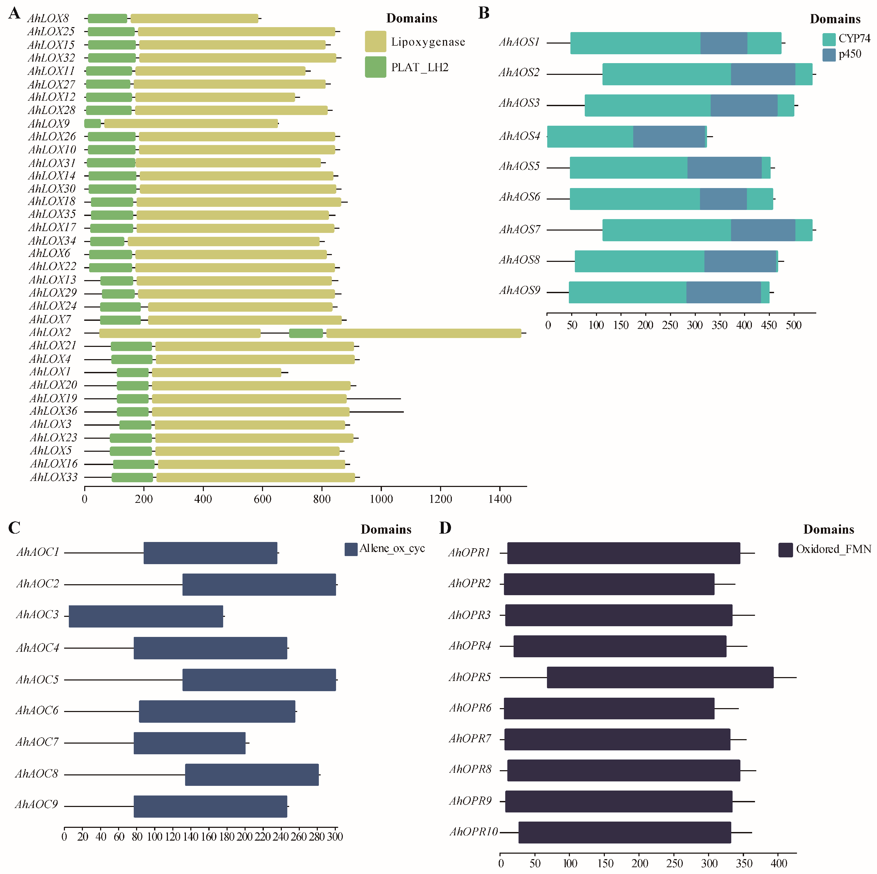
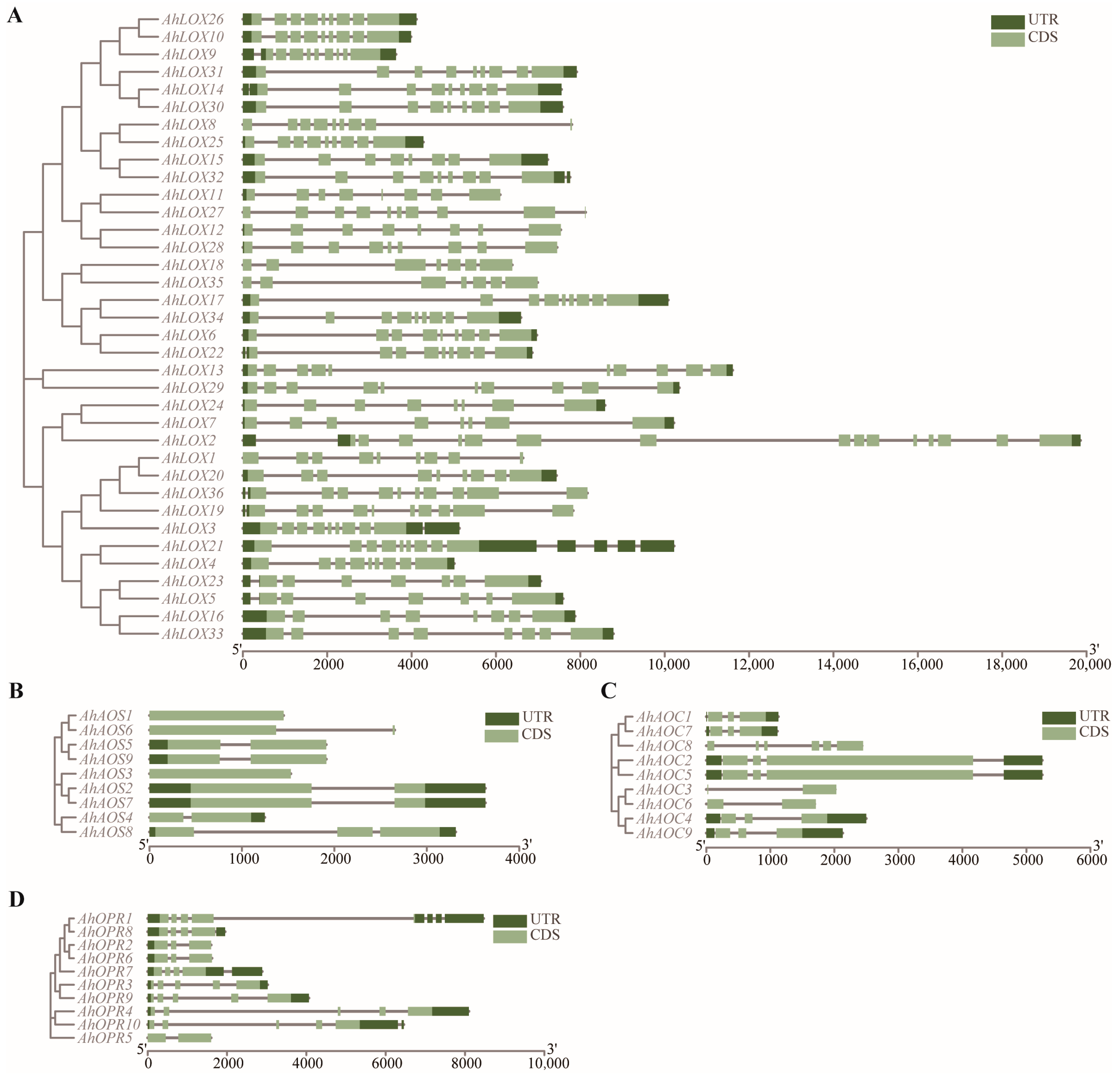
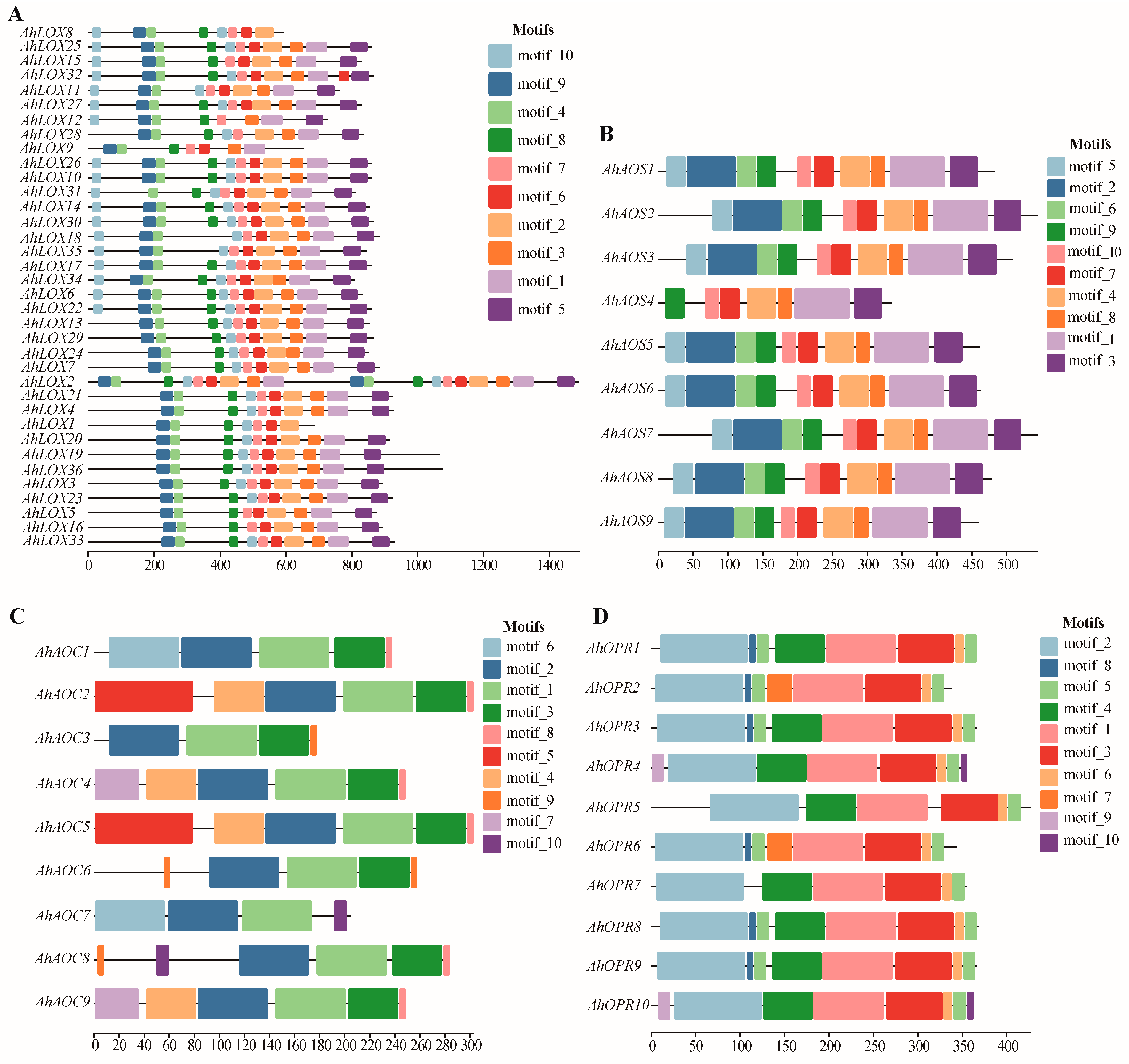
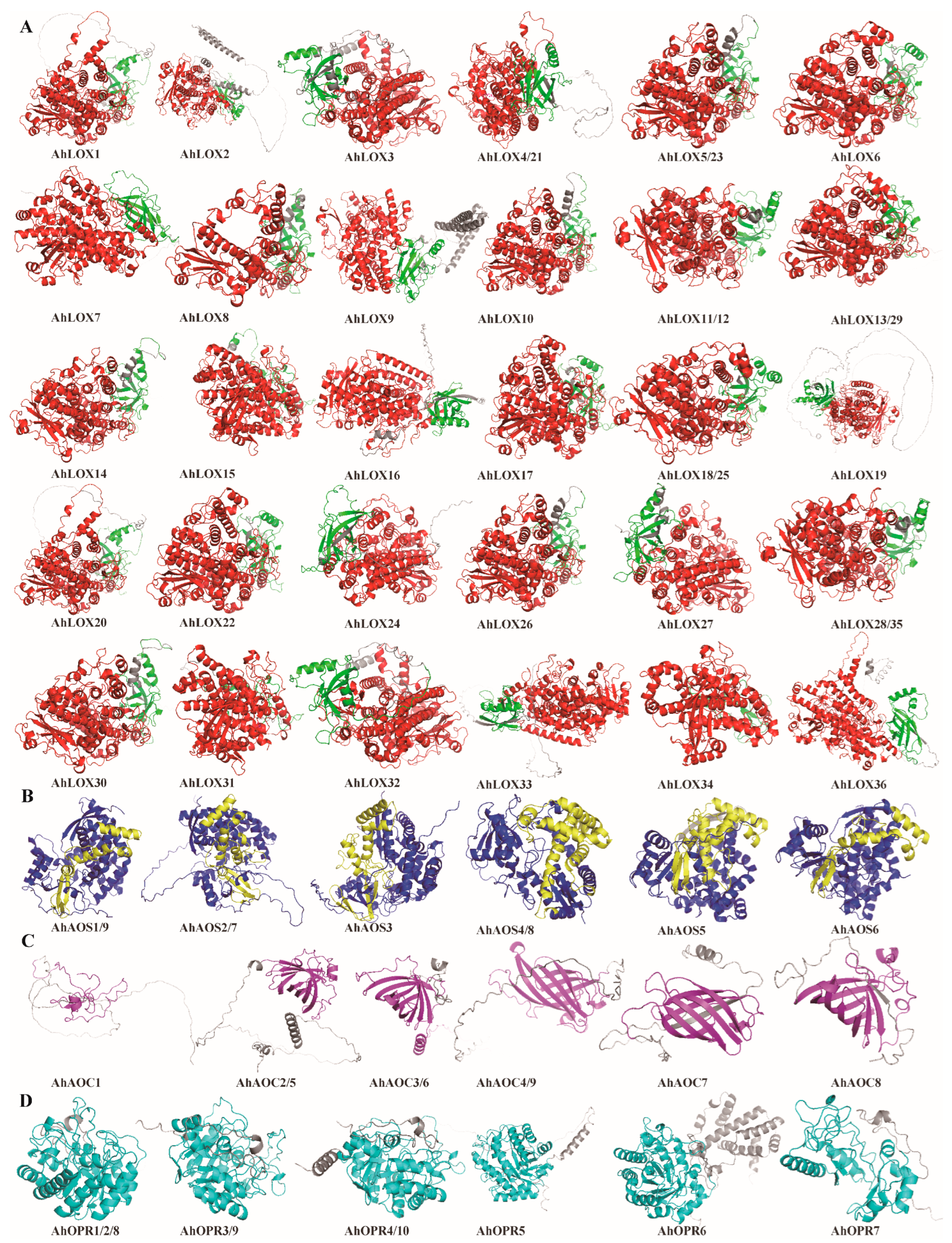
2.4. Domain Characterization, Gene Structure, Conserved Motif Distribution, and Protein Structure of JA Biosynthesis Gene Families

In order to determine the presence of conserved domains, key enzyme genes of JA biosynthesis were filtrated by the CDD database (Figure 3). The results confirmed that the lipoxygenase domain, p450 domain, Allene_ox_cyc domain, and Oxidored_FMN domain were successfully detected in AhLOXs, AhAOSs, AhAOCs, and AhOPRs, respectively. As one of the branches of the p450 superfamily, the CYP74 domain is the typical characteristic and distinguishing basis of the AOS gene family. At the same time, a lipoxygenase domain and a PLAT_LH2 domain exist simultaneously to be considered a member of the LOX family. The analysis revealed that all members of AhLOXs, AhAOSs, AhAOCs, and AhOPRs contained characteristic domains of their family.
The number of introns and the way introns are inserted may be the cause of gene evolution and functional differentiation. In this study, the exon–intron organizations of all the identified JA biosynthesis genes were examined to gain more insight into the evolution in allotetraploid peanut. The phylogenetic tree and gene structure were integrated together as shown in Figure 4. The number of introns of AhLOX varied from 6 to 14 (Figure 4A). Eight AhAOS genes were with or without one intron (Figure 4B). In addition to AhAOC8, the other eight AhAOC genes contained one or two introns (Figure 4C). All OPR genes had fewer than five introns (Figure 4D). The above results implied that the genes within the same family had similar genetic organization. The high number of introns in the AhLOX gene indicated that the biological functions of this family might be more complex than those of the other three families.
To study the structural features of key enzyme proteins of JA biosynthesis, the MEME (Multiple Em for Motif Elicitation) program was utilized to identify the conserved motifs of AhLOXs, AhAOSs, AhAOCs, and AhOPRs in peanut (Figure 5; Tables S6–S9). Ten conserved motifs were detected in each family and distinguished by different colors. The 26 AhLOX proteins contained all motifs. AhLOX9 and AhLOX12 lacked motif 2; AhLOX1, AhLOX8, and AhLOX9 lacked motif 5; motif 6 was not found in AhLOX12 and AhLOX28; AhLOX12 and AhLOX18 lacked motif 8; and AhLOX6, AhLOX9, and AhLOX16 lost motif 10. In the AhAOS family, the composition of conservative motifs is relatively complete. Only AhAOS4 lost motif 2, 5, and 6. Motif 1 and 2 were detected in all AhAOC members. The MEME analysis indicated that only AhAOC7 lacked motif 3 and 7, AhAOC2 and AhAOC5 contained motif 5, AhAOC6 and AhAOC8 contained motif 9, and motif 1, 2, 3, and 8 might be the core components of the Allene_ox_cyc domain. The same trend appeared in the AhOPR family. Motif 1, 2, 3, 5, and 6 were found in all members; AhOPR2 and AhOPR6 lacked motif 4; and motif 7 was detected only in AhOPR2 and AhOPR6. AhOPR4-5, AhOPR7, and AhOPR10 were missing motif 8, and motif 9 and 10 were not discovered in AhOPR4 and AhOPR10. In general, the conserved motifs of protein members within the same family are similar in composition, with some conserved motifs constituting the core components of the characteristic domain, while others show the biological functional diversity of different members.
Structure and function are compatible, and the structure of a protein determines its biological functional properties. Protein secondary structure prediction showed that all key enzyme proteins of JA biosynthesis have four structures, the alpha helix (Hh), extended strand (Ee), beta turn (Tt), and random coil (Cc), with Cc having the highest percentage of 59.04–36.01% and 46.13–10.04%, followed by Hh and Ee with 10.04–46.13% and 11.65–34.87%, respectively, and Tt with the lowest percentage of 9.76–4.04% (Table S10). In addition, the 3D structures of all proteins were predicted using the protein structure database AlphaFold and visualized using PyMOL software (version 2.5.5; https://pymol.org/2/; accessed on 12 April 2023), and it was found that all proteins contained conserved subunits of subfamilies to which they belonged (Figure 6). The results of the 3D protein structure prediction analyses confirmed that the key enzymes of JA biosynthesis showed a high degree of conservatism among and within the subgroups. The key enzyme proteins of JA biosynthesis were categorized into four types based on their 3D structures and six core structural domains were defined: the PLAT_LH2 structural domain located at the N-terminal and lipoxygenase domain located at the C-terminal of AhLOX protein, the CYP74 structural domain presented in AhAOS protein and p450 domain in the middle, and the Allene_ox_cyc domain at the C-terminal of AOC protein and Oxidored_FMN domain anchored in the middle of OPR protein. This suggested that the higher the amino acid sequence similarity between the proteins, the more similar their 3D structures and the more similar their functions were.

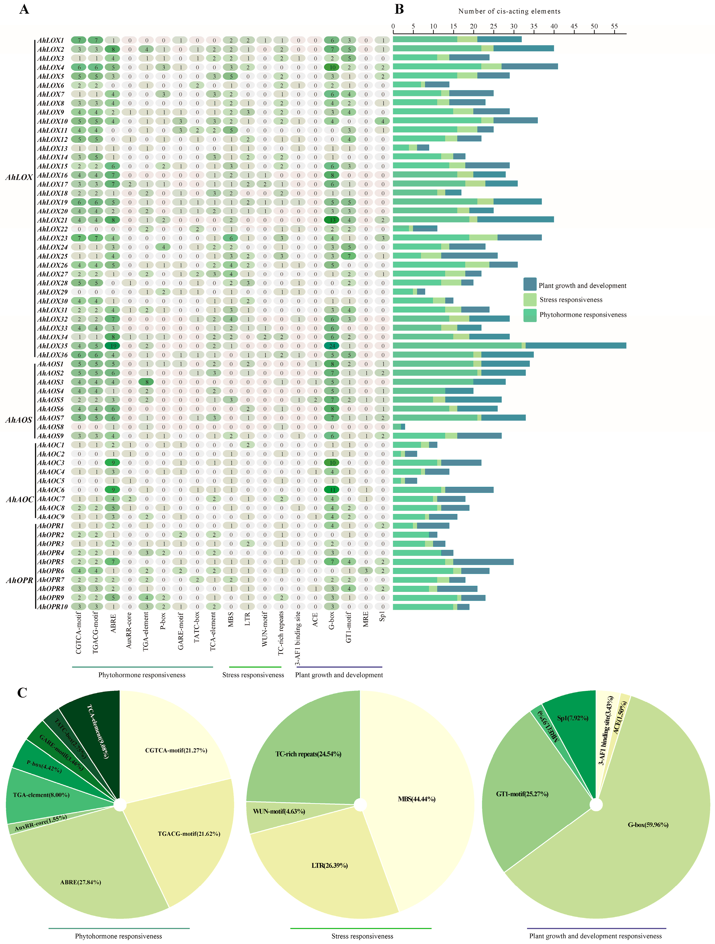
2.5. Cis-Regulatory Elements in the Promoter Regions of JA Biosynthesis Genes

Cis-regulatory elements as binding sites of transcription factors connect gene structure and function and play important roles in gene expression [41]. To further analyze the biological function of JA biosynthesis genes, the regions about 2000 bp upstream of the start codon (ATG) were used, analyzing cis-regulatory elements of AhLOXs, AhAOSs, AhAOCs, and AhOPRs (Figure 7; Table S11). After counting the number of cis-regulatory elements, we found that AhLOXs had the largest number of elements compared to other families, of which AhLOX35 contains 57 elements, and 29 AhLOX members contain more than 20 elements (Figure 7A,B). The same situation appeared as a result for the AhAOS family; 8 AhAOS members had more than 20 elements. AhAOC members contain a minimum of 6 and a maximum of 25 elements and the number of cis-acting elements in an AhOPR member ranges from 11 to 30.
The detected cis-regulatory elements could be divided into three categories according to their functional annotations including phytohormone responsiveness, stress responsiveness, and plant growth and development responsiveness (Figure 7C). The CGTCA motif and TGACG motif, as MeJA responsive elements, accounted for 21.27% and 21.62%, respectively. The promoter regions of some genes such as AhLOX35, AhLOX2, AhLOX4, AhLOX16, AhLOX19, AhLOX35, and AhAOS7 contain a large number of abscisic acid responsive elements, ABRE, AuxRR-core, and TGA-element, suggesting that these genes may be subject to ABA-induced expression. AhLOX4, AhLOX7, AhLOX10, AhLOX11, AhLOX15, AhLOX24, AhLOX29, and AhOPR10 contain more than one gibberellin responsive element including P-box, GARE-motif, and TATC-box. AhLOX5, AhLOX7, AhLOX10, AhLOX11, AhLOX14, AhLOX18, AhLOX27, AhAOS2, and AhAOS7 contain three salicylic acid response elements (TCA-element). In total, 44 of the 64 key enzyme genes of JA biosynthesis contained all classes of hormone response elements, and these findings emphasized the possible involvement of these genes in the crosstalk mechanism of various phytohormones.
MBS, a drought responsive element, accounts for 44.44%, which is the largest among all stress-related elements, indicating that these genes may be involved in the response of drought stress signals (Figure 7C). Forty-eight genes contained one or more MBS and forty-three genes contained both ABREs, such as AhLOX5, AhLOX7, AhLOX8, AhLOX9, AhLOX16, AhAOS5, AhAOC5, AhOPR1, AhOPR5, AhOPR6, and AhOPR7, which signified that these genes may be present in the ABA-dependent drought stress response pathway. Regarding LTR, accounting for 26.39%, 40 genes contained LTRs, of which AhLOX9, AhAOS6, AhAOC1, and AhOPR3 contained at least two LTRs, demonstrating that JA biosynthesis genes may be induced by a low-temperature signal. AhLOX1, AhLOX17, AhLOX19, AhLOX3, and AhLOX36 had WUN-motif and TC-rich repeats in addition to the above-mentioned, which proved that these genes may take a crucial part in the resistance mechanism of various stresses.
Plant growth and development response elements, including the 3-AF1 binding site, ACE, G-box, GT1-motif, MRE, and Sp1, were also detected in AhLOX2, AhLOX4, AhLOX21, AhLOX35, AhAOS1, AhAOS5, AhAOC6, and AhOPR5, confirming that these genes may also be involved in peanut growth and development.

2.6. Protein–Protein Interactions and miRNA Genes’ Regulatory Networks of JA Biosynthesis Gene Families

To investigate the biological functional properties and metabolic pathways of key enzyme genes of JA biosynthesis and to reveal the possible potential interactions of these proteins, interacting proteins of peanut AhLOXs, AhAOSs, AhAOCs, and AhOPRs were searched and networked by the STRING online database (Figure 8A; Table S12). The analysis revealed that 64 peanut key enzyme proteins of JA biosynthesis interacted with 35 functional proteins. Functional prediction revealed that proteins interacting with peanut key enzyme proteins of JA biosynthesis were mainly involved in the JA biosynthesis pathway, including LOX2-4, LOX6, CYP74A (AOS), hydroperoxide lyase CYP74B2 (HPL1), AOC1-4, and OPR1-3, and transcription factors MYC2, MYC3, jasmonate ZIM-domain protein TIFY3A/JAZ11, TIFY3B/JAZ12, TIFY5A/JAZ8, TIFY5B/JAZ7, TIFY6B/JAZ3, TIFY7/JAZ9, TIFY9/JAZ10, TIFY10A/JAZ1, TIFY10B/JAZ2, TIFY11A/JAZ5, and TIFY11B/JAZ6. Coronatine insensitive 1 (COI1) is mainly involved in hormone response, plant development, and signaling in JA. In addition, peanut key enzyme proteins of JA biosynthesis interacted with 4-coumarate/CoA ligase 5 (4CL5), phospholipase A2-alpha (PLA2-ALPHA), alcohol dehydrogenase 1 (ADH1), defective anther dehiscence 1-2 (DAD1-2), sugar-dependent 1 (SDP1), SDP1-like (SDPL), alpha-dioxygenase 1 (DOX1), and DOX2. These proteins may be involved in lipid synthesis, transport, and catabolism during plant development and reproduction, such as in the hydrolysis of phosphatidylcholine, glycolipids, and triacylglycerols, and may be involved in catalyzing acetaldehyde reduction. Notably, proteins interacting with peanut key enzyme proteins of JA biosynthesis are also involved in responding to external stimuli, stresses, and other phytohormone induction. AhLOX interacts with phospholipase A2-alpha (PLA2-ALPHA) and responds to hormone induction and other external stimuli. All three families (except AhOPRs) interact with DOX1, suggesting that they are involved in defense against oxidative stress due to abiotic stress, prevent oxidative stress and cell death, and can be induced by salicylic acid. AhAOC protein interacted with ABI five binding protein homolog 2 (AFPH2) to mediate germination and response to osmotic stress and sugar stress. The AhOPR protein interacted with nine JAZ proteins, which act as direct targets of SCF/COI1 E3 ubiquitin-ligase and mediate the degradation of this proteasome induced by JA treatment.
To explore the micro-RNA (miRNA) regulation by the JA biosynthesis pathway, the relationship between miRNA-targeted key enzyme proteins of JA biosynthesis and miRNAs was predicted by psRNATarget Server (Figure 8B; Table S13). A total of 16 miRNAs targeted 35 genes, of which 6 LOX and 5 AOC were targeted by 1 miRNA at the same time; ahy-miR3509-3p, ahy-miR3515, ahy-miR3518, and ahy-miR394 could only target 1 gene; and ahy-miR159, ahy-miR3511-3p, ahy-miR3513-3p, ahy-miR3520-5p, and ahy-miR408-5p targeted more than 5 genes, suggesting that these miRNAs may play a key role in the JA biosynthesis pathway.

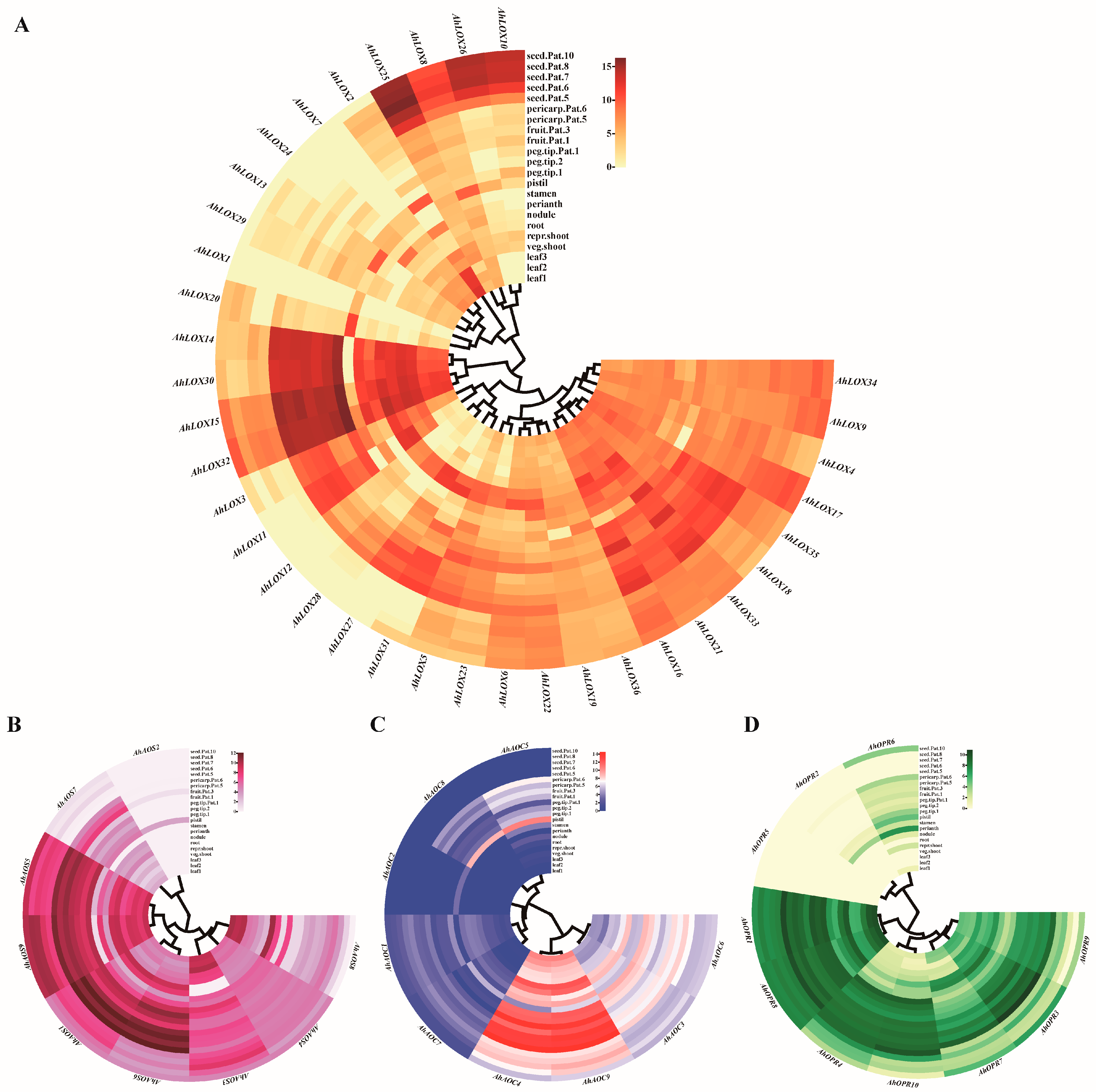
2.7. Expression Profiles of JA Biosynthesis Genes in Different Tissues

To predict the possible biological functions and effects of JA biosynthesis genes on peanut growth and development, RNA-seq data of 22 peanut tissues [42] were utilized, detecting the expression pattern and drawing a heatmap. The analysis verified that most of the genes were expressed to different degrees in 22 tissues (Figure 9; Table S14). AhAOS2, AhAOC2, AhOPR2, and AhOPR5 were the lower expressed or undetected in most tissues. The heatmap results confirmed that 41 out of 64 peanut JA biosynthesis genes were highly expressed in the leaves, vegetative shoot tip, reproductive shoot tip, and root. Some genes showed tissue specificity; for example, AhLOX11 was highly expressed in the vegetative shoot tip, reproductive shoot tip, and root; AhLOX12, AhLOX23, AhLOX27, AhLOX28, AhLOX31, AhAOC7, and AhOPR10 had tissue-specific expression in roots, and these genes were likely to be closely related to stress response.
During peanut growth and development, roots, shoots, leaves, and nodules act as the nutrient organs of the peanut, controlling the transportation of water and nutrients. Among them, roots, shoots, and leaves, as key parts of water transport, are more sensitive to abiotic stress signals. AhLOX2, AhLOX15, AhLOX30, AhLOX32, and AhAOS8 were highly expressed in leaves; AhLOX14-15, AhLOX30, and AhLOX32 were highly expressed in shoot tissues. AhLOX11, AhLOX30, AhLOX32, AhAOC4, and AhAOC9 were expressed in the peanut root. AhLOX11, AhLOX14-15, AhLOX31, and AhLOX32 were expressed in the nodule. This suggested that the above genes played a positive role in the growth and development of peanut and may be involved in the response mechanism to abiotic stresses.
While perianths, stamens, pistils, pegs, pods, and seeds play important reproductive development roles as reproductive organs, AhLOX3-6, AhLOX8-11, AhLOX14-19, AhLOX21-23, AhLOX25-26, AhLOX30, AhLOX32-36, AhAOS1, AhAOS3-6, AhAOS9, AhAOC3-4, AhAOC6, AhAOC9, AhOPR1, AhOPR3-4, and AhOPR7-10 were highly expressed in these tissues, demonstrating that these genes may control reproductive development of peanut.

2.8. Expression Patterns of JA Biosynthesis Genes in Response to Different Abiotic Stress Treatments

To investigate whether the expression of JA biosynthesis genes was influenced by abiotic stress and related signaling at the transcriptional level, further expression profiling was discovered under drought, cold, and salt stress for 0 h, 6 h, 12 h, 24 h, and 48 h (Figure 10; Table S15). The results are consistent with our hypothesis; the expression of many JA genes demonstrated significant changes under abiotic stress.
It was found that the expression of most of the genes increased under drought and cold stress treatments. AhLOX3, AhLOX5, AhLOX7, AhLOX8, AhAOS5, AhAOS8, AhAOC5, and tested AhOPR1, AhOPR3, AhOPR4, AhOPR5, AhOPR6, and AhOPR7 were rapidly up-regulated about 2-fold in expression when they were subjected to drought stress treatment for 6 h, of which AhOPR1 and AhOPR3 were up-regulated more than 10-fold at 6 h and AhOPR3 was even up-regulated more than 100-fold at 6 h, and this trend did not decrease until 48 h. The expression of some genes such as AhLOX9 and AhLOX16 did not increase significantly until 24 h after treatment.
Most of the genes were up-regulated under cold stress. AhLOX11, AhLOX19, AhLOX20, AhLOX29, AhAOS3, AhAOS6, AhAOS7, AhAOC1, AhAOC4, AhAOC8, AhOPR5, and AhOPR7 were up-regulated about 2-fold at 6 h under cold stress; AhLOX2, AhLOX6, AhLOX11, AhLOX17, AhLOX19, AhLOX26, AhLOX29, AhAOS7, AhAOC1, AhAOC8, AhOPR1, and AhOPR5-7 were up-regulated to more than 2-fold transcript levels at 12 h of cold stress treatment compared to the control. The expression of AhLOX20, AhAOS5, AhAOS7, AhOPR1, AhAOC1, AhAOC4-5, AhAOS3, and AhOPR5-7 was up-regulated more than 2-fold at 24 h of cold stress treatment, with more than a 5-fold up-regulation of AhOPR1 and AhAOS3. The transcript levels of AhLOX2, AhLOX5-6, AhLOX11, AhLOX16, AhLOX19-20, AhLOX26, AhLOX28, AhAOS3, AhAOS5-7, AhAOC4, AhOPR1, and AhOPR5-6 were more than 2-fold compared to those of the control at 48 h of cold stress, with more than 5-fold for AhOPR1 and AhLOX16. It is a remarkable fact that the AhOPR1, which performed well under drought stress, showed a trend of up-regulation at 12 h, while AhOPR3 was not slightly expressed until 48 h.
AhLOX7 and AhLOX16 were down-regulated at 6 h, followed by an overall up-regulated trend. AhLOX5, AhLOX6, AhLOX7, AhAOS3, AhAOC3, AhAOC8, AhOPR1, and AhOPR3 transcript levels were up-regulated under salt stress treatments, with AhAOC3, AhAOC8, AhOPR1, and AhOPR3 rapidly up-regulated at 6 h, while the remaining genes were not significantly up-regulated (less than 1.5-fold) or down-regulated. Notably, AhOPR1 and AhOPR3 were transcribed at more than a 30-fold level at 12 h of salt stress. AhLOX8, AhLOX18-19, AhLOX32, AhAOS5, and AhAOC4 were down-regulated at 6 h under salt stress, up-regulated after 12 h, and down-regulated again at 24–48 h. AhLOX17, AhLOX20, AhLOX26, AhLOX29, and AhOPR4 were rapidly downgraded at 6 h and not upgraded to around background levels until 48 h. These results suggested that these genes may be negative regulators in the pathway against salt stress and that some of the genes may be upstream and can cascade signals.
Taken together, the results indicated that most of the genes were up-regulated in transcript levels under drought, cold, and salt stresses, which might be involved in the defense mechanism against stress signals, and some genes were down-regulated in expression, which might play the role of negative feedback regulation. Individual genes were first up-regulated and then down-regulated, which may be involved in the transduction and regulation of stress signals. Interestingly, these key enzyme genes of JA biosynthesis (except AhOPR1 and AhOPR3) were expressed completely differently under different abiotic stress treatments, and did not appear to be highly expressed under both stresses at the same time, suggesting that these genes may exercise different biological functions in response to different abiotic stresses.

3. Discussion

Plant growth, development, and survival depend on the natural environment in a complex and strictly interconnected network, and environmental factors unfavorable to growth and survival become the main threats to this network [43]. Among these environmental factors, abiotic stresses represented by drought, extreme temperatures, and saline conditions are considered as the main abiotic sources of stimuli [44]. Plants have developed complex mechanisms to resist and mitigate unfavorable environments over a long evolutionary process, including the intrinsic regulation of phytohormones [45]. JA, as an important signaling and regulatory molecule, affects plant growth, development, and yield, including the regulation of stomatal opening and closing, cell cycle, and uptake of glucose and nutrients such as nitrogen and phosphorus [2,8]. Meanwhile, JA is involved in the perception and transmission of various environmental signals and makes defense responses to adversity stress, in which the key enzyme genes of the JA biosynthesis pathway play a crucial leading role [46]. Therefore, a genome-wide analysis of gene families of key enzymes for JA biosynthesis can be an effective tool for mining key resistance genes and exploring mechanisms of resistance to abiotic stresses.
The genome-wide identification and bioinformatics analysis of LOX, AOS, AOC, and OPR have been studied in different species such as Arabidopsis, rice, wheat, maize, and soybean [47,48,49,50,51,52,53]. In this study, key enzyme genes of JA biosynthesis within the genomes of two wild species of peanut, cultivated peanut, model plants of Arabidopsis thaliana, rice, maize, and near-origin species of soybean, chickpea, and common bean were rescreened and identified by a hidden Markov model search, and all members were re-determined based on the results of existing studies and structural domain searches [28,54,55,56]. Although the cultivated peanut is a heterotetraploid cross between two wild species of peanut, the number of key enzyme genes of JA biosynthesis did not follow this principle exactly; not all AOS, AOC, and OPR genes on the allotetraploid peanut genome find their corresponding homolog genes in the A and B genome, and the total number of them is less than the sum of the number of genes within the A and B genomes. This may be due to the fact that these gene families lost some genes during the evolutionary process, or some of the functionally redundant genes were discarded while a doubling replication event occurred. A similar situation was found in other gene families of peanut. The natural resistance-associated macrophage protein (NRAMP) family has 7 AdNRAMPs, 6 AiNRAMPs, and 15 AhNRAMPs (Tan et al., 2023). In addition, only 14 AdSAURs and 19 AiSAURs were found, the number of which was much smaller than that of AhSAURs (162) [57]; 68, 36, and 31 major latex-like proteins (MLPs) were obtained from A. hypogaea, A. duranensis, and A. ipaensis, respectively [58].
However, studies on key enzyme genes of JA biosynthesis within a single species are limited. In this study, we conducted the first comprehensive and systematic identification and bioinformatics analysis of peanut key enzyme genes of JA biosynthesis, and explored their roles in the stress resistance mechanism of peanut through the application of abiotic stresses (drought stress, cold stress, and salt stress). A phylogenetic relationship analysis with key enzyme genes of JA biosynthesis in the genomes of other species revealed that members of the four families, LOX, AOS, AOC, and OPR, in peanut were all more related to soybean, chickpea, and common bean key enzyme genes of JA biosynthesis, which may be attributed to the fact that peanut belongs to the same family of legume plants as these three species. Overall, members of each family contain conserved structural domains characteristic of the family, notably the presence of two lipoxygenase domains in AhLOX2 (Figure 3).
Initially, we hypothesized that one additional conserved domain might enhance its defense against abiotic stresses or be able to respond faster than other members, but subsequent qRT-PCR results showed that it did not perform well (Figure 8). Differences in the number and mode of insertion of introns in the gene structure, as well as the type and number of conserved motifs, may lead to genes with different biological functions. In our study, the number of introns in AhLOX family members is around 6–14, while the number of introns in AhAOS, AhAOC, and AhOPR family members is less than 5, which indirectly proves the complexity and diversity of functions of AhLOX family members. Through the conserved motif analysis, we found that 26 out of 36 members of the AhLOX family have similar conserved motif composition, and the conserved motif composition of the AhAOS, AhAOC, and AhOPR families is relatively complete, with only some of the members missing 1–2 motifs, which confirms that the same family has similar biological functions, but the difference of some motifs leads to the convergence of the functions.
Based on the results of a bioinformatic analysis, including a cis-regulatory element analysis, protein–protein interaction analysis, and tissue expression analysis, 35 potential responsive abiotic stress genes were screened and their expression under drought, cold, and salt stresses was examined by qRT-PCR. A large number of cis-regulatory elements responsive to abiotic stresses are present in the promoter regions of these genes, which are highly expressed in water-transporting tissues and are predicted to interact with proteins that defend against abiotic stresses. Inconsistent with our expectation, it was found that most of the genes were not up-regulated under these three stress conditions as we expected. In yellow horn, XsLOX1-2, XsLOX5-8, and XsLOX10 were up-regulated in cold stress and down-regulated after salt stress treatment [59]. In wheat, TaLOX16 and TaLOX18 were up-regulated after drought stress and down-regulated under salt stress [53]. In this study, AhLOX17, as a homolog gene of TaLOX16 and TaLOX18, was first down-regulated and then slightly up-regulated under drought stress treatment and down-regulated under salt stress treatment. A total of 10 GhOPR genes were discovered in cotton, which showed opposite expression trends under drought stress and salt stress, among which GhOPR1-2, GhOPR4-5, GhOPR8, and GhOPR9 were down-regulated under drought stress, and then up-regulated and then down-regulated under salt stress. GhOPR3, GhOPR6, and GhOPR10 were down-regulated under drought stress and remained up-regulated under salt stress. GhOPR7 was up-regulated under drought stress and then down-regulated under salt stress [60]. It can be hypothesized from these results that key enzyme genes of JA biosynthesis respond differently in the face of different abiotic stresses, and that some genes that exhibit resistance under one stress may turn into negative regulators involved in stress signaling when under another stress, thereby engaging more functional genes in resistance and preventing functional redundancy.
To deepen the understanding of the JA biosynthesis pathway, this study presents a schematic diagram of the JA biosynthesis pathway to gain insights into the key genes involved in defense against abiotic stresses and their potential roles (Figure 11). Under drought stress, AhLOX3, AhLOX5, AhLOX7, AhLOX8, AhLOX9, and AhLOX16 in the JA biosynthesis pathway were induced to be up-regulated and expressed, together with PLA2-ALPHA interacting with them, in response to phytohormone induction and other exogenous stimuli. The expression of AhAOS5, AhAOS8, and AhAOC4 was also rapidly elevated, and they interacted with DOX1, thereby protecting against oxidative stress and accompanying cell death (Figure 11A). When cold stress occurred, JA biosynthesis was inhibited; AhOPR1, AhOPR3, AhOPR5, and AhOPR7 were induced to be up-regulated for expression along with AhLOXs, AhAOSs, and AhAOCs; and increased JA concentration may have inhibited the AhJAZs that interact with them, releasing AhMYC2 to initiate the transport JA signaling and induce cold resistance genes (Figure 11B). AhAOCs are critical in the mechanism of resistance to salt stress. Upon exposure to salt stress, AhAOC3 and AhAOC8 transcript levels were up-regulated and interacted with AFPH2 in response to osmotic stress (Figure 11C).

4. Materials and Methods

4.1. Identification of Key Enzyme Genes of JA Biosynthesis in Peanut

Peanut genome and annotation files were obtained from a peanut genomics resource (https://peanutbase.org/; accessed on 26 June 2023). The key enzyme gene family of JA biosynthesis in the Arachis hypogaea genome was identified by the HMMER-3.0 program. The hidden Markov model (HMM) profiles of the lipoxygenase (PF00305) and PLAT_LH2 domain (PF01477) of LOXs, P450 domain (PF00067) of AOSs, Allene_ox_cyc domain (PF06351) of AOCs, and Oxidored_FMN domain (PF00724) of OPRs were retrieved from the Pfam website and were used to identify the putative proteins with the best domain e-value cutoffs of <1 × 10⁻³. To validate the accuracy of these predicted genes, we utilized the online tool SMART (http://smart.embl-heidelberg.de/; accessed on 6 January 2024), pfam (version 36.0; http://pfam.xfam.org/; accessed on 3 January 2024), and NCBI-CDD (https://www.ncbi.nlm.nih.gov/cdd; accessed on 6 January 2024) to confirm that all candidate genes had conserved domains [61,62]. The online tool ProtParam (http://web.expasy.org/protparam/; accessed on 7 January 2024) was utilized, analyzing typical characters, including the theoretical isoelectric point (PI), the instability index, the aliphatic index, and the molecular weight. The Plant-mPLoc online database (http://www.csbio.sjtu.edu.cn/bioinf/plant-multi/; accessed on 15 January 2024) was utilized, predicting the subcellular localization of LOX, AOS, AOC, and OPR proteins [63]. The amino acid, genome, and coding sequences of key enzyme genes of JA biosynthesis in A. duranensis, A. ipaensis, and A. hypogaea are shown in Table S16.

4.2. Gene Structure and Conserved Motifs and Protein Structures

The Gene Structure Display Server (GSDS 2.0) (http://gsds.gao-lab.org; accessed on 10 December 2023) was used, analyzing the composition of exons and introns based on the peanut genome annotation file and to display the structure [64]. MEME [65] was applied to predict the conserved motifs with the following parameter settings: number of repetitions—any, maximum number of motifs—10, minimum motif width—6, and maximum motif width—100. Graphic beautification was performed by TBtools software (version v2.096; https://github.com/CJ-Chen/TBtools-II/releases; accessed on 12 May 2024) [66]. Protein secondary structure prediction was performed by the online tool SOPMA (https://npsa-pbil.ibcp.fr/cgi-bin/npsa_automat.pl?page=npsa_sopma.html; accessed on 6 March 2024), and the 3D structures of all key enzyme proteins of JA biosynthesis were predicted using the protein structure database AlphaFold (https://alphafold.ebi.ac.uk/; accessed on 26 October 2023) and visualized using PyMOL software (version 2.5.5; https://pymol.org/2/; accessed on 12 April 2023).

4.3. Phylogenetic Analysis and Classification

To understand the phylogenetic relationship and to classify the key enzyme genes of JA biosynthesis in peanut, maximum-likelihood trees were constructed based on the full-length amino acid sequences of LOXs, AOSs, AOCs, and OPRs from Arachis duranensis, Arachis ipaensis, Arachis hypogaea, Arabidopsis thaliana, Oryza sativa, Zea mays, Glycine max, Cicer arietinum, and Phaseolus vulgaris (https://phytozome-next.jgi.doe.gov/; accessed on 16 April 2024) [67]. All of the above proteins have been identified to contain the characteristic structural domains of the family by the HMMER web server (https://www.ebi.ac.uk/Tools/hmmer/; accessed on 28 April 2024) [38]. Phylogenetic trees were constructed by MEGA11 software [68].

4.4. Chromosomal Location

Key enzyme genes of JA biosynthesis were mapped to the chromosomes of Arachis duranensis, Arachis ipaensis, and Arachis hypogaea, according to genome annotation GFF files (https://phytozome-next.jgi.doe.gov/; accessed on 16 April 2024), and the results were visualized by MG2C online software (http://mg2c.iask.in/mg2c_v2.1/; accessed on 16 March 2024) [69].

4.5. Cis-Regulatory Element Analysis of Promoter Region

Cis-regulatory elements as binding sites of transcription factors connect gene structure and function and play important roles in gene expression. The cis-regulatory elements in key enzyme gene promoter regions of JA biosynthesis were predicted using PlantCARE (http://bioinformatics.psb.ugent.be/webtools/plantcare/html/; accessed on 19 April 2024) [70] and visualized by TBtools software (version v2.096; https://github.com/CJ-Chen/TBtools-II/releases; accessed on 12 May 2024). The promoter sequences of key enzyme genes of JA biosynthesis in A. hypogaea are shown in Table S17.

4.6. Protein–Protein Interaction and miRNA Genes’ Regulatory Network

The interaction of key enzyme proteins of JA biosynthesis with Arabidopsis proteins was predicted by the online database STRING (https://version-12-0.string-db.org/; accessed on 18 April 2024) with a confidence threshold set at 0.4 and visualized by native software Cytoscape 3.7.2 (version 3.7.2). Peanut miRNA sequences were obtained from the online database miRbase (http://www.mirbase.org/; accessed on 18 April 2024). Extracted cDNA sequences were used, predicting miRNA targets, and miRNA regulatory network maps were constructed using Cytoscape software (version 3.7.2) [71].

4.7. Tissue Expression Pattern

The expression of JA genes in 22 tissues in allotetraploid peanut was obtained from the peanut genome resource PeanutBase (https://peanutbase.org/; accessed on 18 January 2024), FPKM values were calculated and normalized, and genes with the same expression pattern within each family were clustered and visualized using TBTools software (version v2.096; https://github.com/CJ-Chen/TBtools-II/releases; accessed on 12 May 2024).

4.8. Plant Materials and Abiotic Stress Treatment

Mature seeds of A. hypogaea L. Cultivar NongHua 5 (NH5) were provided by the Peanut Research Institute of Shenyang Agricultural University. Peanut seeds of uniform size, fullness, and viability were selected, sterilized by 75% ethanol (v:v) for 10 s, and soaked in deionized water for 12 h. The seeds were sown into individual pots (10 cm in diameter and 20 cm in depth) containing vermiculite and nutrient soil (1:1; v:v), and placed in an intelligent artificial climate incubator for cultivation at the following conditions: temperature of 28 °C/23 °C (day/night), light duration of 16 h/8 h (day/night), light intensity of 600 μmol m−2 s−1, and 60% relative humidity. When the seedlings were cultivated to the three-leaf–one-center stage, seedlings with consistent growth were selected for abiotic stress treatment. Drought stress: seedlings were removed from the substrate, cleaned with deionized water and dried, and immersed in a 20% (v:v) PEG-6000 solution. Salt stress: seedlings were incubated in a 200 mmol·L−1 sodium chloride (NaCl) solution. Cold stress: the incubator was set at 6 °C/6 °C (day/night). Samples were collected from the third leaves from top to bottom at 6 h, 12 h, 24 h, and 48 h, respectively, with 0 h as the control. Three biological and three technical replicates were set up. Seedlings were quickly frozen in liquid nitrogen and stored at −80 °C.

4.9. RNA Extraction and Gene Expression Analysis under Abiotic Stress

RNA was extracted by the Trizol–chloroform method, and all RNAs were analyzed by agarose gel electrophoresis, and then the RNA concentration was determined by a Nanodrop ND-1000 spectrophotometer. The reverse transcription PCR system was as follows: 5×RT Buffer at 10 μL, RT Enzyme Mix at 2.5 μL, dNTPs at 2.5 μL, Oligo (dT) at 1.25 μL, random hexamers at 1.25 μL, and Template RNA at 3 μg; and the system was supplemented with RNase-free ddH2O to 50 μL. The reaction was carried out as follows: 50 °C for 60 min and 85 °C for 5 min. Primers were designed for the coding sequences of 35 genes of the key enzyme gene family of JA biosynthesis with Ahactin11 as an internal reference, and the length of primers was limited to 80–200 bp. qRT-PCR amplification was performed in the following system: 10 μL of template cDNA, 0.1 μL of each primer in the forward and reverse directions, 5 μL of 2×SYBR Green qPCR Master Mix, and 4.8 μL of ddH2O. The reaction was carried out at 95 °C for 10 min, followed by 40 cycles at 95 °C for 15 s and 60 °C for 30 s. Processing of qRT-PCR data was performed by the 2−△△CT method. Primer sequences are shown in Table S18.

5. Conclusions

In conclusion, 64 key enzyme genes of JA biosynthesis were identified and classified according to phylogenetic relationship, gene structure, conserved motif, and protein structure analyses in peanut. They had a large number of growth and development-associated, abiotic stress responsive, and phytohormone responsive cis-regulatory elements and interacted with proteins that defend against abiotic stresses. Five miRNAs were found to target more than five genes, suggesting that these miRNAs may play a key role in the JA biosynthesis pathway. AhLOX32 was highly expressed in perianths, stamens, pistils, pegs, pods, and seeds that affect the reproductive development of peanut. AhLOX16, AhOPR1, and AhOPR3 were up-regulated for expression after drought stress treatment. AhAOS3, AhOPR1, and AhAOC4 had elevated transcript levels in response to cold stress treatment. AhLOX5, AhLOX16, AhAOC3, AhOPR1, and AhOPR3 were up-regulated for expression under salt stress. The data obtained in this study came from the artificial climate incubator, and the results need to be verified by field experiments. The results of these analyses provide a new vision to study the regulation of JA biosynthesis in peanut against abiotic stress, and the candidate genes screened based on a bioinformatics analysis also provide a theoretical basis for peanut to resist adversity.

Supplementary Materials

The following supporting information can be downloaded at: https://www.mdpi.com/article/10.3390/ijms25137054/s1, Table S1: Basic information on LOX family members in A. duranensis, A. ipaensis and A. hypogaea; Table S2: Basic information on AOS family members in A. duranensis, A. ipaensis and A. hypogaea; Table S3: Basic information on AOC family members in A. duranensis, A. ipaensis and A. hypogaea; Table S4: Basic information on OPR family members in A. duranensis, A. ipaensis and A. hypogaea; Table S5: Sequence similarity of JA biosynthesis homologous gene pair in A. duranensis, A. ipaensis and A. hypogaea; Table S6: The sequences of the 10 motifs by motif analysis of AhLOXs; Table S7: The sequences of the 10 motifs by motif analysis of AhAOSs; Table S8: The sequences of the 10 motifs by motif analysis of AhAOCs; Table S9: The sequences of the 10 motifs by motif analysis of AhOPRs; Table S10: Proportion of protein secondary structure of AhLOXs, AhAOSs, AhAOCs and AhOPRs; Table S11: Quantity statistics of different cis-acting elements presented in AhLOX, AhAOS, AhAOC and AhOPR promoters; Table S12: Details of the proteins that interacted with AhLOXs, AhAOSs, AhAOCs and AhOPRs in protein-protein interaction network; Table S13: Details of the targeting relationship between AhLOXs, AhAOSs, AhAOCs, AhOPRs and miRNA; Table S14: The expression levels of AhLOXs, AhAOSs, AhAOCs and AhOPRs in 22 different tissues; Table S15: The relative expression levels of AhLOXs, AhAOSs, AhAOCs and AhOPRs under multiple abiotic stresses; Table S16: The amino acid, genome and coding sequences of key enzyme genes of JA biosynthesis in A. duranensis, A. ipaensis and A. hypogaea; Table S17: The promoter sequences of key enzyme genes of JA biosynthesis in A. hypogaea; Table S18: Quantitative primers of AhLOXs, AhAOSs, AhAOCs, AhOPRs.

Author Contributions

H.Y., H.Z. and X.M. conceived this research; X.M. conducted the research and drafted the manuscript; X.A., C.L., S.W., N.Z., J.R., J.W., C.Z. and X.Z. assisted in data collection and analysis. All authors have read and agreed to the published version of the manuscript.

Funding

This research was funded by the National Natural Science Foundation of China, grant number 32201913; Science and Technology Planning Project by Liaoning province, grant number 2023JH1/10200002; and Science and Technology Program of Shenyang, grant number 23-410-2-08.

Institutional Review Board Statement

Not applicable.

Informed Consent Statement

Not applicable.

Data Availability Statement

All data in this study are available in this article and its Supplementary Files.

Conflicts of Interest

The authors declare no conflicts of interest.

References

  1. Zhu, J.K. Abiotic Stress Signaling and Responses in Plants. Cell 2016, 167, 313–324. [Google Scholar] [CrossRef] [PubMed]
  2. Ruan, J.; Zhou, Y.; Zhou, M.; Yan, J.; Khurshid, M.; Weng, W.; Cheng, J.; Zhang, K. Jasmonic Acid Signaling Pathway in Plants. Int. J. Mol. Sci. 2019, 20, 2479. [Google Scholar] [CrossRef] [PubMed]
  3. Wasternack, C. Jasmonates: An Update on Biosynthesis, Signal Transduction and Action in Plant Stress Response, Growth and Development. Ann. Bot. 2007, 100, 681–697. [Google Scholar] [CrossRef] [PubMed]
  4. Xue, X.; Li, R.; Zhang, C.; Li, W.; Li, L.; Hu, S.; Niu, J.; Cao, X.; Wang, D.; Wang, Z. Identification and Characterization of Jasmonic Acid Biosynthetic Genes in Salvia miltiorrhiza Bunge. Int. J. Mol. Sci. 2022, 23, 9384. [Google Scholar] [CrossRef] [PubMed]
  5. Ali, M.S.; Baek, K.H. Jasmonic Acid Signaling Pathway in Response to Abiotic Stresses in Plants. Int. J. Mol. Sci. 2020, 21, 621. [Google Scholar] [CrossRef] [PubMed]
  6. Wang, J.; Song, L.; Gong, X.; Xu, J.; Li, M. Functions of Jasmonic Acid in Plant Regulation and Response to Abiotic Stress. Int. J. Mol. Sci. 2020, 21, 1446. [Google Scholar] [CrossRef] [PubMed]
  7. Jimenez-Aleman, G.H.; Scholz, S.S.; Heyer, M.; Reichelt, M.; Mithöfer, A.; Boland, W. Synthesis, Metabolism and Systemic Transport of a Fluorinated Mimic of the Endogenous Jasmonate Precursor OPC-8:0. Biochim. Biophys. Acta 2015, 1851, 1545–1553. [Google Scholar] [CrossRef] [PubMed]
  8. Wasternack, C.; Hause, B. Jasmonates: Biosynthesis, Perception, Signal Transduction and Action in Plant Stress Response, Growth and Development. Ann. Bot. 2013, 111, 1021–1058. [Google Scholar] [CrossRef]
  9. Lahari, Z.; van Boerdonk, S.; Omoboye, O.O.; Reichelt, M.; Höfte, M.; Gershenzon, J.; Gheysen, G.; Ullah, C. Strigolactone Deficiency Induces Jasmonate, Sugar and Flavonoid Phytoalexin Accumulation Enhancing Rice Defense against the Blast Fungus Pyricularia Oryzae. New Phytol. 2024, 241, 827–844. [Google Scholar] [CrossRef]
  10. Farmer, E.E.; Goossens, A. Jasmonates: What ALLENE OXIDE SYNTHASE Does for Plants. J. Exp. Bot. 2019, 70, 3373–3378. [Google Scholar] [CrossRef]
  11. Sun, T.; Cen, G.; You, C.; Lou, W.; Wang, Z.; Su, W.; Wang, W.; Li, D.; Que, Y.; Su, Y. ScAOC1, an Allene Oxide Cyclase Gene, Confers Defense Response to Biotic and Abiotic Stresses in Sugarcane. Plant Cell Rep. 2020, 39, 1785–1801. [Google Scholar] [CrossRef] [PubMed]
  12. Chini, A.; Monte, I.; Zamarreño, A.M.; García-Mina, J.M.; Solano, R. Evolution of the Jasmonate Ligands and Their Biosynthetic Pathways. New Phytol. 2023, 238, 2236–2246. [Google Scholar] [CrossRef] [PubMed]
  13. Wang, Z.; Luo, Y.; Yu, J.; Kou, X.; Xie, L.; Deng, P.; Li, T.; Chen, C.; Ji, W.; Liu, X. Genome-Wide Identification and Characterization of Lipoxygenase Genes Related to the English Grain Aphid Infestation Response in Wheat. Planta 2023, 257, 84. [Google Scholar] [CrossRef] [PubMed]
  14. Tu, H.A.T.; Dobson, E.P.; Henderson, L.C.; Barrow, C.J.; Adcock, J.L. Soy Flour as an Alternative to Purified Lipoxygenase for the Enzymatic Synthesis of Resolvin Analogues. New Biotechnol. 2018, 41, 25–33. [Google Scholar] [CrossRef] [PubMed]
  15. Feussner, I.; Wasternack, C. The Lipoxygenase Pathway. Annu. Rev. Plant Biol. 2002, 53, 275–297. [Google Scholar] [CrossRef] [PubMed]
  16. Uehara, S.; Uno, Y.; Yamazaki, H. The Marmoset Cytochrome P450 Superfamily: Sequence/Phylogenetic Analyses, Genomic Structure, and Catalytic Function. Biochem. Pharmacol. 2020, 171, 113721. [Google Scholar] [CrossRef] [PubMed]
  17. Pazhamala, L.T.; Giri, J. Plant Phosphate Status Influences Root Biotic Interactions. J. Exp. Bot. 2023, 74, 2829–2844. [Google Scholar] [CrossRef] [PubMed]
  18. Wang, X.; Chen, H.; Shan, Z.; Hao, Q.; Zhang, C.; Yang, Z.; Zhang, X.; Yuan, S.; Qiu, D.; Chen, S.; et al. Herbivore Defense Responses and Associated Herbivore Defense Mechanism as Revealed by Comparing a Resistant Wild Soybean with a Susceptible Cultivar. Crop J. 2015, 3, 451–467. [Google Scholar] [CrossRef]
  19. Ziegler, J.; Stenzel, I.; Hause, B.; Maucher, H.; Hamberg, M.; Grimm, R.; Ganal, M.; Wasternack, C. Molecular Cloning of Allene Oxide Cyclase: The Enzyme Establishing the Stereochemistry of Octadecanoids and Jasmonates. J. Biol. Chem. 2000, 275, 19132–19138. [Google Scholar] [CrossRef]
  20. Agrawal, G.K.; Jwa, N.S.; Agrawal, S.K.; Tamogami, S.; Iwahashi, H.; Rakwal, R. Cloning of Novel Rice Allene Oxide Cyclase (OsAOC): MRNA Expression and Comparative Analysis with Allene Oxide Synthase (OsAOS) Gene Provides Insight into the Transcriptional Regulation of Octadecanoid Pathway Biosynthetic Genes in Rice. Plant Sci. 2003, 164, 979–992. [Google Scholar] [CrossRef]
  21. Stenzel, I.; Hause, B.; Miersch, O.; Kurz, T.; Maucher, H.; Weichert, H.; Ziegler, J.; Feussner, I.; Wasternack, C. Jasmonate Biosynthesis and the Allene Oxide Cyclase Family of Arabidopsis thaliana. Plant Mol. Biol. 2003, 51, 895–911. [Google Scholar] [CrossRef]
  22. Wu, Q.; Wu, J.; Sun, H.; Zhang, D.; Yu, D. Sequence and Expression Divergence of the AOC Gene Family in Soybean: Insights into Functional Diversity for Stress Responses. Biotechnol. Lett. 2011, 33, 1351–1359. [Google Scholar] [CrossRef]
  23. Zhao, Y.; Dong, W.; Zhang, N.; Ai, X.; Wang, M.; Huang, Z.; Xiao, L.; Xia, G. A Wheat Allene Oxide Cyclase Gene Enhances Salinity Tolerance via Jasmonate Signaling. Plant Physiol. 2014, 164, 1068–1076. [Google Scholar] [CrossRef]
  24. Borrego, E.J.; Robertson, M.; Taylor, J.; Schultzhaus, Z.; Espinoza, E.M. Oxylipin Biosynthetic Gene Families of Cannabis sativa. PLoS ONE 2023, 18, e0272893. [Google Scholar] [CrossRef]
  25. Wang, X.; Wu, L.; Xiang, L.; Gao, R.; Yin, Q.; Wang, M.; Liu, Z.; Leng, L.; Su, Y.; Wan, H.; et al. Promoter Variations in DBR2-like Affect Artemisinin Production in Different Chemotypes of Artemisia Annua. Hortic. Res. 2023, 10, uhad164. [Google Scholar] [CrossRef]
  26. Wang, Y.; Lu, X.; Fu, Y.; Wang, H.; Yu, C.; Chu, J.; Jiang, B.; Zhu, J. Genome-Wide Identification and Expression Analysis of VQ Gene Family under Abiotic Stress in Coix lacryma-jobi L. BMC Plant Biol. 2023, 23, 327. [Google Scholar] [CrossRef]
  27. Schaller, F.; Weiler, E.W. Molecular cloning and characterization of 12-oxophytodienoate reductase, an enzyme of the octadecanoid signaling pathway from Arabidopsis thaliana. Structural and functional relationship to yeast old yellow enzyme. J. Biol. Chem. 1997, 272, 28066–28072. [Google Scholar] [CrossRef]
  28. Li, W.; Zhou, F.; Liu, B.; Feng, D.; He, Y.; Qi, K.; Wang, H.; Wang, J. Comparative Characterization, Expression Pattern and Function Analysis of the 12-Oxo-Phytodienoic Acid Reductase Gene Family in Rice. Plant Cell Rep. 2011, 30, 981–995. [Google Scholar] [CrossRef]
  29. Lian, H.; Qin, C.; Shen, J.; Ahanger, M.A. Alleviation of Adverse Effects of Drought Stress on Growth and Nitrogen Metabolism in Mungbean (Vigna radiata) by Sulphur and Nitric Oxide Involves Up-Regulation of Antioxidant and Osmolyte Metabolism and Gene Expression. Plants 2023, 12, 3082. [Google Scholar] [CrossRef]
  30. Chauvin, A.; Caldelari, D.; Wolfender, J.L.; Farmer, E.E. Four 13-Lipoxygenases Contribute to Rapid Jasmonate Synthesis in Wounded Arabidopsis Thaliana Leaves: A Role for Lipoxygenase 6 in Responses to Long-Distance Wound Signals. New Phytol. 2013, 197, 566–575. [Google Scholar] [CrossRef] [PubMed]
  31. Wang, L.; Ma, C.; Wang, S.; Yang, F.; Sun, Y.; Tang, J.; Luo, J.; Wu, J. Ethylene and Jasmonate Signaling Converge on Gibberellin Catabolism during Thigmomorphogenesis in Arabidopsis. Plant Physiol. 2023, 194, 758–773. [Google Scholar] [CrossRef]
  32. Walper, E.; Weiste, C.; Mueller, M.J.; Hamberg, M.; Dröge-Laser, W. Screen Identifying Arabidopsis Transcription Factors Involved in the Response to 9-Lipoxygenase-Derived Oxylipins. PLoS ONE 2016, 11, e0153216. [Google Scholar] [CrossRef]
  33. Christensen, S.A.; Huffaker, A.; Kaplan, F.; Sims, J.; Ziemann, S.; Doehlemann, G.; Ji, L.; Schmitz, R.J.; Kolomiets, M.V.; Alborn, H.T.; et al. Maize Death Acids, 9-Lipoxygenase-Derived Cyclopente(a)Nones, Display Activity as Cytotoxic Phytoalexins and Transcriptional Mediators. Proc. Natl. Acad. Sci. USA 2015, 112, 11407–11412. [Google Scholar] [CrossRef]
  34. Luo, H.; Xu, Z.; Li, Z.; Li, X.; Lv, J.; Ren, X.; Huang, L.; Zhou, X.; Chen, Y.; Yu, J.; et al. Development of SSR Markers and Identification of Major Quantitative Trait Loci Controlling Shelling Percentage in Cultivated Peanut (Arachis hypogaea L.). Theor. Appl. Genet. 2017, 130, 1635–1648. [Google Scholar] [CrossRef]
  35. Huai, D.; Zhi, C.; Wu, J.; Xue, X.; Hu, M.; Zhang, J.; Liu, N.; Huang, L.; Yan, L.; Chen, Y.; et al. Unveiling the molecular regulatory mechanisms underlying sucrose accumulation and oil reduction in peanut kernels through genetic mapping and transcriptome analysis. Plant Physiol. Biochem. 2024, 208, 108448. [Google Scholar] [CrossRef]
  36. Lu, J.; Liu, Y.; Zou, X.; Zhang, X.; Yu, X.; Wang, Y.; Si, T. Rotational Strip Peanut/Cotton Intercropping Improves Agricultural Production through Modulating Plant Growth, Root Exudates, and Soil Microbial Communities. Agric. Ecosyst. Environ. 2024, 359, 108767. [Google Scholar] [CrossRef]
  37. Lawas, L.M.F.; Erban, A.; Kopka, J.; Jagadish, S.V.K.; Zuther, E.; Hincha, D.K. Metabolic Responses of Rice Source and Sink Organs during Recovery from Combined Drought and Heat Stress in the Field. Gigascience 2019, 8, giz102. [Google Scholar] [CrossRef]
  38. Potter, S.C.; Luciani, A.; Eddy, S.R.; Park, Y.; Lopez, R.; Finn, R.D. HMMER Web Server: 2018 Update. Nucleic Acids Res. 2018, 46, W200–W204. [Google Scholar] [CrossRef]
  39. Dong, W.; Jiao, B.; Wang, J.; Sun, L.; Li, S.; Wu, Z.; Gao, J.; Zhou, S. Genome-Wide Identification and Expression Analysis of Lipoxygenase Genes in Rose (Rosa chinensis). Genes 2023, 14, 1957. [Google Scholar] [CrossRef] [PubMed]
  40. Huang, P.C.; Tate, M.; Berg-Falloure, K.M.; Christensen, S.A.; Zhang, J.; Schirawski, J.; Meeley, R.; Kolomiets, M.V. A Non-JA Producing Oxophytodienoate Reductase Functions in Salicylic Acid-Mediated Antagonism with Jasmonic Acid during Pathogen Attack. Mol. Plant Pathol. 2023, 24, 725–741. [Google Scholar] [CrossRef]
  41. Rani, V. Computational Methods to Dissect Cis-Regulatory Transcriptional Networks. J. Biosci. 2007, 32, 1325–1330. [Google Scholar] [CrossRef]
  42. Clevenger, J.; Chu, Y.; Scheffler, B.; Ozias-Akins, P. A Developmental Transcriptome Map for Allotetraploid Arachis hypogaea. Front. Plant Sci. 2016, 7, 1446. [Google Scholar] [CrossRef]
  43. Ghorbel, M.; Brini, F.; Sharma, A.; Landi, M. Role of Jasmonic Acid in Plants: The Molecular Point of View. Plant Cell Rep. 2021, 40, 1471–1494. [Google Scholar] [CrossRef]
  44. Alonso, C.; Ramos-Cruz, D.; Becker, C. The Role of Plant Epigenetics in Biotic Interactions. New Phytol. 2019, 221, 731–737. [Google Scholar] [CrossRef]
  45. Pottosin, I.; Velarde-Buendía, A.M.; Bose, J.; Zepeda-Jazo, I.; Shabala, S.; Dobrovinskaya, O. Cross-Talk between Reactive Oxygen Species and Polyamines in Regulation of Ion Transport across the Plasma Membrane: Implications for Plant Adaptive Responses. J. Exp. Bot. 2014, 65, 1271–1283. [Google Scholar] [CrossRef]
  46. Gupta, A.; Hisano, H.; Hojo, Y.; Matsuura, T.; Ikeda, Y.; Mori, I.C.; Senthil-Kumar, M. Global Profiling of Phytohormone Dynamics during Combined Drought and Pathogen Stress in Arabidopsis Thaliana Reveals ABA and JA as Major Regulators. Sci. Rep. 2017, 7, 4017. [Google Scholar] [CrossRef]
  47. Dhakarey, R.; Raorane, M.L.; Treumann, A.; Peethambaran, P.K.; Schendel, R.R.; Sahi, V.P.; Hause, B.; Bunzel, M.; Henry, A.; Kohli, A.; et al. Physiological and Proteomic Analysis of the Rice Mutant Cpm2 Suggests a Negative Regulatory Role of Jasmonic Acid in Drought Tolerance. Front. Plant Sci. 2017, 8, 1903. [Google Scholar] [CrossRef]
  48. Dumin, W.; Rostas, M.; Winefield, C. Identification and Functional Characterisation of an Allene Oxide Synthase from Grapevine (Vitis vinifera L. Sauvignon Blanc). Mol. Biol. Rep. 2018, 45, 263–277. [Google Scholar] [CrossRef]
  49. Mou, Y.; Liu, Y.; Tian, S.; Guo, Q.; Wang, C.; Wen, S. Genome-Wide Identification and Characterization of the OPR Gene Family in Wheat (Triticum aestivum L.). Int. J. Mol. Sci. 2019, 20, 1914. [Google Scholar] [CrossRef]
  50. Guang, Y.; Luo, S.; Ahammed, G.J.; Xiao, X.; Li, J.; Zhou, Y.; Yang, Y. The OPR Gene Family in Watermelon: Genome-Wide Identification and Expression Profiling under Hormone Treatments and Root-Knot Nematode Infection. Plant Biol. 2021, 23, 80–88. [Google Scholar] [CrossRef]
  51. Nie, W.F.; Chen, Y.; Tao, J.; Li, Y.; Liu, J.; Zhou, Y.; Yang, Y. Identification of the 12-Oxophytoeienoic Acid Reductase (OPR) Gene Family in Pepper (Capsicum annuum L.) and Functional Characterization of CaOPR6 in Pepper Fruit Development and Stress Response. Genome 2022, 65, 537–545. [Google Scholar] [CrossRef]
  52. Zhang, J.; Ng, C.; Jiang, Y.; Wang, X.; Wang, S.; Wang, S. Genome-Wide Identification and Analysis of LOX Genes in Soybean Cultivar “Zhonghuang 13”. Front. Genet. 2022, 13, 1020554. [Google Scholar] [CrossRef]
  53. Wang, Q.; Zhang, Z.; Guo, C.; Zhao, X.; Li, Z.; Mou, Y.; Sun, Q.; Wang, J.; Yuan, C.; Li, C.; et al. Hsf Transcription Factor Gene Family in Peanut (Arachis hypogaea L.): Genome-Wide Characterization and Expression Analysis under Drought and Salt Stresses. Front. Plant Sci. 2023, 14, 1214732. [Google Scholar] [CrossRef]
  54. Laudert, D.; Weiler, E.W. Allene Oxide Synthase: A Major Control Point in Arabidopsis Thaliana Octadecanoid Signalling. Plant J. 1998, 15, 675–684. [Google Scholar] [CrossRef]
  55. Bannenberg, G.; Martínez, M.; Hamberg, M.; Castresana, C. Diversity of the Enzymatic Activity in the Lipoxygenase Gene Family of Arabidopsis Thaliana. Lipids 2009, 44, 85–95. [Google Scholar] [CrossRef]
  56. Stenzel, I.; Otto, M.; Delker, C.; Kirmse, N.; Schmidt, D.; Miersch, O.; Hause, B.; Wasternack, C. ALLENE OXIDE CYCLASE (AOC) Gene Family Members of Arabidopsis Thal Thaliana: Tissue-and Organ-Specific Promoter Activities and in Vivo Heteromerization. J. Exp. Bot. 2012, 63, 6125–6138. [Google Scholar] [CrossRef]
  57. Liu, Y.; Xiao, L.; Chi, J.; Li, R.; Han, Y.; Cui, F.; Peng, Z.; Wan, S.; Li, G. Genome-Wide Identification and Expression of SAUR Gene Family in Peanut (Arachis hypogaea L.) and Functional Identification of AhSAUR3 in Drought Tolerance. BMC Plant Biol. 2022, 22, 178. [Google Scholar] [CrossRef]
  58. Li, Q.; Gao, L.; Yu, F.; Lü, S.; Yang, P. Evolution and Diversification of CaM/CML Gene Family in Green Plants. Plant Physiol. Biochem. 2023, 202, 107922. [Google Scholar] [CrossRef]
  59. Hu, J.; Liu, T.; Huo, H.; Liu, S.; Liu, M.; Liu, C.; Zhao, M.; Wang, K.; Wang, Y.; Zhang, M. Genome-Wide Characterization, Evolutionary Analysis, and Expression Pattern Analysis of the Trihelix Transcription Factor Family and Gene Expression Analysis under MeJA Treatment in Panax Ginseng. BMC Plant Biol. 2023, 23, 376. [Google Scholar] [CrossRef]
  60. Liu, X.; Li, L.; Zhang, B.; Zeng, L.; Li, L. AhHDA1-Mediated AhGLK1 Promoted Chlorophyll Synthesis and Photosynthesis Regulates Recovery Growth of Peanut Leaves after Water Stress. Plant Sci. 2020, 294, 110461. [Google Scholar] [CrossRef]
  61. Lu, S.; Wang, J.; Chitsaz, F.; Derbyshire, M.K.; Geer, R.C.; Gonzales, N.R.; Gwadz, M.; Hurwitz, D.I.; Marchler, G.H.; Song, J.S.; et al. CDD/SPARCLE: The Conserved Domain Database in 2020. Nucleic Acids Res. 2020, 48, D265–D268. [Google Scholar] [CrossRef]
  62. Letunic, I.; Khedkar, S.; Bork, P. SMART: Recent Updates, New Developments and Status in 2020. Nucleic Acids Res. 2021, 49, D458–D460. [Google Scholar] [CrossRef]
  63. Chou, K.C.; Shen, H. Bin Plant-MPLoc: A Top-down Strategy to Augment the Power for Predicting Plant Protein Subcellular Localization. PLoS ONE 2010, 5, e11335. [Google Scholar] [CrossRef]
  64. Hu, B.; Jin, J.; Guo, A.Y.; Zhang, H.; Luo, J.; Gao, G. GSDS 2.0: An Upgraded Gene Feature Visualization Server. Bioinformatics 2015, 31, 1296–1297. [Google Scholar] [CrossRef]
  65. Bailey, T.L.; Boden, M.; Buske, F.A.; Frith, M.; Grant, C.E.; Clementi, L.; Ren, J.; Li, W.W.; Noble, W.S. MEME Suite: Tools for Motif Discovery and Searching. Nucleic Acids Res. 2009, 37, W202–W208. [Google Scholar] [CrossRef]
  66. Chen, Y.; Yan, Y.; Wu, T.T.; Zhang, G.L.; Yin, H.; Chen, W.; Wang, S.; Chang, F.; Gou, J.Y. Cloning of Wheat Keto-Acyl Thiolase 2B Reveals a Role of Jasmonic Acid in Grain Weight Determination. Nat. Commun. 2020, 11, 6266. [Google Scholar] [CrossRef]
  67. Goodstein, D.M.; Shu, S.; Howson, R.; Neupane, R.; Hayes, R.D.; Fazo, J.; Mitros, T.; Dirks, W.; Hellsten, U.; Putnam, N.; et al. Phytozome: A Comparative Platform for Green Plant Genomics. Nucleic Acids Res. 2012, 40, D1178–D1186. [Google Scholar] [CrossRef]
  68. Tamura, K.; Stecher, G.; Kumar, S. MEGA11: Molecular Evolutionary Genetics Analysis Version 11. Mol. Biol. Evol. 2021, 38, 3022–3027. [Google Scholar] [CrossRef]
  69. Chao, J.; Li, Z.; Sun, Y.; Aluko, O.O.; Wu, X.; Wang, Q.; Liu, G. MG2C: A User-Friendly Online Tool for Drawing Genetic Maps. Mol. Hortic. 2021, 1, 16. [Google Scholar] [CrossRef]
  70. Lescot, M.; Déhais, P.; Thijs, G.; Marchal, K.; Moreau, Y.; Van De Peer, Y.; Rouzé, P.; Rombauts, S. PlantCARE, a Database of Plant Cis-Acting Regulatory Elements and a Portal to Tools for in Silico Analysis of Promoter Sequences. Nucleic Acids Res. 2002, 30, 325–327. [Google Scholar] [CrossRef]
  71. Shannon, P.; Markiel, A.; Ozier, O.; Baliga, N.S.; Wang, J.T.; Ramage, D.; Amin, N.; Schwikowski, B.; Ideker, T. Cytoscape: A Software Environment for Integrated Models of Biomolecular Interaction Networks. Genome Res. 2003, 13, 2498–2504. [Google Scholar] [CrossRef]
Figure 1. The distribution of the LOX, AOS, AOC, and OPR genes in A. duranensis, A. ipaensis (A), and A. hypogaea (B).
Ijms 25 07054 g001
Figure 2. The phylogenetic analysis of proteins implicated in JA biosynthesis-related enzymes in Arabidopsis thaliana, Oryza sativa, Zea mays, Glycine max, Phaseolus vulgaris, Cicer arietinum, A. duranensis, A. ipaensis, and A. hypogaea. A phylogenetic tree was created using the maximum-likelihood (ML) method with 1000 replications by MEGA11 software (version v11.0.13). (A) AhLOXs; (B) AhAOSs; (C) AhAOCs; (D) AhOPRs.
Ijms 25 07054 g002
Figure 3. Distribution of conserved domains of JA biosynthesis-related enzyme genes in A. hypogaea. (A) AhLOXs; (B) AhAOSs; (C) AhAOCs; (D) AhOPRs.
Ijms 25 07054 g003
Figure 4. Distribution and composition of exons–introns of JA biosynthesis-related enzyme genes in A. hypogaea. (A) AhLOXs; (B) AhAOSs; (C) AhAOCs; (D) AhOPRs.
Ijms 25 07054 g004
Figure 5. Distribution of conserved motifs of JA biosynthesis-related enzyme genes in A. hypogaea. (A) AhLOXs; (B) AhAOSs; (C) AhAOCs; (D) AhOPRs. Various conservative motifs are represented by different colors.
Ijms 25 07054 g005
Figure 6. Protein structure and 3D analysis of JA biosynthesis genes in peanut. (A) AhLOXs; (B) AhAOSs; (C) AhAOCs; (D) AhOPRs. Three-dimensional structure predictions of 64 proteins; domains are represented by different colors, with PLAT_LH2 in green, lipoxygenase in red, CYP74 in blue, p450 in yellow, Allene_ox_cyc in purple, Oxidored_FMN in light blue, and uncharacterized regions are shown in gray.
Ijms 25 07054 g006
Figure 7. Cis-regulatory elements in the promoters of peanut JA biosynthesis-related enzyme genes. (A) Distribution of various cis-regulatory elements in AhLOX, AhAOS, AhAOC, and AhOPR promoter regions. (B) Number of elements in different families is counted. (C) Classification and percentage of different cis-regulatory elements.
Ijms 25 07054 g007
Figure 8. Interactions and regulation networks of genes involved in JA biosynthesis of peanut. (A) Predicted protein–protein interaction network of AhLOXs, AhAOSs, AhAOCs, and AhOPRs. (B) Regulatory network of putative miRNAs and their targeted candidate genes. Yellow, green, blue, and orange circles represent target genes. The pink circles represent miRNAs. The putative regulatory relationships between miRNAs and their targeted JA biosynthesis-related enzyme genes are shown as gray lines.
Ijms 25 07054 g008
Figure 9. Heatmap of JA biosynthesis-related enzyme genes in 22 peanut tissues by RNA-seq. (A) AhLOXs; (B) AhAOSs; (C) AhAOCs; (D) AhOPRs. Leaf 1: Lateral stem leaf; leaf 2: Mainstem leaf; leaf 3: Seedling leaf; veg shoot: Vegetative shoot tip; repr shoot: Reproductive shoot tip; root: Root; nodule: Module; perianth: Perianth; stamen: Stamen; pistil: Pistils; peg tip 1: Peg tip aerial; peg tip 2: Peg tip below soil; peg tip Pat. 1: Peg tip to fruit Pattee 1; fruit Pat. 1: Fruit Pattee 1; fruit Pat. 3: Fruit Pattee 3; pericarp Pat. 5: Pericarp Pattee 5; pericarp Pat. 6: Pericarp Pattee 6; seed Pat. 5: Seed Pattee 5; seed Pat. 6: Seed Pattee 6; seed Pat. 7: Seed Pattee 7; seed Pat. 8: Seed Pattee 8; seed Pat 10: Seed Pattee 10. The relative expression values are shown on left side of heatmap.
Ijms 25 07054 g009
Figure 10. Heatmap of JA biosynthesis-related enzyme genes under drought stress by qRT-PCR. (A) AhLOXs; (B) AhAOSs; (C) AhAOCs; (D) AhOPRs. Relative expression values of green (low) to purple (high) are shown in right side of heatmap. Expression levels above 30 times were shown in red.
Ijms 25 07054 g010
Figure 11. Key enzyme proteins of JA biosynthesis-mediated drought (A), cold (B), and salt stress (C) responses in peanut. Solid arrows represent direct promotion and solid horizontal bars represent suppression of expression. Gene expression range increasing from blue to red and placed on right side of figure. Solid arrows indicate promotion.
Ijms 25 07054 g011
Disclaimer/Publisher’s Note: The statements, opinions and data contained in all publications are solely those of the individual author(s) and contributor(s) and not of MDPI and/or the editor(s). MDPI and/or the editor(s) disclaim responsibility for any injury to people or property resulting from any ideas, methods, instructions or products referred to in the content.

