

Hyperthermia Intensifies α-Mangostin and Synthetic Xanthones’ Antimalignancy Properties

 , , , and
Abstract
1. Introduction
2. Results and Discussion
2.1. Chemistry
2.2. Novel Xanthone Derivatives’ Cytotoxicity and Hyperthermia Conditions Establishment
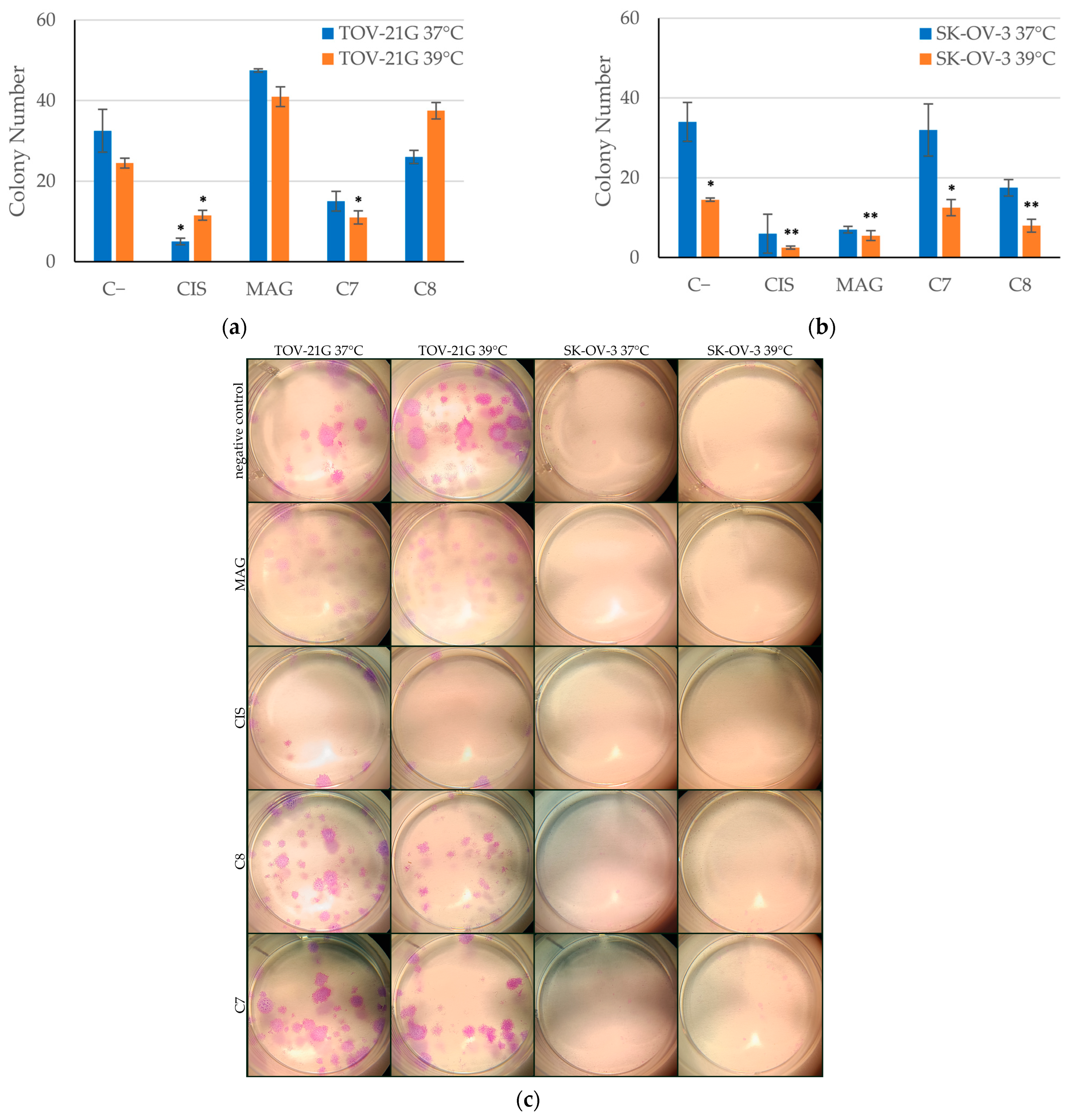
2.3. Hyperthermia Increases the Long-Term Cytotoxicity of Novel Xanthone Derivatives
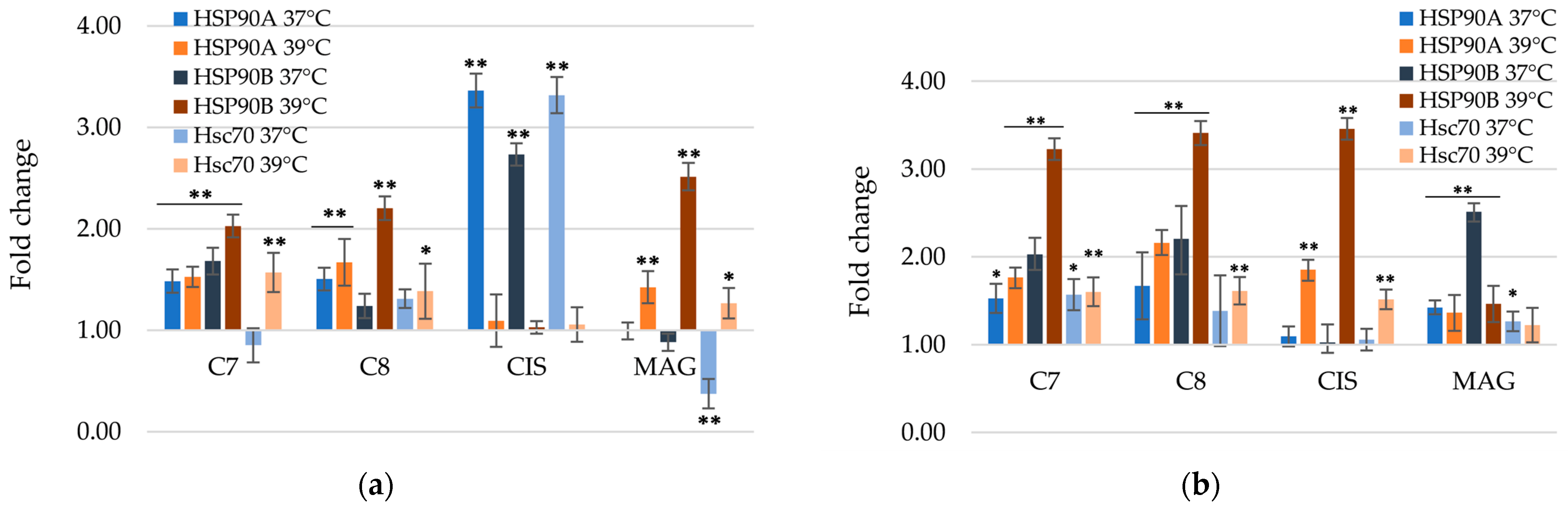
2.4. Hyperthermia Induces Apoptosis
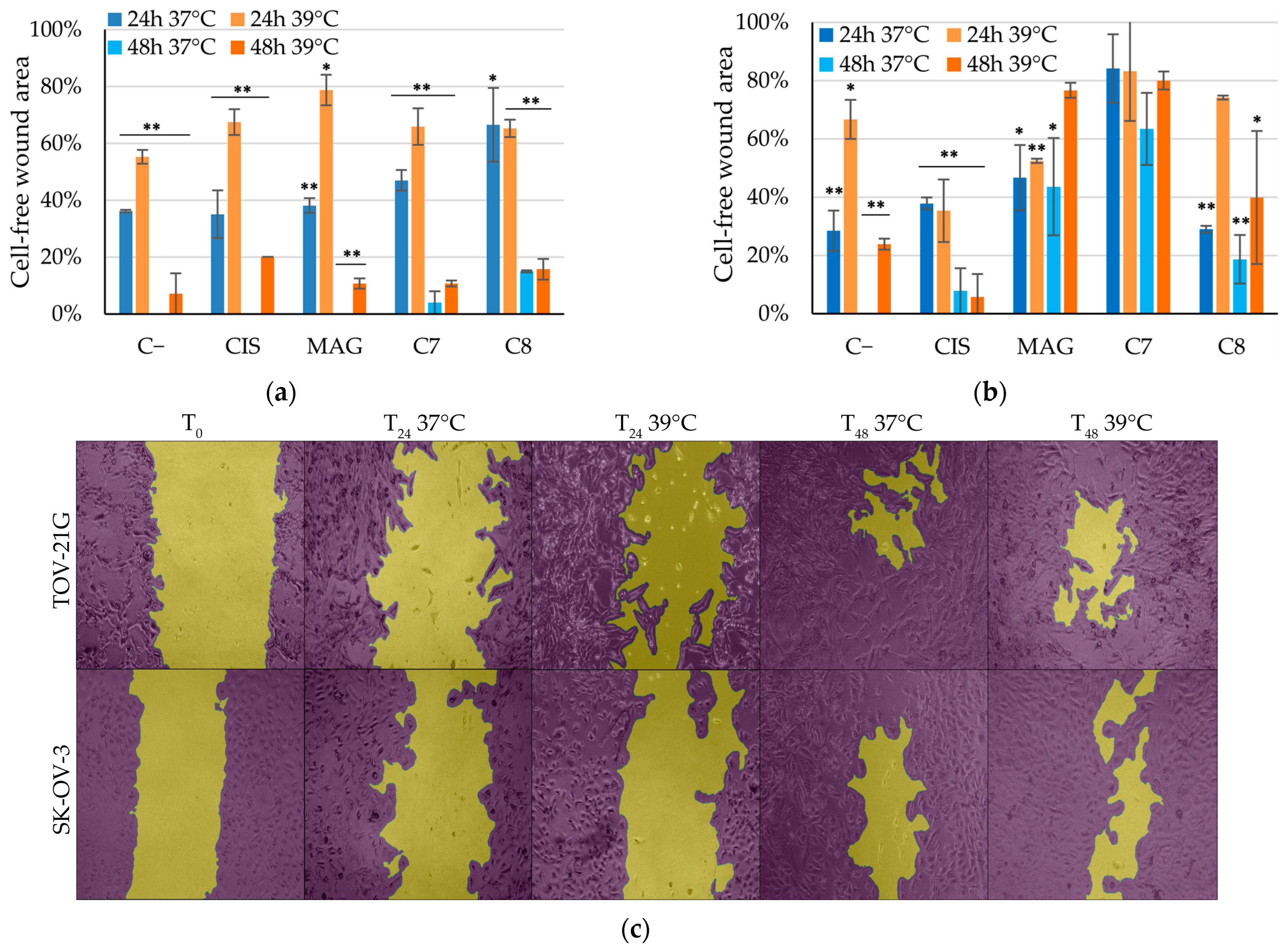
2.5. Hyperthermia Boosts Xanthones’ Anti-Migratory Effects
2.6. Mitochondrial Mass and Membrane Potential
2.7. RNA Extraction and Real-Time RT PCR
3. Materials and Methods
3.1. Chemistry
3.1.1. Chemicals and Equipment
3.1.2. The Synthesis and Characterization of Intermediate Products (1–5)
6-Chloro-9-oxo-9H-xanthene-2-carbonyl chloride (1)
7-Chloro-9-oxo-9H-xanthene-2-carbonyl chloride (2)
2-(Bromomethyl)-4,6-dichloro-9H-xanthen-9-one (4)
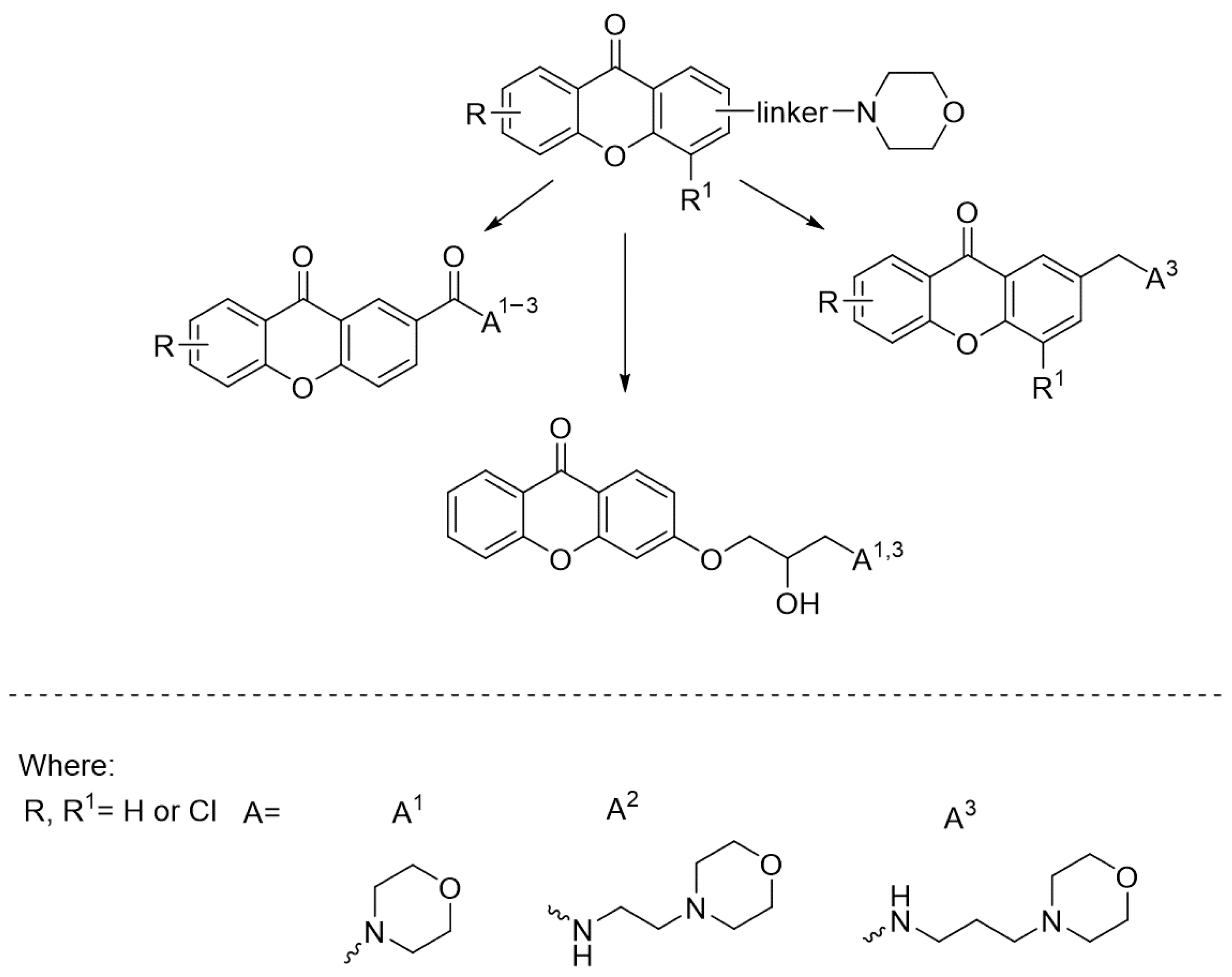
3.1.3. The Synthesis of Final Compounds (C1–C8)
Amide Derivatives (C1–C4)
3-(2-Hydroxypropoxy) Amine Xanthone Derivatives (C5–C6)
2-Methyl Amine Xanthone Derivatives (C7–C8)
3.1.4. Characterization Data for Final Compounds (C1–C8)
6-Chloro-2-(morpholine-4-carbonyl)-9H-xanthen-9-one (C1)
6-Chloro-N-(2-morpholinoethyl)-9-oxo-9H-xanthene-2-carboxamide (C2)
6-Chloro-N-(3-morpholinopropyl)-9-oxo-9H-xanthene-2-carboxamide (C3)
7-Chloro-N-(3-morpholinopropyl)-9-oxo-9H-xanthene-2-carboxamide (C4)
(R/S)-3-(2-Hydroxy-3-morpholinopropoxy)-9H-xanthen-9-one Hydrochloride (C5)
(R/S)-3-(2-Hydroxy-3-((3-morpholinopropyl)amino)propoxy)-9H-xanthen-9-one Dihydrochloride (C6)
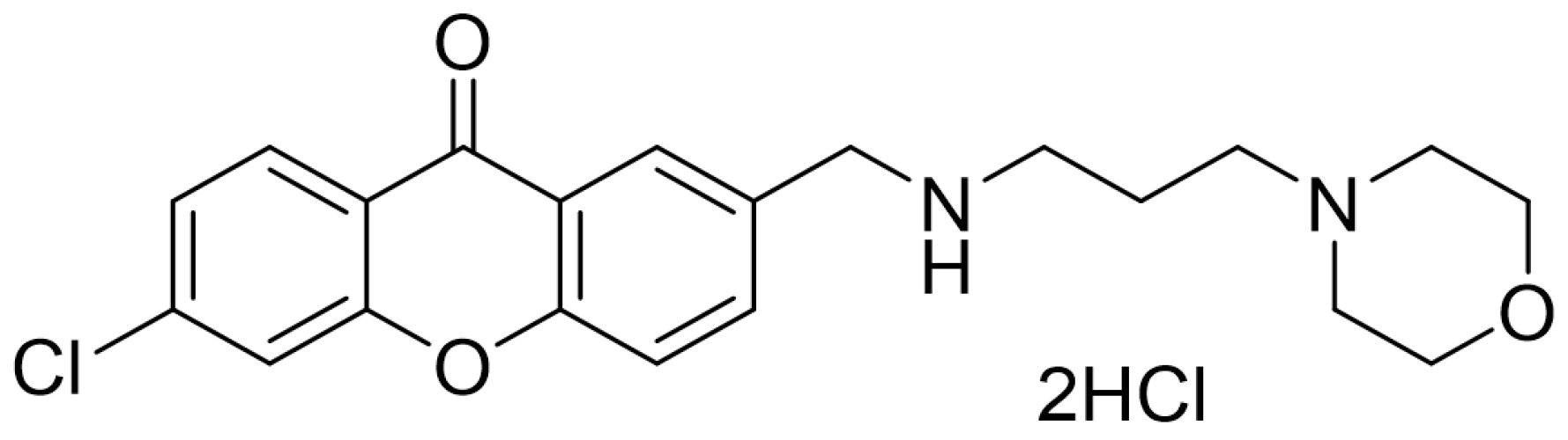
4,6-Dichloro-2-(((3-morpholinopropyl)amino)methyl)-9H-xanthen-9-one Dihydrochloride (C7)
7-Chloro-2-(((3-morpholinopropyl)amino)methyl)-9H-xanthen-9-one Dihydrochloride (C8)
3.2. Chemicals for Biological Evaluations
3.3. Cell Culture and Treatment Conditions
3.4. Cell Viability Assay
3.5. Clonogenic Assay
3.6. Scratch Assay
3.7. AO/PI, NAO, and JC-1 Staining
3.8. RNA Extraction and Real-Time™ RT PCR
3.9. Statistics
4. Conclusions
Supplementary Materials
Author Contributions
Funding
Institutional Review Board Statement
Informed Consent Statement
Data Availability Statement
Acknowledgments
Conflicts of Interest
References
- Siegel, R.L.; Miller, K.D.; Fuchs, H.E.; Jemal, A. Cancer statistics, 2022. CA Cancer J. Clin. 2022, 72, 7–33. [Google Scholar] [CrossRef] [PubMed]
- Bray, F.; Laversanne, M.; Sung, H.; Ferlay, J.; Siegel, R.L.; Soerjomataram, I.; Jemal, A. Global cancer statistics 2022: GLOBOCAN estimates of incidence and mortality worldwide for 36 cancers in 185 countries. CA Cancer J. Clin. 2024, 74, 229–263. [Google Scholar] [CrossRef]
- Kurnit, K.C.; Fleming, G.F.; Lengyel, E. Updates and New Options in Advanced Epithelial Ovarian Cancer Treatment. Obstet. Gynecol. 2021, 137, 108–121. [Google Scholar] [CrossRef]
- Issels, R.; Kampmann, E.; Kanaar, R.; Lindner, L.H. Hallmarks of hyperthermia in driving the future of clinical hyperthermia as targeted therapy: Translation into clinical application. Int. J. Hyperth. 2016, 32, 89–95. [Google Scholar] [CrossRef]
- Huffman, O.G.; Chau, D.B.; Dinicu, A.I.; DeBernardo, R.; Reizes, O. Mechanistic Insights on Hyperthermic Intraperitoneal Chemotherapy in Ovarian Cancer. Cancers 2023, 15, 1402. [Google Scholar] [CrossRef]
- Dellinger, T.H.; Han, E.S. State of the Science: The role of HIPEC in the treatment of ovarian cancer. Gynecol. Oncol. 2021, 160, 364–368. [Google Scholar] [CrossRef]
- Kopacz-Bednarska, A.; Król, T. Cisplatin—properties and clinical application. Oncol. Clin. Pract. 2022, 18, 166–176. [Google Scholar] [CrossRef]
- Grabosch, S.; Bulatovic, M.; Zeng, F.; Ma, T.; Zhang, L.; Ross, M.; Brozick, J.; Fang, Y.S.; Tseng, G.; Kim, E.; et al. Cisplatin-induced immune modulation in ovarian cancer mouse models with distinct inflammation profiles. Oncogene 2019, 38, 2380–2393. [Google Scholar] [CrossRef] [PubMed]
- Rech, J.; Sypniewski, D.; Żelaszczyk, D.; Szkaradek, N.; Rogóż, W.; Waszkielewicz, A.; Marona, H.; Bednarek, I. Novel Xanthone Derivatives Impair Growth and Invasiveness of Colon Cancer Cells In Vitro. Biomolecules 2021, 11, 1480. [Google Scholar] [CrossRef]
- Klein-Júnior, L.C.; Campos, A.; Niero, R.; Corrêa, R.; Vander Heyden, Y.; Filho, V.C. Xanthones and Cancer: From Natural Sources to Mechanisms of Action. Chem. Biodivers. 2020, 17, e1900499. [Google Scholar] [CrossRef]
- Lemos, A.; Gomes, A.S.; Loureiro, J.B.; Brandão, P.; Palmeira, A.; Pinto, M.M.M.; Saraiva, L.; Sousa, M.E. Synthesis, biological evaluation, and in silico studies of novel aminated xanthones as potential p53-activating agents. Molecules 2019, 24, 1975. [Google Scholar] [CrossRef]
- Vieira, S.F.; Araújo, J.; Gonçalves, V.M.F.; Fernandes, C.; Pinto, M.; Ferreira, H.; Neves, N.M.; Tiritan, M.E. Synthesis and Anti-Inflammatory Evaluation of a Library of Chiral Derivatives of Xanthones Conjugated with Proteinogenic Amino Acids. Int. J. Mol. Sci. 2023, 24, 10357. [Google Scholar] [CrossRef] [PubMed]
- Arshad, F.; Khan, M.F.; Akhtar, W.; Alam, M.M.; Nainwal, L.M.; Kaushik, S.K.; Akhter, M.; Parvez, S.; Hasan, S.M.; Shaquiquzzaman, M. Revealing quinquennial anticancer journey of morpholine: A SAR based review. Eur. J. Med. Chem. 2019, 167, 324–356. [Google Scholar] [CrossRef]
- Szkaradek, N.; Sypniewski, D.; Żelaszczyk, D.; Gałka, S.; Borzdziłowska, P.; Marona, H.; Bednarek, I. Influence of New Synthetic Xanthones on the Proliferation and Migration Potential of Cancer Cell Lines In Vitro. Anticancer. Agents Med. Chem. 2020, 19, 1949–1965. [Google Scholar] [CrossRef] [PubMed]
- Kumari, A.; Singh, R.K. Morpholine as ubiquitous pharmacophore in medicinal chemistry: Deep insight into the structure-activity relationship (SAR). Bioorg. Chem. 2020, 96, 103578. [Google Scholar] [CrossRef] [PubMed]
- Szkaradek, N.; Sypniewski, D.; M Waszkielewicz, A.; Gunia-Krzy|ak, A.; Galilejczyk, A.; Ga|ka, S.; Marona, H.; Bednarek, I. Synthesis and in vitro Evaluation of the Anticancer Potential of New Aminoalkanol Derivatives of Xanthone. Anticancer. Agents Med. Chem. 2016, 16, 1587–1604. [Google Scholar] [CrossRef]
- Turek, A.; Cwalina, B.; Kobielarz, M. Radioisotopic investigation of crosslinking density in bovine pericardium used as a biomaterial. Nukleonika 2013, 58, 511–517. [Google Scholar]
- Rech, J.; Wilińska, J.; Borecka, A.; Turek, A. Application of fibrin in drug technology: Achievements and perspectives. Postepy Hig. Med. Dosw. 2020, 74, 322–330. [Google Scholar] [CrossRef]
- Olakowska, E.; Właszczuk, A.; Turek, A.; Borecka, A.; Liśkiewicz, A.; Wawro, D.; Kasperczyk, J.; Jędrzejowska-Szypułka, H. Effects of 17-β-estradiol released from shape-memory terpolymer rods on sciatic nerve regeneration after injury and repair with chitosan nerve conduit in female rats. J. Appl. Biomed. 2022, 20, 87–97. [Google Scholar] [CrossRef]
- Rech, J.; Getinger-Panek, A.; Gałka, S.; Bednarek, I. Origin and Composition of Exosomes as Crucial Factors in Designing Drug Delivery Systems. Appl. Sci. 2022, 12, 12259. [Google Scholar] [CrossRef]
- Dhayapulay, A.; Kanapathipillai, M. Exosomes Based Geldanamycin Delivery to Cancer Cells with Increased Therapeutic Efficacy. J. Biomed. Nanotechnol. 2019, 15, 2202–2208. [Google Scholar] [CrossRef]
- Wust, P.; Hildebrandt, B.; Sreenivasa, G.; Rau, B.; Gellermann, J.; Riess, H.; Felix, R.; Schlag, P. Hyperthermia in combined treatment of cancer. Lancet Oncol. 2002, 3, 487–497. [Google Scholar] [CrossRef]
- Nowak, E.; Sypniewski, D.; Bednarek, I. Morin exerts anti-metastatic, anti-proliferative and anti-adhesive effect in ovarian cancer cells: An in vitro studies. Mol. Biol. Rep. 2020, 47, 1965–1978. [Google Scholar] [CrossRef]
- Bagheri, E.; Hajiaghaalipour, F.; Nyamathulla, S.; Salehen, N. The apoptotic effects of Brucea javanica fruit extract against HT29 cells associated with p53 upregulation and inhibition of NF-κB translocation. Drug Des. Devel. Ther. 2018, 12, 657–671. [Google Scholar] [CrossRef]
- Bhattiprolu, S. Python for Microscopists. Available online: https://github.com/bnsreenu/python_for_microscopists/blob/master/021-scratch_assay_using_scikit_image.py (accessed on 8 February 2024).
- Jin, P.; Jiang, J.; Zhou, L.; Huang, Z.; Nice, E.C.; Huang, C.; Fu, L. Mitochondrial adaptation in cancer drug resistance: Prevalence, mechanisms, and management. J. Hematol. Oncol. 2022, 15, 97. [Google Scholar] [CrossRef] [PubMed]
- Wang, P.; Chen, B.; Zhan, Y.; Wang, L.; Luo, J.; Xu, J.; Zhan, L.; Li, Z.; Liu, Y.; Wei, J. Enhancing the Efficiency of Mild-Temperature Photothermal Therapy for Cancer Assisting with Various Strategies. Pharmaceutics 2022, 14, 2279. [Google Scholar] [CrossRef] [PubMed]
- Ibrahim, M.Y.; Ameen Abdulla, M.; Mohd Ali, H.; Ee Cheng Lian, G.; Arbab, I.A.; Yahayu, M.; Dehghan, F.; Zeenelabdin Ali, L.; Kamalidehghan, B.; Ghaderian, M.; et al. α-Mangostin from Cratoxylum arborescens demonstrates apoptogenesis in MCF-7 with regulation of NF-κB and Hsp70 protein modulation in vitro, and tumor reduction in vivo. Drug Des. Devel. Ther. 2014, 8, 1629. [Google Scholar] [CrossRef] [PubMed]
- Yi, G.Y.; Kim, M.J.; Kim, H.I.; Park, J.; Baek, S.H. Hyperthermia Treatment as a Promising Anti-Cancer Strategy: Therapeutic Targets, Perspective Mechanisms and Synergistic Combinations in Experimental Approaches. Antioxidants 2022, 11, 625. [Google Scholar] [CrossRef]
- Yin, L.; Yang, Y.; Zhu, W.; Xian, Y.; Han, Z.; Huang, H.; Peng, L.; Zhang, K.; Zhao, Y. Heat Shock Protein 90 Triggers Multi-Drug Resistance of Ovarian Cancer via AKT/GSK3β/β-Catenin Signaling. Front. Oncol. 2021, 11, 620907. [Google Scholar] [CrossRef]
- Eckstein, M.; Marona, H.; Mazur, J. Synthesis and some biological properties of substituted xanthone-2-carboxylic acids. Pol. J. Pharmacol. Pharm. 1983, 35, 159–167. [Google Scholar]
- Lin, C.-N.; Liou, S.-S.; Ko, F.-N.; Teng, C.-M. γ-pyrone compounds. IV: Synthesis and antiplatelet effects of mono- and dioxygenated xanthones and xanthonoxypropanolamine. J. Pharm. Sci. 1993, 82, 11–16. [Google Scholar] [CrossRef] [PubMed]
- Eckstein, M.; Marona, H. Preparation and properties of some 2-mercaptomethylxanthones. Pol. J. Chem. 1980, 54, 1281–1285. [Google Scholar]
- Marona, H.; Pękala, E.; Antkiewicz-Michaluk, L.; Walczak, M.; Szneler, E. Anticonvulsant activity of some xanthone derivatives. Bioorg. Med. Chem. 2008, 16, 7234–7244. [Google Scholar] [CrossRef]
- Marona, H.; Pekala, E.; Filipek, B.; Maciag, D.; Szneler, E. Pharmacological properties of some aminoalkanolic derivatives of xanthone. Pharmazie 2001, 56, 567–572. [Google Scholar] [PubMed]
- Rech, J. WHA Project. Available online: https://github.com/JakRec/WHA_project (accessed on 8 February 2024).


| Compound No. | TOV-21G GI50 [μM] | SK-OV-3 GI50 [μM] |
|---|---|---|
| C1 | 15.58 | >150 |
| C2 | 126.43 | >150 |
| C3 | 134.20 | >150 |
| C4 | 101.63 | 141.80 |
| C5 | >150 | >150 |
| C6 | 82.87 | 87.93 |
| C7 | 16.17 | 28.30 |
| C8 | 48.34 | 67.37 |
| Compound | Synthetic xanthone derivative structures |
| C7 | 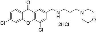 |
| C8 | 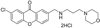 |
| Reference compound structures | |
| MAG | 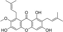 |
| CIS |  |
| TOV-21G | SK-OV-2 | ||||||||||||
|---|---|---|---|---|---|---|---|---|---|---|---|---|---|
| GI50 [μM] | GI25 [μM] | GI10 [μM] | GI50 [μM] | GI25 [μM] | GI10 [μM] | ||||||||
| 24 h | 48 h | 24 h | 48 h | 24 h | 48 h | 24 h | 48 h | 24 h | 48 h | 24 h | 48 h | ||
| 37 °C | C7 | 16.17 | 15.77 | 10.94 | 11.01 | 7.81 | 8.16 | 28.30 | 16.35 | 18.03 | 10.63 | 11.87 | 7.19 |
| C8 | 48.34 | 32.20 | 23.01 | 19.75 | 7.81 | 12.28 | 67.37 | 53.13 | 45.12 | 33.21 | 31.78 | 21.26 | |
| MAG | 10.24 | 10.85 | 5.24 | 6.16 | 2.25 | 3.66 | 67.31 | 38.84 | 50.25 | 32.62 | 40.02 | 28.89 | |
| CIS | 46.56 | 54.22 | 31.51 | 28.46 | 22.49 | 13.01 | 27.30 | 9.91 | 12.00 | 5.33 | 2.82 | 2.58 | |
| 39 °C | C7 | 25.14 | 16.16 | 13.39 | 9.65 | 6.34 | 5.74 | 27.08 | 26.19 | 15.43 | 15.15 | 8.41 | 8.53 |
| C8 | 49.04 | 22.77 | 24.22 | 15.41 | 9.32 | 10.98 | 59.18 | 52.59 | 27.45 | 25.13 | 8.45 | 8.65 | |
| MAG | 33.72 | 24.27 | 21.56 | 18.94 | 14.27 | 15.75 | 38.53 | 37.22 | 28.38 | 29.97 | 22.29 | 25.62 | |
| CIS | 65.11 | 20.21 | 26.28 | 11.91 | 2.98 | 6.93 | 99.86 | 37.13 | 38.10 | 18.46 | 1.05 | 7.25 | |
Disclaimer/Publisher’s Note: The statements, opinions and data contained in all publications are solely those of the individual author(s) and contributor(s) and not of MDPI and/or the editor(s). MDPI and/or the editor(s) disclaim responsibility for any injury to people or property resulting from any ideas, methods, instructions or products referred to in the content. |
© 2024 by the authors. Licensee MDPI, Basel, Switzerland. This article is an open access article distributed under the terms and conditions of the Creative Commons Attribution (CC BY) license (https://creativecommons.org/licenses/by/4.0/).
Share and Cite
Rech, J.; Żelaszczyk, D.; Marona, H.; Gunia-Krzyżak, A.; Żmudzki, P.; Bednarek, I.A. Hyperthermia Intensifies α-Mangostin and Synthetic Xanthones’ Antimalignancy Properties. Int. J. Mol. Sci. 2024, 25, 8874. https://doi.org/10.3390/ijms25168874
Rech J, Żelaszczyk D, Marona H, Gunia-Krzyżak A, Żmudzki P, Bednarek IA. Hyperthermia Intensifies α-Mangostin and Synthetic Xanthones’ Antimalignancy Properties. International Journal of Molecular Sciences. 2024; 25(16):8874. https://doi.org/10.3390/ijms25168874
Chicago/Turabian StyleRech, Jakub, Dorota Żelaszczyk, Henryk Marona, Agnieszka Gunia-Krzyżak, Paweł Żmudzki, and Ilona Anna Bednarek. 2024. "Hyperthermia Intensifies α-Mangostin and Synthetic Xanthones’ Antimalignancy Properties" International Journal of Molecular Sciences 25, no. 16: 8874. https://doi.org/10.3390/ijms25168874
APA StyleRech, J., Żelaszczyk, D., Marona, H., Gunia-Krzyżak, A., Żmudzki, P., & Bednarek, I. A. (2024). Hyperthermia Intensifies α-Mangostin and Synthetic Xanthones’ Antimalignancy Properties. International Journal of Molecular Sciences, 25(16), 8874. https://doi.org/10.3390/ijms25168874