Review

Mussel-Inspired Injectable Adhesive Hydrogels for Biomedical Applications

1
School of Chemistry & Chemical Engineering, Yantai University, Yantai 264005, China
2
Shandong Laboratory of Yantai Advanced Materials and Green Manufacturing, Yantai 265503, China
*
Authors to whom correspondence should be addressed.
Int. J. Mol. Sci. 2024, 25(16), 9100; https://doi.org/10.3390/ijms25169100 (registering DOI)
Submission received: 31 July 2024 / Revised: 19 August 2024 / Accepted: 20 August 2024 / Published: 22 August 2024
(This article belongs to the Special Issue Bioinspired Functional Materials for Biomedical Applications 2.0)

Abstract

:
The impressive adhesive capacity of marine mussels has inspired various fascinating designs in biomedical fields. Mussel-inspired injectable adhesive hydrogels, as a type of promising mussel-inspired material, have attracted much attention due to their minimally invasive property and desirable functions provided by mussel-inspired components. In recent decades, various mussel-inspired injectable adhesive hydrogels have been designed and widely applied in numerous biomedical fields. The rational incorporation of mussel-inspired catechol groups endows the injectable hydrogels with the potential to exhibit many properties, including tissue adhesiveness and self-healing, antimicrobial, and antioxidant capabilities, broadening the applications of injectable hydrogels in biomedical fields. In this review, we first give a brief introduction to the adhesion mechanism of mussels and the characteristics of injectable hydrogels. Further, the typical design strategies of mussel-inspired injectable adhesive hydrogels are summarized. The methodologies for integrating catechol groups into polymers and the crosslinking methods of mussel-inspired hydrogels are discussed in this section. In addition, we systematically overview recent mussel-inspired injectable adhesive hydrogels for biomedical applications, with a focus on how the unique properties of these hydrogels benefit their applications in these fields. The challenges and perspectives of mussel-inspired injectable hydrogels are discussed in the last section. This review may provide new inspiration for the design of novel bioinspired injectable hydrogels and facilitate their application in various biomedical fields.

1. Introduction

In nature, marine mussels secrete proteinaceous glues to tightly adhere to wet rock surfaces [1]. These proteinaceous glues quickly solidify upon contact with surfaces, forming adhesive plaques featuring remarkable interfacial strength and resilience [2]. Over recent decades, extensive efforts have been made to understand the intricate compositions and adhesive mechanisms of these mussel-derived proteins [3,4,5]. These works reveal that a noncanonical amino acid, namely, 3,4-dihydroxyphenylalanine (DOPA) exists in the mussel adhesive proteins [6,7]. In the proteins on the surface of mussel adhesive plaques (Mfps 3 and Mfps 5), the contents of DOPA are higher and reach 20% and 28%, respectively [8]. This phenomenon indicates that DOPA plays an essential role in the adhesion of mussels.
The main functional group in DOPA is the catechol group, which can form multiple covalent and noncovalent interactions with specific surfaces [9,10,11] (Figure 1). For mineral or metal surfaces, the adhesion interactions mainly include hydrogen bonding (H-bonding) and coordination interaction. For organic surfaces, especially biological tissue surfaces, more adhesion interactions may form due to the complicated components of organic surfaces. In addition to H-bonding, π-π interactions and π-cation interactions may exist. If the catechol groups are oxidized by oxidizing agents, such as sodium periodate (NaIO4), the catechol group will transfer to a quinone structure [12]. Under this circumstance, covalent bonding can form with the amino or sulfhydryl groups on the surface of biological tissues through Schiff base reaction or Michael addition [13]. Considering the versatile roles of the catechol group in adhesion, in recent decades, a large number of studies have introduced the catechol group into polymer chains to develop adhesives. These mussel-inspired glues exhibit desirable adhesive performance and are applied in numerous fields, including biomedical adhesives [14,15], antifouling coatings [16,17], flexible electronics [18,19], drug delivery [20,21], cell encapsulation and delivery [22,23], etc.
A hydrogel is a three-dimensional (3D) polymeric network formed by the physical or chemical crosslinking of hydrophilic polymers [5,24]. By virtue of their biological tissue-similar structure, biocompatibility, and tunable physical properties, hydrogels are widely applied in various biomedical applications [3,25,26]. Regarding the formation site, hydrogels can be divided into preformed hydrogels and in situ injectable hydrogels [27]. These two types of hydrogels have their characteristics (Table 1). In general, preformed hydrogels form in specific reactors or molds. Some complicated conditions, such as high temperature, long reaction time, and oxygen-free conditions, can be satisfied for the formation of preformed hydrogels [28]. Therefore, preformed hydrogels usually exhibit high physical performance, such as high mechanical properties, strong adhesive properties, etc. However, since preformed hydrogels are formed in molds, they find it difficult to precisely fill irregular defects [29]. In addition, for internal applications, a relatively large incision is usually required to implant the preformed hydrogel into the body. Different from preformed hydrogels, in situ injectable hydrogels utilize the sol–gel transition process to achieve a minimally invasive way of application [30]. The precursor solution of these hydrogels is injectable, and after the precursor is injected into the target site of the body, the precursor transforms into a gel state. Therefore, the injectable hydrogels can fill irregular shapes well. However, due to limited formation conditions, such as 37 °C and physiological pH, injectable hydrogels usually exhibit weak mechanical and adhesive properties compared with preformed hydrogels [31]. Nevertheless, the minimally invasive delivery method enables injectable hydrogels to be a promising tool in clinical practice [32].
To improve the properties of injectable hydrogels, in recent decades, various mussel-inspired injectable hydrogels have been developed. Different from other related reviews [33,34,35], this review mainly focuses on mussel-inspired hydrogels with injectability. This type of hydrogel simultaneously possesses the minimally invasive property of injectable hydrogels and the desirable functions provided by mussel-inspired components, which may include tissue adhesiveness and self-healing, antimicrobial, and antioxidant capabilities [33]. Their fascinating properties benefit their application in numerous biomedical fields, such as wound closure and healing [36,37], hemostasis [38,39], bone repair [40,41], drug delivery [42,43], smart sensors [44], biological coatings [45], etc. (Figure 2). Typical examples are summarized in Table 2.
In the following review, we summarize the typical design strategies of these mussel-inspired injectable adhesive hydrogels. Further, we systematically overview the recently designed mussel-inspired injectable adhesive hydrogels. In addition, we mainly focus on how the specific properties of the hydrogels facilitate their application in specific fields. The challenges and perspectives of mussel-inspired injectable adhesive hydrogels are also discussed in the last section of this review.

2. Design Strategy of Mussel-Inspired Injectable Adhesive Hydrogels

2.1. Incorporation of Catechol Group into Polymers

The yield of extraction of mussel adhesive proteins is extremely low [46]. Therefore, it is difficult to directly use natural mussel adhesive proteins to develop injectable adhesive hydrogels. Due to the essential role of DOPA in mussel adhesion, mussel-inspired injectable adhesive hydrogels are usually developed by engineering the catechol group into the gel network. Incorporation of the catechol groups into the polymers is usually the first step to developing mussel-inspired injectable hydrogels. During recent decades, many methods have been developed to incorporate the catechol group into polymers [4,47], including the incorporation of the catechol group by classic organic reactions, polymerization of catechol-based monomers, and biosynthesis of catechol-containing proteins (Figure 3).

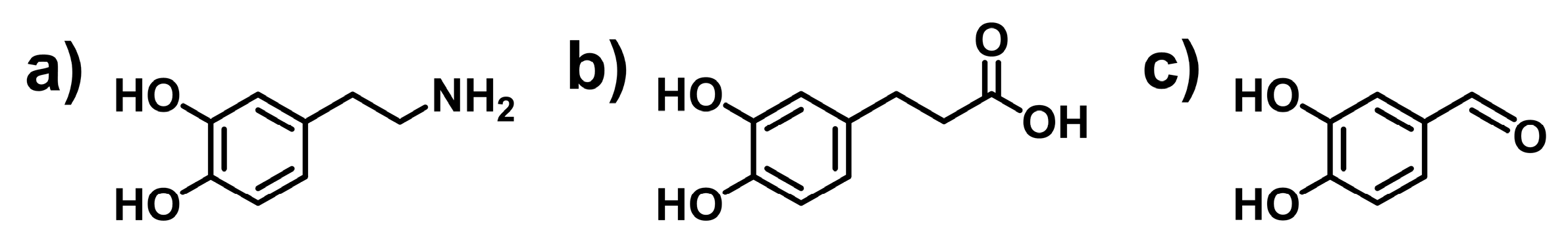
2.1.1. Incorporation of Catechol Groups by Classic Organic Reactions

Since there are many commercial DOPA derivatives (Figure 4), the incorporation of the catechol group into polymers by classic organic reactions is easy to perform. The coupled reaction between amino or carboxyl groups is the most used. Many natural polymers (such as ε-polylysine (PL), chitosan, hyaluronic acid (HA), sodium alginate (SA), etc.) and DOPA derivatives (3,4-dihydroxyphenethylamine and 3-(3,4-dihydroxyphenyl) propionic acid) contain amino or carboxyl groups. Therefore, the catechol group can be easily incorporated as a pendant group through a coupled reaction. Another reaction capable of the incorporation of the catechol group into a polymer chain is the Schiff base reaction. This method utilizes the Schiff base reactions between the aldehyde polysaccharides and 3,4-dihydroxyphenethylamine [48] or between 3,4-dihydroxybenzaldehyde and amino-containing polysaccharides [49]. Due to the instability of the formed imine bond, the sodium borohydride (NaBH4)-mediated reductive amination reaction is usually necessary [49,50]. Using these classic organic reactions, the catechol group has been successfully incorporated into a series of natural polymers, such as PL, HA, chitosan, SA, and gelatin (GT), and synthetic polymers, such as polyethylene glycol (PEG)-based polymers [45,47,51,52,53,54,55].

2.1.2. Polymerization of Catechol-Based Monomers

Catechol is a well-known polymerization inhibitor. Therefore, commonly, catechol is protected before polymerization [47]. Even so, unprotected polymerizations of catechol-based monomers have been reported in recent years [56,57]. The rational regulation of polymerization conditions, such as polymerization time, monomer concentration, and oxygen-free atmosphere, was found to be important for successful polymerization [57]. Unprotected polymerizations of catechol-based monomers have been used in the development of injectable hydrogels. For example, Yang and coworkers used methacrylamide dopamine (DMA) as a catechol-containing monomer and mixed it with polyethylene glycol (PEG) monomethyl ether-modified glycidyl methacrylate-functionalized chitosan and zinc ions to prepare a hydrogel precursor [36]. After initiation by ultraviolet light (365 nm), the monomers were polymerized, and the catechol groups were incorporated into the hydrogels.

2.1.3. Biosynthesis of Catechol-Containing Proteins

Biosynthesis is a method of developing catechol-containing proteins similar to mussel adhesive proteins. In this method, mushroom tyrosinase, which can convert tyrosine residues to DOPA, is used [58]. However, the modification yield is reported to be relatively low. Recently, an in vivo residue-specific incorporation strategy was proposed. This strategy created mussel adhesive proteins with a high DOPA content [59], showing promise in the production of mussel-inspired macromolecules.

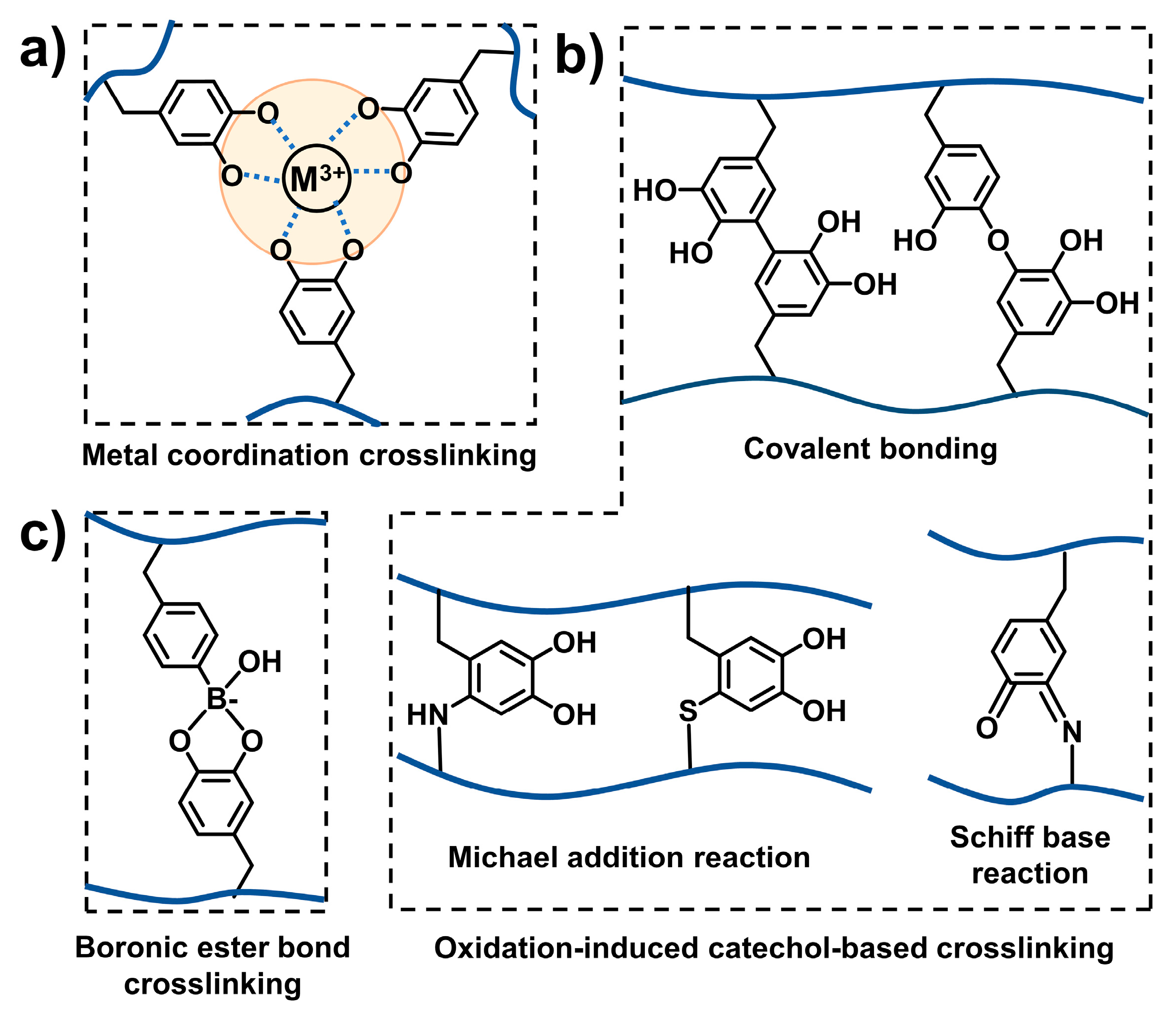
2.2. Crosslinking Strategy of Mussel-Inspired Injectable Adhesive Hydrogels

Crosslinking among catechol-containing polymers is a necessary step for developing mussel-inspired injectable adhesive hydrogels. Various crosslinking strategies have been developed in recent years. When considering whether the catechol group plays a major role in crosslinking, crosslinking strategies can be divided into catechol-mediated crosslinking and other regular crosslinking.

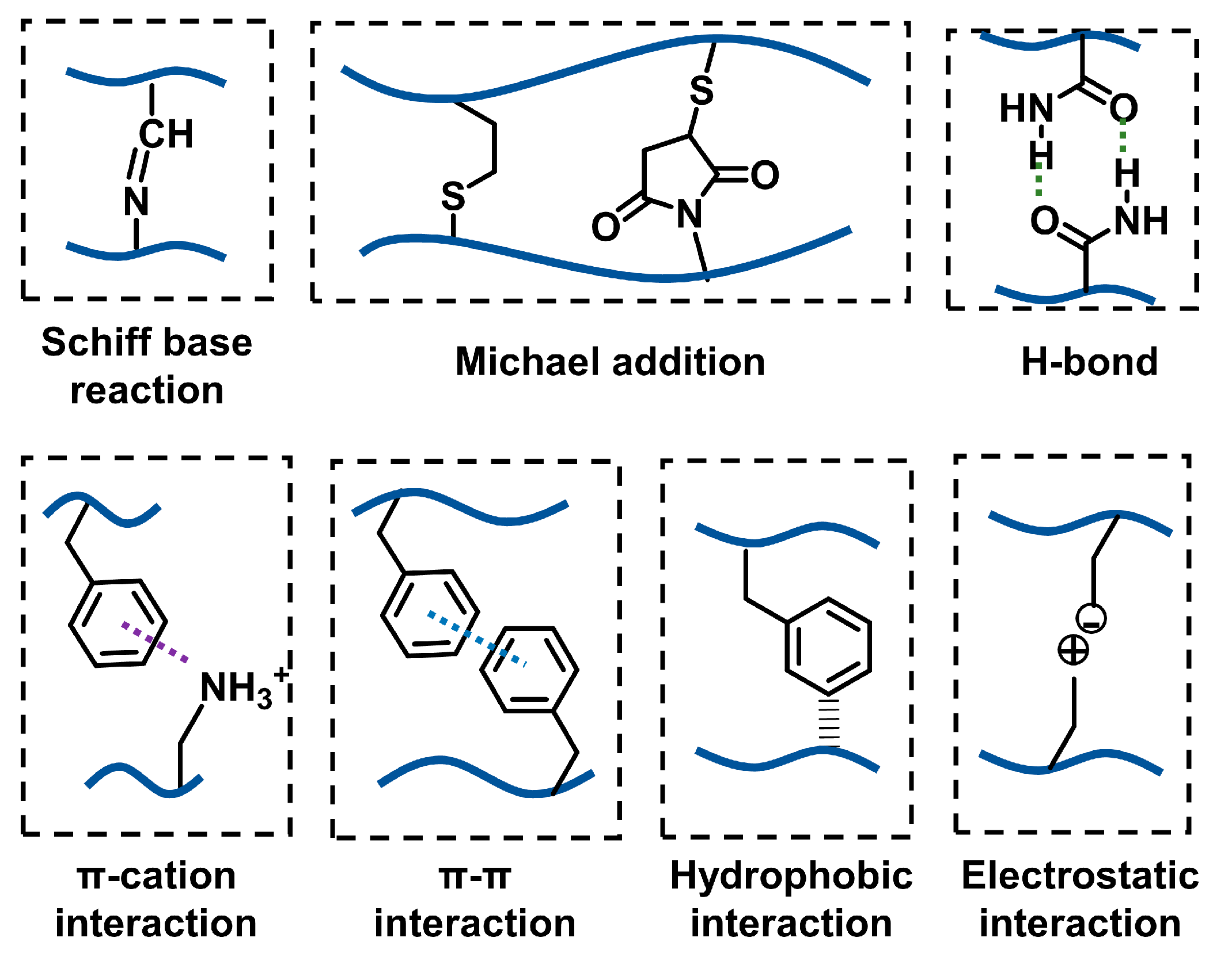
2.2.1. Catechol-Mediated Crosslinking

The catechol group is usually considered an adhesive group for the improvement of the interfacial adhesion property of materials. In the development of mussel-inspired injectable adhesive hydrogels, the catechol group is also widely used as a functional group for crosslinking of hydrogels (Figure 5). Catechol–metal coordination crosslinking, oxidation-induced catechol-based crosslinking, and dynamic boron ester-based crosslinking are the three main crosslinking ways.
The catechol group in polymers can coordinate with metal ions, such as Fe3+, Al3+, Zn2+, Cu2+, and Ti3+, to form coordination crosslinking [12]. Among these metal ions, Fe3+ is widely studied and used in the preparation of mussel-inspired injectable adhesive hydrogels. Three types of catechol–Fe complexes exist, which are mono-, bis-, and tri-catechol–Fe complexes. The tri-complex is the most stable form [60]. It has been reported that the coordination of catechol and Fe3+ is strongly influenced by pH [37,61]. The mono-complex dominates when the pH is lower than 5.6. When the pH is in the range of 5.6–9.1, the bis-complex accounts for a large proportion. Tri-complexes dominate only when the pH is larger than 9.1 [62]. In addition, the molar ratio of catechol to Fe3+ influences the coordination types of catechol and Fe3+. When the ratio of catechol to Fe3+ is high (the amount of Fe3+ is low), catechol and Fe3+ are prone to forming more tri-complexes [63,64]. Coordination principles have been utilized in the design of mussel-inspired adhesives. For example, Li and coworkers developed a Fe3+-crosslinked catechol-modified ε-polylysine adhesive hydrogel. By rationally tuning the pH and Fe3+ content in the gelation condition, the resultant adhesive hydrogels exhibited high adhesive strength [65]. In addition, using metal ions with specific functions to crosslink the catechol-containing polymers is found to be effective in developing multifunctional injectable hydrogels. For example, Yang and coworkers utilized the coordination of Zn2+ with catechol to develop a composite injectable hydrogel (CSG-PEG/DMA/Zn). Due to the inherent antibacterial property of Zn2+, the developed injectable hydrogel exhibited antimicrobial properties [36].
In an alkaline environment, catechol groups are prone to oxidation to form catechol–catechol dimers [13]. This phenomenon has been applied to the development of mussel-inspired hydrogels. Sato and coworkers synthesized a series of catechol-modified hyaluronic acid (HA-CA). They showed that in a weakly alkaline environment (pH 7.4), HA-CA, with a high molar mass and catechol content, was capable of self-assembly into a crosslinked network within about three hours without the addition of additional enzymes or oxidizing agents. This gelation process was mainly due to the oxidation-induced coupling of the catechol groups [66].
Adding oxidizing agents can promote the oxidation of the catechol groups, thus benefiting the gelation process of catechol-containing polymers. For example, Shin and coworkers synthesized catechol-modified hyaluronic acid (HA-CA) and showed that HA-CA can form a hydrogel after the addition of NaIO4. Gelation was considered to be induced by the formation of catechol–catechol adducts due to the oxidation of catechol [67]. The incorporation of specific enzymes also contributes to the gelation of catechol-containing polymers by enzymatically crosslinking. For example, Kim and coworkers synthesized catechol-modified poly (γ-glutamic acid) (PGADA). They found that PGADA was able to gel in the presence of horseradish peroxidase (HRP) and hydrogen peroxide (H2O2) [68]. In addition, in oxidation conditions, the catechol group can transfer to a quinone structure. The formed quinone is capable of reacting with the -NH2 group and -SH group, thus improving the crosslinking density [13].
In addition to catechol–metal coordination crosslinking and oxidation-induced catechol-based crosslinking, dynamic boron ester-based crosslinking is used in the preparation of mussel-inspired injectable hydrogels. The boron ester bond is predominantly formed through the complexation of borate with the catechol group, enabling a versatile and robust network formation. For example, Shan and coworkers developed a mussel-inspired polyethylene glycol (PEG)-based hydrogel. The hydrogel contained catechol-modified four-armed PEG (4-arm-PEG-DA) and phenylboronic acid-functionalized four-armed PEG (4-arm-PEG-PBA). At pH 9.0, mixing the two modified PEGs led to the fast formation of a PEG-based hydrogel by boron ester-based crosslinking (borate–catechol complexation) [69].

2.2.2. Other Regular Crosslinking Methods

Mussel-inspired injectable hydrogels can also be crosslinked by other regular covalent or noncovalent interactions (Figure 6). Covalent crosslinking is the predominant method for developing mussel-inspired injectable gels. The most used reactions in covalent crosslinking include the Schiff base reaction between amino and aldehyde groups and the Michael addition between sulfhydryl and double bonds. For example, Zhou and coworkers synthesized sulfhydryl-modified chitosan (CSS) and catechol and maleimide-functionalized ε-polylysine (Catechol-PL-MAL). After mixing CSS and Catechol-PL-MAL at pH 7.2, the Michael addition between the sulfhydryl in CSS and maleimide in Catechol-PL-MAL induced the gelation of the hydrogel [70]. In this type of gelation, the catechol group was not the major group in crosslinking. Therefore, more catechol groups can participate in interfacial adhesion, which benefits the adhesion of the hydrogel.
Noncovalent interactions are frequently introduced in the design of mussel-inspired adhesive hydrogels. Interactions, including hydrogen bonding, π-π stacking, cation-π interactions, electrostatic forces, and hydrophobic effects, are frequently employed to create self-healable hydrogel networks [3,51]. Compared with covalent bonds, noncovalent interactions are usually weak [71]. Therefore, noncovalent interactions usually work with covalent crosslinking. The introduction of noncovalent interactions into the gel network not only enhances the structural integrity of the hydrogel networks but also endows these materials with additional characteristics, such as self-healing capabilities [72].

2.2.3. Combination of Catechol-Mediated and Other Regular Crosslinking Methods

Mussel-inspired injectable adhesive hydrogels are also able to be prepared by the combination of catechol-mediated and other regular crosslinking methods [61]. Typical examples are summarized in Table 3. The multiple crosslinked structures can endow the hydrogel with enhanced properties. For example, Hu and coworkers developed mussel-inspired hydrogels based on chitosan quaternary ammonium salt (HTCC) and oxidized dextran–dopamine (OD-DA). The hydrogel was double-crosslinked by the catechol–catechol adducts and Schiff base reaction between the aldehyde group in OD-DA and the amino group in HTCC. Owing to the double-crosslinking, the hydrogel exhibited great mechanical properties [73].

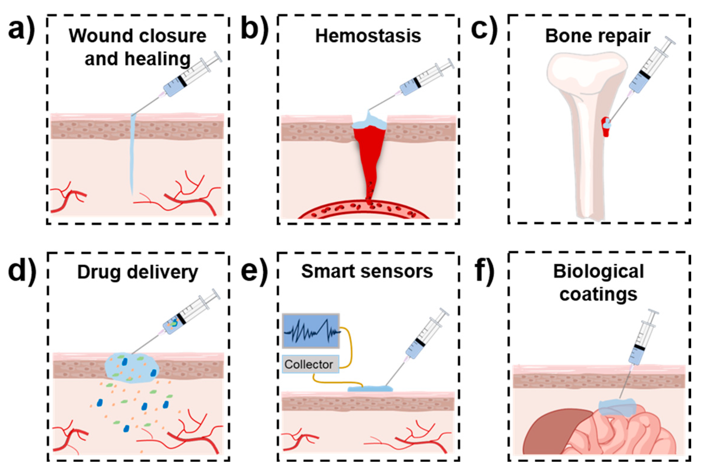
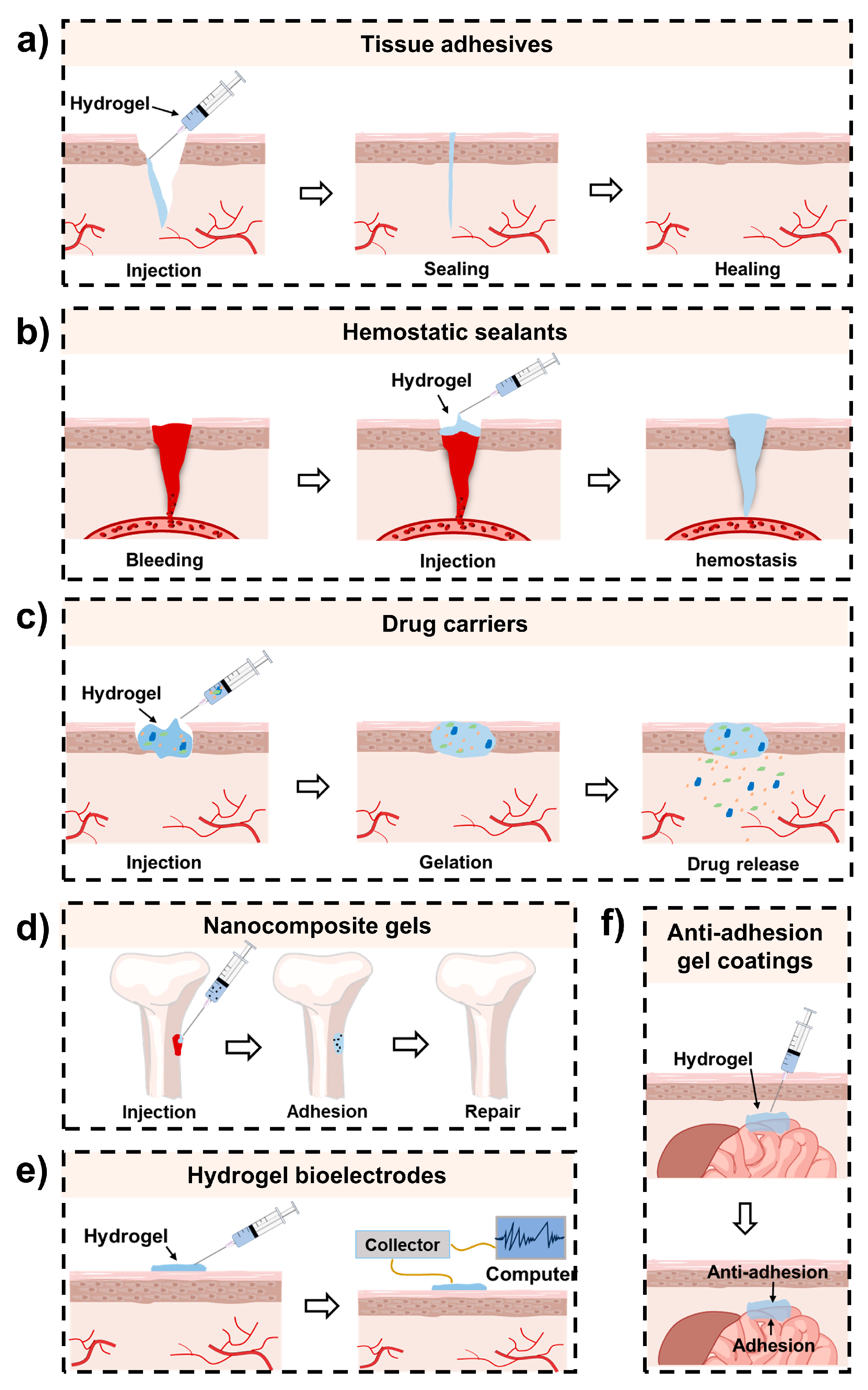
3. Biomedical Applications of Mussel-Inspired Injectable Adhesive Hydrogels

With the development of adhesive hydrogels, in addition to mussel-inspired adhesive hydrogels, many other types of adhesive hydrogels have been reported for biomedical applications [78,79,80]. Nevertheless, mussel-inspired adhesive hydrogels still show their advantages. First, owing to the existence of the catechol group, mussel-inspired adhesive hydrogels can be easily formed by coordination bonds or other noncovalent interactions. Since these interactions are reversible, the obtained hydrogels usually show self-healing properties [81,82]. Second, the catechol group is a reactive oxygen species (ROS) scavenger. Therefore, the mussel-inspired adhesive hydrogels usually exhibit antioxidant properties [83,84]. Additionally, mussel-inspired polydopamine (PDA)-based nanomaterials possess a photothermal effect; therefore, a hydrogel containing PDA-based nanomaterials usually shows photothermal antibacterial capability [85,86]. With multiple desirable properties, mussel-inspired injectable adhesive hydrogels have been widely applied in numerous biomedical fields. These hydrogels are mainly designed as tissue adhesives [61,87] (for wound closure and healing), hemostatic sealants [75,88] (for hemostasis), nanocomposite gels [41,89] (for bone repair), drug carriers [42,90] (for drug delivery), hydrogel bioelectrodes [44] (for smart sensors), and anti-adhesion gel coatings [45] (for biological coatings) (Figure 7).

3.1. Wound Closure and Healing

Millions of wounds, either from accidents or surgery operations, are required to be closed to prevent infection and promote healing each year [91]. In clinical practice, suturing is still the dominant way to close open wounds [13,92]. In recent decades, tissue adhesives have emerged as a new type of tool for wound treatments. The advantages and drawbacks of traditional suturing and tissue adhesive-mediated wound closure are summarized in Table 4. Suturing can stably close wounds. However, it consumes much time and causes secondary damage to tissues surrounding wounds [93,94]. In addition, wounds treated by sutures often have a risk of infection [95] and are prone to leaving scars after healing. Tissue adhesives are capable of closing an open wound by bonding the edges of wound tissues [96]. Compared with clinical sutures, tissue adhesives show advantages in ease of use, sealing of air/fluid leakage, and less pain and scars [97,98]. Therefore, tissue adhesives are becoming attractive alternatives to traditional wound treatment tools. However, current regular tissue adhesives usually show weak mechanical and adhesive strength, and their prices are relatively high.
Due to the ability to adapt to the shapes of wounds, injectable hydrogels have the potential to be used as tissue adhesives. Compared with regular injectable hydrogels, mussel-inspired injectable adhesive hydrogels usually show higher adhesive properties due to the existence of the catechol groups. Their superior adhesive performance has been confirmed by various methods, including the lap shear test, tensile test, 180° peel test, 90° peel test, wound closure test, and burst pressure test (Figure 8). Therefore, mussel-inspired injectable adhesive hydrogels have been widely used as tissue adhesives for wound treatments. For example, Mehdizadeh and coworkers developed an injectable citrate-based mussel-inspired hydrogel bioadhesive (iCMBA) by oxidation-induced crosslinking. The optimized iCMBA bioadhesive showed a strong adhesive strength of about 123 kPa in lap shear tests. Its adhesive strength was about eight times that of commercial fibrin glue. In wound healing experiments, iCMBA hydrogel bioadhesive effectively sealed the wounds on the backs of rats and significantly promoted wound healing [87].
In addition to high adhesive properties, hydrogels for wound treatment should possess high mechanical performance, including toughness and the ability to recover after deformation. The potential of the multiple crosslinking of mussel-inspired hydrogels makes it possible for them to exhibit desirable mechanical performance. Deng and coworkers prepared a mussel-inspired Alg-DA-CATNFC-PAM-Al3+DN hydrogel, which was composed of alginate-modified dopamine (Alg-DA), a copolymer of acrylamide and acrylic acid (PAM), and cationized nanofibrillated cellulose (CATNFC). The Alg-DA-CATNFC-PAM-Al3+DN hydrogel was crosslinked by triple dynamic interactions, including coordination, hydrogen bonding, and electrostatic interaction, and, therefore, exhibited exceptional mechanical properties and self-healing properties. Upon application at the wound site, the hydrogel effectively closed the 3 cm-long incision on the back of a rat and facilitated wound healing [99]. In addition, the multiple crosslinked structures can endow the hydrogel with multiple functions, thus benefiting wound treatment. Since the catechol group can chelate with Fe3+ under weakly alkaline conditions [100], coordination crosslinking is often used in the design of multiple crosslinked hydrogels. For example, Li and coworkers developed a double dynamic bond crosslinked hydrogel adhesive based on catechol-modified ε-polylysine (PL-Cat) and oxidized dextran (ODex). The hydrogel was crosslinked by the dynamic catechol–Fe coordination bond and the dynamic Schiff base bond between the amino group in PL-Cat and the aldehyde group in ODex. This double-dynamic crosslinked characteristic enables the hydrogel to exhibit multiple functions, including on-demand dissolution, repeated adhesion, high adhesive and mechanical properties, injectability, and biocompatibility, which facilitates its application in post-wound closure care [61].
The presence of moisture on tissue surfaces often limits the adhesive strength of traditional hydrogels. It is important for adhesive hydrogels to exhibit robust wet adhesive strength to bond injured tissues. Considering that the marine mussel exhibits robust adhesion to wet rocks, some mussel-inspired adhesive hydrogels were designed and applied in the closure of wet wounds [101]. Recent studies show that the synergistic effect of cation and catechol promoted wet adhesion [102,103]. This principle was applied in developing adhesive hydrogels for wet adhesion. For example, Wang and coworkers developed mussel-inspired catechol-modified ε-polylysine–polyethylene glycol-based hydrogel (PPD hydrogel) by horseradish peroxidase (HRP)-mediated crosslinking. The existence of adjacent catechol–lysine in PPD hydrogel enabled the hydrogel to work in a wet environment. The adhesive strength of optimized PPD hydrogel reached up to about 147 kPa. In vivo studies indicated that the PPD hydrogel was effective in promoting wound repair and skin regeneration [104].
Injured skin is particularly vulnerable to bacterial infections, which result in wound inflammation and delayed healing. The incorporation of DOPA or other catechol compounds endows hydrogels with enhanced adhesion and antimicrobial activity [36,105,106,107]. Therefore, mussel-inspired injectable hydrogel adhesives offer a promising solution for the healing of infected wounds. For example, Guo and coworkers developed antimicrobial mussel-inspired hydrogels based on catechol-functionalized oxidized hyaluronic acid (OHAdop), guar gum (GG), glycol chitosan (GC), borax, and polydopamine nanoparticles (PDA NPs). In addition to their robust tissue adhesive properties, injectability, and self-healing capabilities, due to the photothermal antimicrobial properties of PDA NPs, the developed hydrogels exhibited good antimicrobial properties under near-infrared (NIR) irradiation (808 nm). These desirable properties finally facilitated their application in bacteria-infected wound healing [108].
The healing of diabetic wounds is a significant challenge in clinical practice [73]. Diabetic wounds often involve exacerbated inflammation and an overabundance of reactive oxygen species (ROS) [109,110]. This leads to the leakage of wound tissue fluid, which further fosters pathogen proliferation and results in persistent wound inflammation, non-healing, or recurrence. The development of innovative materials that can effectively relieve the inflammatory response and neutralize ROS is critical for treating diabetic wounds. Since polyphenolic compounds are ROS scavengers, mussel-inspired adhesive hydrogels have the potential to treat diabetic wounds. Fu and coworkers developed a series of tannin–europium coordination complexes (TECs) crosslinked with citrate-based mussel-inspired bioadhesives (TE-CMBAs). The incorporation of TEC improved the antioxidant and anti-inflammatory capabilities of the hydrogel adhesives. In vivo results show that the TE-CMBA hydrogels were able to modulate the inflammatory microenvironment, promote angiogenesis, enhance the production of the extracellular matrix (ECM), and improve re-epithelialization, showing effectiveness in the treatment of diabetic wounds [111].

3.2. Hemostasis

Uncontrolled hemorrhage and subsequent wound infection are major problems in trauma treatment [112,113]. Hemorrhage from irregularly shaped and deep wounds is especially severe in emergency care [114]. Nowadays, traditional surgical suturing remains a dominant method for hemostasis. However, it is often criticized for its time-consuming operations and secondary tissue damage [115]. Injectable hydrogel-based tissue adhesives, with their shape adaptability and minimally invasive characteristics, have become promising alternatives for hemorrhage control [116]. Among them, mussel-inspired injectable hydrogels are particularly suitable for hemostasis due to the following reasons: First, hemorrhage control is typically achieved through the formation of a barrier that effectively seals the site of bleeding. A robust hemostatic barrier requires a hemostatic agent with exceptional adhesive strength in the presence of blood and adequate mechanical strength to firmly seal the wound without breakage. Recent studies have shed light on the remarkable effectiveness of catechol for adhesion to wet tissues [117,118]. The multiple crosslinking ways derived from the catechol groups further endow the hydrogels with the potential to be mechanically strong. Therefore, these requirements can be well met by mussel-inspired injectable hydrogels. In addition, the interaction between the catechol group in mussel-inspired hydrogels and reactive residues in proteins or polysaccharides in the blood is known to accelerate the coagulation process [119], which benefits hemostasis. Moreover, the negative charges of polyphenols are capable of activating coagulation factor XII in vivo, initiating a cascade reaction that enhances coagulation [119]. During recent decades, many mussel-inspired injectable hydrogels have been developed for hemorrhage control [120,121]. For example, Fang and coworkers developed a chitosan-based injectable hydrogel, namely, CCS@gel by Schiff base and catechol-mediated crosslinking. Due to the catechol and remnant aldehyde groups, CCS@gel exhibited a remarkable adhesive strength of about 44.9 kPa. In addition, the hydrogel showed high mechanical performance and self-healing properties. In vivo studies show that CCS@gel rapidly covered the injury site and significantly lowered the loss of bleeding [75]. In addition, Hu and coworkers synthesized PVA-DOPA-Cu (PDPC) hydrogels based on poly(vinyl alcohol) (PVA), DOPA, and Cu by coordination crosslinking. The PDPC hydrogels exhibited multiple functions, including high tissue adhesive performance and photothermal and antibacterial properties. After injection at bleeding sites, the PDPC hydrogels can rapidly absorb blood and adhere tightly to the wound, forming an effective hemostatic barrier. They are effective in treating severe hemorrhages from liver injury, carotid artery rupture, and cardiac penetration injury. In addition, owing to the existence of the DOPA/Cu2+ complex, PDPC hydrogels also exhibit antimicrobial and anti-inflammatory properties, showing great potential in promoting diabetic wound healing [122].
Nanomaterials, with their large surface area, are capable of generating nanoscale effects that significantly enhance blood absorption and cell adhesion [119]. Moreover, their adjustable physical and chemical properties further facilitate their integration with other hemostatic materials, thereby enhancing hemostatic capabilities. Therefore, mussel-inspired nanocomposite hydrogels have been widely studied and emerged as an essential type in mussel-inspired hemostatic materials [123,124,125]. Xu and coworkers incorporated natural clay nanoparticles (Laponite XLG) into a multifunctional mussel-inspired hydrogel composed of polyacrylic acid (PAA)-based copolymers, oxidized carboxymethylcellulose (OCMC), and tannic acid (TA). The incorporation of Laponite XLG benefited the mechanical properties of the hydrogel. In hemostatic experiments, the hydrogel was found to be capable of reducing hemostasis time and lowering the loss of blood in a liver-bleeding mouse model [126].
Employing materials capable of absorbing blood (or eliminating bodily fluids) and adhering to tissues is a promising therapeutic approach to treating uncontrolled bleeding [127]. Hemostatic powders, with their high surface area and exceptional hygroscopic properties, are often used for hemostasis. However, traditional hemostatic powders have some limitations. For example, they may disperse with the flow of blood and, therefore, have the risk of inducing thrombosis. Additionally, their efficacy in addressing bleeding from deep and incompressible wounds still requires improvement. Shape memory hemostatic sponges offer a novel solution to these issues. These sponges can quickly absorb blood, thereby creating a physical barrier that effectively seals wounds. Dong and coworkers prepared a hydroxyethyl cellulose/soy protein isolate composite sponge (EHSS) and then modified the EHSS with dopamine to obtain a polydopamine-coated injectable hemostatic material (EHP). Silver nanoparticles were homogeneously immobilized on the EHP to finally form the shape memory sponge. Its unique ability to re-expand after absorbing blood allowed the shape memory sponge to serve as a robust physical barrier for stopping bleeding. In vivo results show that the shape memory sponge exhibited remarkable hemostatic performance in a rat liver prick hemorrhage model and other deep, narrow, and noncompressible hemorrhage models [112].

3.3. Bone and Cartilage Repair

Bone tissue plays a vital role in bearing weight, shaping physique, and safeguarding vital organs [128]. The occurrence of bone defects due to trauma or tumor removal is common in clinical practice. The reconstruction of such bone injuries is a significant challenge in the medical field. A series of therapeutic strategies have been proposed for bone repair. Autologous and allogeneic bone grafts are the most frequently employed clinical techniques [129,130]. However, there are still some limitations to these treatments, such as the risk of creating new defects at the donor site, potential immune rejection, and the pain associated with surgical procedures [131,132]. In bone tissue engineering, the integration of bioactive growth factors within scaffolds has emerged as an innovative approach to promote bone repair and regeneration [133]. Hydrogels, serving as versatile polymeric scaffolds, are capable of offering robust mechanical support for cell adhesion and can effectively encapsulate bioactive growth factors or cells [133]. Meanwhile, the ideal hydrogel for bone regeneration exhibits exceptional biocompatibility and osteoconductivity. It creates an advantageous microenvironment that fosters cell adhesion, proliferation, and differentiation, which further guides and accelerates the tissue regeneration process [134].
Their robust tissue adhesive properties, minimally invasive abilities, devisable self-healing capabilities, and tunable cell compatibility enable mussel-inspired injectable hydrogels to be applied in bone repair [135,136]. Wang and coworkers developed a mussel-inspired bisphosphorylated injectable composite hydrogel (nHA/PLGA-BP-Dex DC) based on nano-hydroxyapatite (nHA), bisphosphonate–hydrazide-difunctionalized poly(ι-glutamic acid) (PLGA), and aldehyde–catechol-bifunctionalized dextran. The incorporation of catechol and BP groups endowed the composite hydrogel with tissue adhesive properties, self-healing capacity, and osteoconductivity. In vivo cranial bone regeneration experiments confirmed the efficacy of the nHA/PLGA-BP-Dex DC hydrogel in bone repair. Compared with the control group (untreated), the nHA/PLGA-BP-Dex DC hydrogel-treated group showed a higher percentage of bone volume, bone mineral density, and trabecular number [41]. In addition, Ma and coworkers developed mussel-inspired hydrogels (GO-PHA-CPs) based on polydopamine-modified gelatin (Gel-DA), oxidized dextran, polydopamine-functionalized nanohydroxyapatite (PHA), and bioactive cod peptides (CPs). The GO-PHA-CP hydrogels were formed by Schiff base crosslinking and exhibited self-healing abilities, injectability, tissue adhesiveness, antioxidant activity, and osteoinductive properties. In the femoral defect model, GO-PHA-CP hydrogels showed effectiveness in promoting bone regeneration [89].
In addition to nHA, laponite (Lap) [137] was also used with mussel-inspired injectable hydrogels for bone repair. Wu and coworkers developed an injectable and adhesive hydrogel (GMAD/LP) based on gelatin–methacryloyl (GelMA), dopamine-grafted alginate (AD), and polydopamine-functionalized Laponite (Lap@PDA) nanosheets. The GMAD/LP hydrogel exhibited high tissue adhesiveness, robust mechanical strength, injectability, and self-healing abilities. It can serve as an osteoimmune regulator to promote bone regeneration [40].
Cartilage serves as a vital structural tissue, playing a crucial role in minimizing friction, facilitating movement in skeletal joints, and providing structural support across various body regions. Unlike regular bone tissue, however, cartilage lacks blood vessels, cellular reproduction, and regenerative capacity [138]. Chondroitin sulfate (CS), a natural polysaccharide, is usually used as a material to relieve pain and promote cartilage regeneration [139]. Considering the wet adhesion properties of mussels, Zhang and coworkers developed mussel-inspired AD/CS/RSF hydrogels with exceptional adhesion strength on wet surfaces. The AD/CS/RSF hydrogels were prepared by alginate–dopamine (AD), CS, and regenerated silk fibroin (RSF). The hydrogels, after encapsulated exosomes, showed the ability to recruit the migration and inflation of bone marrow stem cells (BMSCs). They also promoted the proliferation and differentiation of BMSCs into chondrocytes, thereby effectively accelerating the healing of cartilage defects in rat knee joints [140].

3.4. Drug Delivery

Among many therapeutic strategies, pharmacological intervention is a prevalent and important method. Traditional administration routes, such as oral or intravenous delivery, may sometimes restrict the efficacy of drugs [141]. In recent years, nanodrug delivery systems (NDDSs) have become innovative tools for targeted drug delivery [142]. These nanocarriers possess the ability to deliver therapeutic agents precisely to the intended target. However, conventional nanocarrier systems often have some limitations, including suboptimal targeting accuracy and the risk of premature drug release. One way to improve efficacy is to deliver therapeutic drugs to the target site and release them efficiently. Hydrogels offer a versatile platform for the creation of drug-carrying systems that are responsive to specific stimuli [143]. For decades, hydrogels have been recognized for their critical role in drug delivery to minimize the drawbacks associated with conventional drug delivery systems [144]. Injectable hydrogels are promising in the fields of drug delivery due to their tunable physical and chemical properties, controlled degradation, high water content, and minimally invasive delivery capabilities. Injectable hydrogels usually enable direct drug administration to the target site. In recent years, researchers have developed numerous injectable gels for drug delivery [145,146,147]. Mussel-inspired adhesive hydrogels with high adhesive properties can allow the gels to remain at a specific site for an extended period. Zhao and coworkers developed a catechol-modified chitosan (CMC)- based hydrogel through mussel-inspired coordination crosslinking or oxidation crosslinking. The CMC-based hydrogel exhibited tissue adhesive properties, and its highest adhesive strength was about 9.7 kPa. It was effective as a wound dressing. After being loaded with PNF (a drug from Panax Notoginseng), the CMC-based hydrogel significantly accelerated wound healing [90]. In addition, Gong and coworkers developed dopamine-modified poly(α,β-aspartic acid) injectable hydrogels (PDAEA-Fe3+) as a bioadhesive drug delivery system. Owing to the strong binding between dopamine and Fe3+, the hydrogels demonstrated excellent adhesive properties. Curcumin was used as a model drug and encapsulated in the hydrogel. The curcumin-loaded PDAEA-Fe3+ hydrogel showed a slow and sustained release profile in 4 weeks, suggesting the potential of PDAEA-Fe3+ hydrogel in drug delivery [42].
Mussel-inspired composite hydrogels containing nanomaterials are an emerging material that has been widely used in drug delivery [148,149]. Yegappan and coworkers developed a thiol-functionalized hyaluronic acid/polydopamine nanoparticle hydrogel (HA-Cys/PDA hydrogel) by Michael-type addition. This injectable composite hydrogel can release dimethyloxalylglycine (DMOG) drugs sustainably for a period of 7 days. In vitro results indicate that human umbilical vein endothelial cells (HUVECs) treated with DMOG-loaded HA-Cys/PDA hydrogel showed an enhanced capillary tube formation, confirming its potential in tissue engineering [149].
Mussel-inspired injectable gels are also used as versatile delivery platforms for oncology therapeutics. Wu and coworkers developed a unique “Jekyll and Hyde” nanoparticle–hydrogel (NP-gel) based on phenylboronic acid-modified mesoporous silica nanoparticles (PBA-MSNs) and dobutamine-conjugated hyaluronic acid (DOP-HA). The PBA-MSNs can load the anticancer drug doxorubicin (DOX), and DOP-HA can crosslink with PBA-MSNs by acid-cleavable boronate bonds. In the tumor microenvironment, which is mildly acidic and rich in hyaluronidase, DOX-loaded PBA-MSNs are released, and in the adjacent normal tissue, the NP-gel remains dormant. In vivo studies show that the NP-gel can effectively inhibit tumor recurrence and avoid severe toxicity to healthy organs [150]. Additionally, Liu and coworkers developed an injectable hydrogel (Bi2Se3-DOX@PDA hydrogel) based on polydopamine-coated Bi2Se3 nanosheets loaded with doxorubicin and dopamine-crosslinked hyaluronic acid (HA-DA) for chemo-photothermal synergistic cancer therapy. The Bi2Se3-DOX@PDA NSs possess a photothermal effect and can release DOX in a controlled manner. The Bi2Se3-DOX@PDA hydrogel can be administered precisely via intra-tumoral injection and remains at the site of injection for at least 12 days. It also demonstrates remarkable therapeutic efficacy in T1 xenograft tumors and minimal systemic side effects [151].

3.5. Others

Stimuli-responsive hydrogels can respond to a variety of environmental stimuli [152,153] and, therefore, can be applied in the detection of some biological signals. Mussel-inspired hydrogels, with their ability to adhere to biological tissues, are suitable for application in flexible sensors. Pan and coworkers prepared composite hydrogels (PC-CNF-GG-glycerol hydrogels) based on proanthocyanin (PC)-coated cellulose nanofibers (CNFs), guar gum (GG), and glycerol. PCs with phenolic hydroxyl groups improved the adhesion properties of the hydrogels. The borax solution was used as a crosslinking agent, which endows the hydrogel with ion-conducting properties. The strain sensor derived from this hydrogel demonstrated a low-weight detection capability (200 mg) and a fast response time (33 ms). Novel electrodes prepared from this hydrogel can also accurately detect the electrophysiological (EP) signals of humans [44].
Biological coatings can be used to reduce the adhesion between biological tissues [154]. However, conventional preformed hydrogels often fail to fully adhere to irregular wound sites, which have the risk of coating detachment. Mussel-inspired injectable hydrogels with strong adhesive capacity can be designed as gel coatings. For example, Hu and coworkers developed a mussel-inspired hydrogel based on dopamine-functionalized oxidized carboxymethylcellulose (OCMC-DA) and carboxymethyl chitosan (CMCS). The hydrogel was formed in situ on polypropylene (PP) mesh to form modified PP mesh (OCMC-DA/CMCS/PP). Through modification, modified PP meshes exhibited effective anti-adhesion properties in vivo in a rat model, showing their potential to be used as postoperative anti-adhesion materials [45].

4. Challenges and Perspectives of Mussel-Inspired Injectable Adhesive Hydrogels

In recent decades, various mussel-inspired injectable adhesive hydrogels have been rationally designed. With their fascinating properties, these hydrogels have gained much attention in many biomedical fields. Although many successes have been achieved, some challenges still exist for mussel-inspired injectable adhesive hydrogels (Table 5). First, the catechol group is prone to oxidation. The oxidation of catechol may reduce the adhesive performance of mussel-inspired hydrogels, especially for some mineral or metal substrates, and is prone to inducing accidental crosslinking [155]. Therefore, commonly, mussel-inspired gels are applied immediately after preparation. The long-term storage of precursor solutions of mussel-inspired hydrogels is usually difficult. There are some strategies to reduce the oxidation of the catechol group. First, utilizing a specific DOPA derivative to fabricate the mussel-inspired hydrogel is effective in lowering the oxidation of the catechol group. For example, methacrylamide dopamine (DMA) can keep catechol stable, even in an oxidation environment [156]. Moreover, the utilization of a reductive group, such as the thiol group, benefits the preservation of the catechol groups. The thiol group has been reported to be able to restore dopa from oxidized dopaquinone [155]. Second, the catechol group is a polymerization inhibitor with the capacity to react with free radicals [47]. On the one hand, this makes the mussel-inspired hydrogel suitable for eliminating ROS in the wound healing process. On the other hand, direct polymerization of catechol-based monomers to form polymers or hydrogels is usually difficult. In general, the catechol group is required to be protected by alkyl silanes and nitrobenzyl before polymerization. However, this method increases the difficulty in the preparation of mussel-inspired materials. Although some studies have successfully polymerized catechol-based monomers by tuning the polymerization conditions [57], the detailed mechanism is still elusive. Efforts can be made to reveal the mechanism of the polymerization of catechol-based monomers. In addition, the catechol group can be used both to enhance the interfacial adhesion and crosslink the hydrogel. Therefore, balancing the content of the catechol groups used for interfacial adhesion and crosslinking the hydrogel is important. Tuning the ratio of catechol to the crosslinking agent or crosslinking hydrogels by other functional groups may be a good solution to this issue [70,100].

5. Conclusions

In recent decades, various mussel-inspired injectable adhesive hydrogels have been developed. These adhesive hydrogels combine the advantage of the minimally invasive property of injectable hydrogels and the fascinating properties of mussel-inspired materials. Nowadays, mussel-inspired injectable adhesive hydrogels have been widely applied in many biomedical fields, including wound closure and healing, hemostasis, bone repair, drug delivery, smart sensors, biological coatings, etc. The desirable properties of mussel-inspired injectable adhesive hydrogels facilitate the development of these fields. This review summarizes the design strategies of these mussel-inspired injectable adhesive hydrogels and overviews mussel-inspired injectable hydrogels for biomedical applications. We focus on how the properties of the mussel-inspired injectable hydrogels benefit their applications in these fields. Additionally, the challenges and perspectives of mussel-inspired injectable hydrogels are discussed. We anticipate that this review can provide new inspiration for the design of the next generation of mussel-inspired smart hydrogels.

Author Contributions

Conceptualization: H.L. and S.L.; Writing—original draft preparation: W.D. and S.L.; Writing—review and editing: W.D., X.Z., S.Z., Y.Z., H.L. and S.L.; Supervision: S.L. and H.L.; Funding acquisition: S.L. and H.L. All authors have read and agreed to the published version of the manuscript.

Funding

This research was funded by the National Natural Science Foundation of China (52203280, 22375172), Natural Science Foundation of Shandong Province (ZR2022QE040), Taishan Young Scholar Program (tsqn202103053), Fundamental Research Projects of Science and Technology Innovation and Development Plan in Yantai City (2022YTJC06002541), and Science Fund of Shandong Laboratory of Advanced Materials and Green Manufacturing at Yantai (AMGM2024F07).

Institutional Review Board Statement

Not applicable.

Informed Consent Statement

Not applicable.

Data Availability Statement

Not applicable.

Conflicts of Interest

The authors declare no conflicts of interest.

Abbreviations

NameAbbreviation
3,4-dihydroxyphenylalanineDOPA
DopamineDA/Dopa
Sodium periodateNaIO4
Hyaluronic acidHA
GelatinGel/GT
Sodium alginateSA
ε-polylysinePL
Polyethylene glycolPEG
Methacrylamide dopamineDMA
Poly (γ-glutamic acid)PGA
Horseradish peroxidaseHRP
Hydrogen peroxideH2O2
Sulfhydryl chitosanCCS
Chitosan quaternary ammonium saltHTCC
Oxidized dextranOD
Copolymer of acrylamide and acrylic acidPAM
Cationized nanofibrillated celluloseCATNFC
DextranDex
Oxidized dextranODex
Oxidized hyaluronic acidOHA
Guar gumGG
Gycol chitosanGC
Reactive oxygen speciesROS
Extracellular matrixECM
Poly (vinyl alcohol)PVA
Polyacrylic acidPAA
Oxidized carboxymethylcelluloseOCMC
Tannic acidTA
Nano-hydroxyapatitenHA
Poly (ι-glutamic acid)PLGA
Cod peptidesCPs
LaponiteLap
Chondroitin sulfateCS
Regenerated silk fibroinRSF
DimethyloxalylglycineDMOG
Human umbilical vein endothelial cellsHUVECs
DoxorubicinDOX
ProanthocyaninPC
Cellulose nanofiberCNF
Carboxymethyl chitosanCMCS
PolypropylenePP
Polyethylene glycol monomethyl ether-modified glycidyl methacrylate-functionalized chitosanCSG-PEG
ProtocatechualdehydePA
PA-Fe3+-TrisTPF
Oxidized alginateOA
Gelatin–methacryloylGelMA
Dopamine-grafted alginateAD
Polydopamine-functionalized LaponiteLap@PDA
Dopamine-modified poly(α,β-aspartic acid) derivativePDAEA
Quaternized chitosanQCS

References

  1. Lee, H.; Dellatore, S.M.; Miller, W.M.; Messersmith, P.B. Mussel-inspired surface chemistry for multifunctional coatings. Science 2007, 318, 426–430. [Google Scholar] [CrossRef]
  2. Saiz-Poseu, J.; Mancebo-Aracil, J.; Nador, F.; Busqué, F.; Ruiz-Molina, D. The chemistry behind catechol-based adhesion. Angew. Chem. Int. Ed. 2019, 58, 696–714. [Google Scholar] [CrossRef]
  3. Zhang, C.; Wu, B.H.; Zhou, Y.S.; Zhou, F.; Liu, W.M.; Wang, Z.K. Mussel-inspired hydrogels: From design principles to promising applications. Chem. Soc. Rev. 2020, 49, 3605–3637. [Google Scholar] [CrossRef] [PubMed]
  4. Forooshani, P.K.; Lee, B.P. Recent approaches in designing bioadhesive materials inspired by mussel adhesive protein. J. Polym. Sci. A Polym. Chem. 2017, 55, 9–33. [Google Scholar] [CrossRef]
  5. Zhang, W.; Wang, R.X.; Sun, Z.M.; Zhu, X.W.; Zhao, Q.; Zhang, T.F.; Cholewinski, A.; Yang, F.; Zhao, B.X.; Pinnaratip, R.; et al. Catechol-functionalized hydrogels: Biomimetic design, adhesion mechanism, and biomedical applications. Chem. Soc. Rev. 2020, 49, 433–464. [Google Scholar] [CrossRef]
  6. Lee, H.; Lee, B.P.; Messersmith, P.B. A reversible wet/dry adhesive inspired by mussels and geckos. Nature 2007, 448, 338–341. [Google Scholar] [CrossRef] [PubMed]
  7. Liu, J.; Song, J.; Zeng, L.; Hu, B. An overview on the adhesion mechanisms of typical aquatic organisms and the applications of biomimetic adhesives in aquatic environments. Int. J. Mol. Sci. 2024, 25, 7994. [Google Scholar] [CrossRef] [PubMed]
  8. Lee, B.P.; Messersmith, P.B.; Israelachvili, J.N.; Waite, J.H. Mussel-inspired adhesives and coatings. Annu. Rev. Mater. Res. 2011, 41, 99–132. [Google Scholar] [CrossRef]
  9. Cui, C.Y.; Liu, W.G. Recent advances in wet adhesives: Adhesion mechanism, design principle and applications. Prog. Polym. Sci. 2021, 116, 101388. [Google Scholar] [CrossRef]
  10. Ye, Q.; Zhou, F.; Liu, W.M. Bioinspired catecholic chemistry for surface modification. Chem. Soc. Rev. 2011, 40, 4244–4258. [Google Scholar] [CrossRef]
  11. Guo, Q.; Chen, J.S.; Wang, J.L.; Zeng, H.B.; Yu, J. Recent progress in synthesis and application of mussel-inspired adhesives. Nanoscale 2020, 12, 1307–1324. [Google Scholar] [CrossRef] [PubMed]
  12. Hofman, A.H.; van Hees, I.A.; Yang, J.; Kamperman, M. Bioinspired underwater adhesives by using the supramolecular toolbox. Adv. Mater. 2018, 30, 1704640. [Google Scholar] [CrossRef]
  13. Nam, S.; Mooney, D. Polymeric tissue adhesives. Chem. Rev. 2021, 121, 11336–11384. [Google Scholar] [CrossRef]
  14. Du, D.Y.; Chen, X.; Shi, C.; Zhang, Z.Y.; Shi, D.J.; Kaneko, D.; Kaneko, T.; Hua, Z. Mussel-inspired epoxy bioadhesive with enhanced interfacial interactions for wound repair. Acta Biomater. 2021, 136, 223–232. [Google Scholar] [CrossRef]
  15. Pan, G.X.; Li, F.H.; He, S.H.; Li, W.D.; Wu, Q.M.; He, J.J.; Ruan, R.J.; Xiao, Z.X.; Zhang, J.; Yang, H.H. Mussel- and barnacle cement proteins-inspired dual-bionic bioadhesive with repeatable wet-tissue adhesion, multimodal self-healing, and antibacterial capability for nonpressing hemostasis and promoted wound healing. Adv. Funct. Mate. 2022, 32, 2200908. [Google Scholar] [CrossRef]
  16. Li, M.J.; Schlaich, C.; Kulka, M.W.; Donskyi, I.S.; Schwerdtle, T.; Unger, W.E.S.; Haag, R. Mussel-inspired coatings with tunable wettability, for enhanced antibacterial efficiency and reduced bacterial adhesion. J. Mater. Chem. B 2019, 7, 3438–3445. [Google Scholar] [CrossRef]
  17. Imbia, A.S.; Ounkaew, A.; Mao, X.H.; Zeng, H.B.; Liu, Y.; Narain, R. Mussel-inspired polymer-based coating technology for antifouling and antibacterial properties. Langmuir 2024, 40, 10957–10965. [Google Scholar] [CrossRef]
  18. Wang, Y.; Yu, Y.R.; Guo, J.H.; Zhang, Z.H.; Zhang, X.X.; Zhao, Y.J. Bio-inspired stretchable, adhesive, and conductive structural color film for visually flexible electronics. Adv. Funct. Mater. 2020, 30, 2000151. [Google Scholar] [CrossRef]
  19. Zhang, X.Y.; Chen, J.S.; He, J.M.; Bai, Y.P.; Zeng, H.B. Mussel-inspired adhesive and conductive hydrogel with tunable mechanical properties for wearable strain sensors. J. Colloid Interface Sci. 2021, 585, 420–432. [Google Scholar] [CrossRef]
  20. Xu, J.K.; Tam, M.F.; Samaei, S.; Lerouge, S.; Barralet, J.; Stevenson, M.M.; Cerruti, M. Mucoadhesive chitosan hydrogels as rectal drug delivery vessels to treat ulcerative colitis. Acta Biomater. 2017, 48, 247–257. [Google Scholar] [CrossRef]
  21. Kastrup, C.J.; Nahrendorf, M.; Figueiredo, J.L.; Lee, H.; Kambhampati, S.; Lee, T.; Cho, S.W.; Gorbatov, R.; Iwamoto, Y.; Dang, T.T.; et al. Painting blood vessels and atherosclerotic plaques with an adhesive drug depot. Proc. Natl. Acad. Sci. USA 2012, 109, 21444–21449. [Google Scholar] [CrossRef]
  22. Lee, C.; Shin, J.; Lee, J.S.; Byun, E.; Ryu, J.H.; Um, S.H.; Kim, D.I.; Lee, H.; Cho, S.W. Bioinspired, calcium-free alginate hydrogels with tunable physical and mechanical properties and improved biocompatibility. Biomacromolecules 2013, 14, 2004–2013. [Google Scholar] [CrossRef] [PubMed]
  23. Park, H.J.; Jin, Y.; Shin, J.; Yang, K.; Lee, C.; Yang, H.S.; Cho, S.W. Catechol-functionalized hyaluronic acid hydrogels enhance angiogenesis and osteogenesis of human adipose-derived stem cells in critical tissue defects. Biomacromolecules 2016, 17, 1939–1948. [Google Scholar] [CrossRef] [PubMed]
  24. Li, J.L.; Xing, R.R.; Bai, S.; Yan, X.H. Recent advances of self-assembling peptide-based hydrogels for biomedical applications. Soft Matter 2019, 15, 1704–1715. [Google Scholar] [CrossRef] [PubMed]
  25. Ouyang, C.G.; Yu, H.J.; Wang, L.; Ni, Z.P.; Liu, X.W.; Shen, D.; Yang, J.; Shi, K.H.; Wang, H.A. Tough adhesion enhancing strategies for injectable hydrogel adhesives in biomedical applications. Adv. Colloid Interface Sci. 2023, 319, 102982. [Google Scholar] [CrossRef]
  26. Mahmoudi, C.; Tahraoui Douma, N.; Mahmoudi, H.; Iurciuc, C.E.; Popa, M. Hydrogels based on proteins cross-linked with carbonyl derivatives of polysaccharides, with biomedical applications. Int. J. Mol. Sci. 2024, 25, 7839. [Google Scholar] [CrossRef]
  27. Vermonden, T.; Censi, R.; Hennink, W.E. Hydrogels for protein delivery. Chem. Rev. 2012, 112, 2853–2888. [Google Scholar] [CrossRef] [PubMed]
  28. Jiang, Z.Y.; Yang, H.; Xu, Y.; Zou, H.J.; Wang, Y.G.; Wang, B.R.; Yu, X.R.; Su, G.S.; Zheng, Y.C. Effect of hydroxy group activity on the hydrogel formation temperature of β-cyclodextrins and its application in profile control. J. Ind. Eng. Chem. 2024, 133, 298–310. [Google Scholar] [CrossRef]
  29. Gao, Y.; Li, Z.; Huang, J.; Zhao, M.; Wu, J. In situ formation of injectable hydrogels for chronic wound healing. J. Mater. Chem. B 2020, 8, 8768–8780. [Google Scholar] [CrossRef]
  30. Yu, L.; Ding, J. Injectable hydrogels as unique biomedical materials. Chem. Soc. Rev. 2008, 37, 1473–1481. [Google Scholar] [CrossRef]
  31. Sun, Y.N.; Nan, D.; Jin, H.Q.; Qu, X.Z. Recent advances of injectable hydrogels for drug delivery and tissue engineering applications. Polym. Test. 2020, 81, 106283. [Google Scholar] [CrossRef]
  32. Li, Y.; Yang, H.Y.; Lee, D.S. Advances in biodegradable and injectable hydrogels for biomedical applications. J. Control. Release 2021, 330, 151–160. [Google Scholar] [CrossRef] [PubMed]
  33. Rahimnejad, M.; Zhong, W. Mussel-inspired hydrogel tissue adhesives for wound closure. RSC Adv. 2017, 7, 47380–47396. [Google Scholar] [CrossRef]
  34. Quan, W.Y.; Hu, Z.; Liu, H.Z.; Ouyang, Q.Q.; Zhang, D.Y.; Li, S.D.; Li, P.W.; Yang, Z.M. Mussel-inspired catechol-functionalized hydrogels and their medical applications. Molecules 2019, 24, 2586. [Google Scholar] [CrossRef] [PubMed]
  35. Li, H.F.; Wang, J.; Yang, G.M.; Pei, X.; Zhang, X. Advances of mussel-inspired hydrogels for bone/cartilage regeneration. Chem. Eng. J. 2024, 487, 150560. [Google Scholar] [CrossRef]
  36. Yang, Y.T.; Liang, Y.P.; Chen, J.Y.; Duan, X.L.; Guo, B.L. Mussel-inspired adhesive antioxidant antibacterial hemostatic composite hydrogel wound dressing via photo-polymerization for infected skin wound healing. Bioact. Mater. 2022, 8, 341–354. [Google Scholar] [CrossRef]
  37. Liang, Y.Q.; Xu, H.R.; Li, Z.L.; Zhangji, A.D.; Guo, B.L. Bioinspired injectable self-healing hydrogel sealant with fault-tolerant and repeated thermo-responsive adhesion for sutureless post-wound-closure and wound healing. Nano-Micro Lett. 2022, 14, 185. [Google Scholar] [CrossRef]
  38. Zhang, W.; Song, S.S.; Huang, J.; Zhang, Z.J. An injectable, robust double network adhesive hydrogel for efficient, real-time hemostatic sealing. Chem. Eng. J. 2023, 476, 146244. [Google Scholar] [CrossRef]
  39. Song, C.K.; Kim, M.K.; Lee, J.; Davaa, E.; Baskaran, R.; Yang, S.G. Dopa-empowered schiff base forming alginate hydrogel glue for rapid hemostatic control. Macromol. Res. 2019, 27, 119–125. [Google Scholar] [CrossRef]
  40. Wu, M.H.; Wang, Y.; Liu, H.F.; Chen, F.X.; Zhang, Y.F.; Wu, P.; Deng, Z.M.; Cai, L. Engineering mussel-inspired multifunctional nanocomposite hydrogels to orchestrate osteoimmune microenvironment and promote bone healing. Mater. Des. 2023, 227, 111705. [Google Scholar] [CrossRef]
  41. Wang, B.; Liu, J.; Niu, D.Y.; Wu, N.A.Q.; Yun, W.T.; Wang, W.D.; Zhang, K.X.; Li, G.F.; Yan, S.F.; Xu, G.H.; et al. Mussel-inspired bisphosphonated injectable nanocomposite hydrogels with adhesive, self-healing, and osteogenic properties for bone regeneration. ACS Appl. Mater. Interfaces 2021, 13, 32673–32689. [Google Scholar] [CrossRef] [PubMed]
  42. Gong, C.; Lu, C.C.; Li, B.Q.; Shan, M.; Wu, G.L. Injectable dopamine-modified poly(α,β-aspartic acid) nanocomposite hydrogel as bioadhesive drug delivery system. J. Biomed. Mater. Res. Part A 2017, 105, 1000–1008. [Google Scholar] [CrossRef] [PubMed]
  43. Ren, Y.Z.; Zhao, X.; Liang, X.F.; Ma, P.X.; Guo, B.L. Injectable hydrogel based on quaternized chitosan, gelatin and dopamine as localized drug delivery system to treat parkinson’s disease. Int. J. Biol. Macromol. 2017, 105, 1079–1087. [Google Scholar] [CrossRef] [PubMed]
  44. Pan, X.F.; Wang, Q.H.; He, P.; Liu, K.; Ni, Y.H.; Ouyang, X.H.; Chen, L.H.; Huang, L.L.; Wang, H.P.; Tan, Y. Mussel-inspired nanocomposite hydrogel-based electrodes with reusable and injectable properties for human electrophysiological signals detection. ACS Sustain. Chem. Eng. 2019, 7, 7918–7925. [Google Scholar] [CrossRef]
  45. Hu, W.J.; Zhang, Z.G.; Lu, S.L.; Zhang, T.Z.; Zhou, N.Z.; Ren, P.F.; Wang, F.M.; Yang, Y.; Ji, Z.L. Assembled anti-adhesion polypropylene mesh with self-fixable and degradable in situ mussel-inspired hydrogel coating for abdominal wall defect repair. Biomater. Sci. 2018, 6, 3030–3041. [Google Scholar] [CrossRef]
  46. Hwang, D.S.; Gim, Y.; Yoo, H.J.; Cha, H.J. Practical recombinant hybrid mussel bioadhesive fp-151. Biomaterials 2007, 28, 3560–3568. [Google Scholar] [CrossRef]
  47. Faure, E.; Falentin-Daudré, C.; Jérôme, C.; Lyskawa, J.; Fournier, D.; Woisel, P.; Detrembleur, C. Catechols as versatile platforms in polymer chemistry. Prog. Polym. Sci. 2013, 38, 236–270. [Google Scholar] [CrossRef]
  48. Zhou, D.; Li, S.Z.; Pei, M.J.; Yang, H.J.; Gu, S.J.; Tao, Y.Z.; Ye, D.Z.; Zhou, Y.S.; Xu, W.L.; Xiao, P. Dopamine-modified hyaluronic acid hydrogel adhesives with fast-forming and high tissue adhesion. ACS Appl. Mater. Interfaces 2020, 12, 18225–18234. [Google Scholar] [CrossRef]
  49. Yan, Y.H.; Rong, L.H.; Ge, J.; Tiu, B.D.B.; Cao, P.F.; Advincula, R.C. Mussel-inspired hydrogel composite with multi-stimuli responsive behavior. Macromol. Mater. Eng. 2019, 304, 1800720. [Google Scholar] [CrossRef]
  50. Yavvari, P.S.; Srivastava, A. Robust, self-healing hydrogels synthesised from catechol rich polymers. J. Mater. Chem. B 2015, 3, 899–910. [Google Scholar] [CrossRef]
  51. Zhang, M.; Li, S.D.; Yuan, X.B.; Zhao, J.; Hou, X. An in situ catechol functionalized ε-polylysine/polyacrylamide hydrogel formed by hydrogen bonding recombination with high mechanical property for hemostasis. Inter. J. Biol. Macromol. 2021, 191, 714–726. [Google Scholar]
  52. Guo, Z.; Mi, S.; Sun, W. A facile strategy for preparing tough, self-healing double-network hyaluronic acid hydrogels inspired by mussel cuticles. Macromol. Mater. Eng. 2019, 304, 1800715. [Google Scholar] [CrossRef]
  53. Hou, M.D.; Wang, X.C.; Yue, O.Y.; Zheng, M.H.; Zhang, H.J.; Liu, X.H. Development of a multifunctional injectable temperature-sensitive gelatin-based adhesive double-network hydrogel. Biomater. Adv. 2022, 134, 112556. [Google Scholar] [CrossRef] [PubMed]
  54. Ryu, J.H.; Hong, S.; Lee, H. Bio-inspired adhesive catechol-conjugated chitosan for biomedical applications: A mini review. Acta Biomater. 2015, 27, 101–115. [Google Scholar] [CrossRef] [PubMed]
  55. Zhong, Y.; Zhao, X.; Li, G.; Zhang, D.; Wang, D. Mussel-inspired hydrogels as tissue adhesives for hemostasis with fast-forming and self-healing properties. Eur. Polym. J. 2021, 148, 110361. [Google Scholar] [CrossRef]
  56. Ham, H.O.; Liu, Z.Q.; Lau, K.H.A.; Lee, H.; Messersmith, P.B. Facile DNA immobilization on surfaces through a catecholamine polymer. Angew. Chem. Int. Ed. 2011, 50, 732–736. [Google Scholar] [CrossRef] [PubMed]
  57. White, E.M.; Seppala, J.E.; Rushworth, P.M.; Sharma, S.; Locklin, J. Switching the adhesive state of catecholic hydrogels using phototitration. Macromolecules 2013, 46, 8882–8887. [Google Scholar] [CrossRef]
  58. Yang, B.; Kang, D.G.; Seo, J.H.; Choi, Y.S.; Cha, H.J. A comparative study on the bulk adhesive strength of the recombinant mussel adhesive protein fp-3. Biofouling 2013, 29, 483–490. [Google Scholar] [CrossRef]
  59. Yang, B.; Ayyadurai, N.; Yun, H.; Choi, Y.S.; Hwang, B.H.; Huang, J.; Lu, Q.Y.; Zeng, H.B.; Cha, H.J. In vivo residue-specific dopa-incorporated engineered mussel bioglue with enhanced adhesion and water resistance. Angew. Chem. Int. Ed. 2014, 53, 13360–13364. [Google Scholar] [CrossRef]
  60. Lee, J.; Chang, K.; Kim, S.; Gite, V.; Chung, H.; Sohn, D. Phase controllable hyaluronic acid hydrogel with iron(iii) ion catechol induced dual cross-linking by utilizing the gap of gelation kinetics. Macromolecules 2016, 49, 7450–7459. [Google Scholar] [CrossRef]
  61. Li, S.D.; Chen, N.; Li, X.P.; Li, Y.; Xie, Z.P.; Ma, Z.Y.; Zhao, J.; Hou, X.; Yuan, X.B. Bioinspired double-dynamic-bond crosslinked bioadhesive enables post-wound closure care. Adv. Funct. Mater. 2020, 30, 2000130. [Google Scholar] [CrossRef]
  62. Holten-Andersen, N.; Harrington, M.J.; Birkedal, H.; Lee, B.P.; Messersmith, P.B.; Lee, K.Y.C.; Waite, J.H. Ph-induced metal-ligand cross-links inspired by mussel yield self-healing polymer networks with near-covalent elastic moduli. Proc. Natl. Acad. Sci. USA 2011, 108, 2651–2655. [Google Scholar] [CrossRef] [PubMed]
  63. Zeng, H.B.; Hwang, D.S.; Israelachvili, J.N.; Waite, J.H. Strong reversible Fe3+-mediated bridging between dopa-containing protein films in water. Proc. Natl. Acad. Sci. USA 2010, 107, 12850–12853. [Google Scholar] [CrossRef] [PubMed]
  64. Dai, F.; Chen, F.; Wang, T.; Feng, S.G.; Hu, C.L.; Wang, X.L.; Zheng, Z. Effects of dopamine-containing curing agents on the water resistance of epoxy adhesives. J. Mater. Sci. 2016, 51, 4320–4327. [Google Scholar] [CrossRef]
  65. Li, S.D.; Chen, N.; Li, Y.; Li, X.P.; Zhan, Q.; Ban, J.M.; Zhao, J.; Hou, X.; Yuan, X.B. Metal-crosslinked ε-poly-l-lysine tissue adhesives with high adhesive performance: Inspiration from mussel adhesive environment. Int. J. Biol. Macromol. 2020, 153, 1251–1261. [Google Scholar] [CrossRef]
  66. Sato, T.; Aoyagi, T.; Ebara, M.; Auzély-Velty, R. Catechol-modified hyaluronic acid: In situ-forming hydrogels by auto-oxidation of catechol or photo-oxidation using visible light. Polym. Bull. 2017, 74, 4069–4085. [Google Scholar] [CrossRef]
  67. Shin, J.; Lee, J.S.; Lee, C.; Park, H.J.; Yang, K.; Jin, Y.; Ryu, J.H.; Hong, K.S.; Moon, S.H.; Chung, H.M.; et al. Tissue adhesive catechol-modified hyaluronic acid hydrogel for effective, minimally invasive cell therapy. Adv. Funct. Mater. 2015, 25, 3814–3824. [Google Scholar] [CrossRef]
  68. Kim, M.H.; Lee, J.N.; Lee, J.; Lee, H.; Park, W.H. Enzymatically cross-linked poly(γ-glutamic acid) hydrogel with enhanced tissue adhesive property. ACS Biomater. Sci. Eng. 2020, 6, 3103–3113. [Google Scholar] [CrossRef] [PubMed]
  69. Shan, M.; Gong, C.; Li, B.Q.; Wu, G.L. A ph, glucose, and dopamine triple-responsive, self-healable adhesive hydrogel formed by phenylborate-catechol complexation. Polym. Chem. 2017, 8, 2997–3005. [Google Scholar] [CrossRef]
  70. Zhou, Y.L.; Zhao, J.; Sun, X.L.; Li, S.D.; Hou, X.; Yuan, X.B.; Yuan, X.Y. Rapid gelling chitosan/polylysine hydrogel with enhanced bulk cohesive and interfacial adhesive force: Mimicking features of epineurial matrix for peripheral nerve anastomosis. Biomacromolecules 2016, 17, 622–630. [Google Scholar] [CrossRef]
  71. Müller-Dethlefs, K.; Hobza, P. Noncovalent interactions:  A challenge for experiment and theory. Chem. Rev. 2000, 100, 143–168. [Google Scholar] [CrossRef] [PubMed]
  72. Han, L.; Lu, X.; Wang, M.H.; Gan, D.L.; Deng, W.L.; Wang, K.F.; Fang, L.M.; Liu, K.Z.; Chan, C.W.; Tang, Y.H.; et al. A mussel-inspired conductive, self-adhesive, and self-healable tough hydrogel as cell stimulators and implantable bioelectronics. Small 2017, 13, 1601916. [Google Scholar] [CrossRef] [PubMed]
  73. Hu, C.; Long, L.Y.; Cao, J.; Zhang, S.M.; Wang, Y.B. Dual-crosslinked mussel-inspired smart hydrogels with enhanced antibacterial and angiogenic properties for chronic infected diabetic wound treatment via pH-responsive quick cargo release. Chem. Eng. J. 2021, 411, 128564. [Google Scholar] [CrossRef]
  74. Chen, X.; Du, D.Y.; Zhang, Z.Y.; Shi, C.; Hua, Z.; Chen, J.H.; Shi, D.J. Injectable dopamine-polysaccharide in situ composite hydrogels with enhanced adhesiveness. ACS Biomater. Sci. Eng. 2023, 9, 427–436. [Google Scholar] [CrossRef] [PubMed]
  75. Fang, W.; Yang, L.; Hong, L.J.; Hu, Q.L. A chitosan hydrogel sealant with self-contractile characteristic: From rapid and long-term hemorrhage control to wound closure and repair. Carbohydr. Polym. 2021, 271, 118428. [Google Scholar] [CrossRef] [PubMed]
  76. Kim, J.Y.; Ryu, S.B.; Park, K.D. Preparation and characterization of dual-crosslinked gelatin hydrogel via dopa-Fe3+ complexation and fenton reaction. J. Ind. Eng. Chem. 2018, 58, 105–112. [Google Scholar] [CrossRef]
  77. Zeng, Z.W.; Liu, D.H.; Li, D.J.; Mo, X.M. An injectable double cross-linked hydrogel adhesive inspired by synergistic effects of mussel foot proteins for biomedical application. Colloids Surf. B Biointerfaces 2021, 204, 111782. [Google Scholar] [CrossRef] [PubMed]
  78. Zhao, Y.; Song, S.L.; Ren, X.Z.; Zhang, J.M.; Lin, Q.; Zhao, Y.L. Supramolecular adhesive hydrogels for tissue engineering applications. Chem. Rev. 2022, 122, 5604–5640. [Google Scholar] [CrossRef]
  79. Yang, Y.; He, G.; Pan, Z.; Zhang, K.W.; Xian, Y.W.; Zhu, Z.R.; Hong, Y.L.; Zhang, C.; Wu, D.C. An injectable hydrogel with ultrahigh burst pressure and innate antibacterial activity for emergency hemostasis and wound repair. Adv. Mater. 2024, 36, 2404811. [Google Scholar] [CrossRef]
  80. Li, S.D.; Dou, W.G.; Ji, W.J.; Li, X.P.; Chen, N.; Ji, Y.P.; Zeng, X.J.; Sun, P.; Li, Y.S.; Liu, C.; et al. Tissue-adhesive, stretchable and compressible physical double-crosslinked microgel-integrated hydrogels for dynamic wound care. Acta Biomater. 2024, 184, 186–200. [Google Scholar] [CrossRef]
  81. Yavvari, P.S.; Pal, S.; Kumar, S.; Kar, A.; Awasthi, A.K.; Naaz, A.; Srivastava, A.; Bajaj, A. Injectable, self-healing chimeric catechol-Fe(III) hydrogel for localized combination cancer therapy. ACS Biomater. Sci. Eng. 2017, 3, 3404–3413. [Google Scholar] [CrossRef] [PubMed]
  82. Lu, L.X.; Tian, T.; Wu, S.S.; Xiang, T.; Zhou, S.B. A pH-induced self-healable shape memory hydrogel with metal-coordination cross-links. Polym. Chem. 2019, 10, 1920–1929. [Google Scholar] [CrossRef]
  83. Zhao, X.; Liang, Y.P.; Huang, Y.; He, J.H.; Han, Y.; Guo, B.L. Physical double-network hydrogel adhesives with rapid shape adaptability, fast self-healing, antioxidant and NIR/pH stimulus-responsiveness for multidrug-resistant bacterial infection and removable wound dressing. Adv. Funct. Mater. 2020, 30, 1910748. [Google Scholar] [CrossRef]
  84. Chen, Y.A.; Zhang, Y.; Chang, L.M.; Sun, W.C.; Duan, W.H.; Qin, J.L. Mussel-inspired self-healing hydrogel form pectin and cellulose for hemostasis and diabetic wound repairing. Int. J. Biol. Macromol. 2023, 246, 125644. [Google Scholar] [CrossRef] [PubMed]
  85. Qi, X.L.; Huang, Y.J.; You, S.Y.; Xiang, Y.J.; Cai, E.Y.; Mao, R.T.; Pan, W.H.; Tong, X.Q.; Dong, W.; Ye, F.F.; et al. Engineering robust Ag-decorated polydopamine nano-photothermal platforms to combat bacterial infection and prompt wound healing. Adv. Sci. 2022, 9, 2106015. [Google Scholar] [CrossRef] [PubMed]
  86. Pang, Y.D.; Wang, H.Y.; Yao, Y.; Chen, D.Y.; Yang, R.; Wang, Z.Y.; Yang, J.H.; Li, Y.M.; Liu, W.G. An injectable self-crosslinked wholly supramolecular polyzwitterionic hydrogel for regulating microenvironment to boost infected diabetic wound healing. Adv. Funct. Mater. 2023, 33, 2303095. [Google Scholar] [CrossRef]
  87. Mehdizadeh, M.; Weng, H.; Gyawali, D.; Tang, L.P.; Yang, J. Injectable citrate-based mussel-inspired tissue bioadhesives with high wet strength for sutureless wound closure. Biomaterials 2012, 33, 7972–7983. [Google Scholar] [CrossRef]
  88. Qiao, Z.W.; Lv, X.L.; He, S.H.; Bai, S.M.; Liu, X.C.; Hou, L.X.; He, J.J.; Tong, D.M.; Ruan, R.J.; Zhang, J.; et al. A mussel-inspired supramolecular hydrogel with robust tissue anchor for rapid hemostasis of arterial and visceral bleedings. Bioact. Mater. 2021, 6, 2829–2840. [Google Scholar] [CrossRef]
  89. Ma, W.C.; Yang, M.L.; Wu, C.; Wang, S.Y.; Du, M. Bioinspired self-healing injectable nanocomposite hydrogels based on oxidized dextran and gelatin for growth-factor-free bone regeneration. Int. J. Biol. Macromol. 2023, 251, 126145. [Google Scholar] [CrossRef]
  90. Zhao, Y.L.; Lu, W.X.; Shen, S.Z.; Wei, L. Chitosan derivative-based mussel-inspired hydrogels as the dressings and drug delivery systems in wound healing. Cellulose 2021, 28, 11429–11450. [Google Scholar] [CrossRef]
  91. Ren, H.; Zhang, Z.; Cheng, X.L.; Zou, Z.; Chen, X.S.; He, C.L. Injectable, self-healing hydrogel adhesives with firm tissue adhesion and on-demand biodegradation for sutureless wound closure. Sci. Adv. 2023, 9, eadh4327. [Google Scholar] [CrossRef]
  92. Bal-Ozturk, A.; Cecen, B.; Avci-Adali, M.; Topkaya, S.N.; Alarcin, E.; Yasayan, G.; Li, Y.C.E.; Bulkurcuoglu, B.; Akpek, A.; Avci, H.; et al. Tissue adhesives: From research to clinical translation. Nano Today 2021, 36, 101049. [Google Scholar] [CrossRef] [PubMed]
  93. Bhagat, V.; Becker, M.L. Degradable adhesives for surgery and tissue engineering. Biomacromolecules 2017, 18, 3009–3039. [Google Scholar] [CrossRef] [PubMed]
  94. Li, S.; Dou, W.; Zhu, S.; Zeng, X.; Ji, W.; Li, X.; Chen, N.; Li, Y.; Liu, C.; Fan, H.; et al. Epidermal growth factor-loaded, dehydrated physical microgel-formed adhesive hydrogel enables integrated care of wet wounds. Int. J. Biol. Macromol. 2024, 275, 133655. [Google Scholar] [CrossRef] [PubMed]
  95. Zou, C.Y.; Lei, X.X.; Hu, J.J.; Jiang, Y.L.; Li, Q.J.; Song, Y.T.; Zhang, Q.Y.; Li-Ling, J.; Xie, H.Q. Multi-crosslinking hydrogels with robust bio-adhesion and pro-coagulant activity for first-aid hemostasis and infected wound healing. Bioact. Mater. 2022, 16, 388–402. [Google Scholar] [CrossRef] [PubMed]
  96. Sun, F.F.; Bu, Y.Z.; Chen, Y.R.; Yang, F.; Yu, J.K.; Wu, D.C. An injectable and instant self-healing medical adhesive for wound sealing. ACS Appl. Mater. Interfaces 2020, 12, 9132–9140. [Google Scholar] [CrossRef]
  97. Taboada, G.M.; Yang, K.S.; Pereira, M.J.N.; Liu, S.H.S.; Hu, Y.S.; Karp, J.M.; Artzi, N.; Lee, Y.H. Overcoming the translational barriers of tissue adhesives. Nat. Rev. Mater. 2020, 5, 310–329. [Google Scholar] [CrossRef]
  98. Li, Q.; Song, W.; Li, J.H.; Ma, C.Y.; Zhao, X.X.; Jiao, J.L.; Mrowczynski, O.; Webb, B.S.; Rizk, E.B.; Lu, D.; et al. Bioinspired super-strong aqueous synthetic tissue adhesives. Matter 2022, 5, 933–956. [Google Scholar] [CrossRef]
  99. Deng, X.Y.; Huang, B.X.; Wang, Q.H.; Wu, W.L.; Coates, P.; Sefat, F.; Lu, C.H.; Zhang, W.; Zhang, X.M. A mussel-inspired antibacterial hydrogel with high cell affinity, toughness, self-healing, and recycling properties for wound healing. ACS Sustain. Chem. Eng. 2021, 9, 3070–3082. [Google Scholar] [CrossRef]
  100. Li, S.D.; Chen, N.; Li, Y.; Zhao, J.; Hou, X.; Yuan, X.B. Environment-dependent adhesive behaviors of mussel-inspired coordinate-crosslinked bioadhesives. Macromol. Mater. Eng. 2020, 305, 1900620. [Google Scholar] [CrossRef]
  101. Zhu, J.Y.; Zhong, K.J.; Zong, Y.; Wang, S.H.; Yang, H.Y.; Zhen, L.; Tao, S.Y.; Sun, L.Z.; Yang, J.J.; Li, J.Y. A mussel-inspired wet-adhesion hydrogel with hemostasis and local anti-inflammation for managing the development of acute wounds. Mater. Des. 2022, 213, 110347. [Google Scholar] [CrossRef]
  102. Maier, G.P.; Rapp, M.V.; Waite, J.H.; Israelachvili, J.N.; Butler, A. Adaptive synergy between catechol and lysine promotes wet adhesion by surface salt displacement. Science 2015, 349, 628–632. [Google Scholar] [CrossRef]
  103. Rapp, M.V.; Maier, G.P.; Dobbs, H.A.; Higdon, N.J.; Waite, J.H.; Butler, A.; Israelachvili, J.N. Defining the catechol-cation synergy for enhanced wet adhesion to mineral surfaces. J. Am. Chem. Soc. 2016, 138, 9013–9016. [Google Scholar] [CrossRef]
  104. Wang, R.; Li, J.Z.; Chen, W.; Xu, T.T.; Yun, S.F.; Xu, Z.; Xu, Z.Q.; Sato, T.; Chi, B.; Xu, H. A biomimetic mussel-inspired ε-poly-l-lysine hydrogel with robust tissue-anchor and anti-infection capacity. Adv. Funct. Mater. 2017, 27, 1604894. [Google Scholar] [CrossRef]
  105. Wang, W.; Jia, B.; Xu, H.; Li, Z.; Qiao, L.; Zhao, Y.; Huang, H.; Zhao, X.; Guo, B. Multiple bonds crosslinked antibacterial, conductive and antioxidant hydrogel adhesives with high stretchability and rapid self-healing for mrsa infected motion skin wound healing. Chem. Eng. J. 2023, 468, 143362. [Google Scholar] [CrossRef]
  106. Sun, A.; Hu, D.R.; He, X.Y.; Ji, X.; Li, T.; Wei, X.W.; Qian, Z.Y. Mussel-inspired hydrogel with injectable self-healing and antibacterial properties promotes wound healing in burn wound infection. NPG Asia Mater. 2022, 14, 86. [Google Scholar] [CrossRef]
  107. He, X.Y.; Sun, A.; Li, T.; Qian, Y.J.; Qian, H.; Ling, Y.F.; Zhang, L.H.; Liu, Q.Y.; Peng, T.; Qian, Z.Y. Mussel-inspired antimicrobial gelatin/chitosan tissue adhesive rapidly activated in situ by H2O2/ascorbic acid for infected wound closure. Carbohydr. Polym. 2020, 247, 116692. [Google Scholar] [CrossRef] [PubMed]
  108. Guo, H.; Huang, S.; Xu, A.; Xue, W. Injectable adhesive self-healing multiple-dynamic-bond crosslinked hydrogel with photothermal antibacterial activity for infected wound healing. Chem. Mater. 2022, 34, 2655–2671. [Google Scholar] [CrossRef]
  109. Zhu, S.Z.; Dou, W.G.; Zeng, X.J.; Chen, X.C.; Gao, Y.L.; Liu, H.L.; Li, S.D. Recent advances in the degradability and applications of tissue adhesives based on biodegradable polymers. Int. J. Mol. Sci. 2024, 25, 5249. [Google Scholar] [CrossRef] [PubMed]
  110. Zhou, X.Y.; Ning, X.Q.; Chen, Y.W.; Chang, H.X.; Lu, D.H.; Pei, D.T.; Geng, Z.J.; Zeng, Z.W.; Guo, C.P.; Huang, J.; et al. Dual glucose/ros-sensitive injectable adhesive self-healing hydrogel with photothermal antibacterial activity and modulation of macrophage polarization for infected diabetic wound healing. ACS Mater. Lett. 2023, 5, 3142–3155. [Google Scholar] [CrossRef]
  111. Fu, M.M.; Zhao, Y.T.; Wang, Y.; Li, Y.; Wu, M.; Liu, Q.; Hou, Z.G.; Lu, Z.H.; Wu, K.K.; Guo, J.S. On-demand removable self-healing and pH-responsive europium-releasing adhesive dressing enables inflammatory microenvironment modulation and angiogenesis for diabetic wound healing. Small 2023, 19, 2205489. [Google Scholar] [CrossRef]
  112. Dong, Q.; Liang, X.; Chen, F.X.; Ke, M.F.; Yang, X.D.; Ai, J.J.; Cheng, Q.Q.; Zhou, Y.; Chen, Y. Injectable shape memory hydroxyethyl cellulose/soy protein isolate based composite sponge with antibacterial property for rapid noncompressible hemorrhage and prevention of wound infection. Int. J. Biol. Macromol. 2022, 217, 367–380. [Google Scholar] [CrossRef]
  113. Li, Z.; Milionis, A.; Zheng, Y.; Yee, M.; Codispoti, L.; Tan, F.; Poulikakos, D.; Yap, C.H. Superhydrophobic hemostatic nanofiber composites for fast clotting and minimal adhesion. Nat. Commun. 2019, 10, 5562. [Google Scholar] [CrossRef] [PubMed]
  114. Peng, X.; Xu, X.; Deng, Y.R.; Xie, X.; Xu, L.M.; Xu, X.Y.; Yuan, W.H.; Yang, B.G.; Yang, X.F.; Xia, X.F.; et al. Ultrafast self-gelling and wet adhesive powder for acute hemostasis and wound healing. Adv. Funct. Mater. 2021, 31, 2102583. [Google Scholar] [CrossRef]
  115. Yang, W.; Kang, X.Y.; Gao, X.; Zhuang, Y.; Fan, C.X.; Shen, H.; Chen, Y.Y.; Dai, J.W. Biomimetic natural biopolymer-based wet-tissue adhesive for tough adhesion, seamless sealed, emergency/nonpressing hemostasis, and promoted wound healing. Adv. Funct. Mater. 2023, 33, 2211340. [Google Scholar] [CrossRef]
  116. Wei, C.Y.; Shi, W.L.; Zhao, C.Q.; Yang, S.; Zheng, J.J.; Zhong, J.P.; Zhao, T.Y.; Kong, S.M.; Gong, X.; Liu, M.J. Superwetting injectable hydrogel with ultrastrong and fast tissue adhesion for minimally invasive hemostasis. Adv. Healthcare Mater. 2023, 12, 2201799. [Google Scholar] [CrossRef]
  117. Cui, C.Y.; Fan, C.C.; Wu, Y.H.; Xiao, M.; Wu, T.L.; Zhang, D.F.; Chen, X.Y.; Liu, B.; Xu, Z.Y.; Qu, B.; et al. Water-triggered hyperbranched polymer universal adhesives: From strong underwater adhesion to rapid sealing hemostasis. Adv. Mater. 2019, 31, 1905761. [Google Scholar] [CrossRef]
  118. Lu, S.C.; Zhang, X.H.; Tang, Z.W.; Xiao, H.; Zhang, M.; Liu, K.; Chen, L.H.; Huang, L.L.; Ni, Y.H.; Wu, H. Mussel-inspired blue-light-activated cellulose-based adhesive hydrogel with fast gelation, rapid haemostasis and antibacterial property for wound healing. Chem. Eng. J. 2021, 417, 129329. [Google Scholar] [CrossRef]
  119. Cui, G.H.; Guo, X.Y.; Su, P.; Zhang, T.S.; Guan, J.; Wang, C.A. Mussel-inspired nanoparticle composite hydrogels for hemostasis and wound healing. Front. Chem. 2023, 11, 1154788. [Google Scholar] [CrossRef]
  120. Kim, Y.M.; Kim, C.H.; Park, M.R.; Song, S.C. Development of an injectable dopamine-conjugated poly(organophophazene) hydrogel for hemostasis. Bull. Korean Chem. Soc. 2016, 37, 372–377. [Google Scholar] [CrossRef]
  121. Ryu, J.H.; Lee, Y.; Kong, W.H.; Kim, T.G.; Park, T.G.; Lee, H. Catechol-functionalized chitosan/pluronic hydrogels for tissue adhesives and hemostatic materials. Biomacromolecules 2011, 12, 2653–2659. [Google Scholar] [CrossRef] [PubMed]
  122. Hu, S.S.; Yang, Z.X.; Zhai, Q.M.; Li, D.Z.; Zhu, X.Y.; He, Q.Q.; Li, L.J.; Cannon, R.D.; Wang, H.A.; Tang, H.; et al. An all-in-one “4a hydrogel”: Through first-aid hemostatic, antibacterial, antioxidant, and angiogenic to promoting infected wound healing. Small 2023, 19, 2207437. [Google Scholar] [CrossRef] [PubMed]
  123. Geng, H.M.; Cui, J.W.; Hao, J.C. Mussel-inspired hydrogels for tissue healing. Acta Chim. Sinica 2020, 78, 105–113. [Google Scholar] [CrossRef]
  124. Wang, L.C.; Wang, Z.Q.; Pan, Y.Y.; Chen, S.S.; Fan, X.; Li, X.L.; Chen, G.Q.; Ma, Y.H.; Cai, Y.J.; Zhang, J.X.; et al. Polycatechol-derived mesoporous polydopamine nanoparticles for combined ros scavenging and gene interference therapy in inflammatory bowel disease. ACS Appl. Mater. Interfaces 2022, 14, 19975–19987. [Google Scholar] [CrossRef]
  125. Li, X.F.; Lu, P.P.; Jia, H.R.; Li, G.F.; Zhu, B.F.; Wang, X.; Wu, F.G. Emerging materials for hemostasis. Coord. Chem. Rev. 2023, 475, 214823. [Google Scholar] [CrossRef]
  126. Xu, L.; Jiao, G.H.; Huang, Y.L.; Ren, P.F.; Liang, M.; Wei, D.D.; Zhang, T.Z. Laponite nanoparticle-crosslinked carboxymethyl cellulose-based injectable hydrogels with efficient underwater-specific adhesion for rapid hemostasis. Inter. J. Biol. Macromol. 2024, 255, 128288. [Google Scholar] [CrossRef] [PubMed]
  127. Lei, X.X.; Zou, C.Y.; Hu, J.J.; Fan, M.H.; Jiang, Y.L.; Xiong, M.; Han, C.; Zhang, X.Z.; Li, Y.X.; Zhao, L.M.; et al. A self-assembly pro-coagulant powder capable of rapid gelling transformation and wet adhesion for the efficient control of non-compressible hemorrhage. Adv. Sci. 2024, 11, 2306289. [Google Scholar] [CrossRef] [PubMed]
  128. Xue, X.; Hu, Y.; Deng, Y.H.; Su, J.C. Recent advances in design of functional biocompatible hydrogels for bone tissue engineering. Adv. Funct. Mater. 2021, 31, 2009432. [Google Scholar] [CrossRef]
  129. Senarath-Yapa, K.; Li, S.L.; Walmsley, G.G.; Zielins, E.; Paik, K.; Britto, J.A.; Grigoriadis, A.E.; Wan, D.C.; Liu, K.J.; Longaker, M.T.; et al. Small molecule inhibition of transforming growth factor beta signaling enables the endogenous regenerative potential of the mammalian calvarium. Tissue Eng. Part A 2016, 22, 707–720. [Google Scholar] [CrossRef]
  130. Chen, X.; Wu, T.; Bu, Y.; Yan, H.; Lin, Q. Fabrication and biomedical application of alginate composite hydrogels in bone tissue engineering: A review. Int. J. Mol. Sci. 2024, 25, 7810. [Google Scholar] [CrossRef]
  131. Kofron, M.D.; Laurencin, C.T. Bone tissue engineering by gene delivery. Adv. Drug Del. Rev. 2006, 58, 555–576. [Google Scholar] [CrossRef] [PubMed]
  132. Ye, K.; Di Bella, C.; Myers, D.E.; Choong, P.F.M. The osteochondral dilemma: Review of current management and future trends. ANZ J. Surg. 2014, 84, 211–217. [Google Scholar] [CrossRef] [PubMed]
  133. Bai, X.; Gao, M.Z.; Syed, S.; Zhuang, J.; Xu, X.Y.; Zhang, X.Q. Bioactive hydrogels for bone regeneration. Bioact. Mater. 2018, 3, 401–417. [Google Scholar] [CrossRef] [PubMed]
  134. Munarin, F.; Guerreiro, S.G.; Grellier, M.A.; Tanzi, M.C.; Barbosa, M.A.; Petrini, P.; Granja, P.L. Pectin-based injectable biomaterials for bone tissue engineering. Biomacromolecules 2011, 12, 568–577. [Google Scholar] [CrossRef]
  135. Liu, C.; Wu, J.; Gan, D.L.; Li, Z.Q.; Shen, J.; Tang, P.F.; Luo, S.Y.; Li, P.F.; Lu, X.; Zheng, W. The characteristics of mussel-inspired nha/osa injectable hydrogel and repaired bone defect in rabbit. J. Biomed. Mater. Res. Part B Appl. Biomater. 2020, 108, 1814–1825. [Google Scholar] [CrossRef] [PubMed]
  136. Ma, W.C.; Chen, H.R.; Cheng, S.Z.; Wu, C.; Wang, L.S.; Du, M. Gelatin hydrogel reinforced with mussel-inspired polydopamine-functionalized nanohydroxyapatite for bone regeneration. Int. J. Biol. Macromol. 2023, 240, 124287. [Google Scholar] [CrossRef] [PubMed]
  137. Li, T.; Liu, Z.L.; Xiao, M.; Yang, Z.Z.; Peng, M.Z.; Li, C.D.; Zhou, X.J.; Wang, J.W. Impact of bone marrow mesenchymal stem cell immunomodulation on the osteogenic effects of laponite. Stem Cell. Res. Ther. 2018, 9, 100. [Google Scholar] [CrossRef] [PubMed]
  138. Hua, Y.J.; Xia, H.T.; Jia, L.T.; Zhao, J.Z.; Zhao, D.D.; Yan, X.Y.; Zhang, Y.Q.; Tang, S.J.; Zhou, G.D.; Zhu, L.Y.; et al. Ultrafast, tough, and adhesive hydrogel based on hybrid photocrosslinking for articular cartilage repair in water-filled arthroscopy. Sci. Adv. 2021, 7, eabg0628. [Google Scholar] [CrossRef]
  139. Clegg, D.O.; Reda, D.J.; Harris, C.L.; Klein, M.A.; O’Dell, J.R.; Hooper, M.M.; Bradley, J.D.; Bingham, C.O.; Weisman, M.H.; Jackson, C.G.; et al. Glucosamine, chondroitin sulfate, and the two in combination for painful knee osteoarthritis. N. Engl. J. Med. 2006, 354, 795–808. [Google Scholar] [CrossRef]
  140. Zhang, F.X.; Liu, P.; Ding, W.; Meng, Q.B.; Su, D.H.; Zhang, Q.C.; Lian, R.X.; Yu, B.Q.; Zhao, M.D.; Dong, J.; et al. Injectable mussel-inspired highly adhesive hydrogel with exosomes for endogenous cell recruitment and cartilage defect regeneration. Biomaterials 2021, 278, 121169. [Google Scholar] [CrossRef]
  141. Bernhard, S.; Tibbitt, M.W. Supramolecular engineering of hydrogels for drug delivery. Adv. Drug Del. Rev. 2021, 171, 240–256. [Google Scholar] [CrossRef] [PubMed]
  142. Qin, S.Y.; Zhang, A.Q.; Cheng, S.X.; Rong, L.; Zhang, X.Z. Drug self-delivery systems for cancer therapy. Biomaterials 2017, 112, 234–247. [Google Scholar] [CrossRef]
  143. Cheng, Y.; Zhang, H.T.; Wei, H.; Yu, C.Y. Injectable hydrogels as emerging drug-delivery platforms for tumor therapy. Biomater. Sci. 2024, 12, 1151–1170. [Google Scholar] [CrossRef] [PubMed]
  144. Kesharwani, P.; Bisht, A.; Alexander, A.; Dave, V.; Sharma, S. Biomedical applications of hydrogels in drug delivery system: An update. J. Drug Deliv. Sci. Technol. 2021, 66, 102914. [Google Scholar] [CrossRef]
  145. Umesh; Sarkar, S.; Bera, S.; Moitra, P.; Bhattacharya, S. A self-healable and injectable hydrogel for pH-responsive doxorubicin drug delivery in vitro and in vivo for colon cancer treatment. Mater. Today Chem. 2023, 30, 101554. [Google Scholar] [CrossRef]
  146. Qu, J.; Zhao, X.; Ma, P.X.; Guo, B. Injectable antibacterial conductive hydrogels with dual response to an electric field and pH for localized “smart” drug release. Acta Biomater. 2018, 72, 55–69. [Google Scholar] [CrossRef] [PubMed]
  147. Fathi, M.; Alami-Milani, M.; Geranmayeh, M.H.; Barar, J.; Erfan-Niya, H.; Omidi, Y. Dual thermo-and ph-sensitive injectable hydrogels of chitosan/(poly(N-isopropylacrylamide-co-itaconic acid)) for doxorubicin delivery in breast cancer. Int. J. Biol. Macromol. 2019, 128, 957–964. [Google Scholar] [CrossRef] [PubMed]
  148. Chen, Y.J.; Qiu, Y.Y.; Wang, Q.Q.; Li, D.W.; Hussain, T.; Ke, H.Z.; Wei, Q.F. Mussel-inspired sandwich-like nanofibers/hydrogel composite with super adhesive, sustained drug release and anti-infection capacity. Chem. Eng. J. 2020, 399, 125668. [Google Scholar] [CrossRef]
  149. Yegappan, R.; Selvaprithiviraj, V.; Mohandas, A.; Jayakumar, R. Nano polydopamine crosslinked thiol-functionalized hyaluronic acid hydrogel for angiogenic drug delivery. Colloids Surf. B Biointerfaces 2019, 177, 41–49. [Google Scholar] [CrossRef]
  150. Wu, D.; Shi, X.G.; Zhao, F.L.; Chilengue, S.T.F.; Deng, L.D.; Dong, A.J.; Kong, D.L.; Wang, W.W.; Zhang, J.H. An injectable and tumor-specific responsive hydrogel with tissue-adhesive and nanomedicine-releasing abilities for precise locoregional chemotherapy. Acta Biomater. 2019, 96, 123–136. [Google Scholar] [CrossRef]
  151. Liu, Q.Y.; Yang, L.; Wang, L.L.; Li, Z.M.; Yu, Y.K.; Zheng, Y.; Lian, D.Z.; Li, X.M.; Chen, H.Z.; Mei, L.; et al. An injectable hydrogel based on Bi2Se3 nanosheets and hyaluronic acid for chemo-photothermal synergistic therapy. Int. J. Biol. Macromol. 2023, 244, 125064. [Google Scholar] [CrossRef] [PubMed]
  152. Hao, Z.; Li, X.Y.; Zhang, R.Z.; Zhang, L.B. Stimuli-responsive hydrogels for antibacterial applications. Adv. Healthcare Mater. 2024, 2400513. [Google Scholar] [CrossRef] [PubMed]
  153. Zhang, D.; Ren, B.P.; Zhang, Y.X.; Xu, L.J.; Huang, Q.Y.; He, Y.; Li, X.F.; Wu, J.; Yang, J.T.; Chen, Q.; et al. From design to applications of stimuli-responsive hydrogel strain sensors. J. Mater. Chem. B 2020, 8, 3171–3191. [Google Scholar] [CrossRef] [PubMed]
  154. Ward, B.C.; Panitch, A. Abdominal adhesions: Current and novel therapies. J. Surg. Res. 2011, 165, 91–111. [Google Scholar] [CrossRef] [PubMed]
  155. Yu, J.; Wei, W.; Danner, E.; Ashley, R.K.; Israelachvili, J.N.; Waite, J.H. Mussel protein adhesion depends on interprotein thiol-mediated redox modulation. Nat. Chem. Biol. 2011, 7, 588–590. [Google Scholar] [CrossRef]
  156. Qian, Y.A.; Xu, K.J.; Shen, L.L.; Dai, M.L.; Zhao, Z.L.; Zheng, Y.J.; Wang, H.O.; Xie, H.L.; Wu, X.; Xiao, D.C.; et al. Dopamine-based high-transparent hydrogel as bioadhesive for sutureless ocular tissue repair. Adv. Funct. Mater. 2023, 33, 2300707. [Google Scholar] [CrossRef]
Figure 1. Adhesion of mussel-inspired materials. (a) The adhesion of marine mussels by adhesive plaques composed of DOPA. (b) The interactions of catechol group with mineral or metal surfaces. (c) The interactions of catechol group with biological tissue surfaces [9,12].
Ijms 25 09100 g001
Figure 2. Biomedical applications of mussel-inspired injectable adhesive hydrogels. (a) Wound closure and healing; (b) Hemostasis; (c) Bone repair; (d) Drug delivery; (e) Smart sensors; (f) Biological coatings.
Ijms 25 09100 g002
Figure 3. Typical strategies for incorporation of catechol groups into polymers. (a) Incorporation of catechol group by classic organic reactions. (b) Polymerization of catechol-based monomers. (c) Biosynthesis of catechol-containing proteins.
Ijms 25 09100 g003
Figure 4. The chemical structures of typical commercial DOPA derivatives. (a) 3,4-dihydroxyphenethylamine; (b) 3-(3,4-dihydroxyphenyl) propionic acid; (c) 3,4-dihydroxybenzaldehyde.
Ijms 25 09100 g004
Figure 5. Catechol-mediated crosslinking [2,3,5]. (a) Catechol–metal coordination crosslinking. (b) Oxidation-induced catechol-based crosslinking. (c) Dynamic boron ester-based crosslinking.
Ijms 25 09100 g005
Figure 6. Regular covalent or noncovalent interactions used in the development of injectable hydrogels [3,5,10,12].
Ijms 25 09100 g006
Figure 7. Typical examples of mussel-inspired hydrogels in different areas. (a) Tissue adhesives; (b) Hemostatic sealants; (c) Nanocomposite gels; (d) Drug carriers; (e) Hydrogel bioelectrodes; (f) Anti-adhesion gel coatings.
Ijms 25 09100 g007
Figure 8. Methods of evaluation of the adhesive performance. (a) Lap shear test; (b) Tensile test; (c) 180° peel test; (d) 90° peel test; (e) Wound closure test, and (f) Burst pressure test.
Ijms 25 09100 g008
Table 1. Characteristics of preformed hydrogels and in situ injectable hydrogels.
Hydrogel TypesAdvantagesDrawbacks
Preformed hydrogelsHigh physical properties
Stable physicochemical properties
Invasive application
Fail to fill irregular shapes
Injectable hydrogelsAdaption to irregular shapes
Minimally invasive application
Low mechanical properties
Weak adhesion properties
Table 2. The applications of mussel-inspired glues and some reported examples.
ApplicationsExamplesRef.
Wound closure and healingCSG-PEG/DMA/Zn hydrogel
GT-SA-TPFx
[36]
[37]
HemostasisDNAH 1
Dopa-OA glue
[38]
[39]
Bone repairGMAD/LP 2
nHA/PLGA-Dex hydrogel
[40]
[41]
Drug deliveryPDAEA-Fe3+ hydrogel
QCS/GT/DA
[42]
[43]
Smart sensorsPC-CNF-GG-glycerol hydrogel[44]
Biological coatingsOCMC-DA/CMCS hydrogel[45]
1 DNAH: Double network adhesive hydrogel; 2 GMAD/LP: Hydrogel composed of GelMA, AD, and Lap@PDA.
Table 3. Combination of catechol-mediated and other regular crosslinking.
Main Crosslinking MethodsRef.
Schiff base reaction and catechol–Fe coordination[61]
Schiff base reaction and catechol–catechol adducts[73]
Schiff base reaction and catechol–Fe coordination/oxidation-induced catechol-based crosslinking[74]
Schiff base reaction/catechol-based Michael addition and Schiff base reaction[75]
Fenton reaction and Dopa–Fe3+ complexation[76]
Michael addition and catechol–Fe coordination/oxidation-induced catechol-based crosslinking [77]
Table 4. Characteristics of traditional suturing and tissue adhesives.
MethodsAdvantagesDrawbacks
Traditional suturingStable wound closure
Desirable mechanical features
Time-consuming
Secondary tissue damage
Risk of wound infection
Prone to leaving scars
Tissue adhesivesEasy to manipulation
Sealing of air/fluid leakage
Minimal tissue damage
Less pain and scars
Weak mechanical and adhesive strength
Relatively high cost
Table 5. The challenges and strategies for addressing challenges.
ChallengesStrategies
Catechol group is prone to oxidationUtilizing specific DOPA derivative
Utilizing reductive group, such as thiol group
Difficulty in polymerization of catechol-based monomersProtection of catechol groups by alkylsilanes or nitrobenzyl group
Tuning the polymerization conditions
Balancing interfacial adhesion and crosslinkingTuning the ratio of catechol group to crosslinkers
Crosslinking by other functional groups
Disclaimer/Publisher’s Note: The statements, opinions and data contained in all publications are solely those of the individual author(s) and contributor(s) and not of MDPI and/or the editor(s). MDPI and/or the editor(s) disclaim responsibility for any injury to people or property resulting from any ideas, methods, instructions or products referred to in the content.

