Article

Multi-Omics Integrative Analysis to Reveal the Impacts of Shewanella algae on the Development and Lifespan of Marine Nematode Litoditis marina

1
CAS and Shandong Province Key Laboratory of Experimental Marine Biology, Institute of Oceanology, Chinese Academy of Sciences, Qingdao 266071, China
2
Laboratory for Marine Biology and Biotechnology, Qingdao Marine Science and Technology Center, Qingdao 266237, China
3
Center for Ocean Mega-Science, Chinese Academy of Sciences, 7 Nanhai Road, Qingdao 266071, China
4
University of Chinese Academy of Sciences, Beijing 100049, China
*
Author to whom correspondence should be addressed.
These authors contributed equally to this work.
Int. J. Mol. Sci. 2024, 25(16), 9111; https://doi.org/10.3390/ijms25169111 (registering DOI)
Submission received: 23 July 2024 / Revised: 19 August 2024 / Accepted: 20 August 2024 / Published: 22 August 2024
(This article belongs to the Special Issue Host-Pathogen Interaction, 6th Edition)

Abstract

:
Understanding how habitat bacteria affect animal development, reproduction, and aging is essential for deciphering animal biology. Our recent study showed that Shewanella algae impaired Litoditis marina development and lifespan, compared with Escherichia coli OP50 feeding; however, the underlying mechanisms remain unclear. Here, multi-omics approaches, including the transcriptome of both L. marina and bacteria, as well as the comparative bacterial metabolome, were utilized to investigate how bacterial food affects animal fitness and physiology. We found that genes related to iron ion binding and oxidoreductase activity pathways, such as agmo-1, cdo-1, haao-1, and tdo-2, were significantly upregulated in L. marina grown on S. algae, while extracellular structural components-related genes were significantly downregulated. Next, we observed that bacterial genes belonging to amino acid metabolism and ubiquinol-8 biosynthesis were repressed, while virulence genes were significantly elevated in S. algae. Furthermore, metabolomic analysis revealed that several toxic metabolites, such as puromycin, were enriched in S. algae, while many nucleotides were significantly enriched in OP50. Moreover, we found that the “two-component system” was enriched in S. algae, whereas “purine metabolism” and “one-carbon pool by folate” were significantly enriched in E. coli OP50. Collectively, our data provide new insights to decipher how diet modulates animal fitness and biology.

1. Introduction

The study of microbe–host interactions is crucial for understanding animal biology and evolution [1,2]. It has been reported that different habitat bacteria have varying effects on nematode physiology [3,4]. However, the mechanisms underpinning how bacterial food affects nematode fitness and physiology remain elusive.
The diet of marine organisms can have profound effects on their overall physiology. It has been reported that in marine invertebrates like sea urchins, diet composition is linked to significant changes in metabolic pathways, including lipid metabolism, which in turn affect animal growth, reproduction, and stress responses [5,6,7]. It has been shown that the postlarvae of American lobster (Homarus americanus) fed with zooplankton are significantly larger and heavier than animals fed with brine shrimp [8]. In addition, it has been described that dietary bile acids significantly promote development, antioxidant capacity, immunity, and intestinal health in abalone (Haliotis discus hannai) [9]. It has been reported that dietary inclusion of 4.1% Clostridium autoethanogenum protein enhance immunity and disease resistance in H. discus hannai, while higher levels (16.25%) reduce oxidative stress resistance [10]. Additionally, a diet containing 41% protein significantly promotes the growth of Australian hybrid abalone (H. rubra × H. laevigata) at warmer temperatures [11]. Comprehensive omics studies, including transcriptomic and metabolomic analyses, have been conducted to understand how diets influence gene expression profiles and metabolic changes related to nutrient absorption, immune responses, and developmental processes in marine organisms. For example, dietary supplementation with Bacillus velezensis significantly promotes the growth and survival rate and increases the expression of genes encoding superoxide dismutase and serine proteinase in Pacific white shrimp, Litopenaeus vannamei [12]. Dietary 5-aminolevulinic acid has been reported to significantly promote the growth and immunity of L. vannamei by increasing the expression of genes associated with antioxidant immune functions [13]. It has been reported that dietary glycerol monolaurate promotes shrimp growth and lipid metabolism [14]. Another study revealed that astaxanthin feeding, upregulated amino acid, and energy metabolism in the muscles of Exopalaemon carinicauda enhanced antioxidant capacity and promoted ATP and unsaturated fatty acid production [15]. These findings underscore the importance of diet as a key factor in the regulation of development and growth, as well as the metabolic and gene expression networks in marine animals, highlighting the need for a deeper understanding of diet-induced physiological and molecular changes.
Litoditis marina, a widely distributed free-living bacterivore marine nematode, is an emerging model organism in marine biology [4,16,17]. L. marina has a short life cycle, multiple inbred lines with clear genetic backgrounds, high-quality genome assemblies, including annotations and functional genomics resources, and can be easily cultivated in laboratory conditions [16,17]. We characterized the composition of the L. marina habitat microbial communities and the natural microbiome-mediated functions on L. marina growth and lifespan and identified that CoQ10, heme b, acetyl-CoA, and acetaldehyde promoted L. marina development, while vitamin B6 attenuated nematode growth [4].
Shewanella algae is a mesophilic marine bacteria with a worldwide distribution [18] and plays an important role in the turnover of inorganic material by reducing Fe(III) during anaerobic respiration [19,20,21]. S. algae is an emerging opportunistic human pathogen and has been implicated in various human disease cases, including skin and soft tissue infections, otitis media, biliary tract infections, vertebral discitis, and bloodstream infections [22,23,24,25,26,27,28,29]. We recently reported that the development and lifespan of C. elegans and L. marina were significantly attenuated by feeding S. algae compared with Escherichia coli OP50, the standard laboratory food for C. elegans [4]; however, the underlying mechanisms remain incompletely understood.
To further explore the mechanisms underlying the distinct physiological effects of S. algae on L. marina, compared with E. coli OP50 feeding, a combinatorial multi-omics approach was deployed to investigate how different bacterial foods affect the development and aging of the marine nematode L. marina. Our results provide novel insights into the molecular and metabolic mechanisms underlying how bacterial food modulates animal fitness and physiology.

2. Results

2.1. S. algae Delayed Development and Shortened Lifespan of L. marina

Our previous study found that S. algae in 2216E media as a food source significantly attenuated L. marina development and promoted aging compared with feeding with E. coli OP50 [4]. In the present study, we evaluated the development and lifespan of L. marina fed with S. algae and E. coli OP50 in LB media. In line with our previous study, we found that S. algae in LB media significantly slowed L. marina development in comparison to E. coli OP50 (Figure 1A). After 5 days post-hatching, 76% of animals reached the L4 larvae stage when fed with E. coli OP50, whereas only 28.67% of animals reached the L4 stage when fed with S. algae (Table S1), which is in accordance with our previous report that 37.62% developed to L4 stage when fed with S. algae in 2216E media (t-test p-value = 0.1286) [4]. In addition, we observed that 82.67% of L. marina developed into the L4 stage larvae when fed with E. coli OP50, whereas only 48.67% reached the L4 stage when fed with S. algae on Day 10 (Table S1), which is also similar to the result when fed with S. algae in 2216E media (42.86%, t-test p-value = 0.1801) [4]. In accordance with our previous report, we found that S. algae feeding promoted L. marina aging in comparison to E. coli OP50 (Figure 1B). L. marina can survive up to 22 days on E. coli OP50, with an average lifespan of about 16 days, in contrast to the maximum lifespan of 15 days and an average lifespan of 8 days when fed with S. algae in LB media (Table S1), which is in line with our previous report that the average lifespan was 7 days when fed with S. algae in 2216E media (t-test p-value = 0.3837) [4].

2.2. Transcriptomic Analysis of L. marina Feeding with S. algae versus E. coli OP50

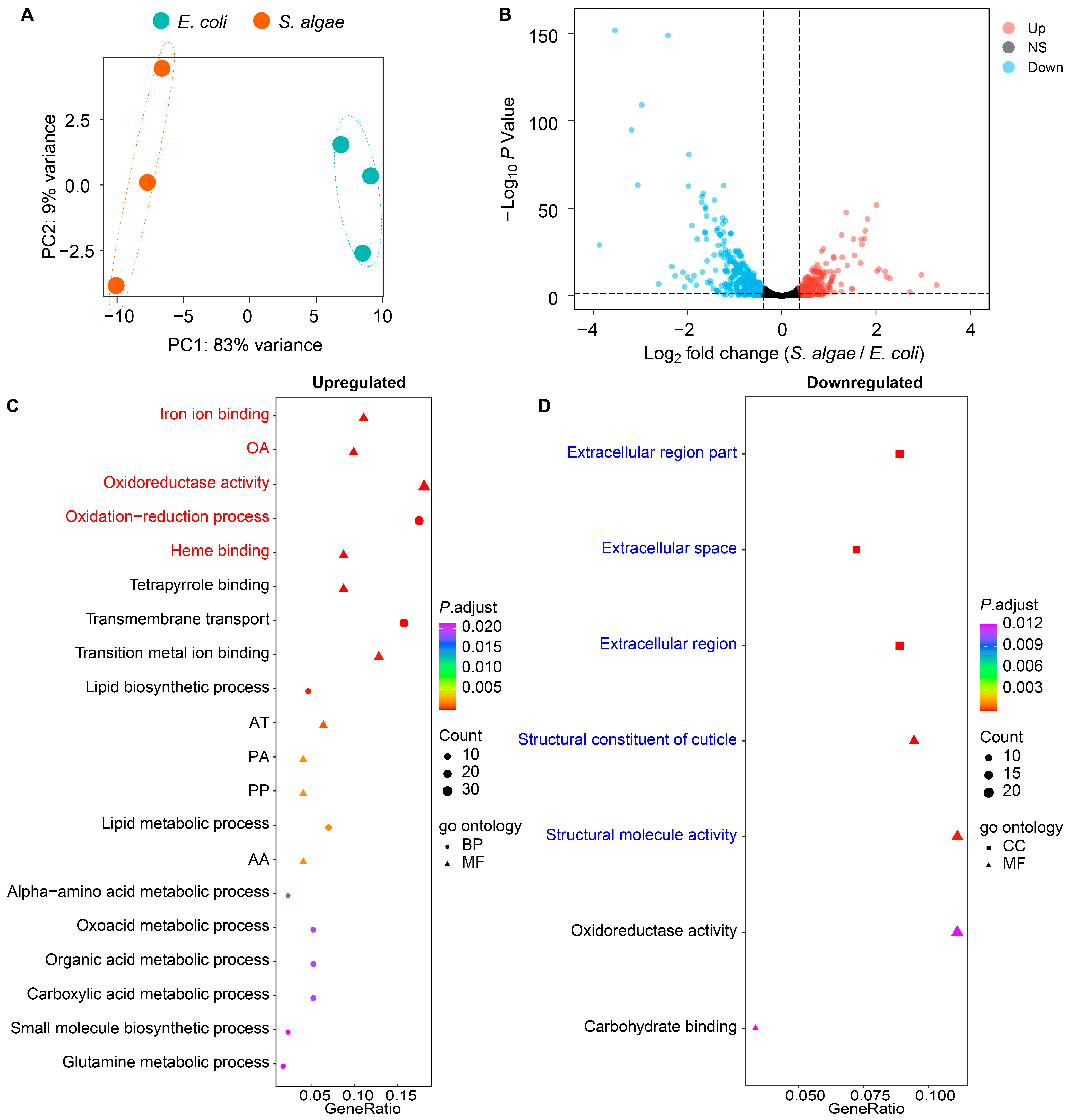
To identify the transcriptional characteristics of L. marina growing on S. algae versus E. coli OP50, we performed Illumina RNA sequencing on synchronized L1 worms. Principal component analysis (PCA) showed a clear separation of L. marina between feeding with S. algae and E. coli OP50 (Figure 2A). In total, we identified 703 differentially expressed genes (DEGs), among which 286 were upregulated and 417 were downregulated in L. marina fed with S. algae compared with E. coli OP50 (Figure 2B, Table S2).
Through GO enrichment analysis, we found that upregulated genes in L. marina growing on S. algae were significantly enriched in terms such as iron ion binding (C08G9.2, cyp-29A2, cyp-14A5, cyp-14A2, dach-1, agmo-1, cyp-25A2, cdo-1, cyp-42A1, cyp-36A1, drd-1, and haao-1), oxidoreductase activity, acting on paired donors, with the incorporation or reduction of molecular oxygen (C08G9.2, cyp-29A2, cyp-14A5, cyp-14A2, dach-1, cyp-25A2, fmo-1, C01H6.4, cyp-42A1, cyp-36A1, and fmo-2), oxidoreductase activity (hacd-1, C08G9.2, cyp-29A2, cyp-14A5, daao-1, cyp-14A2, dach-1, agmo-1, cyp-25A2, fmo-1, C01H6.4, C15B12.1, tdo-2, hsd-2, cdo-1, T10H10.2, ldp-1, cyp-42A1, cyp-36A1, and acox-1.1), and heme binding (C08G9.2, cyp-29A2, cyp-14A5, cyp-14A2, dach-1, cyp-25A2, tdo-2, cyp-42A1, and cyp-36A1) (Figure 2C; Table S3).
When GO analysis was applied to the downregulated DEGs, we observed that terms such as extracellular region part (zmp-3, ttr-30, tep-1, ttr-8, ttr-59, ttr-7, ttr-32, zmp-4, ttr-15, test-1, ttr-27, and ttr-46), structural constituent of cuticle (col-141, col-68, col-142, col-166, col-117, col-73, col-176, kcc-3, col-130, col-34, col-155, and sqt-2) and structural molecule activity (col-141, col-68, col-142, col-166, col-117, col-73, col-176, kcc-3, col-130, rpl-35, col-34, mrpl-51, col-155, let-2, and sqt-2) were significantly enriched in worms fed with S. algae compared with E. coli OP50 (Figure 2D; Table S4).
Additionally, through KEGG analysis, we observed that upregulated DEGs in L. marina growing on S. algae were significantly enriched in pathways such as tryptophan metabolism (hacd-1, tdo-2, alh-11, nkat-3, and haao-1) and fatty acid metabolism (hacd-1, pod-2, acox-1.1, elo-6, elo-5, and fat-6); acox-1.1, elo-6, elo-5, and fat-6 are involved in biosynthesis of unsaturated fatty acids, while acox-1.1, elo-6, and elo-5 are essential for fatty acid elongation (Figure S1; Table S5). When KEGG analysis was applied to the downregulated DEGs, none of the pathways were significantly enriched.

2.3. Transcriptomic Analysis of S. algae versus E. coli OP50

Since feeding with S. algae attenuated the development and shortened lifespan of L. marina compared with E. coli OP50 as a food source, we further investigated the transcriptomic characteristics of S. algae versus E. coli OP50 (Figure S2A). Among a total of 1806 single copy ortholog genes between S. algae and E. coli OP50, we identified 1299 DEGs, with 660 showing upregulation and 639 displaying downregulation in S. algae compared with E. coli OP50 (Figure S2B; Table S6). From BioCyc functional enrichment, no pathway was enriched in upregulated DEGs. As for the downregulated DEGs, we found that amino acid metabolism-related pathways were significantly enriched in S. algae compared with E. coli OP50, such as the superpathway of histidine, purine, and pyrimidine biosynthesis (deoB, nrdD, gmk, hisG, hisD, hisC, prs, hisB, purH, hisI, hisF, purD, hisA, prs, hisH, atpF, guaA, nrdB, pyrH, and purK), the superpathway of L-lysine, L-threonine, and L-methionine biosynthesis I (thrA, thrC, thrB, metH, metL, aspC, dapA, dapF, metE, metB, dapD, and serC), the superpathway of L-threonine metabolism (ilvB, tdh, adhE, ilvC, pflB, kbl, ilvD, ilvA, pta, and ackA), and the L-arginine biosynthesis I pathway (via L-ornithine) (argG, gabT, argE, argC, argB, argH, and argA) (Figure 3A; Table S7). Interestingly, genes in the ubiquinol-8 biosynthesis (prokaryotic) pathway (ubiD, ubiE, ubiF, ubiH, and ubiA), which has been reported to be positively correlated with L. marina growth [4], were significantly downregulated in S. algae in comparison to E. coli OP50 (Figure 3A; Table S7).
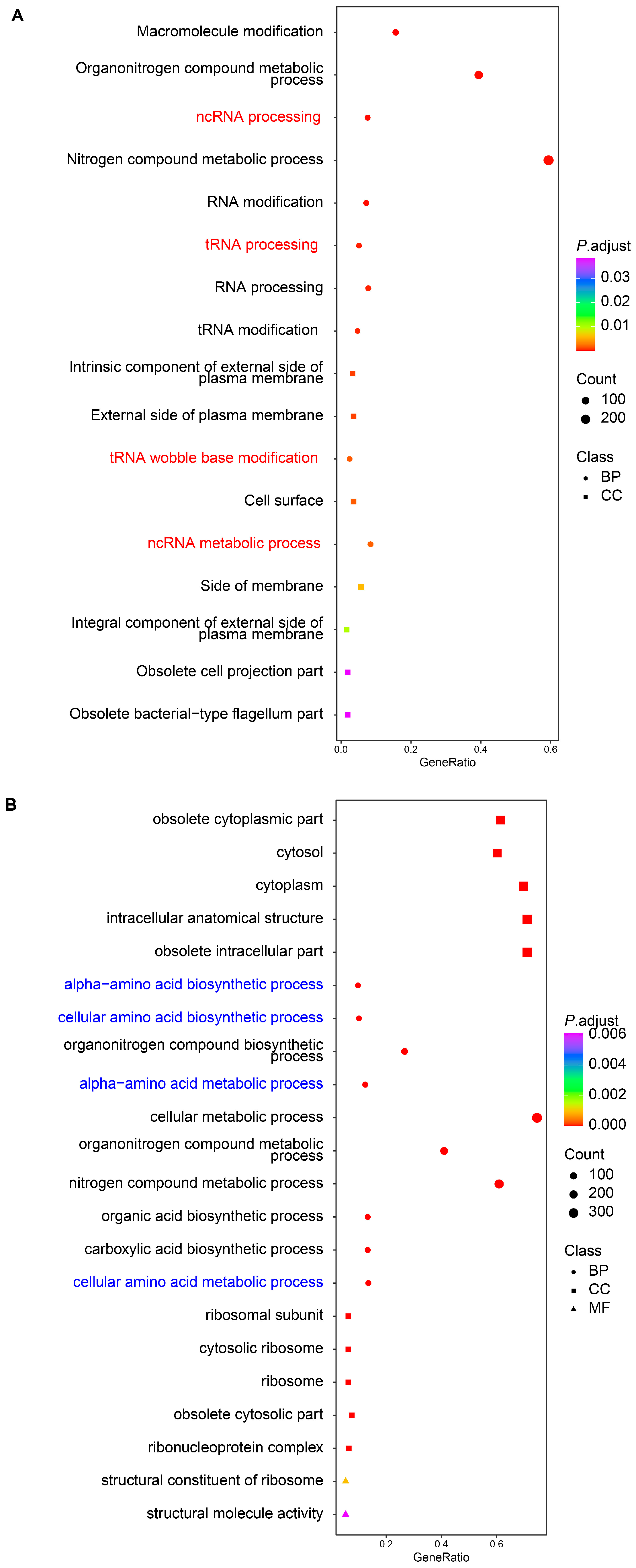
Through GO enrichment analysis, we observed that upregulated genes in S. algae were significantly enriched in terms related to RNA processing, such as ncRNA processing (rluC, tilS, truC, rlmF, rsmF, selU, rlmL, thiI, tsaD, and rlmN), tRNA processing (tilS, truC, selU, thiI, tsaD, rlmN, dusC, tsaE, cmoB, and rne), tRNA wobble base modification (mnmE, tadA, mnmA, gluQ, cmoA, selU, tusD, tusC, tilS, mnmC, tusE, cmoB, and cmoB), and ncRNA metabolic process (rluC, tilS, truC, rlmF, rsmF, selU, rlmL, thiI, cysS, and selA) (Figure 4A; Table S8). When GO analysis was applied to the downregulated DEGs in S. algae, we found that terms such as alpha-amino acid biosynthetic process (ilvB, tyrA, thrA, thrC, tdh, thrB, metH, glyA, hisG and gltB), cellular amino acid biosynthetic process (ilvB, aroF, tyrA, thrA, thrC, tdh, thrB, metH, glyA, and hisG), alpha-amino acid metabolic process (ilvB, tyrA, dadA, thrA, thrC, tdh, thrB, metH, gcvP, and glyA), and cellular amino acid metabolic process (ilvB, aroF, tyrA, dadA, thrA, thrC, tdh, thrB, metH, and gcvP) were significantly enriched (Figure 4B; Table S9).
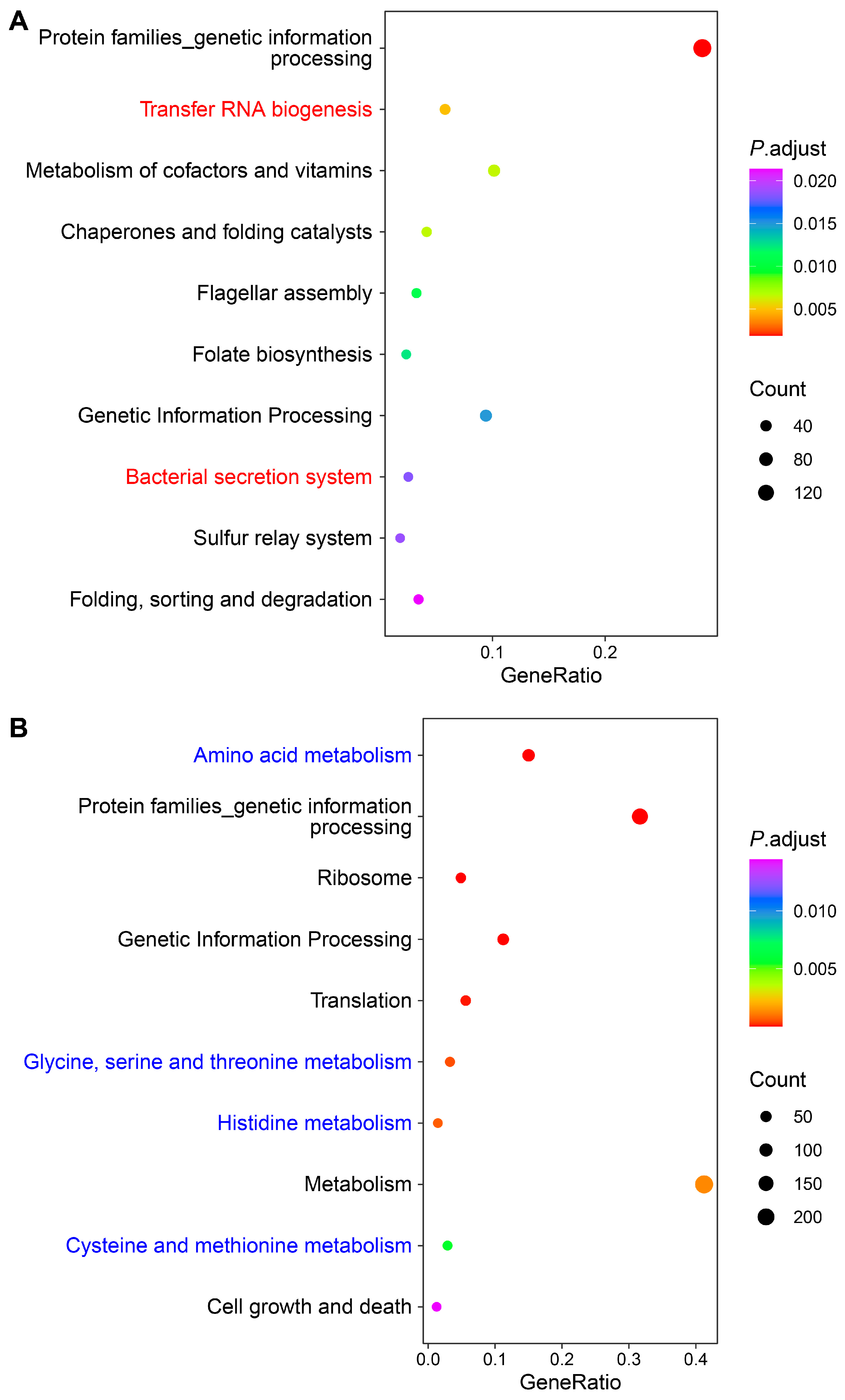
Through KEGG annotation, we observed that upregulated DEGs in S. algae were significantly enriched in pathways such as transfer RNA biogenesis (tilS, truC, selU, thiI, cysS, tsaD, dusC, tsaE, cmoB, and mnmA), and bacterial secretion system (gspD, gspE, gspF, gspK, gspJ, gspH, secA, gspG, epsM, and gspI) (Figure 5A; Table S10). Consistent with the GO analysis, downregulated DEGs in S. algae were enriched in several amino acid metabolism process pathways, such as amino acid metabolism (aroF, tyrA, ilvB, dadA, thrA, katG, puuA, glmS, thrC, and tdh), glycine, serine, and threonine metabolism (thrA, thrC, tdh_1, thrB, gcvP, glyA, metL, pssA, kbl, and ilvA), histidine metabolism (hisG, hisD, hisC, hisB, hisI, hisF, hisA, and hisH), and cysteine and methionine metabolism (thrA, metH, metL, aspC, gshB, sseA, mdh_1, speE, metE, and metB) (Figure 5B; Table S11).
We subsequently focused on the genes apart from the single copy ortholog genes between S. algae and E. coli OP50, and selected the top 10% of genes with the highest median absolute deviation (MAD) of expression for downstream analysis, for S. algae and E. coli OP50, respectively (Figure 3B,C). Through GO enrichment analysis, we observed that terms such as response to xenobiotic stimulus (prpB, acnD, prpC, nqrD, sirA, gbpA, and DPLKCNFD_00060) and energy derivation by oxidation of organic compounds (prpB, acnD, prpC, nqrD, and fdhE) were significantly enriched in S. algae (Figure 3B; Table S12). For the top 10% of genes with the highest MAD of expression in E. coli OP50, we observed that GO terms such as sugar transmembrane transporter activity (exuT, rbsB, gatZ, rbsC, rbsA, rbsD, and kdgT), NAD(P)H dehydrogenase (quinone) activity (wrbA, nuoK, nuoJ, nuoH, and nuoE) and oxidoreductase activity, acting on NAD(P)H, quinone, or a similar compound as acceptor (wrbA, nuoK, nuoJ, nuoH, nuoE, and qorA), were significantly enriched (Figure 3C; Table S13).
KEGG analysis of the selected top 10% of genes revealed that pathways such as propanoate metabolism (acs, prpB, acnD, DPLKCNFD_02174, prpC, mmsA, pdhA, and bkdA2) and biofilm formation-pseudomonas aeruginosa (hcpA, DPLKCNFD_03118, DPLKCNFD_00897, DPLKCNFD_01441, and ntrC) were significantly enriched in S. algae, while the beta-Lactam resistance pathway (oppB, oppA, btuD, oppD, oppC, yejF, ompC, and ompF) was significantly enriched in E. coli OP50 (Figure S3; Tables S14 and S15).

2.4. Metabolic Profiling of S. algae- and E. coli OP50-Conditioned Media

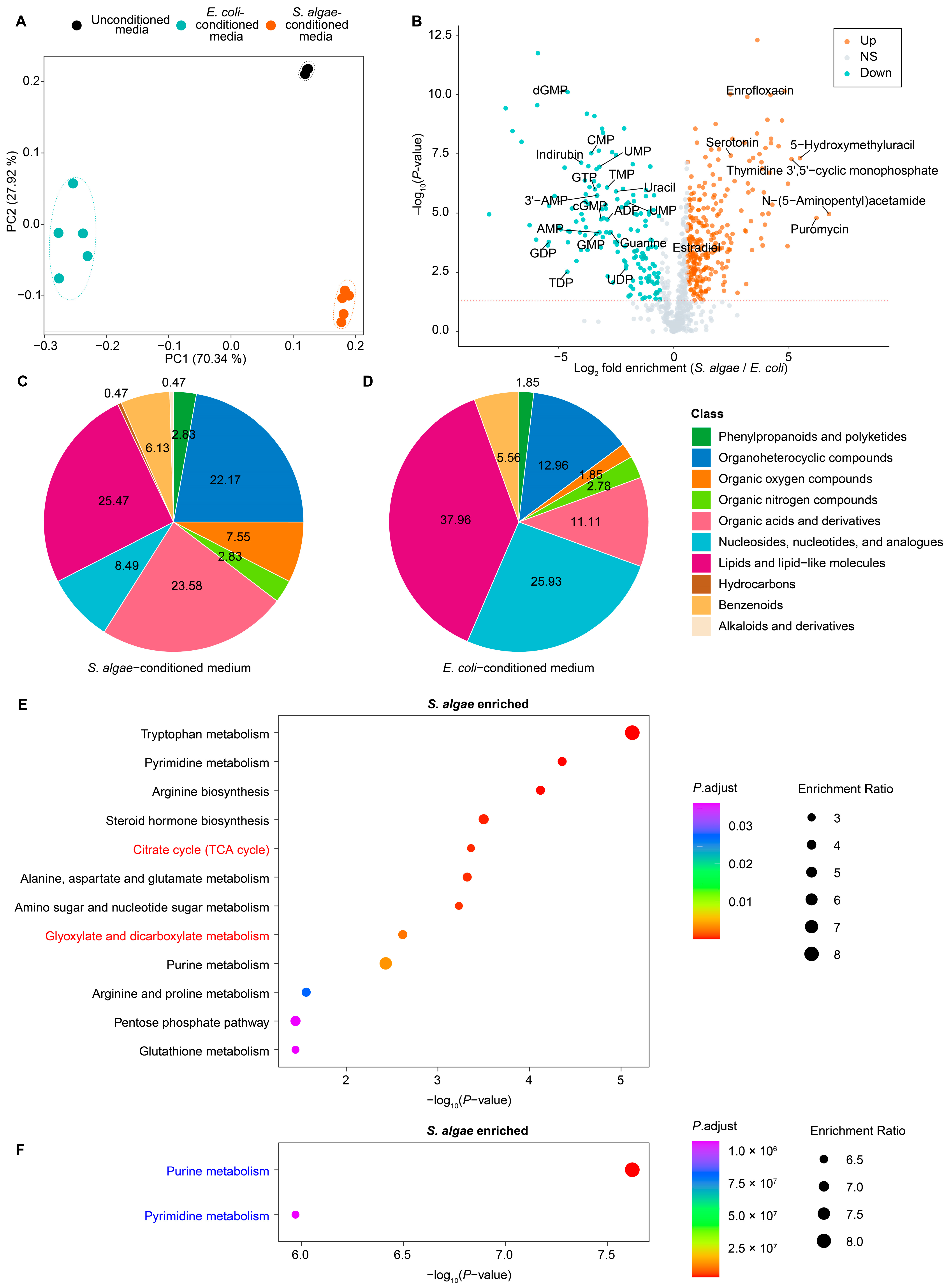
LC-MS-based untargeted metabolomic was conducted to profile the metabolic characterization of S. algae versus E. coli OP50. PCA revealed that media conditioned by S. algae and E. coli OP50 were separated from each other, and also from the unconditioned media (Figure 6A). We found that 275 differentially accumulated metabolites (DAMs) were significantly enriched by at least 1.5-fold in S. algae-conditioned medium, while 157 metabolites were significantly enriched by at least 1.5-fold in E. coli OP50-conditioned medium (Figure 6B; Table S16). In the S. algae-conditioned medium, N-(5-Aminopentyl)acetamide, puromycin, 5-hydroxymethyluracil, and thymidine 3′,5′-cyclic monophosphate were among the most enriched metabolites, showing a log2 fold enrichment above 5 (Figure 6B; Table S16). On the other hand, the levels of various nucleotides were significantly decreased in the S. algae-conditioned medium, including GDP, TDP, dGMP, UDP-galactose, GMP, CMP, GTP, 3′-AMP, UMP, AMP, cGMP, TMP, ADP, guanine, uracil, 5′-AMP, UDP, and UMP, which were enriched in the E. coli OP50-conditioned medium (Figure 6B; Table S16).
In the S. algae-conditioned medium, the most enriched DAMs belonged to lipids and lipid-like molecules (25.47%), organic acids and derivatives (23.58%), organoheterocyclic compounds (22.17%), nucleosides, nucleotides, and analogues (8.49%), and organic oxygen compounds (7.55%) (Figure 6C). While the most enriched DAMs in the E. coli OP50-conditioned medium included lipids and lipid-like molecules (37.96%), nucleosides, nucleotides, and analogues (25.93%), organoheterocyclic compounds (12.96%), organic acids and derivatives (11.11%), and benzenoids (5.56%) (Figure 6D). The percentages of organic acids and derivatives, organoheterocyclic compounds, and organic oxygen compounds were more enriched in the S. algae-conditioned medium; by contrast, the percentages of lipids and lipid-like molecules, nucleotides, and analogues were more enriched in the E. coli OP50-conditioned medium (Figure 6C,D).
Through KEGG pathway-based enrichment analysis on the DAMs between the S. algae- and E. coli OP50-conditioned media, we found that KEGG pathways such as citrate cycle (TCA cycle) (alpha-ketoglutaric acid, cis-aconitic acid, and citric acid) and glyoxylate and dicarboxylate metabolism (cis-aconitic acid, 4-hydroxy-2-oxoglutaric acid, citric acid, and serine) were significantly enriched in the S. algae-conditioned medium (Figure 6E; Table S16), while pathways including purine metabolism (2,6-dihydroxypurine, adenosine 5′-monophosphate, adenylsuccinic acid, guanosine monophosphate, guanine, dGDP, cGMP, adenosine diphosphate ribose, and adenine) and pyrimidine metabolism (UMP, cytidine-5′-monophosphate, dUDP, dUMP, dTDP, thymidine 5′-monophosphate, and uracil) were significantly enriched in the E. coli OP50-conditioned medium (Figure 6F; Table S16).

2.5. Joint Pathway Analysis of DAMs and DEGs in S. algae versus E. coli OP50

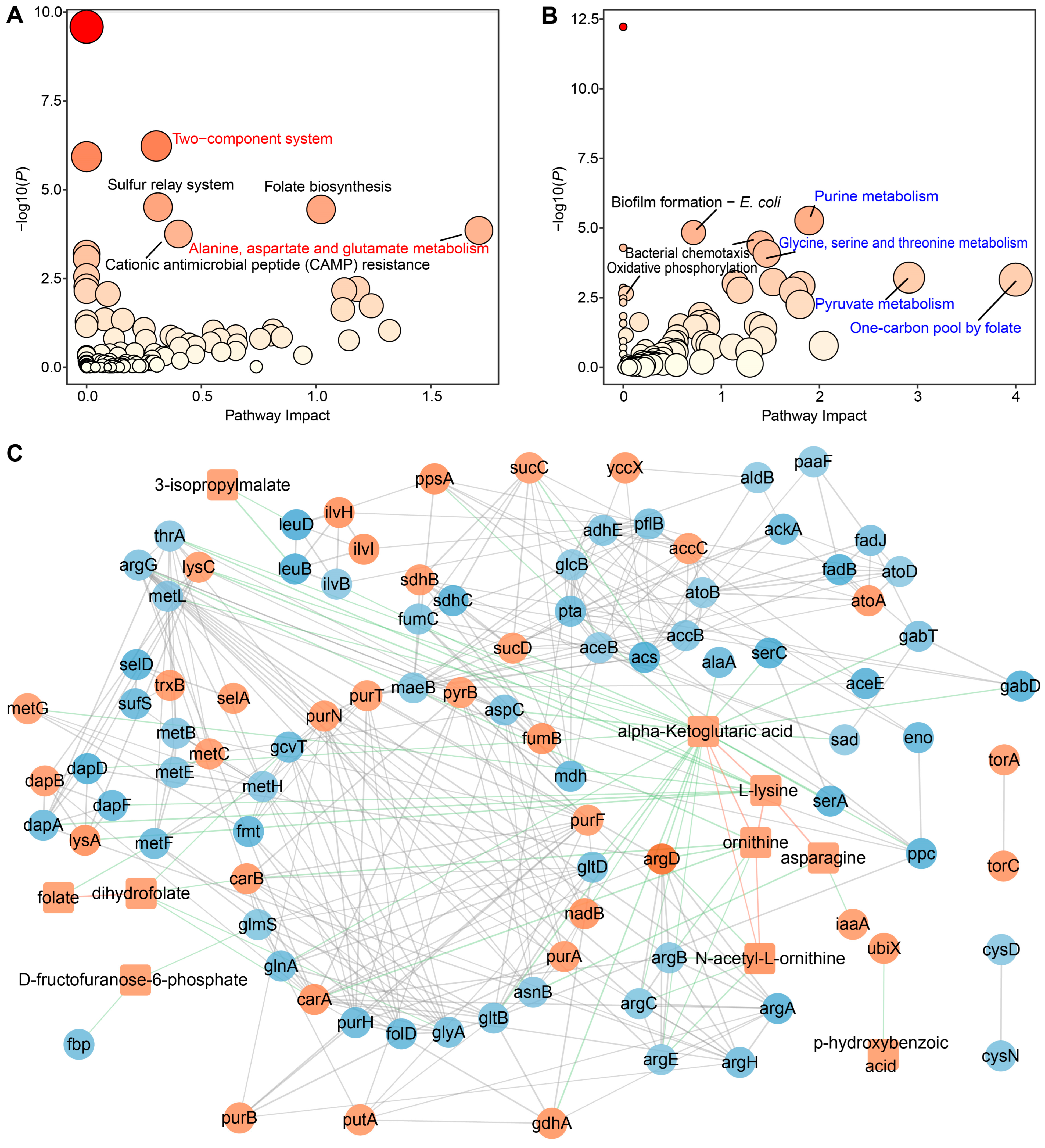
To further integrate the DEGs and DAMs in S. algae versus E. coli OP50, a Joint Pathway Analysis was performed using MetaboAnalyst 6.0 [30]. Joint Pathway Analysis identified 100 and 95 altered pathways in the upregulated and downregulated DEGs and DAMs in S. algae, respectively, with five and eight of them showing significant changes (Figure 7A,B; Table S17). We found that the “two-component system” (TCS) was the most significant upregulated pathway in S. algae compared with E. coli OP50 (Figure 7A), which is an essential prerequisite for many bacterial pathogenicities [31]. Furthermore, “alanine, aspartate and glutamate metabolism” showed the highest impact score in S. algae. In contrast, “pyruvate metabolism”, “glycine, serine and threonine metabolism”, “purine metabolism”, and “one-carbon pool by folate” were significantly enriched in E. coli OP50 (Figure 7B).
STITCH interaction analysis revealed that the transcript-metabolite interactions consisted of 104 nodes, which were connected by 381 edges (Figure 7C; Table S18). Significant interaction was observed only in the enriched metabolites in S. algae. Alpha-ketoglutaric acid, ornithine, L-lysine, and N-acetyl-L-ornithine exhibited the highest number of associations (n ≥ 6) (Figure 7C). Of note, alpha-ketoglutaric acid was significantly connected to lysine, ornithine, N-acetyl-L-ornithine, and 23 DEGs (aspC, gltB, gdhA, serA, gabT, serC, sucC, argD, sucD, alaA, gabD, ppc, sad, glnA, ppsA, lysC, thrA, gltD, argC, metL, dapD, nadB, and argE) (Table S18).

3. Discussion

Bacteria can modulate animal fitness and physiology through bacterially-produced metabolites and RNA [32,33]. For example, it has been reported that bacterial siderophore enterobactin promotes C. elegans iron acquisition and development [34]. Zhang et al. identified 244 bacterial mutants that attenuated C. elegans development, with several of the causal bacterial genes encoding the bo oxidase of the electron transport chain and iron transporters [35]. In addition, bacterial peptidoglycan muropeptides have been described to support the development of C. elegans, by reducing mitochondrial oxidative stress and enhancing ATP synthase activity [36]. Different bacterial diets, such as Methylobacterium braciatum, Xanthomonas citri, and Sphingomonas aquatilis, could uniquely alter the development, longevity, and transcriptomic characteristics of C. elegans, highlighting the impact of bacterial diet on animal physiological outcomes [37]. In addition, bacterial polysaccharide colanic acid has been identified to extend C. elegans lifespan by modulating mitochondrial homeostasis [38]. It has been reported that E. coli mutants with reduced levels of methylglyoxal biosynthesis, promote C. elegans longevity via suppression of TORC2/SGK-1 and induction of DAF-16 [39]. Like model nematode C. elegans, many marine nematodes utilize bacteria as their primary food source; however, how bacterial diet affects the development and aging of marine nematode L. marina remains largely unexplored.

3.1. Higher Expression of Genes in Iron Ion Binding and Oxidoreductase Activity Pathways Might Promote Aging of L. marina When Grown on S. algae

For the upregulated DEGs in marine nematode L. marina grown on S. algae compared with E. coli OP50, several genes in GO terms, such as iron ion binding (agmo-1, cdo-1, and haao-1), oxidoreductase activity (agmo-1, tdo-2, cdo-1), and heme binding (tdo-2) were particularly interesting (Figure 2C; Table S4). agmo-1 encodes alkylglycerol monooxygenase for ether-linked lipids degradation, and agmo-1 mutants exhibit bacterial infection resistance and contain ether-linked (O-alkyl chain) lipids in comparison to exclusively ester-linked (O-acyl) lipids in wild-type animals, which might provide a more resilient cuticle for agmo-1 mutants [40]. cdo-1 is predicted to enable cysteine dioxygenase activity and ferrous iron binding, with its high expression resulting in the shortened lifespan of C. elegans [41]. haao-1 encodes 3-hydroxyanthranilic acid (3HAA) dioxygenase (HAAO), and its knockdown extends the lifespan of C. elegans and promotes healthy aging [42]. It has been reported that RNAi knockdown of tdo-2, which encodes tryptophan 2,3-dioxygenase (TDO), extends the lifespan and ameliorates neurodegenerative pathology in C. elegans [43]. Given that RNAi knockdown of cdo-1, haao-1, and tdo-2 extend the lifespan and promote healthy aging in C. elegans, we speculated that the higher expression of cdo-1, haao-1, and tdo-2 might contribute to the shortened lifespan in L. marina.

3.2. Decreased Expression of Extracellular Structural Components-Related Genes Might Delay the Development of L. marina fed with S. algae

For the downregulated DEGs in L. marina fed on S. algae compared with E. coli OP50, we observed that terms such as extracellular region part and structural constituent of cuticle were significantly enriched in worms when fed with S. algae in comparison to E. coli OP50 (Figure 2D; Table S4). Among the extracellular region part pathway, eight genes were identified as transthyretin-like family genes (TTLs), including ttr-46, ttr-15, ttr-7, ttr-8, ttr-32, ttr-27, ttr-59, and ttr-30 (Table S4). C. elegans TTLs exhibited sensitivity to a variety of environmental stressors, such as reactive oxygen species (ROS) stress, exposure to pathogens, and osmotic imbalances [44,45,46,47]. Among the structural constituent of the cuticle pathway, most genes encode collagens, such as col-141, col-68, col-142, col-166, col-117, col-73, col-176, col-130, col-34, and col-155 (Table S4). Collagens are the primary components of nematode cuticles, which are shed to allow for growth when animals molt, and protect the nematode as a physical barrier [48]. RNAi knockdown of col-141 expression has been reported to promote aging in long-lived daf-2 and eat-2 mutants [49]. Thus, the downregulation of genes in extracellular structural components-related pathways might impair nematode cuticle structure and function, and delay the development of L. marina when fed with S. algae.

3.3. Downregulated Bacterial Amino Acid Metabolism Genes in S. algae Might Delay Development and Shorten the Lifespan of L. marina

Through BioCyc functional enrichment of downregulated DEGs in S. algae compared with E. coli OP50, we found that amino acid metabolism-related pathways were significantly enriched, such as the superpathway of histidine, purine, and pyrimidine biosynthesis (deoB, nrdD, gmk, hisG, hisD, hisC, prs, hisB, purH, hisI, hisF, purD, hisA, prs, hisH, atpF, guaA, nrdB, pyrH, and purK), the superpathway of L-lysine, L-threonine, and L-methionine biosynthesis I (thrA, thrC, thrB, metH, metL, aspC, dapA, dapF, metE, metB, dapD, and serC), the superpathway of L-threonine metabolism (ilvB, tdh, adhE, ilvC, pflB, kbl, ilvD, ilvA, pta, and ackA), and the L-arginine biosynthesis I pathway (via L-ornithine) (argG, gabT, argE, argC, argB, argH, and argA) (Figure 3A; Table S7).
Most of these downregulated genes in S. algae versus E. coli OP50 contribute to amino acid biosynthesis, which indicates that E. coli OP50 feeding might provide more abundant amino acids, promoting the development of L. marina, compared with using S. algae as the food source [50]. It has been reported that individual supplementation with 18 of the 20 essential amino acids could increase C. elegans longevity [51]. Additionally, most of the amino acid pool sizes increased in the long-lived C. elegans [52]. Proline has been found to prolong the longevity of C. elegans by triggering a temporary surge in ROS production, which originates from the mitochondrial electron transport chain [53]. Similarly, elevated tryptophan levels can also boost longevity by silencing an enzyme responsible for tryptophan breakdown, resulting in a longer lifespan [54]. Our GO and KEGG analysis also revealed that genes in several amino acid biosynthesis terms or pathways, such as histidine, L-threonine, L-ornithine, arginine, polyamine, glycine, serine, threonine, cysteine, and methionine, were significantly downregulated in S. algae compared with E. coli OP50, indicating that E. coli OP50 might supply more abundant amino acids to promote the development and extend lifespan of L. marina compared with using S. algae as the food source.

3.4. Higher Expression of Virulence-Related Genes in S. algae Might Contribute to the Delayed Development and Shortened Lifespan of L. marina

For the upregulated genes in S. algae, we observed that GO terms such as ncRNA processing, tRNA processing, tRNA wobble base modification, and ncRNA metabolic process were significantly enriched (Figure 4A; Table S8). It has been reported that E. coli ncRNA DsrA promotes C. elegans aging, through suppressing the expression of diacylglycerol lipase encoding gene dagl-2 [55]. mnmA is involved in tRNA modification and could impact protein synthesis, influencing the expression of virulence factors [56,57]. selU has been reported as a virulence gene in the enterotoxin gene cluster [58]. rlmN, a key methyltransferase encoding gene, can alter ribosomal RNA to regulate bacterial translation efficacy, affecting traits such as virulence, infectiousness, organismal health, and resilience to environmental pressures [59]. The increased expression of mnmA, selU, and rlmN in S. algae might explain the developmental delay and shortened lifespan of L. marina.
For the top 10% of genes with the highest MAD of expression in S. algae, we found that several genes, such as sirA, gbpA, hcpA, and ntrC, might be related to bacterial virulence. For example, sirA acts as a positive regulator for the SPI1 pathogenicity island, encoding a Type III secretion system that injects effector proteins directly into the host cell’s cytoplasm [60]. GbpA, a recognized virulence factor, encoded by gbpA, was reported to facilitate bacterial colonization by interacting with GlcNAc residues in mucin, which upregulates their activity cooperatively and ultimately produces virulence [61]. hcpA has been reported to encode HcpA subunits in Type IV pili (TFP), which are essential for virulence in several Gram-negative bacteria [62]. Furthermore, the ntrC gene is crucial for nitrogen assimilation, stress resilience, and bacterial virulence [63]. We thus propose that these aforementioned upregulated virulence genes of S. algae might induce growth retardation and aging of L. marina.

3.5. Impact of Toxic Metabolites and Nucleotide on L. marina Development and Lifespan

We observed that several toxic metabolites, which potentially impair L. marina development and lifespan, were significantly enriched in the S. algae-conditioned medium, such as puromycin (73-fold) and enrofloxacin (28-fold) (Figure 6B; Table S16). Puromycin has been reported to interfere with the function of ribosomes and block protein synthesis in both eukaryotes and prokaryotes and is toxic to C. elegans in both liquid and solid cultures [64]. Enrofloxacin induces oxidative stress by stimulating peroxyl radicals (H2O2) and lipid peroxidation [65,66].
It is known that nucleotides are essential for nematode cellular function and development, acting as a crucial nutrient source [67,68]. Previous studies have shown that the supplementation of some nucleotides, such as uridine, thymine, cytidine, orotate, β-aminoisobutyrate, and pyrimidine intermediates, extends C. elegans lifespan [69]. We supposed that nucleotides enriched in the E. coli OP50-conditioned medium benefit L. marina physiology.
It has been reported that the glyoxylate cycle plays a crucial role in the pathogenicity of Mycobacterium tuberculosis and is essential for the full virulence of Candida albicans [70]. Additionally, a complete TCA cycle has proven to be indispensable for the full virulence of serovar Typhimurium strain SR-11; the deletion of various genes involved in the TCA cycle has been reported to lead to reduced virulence [71]. According to our KEGG analysis, the TCA cycle and glyoxylate and dicarboxylate metabolism pathways were significantly enriched in the S. algae-conditioned medium, which might contribute to the developmental delay and short lifespan of L. marina when fed on S. algae. On the other hand, we observed that the purine and pyrimidine metabolism pathways were significantly enriched in the E. coli-conditioned medium, which could explain why E. coli OP50 promoted L. marina development and longevity compared with feeding with S. algae.

3.6. “Two-Component System” and “One-Carbon Pool by Folate” Pathways Might Modulate L. marina Physiology

Through Joint Pathway Analysis, we found that the “two-component system” (TCS) was the most significant pathway among the upregulated pathways in S. algae compared with E. coli OP50. TCS has been reported as a vital signal transduction mechanism in bacteria, participating in a multitude of gene regulatory systems that adapt to fluctuating environmental conditions [31]. Thus, TCS is crucial for pathogenic bacteria’s ability to efficiently adapt to diverse microenvironments both inside and outside their hosts, facilitating their pathogenicity [31]. A previous study showed that two-component sensor kinase KdpD (7.4-fold change in S. algae versus E. coli OP50) played a crucial role in the pathogenesis of Salmonella typhimurium to C. elegans through the TCS pathway [72]. Our data suggest that the upregulation of the TCS pathway in S. algae might enhance its virulence and impair L. marina physiology. In contrast, the “one-carbon pool by folate” pathway showed the largest impact score among the downregulated pathways in S. algae. It was described that metformin repressed the folate metabolism of E. coli, which then modulated methionine metabolism in C. elegans, and extended worm lifespan [73]. In addition, it was reported that the one-carbon folate cycle is involved in several long-lived C. elegans mutants, and reduced 5 methyl tetrahydrofolate (5MTHF) extends the nematode lifespan [74]. Thus, upregulation of “two-component system” and downregulation of “one-carbon pool by folate” might explain why S. algae attenuated L. marina development and promoted aging, compared with feeding with E. coli OP50.

4. Materials and Methods

4.1. Worm and Culture

The wild strain of the marine nematode L. marina was isolated from intertidal sediments in Huiquan Bay, Qingdao [75]. L. marina used in this study was a 23rd generation inbred line (F23) that was cultivated through successive full-sibling crosses within our laboratory, and its cultivation follows previous publications [4,16]. In brief, L. marina strains were maintained on seawater nematode growth media (SW-NGM). For regular maintenance, SW-NGM was supplemented with E. coli OP50 as their diet, and the worms were cultured at 20 °C.

4.2. Bacteria

E. coli OP50 was obtained from the Caenorhabditis Genetics Center (CGC, https://cgc.umn.edu/, accessed on 19 August 2024). S. algae was isolated from Huiquan Bay, Qingdao, China, and stored at 80 °C as glycerol stocks [4]. Both bacteria were stamped out fresh cultures from glycerol stocks onto a rectangular LB plate and then incubated overnight at 37 °C. The colonies on the plate were then used to inoculate in LB media.

4.3. Development and Lifespan Assays

To assess the influence of S. algae and E. coli OP50 on L. marina development and lifespan, we examined the growth rate and lifespan as previously reported [4].

4.4. Sample Collection

The synchronized L1 larvae fed with S. algae and E. coli OP50 were collected as previously reported [76]. L. marina F23 worms cultured on SW-NGM plates were permitted to lay eggs overnight at 20 °C. Once a substantial quantity of eggs had been deposited on the culture plates, adult worms were harvested by gently rinsing the plates with sterilized seawater. Following this, the eggs situated on the plates underwent a thorough rinse into a collection tube and were subjected to bleaching utilizing Worm Bleaching Solution (comprising Bleach: 10M NaOH: H2O in a ratio of 4:1:10) to procure sterile eggs, which were subsequently incubated in sterilized seawater overnight, hatching into synchronized L1 larvae. The synchronized L1 larvae were placed onto plates containing S. algae or E. coli OP50 as a food source, with three replicates for each treatment. Following 4 h of feeding, conditioned media were washed off using M9 solution, followed by the rapid separation and collection of both nematodes and conditioned media. Animal bodies and conditioned medium were frozen in liquid nitrogen for approximately half an hour before being stored at −80 °C and processed separately.

4.5. RNA Library Preparation, RNA Sequencing and Transcriptional Analysis of L. marina

Total RNA extraction was performed using Trizol (Invitrogen, Carlsbad, CA, USA). Then, six RNA libraries, three biological replicates for each bacterial feeding, were constructed using NEBNext® UltraTM RNA Library Prep Kit for Illumina® (New England Biolabs, Ipswich, MA, USA), following the manufacturer’s guidelines. Subsequently, we sequenced the RNA libraries on an Illumina NovaSeq 6000 platform, producing 150 bp paired-end reads.
Quality control and adapter trimming were performed on raw reads using fastp [77]. The minimum base score of Q20 was above 98.87%, and Q30 was above 96.95%. Resulting clean reads were mapped to L. marina Genome v1.0 [16] using HISAT2 v2.1.0 (--no-discordant, --no-mixed) [78]. Mapped reads were quantified using featureCounts [79] from Subread v2.0.3 with L. marina Geneset v1.0 as annotation (-B, -p). Differential gene expression analysis was conducted with R package DEseq2 v1.38 [80]. DEGs with an adjusted p-value < 0.05 and a fold change > 1.3 were regarded as significantly differentially expressed genes. GO and KEGG enrichment analysis was performed using R package clusterProfiler v4.6.2 [81] and Geneset v1.0.

4.6. RNA Library Preparation, RNA Sequencing, and Transcriptional Analysis of Bacteria

Microbe-conditioned media were washed off using M9 solution, as described above, and were frozen in liquid nitrogen for approximately half an hour before being stored at −80 °C. Library preparation and RNA sequencing were also performed as described above. For differentially expressed gene (DEG) analysis, only counts from single-copy ortholog genes were used and gene symbols of E. coli OP50 were adopted as the representative gene symbols for downstream analysis. Differential gene expression analysis was conducted with R package DEseq2 v1.38 [80]. DEGs with an adjusted p-value < 0.05 and a fold change > 1.5 were regarded as significantly differentially expressed genes. Enrichment analysis was performed on KOBAS 3.0 [82].
Protein sequences and gene annotation of S. algae and E. coli OP50 were obtained from Prokka [83] outputs. Reciprocal best hit search was performed using MMseq2 easy-rbh with default parameters [84]. The non-redundant result of MMseq2 easy-rbh output was used as single-copy orthologs between S. algae and E. coli OP50.
In addition, counts of genes apart from single-copy ortholog genes were normalized to FPKM using DEseq2 v1.38 [80]. The top 10% of these genes were selected using median absolute deviation (MAD); 230 and 237 genes were selected for S. algae and E. coli OP50 respectively, which were further enriched using clusterProfiler v4.6.2 [81] for KEGG enrichment analysis and TBtools [85] for GO enrichment analysis. All proteins of E. coli OP50 and S. algae were annotated using eggNOG-mapper version 2.1.6 [86]. Gene Ontology annotations were extracted from the results under the name of GOs.

4.7. Metabolomics

The overnight cultures of E. coli and S. algae in LB medium were diluted 1:100 in LB medium and incubated at 37 °C for 4–5 h until they reached an OD600 of 0.4. The bacterial samples were flash-frozen in liquid nitrogen for 30 m before being transferred to a −80 °C freezer. Five replicates of bacteria-conditioned media and three replicates of unconditioned LB medium were collected. The samples underwent freeze-drying and were reconstituted using prechilled 80% methanol with a through vortex. Afterward, the samples were left to incubate on ice for 5 min and then centrifuged (15,000× g, 4 °C for 15 min). A portion of the supernatant was further diluted to a final concentration comprising 53% methanol using LC-MS grade water. The resultant solution was then transferred to a fresh Eppendorf tube and subjected to another round of centrifugation (15,000× g, 4 °C for 15 min). Finally, the supernatant was injected into the LC-MS/MS system for analysis.
UHPLC-MS/MS analyses were conducted utilizing a Vanquish UHPLC system (ThermoFisher, Dreieich, Germany), seamlessly coupled with an Orbitrap Q ExactiveTM HF-X mass spectrometer (Thermo Fisher). Samples were injected onto a Hypersil Gold column (100 × 2.1 mm, 1.9 μm) employing a 12-min linear gradient at a flow rate of 0.2 mL/min. For the positive and negative polarity modes, eluent A (0.1% FA in water) and eluent B (methanol) were employed, respectively. The solvent gradient was meticulously programmed as follows: starting at 2% B for 1.5 min, transitioning to 2–85% B for 3 min, then ramping up to 85–100% B for 10 min, and finally reverting to 2% B for 12 min. Operation of the Q ExactiveTM HF mass spectrometer was optimized for both positive and negative polarity modes, featuring a spray voltage of 3.5 kV, a capillary temperature of 320 °C, a sheath gas flow rate of 35 psi, an auxiliary gas flow rate of 10 L/min, an S-lens RF level of 60, and an auxiliary gas heater temperature of 350 °C.
The Thermo RAW files were processed using Compound Discoverer v3.3 (ThermoFisher) to execute peak alignment, peak picking, and quantitation for each metabolite. We established the primary parameters as follows: correcting peak area with the first QC, maintaining an actual mass tolerance of 5 ppm, ensuring a signal intensity tolerance of 30%, and using a minimum intensity threshold. Following this, we normalized peak intensities against the total spectral intensity. Next, leveraging the normalized data, we employed predictive methods to discern molecular formulas based on additive ions, molecular ion peaks, and fragment ions. Subsequently, we cross-referenced these peaks with mzCloud, mzVault, and the MassList database to secure precise qualitative and relative quantitative measurements. Annotations were performed using KEGG, HMDB, and LIPIDMaps database. The metabolites with adjusted p-value < 0.05 and fold change > 1.5 were considered to be differential metabolites. KEGG enrichment pathways were analyzed using Metaboanalyst 6.0, and only used metabolite sets containing at least 3 entries [30].

4.8. Bacterial Transcriptome and Metabolism Conjoint Analysis

Integrated transcriptomic and metabolic analysis was carried out using the Joint Pathway Analysis module of MetaboAnalyst v6.0 [30]. Both metabolic (adjusted p-value < 0.05, fold change > 1.5) and transcriptomic (adjusted p-value  < 0.05, fold change  > 1.5) datasets were utilized for Joint Pathway Analysis. To assess the potential importance of individual molecules within a network, we uploaded the Entrez IDs of DEGs and the names of metabolites, along with their optional fold changes. In the Joint Pathway Analysis module, various parameters were selected: (i) in the pathway database, metabolic pathways (integrated) were chosen; and (ii) in the algorithm selection, the enrichment analysis using the hypergeometric test, topology measure using degree centrality, and the integration method combining queries were applied. Pathways with an adjusted p-value less than 0.05 were considered significant. The pathway impact score summarized the normalized topological measure of altered genes or metabolites present in each metabolic pathway, while the −log10(adjusted p-value) indicated the results of the enrichment analysis.
Furthermore, the interaction network between the key metabolites and DEGs involved in the Joint Pathway Analysis was established using STITCH [87,88], an online tool for visualizing biological relationships between metabolites and genes, as well as between metabolites and metabolites, and genes and genes. A bipartite subgraph was generated by STITCH due to the presence of edges only between proteins and chemicals. The interactions obtained were then used to construct the final differential network image using Cytoscape software v3.10.2 [89].

5. Conclusions

In conclusion, we performed a comparative metabolome and transcriptome analysis to uncover the impacts of S. algae and E. coli on L. marina growth and lifespan. We found that S. algae potentially impaired L. marina development and longevity by downregulating the expression of amino acid metabolism genes, increasing the expression of virulence-related genes, releasing potential toxic metabolites, and reducing nucleotide metabolism. Future research should focus on unraveling the molecular mechanisms underlying how key nematode genes, bacterial genes, and metabolites modulate animal development, aging, and behavior when grown on different bacterial foods, using CRISPR genome editing, RNAi, and diet supplementation with candidate bacterial metabolites.

Supplementary Materials

The following supporting information can be downloaded at: https://www.mdpi.com/article/10.3390/ijms25169111/s1.

Author Contributions

Conceptualization, Y.X. and L.Z.; methodology, Y.X. and B.X.; software, Y.X. and B.X.; validation, Y.X. and B.X.; formal analysis, Y.X. and B.X.; investigation, Y.X.; resources, Y.X.; data curation, Y.X.; writing—original draft, Y.X.; writing—review and editing, Y.X., B.X. and L.Z.; visualization, Y.X. and B.X.; supervision, L.Z.; project administration, L.Z.; funding acquisition, L.Z. All authors have read and agreed to the published version of the manuscript.

Funding

This work was supported by the National Natural Science Foundation of China [No. 32170537]; the Key deployment project of Centre for Ocean Mega–Research of Science, Chinese Academy of Sciences [COMS2019Q16]; the “Talents from overseas Program, IOCAS” of the Chinese Academy of Sciences; and the “Qingdao Innovation Leadership Program” [Grant 16-8-3-19-zhc].

Institutional Review Board Statement

Not applicable.

Informed Consent Statement

Not applicable.

Data Availability Statement

The datasets used in this study have been deposited in the NCBI Sequence Read Archive under the BioProject ID: PRJNA1138507. Scripts used for custom computational methods are available at: https://github.com/Erique29/Multi-omics-integrative-analysis (accessed on 19 August 2024).

Acknowledgments

We thank all the members of the L.Z. lab for their valuable insights and discussion. Thanks also go to Jianhai Xiang, Guofan Zhang, Fuhua Li, Li Li, Li Sun, Guangce Wang, Chaomin Sun, and Baozhong Liu for their help, suggestions, reagents and facilities.

Conflicts of Interest

The authors declare no conflicts of interest.

References

  1. Kumar, A.; Baruah, A.; Tomioka, M.; Iino, Y.; Kalita, M.C.; Khan, M. Caenorhabditis elegans: A model to understand host–microbe interactions. Cell. Mol. Life Sci. 2020, 77, 1229–1249. [Google Scholar] [CrossRef]
  2. Zhang, F.; Berg, M.; Dierking, K.; Félix, M.-A.; Shapira, M.; Samuel, B.S.; Schulenburg, H. Caenorhabditis elegans as a model for microbiome research. Front. Microbiol. 2017, 8, 485. [Google Scholar] [CrossRef]
  3. Samuel, B.S.; Rowedder, H.; Braendle, C.; Felix, M.A.; Ruvkun, G. Caenorhabditis elegans responses to bacteria from its natural habitats. Proc. Natl. Acad. Sci. USA 2016, 113, E3941–E3949. [Google Scholar] [CrossRef]
  4. Xue, Y.; Xie, Y.; Cao, X.; Zhang, l. The marine natural microbiome mediates physiological outcomes in host nematodes. bioRxiv 2023. [Google Scholar] [CrossRef]
  5. Poorbagher, H.; Lamare, M.D.; Barker, M.F. Effects of nutrition on somatic growth and reproductive strategy of the sea urchin Pseudechinus huttoni. Mar. Biol. Res. 2010, 6, 292–301. [Google Scholar] [CrossRef]
  6. Wang, H.; Ding, J.; Ding, S.; Chang, Y. Metabolomic changes and polyunsaturated fatty acid biosynthesis during gonadal growth and development in the sea urchin Strongylocentrotus intermedius. Comp. Biochem. Physiol. Part D Genom. Proteom. 2019, 32, 100611. [Google Scholar] [CrossRef]
  7. del Mar Otero-Villanueva, M.; Kelly, M.S.; Burnell, G. How diet influences energy partitioning in the regular echinoid Psammechinus miliaris; constructing an energy budget. J. Exp. Mar. Biol. Ecol. 2004, 304, 159–181. [Google Scholar] [CrossRef]
  8. Jane, A.; Rasher, D.B.; Waller, J.; Annis, E.; Frederich, M. Rearing condition influences gene expression in postlarval American lobster (Homarus americanus). PLoS ONE 2024, 19, e0307169. [Google Scholar] [CrossRef]
  9. Chen, P.; Wu, Z.; Cui, Z.; Liu, C.; Lei, K.; Tian, S.; Mai, K.; Zhang, W. Effects of dietary bile acids levels on growth performance, anti-oxidative capacity, immunity and intestinal microbiota of abalone Haliotis discus hannai. Fish Shellfish Immunol. 2023, 142, 109114. [Google Scholar] [CrossRef] [PubMed]
  10. Wu, Z.; Yu, X.; Chen, P.; Pan, M.; Liu, J.; Sahandi, J.; Zhou, W.; Mai, K.; Zhang, W. Dietary Clostridium autoethanogenum protein has dose-dependent influence on the gut microbiota, immunity, inflammation and disease resistance of abalone Haliotis discus hannai. Fish Shellfish Immunol. 2024, 151, 109737. [Google Scholar] [CrossRef] [PubMed]
  11. Hassan, A.L.I.; Mock, T.S.; Searle, K.; Rocker, M.M.; Turchini, G.M.; Francis, D.S. Growth performance and feed utilisation of Australian hybrid abalone (Haliotis rubra × Haliotis laevigata) fed increasing dietary protein levels at three water temperatures. Br. J. Nutr. 2024, 131, 944–955. [Google Scholar] [CrossRef] [PubMed]
  12. Abdelsamad, A.E.M.; Said, R.E.M.; Assas, M.; Gaafar, A.Y.; Hamouda, A.H.; Mahdy, A. Effects of dietary supplementation with Bacillus velezensis on the growth performance, body composition, antioxidant, immune-related gene expression, and histology of Pacific white shrimp, Litopenaeus vannamei. BMC Vet. Res. 2024, 20, 368. [Google Scholar] [CrossRef]
  13. Ye, Y.; Li, S.; Du, X.; Zhang, L.; Bao, N.; Li, Y.; Zhao, Y. Effects of dietary 5-aminolevulinic acid on growth performance and nonspecific immunity of Litopenaeus vannamei, as determined by transcriptomic analysis. Fish Shellfish Immunol. 2024, 151, 109746. [Google Scholar] [CrossRef]
  14. Liu, R.; Ding, Y.; Jing, F.; Chen, Z.; Su, C.; Pan, L. Effects of dietary glycerol monolaurate on growth and digestive performance, lipid metabolism, immune defense and gut microbiota of shrimp (Penaeus vannamei). Fish Shellfish Immunol. 2024, 151, 109666. [Google Scholar] [CrossRef]
  15. Li, W.; Wang, J.; Li, J.; Liu, P.; Li, J.; Zhao, F. Antioxidant, transcriptome and the metabolome response to dietary astaxanthin in Exopalaemon carinicauda. Front. Physiol. 2022, 13, 859305. [Google Scholar] [CrossRef]
  16. Xie, Y.; Zhang, P.; Xue, B.; Cao, X.; Ren, X.; Wang, L.; Sun, Y.; Yang, H.; Zhang, L. Establishment of a marine nematode model for animal functional genomics, environmental adaptation and developmental evolution. bioRxiv 2020. [Google Scholar] [CrossRef]
  17. Zhao, L.; Gao, F.; Gao, S.; Liang, Y.; Long, H.; Lv, Z.; Su, Y.; Ye, N.; Zhang, L.; Zhao, C.; et al. Biodiversity-based development and evolution: The emerging research systems in model and non-model organisms. Sci. China Life Sci. 2021, 64, 1236–1280. [Google Scholar] [CrossRef]
  18. Vogel, B.F.; Jørgensen, K.; Christensen, H.; Olsen, J.E.; Gram, L. Differentiation of Shewanella putrefaciens and Shewanella alga on the basis of whole-cell protein profiles, ribotyping, phenotypic characterization, and 16S rRNA gene sequence analysis. Appl. Environ. Microbiol. 1997, 63, 2189–2199. [Google Scholar] [CrossRef] [PubMed]
  19. Rossello-Mora, R.A.; Caccavo Jr, F.; Osterlehner, K.; Springer, N.; Spring, S.; Schüler, D.; Ludwig, W.; Amann, R.; Vanncanneyt, M.; Schleifer, K.H. Isolation and taxonomic characterization of a halotolerant, facultatively iron-reducing bacterium. Syst. Appl. Microbiol. 1995, 17, 569–573. [Google Scholar] [CrossRef]
  20. Martín-Rodríguez, A.J.; Higdon, S.M.; Thorell, K.; Tellgren-Roth, C.; Sjöling, Å.; Galperin, M.Y.; Krell, T.; Römling, U. Comparative genomics of cyclic di-GMP metabolism and chemosensory pathways in Shewanella algae strains: Novel bacterial sensory domains and functional insights into lifestyle regulation. mSystems 2022, 7, e01518–e01521. [Google Scholar] [CrossRef] [PubMed]
  21. Jatuyosporn, T.; Laohawutthichai, P.; Romo, J.P.O.; Gallardo-Becerra, L.; Lopez, F.S.; Tassanakajon, A.; Ochoa-Leyva, A.; Krusong, K. White spot syndrome virus impact on the expression of immune genes and gut microbiome of black tiger shrimp Penaeus monodon. Sci. Rep. 2023, 13, 996. [Google Scholar] [CrossRef] [PubMed]
  22. Nozue, H.; Hayashi, T.; Hashimoto, Y.; Ezaki, T.; Hamasaki, K.; Ohwada, K.; Terawaki, Y. Isolation and characterization of Shewanella alga from human clinical specimens and emendation of the description of S. alga Simidu et al., 1990, 335. Int. J. Syst. Evol. Microbiol. 1992, 42, 628–634. [Google Scholar] [CrossRef]
  23. Dominguez, H.; Vogel, B.F.; Gram, L.; Hoffmann, S.; Schaebel, S. Shewanella alga bacteremia in two patients with lower leg ulcers. Clin. Infect. Dis. 1996, 22, 1036–1039. [Google Scholar] [CrossRef]
  24. Holt, H.M.; Søgaard, P.; Gahrn-Hansen, B. Ear infections with Shewanella alga: A bacteriologic, clinical and epidemiologic study of 67 cases. Clin. Microbiol. Infect. 1997, 3, 329–334. [Google Scholar] [CrossRef]
  25. Botelho-Nevers, E.; Gouriet, F.; Rovery, C.; Paris, P.; Roux, V.; Raoult, D.; Brouqui, P. First case of osteomyelitis due to Shewanella algae. J. Clin. Microbiol. 2005, 43, 5388–5390. [Google Scholar] [CrossRef]
  26. Serrano-Coll, H.; Arrieta, A.; Miranda, J.; López, A.; Mattar, A.S.; Arrieta, G.; Mattar, S. Case Report: A Case of Sepsis due to Shewanella algae Infection in the Colombian Caribbean. Am. J. Trop. Med. Hyg. 2023, 109, 35–37. [Google Scholar] [CrossRef]
  27. Fernandes, S.; Sérvio, R.; Silva, A.R.; Tavares, R.; Rodrigues, P. Shewanella algae, an emerging human pathogen: A series of four cases from a Portuguese hospital. Cureus 2023, 15, e33686. [Google Scholar] [CrossRef]
  28. Symanzik, C.; Esser, J.; Pfennigwerth, N.; Reuter, C.; Bronnert, J.; Grade, M. Shewanella algae bacteraemia in a patient with a chronic ulcer after contact with seawater on vacation in turkey: A case report from a German maximum-care hospital. New Microbes New Infect. 2022, 48, 101016. [Google Scholar] [CrossRef]
  29. Ainoda, Y.; Tanaka, E.; Wajima, T.; Nakaminami, H.; Hirota, Y.; Matsushita, T.; Hirai, Y. A case of Shewanella algae-induced bacteremia in Japan: Case report and literature review. J. Infect. Chemother. 2022, 28, 1430–1432. [Google Scholar] [CrossRef] [PubMed]
  30. Pang, Z.; Lu, Y.; Zhou, G.; Hui, F.; Xu, L.; Viau, C.; Spigelman, A.F.; MacDonald, P.E.; Wishart, D.S.; Li, S. MetaboAnalyst 6.0: Towards a unified platform for metabolomics data processing, analysis and interpretation. Nucleic Acids Res. 2024, 52, gkae253. [Google Scholar] [CrossRef] [PubMed]
  31. Beier, D.; Gross, R. Regulation of bacterial virulence by two-component systems. Curr. Opin. Microbiol. 2006, 9, 143–152. [Google Scholar] [CrossRef]
  32. Feng, M.; Gao, B.; Garcia, L.R.; Sun, Q. Microbiota-derived metabolites in regulating the development and physiology of Caenorhabditis elegans. Front. Microbiol. 2023, 14, 1035582. [Google Scholar] [CrossRef] [PubMed]
  33. Gao, B.; Ruiz, D.; Case, H.; Jinkerson, R.E.; Sun, Q. Engineering bacterial warriors: Harnessing microbes to modulate animal physiology. Curr. Opin. Biotechnol. 2024, 87, 103113. [Google Scholar] [CrossRef] [PubMed]
  34. Qi, B.; Han, M. Microbial siderophore enterobactin promotes mitochondrial iron uptake and development of the host via interaction with ATP synthase. Cell 2018, 175, 571–582.e11. [Google Scholar] [CrossRef]
  35. Zhang, J.; Li, X.; Olmedo, M.; Holdorf, A.D.; Shang, Y.; Artal-Sanz, M.; Yilmaz, L.S.; Walhout, A.J. A delicate balance between bacterial iron and reactive oxygen species supports optimal C. elegans development. Cell Host Microbe 2019, 26, 400–411.e3. [Google Scholar] [CrossRef]
  36. Tian, D.; Han, M. Bacterial peptidoglycan muropeptides benefit mitochondrial homeostasis and animal physiology by acting as ATP synthase agonists. Dev. Cell 2022, 57, 361–372.e5. [Google Scholar] [CrossRef]
  37. Stuhr, N.L.; Curran, S.P. Bacterial diets differentially alter lifespan and healthspan trajectories in C. elegans. Commun. Biol. 2020, 3, 653. [Google Scholar] [CrossRef]
  38. Han, B.; Sivaramakrishnan, P.; Lin, C.J.; Neve, I.A.A.; He, J.; Tay, L.W.R.; Sowa, J.N.; Sizovs, A.; Du, G.; Wang, J.; et al. Microbial genetic composition tunes host longevity. Cell 2017, 169, 1249–1262.e13. [Google Scholar] [CrossRef]
  39. Shin, M.G.; Lee, J.W.; Han, J.S.; Lee, B.; Jeong, J.H.; Park, S.H.; Kim, J.H.; Jang, S.; Park, M.; Kim, S.Y.; et al. Bacteria-derived metabolite, methylglyoxal, modulates the longevity of C. elegans through TORC2/SGK-1/DAF-16 signaling. Proc. Natl. Acad. Sci. USA 2020, 117, 17142–17150. [Google Scholar] [CrossRef]
  40. Sailer, S.; Keller, M.A.; Werner, E.R.; Watschinger, K. The emerging physiological role of AGMO 10 years after its gene identification. Life 2021, 11, 88. [Google Scholar] [CrossRef]
  41. Michels, H.; Koopman, M.; Seinstra, R.I.; van Faassen, M. Tryptophan Metabolism in Aging and Disease: Transcriptome Profiling in C. elegans Reveals That TDO and Other Amino Acid Dioxygenases Regulate Mitochondrial Capacity and Healthspan; University of Groningen: Groningen, The Netherlands, 2017. [Google Scholar]
  42. Dang, H.; Castro-Portuguez, R.; Espejo, L.; Backer, G.; Freitas, S.; Spence, E.; Meyers, J.; Shuck, K.; Gardea, E.A.; Chang, L.M.; et al. On the benefits of the tryptophan metabolite 3-hydroxyanthranilic acid in Caenorhabditis elegans and mouse aging. Nat. Commun. 2023, 14, 8338. [Google Scholar] [CrossRef] [PubMed]
  43. Sutphin, G.L.; Backer, G.; Sheehan, S.; Bean, S.; Corban, C.; Liu, T.; Peters, M.J.; van Meurs, J.B.; Murabito, J.M.; Johnson, A.D. Caenorhabditis elegans orthologs of human genes differentially expressed with age are enriched for determinants of longevity. Aging Cell 2017, 16, 672–682. [Google Scholar] [CrossRef] [PubMed]
  44. Oliveira, R.P.; Abate, J.P.; Dilks, K.; Landis, J.; Ashraf, J.; Murphy, C.T.; Blackwell, T.K. Condition-adapted stress and longevity gene regulation by Caenorhabditis elegans SKN-1/Nrf. Aging Cell 2009, 8, 524–541. [Google Scholar] [CrossRef]
  45. Engelmann, I.; Griffon, A.; Tichit, L.; Montanana-Sanchis, F.; Wang, G.; Reinke, V.; Waterston, R.H.; Hillier, L.W.; Ewbank, J.J. A comprehensive analysis of gene expression changes provoked by bacterial and fungal infection in C. elegans. PLoS ONE 2011, 6, e19055. [Google Scholar] [CrossRef]
  46. Schmeisser, S.; Priebe, S.; Groth, M.; Monajembashi, S.; Hemmerich, P.; Guthke, R.; Platzer, M.; Ristow, M. Neuronal ROS signaling rather than AMPK/sirtuin-mediated energy sensing links dietary restriction to lifespan extension. Mol. Metab. 2013, 2, 92–102. [Google Scholar] [CrossRef]
  47. Treitz, C.; Cassidy, L.; Höckendorf, A.; Leippe, M.; Tholey, A. Quantitative proteome analysis of Caenorhabditis elegans upon exposure to nematicidal Bacillus thuringiensis. J. Proteom. 2015, 113, 337–350. [Google Scholar] [CrossRef]
  48. Sundaram, M.V.; Pujol, N. The Caenorhabditis elegans cuticle and precuticle: A model for studying dynamic apical extracellular matrices in vivo. Genetics 2024, 227, iyae072. [Google Scholar] [CrossRef]
  49. Ewald, C.Y.; Landis, J.N.; Abate, J.P.; Murphy, C.T.; Blackwell, T.K. Dauer-independent insulin/IGF-1-signalling implicates collagen remodelling in longevity. Nature 2015, 519, 97–101. [Google Scholar] [CrossRef]
  50. Zečić, A.; Dhondt, I.; Braeckman, B.P. The nutritional requirements of Caenorhabditis elegans. Genes Nutr. 2019, 14, 15. [Google Scholar] [CrossRef]
  51. Edwards, C.; Canfield, J.; Copes, N.; Brito, A.; Rehan, M.; Lipps, D.; Brunquell, J.; Westerheide, S.D.; Bradshaw, P.C. Mechanisms of amino acid-mediated lifespan extension in Caenorhabditis elegans. BMC Genet. 2015, 16, 8. [Google Scholar] [CrossRef]
  52. Fuchs, S.; Bundy, J.G.; Davies, S.K.; Viney, J.M.; Swire, J.S.; Leroi, A.M. A metabolic signature of long life in Caenorhabditis elegans. BMC Biol. 2010, 8, 14. [Google Scholar] [CrossRef] [PubMed]
  53. Zarse, K.; Schmeisser, S.; Groth, M.; Priebe, S.; Beuster, G.; Kuhlow, D.; Guthke, R.; Platzer, M.; Kahn, C.R.; Ristow, M. Impaired insulin/IGF1 signaling extends life span by promoting mitochondrial L-proline catabolism to induce a transient ROS signal. Cell Metab. 2012, 15, 451–465. [Google Scholar] [CrossRef]
  54. Van Der Goot, A.T.; Zhu, W.; Vázquez-Manrique, R.P.; Seinstra, R.I.; Dettmer, K.; Michels, H.; Farina, F.; Krijnen, J.; Melki, R.; Buijsman, R.C. Delaying aging and the aging-associated decline in protein homeostasis by inhibition of tryptophan degradation. Proc. Natl. Acad. Sci. USA 2012, 109, 14912–14917. [Google Scholar] [CrossRef]
  55. Liu, H.; Wang, X.; Wang, H.D.; Wu, J.; Ren, J.; Meng, L.; Wu, Q.; Dong, H.; Wu, J.; Kao, T.Y.; et al. Escherichia coli noncoding RNAs can affect gene expression and physiology of Caenorhabditis elegans. Nat. Commun. 2012, 3, 1073. [Google Scholar] [CrossRef]
  56. Shippy, D.C.; Fadl, A.A. RNA modification enzymes encoded by the gid operon: Implications in biology and virulence of bacteria. Microb. Pathog. 2015, 89, 100–107. [Google Scholar] [CrossRef]
  57. Shippy, D.C.; Fadl, A.A. tRNA modification enzymes GidA and MnmE: Potential role in virulence of bacterial pathogens. Int. J. Mol. Sci. 2014, 15, 18267–18280. [Google Scholar] [CrossRef] [PubMed]
  58. Kwiatkowski, P.; Masiuk, H.; Pruss, A.; Łopusiewicz, Ł.; Sienkiewicz, M.; Wojciechowska-Koszko, I.; Roszkowska, P.; Bania, J.; Guenther, S.; Dołęgowska, B. Clonal diversity, antimicrobial susceptibility and presence of genes encoding virulence factors in Staphylococcus aureus strains isolated from cut wound infections. Curr. Microbiol. 2022, 79, 144. [Google Scholar] [CrossRef] [PubMed]
  59. Benítez-Páez, A.; Villarroya, M.; Armengod, M.-E. The Escherichia coli RlmN methyltransferase is a dual-specificity enzyme that modifies both rRNA and tRNA and controls translational accuracy. RNA 2012, 18, 1783–1795. [Google Scholar] [CrossRef]
  60. Goodier, R.I.; Ahmer, B.M. SirA orthologs affect both motility and virulence. J. Bacteriol. 2001, 183, 2249–2258. [Google Scholar] [CrossRef]
  61. Stauder, M.; Huq, A.; Pezzati, E.; Grim, C.J.; Ramoino, P.; Pane, L.; Colwell, R.R.; Pruzzo, C.; Vezzulli, L. Role of GbpA protein, an important virulence-related colonization factor, for Vibrio cholerae’s survival in the aquatic environment. Environ. Microbiol. Rep. 2012, 4, 439–445. [Google Scholar] [CrossRef]
  62. Cadona, J.S.; Bustamante, A.V.; Parma, A.E.; Lucchesi, P.M.A.; Sanso, A.M. Distribution of additional virulence factors related to adhesion and toxicity in Shiga toxin-producing Escherichia coli isolated from raw products in Argentina. Lett. Appl. Microbiol. 2013, 56, 449–455. [Google Scholar] [CrossRef]
  63. Dorrell, N.; Guigue-Talet, P.; Spencer, S.; Foulonge, V.; David, O.; Wren, B.W. Investigation into the role of the response regulator NtrC in the metabolism and virulence of Brucella suis. Microb. Pathog. 1999, 27, 1–11. [Google Scholar] [CrossRef]
  64. Giordano-Santini, R.; Dupuy, D. Selectable genetic markers for nematode transgenesis. Cell. Mol. Life Sci. 2011, 68, 1917–1927. [Google Scholar] [CrossRef]
  65. Gomes, M.P.; Brito, J.C.M.; Rocha, D.C.; Navarro-Silva, M.A.; Juneau, P. Individual and combined effects of amoxicillin, enrofloxacin, and oxytetracycline on Lemna minor physiology. Ecotoxicol. Environ. Saf. 2020, 203, 111025. [Google Scholar] [CrossRef] [PubMed]
  66. Sehonova, P.; Tokanova, N.; Hodkovicova, N.; Kroupova, H.K.; Tumova, J.; Blahova, J.; Marsalek, P.; Plhalova, L.; Doubkova, V.; Dobsikova, R. Oxidative stress induced by fluoroquinolone enrofloxacin in zebrafish (Danio rerio) can be ameliorated after a prolonged exposure. Environ. Toxicol. Pharmacol. 2019, 67, 87–93. [Google Scholar] [CrossRef] [PubMed]
  67. Ding, T.; Song, G.; Liu, X.; Xu, M.; Li, Y. Nucleotides as optimal candidates for essential nutrients in living organisms: A review. J. Funct. Foods 2021, 82, 104498. [Google Scholar] [CrossRef]
  68. Kulkarni, A.D.; Rudolph, F.B.; Van Buren, C.T. The role of dietary sources of nucleotides in immune function: A review. J. Nutr. 1994, 124, 1442S–1446S. [Google Scholar] [CrossRef]
  69. Wan, Q.-L.; Meng, X.; Fu, X.; Chen, B.; Yang, J.; Yang, H.; Zhou, Q. Intermediate metabolites of the pyrimidine metabolism pathway extend the lifespan of C. elegans through regulating reproductive signals. Aging 2019, 11, 3993. [Google Scholar] [CrossRef]
  70. Lorenz, M.C.; Fink, G.R. Life and death in a macrophage: Role of the glyoxylate cycle in virulence. Eukaryot. Cell 2002, 1, 657–662. [Google Scholar] [CrossRef]
  71. Mercado-Lubo, R.; Leatham, M.P.; Conway, T.; Cohen, P.S. Salmonella enterica serovar Typhimurium mutants unable to convert malate to pyruvate and oxaloacetate are avirulent and immunogenic in BALB/c mice. Infect. Immun. 2009, 77, 1397–1405. [Google Scholar] [CrossRef] [PubMed]
  72. Alegado, R.A.; Chin, C.Y.; Monack, D.M.; Tan, M.W. The two-component sensor kinase KdpD is required for Salmonella typhimurium colonization of Caenorhabditis elegans and survival in macrophages. Cell. Microbiol. 2011, 13, 1618–1637. [Google Scholar] [CrossRef] [PubMed]
  73. Cabreiro, F.; Au, C.; Leung, K.-Y.; Vergara-Irigaray, N.; Cochemé, H.M.; Noori, T.; Weinkove, D.; Schuster, E.; Greene, N.D.; Gems, D. Metformin retards aging in C. elegans by altering microbial folate and methionine metabolism. Cell 2013, 153, 228–239. [Google Scholar] [CrossRef] [PubMed]
  74. Annibal, A.; Tharyan, R.G.; Schonewolff, M.F.; Tam, H.; Latza, C.; Auler, M.M.K.; Grönke, S.; Partridge, L.; Antebi, A. Regulation of the one carbon folate cycle as a shared metabolic signature of longevity. Nat. Commun. 2021, 12, 3486. [Google Scholar] [CrossRef]
  75. Zhang, P.; Xue, B.; Yang, H.; Zhang, L. Transcriptome Responses to Different Salinity Conditions in Litoditis marina, Revealed by Long-Read Sequencing. Genes 2024, 15, 317. [Google Scholar] [CrossRef]
  76. Xie, Y.; Zhang, L. Transcriptomic and proteomic analysis of marine nematode Litoditis marina acclimated to different salinities. Genes 2022, 13, 651. [Google Scholar] [CrossRef]
  77. Chen, S.; Zhou, Y.; Chen, Y.; Gu, J. fastp: An ultra-fast all-in-one FASTQ preprocessor. Bioinformatics 2018, 34, i884–i890. [Google Scholar] [CrossRef] [PubMed]
  78. Kim, D.; Paggi, J.M.; Park, C.; Bennett, C.; Salzberg, S.L. Graph-based genome alignment and genotyping with HISAT2 and HISAT-genotype. Nat. Biotechnol. 2019, 37, 907–915. [Google Scholar] [CrossRef]
  79. Liao, Y.; Smyth, G.K.; Shi, W. featureCounts: An efficient general purpose program for assigning sequence reads to genomic features. Bioinformatics 2014, 30, 923–930. [Google Scholar] [CrossRef] [PubMed]
  80. Love, M.I.; Huber, W.; Anders, S. Moderated estimation of fold change and dispersion for RNA-seq data with DESeq2. Genome Biol. 2014, 15, 550. [Google Scholar] [CrossRef]
  81. Wu, T.; Hu, E.; Xu, S.; Chen, M.; Guo, P.; Dai, Z.; Feng, T.; Zhou, L.; Tang, W.; Zhan, L.; et al. clusterProfiler 4.0: A universal enrichment tool for interpreting omics data. Innovation 2021, 2, 100141. [Google Scholar] [CrossRef]
  82. Mao, X.; Cai, T.; Olyarchuk, J.G.; Wei, L. Automated genome annotation and pathway identification using the KEGG Orthology (KO) as a controlled vocabulary. Bioinformatics 2005, 21, 3787–3793. [Google Scholar] [CrossRef]
  83. Seemann, T. Prokka: Rapid prokaryotic genome annotation. Bioinformatics 2014, 30, 2068–2069. [Google Scholar] [CrossRef]
  84. Mirdita, M.; Steinegger, M.; Söding, J. MMseqs2 desktop and local web server app for fast, interactive sequence searches. Bioinformatics 2019, 35, 2856–2858. [Google Scholar] [CrossRef]
  85. Chen, C.; Chen, H.; Zhang, Y.; Thomas, H.R.; Frank, M.H.; He, Y.; Xia, R. TBtools: An integrative toolkit developed for interactive analyses of big biological data. Mol. Plant 2020, 13, 1194–1202. [Google Scholar] [CrossRef]
  86. Cantalapiedra, C.P.; Hernández-Plaza, A.; Letunic, I.; Bork, P.; Huerta-Cepas, J. eggNOG-mapper v2: Functional annotation, orthology assignments, and domain prediction at the metagenomic scale. Mol. Biol. Evol. 2021, 38, 5825–5829. [Google Scholar] [CrossRef]
  87. Kuhn, M.; von Mering, C.; Campillos, M.; Jensen, L.J.; Bork, P. STITCH: Interaction networks of chemicals and proteins. Nucleic Acids Res. 2007, 36, D684–D688. [Google Scholar] [CrossRef]
  88. Szklarczyk, D.; Santos, A.; Von Mering, C.; Jensen, L.J.; Bork, P.; Kuhn, M. STITCH 5: Augmenting protein–chemical interaction networks with tissue and affinity data. Nucleic Acids Res. 2016, 44, D380–D384. [Google Scholar] [CrossRef] [PubMed]
  89. Shannon, P.; Markiel, A.; Ozier, O.; Baliga, N.S.; Wang, J.T.; Ramage, D.; Amin, N.; Schwikowski, B.; Ideker, T. Cytoscape: A software environment for integrated models of biomolecular interaction networks. Genome Res. 2003, 13, 2498–2504. [Google Scholar] [CrossRef] [PubMed]
Figure 1. S. algae delayed development and shortened the lifespan of L. marina. (A) S. algae significantly attenuated L. marina development. p-value (Day 5) = 0.00136. p-value (Day 10) = 0.00093. p-values were calculated with the two-tailed Student’s t-test. 70 hatched L1s were transferred onto each conditioned media. The number of L4 worms was scored every day. (B) S. algae significantly shortened the lifespan of L. marina. Log-rank test was applied for the significance.
Ijms 25 09111 g001
Figure 2. Transcriptional characteristics of L. marina growing on S. algae versus E. coli OP50. (A) Principal component analysis (PCA) of L. marina gene expression changes growing on S. algae and E. coli OP50. (B) Volcano plot showing differentially expressed L. marina genes growing on S. algae versus E. coli OP50. Up, upregulated genes of S. algae vs. E. coli OP50; NS, genes with no significant changes; Down, downregulated genes of S. algae vs. E. coli OP50. (C,D) GO enrichment analysis for DEGs of L. marina growing on S. algae versus E. coli OP50. BP, biological process; CC, cellular component; MF, molecular function. The color from red to purple represents the significance of the enrichment. GeneRatio is calculated as the ratio of annotated differential genes to the total number of differential genes within a given GO term. OA: oxidoreductase activity, acting on paired donors, with incorporation or reduction of molecular oxygen. AT: active transmembrane transporter activity. PA: primary active transmembrane transporter activity. PP: P-P-bond-hydrolysis-driven transmembrane transporter activity. AA: ATPase activity, coupled to transmembrane movement of substances. The details of GO enrichment analysis are shown in Tables S3 and S4.
Ijms 25 09111 g002
Figure 3. Transcriptional characteristics of S. algae versus E. coli OP50. (A) BioCyc enrichment analysis for downregulated DEGs of S. algae versus E. coli OP50. The color from red to purple represents the significance of the enrichment. SOL: superpathway of L-lysine, L-threonine, and L-methionine biosynthesis I. SOG: superpathway of glycolysis, pyruvate dehydrogenase, TCA, and glyoxylate bypass. The details of BioCyc enrichment analysis are shown in Table S7. (B,C) GO enrichment analysis for the top 10% MAD genes of S. algae and E. coli OP50, respectively. BP, biological process; MF, molecular function. OAA: oxidoreductase activity, acting on NAD(P)H, quinone, or similar compound as acceptor.
Ijms 25 09111 g003
Figure 4. GO enrichment analysis of DEGs in S. algae versus E. coli OP50. (A) GO enrichment of upregulated DEGs in S. algae compared with E. coli OP50. (B) GO enrichment of downregulated DEGs in S. algae compared with E. coli OP50. The color from red to purple represents the significance of the enrichment. BP, biological process; CC, cellular component; MF, molecular function. Details are shown in Tables S8 and S9.
Ijms 25 09111 g004
Figure 5. KEGG enrichment analysis of DEGs in S. algae versus E. coli OP50. (A) KEGG enrichment of upregulated DEGs in S. algae compared with E. coli OP50. (B) KEGG enrichment of downregulated DEGs in S. algae compared with E. coli OP50. The color from red to purple represents the significance of the enrichment. Details are shown in Tables S11 and S12.
Ijms 25 09111 g005
Figure 6. Metabolomics profiling of the S. algae- and E. coli OP50-conditioned media. (A) PCA of the metabolomes of unconditioned medium, S. algae-conditioned medium, and E. coli OP50-conditioned medium. Five replicates of S. algae-conditioned medium and E. coli OP50-conditioned medium, as well as three replicates of unconditioned medium were analyzed. (B) Volcano plot comparing S. algae- and E. coli-conditioned media. Log2 fold enrichment in S. algae over E. coli OP50 is plotted on the horizontal axis, and the associated p-value is plotted on the vertical axis. The red dashed line shows p-value = 0.05. (C,D) Pie charts show the category of DAMs in S. algae (C)- and E. coli OP50 (D)-conditioned media. (E,F) KEGG enrichment of the significantly changed metabolites. (E) Upregulated pathways. (F) Downregulated pathways. The color from red to purple represents the significance of the enrichment.
Ijms 25 09111 g006
Figure 7. The integrative analysis of transcriptome and metabolome. (A) Joint Pathway Analysis between the upregulated DAMs and DEGs in S. algae versus E. coli OP50. (B) Joint Pathway Analysis between the downregulated DAMs and DEGs in S. algae versus E. coli OP50. Each circle represents a single metabolic pathway, with the size of the circle proportional to the pathway’s impact. The color indicates the pathway’s significance, ranging from highest (red) to lowest (yellow). The enrichment analysis was performed using a hypergeometric test, and the topology measure was assessed by degree centrality. (C) The network visualization of STITCH interactions was generated using Cytoscape. Interactions between significant DAMs (square) and DEGs (circle) are displayed, with the edge thickness proportional to the interaction score in the STITCH database. The orange and blue node color indicates the upregulated and downregulated DEGs/DAMs in S. algae compared with E. coli OP50. Green, red, and gray edges represent interactions between metabolite–gene, metabolite–metabolite, and gene–gene, respectively.
Ijms 25 09111 g007
Disclaimer/Publisher’s Note: The statements, opinions and data contained in all publications are solely those of the individual author(s) and contributor(s) and not of MDPI and/or the editor(s). MDPI and/or the editor(s) disclaim responsibility for any injury to people or property resulting from any ideas, methods, instructions or products referred to in the content.