Abstract
MicroRNAs (miRNAs) are molecules that influence messenger RNA (mRNA) expression levels by binding to the 3′ untranslated region (3′ UTR) of target genes. Host miRNAs can influence flavivirus replication, either by inducing changes in the host transcriptome or by directly binding to viral genomes. The 3′ UTR of the flavivirus genome is a conserved region crucial for viral replication. Cells might exploit this well-preserved region by generating miRNAs that interact with it, ultimately impacting viral replication. Despite significant efforts to identify miRNAs capable of arresting viral replication, the potential of all these miRNAs to interact with the flavivirus 3′ UTR is still poorly characterised. In this context, bioinformatic tools have been proposed as a fundamental part of accelerating the discovery of interactions between miRNAs and the 3′ UTR of viral genomes. In this study, we performed a computational analysis to reveal potential miRNAs from human and mosquito species that bind to the 3′ UTR of flaviviruses. In humans, miR-6842 and miR-661 were found, while in mosquitoes, miR-9-C, miR-2945-5p, miR-11924, miR-282-5p, and miR-79 were identified. These findings open new avenues for studying these miRNAs as antivirals against flavivirus infections.
1. Introduction
Flaviviruses are characterised by a single-stranded RNA genome of approximately 11 kb with positive polarity []. Some of these, called mosquito-borne flaviviruses (MBFV), include pathogens such as dengue virus (DENV), Zika virus (ZIKV), yellow fever virus (YFV), West Nile virus (WNV), Japanese encephalitis virus (JEV), Saint Louis encephalitis virus (SLEV), Usutu virus (USUV), and Murray Valley encephalitis virus (MVEV) []. The viral genome consists of a single open reading frame (ORF) flanked by 5′ and 3′ untranslated regions (UTRs) []. The 3′ UTR is highly conserved across flaviviruses and comprises an initial variable region (VR), a central core, and terminal 3′-end regions [,]. This region plays a critical role in viral translation, replication, adaptation, fitness, virulence, and tissue tropism [,].
MicroRNAs (miRNAs) are a class of noncoding RNAs (ncRNAs), approximately 22 nucleotides (nt) in length, that are derived from longer primary miRNA (pri-miRNA) transcripts or processed by endogenous introns from snoRNAs, tRNAs, and shRNAs bearing one or more hairpins []. They are processed by two cellular RNase III enzymes, Drosha and Dicer, to generate mature miRNAs capable of controlling gene expression at the post-transcriptional level [,,]. Mature miRNAs can be loaded onto Argonaute (AGO) proteins, allowing gene repression. Interestingly, miRNA target sites are typically located in the 3′ UTRs of mRNAs with strong complementarity to the seed region, which is the principal criterion for target-site prediction [,,]. The binding of AGO–miRNA to the 3′ UTR of mRNAs leads to gene silencing by causing translational repression and promoting mRNA decay [,,].
Indeed, miRNAs can interact with the 3′ UTR of the viral genome, exerting a significant influence on the viral replication cycle []. For instance, miR-484, miR-744 [], and miR-133a [] possess specific target sequences within the 3′ UTR of dengue virus (DENV) serotypes, and their overexpression inhibits viral replication in mammalian cell lines. Moreover, experimental results indicate that the introduction of miRNA recognition elements (MREs) into the 3′ UTRs of genetically modified flaviviruses has important implications for viral attenuation []. For instance, the incorporation of MREs for miR-122 [] and miR-142 [] into genetically modified dengue virus vaccine candidates increases the susceptibility of the virus to infection inhibition in cell models that overexpress these miRNAs. In addition, the insertion of miR-124 MRE into the JEV genome results in the inhibition of its replication and translation. This modified virus exhibits an attenuated phenotype in mice inoculated either intraperitoneally or intracerebrally and replicates inefficiently in the brain, where miR-124 is highly expressed, but shows no significant impact in the spleen or liver [].
Furthermore, exploring interactions between flavivirus 3′ UTRs and host miRNAs provides promising avenues for pioneering strategies that harness the potential of these small RNAs to modulate viral replication [,,,,]. Recently, 30 human microRNAs capable of recognising the 3′ UTRs of all four serotypes of DENV were reported []. However, it remains unknown whether these endogenous miRNAs can also recognise the 3′ UTRs of different flaviviruses or recognise the 3′ UTR in the transmission vector.
This ongoing study employs computational analyses to scrutinise the interactions between diverse human and mosquito host microRNAs and the 3′ UTRs of various members of the Flaviviridae family with medical importance. The goal is this study is to develop a fast and reliable approach for identifying new miRNAs in human and mosquito cells with therapeutic potential for regulating viral replication by interacting with the 3′ UTRs of several flaviviruses.
2. Results
2.1. Data Filtering of miRNA–Flavivirus Interactions
A dataset of 2693 mature human miRNAs, 165 from Aedes (Ae.) aegypti and 93 from Culex (Cu.) quinquefasciatus, was obtained from miRBase. Interactions between these miRNAs and flavivirus 3′ UTRs were assessed using RNAhybrid. Interactions with scores below −20 kcal/mol, utilising MFE as a stability metric, were considered. As a negative control, a dataset from Caenorhabditis elegans, which is not a natural host for these viruses, was incorporated. The results from this control were treated as algorithmic noise, and these miRNAs were excluded from further analyses. Supplementary Table S1 presents the results of the miRNAs identified by the RNAhybrid algorithm. An overview of our proposed methodology is illustrated in Figure 1. Remarkably, 29.40% of human miRNAs (792 miRNAs) exhibited interactions with at least one of the eleven flavivirus 3′ UTRs. In mosquitoes, the percentage was notably higher. For instance, of the total miRNAs present in Ae. aegypti and Cu. quinquefasciatus, 89.69% (148 miRNAs) and 73.34% (71 miRNAs), respectively, target at least one flavivirus 3′ UTR.
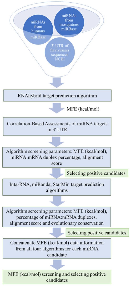
Figure 1.
Workflow diagram for identifying new miRNAs in human and mosquito cells that can interact with the 3′ UTR of flaviviruses.
2.2. Human miRNA Interactions with the Flavivirus 3′ UTRs
The RNAhybrid MFE (kcal/mol) data were used to perform correlation analyses using Spearman’s correlation coefficient to evaluate which 3′ UTRs among the diverse flaviviruses exhibited stronger affinities for host miRNAs. Correlation values close to 1 indicate a strong positive association, suggesting that consistently lower MFE values, which reflect stronger binding, are observed among certain flaviviruses. This implies the presence of shared miRNA binding sites across these viruses, highlighting consistent patterns of interaction strength across different flaviviruses.
Notable correlations were evident in human miRNA–3′ UTR interactions (Figure 2a), such as the significant correlation between DENV1 and DENV3 (0.56), and the strong correlation between JEV and DENV1 (0.71). Additionally, DENV3 exhibited a strong correlation with ZIKV (0.43), while YFV displayed positive correlations with DENV2 (0.40) and WNV1 (0.18), indicating their active involvement in these interactions. MVEV exhibited a correlation with DENV1 (0.45) and WNV2 (0.50). Remarkably, WNV1 displayed a strong correlation with JEV (0.58). In addition to being positively correlated with DENV3, ZIKV was also positively correlated with JEV, MVEV (0.46), and WNV1 (0.51). All these correlations suggest possible similarities in miRNAs with affinity for the 3′ UTRs of the flavivirus genome.
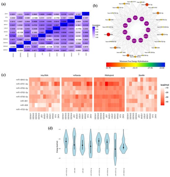
Figure 2.
Human miRNA targets in flavivirus 3′ UTRs. (a) Spearman’s correlation coefficient analysis of the MFE for miRNAs targeting at least one 3′ UTR of the eleven flaviviruses. MFE values were calculated using the RNAhybrid algorithm, revealing values lower than −20 kcal/mol. (b) Network of miRNA candidates identified by RNAhybrid as targeting the genomes of all flaviviruses, with MFE values represented on a gradient from lower values in yellow dots to higher values in red dots. The miRNAs represented by the largest red dots correspond to those with the highest MFE scores, indicating potentially stronger binding interactions. (c) miRNA candidates selected from the results of the four algorithms. MFE values are represented in kcal/mol, with lower values shown in intense red. (d) Violin plot depicting the distribution of MFE values for miRNA candidates, featuring the mean (represented by black dots) and quartiles (Q3 and Q4) as intersecting lines. The MFE distribution was calculated using data from the four algorithms. The width of each violin represents the density of data points at different MFE values, with wider sections indicating a higher concentration of values in that range.
2.3. Selecting the Optimal Human miRNA That Targets Flavivirus 3′ UTRs
We further evaluated 23 human miRNAs that exhibited binding to the 3′ UTRs of all eleven flaviviruses (Figure 2b). However, our analysis with three additional programs unexpectedly revealed that only eight human miRNAs consistently interacted with all eleven 3′ UTR sequences (Figure 2c). The target positions and corresponding 3′ UTR target sequences for these eight candidates are presented in Table 1. The distinct methodologies of each algorithm to determine the MFE in the miRNA–3′ UTR interactions allowed us to explore the MFE distribution for these eight miRNAs (Figure 2d). As anticipated, due to algorithmic differences, the MFE data showed variability. Notably, miR-6842-5p and miR-661 exhibited a greater degree of similarity in MFE predictions. This discovery emphasises the potential importance of miR-6842-5p and miR-661 in targeting the flavivirus 3′ UTR, making them compelling candidates for further research and potential therapeutic applications.

Table 1.
Position of human miRNA candidates in flavivirus genome 3′ UTRs.
2.4. Mosquito miRNA Interactions with Flavivirus 3′ UTRs
In our analysis of mosquito miRNAs and their interactions with flaviviruses, we segregated the data based on the mosquito species (Figure 3 and Figure 4). Unexpectedly, positive correlations in MFE values were observed for most viruses in both mosquito species, as shown in Figure 3a and Figure 4a. This indicates a strong affinity between mosquito miRNAs and the 3′ UTRs of these viruses. Several of these correlations were detected in both vertebrate and invertebrate organisms. For example, the correlation of DENV1 with DENV3 and DENV4 in humans and Ae. aegypti mosquitoes showed the highest correlation coefficient with DENV3 (0.56 vs. 0.59; compare Figure 2a with Figure 3a). DENV2 showed positive and significant correlation with DENV4 and YFV in both species, with the highest correlation being with DENV4 (0.46 vs. 0.61; compare Figure 2a with Figure 3a). A stronger correlation was observed between miRNAs in humans and Cu. quinquefasciatus mosquitoes (compare Figure 2a with Figure 4a). For example, WNV1 and 2 are strongly correlated with JEV, MVEV, and USUV in both organisms.
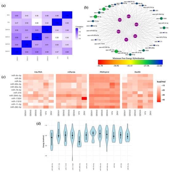
Figure 3.
Ae. aegypti miRNA targets in flavivirus 3′ UTRs. (a) Spearman’s correlation coefficient analysis of the MFE for miRNAs targeting at least one 3′ UTR of the six flaviviruses. MFE values were calculated using the RNAhybrid algorithm, revealing values lower than −20 kcal/mol. (b) Network of miRNA candidates identified by RNAhybrid as targeting the genomes of all flaviviruses, with MFE values represented on a gradient from lower values in blue dots to higher values in green dots. The miRNAs represented by the largest green dots correspond to those with the highest MFE scores, indicating potentially stronger binding interactions. (c) miRNA candidates selected from the results of the four algorithms. MFE values are represented in kcal/mol, with lower values shown in intense red. (d) Violin plot depicting the distribution of MFE values for miRNA candidates, featuring the mean (represented by black dots) and quartiles (Q3 and Q4) as intersecting lines. The MFE distribution was calculated using the results from all four algorithms. The width of each violin represents the density of data points at different MFE values, with wider sections indicating a higher concentration of values in that range.
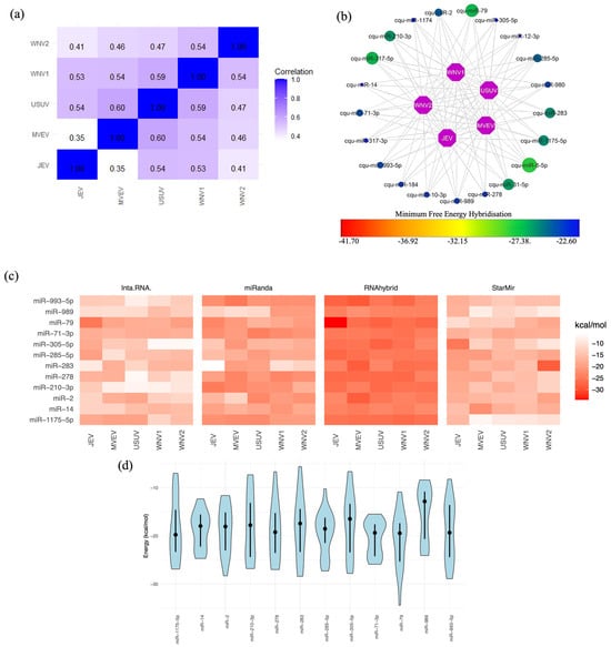
Figure 4.
Cu. quinquefasciatus miRNA targets in flavivirus 3′ UTRs. (a) Spearman’s correlation coefficient analysis of MFEs for miRNAs targeting at least one 3′ UTR of the five flaviviruses. MFE values were calculated using the RNAhybrid algorithm, revealing values lower than −20 kcal/mol. (b) Network of miRNA candidates identified by RNAhybrid as targeting the genomes of all flaviviruses, with MFE values represented on a gradient from lower values in blue dots to higher values in green dots. The miRNAs represented by the largest green dots correspond to those with the highest MFE scores, indicating potentially stronger binding interactions. (c) miRNA candidates selected from the results of the four algorithms. MFE values are represented in kcal/mol, with lower values shown in intense red. (d) Violin plot depicting the distribution of MFE values for miRNA candidates, featuring the mean (represented by black dots) and quartiles (Q3 and Q4) as intersecting lines. The MFE distribution was calculated using the results from all four algorithms. The width of each violin represents the density of data points at different MFE values, with wider sections indicating a higher concentration of values in that range.
2.5. Selecting the Optimal Mosquito miRNA That Targets Flavivirus 3′ UTRs
In Ae. aegypti, we identified 33 miRNAs that target the 3′ UTRs of six flaviviruses (Figure 3b). In Cu. quinquefasciatus, 21 miRNAs interacted with the 3′ UTRs of five flaviviruses (Figure 4b). In both cases, 12 miRNAs were predicted by the four programs to interact with the 3′ UTRs of flaviviruses (Figure 3c and Figure 4c). The target positions and corresponding 3′ UTR target sequences for these 12 miRNA candidates are presented in Table 2 for Ae. aegypti and Table 3 for Cu. quinquefasciatus. Finally, the distribution of MFEs for these miRNAs is displayed, revealing variability and, in some cases, MFE values exceeding −20 kcal/mol. This discrepancy might be attributable to the fact that the programs were not originally designed for invertebrate miRNAs. Nonetheless, the average MFEs for certain miRNAs remained below −20 kcal/mol, indicating their potential as promising candidates. Additionally, some miRNAs displayed consistent MFE predictions across different programs. Notable candidates among Ae. aegypti mosquito miRNAs include miR-9-C-5p, miR-2945-5p, miR-11924, and miR-282-5p (Figure 3d). In the case of Cu. quinquefasciatus, the standout candidate was miR-79 (Figure 4d).
Table 2.
Position of Ae. aegypti miRNA candidates in flavivirus genome 3′ UTRs.
Table 3.
Position of Cu. quinquefasciatus miRNA candidates in flavivirus genome 3′ UTRs.
3. Discussion
miRNAs are considered good therapeutic agents because they are small molecules, have an endogenous origin and flexible functions, do not induce a relevant immune response, do not have important side effects and, since their mechanism of action does not require full complementarity to the target sequence, they tolerate mutations outside the seed region [,,]. Additionally, synthetic miRNAs, contrary to cellular RNAs, are more stable and resistant to degradation by environment deleterious conditions [].
There are several studies about the participation of miRNAs in the flavivirus replicative cycle using human cell lines as a model. The most common process regulated by miRNAs during flavivirus infection is the innate immune response and inflammation and includes miR-146a-5p [] in DENV infection; miR-146a in DENV [] and ZIKV [] infections; miR-532-5p in WNV infection []; and miR-19b-3p [], miR-9-5p [], and miR-15b [] in JEV neuroinflammation. Finally, BACH1, a transcriptional repressor of HO-1 that participates in the regulation of the IFN response, is the target gene of miRNAs that are dysregulated in DENV infections, like let-7c [] and miR-155 []. miR-155 also participates in the inflammatory process during JEV infection [].
Human miRNAs also participate in other processes required for flavivirus infection, like miR-383-5p in lipid metabolism during DENV infection, which affects viral particle production []; miR-15 and -16, which increase the activity of caspases 3/7, indicating a probable relationship with apoptosis, also in DENV infection []; miR-3614-5p, which reduces DENV2 and WNV infection by inhibiting the action of adenosine deaminase on RNA 1 (ADAR1), a factor that promotes viral infectivity in early stages of infection []; miR-532-5p, which reduces the expression of TAB3, a factor involved in cell survival, proliferation, differentiation, embryonic development, inflammation, and carcinogenesis, and SESTD1, a phospholipid-binding protein essential for the efficient activation of the calcium channels TRPC4 and TRPC5, which is required for efficient propagation of WNV []; miR-33a-5p, which has an inhibitory effect on viral replication by silencing the EEF1A1 factor, a component of the JEV replication complex that avoids NS3 and NS5 proteasome degradation []; and, finally, miR-204-5p and miR-103a-3p, whose expression is induced by ZIKV E protein [] and infection [], respectively. miR-204-5p downregulates WNT2, a growth factor that is involved in brain development [], and miR-103a-3p promotes the phosphorylation of p38 MAPK and HSP2 through the inhibition of OTUD4 [].
The information about the participation of miRNAs in flavivirus infections in mosquitoes is more limited. Again, the innate immune response is regulated by miRNAs like miR-375 in Ae. aegypti mosquitoes infected with DENV2 []. miR-252 is downregulated in Ae. albopictus mosquitoes infected with DENV2, and this miRNA has a target sequence in the viral E protein, suggesting an antiviral role []. Finally, miRNA-240-5p is specifically downregulated in the Ae. albopictus cell line C6/36 when infected by WNV in a time-dependent manner. This miRNA is involved in the translation regulation of m41 FtsH, an ATP-dependent metalloprotease that localises to the inner membrane of mitochondria and that is responsible for the degradation of misfolded proteins. The silencing of this protein results in the reduction of both the viral titre and the quantity of viral genomes in infected cells, indicating its relevance in the WNV replicative cycle [].
Since miRNAs play important roles in viral infections, they have been seriously considered for the treatment of several viral infections, and they have been tested in in vivo preclinical trials []. For example, the design of synthetic miRNAs against the 3′ and 5′ UTRs and ORF9 of Severe Acute Respiratory Syndrome Coronavirus 2 (SARS-CoV2) has been proposed to inhibit the translation process []. A similar approach has been used to reduce coxsackievirus B3 (CVB3) infection in HeLa cells. In this case, two artificial miRNAs (AmiR) against the Y loop of the viral 3′ UTR, delivered to HeLa cells by folate-mediated internalisation via the folate receptor, demonstrated their antiviral effect []. The transfection of miR-199a-3p and miR-210 into HepG2 2.2.15 cells, which target the S and P regions of the genome of Hepatitis B virus (HBV), reduced the expression of the S antigen (HBsAg) and viral replication [].
Finally, miRNAs have been proposed for the design of viral vaccines. The insertion of an MRE into the ORF of the nucleoprotein of influenza virus A H1N1 and H5NI allowed the generation of an influenza vaccine that displays an attenuated phenotype in mice but not in eggs []. The insertion of MREs for miR-133 and miR-206 into the 5′ UTR of CVB3 resulted in the generation of engineered viruses that could replicate efficiently in HeLa but not in TE671 cells or mice cardiac muscle. Additionally, these viruses were able to induce neutralising anti-CVB3 antibodies and protect against wild-type virus challenge in mice []. Similar results were obtained with engineered viruses that included MREs specific for different tissues (miR-206, specific for muscle; miR-29a, specific for pancreas; and miR-124-3p, abundant in the central nervous system) [].
Bioinformatic analysis is a time-efficient approach for approximating miRNA interactions with 3′ UTRs of the viral genome []. This computational approach has been successfully applied to various RNA viruses, including influenza C virus [] and DENV []. Our research extends the analysis to various mosquito-borne flaviviruses, including both their vectors and human hosts. The most crucial step for gene or mRNA silencing is the effective hybridisation and heteroduplex formation between miRNAs and 3′ UTRs []. Seed types (8mer, 7mer-A1, and 7mer-m8) have been noted to be particularly recognisable by the RNA-induced silencing complex (RISC), increasing the degree of gene silencing [,,].
The target prediction algorithms employed in this study are considered the most suitable for achieving the effective identification of miRNA binding sites on the 3′ UTRs of the flavivirus genome. The MFE and seed region of the miRNA–target hybrid are consistently recognised as the most widely exploited parameters in all these miRNA target prediction algorithms [,,,,]; therefore, these parameters were selected to process the data with the utmost rigor to avoid false-positive candidates. The information obtained from the four algorithms was concatenated to identify the miRNAs with the highest scores and the potential for binding to the 3′ UTRs.
Among the identified interactions, the human miRNAs miR-6842 and miR-661 demonstrated robust targeting of flavivirus 3′ UTRs. Notably, miR-661, previously identified as a promising miRNA in interactions with all four DENV serotypes [], retained its importance in our study. Additionally, miR-484, miR-744 [], and miR-133a [] have been reported to possess specific target sequences within the DENV 3′ UTR. While our initial analysis detected these miRNAs as targets of at least one flavivirus (see Supplementary Table S1), they did not exhibit interactions with all 11 proposed flaviviruses. This finding underscores the potential of other miRNAs with broader binding capabilities to exert effects against these viruses.
However, all these predictions should be validated by experimental approaches. For example, miR-532 has two putative binding sites in the WNV genome predicted by the RNAhybrid algorithm that are not functional in vitro []. For these validations, a common approach is to clone the putative target sequence in the 3′ UTR of a luciferase gene to perform a double-luciferase reporter gene assay [,,,,,,,]. Then, the antiviral effect of the miRNA should be evaluated during a flavivirus infection. For that, a miRNA mimic is transfected into a suitable cell line and then infected with a flavivirus. There are several suitable human cell lines that can be used for this purpose, like Huh-7 [,,,,,], THP1 [], HepG2 [], HEK293 [,], HCM3 [,], U251 [,], SH-SY5Y [], and A549 cells []. The C6/36 cell line from Ae albopictus is a suitable model for performing these experiments with mosquito miRNAs []. The infection can be evaluated by determining viral titres by the plaque assay [,,,,], the viral genome copy number by RT-qPCR [,,,,], and viral protein synthesis by Western blotting [,,,,,,].
However, our mosquito-focused analysis faced challenges, because most algorithms have been created for mammalian miRNA interactions, and mosquito miRNA processing differs substantially [,,]. Despite these limitations, the positive MFE correlations observed for the interactions between mosquito miRNAs and the 3′ UTRs of flaviviruses are particularly intriguing, indicating a substantial affinity between mosquito miRNAs and the 3′ UTRs of various flaviviruses. In contrast, human interactions demonstrated negative correlations when evaluating different flaviviruses. These differences may have significant biological implications for mosquito vectors and warrant further experimental exploration. Additionally, we identified miRNAs that potentially interact with Aedes and Culex mosquitoes, including miR-9c, miR-2945-5p, miR-11924, and miR-282-5p, while miR-79 emerged as a noteworthy candidate in the context of flavivirus infections. Given that not all algorithms were created with insects in mind, it is important to highlight that future work can further develop this approach to define more precisely the interactions of mosquito miRNAs with the 3′ UTRs.
On the other hand, there are no experimental reports of the participation of these miRNAs in flavivirus infections, and only human miR-661 has been determined to be notably increased in the serum of patients with herpes zoster infection. Using TargetScan (Version 7.1) software, several target genes of this miRNA were identified; these were associated with the nervous and immune systems, but none of them were validated experimentally []. However, the functions of some of these miRNAs have been reported. For example, in invertebrates (see Table 4), miR-9c is involved in the development of the fruit fly Drosophila melanogaster [,,] and the mud crab Scylla paramamosain [,]; miR-282-5p is involved in the moulting process of the silkworm Bombyx mori []; and finally, miR-79, the orthologue of miR-9 in humans [], participates in cell proliferation and development in several organisms such as the fruit fly [,], worm [], silkworm [], and sea cucumber []. Interestingly, it has been reported that miR-79 is overexpressed in ISE6 cells from the tick I. scapularis infected with the bacterium Anaplasma phagocytophilum. This miRNA suppresses the expression of Roundabout protein 2 (Robo2), a molecule involved in the proinflammatory response, thereby promoting infection []. Additionally, miR-79 has been shown to be upregulated in exosomes from patients with chronic rhinosinusitis with nasal polyps [].
Table 4.
Functions of miRNAs detected in mosquitoes that interact with 3′ UTR of flaviviruses.
More information is available for human miRNAs. For example, miR-6842-5p is involved in glucose metabolism through the inhibition of AKT2 and has a negative effect on proliferation and migration in endothelial cells during persistent high-glucose exposure, and miR-661, one of the most promising miRNAs identified in this work, has been implicated in several types of cancer as well as some diseases, such as diabetes mellitus 2 and Alzheimer’s disease (see Table 5). The present work identified new potential functions of these miRNAs through the proposed computational workflow. These miRNAs have the potential to be utilised as tools in the development of antiviral therapies, as both miRNAs have binding sites in the 3′ UTRs of flaviviruses []. These regions play crucial role in the post-transcriptional repression and decay of RNAs [,,,], so defining their functions within virus infections will have important implications for future therapeutic endeavours.
Table 5.
Functions of miRNAs detected in humans that interact with the 3′ UTR of flaviviruses.
4. Materials and Methods
4.1. Retrieval of the Viral Genome and Mature miRNA Sequences
The genomic sequences of the flaviviruses DENV1 (accession NC_001477.1), DENV2 (accession NC_001474.2), DENV3 (accession NC_001475.2), DENV4 (accession NC_002640.1), WNV1 (accession NC_009942.1), WNV2 (accession NC_001563.2), ZIKV (accession NC_035889.1), YFV (accession NC_002031.1), JEV (accession NC_001437.1), MVEV (accession NC_000943.1), and USUV (accession NC_006551.1) were retrieved from the National Center for Biotechnology Information (NCBI) GenBank platform “https://www.ncbi.nlm.nih.gov/ (accesed on 27 August 2024)”. FASTA-formatted genomic sequences of flaviviruses were processed using bedtools “https://bedtools.readthedocs.io/en/latest/ (accesed on 27 August 2024)” to extract their 3′ UTRs.
miRNA sequences from humans (Homo sapiens; hsa.gff3); two mosquito species, Aedes aegypti (aae.gff3) and Culex quinquefasciatus (cfa.gff3); and Caenorhabditis elegans (cel.ggf3) were obtained from the miRBase database “https://www.mirbase.org (accesed on 27 August 2024).
4.2. miRNA Target Site Algorithms
To identify endogenous miRNA target sites in the 3′ UTR sequences of different flaviviruses, four reliable target prediction algorithms were employed in this study:
- RNAhybrid: This algorithm calculates the minimum free energy (MFE) for miRNA–target hybrids using thermodynamic principles. It integrates helix parameters and loop constraints and accounts for G:U wobbles within the seed region. A favourable free energy for hybridisation is typically approximately −20 kcal/mol [].
- Inta-RNA: Using an enhanced scoring system, this algorithm predicts RNA–RNA interactions. It assesses the thermodynamic stability of interaction duplexes, site accessibility, and seed region attributes. Interactions are predicted when both the total energy and hybridisation energy are less than zero, with scores greater than 140 indicating optimal interactions [].
- miRanda: This algorithm identifies miRNA–mRNA target duplexes, accommodating mismatches, gaps, and wobble base pairings. It extends beyond the seed region to predict all possible miRNA target sites. We adjusted the threshold binding energy to −20 kcal/mol, set a score threshold of 100, and applied a gap-opening penalty (GOP) of −9 and a gap-extension penalty (GEP) of −4 [].
- StarMir: Uses miRNA binding data from CLIP studies in non-linear logistic prediction models. It excels at identifying seeded and unseeded target sites by considering thermodynamic, structural, and sequence features from SFold 2.2. The algorithm considers factors, such as the type of seed and site accessibility, and incorporates several parameters, including the Gibbs free energy change of the miRNA–mRNA target hybrid (ΔGhybrid) [], the miRNA–mRNA target hybridisation (ΔGnucl), the total energy change of the hybridisation (ΔGtotal), and the LogitProb score [,].
4.3. Correlation-Based Assessments of miRNA Targets in the 3′ UTR
Using RNAhybrid “https://bibiserv.cebitec.uni-bielefeld.de/rnahybrid (accesed on 27 August 2024)”, 3′ UTR sequences were employed to predict the targets of host miRNAs from both humans and the mosquito species Ae. aegypti and Cu. quinquefasciatus. To assess which 3′ UTRs of flaviviruses exhibit a stronger affinity for host microRNAs, correlation analyses were performed based on the MFE hybridisation (kcal/mol). Spearman’s correlation coefficient was used to compare the interactions between miRNAs and the 3′ UTRs of the different flaviviruses.
Correlations approaching 1 indicated positive associations, suggesting that a lower MFE led to stronger interactions with specific viruses, suggesting shared miRNA binding sites among them. Conversely, correlations near −1 implied negative associations, indicating that a higher MFE resulted in weaker interactions between the miRNA and the correlated viruses, likely due to a lack of common binding sites. A correlation close to zero indicated an absence of a clear relationship between the MFE and miRNA–3′ UTR interactions.
4.4. Identification of miRNA Binding Sites
Following the initial data grouping, miRNAs that exhibited binding to the 3′ UTRs of all flaviviruses were subjected to further analyses. Typically, prevailing target prediction algorithms initiate a sequence search on 3′ UTRs, seeking regions with complementarity to miRNAs, ideally at their seed sites. However, this initial phase often results in thousands of potential target sites, accompanied by many false positives. To address this issue, most algorithms incorporate additional features such as MFE filters, the % mRNA–miRNA duplex complementarity, and evolutionary conservation to increase the specificity and to reduce false positives. Taking advantage of each algorithm, the candidates obtained from RNAhybrid were evaluated for their affinity for the 3′ UTRs of flaviviruses using three distinct algorithms: Inta-RNA “https://rna.informatik.uni-freiburg.de (accesed on 27 August 2024)”, miRanda “http://multimir.ucdenver.edu/ (accesed on 27 August 2024)”, and StarMir “https://sfold.wadsworth.org/cgi-bin/index.pl (accesed on 27 August 2024)”.
Positive candidates from the four algorithms were grouped, and miRNAs targeting all the 3′ UTRs of flaviviruses were selected as potential candidates. Finally, using MFE data information, we concatenated the information from each miRNA candidate to identify the optimal miRNA capable of binding the 3′ UTR of flavivirus genomes.
4.5. Computational Environments and Software
All data processing in this study was conducted in R and UNIX environments using specific packages. The miRNA network for the 3′ UTR of flavivirus genomes was created using Cytoscape 3.10.1 “https://cytoscape.org/ (accesed on 27 August 2024)”.
5. Conclusions
An analysis of human interactions revealed promising candidates, namely miR-6842 and miR-661, for the therapeutic targeting of flavivirus 3′ UTRs. In mosquito miRNA–flavivirus interactions, positive correlations suggest a strong affinity, whereas human interactions with various flaviviruses display negative correlations. Potential mosquito miRNA candidates, including miR-9-C, miR-2945-5p, miR-11924, miR-282-5p, and miR-79, warrant further exploration, offering the potential for viral transmission control strategies.
Supplementary Materials
The following supporting information can be downloaded at: https://www.mdpi.com/article/10.3390/ijms251810135/s1.
Author Contributions
Conceptualisation, R.G.A.-B. and J.S.S.-B.; methodology, R.G.A.-B.; software, R.G.A.-B.; formal analysis, R.G.A.-B.; investigation, R.G.A.-B. and J.S.S.-B.; writing—original draft preparation, R.G.A.-B. and J.S.S.-B.; writing—review and editing, R.G.A.-B. and J.S.S.-B.; funding acquisition, J.S.S.-B. All authors have read and agreed to the published version of the manuscript.
Funding
This research was funded by SECRETARIA DE INVESTIGACIÓN Y POSGRADO of INSTITUTO POLITÉCNICO NACIONAL (grant number SIP 20242215).
Institutional Review Board Statement
Not applicable.
Informed Consent Statement
Not applicable.
Data Availability Statement
Data generated or analysed during this study are included in this article. Further inquiries can be directed to the corresponding authors.
Acknowledgments
We thank the Instituto Politécnico Nacional and Centro de Investigación y de Estudios Avanzados del IPN for their support. Salas-Benito has fellowships from the Comisión de Operación y Fomento de Actividades Académicas (COFAA) and Estimulo al Desempeño a los Investigadores (EDI) of Instituto Politécnico Nacional. Avila-Bonilla and Salas-Benito have fellowships from the Sistema Nacional de Investigadoras e Investigadores (SNII) of Consejo Nacional de Humanidades, Ciencias y Tecnologías (Conahcyt).
Conflicts of Interest
The authors declare no conflicts of interest.
References
- van den Elsen, K.; Quek, J.P.; Luo, D. Molecular Insights into the Flavivirus Replication Complex. Viruses 2021, 13, 956. [Google Scholar] [CrossRef] [PubMed]
- Ochsenreiter, R.; Hofacker, I.L.; Wolfinger, M.T. Functional RNA Structures in the 3′ UTR of Tick-Borne, Insect-Specific and No-Known-Vector Flaviviruses. Viruses 2019, 11, 298. [Google Scholar] [CrossRef] [PubMed]
- Harapan, H.; Michie, A.; Sasmono, R.T.; Imrie, A. Dengue: A Minireview. Viruses 2020, 12, 829. [Google Scholar] [CrossRef] [PubMed]
- Markoff, L. 5′- and 3′-noncoding regions in flavivirus RNA. Adv. Virus Res. 2003, 59, 177–228. [Google Scholar] [CrossRef]
- Romero, T.A.; Tumban, E.; Jun, J.; Lott, W.B.; Hanley, K.A. Secondary structure of dengue virus type 4 3′ untranslated region: Impact of deletion and substitution mutations. J. Gen. Virol. 2006, 87, 3291–3296. [Google Scholar] [CrossRef]
- Wei, Y.; Qin, C.; Jiang, T.; Li, X.; Zhao, H.; Liu, Z.; Deng, Y.; Liu, R.; Chen, S.; Yu, M.; et al. Translational regulation by the 3′ untranslated region of the dengue type 2 virus genome. Am. J. Trop. Med. Hyg. 2009, 81, 817–824. [Google Scholar] [CrossRef]
- Ng, W.C.; Soto-Acosta, R.; Bradrick, S.S.; Garcia-Blanco, M.A.; Ooi, E.E. The 5′ and 3′ Untranslated Regions of the Flaviviral Genome. Viruses 2017, 9, 137. [Google Scholar] [CrossRef]
- Shang, R.; Lee, S.; Senavirathne, G.; Lai, E.C. microRNAs in action: Biogenesis, function and regulation. Nat. Rev. 2023, 24, 816–833. [Google Scholar] [CrossRef]
- Maurin, T.; Cazalla, D.; Yang, S.; Jr Bortolamiol-Becet, D.; Lai, E.C. RNase III-independent microRNA biogenesis in mammalian cells. RNA 2012, 18, 2166–2173. [Google Scholar] [CrossRef][Green Version]
- Meister, G.; Landthaler, M.; Patkaniowska, A.; Dorsett, Y.; Teng, G.; Tuschl, T. Human Argonaute2 mediates RNA cleavage targeted by miRNAs and siRNAs. Mol. Cell 2004, 15, 185–197. [Google Scholar] [CrossRef]
- Gebert, L.F.R.; MacRae, I.J. Regulation of microRNA function in animals. Nat. Rev. 2019, 20, 21–37. [Google Scholar] [CrossRef] [PubMed]
- Betel, D.; Koppal, A.; Agius, P.; Sander, C.; Leslie, C. Comprehensive modeling of microRNA targets predicts functional non-conserved and non-canonical sites. Genome Biol. 2010, 11, R90. [Google Scholar] [CrossRef] [PubMed]
- Bartel, D.P. MicroRNAs: Target recognition and regulatory functions. Cell 2009, 136, 215–233. [Google Scholar] [CrossRef] [PubMed]
- Schirle, N.T.; Sheu-Gruttadauria, J.; MacRae, I.J. Structural basis for microRNA targeting. Science 2014, 346, 608–613. [Google Scholar] [CrossRef] [PubMed]
- Guo, H.; Ingolia, N.T.; Weissman, J.S.; Bartel, D.P. Mammalian microRNAs predominantly act to decrease target mRNA levels. Nature 2010, 466, 835–840. [Google Scholar] [CrossRef]
- Moore, M.J.; Scheel, T.K.; Luna, J.M.; Park, C.Y.; Fak, J.J.; Nishiuchi, E.; Rice, C.M.; Darnell, R.B. miRNA-target chimeras reveal miRNA 3′-end pairing as a major determinant of Argonaute target specificity. Nat. Commun. 2015, 6, 8864. [Google Scholar] [CrossRef]
- Helwak, A.; Kudla, G.; Dudnakova, T.; Tollervey, D. Mapping the human miRNA interactome by CLASH reveals frequent noncanonical binding. Cell 2013, 153, 654–665. [Google Scholar] [CrossRef]
- Avila-Bonilla, R.G.; Salas-Benito, J.S. Interactions of host miRNAs in the flavivirus 3´UTR genome: From bioinformatics predictions to practical approaches. Front. Cell. Infect. Microbiol. 2022, 12, 976843. [Google Scholar] [CrossRef]
- Castrillón-Betancur, J.C.; Urcuqui-Inchima, S. Overexpression of miR-484 and miR-744 in Vero cells alters Dengue virus replication. Mem. Inst. Oswaldo Cruz 2017, 112, 281–291. [Google Scholar] [CrossRef]
- Castillo, J.A.; Castrillón, J.C.; Diosa-Toro, M.; Betancur, J.G.; St Laurent, G., 3rd; Smit, J.M.; Urcuqui-Inchima, S. Complex interaction between dengue virus replication and expression of miRNA-133a. BMC Infect. Dis. 2016, 16, 29. [Google Scholar] [CrossRef]
- Cai, W.; Pan, Y.; Cheng, A.; Wang, M.; Yin, Z.; Jia, R. Regulatory Role of Host MicroRNAs in Flaviviruses Infection. Front. Microbiol. 2022, 13, 869441. [Google Scholar] [CrossRef] [PubMed]
- Lee, T.C.; Lin, Y.L.; Liao, J.T.; Su, C.M.; Lin, C.C.; Lin, W.P.; Liao, C.L. Utilizing liver-specific microRNA-122 to modulate replication of dengue virus replicon. Biochem. Biophys. Res. Commun. 2010, 396, 596–601. [Google Scholar] [CrossRef] [PubMed]
- Pham, A.M.; Langlois, R.A.; TenOever, B.R. Replication in cells of hematopoietic origin is necessary for Dengue virus dissemination. PLoS Pathog. 2012, 8, e1002465. [Google Scholar] [CrossRef] [PubMed]
- Yen, L.C.; Lin, Y.L.; Sung, H.H.; Liao, J.T.; Tsao, C.H.; Su, C.M.; Lin, C.K.; Liao, C.L. Neurovirulent flavivirus can be attenuated in mice by incorporation of neuron-specific microRNA recognition elements into viral genome. Vaccine 2013, 31, 5915–5922. [Google Scholar] [CrossRef] [PubMed]
- Hum, C.; Loiselle, J.; Ahmed, N.; Shaw, T.A.; Toudic, C.; Pezacki, J.P. MicroRNA Mimics or Inhibitors as Antiviral Therapeutic Approaches Against COVID-19. Drugs 2021, 81, 517–531. [Google Scholar] [CrossRef] [PubMed]
- Bauer, A.N.; Majumdar, N.; Williams, F.; Rajput, S.; Pokhrel, L.R.; Cook, P.P.; Akula, S.M. MicroRNAs: Small but Key Players in Viral Infections and Immune Responses to Viral Pathogens. Biology 2023, 12, 1334. [Google Scholar] [CrossRef]
- Xu, T.; Li, L.X.; Jia, Y.; Wu, Q.; Zhu, W.; Xu, Z.; Zheng, B.; Lu, X. One microRNA has the potential to target whole viral mRNAs in a given human coronavirus. Front. Microbiol. 2022, 13, 1035044. [Google Scholar] [CrossRef]
- Park, J.H.; Moon, J. Conserved 3′ UTR of Severe Acute Respiratory Syndrome Coronavirus 2: Potential Therapeutic Targets. Front. Genet. 2022, 13, 893141. [Google Scholar] [CrossRef]
- Baig, M.S.; Krishnan, A. A bioinformatics approach to investigate serum and hematopoietic cell-specific therapeutic microRNAs targeting the 3′ UTRs of all four Dengue virus serotypes. Pathog. Dis. 2021, 79, ftab050. [Google Scholar] [CrossRef]
- El-Nabi, S.H.; Elhiti, M.; El-Sheekh, M. A new approach for COVID-19 treatment by micro-RNA. Med. Hypotheses 2020, 143, 110203. [Google Scholar] [CrossRef]
- Hemida, M.G.; Ye, X.; Thair, S.; Yang, D. Exploiting the therapeutic potential of microRNAs in viral diseases: Expectations and limitations. Mol. Diagn. Ther. 2010, 14, 271–282. [Google Scholar] [CrossRef] [PubMed]
- Ye, X.; Liu, Z.; Hemida, M.G.; Yang, D. Targeted delivery of mutant tolerant anti-coxsackievirus artificial microRNAs using folate conjugated bacteriophage Phi29 pRNA. PLoS ONE 2011, 6, e21215. [Google Scholar] [CrossRef] [PubMed]
- Pradhan, A.; Aneja, A.; Ghosh, S.; Devvanshi, H.C.D.; Sahu, R.; Ross, C.; Kshetrapal, P.; Maitra, A.; Das, S. Association of exosomal miR-96-5p and miR-146a-5p with the disease severity in dengue virus infection. J. Med. Virol. 2023, 95, e28614. [Google Scholar] [CrossRef] [PubMed]
- Wu, S.; He, L.; Li, Y.; Wang, T.; Feng, L.; Jiang, L.; Zhang, P.; Huang, X. miR-146a facilitates replication of dengue virus by dampening interferon induction by targeting TRAF6. J. Infect. 2013, 67, 329–341. [Google Scholar] [CrossRef] [PubMed]
- Shukla, A.; Rastogi, M.; Singh, S.K. Zika virus NS1 suppresses the innate immune responses via miR-146a in human microglial cells. Int. J. Biol. Macromol. 2021, 193 Pt B, 2290–2296. [Google Scholar] [CrossRef]
- Slonchak, A.; Shannon, R.P.; Pali, G.; Khromykh, A.A. Human MicroRNA miR-532-5p Exhibits Antiviral Activity against West Nile Virus via Suppression of Host Genes SESTD1 and TAB3 Required for Virus Replication. J. Virol. 2015, 90, 2388–2402. [Google Scholar] [CrossRef]
- Ashraf, U.; Zhu, B.; Ye, J.; Wan, S.; Nie, Y.; Chen, Z.; Cui, M.; Wang, C.; Duan, X.; Zhang, H.; et al. MicroRNA-19b-3p Modulates Japanese Encephalitis Virus-Mediated Inflammation via Targeting RNF11. J. Virol. 2016, 90, 4780–4795. [Google Scholar] [CrossRef]
- Sharma, S.; Majumdar, A.; Basu, A. Regulation of Onecut2 by miR-9-5p in Japanese encephalitis virus infected neural stem/progenitor cells. Microbiol. Spectr. 2024, 12, e0323823. [Google Scholar] [CrossRef]
- Zhu, B.; Ye, J.; Nie, Y.; Ashraf, U.; Zohaib, A.; Duan, X.; Fu, Z.F.; Song, Y.; Chen, H.; Cao, S. MicroRNA-15b Modulates Japanese Encephalitis Virus-Mediated Inflammation via Targeting RNF125. J. Immunol. 2015, 195, 2251–2262. [Google Scholar] [CrossRef]
- Escalera-Cueto, M.; Medina-Martínez, I.; del Angel, R.M.; Berumen-Campos, J.; Gutiérrez-Escolano, A.L.; Yocupicio-Monroy, M. Let-7c overexpression inhibits dengue virus replication in human hepatoma Huh-7 cells. Virus Res. 2015, 196, 105–112. [Google Scholar] [CrossRef]
- Su, Y.C.; Huang, Y.F.; Wu, Y.W.; Chen, H.F.; Wu, Y.H.; Hsu, C.C.; Hsu, Y.C.; Lee, J.C. MicroRNA-155 inhibits dengue virus replication by inducing heme oxygenase-1-mediated antiviral interferon responses. FASEB J. 2020, 34, 7283–7294. [Google Scholar] [CrossRef] [PubMed]
- Pareek, S.; Roy, S.; Kumari, B.; Jain, P.; Banerjee, A.; Vrati, S. MiR-155 induction in microglial cells suppresses Japanese encephalitis virus replication and negatively modulates innate immune responses. J. Neuroinflamm. 2014, 11, 97. [Google Scholar] [CrossRef] [PubMed]
- Ahmed, N.; Ahmed, N.; Pezacki, J.P. miR-383 Regulates Hepatic Lipid Homeostasis and Response to Dengue Virus Infection. ACS Infect. Dis. 2022, 8, 928–941. [Google Scholar] [CrossRef] [PubMed]
- Casseb, S.M.M.; Melo, K.F.L.; Carvalho, C.A.M.; Santos, C.R.D.; Franco, E.C.S.; Vasconcelos, P.F.D.C. Experimental Dengue Virus Type 4 Infection Increases the Expression of MicroRNAs-15/16, Triggering a Caspase-Induced Apoptosis Pathway. Curr. Issues Mol. Biol. 2023, 45, 4589–4599. [Google Scholar] [CrossRef] [PubMed]
- Diosa-Toro, M.; Echavarría-Consuegra, L.; Flipse, J.; Fernández, G.J.; Kluiver, J.; van den Berg, A.; Urcuqui-Inchima, S.; Smit, J.M. MicroRNA profiling of human primary macrophages exposed to dengue virus identifies miRNA-3614-5p as antiviral and regulator of ADAR1 expression. PLoS Negl. Trop. Dis. 2017, 11, e0005981. [Google Scholar] [CrossRef]
- Chen, Z.; Ye, J.; Ashraf, U.; Li, Y.; Wei, S.; Wan, S.; Zohaib, A.; Song, Y.; Chen, H.; Cao, S. MicroRNA-33a-5p Modulates Japanese Encephalitis Virus Replication by Targeting Eukaryotic Translation Elongation Factor 1A1. J. Virol. 2016, 90, 3722–3734. [Google Scholar] [CrossRef]
- Bhagat, R.; Rajpara, P.; Kaur, G.; Gupta, K.; Seth, P. Zika virus E protein dysregulate mir-204/WNT2 signalling in human fetal neural stem cells. Brain Res. Bull. 2021, 176, 93–102. [Google Scholar] [CrossRef]
- Ye, H.; Kang, L.; Yan, X.; Li, S.; Huang, Y.; Mu, R.; Duan, X.; Chen, L. MiR-103a-3p Promotes Zika Virus Replication by Targeting OTU Deubiquitinase 4 to Activate p38 Mitogen-Activated Protein Kinase Signaling Pathway. Front. Microbiol. 2022, 13, 862580. [Google Scholar] [CrossRef]
- Hussain, M.; Walker, T.; O’Neill, S.L.; Asgari, S. Blood meal induced microRNA regulates development and immune associated genes in the Dengue mosquito vector, Aedes aegypti. Insect Biochem. Mol. Biol. 2013, 43, 146–152. [Google Scholar] [CrossRef]
- Yan, H.; Zhou, Y.; Liu, Y.; Deng, Y.; Chen, X. miR-252 of the Asian tiger mosquito Aedes albopictus regulates dengue virus replication by suppressing the expression of the dengue virus envelope protein. J. Med. Virol. 2014, 86, 1428–1436. [Google Scholar] [CrossRef]
- Slonchak, A.; Hussain, M.; Torres, S.; Asgari, S.; Khromykh, A.A. Expression of mosquito microRNA Aae-miR-2940-5p is downregulated in response to West Nile virus infection to restrict viral replication. J. Virol. 2014, 88, 8457–8467. [Google Scholar] [CrossRef] [PubMed]
- Procyk, G.; Grodzka, O.; Procyk, M.; Gąsecka, A.; Głuszek, K.; Wrzosek, M. MicroRNAs in Myocarditis-Review of the Preclinical In Vivo Trials. Biomedicines 2023, 11, 2723. [Google Scholar] [CrossRef] [PubMed]
- Zhang, G.L.; Li, Y.X.; Zheng, S.Q.; Liu, M.; Li, X.; Tang, H. Suppression of hepatitis B virus replication by microRNA-199a-3p and microRNA-210. Antivir. Res. 2010, 88, 169–175. [Google Scholar] [CrossRef] [PubMed]
- Perez, J.T.; Pham, A.M.; Lorini, M.H.; Chua, M.A.; Steel, J.; tenOever, B.R. MicroRNA-mediated species-specific attenuation of influenza A virus. Nat. Biotechnol. 2009, 27, 572–576. [Google Scholar] [CrossRef]
- He, F.; Yao, H.; Wang, J.; Xiao, Z.; Xin, L.; Liu, Z.; Ma, X.; Sun, J.; Jin, Q.; Liu, Z. Coxsackievirus B3 engineered to contain microRNA targets for muscle-specific microRNAs displays attenuated cardiotropic virulence in mice. J. Virol. 2015, 89, 908–916. [Google Scholar] [CrossRef]
- Xiao, Z.; He, F.; Feng, M.; Liu, Z.; Liu, Z.; Li, S.; Wang, W.; Yao, H.; Wu, J. Engineered coxsackievirus B3 containing multiple organ-specific miRNA targets showed attenuated viral tropism and protective immunity. Infect. Genet. Evol. J. Mol. Epidemiol. Evol. Genet. Infect. Dis. 2022, 103, 105316. [Google Scholar] [CrossRef]
- Hassan, M.; Iqbal, M.S.; Naqvi, S.; Alashwal, H.; Moustafa, A.A.; Kloczkowski, A. Prediction of Site Directed miRNAs as Key Players of Transcriptional Regulators Against Influenza C Virus Infection Through Computational Approaches. Front. Mol. Biosci. 2022, 9, 866072. [Google Scholar] [CrossRef]
- Witkos, T.M.; Koscianska, E.; Krzyzosiak, W.J. Practical Aspects of microRNA Target Prediction. Curr. Mol. Med. 2011, 11, 93–109. [Google Scholar] [CrossRef]
- Saito, T.; Saetrom, P. MicroRNAs--targeting and target prediction. New Biotechnol. 2010, 27, 243–249. [Google Scholar] [CrossRef]
- Maragkakis, M.; Alexiou, P.; Papadopoulos, G.L.; Reczko, M.; Dalamagas, T.; Giannopoulos, G.; Goumas, G.; Koukis, E.; Kourtis, K.; Simossis, V.A.; et al. Accurate microRNA target prediction correlates with protein repression levels. BMC Bioinform. 2009, 10, 295. [Google Scholar] [CrossRef]
- Grimson, A.; Farh, K.K.; Johnston, W.K.; Garrett-Engele, P.; Lim, L.P.; Bartel, D.P. MicroRNA targeting specificity in mammals: Determinants beyond seed pairing. Mol. Cell 2007, 27, 91–105. [Google Scholar] [CrossRef] [PubMed]
- Rehmsmeier, M.; Steffen, P.; Hochsmann, M.; Giegerich, R. Fast and effective prediction of microRNA/target duplexes. RNA 2004, 10, 1507–1517. [Google Scholar] [CrossRef] [PubMed]
- Mann, M.; Wright, P.R.; Backofen, R. IntaRNA 2.0: Enhanced and customizable prediction of RNA-RNA interactions. Nucleic Acids Res. 2017, 45, W435–W439. [Google Scholar] [CrossRef] [PubMed]
- Enright, A.J.; John, B.; Gaul, U.; Tuschl, T.; Sander, C.; Marks, D.S. MicroRNA targets in Drosophila. Genome Biol. 2003, 5, R1. [Google Scholar] [CrossRef] [PubMed]
- Long, D.; Lee, R.; Williams, P.; Chan, C.Y.; Ambros, V.; Ding, Y. Potent effect of target structure on microRNA function. Nat. Struct. Mol. Biol. 2007, 14, 287–294. [Google Scholar] [CrossRef]
- Rennie, W.; Liu, C.; Carmack, C.S.; Wolenc, A.; Kanoria, S.; Lu, J.; Long, D.; Ding, Y. STarMir: A web server for prediction of microRNA binding sites. Nucleic Acids Res. 2014, 42, W114–W118. [Google Scholar] [CrossRef]
- Avila-Bonilla, R.G.; Yocupicio-Monroy, M.; Marchat, L.A.; Pérez-Ishiwara, D.G.; Cerecedo-Mercado, D.A.; Del Ángel, R.M.; Salas-Benito, J.S. miR-927 has pro-viral effects during acute and persistent infection with dengue virus type 2 in C6/36 mosquito cells. J. Gen. Virol. 2020, 101, 825–839. [Google Scholar] [CrossRef]
- Asgari, S. Role of microRNAs in arbovirus/vector interactions. Viruses 2014, 6, 3514–3534. [Google Scholar] [CrossRef]
- Lucas, K.; Raikhel, A.S. Insect microRNAs: Biogenesis, expression profiling and biological functions. Insect Biochem. Mol. Biol. 2013, 43, 24–38. [Google Scholar] [CrossRef]
- Ylla, G.; Fromm, B.; Piulachs, M.D.; Belles, X. The microRNA toolkit of insects. Sci. Rep. 2016, 6, 37736. [Google Scholar] [CrossRef]
- Li, X.; Huang, Y.; Zhang, Y.; He, N. Evaluation of microRNA Expression in Patients with Herpes Zoster. Viruses 2016, 8, 326. [Google Scholar] [CrossRef] [PubMed]
- Busto, G.U.; Guven-Ozkan, T.; Fulga, T.A.; Van Vactor, D.; Davis, R.L. microRNAs That Promote or Inhibit Memory Formation in Drosophila melanogaster. Genetics 2015, 200, 569–580. [Google Scholar] [CrossRef] [PubMed]
- Marco, A. Selection Against Maternal microRNA Target Sites in Maternal Transcripts. G3 2015, 5, 2199–2207. [Google Scholar] [CrossRef] [PubMed]
- Ito, T.; Igaki, T. Yorkie drives Ras-induced tumor progression by microRNA-mediated inhibition of cellular senescence. Sci. Signal. 2021, 14, eaaz3578. [Google Scholar] [CrossRef] [PubMed]
- Zhou, M.; Jia, X.; Wan, H.; Wang, S.; Zhang, X.; Zhang, Z.; Wang, Y. miR-9 and miR-263 Regulate the Key Genes of the ERK Pathway in the Ovary of Mud Crab Scylla paramamosain. Mar. Biotechnol. 2020, 22, 594–606. [Google Scholar] [CrossRef]
- Liu, J.; Zeng, X.; Han, K.; Jia, X.; Zhou, M.; Zhang, Z.; Wang, Y. The expression regulation of Cyclins and CDKs in ovary via miR-9c and miR-263a of Scylla paramamosain. Comp. Biochem. Physiol. Part B Biochem. Mol. Biol. 2021, 254, 110567. [Google Scholar] [CrossRef]
- Wang, S.; Zheng, B.; Zhao, H.; Li, Y.; Zhang, X.; Wen, J. Downregulation of lncRNA MIR181A2HG by high glucose impairs vascular endothelial cell proliferation and migration through the dysregulation of the miRNAs/AKT2 axis. Int. J. Mol. Med. 2021, 47, 35. [Google Scholar] [CrossRef]
- Pedersen, M.E.; Snieckute, G.; Kagias, K.; Nehammer, C.; Multhaupt, H.A.; Couchman, J.R.; Pocock, R. An epidermal microRNA regulates neuronal migration through control of the cellular glycosylation state. Science 2013, 341, 1404–1408. [Google Scholar] [CrossRef]
- Wang, Z.; Xia, X.; Li, J.; Igaki, T. Tumor elimination by clustered microRNAs miR-306 and miR-79 via noncanonical activation of JNK signaling. eLife 2022, 11, e77340. [Google Scholar] [CrossRef]
- Xu, X.; Zhu, H.; Yang, F.; Wu, C.; Jiang, C.; Yu, W.; Liu, K.; Sheng, Q.; Nie, Z. Bmo-miR-79 downregulates the expression of BmEm4 in the silkworm, Bombyx mori. Gene 2019, 690, 113–119. [Google Scholar] [CrossRef]
- Chen, M.; Storey, K.B. Large-scale identification and comparative analysis of miRNA expression profile in the respiratory tree of the sea cucumber Apostichopus japonicus during aestivation. Mar. Genom. 2014, 13, 39–44. [Google Scholar] [CrossRef] [PubMed]
- Artiagas-Jerónimo, S.; Alberdi, P.; Villar, R.M.; Cabezas-Cruz, A.; Espinosa, P.J.P.; Mateos-Hernámdez, L.; de la Fuente, J. Anaplasma phagocytophilum modifies tick cell microRNA expression and upregulates isc-mir-79 to facilitate infection by targeting the Roundabout protein 2 pathway. Sci. Rep. 2019, 9, 9073. [Google Scholar] [CrossRef]
- He, S.; Wu, J.; Han, D.; Li, Y.; Wang, T.; Wei, H.; Pan, Y.; Zang, H. Differential expression profile of plasma exosomal microRNAs in chronic rhinosinusitis with nasal polyps. Exp. Biol. Med. 2022, 247, 1039–1046. [Google Scholar] [CrossRef] [PubMed]
- Yang, Z.; Zhang, Y.; Wang, X.; Huang, J.; Guo, W.; Wei, P.; Li, G.; Wang, Z.; Huang, Z.; Zhang, L. Putative biomarkers of malignant transformation of sinonasal inverted papilloma into squamous cell carcinoma. J. Int. Med. Res. 2019, 47, 2371–2380. [Google Scholar] [CrossRef]
- Wang, Y.; Li, Y.; Wu, B.; Shi, C.; Li, C. MicroRNA-661 promotes non-small cell lung cancer progression by directly targeting RUNX3. Mol. Med. Rep. 2017, 16, 2113–2120. [Google Scholar] [CrossRef]
- Zhou, G.H.; Yang, W.H.; Sun, B. Clinical impact of serum miR-661 in diagnosis and prognosis of non-small cell lung cancer. Eur. Rev. Med. Pharmacol. Sci. 2017, 21, 5696–5701. [Google Scholar] [CrossRef]
- Liu, F.; Cai, Y.; Rong, X.; Chen, J.; Zheng, D.; Chen, L.; Zhang, J.; Luo, R.; Zhao, P.; Ruan, J. MiR-661 promotes tumor invasion and metastasis by directly inhibiting RB1 in non small cell lung cancer. Mol. Cancer 2017, 16, 122. [Google Scholar] [CrossRef]
- Bao, Y.; Yu, Y.; Hong, B.; Lin, Z.; Qi, G.; Zhou, J.; Liu, K.; Zhang, X. Hsa_Circ_0001947/MiR-661/DOK7 Axis Restrains Non-Small Cell Lung Cancer Development. J. Microbiol. Biotechnol. 2021, 31, 1508–1518. [Google Scholar] [CrossRef]
- Ren, C.; Cui, L.; Li, R.; Song, X.; Li, J.; Xi, Q.; Zhang, Z.; Zhao, L. Hsa_circ_0080608 Attenuates Lung Cancer Progression by Functioning as a Competitive Endogenous RNA to Regulate the miR-661/ADRA1A Pathway. Horm. Metab. Res. 2023, 55, 876–884. [Google Scholar] [CrossRef]
- Liu, F.; Gong, R.; He, B.; Chen, F.; Hu, Z. TUSC2P suppresses the tumor function of esophageal squamous cell carcinoma by regulating TUSC2 expression and correlates with disease prognosis. BMC Cancer 2018, 18, 894. [Google Scholar] [CrossRef]
- Long, H.; Li, Y.; Wang, H.; Guo, B.; Song, S.; Zhe, X.; Li, H.; Li, D.; Shao, R.; Pan, Z. C/EBPβ expression decreases in cervical cancer and leads to tumorigenesis. BMC Cancer 2013, 23, 79. [Google Scholar] [CrossRef] [PubMed]
- Fan, L.; Zhu, C.; Qiu, R.; Zan, P.; Zheng, Z.; Xu, T.; Li, G. MicroRNA-661 Enhances TRAIL or STS Induced Osteosarcoma Cell Apoptosis by Modulating the Expression of Cytochrome c1. Cell. Physiol. Biochem. Int. J. Exp. Cell. Physiol. Biochem. Pharmacol. 2017, 41, 1935–1946. [Google Scholar] [CrossRef] [PubMed]
- Reddy, S.D.; Pakala, S.B.; Ohshiro, K.; Rayala, S.K.; Kumar, R. MicroRNA-661, a c/EBPalpha target, inhibits metastatic tumor antigen 1 and regulates its functions. Cancer Res. 2009, 69, 5639–5642. [Google Scholar] [CrossRef] [PubMed]
- Hoffman, Y.; Bublik, D.R.; Pilpel, Y.; Oren, M. miR-661 downregulates both Mdm2 and Mdm4 to activate p53. Cell Death Differ. 2014, 21, 302–309. [Google Scholar] [CrossRef] [PubMed]
- Zhang, Z.; Xu, L.; He, L.; Wang, J.; Shi, X.; Li, Z.; Shi, S.; Hou, K.; Teng, Y.; Qu, X. MiR-891a-5p as a prognostic marker and therapeutic target for hormone receptor-positive breast cancer. J. Cancer 2020, 11, 3771–3782. [Google Scholar] [CrossRef]
- Sui, C.; Qu, W.; Lian, Y.; Feng, C.; Zhan, Y. Hsa_circ_0069094 knockdown inhibits cell proliferation, migration, invasion and glycolysis, while induces cell apoptosis by miR-661/HMGA1 axis in breast cancer. Anti-Cancer Drugs 2021, 32, 829–841. [Google Scholar] [CrossRef]
- Almohaywi, M.; Sugita, B.M.; Centa, A.; Fonseca, A.S.; Antunes, V.C.; Fadda, P.; Mannion, C.M.; Abijo, T.; Goldberg, S.L.; Campbell, M.C.; et al. Deregulated miRNA Expression in Triple-Negative Breast Cancer of Ancestral Genomic-Characterized Latina Patients. Int. J. Mol. Sci. 2023, 24, 13046. [Google Scholar] [CrossRef]
- Surmiak, M.; Kosałka-Węgiel, J.; Polański, S.; Sanak, M. Endothelial cells response to neutrophil-derived extracellular vesicles miRNAs in anti-PR3 positive vasculitis. Clin. Exp. Immunol. 2021, 204, 267–282. [Google Scholar] [CrossRef]
- Kang, S.H.; Choi, J.S. MicroRNA-661 upregulation in myelodysplastic syndromes induces apoptosis through p53 activation and associates with decreased overall survival. Leuk. Lymphoma 2019, 60, 2779–2786. [Google Scholar] [CrossRef]
- Sun, J.; Zhang, Z.; Yang, S. Circ_RUSC2 upregulates the expression of miR-661 target gene SYK and regulates the function of vascular smooth muscle cells. Biochem. Cell Biol. 2019, 97, 709–714. [Google Scholar] [CrossRef]
- Biranvand, A.S.; Khosravi, M.; Esfandiari, G.; Poursaleh, A.; Hosseini-Fard, S.R.; Amirfarhangi, A.; Najafi, M. Associations between miR-661, miR-1202, lncRNA-HOTAIR, lncRNA-GAS5 and MMP9 in differentiated M2-macrophages of patients with varicose veins. Int. Angiol. J. Int. Union Angiol. 2018, 37, 451–456. [Google Scholar] [CrossRef] [PubMed]
- Xie, G. Circular RNA hsa-circ-0012129 Promotes Cell Proliferation and Invasion in 30 Cases of Human Glioma and Human Glioma Cell Lines U373, A172, and SHG44, by Targeting MicroRNA-661 (miR-661). Med. Sci. Monit. Int. Med. J. Exp. Clin. Res. 2018, 24, 2497–2507. [Google Scholar] [CrossRef] [PubMed]
- Jin, T.; Liu, M.; Liu, Y.; Li, Y.; Xu, Z.; He, H.; Liu, J.; Zhang, Y.; Ke, Y. Lcn2-derived Circular RNA (hsa_circ_0088732) Inhibits Cell Apoptosis and Promotes EMT in Glioma via the miR-661/RAB3D Axis. Front. Oncol. 2020, 10, 170. [Google Scholar] [CrossRef] [PubMed]
- Qin, Y.; Qi, Y.; Zhang, X.; Guan, Z.; Han, W.; Peng, X. Production and Stabilization of Specific Upregulated Long Noncoding RNA HOXD-AS2 in Glioblastomas Are Mediated by TFE3 and miR-661, Respectively. Int. J. Mol. Sci. 2022, 23, 2828. [Google Scholar] [CrossRef] [PubMed]
- Lv, F.; Zheng, K.; Yu, J.; Huang, Z. MicroRNA-661 expression is upregulated in pancreatic ductal adenocarcinoma and promotes cell proliferation. Oncol. Lett. 2018, 16, 6293–6298. [Google Scholar] [CrossRef]
- Lee, J.; Lee, H.S.; Park, S.B.; Kim, C.; Kim, K.; Jung, D.E.; Song, S.Y. Identification of Circulating Serum miRNAs as Novel Biomarkers in Pancreatic Cancer Using a Penalized Algorithm. Int. J. Mol. Sci. 2021, 22, 1007. [Google Scholar] [CrossRef]
- Hojati, Z.; Omidi, F.; Dehbashi, M.; Mohammad Soltani, B. The Highlighted Roles of Metabolic and Cellular Response to Stress Pathways Engaged in Circulating hsa-miR-494-3p and hsa-miR-661 in Alzheimer’s Disease. Iran. Biomed. J. 2021, 25, 62–67. [Google Scholar] [CrossRef]
- Ali, M.A.; Matboli, M.; El-Khazragy, N.; Saber, O.; El-Nakeep, S.; Abdelzaher, H.M.; Shafei, A.E.; Mostafa, R. Investigating miRNA-661 and ATG4-B mRNA expression as potential biomarkers for hepatocellular carcinoma. Biomark. Med. 2018, 12, 245–256. [Google Scholar] [CrossRef]
- Matboli, M.; Hassan, M.K.; Ali, M.A.; Mansour, M.T.; Elsayed, W.; Atteya, R.; Aly, H.S.; Meteini, M.E.; Elghazaly, H.; El-Khamisy, S.; et al. Impact of circ-0000221 in the Pathogenesis of Hepatocellular via Modulation of miR-661-PTPN11 mRNA Axis. Pharmaceutics 2022, 14, 138. [Google Scholar] [CrossRef]
- Rodosthenous, R.S.; Burris, H.H.; Sanders, A.P.; Just, A.C.; Dereix, A.E.; Svensson, K.; Solano, M.; Téllez-Rojo, M.M.; Wright, R.O.; Baccarelli, A.A. Second trimester extracellular microRNAs in maternal blood and fetal growth: An exploratory study. Epigenetics 2017, 12, 804–810. [Google Scholar] [CrossRef]
- Cuman, C.; Van Sinderen, M.; Gantier, M.P.; Rainczuk, K.; Sorby, K.; Rombauts, L.; Osianlis, T.; Dimitriadis, E. Human Blastocyst Secreted microRNA Regulate Endometrial Epithelial Cell Adhesion. EBioMedicine 2015, 2, 1528–1535. [Google Scholar] [CrossRef] [PubMed]
- Esmaeilivand, M.; Fattahi, A.; Abedelahi, A.; Hamdi, K.; Farzadi, L.; Goharitaban, S.; Niknafs, B. microRNAs in the blastocoel fluid as accessible indicators of chromosomal normality. Reprod. Biol. 2022, 22, 100695. [Google Scholar] [CrossRef] [PubMed]
- Lopes, A.P.; Hillen, M.R.; Chouri, E.; Blokland, S.L.M.; Bekker, C.P.J.; Kruize, A.A.; Rossato, M.; van Roon, J.A.G.; Radstake, T.R.D.J. Circulating small non-coding RNAs reflect IFN status and B cell hyperactivity in patients with primary Sjögren’s syndrome. PLoS ONE 2018, 13, e0193157. [Google Scholar] [CrossRef] [PubMed]
- Wu, F.; He, H.; Chen, Y.; Zhu, D.; Jiang, T.; Wang, J. CircPDE7B/miR-661 axis accelerates the progression of human keloid fibroblasts by upregulating fibroblast growth factor 2 (FGF2). Mol. Cell. Biochem. 2022, 477, 1113–1126. [Google Scholar] [CrossRef] [PubMed]
- Wang, C.; Wan, S.; Yang, T.; Niu, D.; Zhang, A.; Yang, C.; Cai, J.; Wu, J.; Song, J.; Zhang, C.Y.; et al. Increased serum microRNAs are closely associated with the presence of microvascular complications in type 2 diabetes mellitus. Sci. Rep. 2016, 6, 20032. [Google Scholar] [CrossRef]
- VatanIman, R.; Malekpour, S.H.; Afshari, A.; Zare, M. MiR-770-5p, miR-661 and miR-571 expression level in serum and tissue samples of foot ulcer caused by diabetes mellitus type II in Iranian population. Mol. Biol. Rep. 2021, 48, 7811–7818. [Google Scholar] [CrossRef]
- Zhu, T.; Yuan, J.; Wang, Y.; Gong, C.; Xie, Y.; Li, H. MiR-661 contributed to cell proliferation of human ovarian cancer cells by repressing INPP5J expression. Biomed. Pharmacother. 2015, 75, 123–128. [Google Scholar] [CrossRef]
Disclaimer/Publisher’s Note: The statements, opinions and data contained in all publications are solely those of the individual author(s) and contributor(s) and not of MDPI and/or the editor(s). MDPI and/or the editor(s) disclaim responsibility for any injury to people or property resulting from any ideas, methods, instructions or products referred to in the content. |
© 2024 by the authors. Licensee MDPI, Basel, Switzerland. This article is an open access article distributed under the terms and conditions of the Creative Commons Attribution (CC BY) license (https://creativecommons.org/licenses/by/4.0/).