
Chitosan–Clay Mineral Nanocomposites with Antibacterial Activity for Biomedical Application: Advantages and Future Perspectives


Abstract
1. Introduction
2. Chitosan–Clay Nanocomposites
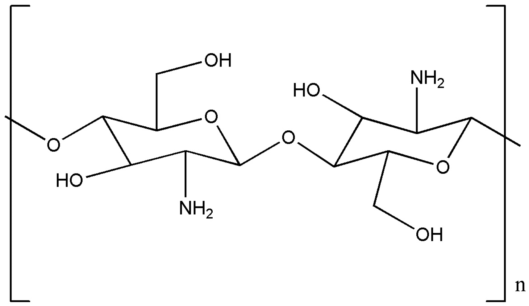
2.1. Chitosan
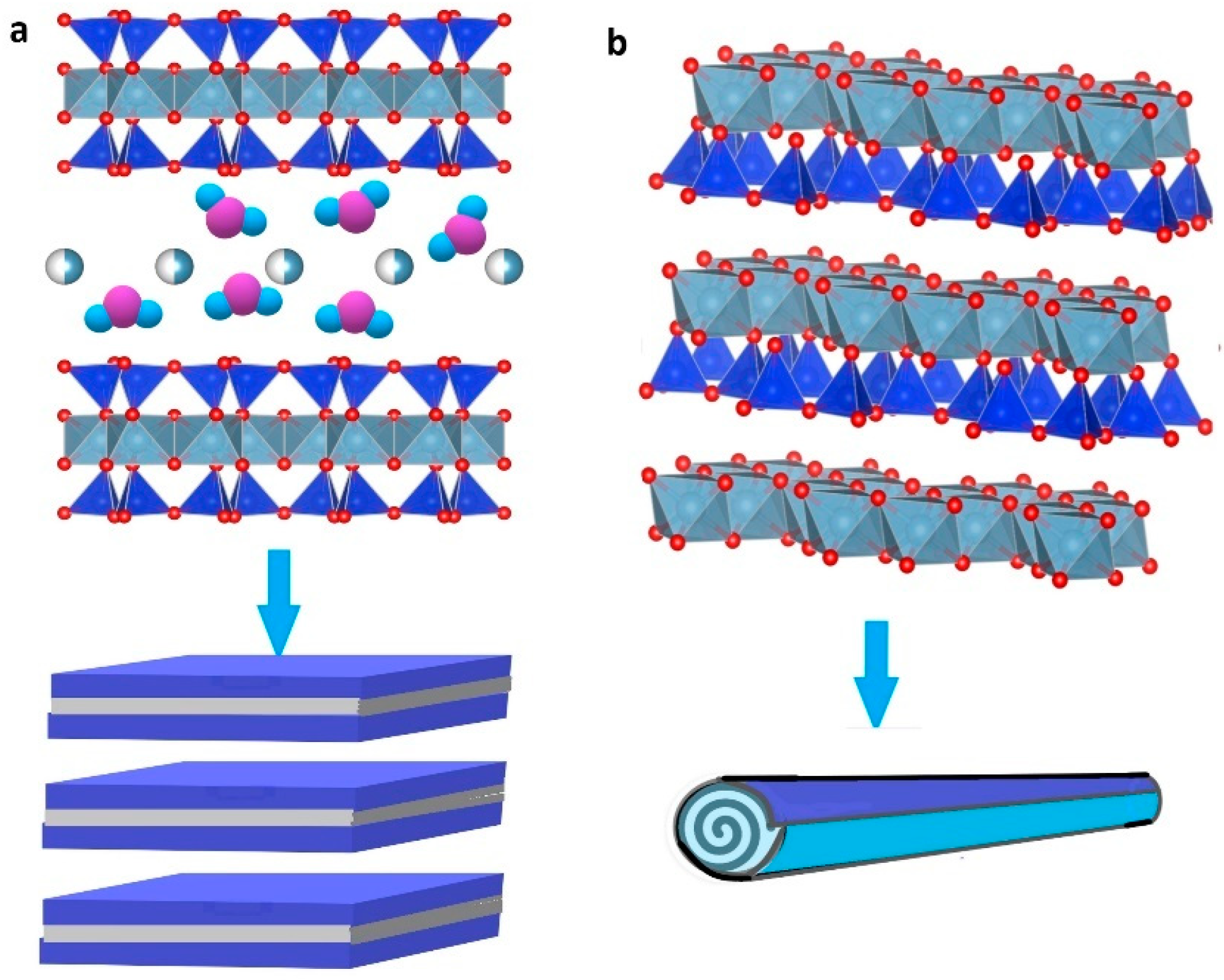
2.2. Nanoclays
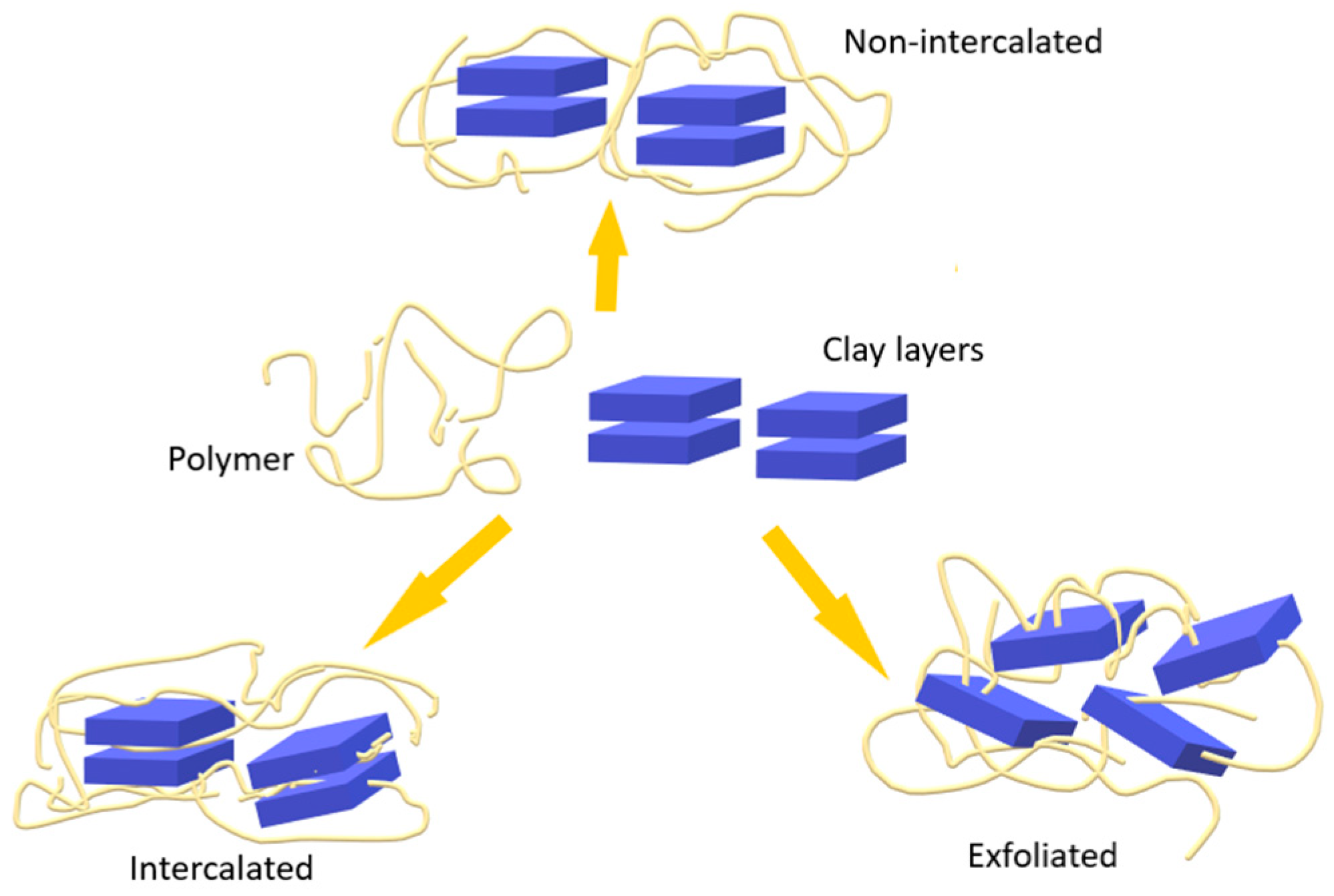
2.3. Preparation and Characterization of Chitosan–Clay Nanocomposites
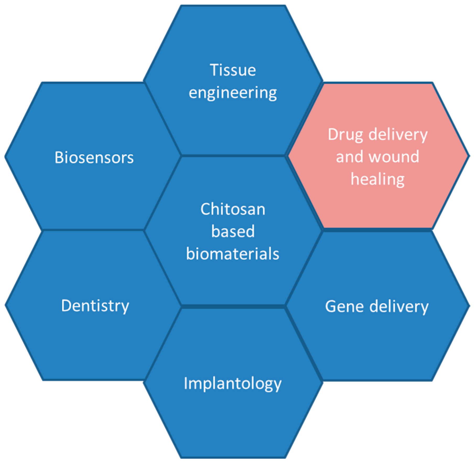
3. Chitosan–Clay Nanocomposites for Biomedical Applications
3.1. Drug Delivery
3.2. Wound Healing
4. Challenges and Future Perspectives
5. Conclusions
Author Contributions
Funding
Conflicts of Interest
References
- IUPAC Gold Book. In International Union of Pure and Applied Chemistry (IUPAC) eBooks; International Union of Pure and Applied Chemistry: Zürich, Switzerland, 2014. [CrossRef]
- Gebai, S.; Hallal, A.; Hammoud, M. Composite materials types and applications: A review on composite materials. In Mechanical Properties of Natural Fiber Reinforced Polymers. Emerging Research and Opportunities; IGI Global: Hershey, PA, USA, 2018; pp. 1–29. [Google Scholar] [CrossRef]
- Egbo, M.K. A fundamental review on composite materials and some of their applications in biomedical engineering. J. King Saud Univ.-Eng. Sci. 2021, 33, 557–568. [Google Scholar] [CrossRef]
- Gao, F. Clay/polymer composites: The story. Mater. Today 2004, 7, 50–55. [Google Scholar] [CrossRef]
- Lunetto, V.; Galati, M.; Settineri, L.; Iuliano, L. Sustainability in the manufacturing of composite materials: A literature review and directions for future research. J. Manuf. Process. 2023, 85, 858–874. [Google Scholar] [CrossRef]
- Ramakrishna, S.; Mayer, J.; Wintermantel, E.; Leong, K.W. Biomedical applications of polymer-composite materials: A review. Compos. Sci. Technol. 2001, 61, 1189–1224. [Google Scholar] [CrossRef]
- Paul, D.; Robeson, L. Polymer nanotechnology: Nanocomposites. Polymer 2008, 49, 3187–3204. [Google Scholar] [CrossRef]
- Rahaman, M.N.; Brown, R.F. Materials for Biomedical Engineering: Fundamentals and Applications; John Wiley & Sons: Hoboken, NJ, USA, 2021; pp. 307–314. [Google Scholar]
- Gu, H.; Liu, C.; Zhu, J.; Gu, J.; Wujcik, E.K.; Shao, L.; Wang, N.; Wei, H.; Scaffaro, R.; Zhang, J.; et al. Introducing advanced composites and hybrid materials. Adv. Compos. Hybrid Mater. 2017, 1, 1–5. [Google Scholar] [CrossRef]
- Mascarenhas, R.; Hegde, S.; Manaktala, N. Chitosan nanoparticle applications in dentistry: A sustainable biopolymer. Front. Chem. 2024, 12, 1362482. [Google Scholar] [CrossRef]
- Nomicisio, C.; Ruggeri, M.; Bianchi, E.; Vigani, B.; Valentino, C.; Aguzzi, C.; Viseras, C.; Rossi, S.; Sandri, G. Natural and synthetic clay minerals in the pharmaceutical and biomedical fields. Pharmaceutics 2023, 15, 1368. [Google Scholar] [CrossRef] [PubMed]
- Zagho, M.; Hussein, E.; Elzatahry, A. Recent overviews in functional polymer composites for biomedical applications. Polymers 2018, 10, 739. [Google Scholar] [CrossRef]
- Akgöl, S.; Ulucan-Karnak, F.; Kuru, C.İ.; Kuşat, K. The Usage of Composite Nanomaterials in Biomedical Engineering Applications. Biotechnol. Bioeng. 2021, 118, 2906–2922. [Google Scholar] [CrossRef]
- Nandhini, J.; Karthikeyan, E.; Rajeshkumar, S. Eco-friendly bio-nanocomposites: Pioneering sustainable biomedical advancements in engineering. Discov. Nano 2024, 19, 86. [Google Scholar] [CrossRef] [PubMed]
- Jena, G.K.; Parhi, R. Applications of composite materials in drug delivery systems. In Elsevier eBooks; Elsevier: Amsterdam, The Netherlands, 2023; pp. 111–130. [Google Scholar] [CrossRef]
- Peltonen, L.; Singhal, M.; Hirvonen, J. Principles of nanosized drug delivery systems. In Elsevier eBooks; Elsevier: Amsterdam, The Netherlands, 2020; pp. 3–25. [Google Scholar] [CrossRef]
- Baptista, P.V.; McCusker, M.P.; Carvalho, A.; Ferreira, D.A.; Mohan, N.M.; Martins, M.; Fernandes, A.R. Nano-Strategies to fight Multidrug Resistant Bacteria—“A Battle of the Titans”. Front. Microbiol. 2018, 9, 1441. [Google Scholar] [CrossRef] [PubMed]
- Ke, C.; Deng, F.; Chuang, C.; Lin, C. Antimicrobial actions and applications of chitosan. Polymers 2021, 13, 904. [Google Scholar] [CrossRef]
- Picos-Corrales, L.; Morales-Burgos, A.; Ruelas-Leyva, J.; Crini, G.; García-Armenta, E.; Jimenez-Lam, S.; Ayón-Reyna, L.; Rocha-Alonzo, F.; Calderón-Zamora, L.; Osuna-Martínez, U.; et al. Chitosan as an outstanding polysaccharide improving Health-Commodities of Humans and environmental Protection. Polymers 2023, 15, 526. [Google Scholar] [CrossRef]
- Kravanja, G.; Primožič, M.; Knez, Ž.; Leitgeb, M. Chitosan-Based (Nano)Materials for novel biomedical applications. Molecules 2019, 24, 1960. [Google Scholar] [CrossRef]
- Lee, N.Y.; Ko, W.C.; Hsueh, P.R. Nanoparticles in the treatment of infections caused by Multidrug-Resistant organisms. Front. Pharmacol. 2019, 10, 1153. [Google Scholar] [CrossRef] [PubMed]
- Hetta, H.F.; Ramadan, Y.N.; Al-Harbi, A.I.; Ahmed, E.A.; Battah, B.; Ellah, N.H.A.; Zanetti, S.; Donadu, M.G. Nanotechnology as a Promising approach to combat multidrug Resistant bacteria: A Comprehensive review and future perspectives. Biomedicines 2023, 11, 413. [Google Scholar] [CrossRef]
- Reddy, A.B.; Manjula, B.; Jayaramudu, T.; Owonubi, S.J.; Sadiku, E.R.; Agboola, O.; Sivanjineyulu, V.; Molelekwa, G.F. Biocomposites from renewable resources: Preparation and applications of chitosan-clay nanocomposites. In Handbook of Composites from Renewable Materials, Nanocomposites: Advanced Applications; Wiley: Hoboken, NJ, USA, 2017; Volume 8, pp. 275–303. [Google Scholar]
- Mousa, M.H.; Dong, Y.; Davies, I.J. Recent advances in bionanocomposites: Preparation, properties, and applications. Int. J. Polym. Mater. 2016, 65, 225–254. [Google Scholar] [CrossRef]
- Ahmad, M.; Manzoor, K.; Ikram, S. Chitosan based nanocomposites for drug, gene delivery, and bioimaging applications. In Applications of Nanocomposite Materials in Drug Delivery; Woodhead Publishing: Cambridge, UK, 2018; pp. 27–38. [Google Scholar]
- Noreen, A.; Sultana, S.; Sultana, T.; Tabasum, S.; Zia, K.M.; Muzammil, Z.; Jabeen, M.; Lodhi, A.Z.; Sultana, S. Natural polymers as constituents of bionanocomposites. In Bionanocomposites; Elsevier: Amsterdam, The Netherlands, 2020; pp. 55–85. [Google Scholar]
- Onnainty, R.; Granero, G. Chitosan-based nanocomposites: Promising materials for drug delivery applications. In Biomedical Applications of Nanoparticles; William Andrew Publishing: Norwich, NY, USA, 2019; pp. 375–407. [Google Scholar]
- ul Haque, S.; Nasar, A. Montmorillonite clay nanocomposites for drug delivery. In Applications of Nanocomposite Materials in Drug Delivery; Woodhead Publishing: Cambridge, UK, 2018; pp. 633–648. [Google Scholar]
- Silva-Castro, I.; Martín-Ramos, P.; Matei, P.M.; Fernandes-Correa, M.; Hernánez-Navarro, S.; Martín-Gil, J. Eco-friendly nanocomposites of chitosan with natural extracts, antimicrobial agents, and nanometals. In Handbook of Composites from Renewable Materials; Thakur, V.K., Thakur, M.K., Kessler, M.R., Eds.; John Wiley & Sons: Hoboken, NJ, USA, 2017; pp. 35–60. [Google Scholar]
- Kean, T.; Thanou, M. Biodegradation, biodistribution and toxicity of chitosan. Adv. Drug Deliv. Rev. 2010, 62, 3–11. [Google Scholar] [CrossRef]
- Moss, G.P. American Pharmacists Association. In Handbook of Pharmaceutical Excipients, 9th ed.; Sheskey, P.J., Hancock, B.C., Eds.; Pharmaceutical Press: London, UK; American Pharmacists Association: Washington, DC, USA, 2020; pp. 269–272. [Google Scholar]
- Sorlier, P.; Denuzière, A.; Viton, C.; Domard, A. Relation between the degree of acetylation and the electrostatic properties of chitin and chitosan. Biomacromolecules 2001, 2, 765–772. [Google Scholar] [CrossRef]
- Yassue-Cordeiro, P.H.; Zandonai, C.H.; Genesi, B.P.; Lopes, P.S.; Sanchez-Lopez, E.; Garcia, M.L.; Fernandes-Machado, N.R.C.; Severino, P.; Souto, E.B.; Da Silva, C.F. Development of Chitosan/Silver Sulfadiazine/Zeolite composite films for wound dressing. Pharmaceutics 2019, 11, 535. [Google Scholar] [CrossRef]
- Thakur, V.K.; Thakur, M.K. Recent Advances in graft copolymerization and Applications of Chitosan: A review. ACS Sustain. Chem. Eng. 2014, 2, 2637–2652. [Google Scholar] [CrossRef]
- Seyfarth, F.; Schliemann, S.; Elsner, P.; Hipler, U. Antifungal effect of high- and low-molecular-weight chitosan hydrochloride, carboxymethyl chitosan, chitosan oligosaccharide and N-acetyl-d-glucosamine against Candida albicans, Candida krusei and Candida glabrata. Int. J. Pharm. 2007, 353, 139–148. [Google Scholar] [CrossRef] [PubMed]
- Mishra, M. (Ed.) Handbook of Encapsulation and Controlled Release; CRC Press: Boca Raton, FL, USA, 2015. [Google Scholar]
- Boamah, P.O.; Onumah, J.; Agolisi, M.H.; Idan, F. Application of low molecular weight chitosan in animal nutrition, husbandry, and health: A review. Carbohydr. Polym. Technol. Appl. 2023, 6, 100329. [Google Scholar] [CrossRef]
- FDA, GRAS Substances (SCOGS) Database. Available online: http://www.fda.gov/Food/IngredientsPackagingLabeling/GRAS/SCOGS/default.htm (accessed on 16 September 2024).
- Krajišnik, D.; Čalija, B.; Milić, J. Aluminosilicate-based composites functionalized with cationic materials: Possibilities for drug-delivery applications. In Elsevier eBooks; Elsevier: Amsterdam, The Netherlands, 2019; pp. 285–327. [Google Scholar] [CrossRef]
- Safdar, R.; Omar, A.A.; Arunagiri, A.; Regupathi, I.; Thanabalan, M. Potential of Chitosan and its derivatives for controlled drug release applications—A review. J. Drug Deliv. Sci. Technol. 2019, 49, 642–659. [Google Scholar] [CrossRef]
- Ways, T.M.; Lau, W.; Khutoryanskiy, V. Chitosan and its derivatives for application in mucoadhesive drug delivery systems. Polymers 2018, 10, 267. [Google Scholar] [CrossRef]
- Jagdale, S.; Agarwal, B.; Dixit, A.; Gaware, S. Chitosan as excellent bio-macromolecule with myriad of anti-activities in biomedical applications—A review. Int. J. Biol. Macromol. 2024, 257, 128697. [Google Scholar] [CrossRef]
- Anraku, M.; Fujii, T.; Furutani, N.; Kadowaki, D.; Maruyama, T.; Otagiri, M.; Gebicki, J.M.; Tomida, H. Antioxidant effects of a dietary supplement: Reduction of indices of oxidative stress in normal subjects by water-soluble chitosan. Food Chem. Toxicol. 2009, 47, 104–109. [Google Scholar] [CrossRef]
- Shagdarova, B.; Konovalova, M.; Varlamov, V.; Svirshchevskaya, E. Anti-Obesity effects of chitosan and its derivatives. Polymers 2023, 15, 3967. [Google Scholar] [CrossRef]
- Piekarska, K.; Sikora, M.; Owczarek, M.; Jóźwik-Pruska, J.; Wiśniewska-Wrona, M. Chitin and chitosan as polymers of the future—Obtaining, modification, life cycle assessment and main directions of application. Polymers 2023, 15, 793. [Google Scholar] [CrossRef]
- Yadav, H.; Malviya, R.; Kaushik, N. Chitosan in biomedicine: A comprehensive review of recent developments. Carbohydr. Polym. Technol. Appl. 2024, 8, 100551. [Google Scholar] [CrossRef]
- Smola-Dmochowska, A.; Lewicka, K.; Macyk, A.; Rychter, P.; Pamuła, E.; Dobrzyński, P. Biodegradable Polymers and Polymer Composites with Antibacterial Properties. Int. J. Mol. Sci. 2023, 24, 7473. [Google Scholar] [CrossRef] [PubMed]
- Nasaj, M.; Chehelgerdi, M.; Asghari, B.; Ahmadieh-Yazdi, A.; Asgari, M.; Kabiri-Samani, S.; Sharifi, E.; Arabestani, M. Factors influencing the antimicrobial mechanism of chitosan action and its derivatives: A review. Int. J. Biol. Macromol. 2024, 277, 134321. [Google Scholar] [CrossRef]
- Zou, P.; Yang, X.; Wang, J.; Li, Y.; Yu, H.; Zhang, Y.; Liu, G. Advances in characterisation and biological activities of chitosan and chitosan oligosaccharides. Food Chem. 2016, 190, 1174–1181. [Google Scholar] [CrossRef] [PubMed]
- Davis, S.P. Chitosan: Manufacture, Properties, and Usage; Nova Science Publishers: Hauppauge, NY, USA, 2011. [Google Scholar]
- Motelica-Heino, M.; Predoi, M.V.; Ciobanu, S.C.; Iconaru, S.L.; Predoi, D. Studies of new layer formation on the surface of zinc doped Hydroxyapatite/Chitosan composite coatings in biological medium. Coatings 2023, 13, 472. [Google Scholar] [CrossRef]
- Iconaru, S.L.; Ciobanu, C.S.; Predoi, G.; Rokosz, K.; Chifiriuc, M.C.; Bleotu, C.; Stanciu, G.; Hristu, R.; Raaen, S.; Raita, S.M.; et al. Biological and Physico-Chemical Properties of composite layers based on Magnesium-Doped hydroxyapatite in Chitosan Matrix. Micromachines 2022, 13, 1574. [Google Scholar] [CrossRef]
- Predoi, D.; Ciobanu, C.S.; Iconaru, S.L.; Predoi, S.A.; Chifiriuc, M.C.; Raaen, S.; Badea, M.L.; Rokosz, K. Impact of gamma irradiation on the properties of Magnesium-Doped hydroxyapatite in Chitosan matrix. Materials 2022, 15, 5372. [Google Scholar] [CrossRef]
- Kim, S.; Rajapakse, N. Enzymatic production and biological activities of chitosan oligosaccharides (COS): A review. Carbohydr. Polym. 2005, 62, 357–368. [Google Scholar] [CrossRef]
- Ghadiri, M.; Chrzanowski, W.; Rohanizadeh, R. Biomedical applications of cationic clay minerals. RSC Adv. 2015, 5, 29467–29481. [Google Scholar] [CrossRef]
- Peña-Parás, L.; Sánchez-Fernández, J.A.; Vidaltamayo, R. Nanoclays for biomedical applications. Handb. Ecomater. 2018, 5, 3453–3471. [Google Scholar]
- Murugesan, S.; Scheibel, T. Copolymer/Clay nanocomposites for biomedical applications. Adv. Funct. Mater. 2020, 30, 1908101. [Google Scholar] [CrossRef]
- Helbert, J.; Hauber, E.; Reiss, D. Water on the terrestrial planets. In Elsevier eBooks; Elsevier: Amsterdam, The Netherlands, 2015; pp. 367–409. [Google Scholar] [CrossRef]
- García-Villén, F.; Ruiz-Alonso, S.; Lafuente-Merchan, M.; Gallego, I.; Sainz-Ramos, M.; Saenz-Del-Burgo, L.; Pedraz, J.L. Clay minerals as BioINK ingredients for 3D printing and 3D bioprinting: Application in tissue engineering and regenerative medicine. Pharmaceutics 2021, 13, 1806. [Google Scholar] [CrossRef] [PubMed]
- Belghazdis, M.; Hachem, E. Clay and Clay Minerals: A Detailed review. Int. J. Recent Technol. Appl. Sci. (IJORTAS) 2022, 4, 54–75. [Google Scholar] [CrossRef]
- Giuseppe, N.; Riela, S.; Fakhrullin, R.F. Clay-based drug-delivery systems: What does the future hold? Ther. Deliv. 2017, 8, 633–646. [Google Scholar] [CrossRef] [PubMed]
- Maisanaba, S.; Pichardo, S.; Puerto, M.; Gutiérrez-Praena, D.; Cameán, A.M.; Jos, A. Toxicological evaluation of clay minerals and derived nanocomposites: A review. Environ. Res. 2015, 138, 233–254. [Google Scholar] [CrossRef]
- Katti, K.S.; Jasuja, H.; Jaswandkar, S.V.; Mohanty, S.; Katti, D.R. Nanoclays in medicine: A new frontier of an ancient medical practice. Mater. Adv. 2022, 3, 7484–7500. [Google Scholar] [CrossRef] [PubMed]
- Bergaya, F.; Lagaly, G. General Introduction. In Developments in Clay Science; Elsevier: Amsterdam, The Netherlands, 2013; pp. 1–19. [Google Scholar] [CrossRef]
- De Paiva, L.B.; Morales, A.R.; Díaz, F.R.V. Organoclays: Properties, preparation and applications. Appl. Clay Sci. 2008, 42, 8–24. [Google Scholar] [CrossRef]
- Yang, J.; Lee, J.; Ryu, H.; Elzatahry, A.A.; Alothman, Z.A.; Choy, J. Drug–clay nanohybrids as sustained delivery systems. Appl. Clay Sci. 2016, 130, 20–32. [Google Scholar] [CrossRef]
- Saadh, M.J.; Abdulsahib, W.K.; Mustafa, A.N.; Zabibah, R.S.; Adhab, Z.H.; Rakhimov, N.; Alsaikhan, F. Recent advances in natural nanoclay for diagnosis and therapy of cancer: A review. Colloids Surf. B Biointerfaces 2024, 235, 113768. [Google Scholar] [CrossRef]
- ISO/TS 21236-1:2019(en); Nanotechnologies—Clay Nanomaterials—Part 1: Specification of Characteristics and Measurement Methods for Layered Clay Nanomaterials. International Organization for Standardization (ISO): Geneva, Switzerland, 2019.
- EUON, Definition of Nanomaterial—ECHA. Available online: https://euon.echa.europa.eu/definition-of-nanomaterial (accessed on 16 September 2024).
- FDA. Considering Whether an FDA-Regulated Product Involves the Application of Nanotechnology Guidance for Industry June 2014. Available online: https://www.fda.gov/regulatory-information/search-fda-guidance-documents/considering-whether-fda-regulated-product-involves-application-nanotechnology (accessed on 16 September 2024).
- FDA. Drug Products, Including Biological Products, That Contain Nanomaterials—Guidance for Industry April 2022. Available online: https://www.fda.gov/regulatory-information/search-fda-guidance-documents/drug-products-including-biological-products-contain-nanomaterials-guidance-industry (accessed on 16 September 2024).
- De Melo Barbosa, R.; Ferreira, M.A.; Meirelles, L.M.A.; Zorato, N.; Raffin, F.N. Nanoclays in drug delivery systems. In Elsevier eBooks; Elsevier: Amsterdam, The Netherlands, 2020; pp. 185–202. [Google Scholar] [CrossRef]
- Viseras, C.; Cerezo, P.; Sanchez, R.; Salcedo, I.; Aguzzi, C. Current challenges in clay minerals for drug delivery. Appl. Clay Sci. 2010, 48, 291–295. [Google Scholar] [CrossRef]
- Goo, J.; Kim, B.; Kwon, J.; Jo, H.Y. Bentonite alteration and retention of cesium and iodide ions by Ca-bentonite in alkaline and saline solutions. Appl. Clay Sci. 2023, 245, 107141. [Google Scholar] [CrossRef]
- Lvov, Y.M.; DeVilliers, M.M.; Fakhrullin, R.F. The application of halloysite tubule nanoclay in drug delivery. Expert Opin. Drug Deliv. 2016, 13, 977–986. [Google Scholar] [CrossRef] [PubMed]
- Setter, O.P.; Movsowitz, A.; Goldberg, S.; Segal, E. Antibody-Functionalized halloysite nanotubes for targeting bacterial cells. ACS Appl. Bio Mater. 2021, 4, 4094–4104. [Google Scholar] [CrossRef]
- Du, M.; Guo, B.; Jia, D. Newly emerging applications of halloysite nanotubes: A review. Polym. Int. 2010, 59, 574–582. [Google Scholar] [CrossRef]
- Fahimizadeh, M.; Wong, L.W.; Baifa, Z.; Sadjadi, S.; Auckloo, S.a.B.; Palaniandy, K.; Pasbakhsh, P.; Tan, J.B.L.; Singh, R.R.; Yuan, P. Halloysite clay nanotubes: Innovative applications by smart systems. Appl. Clay Sci. 2024, 251, 107319. [Google Scholar] [CrossRef]
- Bujdáková, H.; Bujdáková, V.; Májeková-Koščová, H.; Gaálová, B.; Bizovská, V.; Boháč, P.; Bujdák, J. Antimicrobial activity of organoclays based on quaternary alkylammonium and alkylphosphonium surfactants and montmorillonite. Appl. Clay Sci. 2018, 158, 21–28. [Google Scholar] [CrossRef]
- Bagchi, B.; Kar, S.; Dey, S.K.; Bhandary, S.; Roy, D.; Mukhopadhyay, T.K.; Das, S.; Nandy, P. In situ synthesis and antibacterial activity of copper nanoparticle loaded natural montmorillonite clay based on contact inhibition and ion release. Colloids Surf. B Biointerfaces 2013, 108, 358–365. [Google Scholar] [CrossRef] [PubMed]
- Meng, N.; Zhou, N.; Zhang, S.; Shen, J. Controlled release and antibacterial activity chlorhexidine acetate (CA) intercalated in montmorillonite. Int. J. Pharm. 2009, 382, 45–49. [Google Scholar] [CrossRef]
- Ghazaie, M.; Ghiaci, M.; Soleimanian-Zad, S.; Behzadi-Teshnizi, S. Preparing natural biocomposites of N-quaternary chitosan with antibacterial activity to reduce consumption of antibacterial drugs. J. Hazard. Mater. 2019, 371, 224–232. [Google Scholar] [CrossRef]
- Zhou, S.Q.; Niu, Y.Q.; Liu, J.H.; Chen, X.X.; Li, C.S.; Gates, W.P.; Zhou, C.H. Functional Montmorillonite/Polymer coatings. Clays Clay Miner. 2022, 70, 209–232. [Google Scholar] [CrossRef]
- Okamoto, M.; Morita, S.; Kim, Y.; Kotaka, T.; Tateyama, H. Dispersed structure change of smectic clay/poly(methyl methacrylate) nanocomposites by copolymerization with polar comonomers. Polymer 2001, 42, 1201–1206. [Google Scholar] [CrossRef]
- Ray, S.S.; Okamoto, M. Polymer/layered silicate nanocomposites: A review from preparation to processing. Prog. Polym. Sci. 2003, 28, 1539–1641. [Google Scholar] [CrossRef]
- Aguzzi, C.; Sandri, G.; Bonferoni, C.; Cerezo, P.; Rossi, S.; Ferrari, F.; Caramella, C.; Viseras, C. Solid state characterisation of silver sulfadiazine loaded on montmorillonite/chitosan nanocomposite for wound healing. Colloids Surf. B Biointerfaces 2014, 113, 152–157. [Google Scholar] [CrossRef]
- Theng, B.K.G. Clay-Polymer Interactions: Summary and Perspectives. Clays Clay Miner. 1982, 30, 1–10. [Google Scholar] [CrossRef]
- Bordes, P.; Pollet, E.; Bourbigot, S.; Avérous, L. Structure and properties of PHA/Clay Nano-Biocomposites prepared by Melt intercalation. Macromol. Chem. Phys. 2008, 209, 1473–1484. [Google Scholar] [CrossRef]
- Peramune, D.; Peduruhewa, P.; Hewawardhana, S.; Perera, W.Y.; Sandaruwan, H.H.P.B.; Manatunga, D.C.; Dassanayake, R.S. Enriched Clay-Polymer Composites and Their Applications. In Clay Composites: Environmental Applications; Springer Nature: Singapore, 2023; pp. 279–295. [Google Scholar]
- Onnainty, R.; Onida, B.; Páez, P.; Longhi, M.; Barresi, A.; Granero, G. Targeted chitosan-based bionanocomposites for controlled oral mucosal delivery of chlorhexidine. Int. J. Pharm. 2016, 509, 408–418. [Google Scholar] [CrossRef]
- Rao, K.M.; Kumar, A.; Suneetha, M.; Han, S.S. pH and near-infrared active; chitosan-coated halloysite nanotubes loaded with curcumin-Au hybrid nanoparticles for cancer drug delivery. Int. J. Biol. Macromol. 2018, 112, 119–125. [Google Scholar] [CrossRef] [PubMed]
- Sandri, G.; Aguzzi, C.; Rossi, S.; Bonferoni, M.C.; Bruni, G.; Boselli, C.; Cornaglia, A.I.; Riva, F.; Viseras, C.; Caramella, C.; et al. Halloysite and chitosan oligosaccharide nanocomposite for wound healing. Acta Biomater. 2017, 57, 216–224. [Google Scholar] [CrossRef] [PubMed]
- Čalija, B.; Milić, J.; Janićijević, J.; Daković, A.; Krajišnik, D. Ionically cross-linked chitosan–halloysite composite microparticles for sustained drug release. Clay Miner. 2017, 52, 413–426. [Google Scholar] [CrossRef]
- Jauković, V.; Krajišnik, D.; Daković, A.; Damjanović, A.; Krstić, J.; Stojanović, J.; Čalija, B. Influence of selective acid-etching on functionality of halloysite-chitosan nanocontainers for sustained drug release. Mater. Sci. Eng. C 2021, 123, 112029. [Google Scholar] [CrossRef]
- Lertsutthiwong, P.; Noomun, K.; Khunthon, S.; Limpanart, S. Influence of chitosan characteristics on the properties of biopolymeric chitosan–montmorillonite. Prog. Nat. Sci. Mater. Int. 2012, 22, 502–508. [Google Scholar] [CrossRef]
- Zhan, J.; Chen, H.; Zhou, H.; Hao, L.; Xu, H.; Zhou, X. Essential oil-loaded chitosan/zinc (II) montmorillonite synergistic sustained-release system as antibacterial material. J. Dispers. Sci. Technol. 2021, 44, 288–298. [Google Scholar] [CrossRef]
- Liu, M.; Zhang, Y.; Wu, C.; Xiong, S.; Zhou, C. Chitosan/halloysite nanotubes bionanocomposites: Structure, mechanical properties and biocompatibility. Int. J. Biol. Macromol. 2012, 51, 566–575. [Google Scholar] [CrossRef] [PubMed]
- Moghadas, B.; Dashtimoghadam, E.; Mirzadeh, H.; Seidi, F.; Hasani-Sadrabadi, M.M. Novel chitosan-based nanobiohybrid membranes for wound dressing applications. RSC Adv. 2016, 6, 7701–7711. [Google Scholar] [CrossRef]
- Cankaya, N.; Sahin, R. Chitosan/clay bionanocomposites: Structural, antibacterial, thermal and swelling properties. Cellul. Chem. Technol. 2019, 53, 537–549. [Google Scholar] [CrossRef]
- Devi, N.; Dutta, J. Preparation and characterization of chitosan-bentonite nanocomposite films for wound healing application. Int. J. Biol. Macromol. 2017, 104, 1897–1904. [Google Scholar] [CrossRef]
- Nozari, M.; Gholizadeh, M.; Oghani, F.Z.; Tahvildari, K. Studies on novel chitosan/alginate and chitosan/bentonite flexible films incorporated with ZnO nano particles for accelerating dermal burn healing: In vivo and in vitro evaluation. Int. J. Biol. Macromol. 2021, 184, 235–249. [Google Scholar] [CrossRef]
- Luo, Y.; Mills, D.K. The effect of halloysite addition on the material properties of Chitosan–Halloysite hydrogel composites. Gels 2019, 5, 40. [Google Scholar] [CrossRef]
- Yilmaz Atay, H. Antibacterial activity of chitosan-based systems. In Functional Chitosan: Drug Delivery and Biomedical Applications; Springer: Berlin/Heidelberg, Germany, 2019; pp. 457–489. [Google Scholar]
- Ambrogi, V.; Pietrella, D.; Nocchetti, M.; Casagrande, S.; Moretti, V.; De Marco, S.; Ricci, M. Montmorillonite–chitosan–chlorhexidine composite films with antibiofilm activity and improved cytotoxicity for wound dressing. J. Colloid Interface Sci. 2017, 491, 265–272. [Google Scholar] [CrossRef]
- Hua, S.; Yang, H.; Wang, W.; Wang, A. Controlled release of ofloxacin from chitosan–montmorillonite hydrogel. Appl. Clay Sci. 2010, 50, 112–117. [Google Scholar] [CrossRef]
- Kelly, H.; Deasy, P.; Ziaka, E.; Claffey, N. Formulation and preliminary in vivo dog studies of a novel drug delivery system for the treatment of periodontitis. Int. J. Pharm. 2004, 274, 167–183. [Google Scholar] [CrossRef] [PubMed]
- Čalija, B.; Milić, J.; Milašinović, N.; Daković, A.; Trifković, K.; Stojanović, J.; Krajišnik, D. Functionality of chitosan-halloysite nanocomposite films for sustained delivery of antibiotics: The effect of chitosan molar mass. J. Appl. Polym. Sci. 2019, 137, 48406. [Google Scholar] [CrossRef]
- Tenci, M.; Rossi, S.; Aguzzi, C.; Carazo, E.; Sandri, G.; Bonferoni, M.; Grisoli, P.; Viseras, C.; Caramella, C.; Ferrari, F. Carvacrol/clay hybrids loaded into in situ gelling films. Int. J. Pharm. 2017, 531, 676–688. [Google Scholar] [CrossRef] [PubMed]
- Salcedo, I.; Aguzzi, C.; Sandri, G.; Bonferoni, M.C.; Mori, M.; Cerezo, P.; Sánchez, R.; Viseras, C.; Caramella, C. In vitro biocompatibility and mucoadhesion of montmorillonite chitosan nanocomposite: A new drug delivery. Appl. Clay Sci. 2012, 55, 131–137. [Google Scholar] [CrossRef]
- Salcedo, I.; Sandri, G.; Aguzzi, C.; Bonferoni, C.; Cerezo, P.; Sánchez-Espejo, R.; Viseras, C. Intestinal permeability of oxytetracycline from chitosan-montmorillonite nanocomposites. Colloids Surf. B Biointerfaces 2014, 117, 441–448. [Google Scholar] [CrossRef]
- Sandri, G.; Bonferoni, M.C.; Ferrari, F.; Rossi, S.; Aguzzi, C.; Mori, M.; Grisoli, P.; Cerezo, P.; Tenci, M.; Viseras, C.; et al. Montmorillonite–chitosan–silver sulfadiazine nanocomposites for topical treatment of chronic skin lesions: In vitro biocompatibility, antibacterial efficacy and gap closure cell motility properties. Carbohydr. Polym. 2014, 102, 970–977. [Google Scholar] [CrossRef]
- García-Villén, F.; Carazo, E.; Borrego-Sánchez, A.; Sánchez-Espejo, R.; Cerezo, P.; Viseras, C.; Aguzzi, C. Clay minerals in drug delivery systems. In Modified Clay and Zeolite Nanocomposite Materials; Elsevier: Amsterdam, The Netherlands, 2019; pp. 129–166. [Google Scholar]
- Kim, M.H.; Choi, G.; Elzatahry, A.; Vinu, A.; Choy, Y.B.; Choy, J. Review of Clay-drug Hybrid Materials for Biomedical Applications: Administration routes. Clays Clay Miner. 2016, 64, 115–130. [Google Scholar] [CrossRef]
- Allen, L.V. Ansel’s Pharmaceutical Dosage Forms and Drug Elivery Systems, 11th International ed.; Wolters Kluwer: Philadelphia, PA, USA, 2018. [Google Scholar]
- Bandyopadhyay, D. Topical antibacterials in dermatology. Indian J. Dermatol. 2021, 66, 117. [Google Scholar] [CrossRef]
- Committee, J.F. BNF 84 (British National Formulary) September 2022: 84: September 2022–March 2023; Pharmaceutical Press: London, UK, 2022. [Google Scholar]
- Buckingham, R. (Ed.) Martindale: The Complete Drug Reference, 40th ed.; Pharmaceutical Press: London, UK, 2020; ISBN 9780857113672, ISBN 0857113674. [Google Scholar]
- Mamun, M.M.; Sorinolu, A.J.; Munir, M.; Vejerano, E.P. Nanoantibiotics: Functions and properties at the nanoscale to combat antibiotic resistance. Front. Chem. 2021, 9, 687660. [Google Scholar] [CrossRef]
- Muzammil, S.; Hayat, S.; Fakhar-E-Alam, M.; Aslam, B.; Siddique, M.H.; Nisar, M.A.; Saqalein, M.; Atif, M.; Sarwar, A.; Khurshid, A.; et al. Nanoantibiotics: Future nanotechnologies to combat antibiotic resistance. Front. Biosci. 2018, 10, 352–374. [Google Scholar] [CrossRef]
- Yeh, Y.; Huang, T.; Yang, S.; Chen, C.; Fang, J. Nano-Based drug delivery or targeting to eradicate bacteria for infection mitigation: A review of recent advances. Front. Chem. 2020, 8, 286. [Google Scholar] [CrossRef] [PubMed]
- Quiñones, J.P.; Peniche, H.; Peniche, C. Chitosan based Self-Assembled nanoparticles in drug delivery. Polymers 2018, 10, 235. [Google Scholar] [CrossRef] [PubMed]
- Chahardahmasoumi, S.; Sarvi, M.N.; Jalali, S.a.H. Modified montmorillonite nanosheets as a nanocarrier with smart pH-responsive control on the antimicrobial activity of tetracycline upon release. Appl. Clay Sci. 2019, 178, 105135. [Google Scholar] [CrossRef]
- Cardoso, H.P.; Rodrigues, J.F.B.; Da Silva, H.N.; Galdino, T.P.; Luna, C.B.B.; Fook, M.V.L.; Montazerian, M.; Baino, F.; De Lima Silva, S.M. Chitosan/montmorillonite nanocomposite film as anticancer drug carrier: A promising biomaterial to treat skin cancers. Ceram. Int. 2024, 50, 18528–18539. [Google Scholar] [CrossRef]
- Hanssen, A.D.; Osmon, D.R.; Patel, R. Local antibiotic delivery systems: Where are we and where are we going? Clin. Orthop. Relat. Res. (1976–2007) 2005, 437, 111–114. [Google Scholar] [CrossRef]
- Liu, Y.; Li, X.; Liang, A. Current research progress of local drug delivery systems based on biodegradable polymers in treating chronic osteomyelitis. Front. Bioeng. Biotechnol. 2022, 10, 1042128. [Google Scholar] [CrossRef]
- Nandi, S.K.; Mukherjee, P.; Roy, S.; Kundu, B.; De, D.K.; Basu, D. Local antibiotic delivery systems for the treatment of osteomyelitis—A review. Mater. Sci. Eng. C 2009, 29, 2478–2485. [Google Scholar] [CrossRef]
- Boles, L.R.; Awais, R.; Beenken, K.E.; Smeltzer, M.S.; Haggard, W.O.; Jessica, A.J. Local Delivery of Amikacin and Vancomycin from Chitosan Sponges Prevent Polymicrobial Implant-Associated Biofilm. Mil. Med. 2018, 183 (Suppl. 1), 459–465. [Google Scholar] [CrossRef]
- Smith, J.K.; Moshref, A.R.; Jennings, J.A.; Courtney, H.S.; Haggard, W.O. Chitosan sponges for local synergistic infection therapy: A pilot study. Clin. Orthop. Relat. Res. 2013, 471, 3158–3164. [Google Scholar] [CrossRef]
- Noel, S.P.; Courtney, H.; Bumgardner, J.D.; Haggard, W.O. Chitosan Films: A potential local drug delivery system for antibiotics. Clin. Orthop. Relat. Res. 2008, 466, 1377–1382. [Google Scholar] [CrossRef]
- Valdés, L.; Pérez, I.; De Ménorval, L.C.; Altshuler, E.; Fossum, J.O.; Rivera, A. A simple way for targeted delivery of an antibiotic: In vitro evaluation of a nanoclay-based composite. PLoS ONE 2017, 12, e0187879. [Google Scholar] [CrossRef] [PubMed]
- Steadman, W.; Chapman, P.R.; Schuetz, M.; Schmutz, B.; Trampuz, A.; Tetsworth, K. Local antibiotic delivery options in prosthetic joint infection. Antibiotics 2023, 12, 752. [Google Scholar] [CrossRef] [PubMed]
- Wassif, R.K.; Elkayal, M.; Shamma, R.N.; Elkheshen, S.A. Recent advances in the local antibiotics delivery systems for management of osteomyelitis. Drug Deliv. 2021, 28, 2392–2414. [Google Scholar] [CrossRef] [PubMed]
- Wu, Q.; Liao, J.; Yang, H. Recent advances in kaolinite Nanoclay as drug carrier for bioapplications: A review. Adv. Sci. 2023, 10, e2300672. [Google Scholar] [CrossRef]
- Lopez-Ojeda, W.; Pandey, A.; Alhajj, M.; Oakley, A.M. Anatomy, Skin (Integument). StatPearls–NCBI Bookshelf. 17 October 2022. Available online: https://www.ncbi.nlm.nih.gov/books/NBK441980/ (accessed on 16 September 2024).
- Chouhan, D.; Dey, N.; Bhardwaj, N.; Mandal, B.B. Emerging and innovative approaches for wound healing and skin regeneration: Current status and advances. Biomaterials 2019, 216, 119267. [Google Scholar] [CrossRef] [PubMed]
- Rodrigues, M.; Kosaric, N.; Bonham, C.A.; Gurtner, G.C. Wound Healing: A Cellular perspective. Physiol. Rev. 2019, 99, 665–706. [Google Scholar] [CrossRef]
- Salcido, R. The cicatrix: The functional stage of wound healing. Adv. Ski. Wound Care 2018, 31, 581. [Google Scholar] [CrossRef]
- Suarato, G.; Bertorelli, R.; Athanassiou, A. Borrowing from nature: Biopolymers and biocomposites as smart wound care materials. Front. Bioeng. Biotechnol. 2018, 6, 137. [Google Scholar] [CrossRef]
- Blanco-Fernandez, B.; Castaño, O.; Mateos-Timoneda, M.Á.; Engel, E.; Pérez-Amodio, S. Nanotechnology approaches in chronic wound healing. Adv. Wound Care 2021, 10, 234–256. [Google Scholar] [CrossRef]
- Verma, D.; Okhawilai, M.; Goh, K.L.; Thakur, V.K.; Senthilkumar, N.; Sharma, M.; Uyama, H. Sustainable functionalized chitosan based nano-composites for wound dressings applications: A review. Environ. Res. 2023, 235, 116580. [Google Scholar] [CrossRef]
- Matica, N.; Aachmann, N.; Tøndervik, N.; Sletta, N.; Ostafe, N. Chitosan as a wound dressing starting material: Antimicrobial properties and mode of action. Int. J. Mol. Sci. 2019, 20, 5889. [Google Scholar] [CrossRef] [PubMed]
- Falanga, V. Wound healing and its impairment in the diabetic foot. Lancet 2005, 366, 1736–1743. [Google Scholar] [CrossRef] [PubMed]
- Jain, A.K.; Thareja, S. In vitro and in vivo characterization of pharmaceutical nanocarriers used for drug delivery. Artif. Cells Nanomed. Biotechnol. 2019, 47, 524–539. [Google Scholar] [CrossRef] [PubMed]
- Manaia, E.B.; Abuçafy, M.P.; Chiari-Andréo, B.G.; Silva, B.L.; Oshiro-Júnior, J.A.; Chiavacci, L. Physicochemical characterization of drug nanocarriers. Int. J. Nanomed. 2017, 12, 4991–5011. [Google Scholar] [CrossRef]
- Naumenko, E.A.; Fakhrullin, R.F. Toxicological evaluation of clay nanomaterials and Polymer–Clay nanocomposites. In The Royal Society of Chemistry eBooks; The Royal Society of Chemistry: London, UK, 2016; pp. 399–419. [Google Scholar] [CrossRef]

| Constituents | Preparation Technique/ Final Carrier Morphology/Type | Main Features | Methods of Characterization | Reference |
|---|---|---|---|---|
| MMT (CEC: 71 meq/100 g)/CS (MW: 71, 220 and 583 kDa; DD: 85–90%) API:/ | Ion-exchange reaction/ composite powder | Antibacterial activity |
| [95] |
| MMT (CEC: n/a)/CS (MW: n/a; DD: 98%) API:/ | Solution intercalation/ composite powder | Mucoadhesivity Wound-healing properties |
| [109] |
| MMT (natural and modified with a quaternary ammonium salt, CEC: n/a)/CS (MW: 310–375 kDa; DD: ≥75%) API:/ | Solution-blending method/composite nanoparticles | Improved heat resistance of bionanocomposites Swelling proportional to the amount of chitosan in the bionanocomposites |
| [99] |
| MMT (CEC: n/a)/CS (MW: 81 kDa; DD: 85%) API:/ | Solvent casting/nanohybrid films based on CS and biofunctionalized MMT with CS-sulfate chains (SMMT) | Improved physicochemical properties compared to plain MMT/CS nanocomposite films Antibacterial activity against S. aureus and E. coli Possible application in wound dressing for burns and chronic and diabetic wound infections with low to moderate exudate. |
| [98] |
| BNT (CEC: n/a)/CS (MW: 100 kDa; DD: 79%) API:/ | Solvent casting/ nanocomposite films | CS-BNT films exhibited good antibacterial activity Hemocompatibility Suitable for wound-care products |
| [100] |
| MMT (CEC: 80.64 meq/100 g)/CS (MW: 251 kDa; DD: 98%) API: silver sulfadiazine | Solution intercalation/ composite powder | Bacteriostatic and bactericidal properties, suitable for skin lesions |
| [111] |
| MMT (CEC: 80.64 meq/100 g)/CS (MW: n/a; DD: 98%) API: silver sulfadiazine | Solution intercalation/ composite powder | The effective interaction between the organic and inorganic components The successful drug loading of clay/chitosan nanostructures |
| [86] |
| MMT (CEC: 80.64 meq/100 g)/CS (Mw: n/a; DD: 98%) API: oxytetracycline hydrochloride | Solution intercalation and lyophilization/ composite powder | Good biocompatibility Enhancement of drug permeation |
| [110] |
| MMT (CEC: n/a)/CS (MW: 50–190 kDa; DD: 85%) API: chlorhexidine digluconate | Solution intercalation and lyophilization/ composite nanoparticles | Sustained release Mucoadhesivity Suitable for treatment of buccal infections |
| [90] |
| MMT (CEC: 120 meq/100 g)/CS (Mw: n/a; DD: n/a) API: chlorhexidine diacetate | Solvent casting/ composite films | Antimicrobial and antibiofilm activity Suitable for wound dressings |
| [104] |
| MMT (CEC: n/a)/CS (Mw: 90 kDa; DD: 81%) API: ofloxacin | Solution intercalation and ionic crosslinking with TPP/ nanocomposite beads | Improved drug loading and sustained drug release. The drug release rate of the beads was influenced by pH of the medium |
| [105] |
| MMT (CEC: n/a)/N-quaternary CS (Mw: 100–300 kDa; DD: ≥ 90%) API: ciprofloxacin | Solution intercalation/ composite powder | Efficient drug encapsulation by N,N,N-triethyl CS composites Prolonged drug release and enhanced antibacterial activity |
| [82] |
| BNT (CEC: n/a)/CS (MW: n/a; DD: 90%) ZnO (nanoparticles) Gelatin (prepared from A. stellatus n. cyrenisis Berg fish waste) | Solvent casting/ composite films | ZnO nanoparticles increased the porosity, hydrophilicity, and water absorption of the composite films High antibacterial activity Wound healing and epithelium regeneration |
| [101] |
| Zinc (II)-MMT-organically modified (CEC: n/a)/CS (MW: n/a; DD: 95%) API: tea tree oil | Solution intercalation and adsorption saturation/ composite powder | Good loading capacity and sustained release for tea tree oil, alongside with good antibacterial effect against E. coli |
| [96] |
| HAL/CS (MW: 1000 Da; DD: 75.4%) API:/ | Solution intercalation and lyophilization/ composite powder | Biocompatibility in vitro towards normal human dermal fibroblasts in an in vitro wound healing test (improved re-epithelialization effect) |
| [92] |
| HAL/CS (MW: medium; DD: n/a) API: tetracycline, tetracycline hydrochloride | Solution blending/thermoresponsive hydrogel containing halloysite–chitosan composite | Sustained drug release and microbiological activity over a 6-week period |
| [106] |
| HAL/CS (Mw: low, medium and high, according to the supplier’s data; DD: >75%) API: tetracycline hydrochloride | Solvent casting and evaporation/ nanocomposite films | Improved thermal stability and mechanical properties in comparison with corresponding CS films Sustained release of tetracycline hydrochloride during 8 h |
| [107] |
| HAL/CS (Mw: low; DD: n/a) API: gentamicin sulfate | Solution blending; hydrogels were formed by crosslinking the mixture solution with 10% TPP/ hydrogel composite | Sustained drug release and efficacy in reducing bacterial growth The addition of HAL improved mechanical properties of hydrogel nanocomposites |
| [102] |
| Antibiotic | Origin | Sensitive Microorganisms | Application for Topical Treatment | Application in Chitosan–Clay Nanocomposites |
|---|---|---|---|---|
| Clindamycin | A semisynthetic derivative of lincomycin | Cutibacterium acnes (Propionibacterium acnes) | Erythrasma, folliculitis, Fox–Fordyce disease, periorificial facial dermatitis, and rosacea | / |
| Fusidic acid | A steroid-like structure derived from the fungus Fusidium coccineum | Gram-positive bacteria such as Staphylococcus sp. and Corynebacterium sp. | Impetigo, erythrasma, and pitted keratolysis | / |
| Gentamicin | An aminoglycoside antibiotic; derived from Micromonospora purpurea | Mostly Gram-negative like Pseudomonas, Proteus, and Escherichia coli, and Gram-positive like S. aureus | Impetigo and folliculitis | [102] |
| Metronidazole | Synthetic nitroimidazole | Most anaerobic bacteria and protozoa | Rosacea, benign and malignant ulcers, including pressure sores infected with anaerobes | / |
| Mupirocin | Derived from Pseudomonas fluorescens; unique structure, distinct from any other antibiotics | Ineffective against most aerobic Gram-negative bacteria and anaerobes, but active against penicillinase-producing and methicillin-resistant strains of S. aureus | Impetigo, secondarily infected eczema, infected wounds with strains of S. aureus or Streptococcus pyogenes; nasal colonization of methicillin-resistant S. aureus (MRSA) | / |
| Nadifloxacin | A synthetic quinolone | Broad-spectrum bactericidal activity (aerobic Gram-positive, Gram-negative, and anaerobic bacteria, including P. acnes and S. epidermidis) | Active against MRSA, high potential as an alternative for topical antibiotic treatment in bacterial skin infection | / |
| Neomycin | A bactericidal aminoglycoside antibiotic, which acts by inhibiting bacterial protein synthesis | Mostly Gram-negative organisms like Proteus, E. coli, Serratia, and H. influenzae | Superficial pyodermas, minor wounds, and secondarily infected dermatitis | / |
| Retapamulin | A semisynthetic antibiotic | Isolates resistant-to-therapy beta-lactams, macrolides, quinolones, topical fusidic acid, and mupirocin | Impetigo, the short-term treatment of impetigo and infected small lacerations, abrasions, and sutured wounds | / |
| Silver sulfadiazine | A topical sulfonamide | A wide spectrum of activity against both Gram-positive and Gram-negative bacteria | Prevention and treatment of wounds caused by serious burns | [86,111] |
| Tetracycline | Derived by Streptomyces genus of Actinobacteria | Exhibits various bacteriostatic effects against many aerobic and anaerobic bacterial genera, both Gram-positive and Gram-negative | Treatment of several infections, including acne and rosacea | [106,107] |
Disclaimer/Publisher’s Note: The statements, opinions and data contained in all publications are solely those of the individual author(s) and contributor(s) and not of MDPI and/or the editor(s). MDPI and/or the editor(s) disclaim responsibility for any injury to people or property resulting from any ideas, methods, instructions or products referred to in the content. |
© 2024 by the authors. Licensee MDPI, Basel, Switzerland. This article is an open access article distributed under the terms and conditions of the Creative Commons Attribution (CC BY) license (https://creativecommons.org/licenses/by/4.0/).
Share and Cite
Krajišnik, D.; Uskoković-Marković, S.; Daković, A. Chitosan–Clay Mineral Nanocomposites with Antibacterial Activity for Biomedical Application: Advantages and Future Perspectives. Int. J. Mol. Sci. 2024, 25, 10377. https://doi.org/10.3390/ijms251910377
Krajišnik D, Uskoković-Marković S, Daković A. Chitosan–Clay Mineral Nanocomposites with Antibacterial Activity for Biomedical Application: Advantages and Future Perspectives. International Journal of Molecular Sciences. 2024; 25(19):10377. https://doi.org/10.3390/ijms251910377
Chicago/Turabian StyleKrajišnik, Danina, Snežana Uskoković-Marković, and Aleksandra Daković. 2024. "Chitosan–Clay Mineral Nanocomposites with Antibacterial Activity for Biomedical Application: Advantages and Future Perspectives" International Journal of Molecular Sciences 25, no. 19: 10377. https://doi.org/10.3390/ijms251910377
APA StyleKrajišnik, D., Uskoković-Marković, S., & Daković, A. (2024). Chitosan–Clay Mineral Nanocomposites with Antibacterial Activity for Biomedical Application: Advantages and Future Perspectives. International Journal of Molecular Sciences, 25(19), 10377. https://doi.org/10.3390/ijms251910377