Review

X-Linked Epilepsies: A Narrative Review

by
Pia Bernardo
1,†,
Claudia Cuccurullo
2,†,
Marica Rubino
3,
Gabriella De Vita
4,
Gaetano Terrone
5,
Leonilda Bilo
3 and
Antonietta Coppola
3,*
1
Pediatric Psychiatry and Neurology Unit, Department of Neurosciences, Santobono-Pausilipon Children’s Hospital, 80129 Naples, Italy
2
Neurology and Stroke Unit, Ospedale del Mare Hospital, ASL Napoli 1 Centro, 80147 Naples, Italy
3
Department of Neurosciences, Reproductive Sciences and Odontostomatology, University Federico II of Naples, 80131 Naples, Italy
4
Department of Molecular Medicine and Medical Biotechnology, University of Naples Federico II, 80131 Naples, Italy
5
Child Neuropsychiatry Units, Department of Translational Medical Sciences, University Federico II of Naples, 80131 Naples, Italy
*
Author to whom correspondence should be addressed.
These authors contributed equally to this work.
Int. J. Mol. Sci. 2024, 25(7), 4110; https://doi.org/10.3390/ijms25074110
Submission received: 11 March 2024 / Revised: 3 April 2024 / Accepted: 6 April 2024 / Published: 8 April 2024
(This article belongs to the Special Issue Molecular Advances in Epilepsy and Seizures)

Abstract

:
X-linked epilepsies are a heterogeneous group of epileptic conditions, which often overlap with X-linked intellectual disability. To date, various X-linked genes responsible for epilepsy syndromes and/or developmental and epileptic encephalopathies have been recognized. The electro-clinical phenotype is well described for some genes in which epilepsy represents the core symptom, while less phenotypic details have been reported for other recently identified genes. In this review, we comprehensively describe the main features of both X-linked epileptic syndromes thoroughly characterized to date (PCDH19-related DEE, CDKL5-related DEE, MECP2-related disorders), forms of epilepsy related to X-linked neuronal migration disorders (e.g., ARX, DCX, FLNA) and DEEs associated with recently recognized genes (e.g., SLC9A6, SLC35A2, SYN1, ARHGEF9, ATP6AP2, IQSEC2, NEXMIF, PIGA, ALG13, FGF13, GRIA3, SMC1A). It is often difficult to suspect an X-linked mode of transmission in an epilepsy syndrome. Indeed, different models of X-linked inheritance and modifying factors, including epigenetic regulation and X-chromosome inactivation in females, may further complicate genotype–phenotype correlations. The purpose of this work is to provide an extensive and updated narrative review of X-linked epilepsies. This review could support clinicians in the genetic diagnosis and treatment of patients with epilepsy featuring X-linked inheritance.

1. Introduction

X-linked epilepsies (XLE) can be considered as an expanding group of genetically heterogeneous and clinically variable conditions, with frequent coexistence of intellectual disability (ID) [1,2]. Particularly, epilepsy can occur as an isolated X-linked trait, as a component of recognizable X-linked syndromes or X-linked developmental and epileptic encephalopathies (DEEs). Moreover, the phenotype of X-linked epilepsy is complex, because of the variability of clinical manifestations that may be attributed to several factors, including the type of genetic mutation, epigenetic regulation, X chromosome random inactivation, and mosaic distribution.
The following is a comprehensive review of X-linked disorders and conditions associated with epilepsy, where epilepsy represents the “core” of the disease. Several important genetic pathways for both known disease genes and novel emerging X-linked genes are described and are discussed with relevance to clinical characteristics. This review might support clinicians in the genetic diagnosis of epileptic syndromes or DEEs when an X-linked mode of transmission is suspected.

2. Materials and Methods

A systematic search on MEDLINE PubMed was performed with the terms “X-linked”, “Epilepsy”, “Developmental and epileptic encephalopathy”, and “Seizures” (last search 31 December 2023). All manuscripts including cases with electro-clinical information were reviewed.

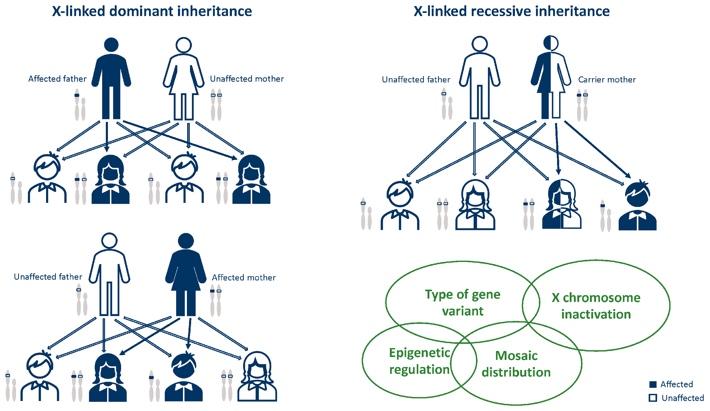
3. General Notes on X-Linked Disease Inheritance

The majority of disorders caused by mutations in genes on the X chromosome are inherited through the maternal germline and their phenotype depends on the sex of the affected individual [3]. The inheritance pattern of X-linked mutations can be dominant or recessive. The former is defined as X-linked dominant vertical transmission in which daughters of affected males are always affected, while the latter is defined as X-linked recessive vertical transmission in which carrier females pass the trait to affected sons (Figure 1) [3]. Females with X-linked dominant disorders typically have variable severity of their phenotype, a feature that is thought to be dependent on the X inactivation pattern [3]. X chromosome inactivation (XCI) is a process by which the dosage of proteins transcribed from genes on the X chromosome is equalized between males (XY) and females (XX) through the silencing of most genes on one of the two X chromosomes in females. Mammalian XCI results in the random silencing of one of the two X chromosomes in females early in development in order to ensure a balance in the dosage of X-linked gene expression between the sexes [4,5]. As the descendants of each cell keep the same pattern of inactivation, an individual heterozygote for an X-linked disease will be a mosaic, with two cell populations, one of which will express the normal and the other one the abnormal X chromosome. Consequently, some disorders demonstrate ‘mosaic’ or ‘patchy’ symptoms in heterozygous females [3].

4. Known X-Linked Developmental and Epileptic Encephalopathies

We reviewed the main electro-clinical features and the known genotype–phenotype correlations, including X-liked genes long known to cause DEEs, genes responsible for genetic and structural epilepsy from X-linked Neuronal Migration disorders and X-linked DEE genes that have emerged in the last few years. Our findings are summarized in Table 1 [6,7,8,9,10,11,12,13,14,15,16,17,18,19,20,21,22,23,24,25,26,27,28,29,30,31,32,33,34,35,36,37,38,39,40,41,42,43,44,45,46,47,48,49,50,51,52,53]. In Supplementary Table S1, we included XLE patients personally evaluated and treated (Supplementary Table S1).

4.1. PCDH19

Mutations in the PCDH19 gene were originally identified by Dibbens et al. [54] in patients with epilepsy and ID limited to females (or Epilepsy, Female-restricted, with Mental Retardation, EFMR; OMIM# 300088).

4.1.1. Genetic Features and Inheritance

Although the PCDH19 gene is located on Xq22, this condition has an unusual X-linked mode of inheritance, sparing transmitting males, who are affected only when somatic mosaicism for the PCDH19 mutation occurs. Males with cellular mosaics for the PCDH19 gene with a similar clinical profile to that of affected females have been described, thus challenging the dogma that this is a disorder limited exclusively to females [55]. Mosaic male patients have the same phenotype as that presented by female patients, as reported in the literature, because they have the same hypothesized pathological mechanism of molecular interference [56].

4.1.2. Epileptic Phenotypes

The hallmark feature of PCDH19-associated epilepsy is that seizures occur in clusters, lasting hours to days, often induced by fever, with onset around the first year of life [9]. A bi-phasic course with several brief clusters of febrile seizures followed by clusters of afebrile seizures has been described. Chemaly et al. proposed that fever sensitivity may be highest after age two years rather than at the onset of seizures [57]. The occurrence of a cluster of seizures is a typical feature, hence the name PCDH19-Girls Clustering Epilepsy (GCE).
Epilepsy is characterized by predominantly focal seizures, with affective symptoms (“fearful expression”) that emerged as a characteristic sign of seizures in this syndrome [6]. Hypo-motor seizures with staring and behavioral arrest are also frequent. Although focal onset is a constant feature, generalized seizures (such as tonic, myoclonic or absence) of-ten happen when rapid bilateral diffusion occurs [7] and are reported in a minority of patients. However, the seizure semiology appears to be highly variable during the disease history, with a prevalence of non-motor seizures in the first years of life. From 10% to 53% of subjects are at risk of progressing into status epilepticus (SE), frequently as convulsive SE consisting of repetitive focal motor seizures. Rarely, Non-Convulsive Status Epilepticus (NCSE) might occur, characterized by unresponsiveness, eye deviation, oral automatisms, and distal myoclonic jerks and, in some patients, vegetative symptoms, such as tachycardia and/or abnormal breathing, might also be associated.

4.1.3. EEG Features

Interictal EEG is highly variable. It is reported to be normal in approximately half of patients or characterized by slowing of background activity and focal epileptiform and/or slow wave abnormalities in the remaining half [6]. A photo-paroxysmal response occurs in about one third of patients. Seizures recorded on ictal EEG often arise from the temporal regions [56]. In a case series, Trivisano et al. [7] reported an ictal EEG with seizures independently arising from both hemispheres, even during the same cluster, and seizures characterized by asynchronous and independent discharges on both hemispheres. This pattern is often observed in other genetic epilepsies and resembles epilepsy with migrating seizures of infancy. An example of ictal and interictal EEG characteristic of PCDH19-related DEE is shown in Figure 2 and Figure 3.

4.1.4. Neurocognitive Profile and Other Disorders

Epilepsy is associated with mild to moderate-severe ID. However, patients with normal psychomotor development have also been reported. Psychiatric comorbidities are the most frequent and include autistic-like disorders, hyperactive and/or attention-deficit and behavioral disturbances. Obsessive traits and a risk of psychotic behavior may also occur [58].

4.1.5. MRI Features

Initial studies reported mostly normal brain MRIs associated with this condition. Lotte et al. noted normal brain MRI in 81% of 58 patients [59]. A focal cortical dysplasia was identified in 4% of patients, with further suspicion of the same diagnosis in an additional 10% of cases. One patient was noted to have hippocampal sclerosis, and another patient had an arachnoid cyst. However, Kurian et al. described five children with a cortical malformation (four of them had FCD I or IIa, and one had sub-ependymal periventricular nodular heterotopia, a disorder of cortical migration rather than cell proliferation). In two of these patients, histopathology confirmed the diagnosis. Seizure control improved in two patients after epilepsy surgery [60].

4.1.6. Treatment

Epilepsy is known to be highly drug resistant especially in the first years of life. These patients would be on monotherapy very rarely. Different combinations of anti-seizure medications (ASMs) have been explored, and none has proven to be definitively superior. Opposite to Dravet syndrome, definite seizure exacerbation from sodium channel blockers, such as lamotrigine (LTG) and carbamazepine (CBZ), has not been proven.
In a multi-center study, the most effective ASMs for patients with PCDH19 mutations were potassium bromide and clobazam [61]. In clinical practice, various combinations of clobazam (CLB), valproic acid (VPA), and stiripentol are also frequently used. Higurashi et al. emphasized the use of low dose midazolam infusion as an effective treatment modality during seizure clusters [62]. Levetiracetam (LEV) is an effective therapy and should be considered early in the management of the highly refractory clusters of seizures [63]. However, the effect might be transient, with potential worsening during dose reduction. Among third generation ASMs, perampanel (PER) has been reported as efficacious [64]. Intravenous phenytoin (PHT) and phenobarbital (PB) were also used successfully to terminate seizure clusters. Corticosteroids have also been used, particularly during seizure clusters. However, though corticosteroids may suppress seizure clusters, rapid recurrence occurs with no definite long-term benefit in seizures control [8]. A randomized, double-blind, placebo-controlled, phase 2 trial with a synthetic analog of endogenous allopregnanolone, namely ganaxolone, was conducted (VIOLET; NCT03865732). Nevertheless, although ganaxolone was well tolerated and led to a greater reduction in the frequency of clustering seizures compared to placebo, the difference did not reach statistical significance. Finally, ketogenic diet (KD) is reported as an effective adjuvant therapy [65,66,67].

4.1.7. Clinical Outcome

The severity of epilepsy is also extremely variable, from drug-resistant and progressive forms to self-limiting ones. Three clinical stages were identified during PCDH19 clustering epilepsy: (I) seizure clusters without fever in the first 2 years of life in healthy girls; (II) seizure clusters during febrile illness between 2 and 10 years; (III) after puberty, most of the patients have a spontaneous reduction of seizure frequency, and the most disabling features are ID and behavioral disturbances [8].

4.2. CDKL5

Mutations in the X-linked cyclin-dependent kinase like 5 (CDKL5, OMIM 300203) gene (previously known as STK9, serine/threonine kinase 9) are responsible for a severe encephalopathy with X-linked infantile spasms (ISSX, OMIM 308350) [68,69,70,71].

4.2.1. Genetic Features and Inheritance

To date, various CDKL5 variants have been identified. Although there are limited data on genotype–phenotype correlation, mutations affecting the catalytic activity of CDKL5 have been associated with a more severe phenotype [72,73] and missense variants have been correlated with a slightly less severe phenotype compared to truncating variants [74]. Furthermore, a common pathway between CDKL5 and MECP2 has been identified, since CDKL5 phosphorylates the product of the MECP2 gene [75].

4.2.2. Epileptic Phenotypes

The core features of CDKL5-related phenotype are an early onset epileptic encephalopathy, severe developmental delay, deceleration of head growth, impaired communication, hand stereotypies and pronounced hypotonia [9,12]. Males are at the more severe end of the phenotypic spectrum with virtually no motor acquisition [10,76,77]. Epilepsy typically manifests with IS and tonic seizures, starting between the first days and fourth month of life (median 6 weeks) [9,72,78]. A peculiar seizure pattern has been described: “prolonged” generalized tonic–clonic events, lasting 2–4 min, consisting of a tonic-vibratory contraction, followed by a clonic phase with series of spasms, gradually translating into repetitive distal myoclonic jerks [10]. However, other seizure types can occur, including autonomic seizures, myoclonic, atonic seizures, and absences [9]. With regard to EEG features, Buoni and colleagues [79] studied the electro-clinical pattern of epilepsy in three CDKL5 patients. They emphasized a specific epileptic pattern that consisted of myoclonic epilepsy with refractory seizures and a “unique electroencephalogram (EEG) pattern”. This included diffuse and high-amplitude continuous sharp waves with multifocal spikes, and interictal quasi-periodic diffuse poly-spike or wave discharges. The ictal EEG during tonic seizures is usually characterized by a bilateral and synchronous initial flattening, followed by fast activity discharge and repetitive spike and wave complexes in frontal or central regions. Early EEG findings vary from normal background to moderate slowing, with superimposed focal or multifocal interictal discharges and, in some cases, a burst-suppression pattern [10]. Atypical hypsarrhythmia is often seen in early infancy, while in older patients the interictal EEG usually shows diffuse and high-amplitude sharp waves with pseudo-periodic multifocal spikes, poly-spikes or spike-wave discharges [72] (Figure 4).

4.2.3. Neurocognitive Profile and Other Disorders

Achievement of developmental milestones is severely impaired, with most patients attaining them at a late age. No clear relationship between variant type and milestone attainment was present, although females with a late truncating mutation attained the most milestones. Functional abilities, particularly motor abilities, were related to seizure frequency and refractory to ASMs [79,80]. Neuroimaging has not been systematically analyzed. Several case reports found normal brain anatomy and, rarely, white matter hyper-intensity and cortical atrophy [72].

4.2.4. Treatment

Treatment with ASMs is similar to other drug-resistant epileptic encephalopathies. Cannabidiol based products have attracted significant attention over recent years [81,82]. Fenfluramine has also been observed to be effective for tonic–clonic and tonic seizures [83]. Non-pharmacologic strategies, such as KD and vagus nerve stimulation (VNS), were also proposed [84,85]. Recently, a phase 3 randomized, double-blind, placebo-control trial showed that ganaxolone was effective in reducing seizure frequency [11].

4.2.5. Clinical Outcome

In CDKL5-DEE, seizures often remain drug-resistant and developmental milestones are achieved in less than one quarter of cases [9]. A three-step epilepsy phenotype associated with CDKL5 variants has been described [72], consisting of early epilepsy (stage I), followed by epileptic encephalopathy (stage II), and finally late multifocal and myoclonic epilepsy (stage III). These stages are interspersed with “honeymoon periods” (defined as a seizure-free period longer than 2 months), with a median duration of 6 months [72]. A longer duration of such honeymoon periods has been correlated with better epilepsy outcome, while the developmental impairment proceeds independently [72,86].

4.3. MECP2

Mutations in Methyl-CpG-Binding protein 2 (MECP2), located on Xq28 and encoding a methyl CpG binding protein, are related to variable clinical phenotypes, based on gender.
In female patients, variants of the MECP2 gene cause a spectrum of phenotypes ranging from severe encephalopathy to asymptomatic carriers, whose carrier status is detected after investigation for familial Rett syndrome (RTT) [12,87]. The asymptomatic carrier status is favored by extreme X-inactivation skewing, with the mutated allele being largely inactivated [12]. The severe phenotypes include boys presenting with severe early progressive encephalopathy [88], early onset myoclonic epilepsy [89], and early death. The type and location of pathogenic variants dramatically affect the male phenotype, with severe inactivating variants causing neonatal encephalopathy [87] and hypo-morphic alleles yielding to X-linked intellectual disability with or without psychosis [90,91]. MECP2 has also been implicated in Angelman-like (AS-like) phenotypes [92]. Epilepsy is also frequently described in the MECP2 Duplication Syndrome, both in males and females. Below, we review epilepsy features in Rett syndrome and in other MECP2-related disorders.

4.3.1. Rett Syndrome (RTT)

Typical Rett syndrome remains by far the most frequent phenotype associated with MECP2 pathogenic variants. It occurs in approximately 1 in 10,000 females and is the second most common cause of severe ID in females. The currently used diagnostic criteria for RTT include an early neurologic regression, occurring after an initially normal development, that severely affects motor, cognitive, and communication skills. Particularly, one of the main features of RTT is the loss of spoken language and hand use, with the development of distinctive hand stereotypies. After the period of regression, a stage of stabilization and potentially even improvement ensues [93]. Motor impairment constitutes one of the core features of RTT, including the development of gait abnormalities, stereotypic hand movement and the progressive deterioration of motor abilities. Impairment of GABAergic inhibitory circuits within the primary motor cortex has been observed in RTT patients through transcranial magnetic stimulation (TMS) protocols and it has been hypothesized as one of the underlying mechanisms of motor dysfunction [94]. Moreover, in RTT mouse models, significant structural and functional impairments affecting the thalamo–cortical circuitry and the developing somatosensory cortex have been observed. These anomalies have been hypothesized as contributing to the sensorimotor stereotypies characteristic of RTT [95].
In addition to typical RTT, “variant” or “atypical” RTT forms have been recognized. In such atypical RTT forms, many of the clinical features of RTT are present, but the diagnostic criteria are not entirely met and pathogenic variants in MECP2 are identified in about 50–70% of cases. These variant forms include the early-seizure onset variant, that has been linked to pathogenic variants in the X-linked CDKL5 gene [72], and the congenital variant, that is distinguished from typical RTT, since development is usually impaired from birth and has been related to FOXG1 pathogenic variants [96]. Although epilepsy is not the core feature of the typical syndrome, it represents a significant clinical problem in RTT and negatively impacts the quality of life of parents and caregivers of patients with RTT [13]. Epilepsy is frequent and affects approximately 60–80% of patients. Seizures usually appear during the regression stage, at a median age of 3–4 years, and may play an encephalopathic effect [12,97,98,99,100]. Early age of onset predicts a more severe course of seizures and represents a risk factor for Electrical Status Epilepticus During Sleep (ESES) occurrence. The BDNF val/met polymorphism has been correlated with earlier onset of seizures, whereas some MECP2 variants have been correlated to increased risk for epilepsy and/or epilepsy occurrence (i.e., p.R133C, p.R255X, p.T158M, p.C306C) [98,99]. There is no specific semiology for seizures in RTT; all seizure type have been reported, particularly focal seizures, followed by tonic–clonic, tonic and myoclonic seizures. Cortical reflex myoclonus, manifested as multifocal, arrhythmic, and asynchronous jerks involving distal limbs, is frequently observed in RTT and exhibits neuro-physiological features, including a prolonged intra-cortical delay of the long-loop reflex [12,101]. Further differentiation between seizures and paroxysmal non-epileptic events is critical and not always simple. Long-term video-EEG monitoring is a safe diagnostic tool that provides a high diagnostic yield. It is essential for characterizing paroxysmal events, differentiating breathing and autonomic dysfunction and motor phenomena, and evaluating the treatment response to ASMs. The EEG patterns are neither diagnostic nor patho-gnomonic of RTT and include progressive generalized background slowing and/or loss of the occipital dominant rhythm, with further theta and delta slowing as these children continue to regress developmentally [92,102] (Figure 5).
Due to limited experience of epilepsy treatment, there are no definitive recommendations regarding treatment approach with the ASMs currently available. CBZ and VPA were the most widely used. However, CBZ has been reported to aggravate ESES pattern [103]. Alternative therapies, such as VNS, KD or cannabinoids and neuro-steroids, are also reported in literature. The relationship between epilepsy and global severity is not clear. In general, individuals with seizures are considered to have greater overall clinical severity, particularly greater impairment of ambulation, hand use, and communication [12,13].
To conclude, additional features may be observed in RTT individuals, partly included within the supportive criteria, such as impaired nociception, sleep disturbances, and hypersalivation due to oral motor dysfunction [104,105].

4.3.2. Other MECP2-Related Disorders

As an X-linked disorder, it had been suspected that mutations in MECP2 led to embryonic lethality in males. However, males harboring MECP2 pathogenic variants have been reported, defining a new entity: Male RTT Encephalopathy” [106], which includes the key RTT diagnostic feature, developmental regression, but also a more severe clinical course than typical RTT in females. A genotype–phenotype study in boys with MECP2 mutations showed a range of severity from fatal neonatal encephalopathy to psychiatric abnormalities (i.e., schizophrenia or bipolar disorder), depending on the type and position of the mutation [107]. Males harboring MECP2 variants fall into four categories:
  • Severe neonatal encephalopathy and infantile death. MECP2 variant is usually passed on by a mildly symptomatic or asymptomatic mother. Clinically the scenarios can be spontaneous miscarriages or, if born, neonatal encephalopathy, respiratory arrest and seizures; death occurs within 2 years.
  • Classical male RTT patients have at least partial Klinefelter’s syndrome (XXY karyotype) or other somatic mosaicism and the symptoms are similar to female RTT patients [108,109].
  • Less severe neuro-psychiatric symptoms. In these cases, the MECP2 variants are less severe than those in female RTT patients, and symptoms are heterogeneous and overlap with features of Angelman syndrome (intellectual disability and motor abnormalities).
  • MECP2 duplication syndrome, with gain in MECP2 dosage. The clinical phenotype is characterized by hypotonia, severe ID, recurrent lung infections, absent or limited speech and walking, seizures, motor spasticity and muscle stiffness, and 50% of affected individuals usually die before the age of 25 years [110,111].
Many males harboring RTT-causing MECP2 variants present with epilepsy, which is often severe and medically refractory. Other symptoms, such as regression, developmental delay, absence of language, and motor deficits, may also occur.

4.3.3. The MECP2 Duplication Syndrome (MDS)

MECP2 duplication syndrome (MDS) is a severe, clinically recognizable X-linked recessive neurodevelopmental syndrome caused by gain-of-function duplications of MECP2 on the long arm of the X chromosome (Xq28). The MDS phenotype shares some features with RTT. It is characterized by neonatal or infantile hypotonia, severe to profound ID with poor speech development, lack of walking acquisition or motor delay with ataxic gait and progressive spasticity, stereotypies, recurrent respiratory infections, gastrointestinal problems, and mild facial dysmorphisms. Developmental regression is sometimes observed [12,14,106]. The main differences with RTT are that children with MDS are much more likely to be male and have recurrent pulmonary infections [106]. Epilepsy has been reported in over approximately 60% of children with MDS [14,106,112], usually with onset later than in RTT, with a median age of 8 years. Seizures tend to be refractory to medications [107,113].
Seizures have been described as focal or generalized, with heterogeneous semiology, including atypical absences, generalized convulsive, myoclonic, and focal seizures, with frequent status epilepticus [112]. However, atonic seizures have been reported as a frequent manifestation, with or without a myoclonic component [15]. Approximately half of MDS individuals meet the criteria for Lennox–Gastaut syndrome [107]. The most frequent EEG pattern was the occurrence of generalized slow spike and wave asynchronous discharges with fronto-temporal predominance. Sleep electroencephalography studies also demonstrated abnormal background activity; spindles and K complex were often abnormal in morphology and amplitude (Figure 6).
The majority of MECP2 duplications are inherited, while de novo duplications are rare [114]. Most cases are inherited maternally; paternal inheritance is exceedingly rare, and there have been only four reported cases of paternal inheritance, in which the male patients inherited the gene duplication from an unbalanced X/Y translocation [115]. As an X-linked disorder, males are predominantly affected, while penetrance in female patients is variable due to skewed X-inactivation [116].
Females with one copy of MECP2 duplication are usually asymptomatic carriers, since XCI is often skewed and preferentially inactivates the duplication-bearing X chromosome. However, female patients with MECP2 duplication have been reported [117]. The molecular genetic pathogenesis of symptomatic female patients included (1) unbalanced translocation between X chromosome and autosome, (2) no skewing of X-chromosome inactivation and (3) skewed XCI where the normal chromosome is preferentially inactivated. The clinical spectrum in rare symptomatic female includes non-specific mild to moderate ID to severe phenotype with drug-resistant epilepsy similar to affected males [118]. Although genotype–phenotype correlations in MDS are limited, larger duplication size has been correlated to increased severity. Additionally, duplication of neighboring genes could contribute to additional clinical phenotypes. For example, the Filamin A gene (FLNA) duplication has been proposed to contribute to intestinal and bladder dysfunction and development of distinct facial features amongst individuals with MDS [119,120].

5. X-Linked Neuronal Migration Disorders

Neuronal Migration Disorders (NMDs) are a group of various malformations of cortical development related to dysregulated neuronal differentiation and migration, including lissencephaly, subcortical band heterotopia (SBH) and periventricular nodular heterotopia (PNH) [121]. Many X-linked NMDs occur because of genetic mutations which are often familial. In female probands, the severity of the phenotype is usually attenuated by X-inactivation. Moreover, skewed X-inactivation may occur and lead to further heterogeneity of phenotype in female probands. Therefore, females with mild symptoms may pass on X-linked NMD mutations to male offspring who then present with a more severe form of the disorder [19].

5.1. ARX

The Xp21.3 gene ARX encodes a transcription factor involved in tangential migration and differentiation of GABAergic interneurons in the cortex. Pathogenic variants in ARX lead to functional alterations affecting microtubule network and RNA metabolism [122]. A spectrum of phenotypes is related to ARX pathogenic variants, which range from non-syndromic X-linked ID (XLID), developmental and epileptic encephalopathies (e.g., EEIE, IESS), Partington and Proud syndromes to X-linked lissencephaly with abnormal genitalia (XLAG) [16,17,18,19]. The ARX-associated disorders may be divided into two categories based on presence or absence of brain malformations. NMDs are present in XLAG, Proud syndrome, and hydranencephaly with abnormal genitalia [1]. Particularly, XLAG is characterized by corpus callosum agenesis, neonatal refractory epilepsy, hormone dysregulation, temperature instability and lissencephaly, which is more severe caudally [123]. Males are primarily affected by XLAG, while females usually show milder phenotype [124].
To date, various ARX variants have been identified, including a 24-bp duplication in Exon 2 (c.428_451dup), expansion in the first or second poly-alanine tract, frameshift, and deletion and point mutation both within and outside of the homeodomain [125,126]. Moreover, some genotype–phenotype correlations have been highlighted. Truncation mutations and missense mutations in the ARX homeodomain result in severe brain malformation phenotypes, while missense mutations outside of the homeodomain and expansion of the poly-alanine tracts are associated with non-malformation phenotypes [125,126,127]. In the context of developmental malformations, ARX related syndromes can be thus considered as “inter-neuronopathies” and the epilepsy and ID would be directly related to the reduction of inhibition [128].

5.2. DCX

The doublecortin (DCX) gene, mapped to Xq23, encodes for a microtubule-associated protein which interacts with lissencephaly 1 protein (LIS1), expressed at high level in the orbitofrontal region [1,19]. In hemizygous males, DCX pathogenic variants are usually responsible for lissencephaly and present with agyria or pachygyria with global developmental delay, severe ID, and seizures. Heterozygous females present with a milder phenotype, showing mixed pachygyria and SBH or SBH alone, presumably due to random X-inactivation [129]. The epileptic phenotype is heterogeneous: most reports suggest an early onset of seizures, during the first year of life in most cases; however, the age at onset varied between the first month of life and 17 years [20]. SBH has been observed in a minority of males harboring DCX variants affecting non-coding gene regions or somatic mosaicism of the DCX mutation, expressed only in a subset of neurons [130]. A wide range of DCX mutation types have been identified in lissencephaly and SBH, including missense, nonsense, frameshift with insertion or deletion, splice site and deletion of exons [20]. Frameshift variants were observed to be more commonly de novo, producing more severe phenotypes, while missense mutations inferred milder developmental and cognitive defects [131].

5.3. FLNA

The filamin-A (FLNA) gene, mapped to Xq28, is the most common genetic cause of X-linked bilateral periventricular nodular heterotopia (BPNH), that usually shows anterior predominance [22,132]. Other genes associated to BPNH in a minority of cases are the Fragile X mental retardation 1 (FMR1) and L1 Cell Adhesion Molecule (L1CAM) genes [133]. FLNA encodes a structural protein involved in connecting membrane proteins to the cytoskeleton. Particularly, in the developing brain, it is involved in radial migration of excitatory neurons [23,134]. FLNA is responsible for 49% of all bilateral PNH cases, and for the 77% of female cases. In familial cases, usually heterozygous female individuals, FLNA pathogenic variants are truncating and cause a complete loss of function. Conversely, in male patients, pathogenic variants are usually de novo and missense, therefore they cause a milder protein dysfunction [19,22]. Systemic manifestations may be present, including facial dysmorphism, cardiac abnormalities, excessive laxity of skin and joints, and intestinal dysfunction. The electro-clinical pattern may be variable and specific features have not been reported. However, in approximately a third of patients, seizures are resistant to multiple ASMs [135].

5.4. X-Linked Polymicrogyria (PMG) Genes

Polymicrogyria (PMG) is a malformation secondary to abnormal post-migrational development, which is heterogeneous in cause and has been reported in association with various genetic disorders [121]. X-linked PMG is usually bilateral and symmetric, particularly bilateral peri-sylvian PMG and bilateral frontal PMG. Epilepsy is observed in approximately 80% of cases. Among identified X-linked PMG-associated genes, the DEAD-Box Helicase 3 X-Linked (DDX3X) gene is associated with the most PMG cases together with Sushi Repeat Containing Protein X-linked 2 (SRPX2). Other genes identified in fewer cases are NSDHL, CUL4B and OFD1. To date, all PMG cases with DDX3X mutations are females with heterozygous de novo variants, while in males DDX3X variants seem to be not compatible with life. Conversely, SRPX2 and NSDHL have been observed to affect males, owing to skewed X-inactivation in females [19]. The SRPX2 gene was initially related to Rolandic seizures, ID, speech delay, oro-facial dyspraxia, and abnormalities in brain speech areas [136].

6. Other Emerging X-Linked DEE Genes

6.1. SLC9A6

The SLC9A6 gene, mapped to Xq26.3, encodes a sodium/hydrogen exchanger (NHE6) localized both on plasma membrane and mitochondrial inner membrane [137]. The NHE6 is involved in many cellular processes critical to normal nervous system development and responsiveness to noxious stimuli [138]. Loss of function variants in this gene have been associated with the Christianson syndrome (OMIM 300243) [139]. This syndrome usually presents with severe ID or regression, early-onset seizures, hypotonia, microcephaly, impaired ocular movements, ataxia and facial dysmorphisms in males. A milder phenotype with learning difficulties, behavior disorder and mild-moderate ID is observed in female carriers. Epilepsy, most often drug-resistant, usually manifests with generalized tonic-clonic seizures, atypical absences, focal-onset seizures with impaired awareness. The EEG pattern in some cases shows slow rhythmic activity of high amplitude, which resembles the pattern in Angelman syndrome [24,25].

6.2. SLC35A2

The SLC35A2 gene, mapped to Xp11.23, encodes a UDP-galactose transporter, required for protein and lipid glycosylation in the Golgi apparatus. Pathogenic variants in this gene are responsible for a congenital disorder of glycosylation (CDG) in females, that comprises early-onset epileptic encephalopathy, ID, hypotonia, facial dysmorphisms, skeletal abnormalities, microcephaly in some cases, abnormal brain imaging. Among these latter, white matter abnormalities and cerebellar atrophy are the most frequent.
The epileptic phenotype is often consistent with infantile epileptic spasms syndrome, with IS beginning within the first year of life, and a hypsarrythmic EEG pattern. Females are inherently mosaic for X-linked genes owing to X-chromosome skewed inactivation. Somatic variants in SLC35A2 have been identified in some cases, both males and females with focal cortical dysplasia [26,27,28].

6.3. SYN1

The synapsin-1 (SYN1) gene, mapped to Xp11.23, encodes the synaptic vesicle protein synapsin I, which is involved in synaptogenesis and regulation of neurotransmitters release. Mutations in this gene are considered responsible for epilepsy, learning difficulties, specific language impairment and mild autistic spectrum disorder (ASD), in male carriers. Particularly, SYN1-associated epilepsy is characterized by reflex seizures triggered by bathing or showering. Ictal EEG recordings and ictal single-photon emission computed tomography (SPECT) have shown that the epileptic focus is localized in the temporo-insular region. Female carriers usually present with a milder phenotype, with reading impairments and febrile seizures but no chronic epilepsy [29,30,31].

6.4. ARHGEF9

ARHGEF9 pathogenic variants have been associated with early onset developmental and epileptic encephalopathy. Epilepsy frequently manifests with tonic-clonic seizures. However, seizure semiology is variable with many patients showing multiple seizure types. Seizures are often drug resistant. Brain MRI may not show anomalies, although polymicrogyria, hippocampal sclerosis and delayed myelination have been described in some cases. Female cases have been reported, usually showing strongly skewed X-inactivation in favor of the abnormal X-chromosome. ARHGEF9 encodes the protein collybistin, a brain-specific GDP/GTP-exchange factor which is involved in inhibitory synaptic transmission, specifically in postsynaptic glycine and gamma-aminobutyric acid receptor clustering via its interaction with the inhibitory receptor anchoring protein gephyrin [32,33,34].

6.5. ATP6AP2

ATP6AP2, mapped to Xp11.4, encodes an accessory unit of vacuolar ATPase, an essential lysosomal enzyme. This gene is responsible for XLID with early epilepsy onset. Seizures usually begin between four and 14 months of age, and consist of primarily generalized tonic-clonic, brief atonic and myoclonic seizures. The ictal EEG shows generalized poly-spike and waves discharges before seizures. Scoliosis, ataxia and hyporeflexia may occur. Parkinsonism has been reported in a minority of cases. Brain MRI may show cerebellar atrophy and/or thinning of the corpus callosum, although it has been described as normal in the majority [35,36,37].

6.6. IQSEC2

The IQSEC2 gene, mapped to Xp11.22, encodes the IQ motif and Sec7 domain-containing protein 2, that is highly expressed in the brain and is clustered at excitatory synapses [140]. The phenotypic spectrum of IQSEC2-associated disorder includes X-linked epileptic encephalopathy and non-syndromic XLID with epilepsy. Particularly, de novo pathogenic variants in this gene have been observed in both males and females presenting with epileptic encephalopathy. In these cases, seizures usually manifest between 8 months and four years of age; seizures are of various type (i.e., atonic, myoclonic, epileptic spasms, atypical absences, generalized tonic–clonic seizures) and resistant to multiple ASMs. In some IQSEC2-related DEE patients a Lennox-Gastaut-like phenotype and a Rett-like phenotype in females have been described. However, in the majority seizure type and EEG pattern are variable and do not meet the criteria for a specific epileptic syndrome. Additional clinical features include autistic features, hypotonia, strabismus, microcephaly, craniofacial dysmorphisms. Brain MRI may show abnormal findings, including white matter changes, thin corpus callosum and cortical atrophy. Conversely, patients harboring inherited IQSEC2 pathogenic variants usually present with moderate to profound ID and generalized epilepsy, sometimes responsive to ASMs [38,39].

6.7. NEXMIF

The neurite extension and migration factor (NEXMIF) gene, previously called KIAA2022, is an X-linked gene involved in early brain development. NEXMIF loss of function variants have been reported in individuals with ID, ASD, and epilepsy. In females, pathogenic variants in this gene are responsible for a DEE overlapping with epilepsy with eyelid myoclonia (EEM) and epilepsy with myoclonic–atonic seizures (EMAtS). Seizures are predominantly generalized, including absences with eyelid myoclonia, myoclonic seizures, myoclonic–atonic or atonic seizures. The EEG predominant findings are generalized spike wave or poly-spike wave discharges. Photosensitivity and/or eye closure sensitivity have been observed as either a clinical or electro-encephalographic phenomenon in about 20–30% of patients with epilepsy. Males are more severely affected than females, showing more severe ID. However, seizures are less common and, when present, usually consist of epileptic spasms. Additional clinical features include infantile hypotonia, ataxia, microcephaly, gastro-esophageal reflux disease, strabismus, and dysmorphic features. Brain MRI does not usually show major anomalies [40,41]. Advanced neuroimaging techniques have revealed brain structural and functional changes, especially in the visual pericalcarine cortex and in the middle frontal gyrus, which are involved in several visuo-motor function and therefore in epilepsy phenotypes with visual sensitivity and eyelid myoclonia [141].

6.8. PIGA

PIGA encodes an enzyme involved in the GPI anchor biosynthesis pathway, which is important for anchoring proteins to the cell membrane. GPI-anchored proteins are thought to be involved in neurogenesis, complement regulation, and embryogenesis [142]. The phenotypic spectrum of PIGA-related congenital disorder of glycosylation (PIGA-CDG) spans from early onset epileptic encephalopathy to multiple congenital anomaly syndrome. Most individuals present with hypotonia, moderate to profound developmental delay and ID, and intractable seizures. Congenital heart anomalies are common. Epilepsy is present in almost all patients, usually with onset within the first year of life. Myoclonic and/or tonic seizures often with apnea, sometimes evolving to bilateral tonic clonic seizures, are common. Focal seizures, migrating focal seizures, atonic seizures and gelastic seizures have also been reported. Epileptic spasms are usually seen in patients with severe or profound developmental delay. Fever-sensitivity may be common. The EEG pattern typically shows diffuse background slowing and high amplitude multifocal or diffuse spike/sharp and slow waves. Brain anomalies usually include cortical and subcortical volume loss, cerebellar and/or brainstem hypoplasia, white matter immaturity, and corpus callosum dysgenesis; focal cortical dysplasia and hippocampal sclerosis have been observed in some cases. Seizures are often resistant to multiple ASMs and sudden unexpected death in epilepsy (SUDEP) may occur. However, seizure freedom achievement has been observed in few cases when adding KD or pyridoxine [42,43,44].

6.9. ALG13

Deficiency of asparagine-linked glycosylation 13 (ALG13) causes a congenital disorder of glycosylation (CDG), usually with normal transferrin electrophoresis and consisting of a DEE. ALG13-related DEE mainly affects females carrying the recurrent ALG13 de novo variant p.Asn107Ser. It usually manifests as IESS, with IS beginning within 6 months of life and an EEG hypsarrythmic pattern. Severe to profound developmental delay with regression is present, as well as hypotonia, movement disorders, and sometimes dysmorphic features. Epileptic spasms persist beyond 2 years of age in 60% of individuals. After the age of 2 years, a distinctive EEG pattern comprising paroxysms of fast activity with electro-decremental response lasting a few seconds without clinical correlate has been observed in some patients. Tonic seizures are also common and evolution to Lennox Gastaut syndrome may occur. Brain MRI usually shows non-specific anomalies, including white matter volume loss, corpus callosum dysgenesis and cortical atrophy [45,46].

6.10. FHF2/FGF13

Fibroblast growth factor homologous factors (FHFs) are intra-cellular proteins which regulate voltage-gated sodium (Nav) channels in the brain and other tissues.
FHF2/FGF13, mapped to Xq26.3-q27.1, is highly expressed in the developing brain and hemi- and heterozygous gain of function missense variants in the N-terminal of the A isoform of this gene cause infantile-onset DEE. Seizures usually have focal onset with motor features, apneas, and oro-alimentary automatisms. Epileptic spasms, absences, atonic and generalized tonic–clonic seizures have also been observed. The EEG has shown a temporal focus in some cases. Seizures tend to resist multiple ASMs. Moreover, in one individual, seizures resisted left anterior temporal lobectomy and partial amygdalohippocampectomy [47,48]. A maternally transmitted balanced translocation between chromosome X and 14 involving FGF13 has been identified in a family with Genetic Epilepsy with Febrile Seizures Plus (GEFS+) and temporal lobe epilepsy [143].

6.11. GRIA3

The X-linked GRIA3 gene encodes for the subunit 3 of the glutamate ionotropic AMPA receptor. Pathogenic variants in this gene mainly affect males and are associated with ID, dysmorphic features, and epilepsy. Epilepsy onset is usually early and myoclonic seizures are frequent. NCSE with subtle myoclonia lasting 20 days has been reported in one case. However, a gain of function variant in GRIA, enhancing synaptic transmission, has been identified in a female affected with DEE [48,49]. No typical electro-physiological or brain MRI findings were reported. Indeed, the latter is often reported as normal. Treatment of seizures remains largely empirical, and individual prescribing based on the mechanism of action is generally not possible. However, a trial with perampanel (PER), the only available drug active on AMPA receptor, is conceivable [49,50].

6.12. SMC1A

Pathogenic variants in SMC1A are often dominant-negative and cause Cornelia de Lange syndrome (CdLS). This syndrome is characterized by growth retardation, ID, distinct facial features, and major malformations, including heart anomalies. Multiple genes are associated with CdLS, including heterozygous pathogenic variants in NIPBL, RAD21, SMC3, and BRD4, or hemizygous pathogenic variants in HDAC8 and SMC1A. Among these, NIPBL is the most common gene, associated with 60–70% of CdLS patients. SMC1A variants are found in about 5% of CdLS female patients, often associated with mild CdLS phenotypes. However, rare SMC1A loss of function variants have been identified in females presenting with a more severe phenotype of early-onset DEE. SMC1A-related DEE in these females manifests with a Rett-like phenotype and drug-resistant clustering seizures. A differential dosage effect of SMC1A loss of function variants has been closely associated with the manifestation of DEE phenotype [51,52,53].

7. Conclusions

In this narrative review, we provide a detailed description of the main electro-clinical features of epilepsies associated with X-linked genes. XLEs are a heterogeneous expanding group of epileptic syndromes and DEEs. They differ from epilepsies caused by pathogenic variants in autosomes because of a greater phenotypic variability, which could hinder their recognition. Different modes of X-linked transmission, different types of pathogenic variants and further modifying factors, including epigenetic regulation and X-chromosome inactivation, underly XLEs’ phenotypic heterogeneity. XLE is well described for some genes, where it represents the main symptom, while it is less characterized for other more recently identified genes. The epileptic phenotype is generally quite severe, with DEE occurring especially in males. The age of onset, albeit variable, is often in infancy and with epileptic spasms. Early onset forms have usually greater risk of drug resistance. ID and neurodevelopmental disorder are constant and, compared to other forms of epilepsy, their progression is usually independent from seizures.
The epilepsy treatment in these conditions is largely based on anti-seizure medication, as tailored treatment is not available to date.
We acknowledge a limitation of this review as it does not cover recurrent CNVs affecting the X chromosome and featuring epilepsy.
Future research will likely lead to the identification of new genes responsible for XLE and to a better characterization of the genotype–phenotype correlations related to known genes. We hope that an updated XLE narrative review will support clinicians in the diagnosis and treatment of patients with epilepsy showing suspected X-linked inheritance.

Supplementary Materials

The following supporting information can be downloaded at: https://www.mdpi.com/article/10.3390/ijms25074110/s1.

Author Contributions

Conceptualization, A.C., P.B. and C.C.; resources, P.B., C.C., M.R. and G.T.; data curation, P.B. and C.C.; writing—original draft preparation, P.B. and C.C.; writing—review and editing, P.B., C.C., A.C., L.B. and G.D.V.; supervision, A.C., L.B. and G.D.V. All authors have read and agreed to the published version of the manuscript.

Funding

This research received no external funding.

Acknowledgments

We thank Santa Pelliccia for Technical support.

Conflicts of Interest

None of the authors has conflicts of interest to declare in relation to the present review.

References

  1. Deng, H.; Zheng, W.; Song, Z. Genetics, molecular biology, and phenotypes of X-linked epilepsy. Mol. Neurobiol. 2014, 49, 1166–1180. [Google Scholar] [CrossRef]
  2. Stevenson, R.E.; Holden, K.R.; Rogers, R.C.; Schwartz, C.E. Seizures and X-linked intellectual disability. Eur. J. Med. Genet. 2012, 55, 307–312. [Google Scholar] [CrossRef] [PubMed]
  3. den Veyver, I.B.V. Skewed X inactivation in X-linked disorders. Semin. Reprod. Med. 2001, 19, 183–191. [Google Scholar] [CrossRef] [PubMed]
  4. Germain, D.P. General aspects of X-linked diseases. In Fabry Disease: Perspectives from 5 Years of FOS; Mehta, A., Beck, M., Sunder-Plassmann, G., Eds.; Oxford PharmaGenesis: Oxford, UK, 2006; Chapter 7. [Google Scholar]
  5. Lyon, M.F. Gene action in the X-chromosome of the mouse (Mus musculus L.). Nature 1961, 190, 372–373. [Google Scholar] [CrossRef] [PubMed]
  6. Marini, C.; Darra, F.; Specchio, N.; Mei, D.; Terracciano, A.; Parmeggiani, L.; Ferrari, A.; Sicca, F.; Mastrangelo, M.; Spaccini, L.; et al. Focal seizures with affective symptoms are a major feature of PCDH19 gene-related epilepsy. Epilepsia 2012, 53, 2111–2119. [Google Scholar] [CrossRef] [PubMed]
  7. Trivisano, M.; Pietrafusa, N.; Terracciano, A.; Marini, C.; Mei, D.; Darra, F.; Accorsi, P.; Battaglia, D.; Caffi, L.; Canevini, M.P.; et al. Defining the electroclinical phenotype and outcome of PCDH19-related epilepsy: A multicenter study. Epilepsia 2018, 59, 2260–2271. [Google Scholar] [CrossRef] [PubMed]
  8. Samanta, D. PCDH19-Related Epilepsy Syndrome: A Comprehensive Clinical Review. Pediatr. Neurol. 2020, 105, 3–9. [Google Scholar] [CrossRef] [PubMed]
  9. Zuberi, S.M.; Wirrell, E.; Yozawitz, E.; Wilmshurst, J.M.; Specchio, N.; Riney, K.; Pressler, R.; Auvin, S.; Samia, P.; Hirsch, E.; et al. ILAE classification and definition of epilepsy syndromes with onset in neonates and infants: Position statement by the ILAE Task Force on Nosology and Definitions. Epilepsia 2022, 63, 1349–1397. [Google Scholar] [CrossRef] [PubMed]
  10. Melani, F.; Mei, D.; Pisano, T.; Savasta, S.; Franzoni, E.; Ferrari, A.R.; Marini, C.; Guerrini, R. CDKL5 gene-related epileptic encephalopathy: Electroclinical findings in the first year of life. Dev. Med. Child. Neurol. 2011, 53, 354–360. [Google Scholar] [CrossRef] [PubMed]
  11. Knight, E.M.P.; Amin, S.; Bahi-Buisson, N.; Benke, T.A.; Cross, J.H.; Demarest, S.T.; Olson, H.E.; Specchio, N.; Fleming, T.R.; Aimetti, A.A.; et al. Safety and efficacy of ganaxolone in patients with CDKL5 deficiency disorder: Results from the double-blind phase of a randomised, placebo-controlled, phase 3 trial. Lancet Neurol. 2022, 21, 417–427. [Google Scholar] [CrossRef]
  12. Guerrini, R.; Parrini, E. Epilepsy in Rett syndrome, and CDKL5- and FOXG1-gene-related encephalopathies. Epilepsia 2012, 53, 2067–2078. [Google Scholar] [CrossRef] [PubMed]
  13. Glaze, D.G.; Percy, A.K.; Skinner, S.; Motil, K.J.; Neul, J.L.; Barrish, J.O.; Lane, J.B.; Geerts, S.P.; Annese, F.; Graham, J.; et al. Epilepsy and the natural history of Rett syndrome. Neurology 2010, 74, 909–912. [Google Scholar] [CrossRef] [PubMed]
  14. Ramocki, M.B.; Tavyev, Y.J.; Peters, S.U. The MECP2 duplication syndrome. Am. J. Med. Genet. Part A 2010, 152A, 1079–1088. [Google Scholar] [CrossRef] [PubMed]
  15. Vignoli, A.; Borgatti, R.; Peron, A.; Zucca, C.; Ballarati, L.; Bonaglia, C.; Bellini, M.; Giordano, L.; Romaniello, R.; Bedeschi, M.F.; et al. Electroclinical pattern in MECP2 duplication syndrome: Eight new reported cases and review of literature. Epilepsia 2012, 53, 1146–1155. [Google Scholar] [CrossRef] [PubMed]
  16. Kitamura, K.; Yanazawa, M.; Sugiyama, N.; Miura, H.; Iizuka-Kogo, A.; Kusaka, M.; Omichi, K.; Suzuki, R.; Kato-Fukui, Y.; Kamiirisa, K.; et al. Mutation of ARX causes abnormal development of forebrain and testes in mice and X-linked lissencephaly with abnormal genitalia in humans. Nat. Genet. 2002, 32, 359–369. [Google Scholar] [CrossRef] [PubMed]
  17. Kato, M.; Das, S.; Petras, K.; Kitamura, K.; Morohashi, K.I.; Abuelo, D.N.; Barr, M.; Bonneau, D.; Brady, A.F.; Carpenter, N.J.; et al. Mutations of ARX are associated with striking pleiotropy and consistent genotype-phenotype correlation. Hum. Mutat. 2004, 23, 147–159. [Google Scholar] [CrossRef] [PubMed]
  18. Marsh, E.D.; Golden, J.A. Developing Models of Aristaless-related homeobox mutations. In Jasper’s Basic Mechanisms of the Epilepsies, 4th ed.; Noebels, J.L., Avoli, M., Rogawski, M.A., Olsen, R.W., Delgado-Escueta, A.V., Eds.; National Center for Biotechnology Information (US): Bethesda, MD, USA, 2012. [Google Scholar]
  19. Edey, J.; Soleimani-Nouri, P.; Dawson-Kavanagh, A.; Imran Azeem, M.S.; Episkopou, V. X-linked neuronal migration disorders: Gender differences and insights for genetic screening. Int. J. Dev. Neurosci. 2023, 83, 581–599. [Google Scholar] [CrossRef]
  20. Bahi-Buisson, N.; Souville, I.; Fourniol, F.J.; Toussaint, A.; Moores, C.A.; Houdusse, A.; Lemaitre, J.Y.; Poirier, K.; Khalaf-Nazzal, R.; Hully, M.; et al. SBH-LIS European Consortium. New insights into genotype-phenotype correlations for the doublecortin-related lissencephaly spectrum. Brain 2013, 136 Pt 1, 223–244. [Google Scholar] [CrossRef]
  21. Chou, A.; Boerkoel, C.; du Souich, C.; Rupps, R. Phenotypic and molecular characterization of a novel DCX deletion and a review of the literature. Clin. Genet. 2009, 76, 214–218. [Google Scholar] [CrossRef] [PubMed]
  22. Parrini, E.; Ramazzotti, A.; Dobyns, W.B.; Mei, D.; Moro, F.; Veggiotti, P.; Marini, C.; Brilstra, E.H.; Dalla Bernardina, B.; Goodwin, L.; et al. Periventricular heterotopia: Phenotypic heterogeneity and correlation with Filamin A mutations. Brain 2006, 129 Pt 7, 1892–1906. [Google Scholar] [CrossRef]
  23. Cannaerts, E.; Shukla, A.; Hasanhodzic, M.; Alaerts, M.; Schepers, D.; Van Laer, L.; Girisha, K.M.; Hojsak, I.; Loeys, B.; Verstraeten, A. FLNA mutations in surviving males presenting with connective tissue findings: Two new case reports and review of the literature. BMC Med. Genet. 2018, 19, 140. [Google Scholar] [CrossRef] [PubMed]
  24. Schroer, R.J.; Holden, K.R.; Tarpey, P.S.; Matheus, M.G.; Griesemer, D.A.; Friez, M.J.; Fan, J.Z.; Simensen, R.J.; Strømme, P.; Stevenson, R.E.; et al. Natural history of Christianson syndrome. Am. J. Med. Genet. Part A 2010, 152A, 2775–2783. [Google Scholar] [CrossRef] [PubMed]
  25. Sinajon, P.; Verbaan, D.; So, J. The expanding phenotypic spectrum of female SLC9A6 mutation carriers: A case series and review of the literature. Hum. Genet. 2016, 135, 841–850. [Google Scholar] [CrossRef] [PubMed]
  26. Kodera, H.; Nakamura, K.; Osaka, H.; Maegaki, Y.; Haginoya, K.; Mizumoto, S.; Kato, M.; Okamoto, N.; Iai, M.; Kondo, Y.; et al. De novo mutations in SLC35A2 encoding a UDP-galactose transporter cause early-onset epileptic encephalopathy. Hum. Mutat. 2013, 34, 1708–1714. [Google Scholar] [CrossRef] [PubMed]
  27. Winawer, M.R.; Griffin, N.G.; Samanamud, J.; Baugh, E.H.; Rathakrishnan, D.; Ramalingam, S.; Zagzag, D.; Schevon, C.A.; Dugan, P.; Hegde, M.; et al. Somatic SLC35A2 variants in the brain are associated with intractable neocortical epilepsy. Ann. Neurol. 2018, 83, 1133–1146. [Google Scholar] [CrossRef]
  28. Ng, B.G.; Sosicka, P.; Agadi, S.; Almannai, M.; Bacino, C.A.; Barone, R.; Botto, L.D.; Burton, J.E.; Carlston, C.; Chung, B.H.; et al. SLC35A2-CDG: Functional characterization, expanded molecular, clinical, and biochemical phenotypes of 30 unreported Individuals. Hum. Mutat. 2019, 40, 908–925. [Google Scholar] [CrossRef] [PubMed]
  29. Garcia, C.C.; Blair, H.J.; Seager, M.; Coulthard, A.; Tennant, S.; Buddles, M.; Curtis, A.; Goodship, J.A. Identification of a mutation in synapsin I, a synaptic vesicle protein, in a family with epilepsy. J. Med. Genet. 2004, 41, 183–186. [Google Scholar] [CrossRef] [PubMed]
  30. Giovedì, S.; Darchen, F.; Valtorta, F.; Greengard, P.; Benfenati, F. Synapsin is a novel Rab3 effector protein on small synaptic vesicles. II. Functional effects of the Rab3A-synapsin I interaction. J. Biol. Chem. 2004, 279, 43769–43779. [Google Scholar] [CrossRef] [PubMed]
  31. Nguyen, D.K.; Rouleau, I.; Sénéchal, G.; Ansaldo, A.I.; Gravel, M.; Benfenati, F.; Cossette, P. X-linked focal epilepsy with reflex bathing seizures: Characterization of a distinct epileptic syndrome. Epilepsia 2015, 56, 1098–1108. [Google Scholar] [CrossRef]
  32. Shimojima, K.; Sugawara, M.; Shichiji, M.; Mukaida, S.; Takayama, R.; Imai, K.; Yamamoto, T. Loss-of-function mutation of collybistin is responsible for X-linked mental retardation associated with epilepsy. J. Hum. Genet. 2011, 56, 561–565. [Google Scholar] [CrossRef]
  33. Alber, M.; Kalscheuer, V.M.; Marco, E.; Sherr, E.; Lesca, G.; Till, M.; Gradek, G.; Wiesener, A.; Korenke, C.; Mercier, S.; et al. ARHGEF9 disease: Phenotype clarification and genotype-phenotype correlation. Neurol. Genet. 2017, 3, e148. [Google Scholar] [CrossRef] [PubMed]
  34. Yang, H.; Liao, H.; Gan, S.; Xiao, T.; Wu, L. ARHGEF9 gene variant leads to developmental and epileptic encephalopathy: Genotypic phenotype analysis and treatment exploration. Mol. Genet. Genom. Med. 2022, 10, e1967. [Google Scholar] [CrossRef]
  35. Hedera, P.; Alvarado, D.; Beydoun, A.; Fink, J.K. Novel mental retardation-epilepsy syndrome linked to Xp21.1-p11.4. Ann. Neurol. 2002, 51, 45–50. [Google Scholar] [CrossRef] [PubMed]
  36. Ramser, J.; Abidi, F.E.; Burckle, C.A.; Lenski, C.; Toriello, H.; Wen, G.; Lubs, H.A.; Engert, S.; Stevenson, R.E.; Meindl, A.; et al. A unique exonic splice enhancer mutation in a family with X-linked mental retardation and epilepsy points to a novel role of the renin receptor. Hum Mol Genet. 2005, 14, 1019–1027. [Google Scholar] [CrossRef]
  37. Gupta, H.V.; Vengoechea, J.; Sahaya, K.; Virmani, T. A splice site mutation in ATP6AP2 causes X-linked intellectual disability, epilepsy, and parkinsonism. Park. Relat. Disord. 2015, 21, 1473–1475. [Google Scholar] [CrossRef]
  38. Zerem, A.; Haginoya, K.; Lev, D.; Blumkin, L.; Kivity, S.; Linder, I.; Shoubridge, C.; Palmer, E.E.; Field, M.; Boyle, J.; et al. The molecular and phenotypic spectrum of IQSEC2-related epilepsy. Epilepsia 2016, 57, 1858–1869. [Google Scholar] [CrossRef]
  39. Choi, M.H.; Yang, J.O.; Min, J.S.; Lee, J.J.; Jun, S.Y.; Lee, Y.J.; Yoon, J.Y.; Jeon, S.J.; Byeon, I.; Kang, J.W.; et al. A Novel X-Linked Variant of IQSEC2 is Associated with Lennox-Gastaut Syndrome and Mild Intellectual Disability in Three Generations of a Korean Family. Genet. Test Mol. Biomark. 2020, 24, 54–58. [Google Scholar] [CrossRef] [PubMed]
  40. Stamberger, H.; Hammer, T.B.; Gardella, E.; Vlaskamp, D.R.M.; Bertelsen, B.; Mandelstam, S.; de Lange, I.; Zhang, J.; Myers, C.T.; Fenger, C.; et al. NEXMIF encephalopathy: An X-linked disorder with male and female phenotypic patterns. Genet. Med. 2021, 23, 363–373. [Google Scholar] [CrossRef] [PubMed]
  41. Coppola, A.; Krithika, S.; Iacomino, M.; Bobbili, D.; Balestrini, S.; Bagnasco, I.; Bilo, L.; Buti, D.; Casellato, S.; Cuccurullo, C.; et al. Dissecting genetics of spectrum of epilepsies with eyelid myoclonia by exome sequencing. Epilepsia 2023. ahead of print. [Google Scholar] [CrossRef]
  42. Kim, Y.O.; Yang, J.H.; Park, C.; Kim, S.K.; Kim, M.K.; Shin, M.G.; Woo, Y.J. A novel PIGA mutation in a family with X-linked, early-onset epileptic encephalopathy. Brain Dev. 2016, 38, 750–754. [Google Scholar] [CrossRef]
  43. Bayat, A.; Knaus, A.; Pendziwiat, M.; Afenjar, A.; Barakat, T.S.; Bosch, F.; Callewaert, B.; Calvas, P.; Ceulemans, B.; Chassaing, N.; et al. Lessons learned from 40 novel PIGA patients and a review of the literature. Epilepsia 2020, 61, 1142–1155. [Google Scholar] [CrossRef] [PubMed]
  44. Ikeda, A.; Nagafuchi, H.; Enomoto, Y.; Kurosawa, K.; Tsuyusaki, Y.; Tsuji, M.; Goto, T. The efficacy of a medium-chain triglyceride ketogenic diet for drug-resistant epilepsy with PIGA germline variant. Seizure 2023, 111, 103–105. [Google Scholar] [CrossRef] [PubMed]
  45. Datta, A.N.; Bahi-Buisson, N.; Bienvenu, T.; Buerki, S.E.; Gardiner, F.; Cross, J.H.; Heron, B.; Kaminska, A.; Korff, C.M.; Lepine, A.; et al. The phenotypic spectrum of X-linked, infantile onset ALG13-related developmental and epileptic encephalopathy. Epilepsia 2021, 62, 325–334. [Google Scholar] [CrossRef] [PubMed]
  46. Berry, G.T.; Freeze, H.H.; Morava, E. Is X-linked, infantile onset ALG13-related developmental and epileptic encephalopathy a congenital disorder of glycosylation? Epilepsia 2021, 62, 335–336. [Google Scholar] [CrossRef] [PubMed]
  47. Fry, A.E.; Marra, C.; Derrick, A.V.; Pickrell, W.O.; Higgins, A.T.; Naude, J.T.W.; McClatchey, M.A.; Davies, S.J.; Metcalfe, K.A.; Tan, H.J.; et al. Missense variants in the N-terminal domain of the A isoform of FHF2/FGF13 cause an X-linked developmental and epileptic encephalopathy. Am. J. Hum. Genet. 2021, 108, 176–185. [Google Scholar] [CrossRef] [PubMed]
  48. Narayanan, D.L.; Majethia, P.; Shrikiran, A.; Siddiqui, S.; Dalal, A.; Shukla, A. Further evidence of affected females with a heterozygous variant in FGF13 causing X-linked developmental and epileptic encephalopathy 90. Eur. J. Med. Genet. 2022, 65, 104403. [Google Scholar] [CrossRef] [PubMed]
  49. Trivisano, M.; Santarone, M.E.; Micalizzi, A.; Ferretti, A.; Dentici, M.L.; Novelli, A.; Vigevano, F.; Specchio, N. GRIA3 missense mutation is cause of an X-linked developmental and epileptic encephalopathy. Seizure 2020, 82, 1–6. [Google Scholar] [CrossRef] [PubMed]
  50. Sun, J.H.; Chen, J.; Valenzuela, F.E.A.; Brown, C.; Masser-Frye, D.; Jones, M.; Romero, L.P.; Rinaldi, B.; Li, W.L.; Li, Q.Q.; et al. X-linked neonatal-onset epileptic encephalopathy associated with a gain-of-function variant p.R660T in GRIA3. PLoS Genet. 2021, 17, e1009608. [Google Scholar] [CrossRef] [PubMed]
  51. Selicorni, A.; Mariani, M.; Lettieri, A.; Massa, V. Cornelia de Lange Syndrome: From a Disease to a Broader Spectrum. Genes 2021, 12, 1075. [Google Scholar] [CrossRef] [PubMed]
  52. Symonds, J.D.; Joss, S.; Metcalfe, K.A.; Somarathi, S.; Cruden, J.; Devlin, A.M.; Donaldson, A.; DiDonato, N.; Fitzpatrick, D.; Kaiser, F.J.; et al. Heterozygous truncation mutations of the SMC1A gene cause a severe early onset epilepsy with cluster seizures in females: Detailed phenotyping of 10 new cases. Epilepsia 2017, 58, 565–575. [Google Scholar] [CrossRef]
  53. Bozarth, X.L.; Lopez, J.; Fang, H.; Lee-Eng, J.; Duan, Z.; Deng, X. Phenotypes and Genotypes in Patients with SMC1A-Related Developmental and Epileptic Encephalopathy. Genes 2023, 14, 852. [Google Scholar] [CrossRef] [PubMed]
  54. Dibbens, L.M.; Tarpey, P.S.; Hynes, K.; Bayly, M.A.; Scheffer, I.E.; Smith, R.; Bomar, J.; Sutton, E.; Vandeleur, L.; Shoubridge, C.; et al. X-linked protocadherin 19 mutations cause female-limited epilepsy and cognitive impairment. Nat. Genet. 2008, 40, 776–781. [Google Scholar] [CrossRef] [PubMed]
  55. Thiffault, I.; Farrow, E.; Smith, L.; Lowry, J.; Zellmer, L.; Black, B.; Abdelmoity, A.; Miller, N.; Soden, S.; Saunders, C. PCDH19-related epileptic encephalopathy in a male mosaic for a truncating variant. Am. J. Med. Genet. Part A 2016, 170, 1585–1589. [Google Scholar] [CrossRef] [PubMed]
  56. Terracciano, A.; Trivisano, M.; Cusmai, R.; De Palma, L.; Fusco, L.; Compagnucci, C.; Bertini, E.; Vigevano, F.; Specchio, N. PCDH19-related epilepsy in two mosaic male patients. Epilepsia 2016, 57, e51–e55. [Google Scholar] [CrossRef] [PubMed]
  57. Chemaly, N.; Losito, E.; Pinard, J.M.; Gautier, A.; Villeneuve, N.; Arbues, A.S.; An, I.; Desguerre, I.; Dulac, O.; Chiron, C.; et al. Early and long-term electroclinical features of patients with epilepsy and PCDH19 mutation. Epileptic Disord. 2018, 20, 457–467. [Google Scholar] [CrossRef] [PubMed]
  58. Kolc, K.L.; Sadleir, L.G.; Scheffer, I.E.; Ivancevic, A.; Roberts, R.; Pham, D.H.; Gecz, J. A systematic review and meta-analysis of 271 PCDH19-variant individuals identifies psychiatric comorbidities, and association of seizure onset and disease severity. Mol. Psychiatry 2019, 24, 241–251. [Google Scholar] [CrossRef] [PubMed]
  59. van Harssel, J.J.; Weckhuysen, S.; van Kempen, M.J.; Hardies, K.; Verbeek, N.E.; de Kovel, C.G.; Gunning, W.B.; van Daalen, E.; de Jonge, M.V.; Jansen, A.C.; et al. Clinical and genetic aspects of PCDH19-related epilepsy syndromes and the possible role of PCDH19 mutations in males with autism spectrum disorders. Neurogenetics 2013, 14, 23–34. [Google Scholar] [CrossRef]
  60. Kurian, M.; Korff, C.M.; Ranza, E.; Bernasconi, A.; Lübbig, A.; Nangia, S.; Ramelli, G.P.; Wohlrab, G.; Nordli, D.R., Jr.; Bast, T. Focal cortical malformations in children with early infantile epilepsy and PCDH19 mutations: Case report. Dev. Med. Child Neurol. 2018, 60, 100–105. [Google Scholar] [CrossRef] [PubMed]
  61. Lotte, J.; Bast, T.; Borusiak, P.; Coppola, A.; Cross, J.H.; Dimova, P.; Fogarasi, A.; Graneß, I.; Guerrini, R.; Hjalgrim, H.; et al. Effectiveness of antiepileptic therapy in patients with PCDH19 mutations. Seizure 2016, 35, 106–110. [Google Scholar] [CrossRef] [PubMed]
  62. Higurashi, N.; Takahashi, Y.; Kashimada, A.; Sugawara, Y.; Sakuma, H.; Tomonoh, Y.; Inoue, T.; Hoshina, M.; Satomi, R.; Ohfu, M.; et al. Immediate suppression of seizure clusters by corticosteroids in PCDH19 female epilepsy. Seizure 2015, 27, 1–5. [Google Scholar] [CrossRef]
  63. Sadleir, L.G.; Kolc, K.L.; King, C.; Mefford, H.C.; Dale, R.C.; Gecz, J.; Scheffer, I.E. Levetiracetam efficacy in PCDH19 Girls Clustering Epilepsy. Eur. J. Paediatr. Neurol. 2020, 24, 142–147. [Google Scholar] [CrossRef] [PubMed]
  64. Nissenkorn, A.; Kluger, G.; Schubert-Bast, S.; Bayat, A.; Bobylova, M.; Bonanni, P.; Ceulemans, B.; Coppola, A.; Di Bonaventura, C.; Feucht, M.; et al. Perampanel as precision therapy in rare genetic epilepsies. Epilepsia 2023, 64, 866–874. [Google Scholar] [CrossRef] [PubMed]
  65. Caraballo, R.; Vaccarezza, M.; Cersósimo, R.; Rios, V.; Soraru, A.; Arroyo, H.; Agosta, G.; Escobal, N.; Demartini, M.; Maxit, C.; et al. Long-term follow-up of the ketogenic diet for refractory epilepsy: Multicenter Argentinean experience in 216 pediatric patients. Seizure 2011, 20, 640–645. [Google Scholar] [CrossRef] [PubMed]
  66. Appavu, B.; Vanatta, L.; Condie, J.; Kerrigan, J.F.; Jarrar, R. Ketogenic diet treatment for pediatric super-refractory status epilepticus. Seizure 2016, 41, 62–65. [Google Scholar] [CrossRef] [PubMed]
  67. Moncayo, J.A.; Vargas, M.N.; Castillo, I.; Granda, P.V.; Duque, A.M.; Argudo, J.M.; Matcheswalla, S.; Lopez Dominguez, G.E.; Monteros, G.; Andrade, A.F.; et al. Adjuvant Treatment for Protocadherin 19 (PCDH19) Syndrome. Cureus 2022, 14, e27154. [Google Scholar] [CrossRef] [PubMed]
  68. Kalscheuer, V.M.; Tao, J.; Donnelly, A.; Hollway, G.; Schwinger, E.; Kübart, S.; Menzel, C.; Hoeltzenbein, M.; Tommerup, N.; Eyre, H.; et al. Disruption of the serine/threonine kinase 9 gene causes severe X-linked infantile spasms and mental retardation. Am. J. Hum. Genet. 2003, 72, 1401–1411. [Google Scholar] [CrossRef] [PubMed]
  69. Tao, J.; Van Esch, H.; Hagedorn-Greiwe, M.; Hoffmann, K.; Moser, B.; Raynaud, M.; Sperner, J.; Fryns, J.P.; Schwinger, E.; Gécz, J.; et al. Mutations in the X-linked cyclin-dependent kinase-like 5 (CDKL5/STK9) gene are associated with severe neurodevelopmental retardation. Am. J. Hum. Genet. 2004, 75, 1149–1154. [Google Scholar] [CrossRef] [PubMed]
  70. Weaving, L.S.; Christodoulou, J.; Williamson, S.L.; Friend, K.L.; McKenzie, O.L.; Archer, H.; Evans, J.; Clarke, A.; Pelka, G.J.; Tam, P.P.; et al. Mutations of CDKL5 cause a severe neurodevelopmental disorder with infantile spasms and mental retardation. Am. J. Hum. Genet. 2004, 75, 1079–1093. [Google Scholar] [CrossRef] [PubMed]
  71. Scala, E.; Ariani, F.; Mari, F.; Caselli, R.; Pescucci, C.; Longo, I.; Meloni, I.; Giachino, D.; Bruttini, M.; Hayek, G.; et al. CDKL5/STK9 is mutated in Rett syndrome variant with infantile spasms. J. Med. Genet. 2005, 42, 103–107. [Google Scholar] [CrossRef]
  72. Bahi-Buisson, N.; Nectoux, J.; Rosas-Vargas, H.; Milh, M.; Boddaert, N.; Girard, B.; Cances, C.; Ville, D.; Afenjar, A.; Rio, M.; et al. Key clinical features to identify girls with CDKL5 mutations. Brain 2008, 131 Pt 10, 2647–2661. [Google Scholar] [CrossRef]
  73. White, R.; Ho, G.; Schmidt, S.; Scheffer, I.E.; Fischer, A.; Yendle, S.C.; Bienvenu, T.; Nectoux, J.; Ellaway, C.J.; Darmanian, A.; et al. Cyclin-dependent kinase-like 5 (CDKL5) mutation screening in Rett syndrome and related disorders. Twin Res. Hum. Genet. 2010, 13, 168–178. [Google Scholar] [CrossRef] [PubMed]
  74. Olson, H.E.; Demarest, S.T.; Pestana-Knight, E.M.; Swanson, L.C.; Iqbal, S.; Lal, D.; Leonard, H.; Cross, J.H.; Devinsky, O.; Benke, T.A. Cyclin-Dependent Kinase-like 5 Deficiency Disorder: Clinical Review. Pediatr. Neurol. 2019, 97, 18–25. [Google Scholar] [CrossRef] [PubMed]
  75. Mari, F.; Azimonti, S.; Bertani, I.; Bolognese, F.; Colombo, E.; Caselli, R.; Scala, E.; Longo, I.; Grosso, S.; Pescucci, C.; et al. CDKL5 belongs to the same molecular pathway of MeCP2 and it is responsible for the early-onset seizure variant of Rett syndrome. Hum. Mol. Genet. 2005, 14, 1935–1946. [Google Scholar] [CrossRef] [PubMed]
  76. Van Esch, H.; Jansen, A.; Bauters, M.; Froyen, G.; Fryns, J.P. Encephalopathy and bilateral cataract in a boy with an interstitial deletion of Xp22 comprising the CDKL5 and NHS genes. Am. J. Med. Genet. Part A 2007, 143, 364–369. [Google Scholar] [CrossRef] [PubMed]
  77. Sartori, S.; Di Rosa, G.; Polli, R.; Bettella, E.; Tricomi, G.; Tortorella, G.; Murgia, A. A novel CDKL5 mutation in a 47,XXY boy with the early-onset seizure variant of Rett syndrome. Am. J. Med. Genet. Part A. 2009, 149A, 232–236. [Google Scholar] [CrossRef] [PubMed]
  78. Archer, H.L.; Evans, J.; Edwards, S.; Colley, J.; Newbury-Ecob, R.; O’Callaghan, F.; Huyton, M.; O’Regan, M.; Tolmie, J.; Sampson, J.; et al. CDKL5 mutations cause infantile spasms, early onset seizures, and severe mental retardation in female patients. J. Med. Genet. 2006, 43, 729–734. [Google Scholar] [CrossRef] [PubMed]
  79. Buoni, S.; Zannolli, R.; Colamaria, V.; Macucci, F.; di Bartolo, R.M.; Corbini, L.; Orsi, A.; Zappella, M.; Hayek, J. Myoclonic encephalopathy in the CDKL5 gene mutation. Clin. Neurophysiol. 2006, 117, 223–227. [Google Scholar] [CrossRef] [PubMed]
  80. Fehr, S.; Wilson, M.; Downs, J.; Williams, S.; Murgia, A.; Sartori, S.; Vecchi, M.; Ho, G.; Polli, R.; Psoni, S.; et al. The CDKL5 disorder is an independent clinical entity associated with early-onset encephalopathy. Eur. J. Hum. Genet. 2013, 21, 266–273. [Google Scholar] [CrossRef] [PubMed]
  81. Devinsky, O.; Verducci, C.; Thiele, E.A.; Laux, L.C.; Patel, A.D.; Filloux, F.; Szaflarski, J.P.; Wilfong, A.; Clark, G.D.; Park, Y.D.; et al. Open-label use of highly purified CBD (Epidiolex®) in patients with CDKL5 deficiency disorder and Aicardi, Dup15q, and Doose syndromes. Epilepsy Behav. 2018, 86, 131–137. [Google Scholar] [CrossRef]
  82. Dale, T.; Downs, J.; Wong, K.; Leonard, H. The perceived effects of cannabis products in the management of seizures in CDKL5 Deficiency Disorder. Epilepsy Behav. 2021, 122, 108152. [Google Scholar] [CrossRef]
  83. Devinsky, O.; King, L.; Schwartz, D.; Conway, E.; Price, D. Effect of fenfluramine on convulsive seizures in CDKL5 deficiency disorder. Epilepsia 2021, 62, e98–e102. [Google Scholar] [CrossRef] [PubMed]
  84. Lim, Z.; Wong, K.; Olson, H.E.; Bergin, A.M.; Downs, J.; Leonard, H. Use of the ketogenic diet to manage refractory epilepsy in CDKL5 disorder: Experience of >100 patients. Epilepsia 2017, 58, 1415–1422. [Google Scholar] [CrossRef] [PubMed]
  85. Lim, Z.; Wong, K.; Downs, J.; Bebbington, K.; Demarest, S.; Leonard, H. Vagus nerve stimulation for the treatment of refractory epilepsy in the CDKL5 Deficiency Disorder. Epilepsy Res. 2018, 146, 36–40. [Google Scholar] [CrossRef] [PubMed]
  86. Bernardo, P.; Ferretti, A.; Terrone, G.; Santoro, C.; Bravaccio, C.; Striano, S.; Coppola, A.; Striano, P. Clinical evolution and epilepsy outcome in three patients with CDKL5-related developmental encephalopathy. Epileptic Disord. 2019, 21, 271–277. [Google Scholar] [CrossRef] [PubMed]
  87. Wan, M.; Lee, S.S.; Zhang, X.; Houwink-Manville, I.; Song, H.R.; Amir, R.E.; Budden, S.; Naidu, S.; Pereira, J.L.; Lo, I.F.; et al. Rett syndrome and beyond: Recurrent spontaneous and familial MECP2 mutations at CpG hotspots. Am. J. Hum. Genet. 1999, 65, 1520–1529. [Google Scholar] [CrossRef] [PubMed]
  88. Kankirawatana, P.; Leonard, H.; Ellaway, C.; Scurlock, J.; Mansour, A.; Makris, C.M.; Dure, L.S., IV; Friez, M.; Lane, J.; Kiraly-Borri, C.; et al. Early progressive encephalopathy in boys and MECP2 mutations. Neurology 2006, 67, 164–166. [Google Scholar] [CrossRef]
  89. Leuzzi, V.; Di Sabato, M.L.; Zollino, M.; Montanaro, M.L.; Seri, S. Early-onset encephalopathy and cortical myoclonus in a boy with MECP2 gene mutation. Neurology 2004, 63, 1968–1970. [Google Scholar] [CrossRef]
  90. Cohen, D.; Lazar, G.; Couvert, P.; Desportes, V.; Lippe, D.; Mazet, P.; Héron, D. MECP2 mutation in a boy with language disorder and schizophrenia. Am. J. Psychiatry 2002, 159, 148–149. [Google Scholar] [CrossRef] [PubMed]
  91. Kleefstra, T.; Yntema, H.G.; Oudakker, A.R.; Romein, T.; Sistermans, E.; Nillessen, W.; van Bokhoven, H.; de Vries, B.B.; Hamel, B.C. De novo MECP2 frameshift mutation in a boy with moderate mental retardation, obesity and gynaecomastia. Clin. Genet. 2002, 61, 359–362. [Google Scholar] [CrossRef]
  92. Tan, W.H.; Bird, L.M.; Thibert, R.L.; Williams, C.A. If not Angelman, what is it? A review of Angelman-like syndromes. Am. J. Med. Genet. Part A 2014, 164A, 975–992. [Google Scholar] [CrossRef]
  93. Neul, J.L.; Kaufmann, W.E.; Glaze, D.G.; Christodoulou, J.; Clarke, A.J.; Bahi-Buisson, N.; Leonard, H.; Bailey, M.E.; Schanen, N.C.; Zappella, M.; et al. Rett syndrome: Revised diagnostic criteria and nomenclature. Ann. Neurol. 2010, 68, 944–950. [Google Scholar] [CrossRef]
  94. Bernardo, P.; Cobb, S.; Coppola, A.; Tomasevic, L.; Di Lazzaro, V.; Bravaccio, C.; Manganelli, F.; Dubbioso, R. Neurophysiological Signatures of Motor Impairment in Patients with Rett Syndrome. Ann. Neurol. 2020, 87, 763–773. [Google Scholar] [CrossRef] [PubMed]
  95. Lee, L.J.; Tsytsarev, V.; Erzurumlu, R.S. Structural and functional differences in the barrel cortex of Mecp2 null mice. J. Comp. Neurol. 2017, 525, 3951–3961. [Google Scholar] [CrossRef] [PubMed]
  96. Ariani, F.; Hayek, G.; Rondinella, D.; Artuso, R.; Mencarelli, M.A.; Spanhol-Rosseto, A.; Pollazzon, M.; Buoni, S.; Spiga, O.; Ricciardi, S.; et al. FOXG1 is responsible for the congenital variant of Rett syndrome. Am. J. Hum. Genet. 2008, 83, 89–93. [Google Scholar] [CrossRef] [PubMed]
  97. Pintaudi, M.; Calevo, M.G.; Vignoli, A.; Parodi, E.; Aiello, F.; Baglietto, M.G.; Hayek, Y.; Buoni, S.; Renieri, A.; Russo, S.; et al. Epilepsy in Rett syndrome: Clinical and genetic features. Epilepsy Behav. 2010, 19, 296–300. [Google Scholar] [CrossRef] [PubMed]
  98. Nissenkorn, A.; Gak, E.; Vecsler, M.; Reznik, H.; Menascu, S.; Ben Zeev, B. Epilepsy in Rett syndrome—The experience of a National Rett Center. Epilepsia 2010, 51, 1252–1258. [Google Scholar] [CrossRef] [PubMed]
  99. Nissenkorn, A.; Levy-Drummer, R.S.; Bondi, O.; Renieri, A.; Villard, L.; Mari, F.; Mencarelli, M.A.; Lo Rizzo, C.; Meloni, I.; Pineda, M.; et al. Epilepsy in Rett syndrome—Lessons from the Rett networked database. Epilepsia 2015, 56, 569–576. [Google Scholar] [CrossRef] [PubMed]
  100. Bernardo, P.; Coppola, A.; Terrone, G.; Riccio, M.P.; Santoro, C.; Del Giudice, E.; Bravaccio, C. Epilepsy in Rett Syndrome: Can seizures play an encephalopathic effect in this disorder? Minerva Pediatr. 2019, 71, 391–393. [Google Scholar] [CrossRef] [PubMed]
  101. Guerrini, R.; Bonanni, P.; Parmeggiani, L.; Santucci, M.; Parmeggiani, A.; Sartucci, F. Cortical reflex myoclonus in Rett syndrome. Ann. Neurol. 1998, 43, 472–479. [Google Scholar] [CrossRef]
  102. Smeets, E.E.; Pelc, K.; Dan, B. Rett Syndrome. Mol. Syndromol. 2012, 2, 113–127. [Google Scholar] [CrossRef]
  103. Gayatri, N.A.; Livingston, J.H. Aggravation of epilepsy by anti-epileptic drugs. Dev. Med. Child Neurol. 2006, 48, 394–398. [Google Scholar] [CrossRef] [PubMed]
  104. Cappuccio, G.; Bernardo, P.; Raiano, E.; Pinelli, M.; Alagia, M.; Esposito, M.; Della Casa, R.; Strisciuglio, P.; Brunetti-Pierri, N.; Bravaccio, C. Pain and sleep disturbances in Rett syndrome and other neurodevelopmental disorders. Acta Paediatr. 2019, 108, 171–172. [Google Scholar] [CrossRef] [PubMed]
  105. Bernardo, P.; Raiano, E.; Cappuccio, G.; Dubbioso, R.; Bravaccio, C.; Vergara, E.; Peluso, S.; Manganelli, F.; Esposito, M. The Treatment of Hypersalivation in Rett Syndrome with Botulinum Toxin: Efficacy and Clinical Implications. Neurol. Ther. 2019, 8, 155–160. [Google Scholar] [CrossRef] [PubMed]
  106. Neul, J.L.; Benke, T.A.; Marsh, E.D.; Skinner, S.A.; Merritt, J.; Lieberman, D.N.; Standridge, S.; Feyma, T.; Heydemann, P.; Peters, S.; et al. The array of clinical phenotypes of males with mutations in Methyl-CpG binding protein 2. Am. J. Med. Genet. Part B Neuropsychiatr. Genet. 2019, 180, 55–67. [Google Scholar] [CrossRef]
  107. Sandweiss, A.J.; Brandt, V.L.; Zoghbi, H.Y. Advances in understanding of Rett syndrome and MECP2 duplication syndrome: Prospects for future therapies. Lancet Neurol. 2020, 19, 689–698. [Google Scholar] [CrossRef] [PubMed]
  108. Schwartzman, J.S.; Bernardino, A.; Nishimura, A.; Gomes, R.R.; Zatz, M. Rett syndrome in a boy with a 47,XXY karyotype confirmed by a rare mutation in the MECP2 gene. Neuropediatrics 2001, 32, 162–164. [Google Scholar] [CrossRef] [PubMed]
  109. Vorsanova, S.G.; Demidova, I.A.; Kolotii, A.D.; Kurinnaia, O.S.; Kravets, V.S.; Soloviev, I.V.; Yurov, Y.B.; Iourov, I.Y. Klinefelter syndrome mosaicism in boys with neurodevelopmental disorders: A cohort study and an extension of the hypothesis. Mol. Cytogenet. 2022, 15, 8. [Google Scholar] [CrossRef] [PubMed]
  110. Shah, R.R.; Bird, A.P. MeCP2 mutations: Progress towards understanding and treating Rett syndrome. Genome Med. 2017, 9, 17. [Google Scholar] [CrossRef] [PubMed]
  111. Kyle, S.M.; Vashi, N.; Justice, M.J. Rett syndrome: A neurological disorder with metabolic components. Open Biol. 2018, 8, 170216. [Google Scholar] [CrossRef]
  112. Echenne, B.; Roubertie, A.; Lugtenberg, D.; Kleefstra, T.; Hamel, B.C.; Van Bokhoven, H.; Lacombe, D.; Philippe, C.; Jonveaux, P.; de Brouwer, A.P. Neurologic aspects of MECP2 gene duplication in male patients. Pediatr. Neurol. 2009, 41, 187–191. [Google Scholar] [CrossRef]
  113. Marafi, D.; Suter, B.; Schultz, R.; Glaze, D.; Pavlik, V.N.; Goldman, A.M. Spectrum and time course of epilepsy and the associated cognitive decline in MECP2 duplication syndrome. Neurology 2019, 92, e108–e114. [Google Scholar] [CrossRef] [PubMed]
  114. Lin, D.S.; Chuang, T.P.; Chiang, M.F.; Ho, C.S.; Hsiao, C.D.; Huang, Y.W.; Wu, T.Y.; Wu, J.Y.; Chen, Y.T.; Chen, T.C.; et al. De novo MECP2 duplication derived from paternal germ line result in dysmorphism and developmental delay. Gene 2014, 533, 78–85. [Google Scholar] [CrossRef] [PubMed]
  115. Hanchard, N.A.; Carvalho, C.M.; Bader, P.; Thome, A.; Omo-Griffith, L.; del Gaudio, D.; Pehlivan, D.; Fang, P.; Schaaf, C.P.; Ramocki, M.B.; et al. A partial MECP2 duplication in a mildly affected adult male: A putative role for the 3′ untranslated region in the MECP2 duplication phenotype. BMC Med. Genet. 2012, 13, 71. [Google Scholar] [CrossRef] [PubMed]
  116. Shimada, S.; Okamoto, N.; Ito, M.; Arai, Y.; Momosaki, K.; Togawa, M.; Maegaki, Y.; Sugawara, M.; Shimojima, K.; Osawa, M.; et al. MECP2 duplication syndrome in both genders. Brain Dev. 2013, 35, 411–419. [Google Scholar] [CrossRef] [PubMed]
  117. Auber, B.; Burfeind, P.; Thiels, C.; Alsat, E.A.; Shoukier, M.; Liehr, T.; Nelle, H.; Bartels, I.; Salinas-Riester, G.; Laccone, F. An unbalanced translocation resulting in a duplication of Xq28 causes a Rett syndrome-like phenotype in a female patient. Clin. Genet. 2010, 77, 593–597. [Google Scholar] [CrossRef] [PubMed]
  118. El Chehadeh, S.; Touraine, R.; Prieur, F.; Reardon, W.; Bienvenu, T.; Chantot-Bastaraud, S.; Doco-Fenzy, M.; Landais, E.; Philippe, C.; Marle, N.; et al. Xq28 duplication including MECP2 in six unreported affected females: What can we learn for diagnosis and genetic counselling? Clin. Genet. 2017, 91, 576–588. [Google Scholar] [CrossRef] [PubMed]
  119. Peters, S.U.; Fu, C.; Suter, B.; Marsh, E.; Benke, T.A.; Skinner, S.A.; Lieberman, D.N.; Standridge, S.; Jones, M.; Beisang, A.; et al. Characterizing the phenotypic effect of Xq28 duplication size in MECP2 duplication syndrome. Clin. Genet. 2019, 95, 575–581. [Google Scholar] [CrossRef] [PubMed]
  120. Clayton-Smith, J.; Walters, S.; Hobson, E.; Burkitt-Wright, E.; Smith, R.; Toutain, A.; Amiel, J.; Lyonnet, S.; Mansour, S.; Fitzpatrick, D.; et al. Xq28 duplication presenting with intestinal and bladder dysfunction and a distinctive facial appearance. Eur. J. Hum. Genet. 2009, 17, 434–443. [Google Scholar] [CrossRef]
  121. Barkovich, A.J.; Guerrini, R.; Kuzniecky, R.I.; Jackson, G.D.; Dobyns, W.B. A developmental and genetic classification for malformations of cortical development: Update 2012. Brain 2012, 135 Pt 5, 1348–1369. [Google Scholar] [CrossRef] [PubMed]
  122. Drongitis, D.; Caterino, M.; Verrillo, L.; Santonicola, P.; Costanzo, M.; Poeta, L.; Attianese, B.; Barra, A.; Terrone, G.; Lioi, M.B.; et al. Deregulation of microtubule organization and RNA metabolism in Arx models for lissencephaly and developmental epileptic encephalopathy. Hum. Mol. Genet. 2022, 31, 1884–1908. [Google Scholar] [CrossRef]
  123. Dobyns, W.B.; Berry-Kravis, E.; Havernick, N.J.; Holden, K.R.; Viskochil, D. X-linked lissencephaly with absent corpus callosum and ambiguous genitalia. Am. J. Med. Genet. 1999, 86, 331–337. [Google Scholar] [CrossRef]
  124. Mattiske, T.; Moey, C.; Vissers, L.E.; Thorne, N.; Georgeson, P.; Bakshi, M.; Shoubridge, C. An Emerging Female Phenotype with Loss-of-Function Mutations in the Aristaless-Related Homeodomain Transcription Factor ARX. Hum. Mutat. 2017, 38, 548–555. [Google Scholar] [CrossRef] [PubMed]
  125. Shoubridge, C.; Tan, M.H.; Seiboth, G.; Gécz, J. ARX homeodomain mutations abolish DNA binding and lead to a loss of transcriptional repression. Hum. Mol. Genet. 2012, 21, 1639–1647. [Google Scholar] [CrossRef] [PubMed]
  126. Cho, G.; Nasrallah, M.P.; Lim, Y.; Golden, J.A. Distinct DNA binding and transcriptional repression characteristics related to different ARX mutations. Neurogenetics 2012, 13, 23–29. [Google Scholar] [CrossRef] [PubMed]
  127. Shoubridge, C.; Fullston, T.; Gécz, J. ARX spectrum disorders: Making inroads into the molecular pathology. Hum. Mutat. 2010, 31, 889–900. [Google Scholar] [CrossRef] [PubMed]
  128. Represa, A. Why Malformations of Cortical Development Cause Epilepsy. Front. Neurosci. 2019, 13, 250. [Google Scholar] [CrossRef]
  129. Matsumoto, N.; Leventer, R.J.; Kuc, J.A.; Mewborn, S.K.; Dudlicek, L.L.; Ramocki, M.B.; Pilz, D.T.; Mills, P.L.; Das, S.; Ross, M.E.; et al. Mutation analysis of the DCX gene and genotype/phenotype correlation in subcortical band heterotopia. Eur. J. Hum. Genet. 2001, 9, 5–12. [Google Scholar] [CrossRef] [PubMed]
  130. D’Agostino, M.D.; Bernasconi, A.; Das, S.; Bastos, A.; Valerio, R.M.; Palmini, A.; Costa da Costa, J.; Scheffer, I.E.; Berkovic, S.; Guerrini, R.; et al. Subcortical band heterotopia (SBH) in males: Clinical, imaging and genetic findings in comparison with females. Brain 2002, 125 Pt 11, 2507–2522. [Google Scholar] [CrossRef] [PubMed]
  131. Gleeson, J.G.; Lin, P.T.; Flanagan, L.A.; Walsh, C.A. Doublecortin is a microtubule-associated protein and is expressed widely by migrating neurons. Neuron 1999, 23, 257–271. [Google Scholar] [CrossRef] [PubMed]
  132. Barkovich, A.J.; Dobyns, W.B.; Guerrini, R. Malformations of cortical development and epilepsy. Cold Spring Harb. Perspect. Med. 2015, 5, a022392. [Google Scholar] [CrossRef]
  133. Moffat, J.J.; Ka, M.; Jung, E.M.; Kim, W.Y. Genes and brain malformations associated with abnormal neuron positioning. Mol. Brain 2015, 8, 72. [Google Scholar] [CrossRef]
  134. Carabalona, A.; Beguin, S.; Pallesi-Pocachard, E.; Buhler, E.; Pellegrino, C.; Arnaud, K.; Hubert, P.; Oualha, M.; Siffroi, J.P.; Khantane, S.; et al. A glial origin for periventricular nodular heterotopia caused by impaired expression of Filamin-A. Hum. Mol. Genet. 2012, 21, 1004–1017. [Google Scholar] [CrossRef]
  135. Lu, Y.T.; Hsu, C.Y.; Liu, Y.T.; Chan, C.K.; Chuang, Y.C.; Lin, C.H.; Chang, K.P.; Ho, C.J.; Ng, C.C.; Lim, K.S.; et al. The clinical and imaging features of FLNA positive and negative periventricular nodular heterotopia. Biomed. J. 2022, 45, 542–548. [Google Scholar] [CrossRef] [PubMed]
  136. Roll, P.; Rudolf, G.; Pereira, S.; Royer, B.; Scheffer, I.E.; Massacrier, A.; Valenti, M.P.; Roeckel-Trevisiol, N.; Jamali, S.; Beclin, C.; et al. SRPX2 mutations in disorders of language cortex and cognition. Hum. Mol. Genet. 2006, 15, 1195–1207. [Google Scholar] [CrossRef]
  137. Numata, M.; Petrecca, K.; Lake, N.; Orlowski, J. Identification of a mitochondrial Na+/H+ exchanger. J. Biol. Chem. 1998, 273, 6951–6959. [Google Scholar] [CrossRef]
  138. Kerner-Rossi, M.; Gulinello, M.; Walkley, S.; Dobrenis, K. Pathobiology of Christianson syndrome: Linking disrupted endosomal-lysosomal function with intellectual disability and sensory impairments. Neurobiol. Learn. Mem. 2019, 165, 106867. [Google Scholar] [CrossRef] [PubMed]
  139. Gilfillan, G.D.; Selmer, K.K.; Roxrud, I.; Smith, R.; Kyllerman, M.; Eiklid, K.; Kroken, M.; Mattingsdal, M.; Egeland, T.; Stenmark, H.; et al. SLC9A6 mutations cause X-linked mental retardation, microcephaly, epilepsy, and ataxia, a phenotype mimicking Angelman syndrome. Am. J. Hum. Genet. 2008, 82, 1003–1010. [Google Scholar] [CrossRef]
  140. Sakagami, H.; Sanda, M.; Fukaya, M.; Miyazaki, T.; Sukegawa, J.; Yanagisawa, T.; Suzuki, T.; Fukunaga, K.; Watanabe, M.; Kondo, H. IQ-ArfGEF/BRAG1 is a guanine nucleotide exchange factor for Arf6 that interacts with PSD-95 at postsynaptic density of excitatory synapses. Neurosci. Res. 2008, 60, 199–212. [Google Scholar] [CrossRef]
  141. Cioclu, M.C.; Coppola, A.; Tondelli, M.; Vaudano, A.E.; Giovannini, G.; Krithika, S.; Iacomino, M.; Zara, F.; Sisodiya, S.M.; Meletti, S. Cortical and Subcortical Network Dysfunction in a Female Patient with NEXMIF Encephalopathy. Front. Neurol. 2021, 12, 722664. [Google Scholar] [CrossRef] [PubMed]
  142. Watanabe, R.; Inoue, N.; Westfall, B.; Taron, C.H.; Orlean, P.; Takeda, J.; Kinoshita, T. The first step of glycosylphosphatidylinositol biosynthesis is mediated by a complex of PIG-A, PIG-H, PIG-C and GPI1. EMBO J. 1998, 17, 877–885. [Google Scholar] [CrossRef]
  143. Puranam, R.S.; He, X.P.; Yao, L.; Le, T.; Jang, W.; Rehder, C.W.; Lewis, D.V.; McNamara, J.O. Disruption of Fgf13 causes synaptic excitatory-inhibitory imbalance and genetic epilepsy and febrile seizures plus. J. Neurosci. 2015, 35, 8866–8881. [Google Scholar] [CrossRef] [PubMed]
Figure 1. X-linked modes of inheritance, influenced by X-chromosome inactivation in females, epigenetic regulation, mosaic distribution of pathogenic variants.
Ijms 25 04110 g001
Figure 2. Poly-graphic recording, including EEG and electromyography (EMG) channels of a focal seizure recorded at 7 year and 3 months of age in a girl with PCDH19-related DEE. Interictal sleep EEG (A) showing slow background activity with poor representation of physiological sleeping figures. Seizure onset (B) with a bilateral ictal theta activity over both parieto-temporal regions, followed by bilateral high amplitude slow waves (C) and a rhythmic fast ictal discharge persisting over the bilateral temporal regions. Twelve seconds later (D), a sudden termination of the ictal discharge with electro-decrementation is visible. Seizure semiology: (1) eyes opening and staring; (2) behavior arrest; (3) right eye deviation, bilateral blinking and minor arm and hand movements, followed by desaturation and post ictal sleep. Poly-graphic recording, including EEG (bipolar montage: blue lines are the right electrodes, black lines are the left electrodes; red lines are the vertex electrodes) and electromyography (EMG1: left deltoid; EMG2: right deltoid) of a focal seizure recorded.
Ijms 25 04110 g002
Figure 3. Poly-graphic recording, including EEG and electromyography (EMG) channels, in a 10 year and 2 months years old girl with PCDH19-gene related DEE. Interictal awake (A,B) and sleep (C,D) EEG study showing slow background activity and bilateral frontal paroxysmal activity, which increases during sleep, spreading over the temporal regions. Poly-graphic recording, including EEG (bipolar montage: blue lines are the right electrodes, black lines are the left electrodes; red lines are the vertex electrodes) and electromyography (EMG1: left deltoid; EMG2: right deltoid) in a 10 year and 2 months.
Ijms 25 04110 g003
Figure 4. EEG recording (bipolar montage) of a 9 year and 2 months old girl with CDKL5-gene related DEE. (A) Interictal EEG during sleep showing continuous spikes and slow wave activity, prominent over the frontal regions. Sleep figures are poorly represented. (B) Ictal recording showing a tonic seizure corresponding to an electro-decremental change (B) followed by low voltage diffuse fast activity and high-voltage sharp waves (C,D), lasting 40 s.
Ijms 25 04110 g004
Figure 5. Video-EEG recording of a 14 year and 6 months old girl with typical Rett Syndrome MECP2-related. Interictal EEG during wakefulness showing a background activity characterized by monomorphic theta activity (6 Hz), topographically undifferentiated (A). During sleep, asymmetrical and disorganized slow activity was present, with poor physiological sleep figures representation (B). Video-EEG recording (bipolar montage: blue lines are the right electrodes, black lines are the left electrodes; red lines are the vertex electrodes).
Ijms 25 04110 g005
Figure 6. Electroencephalographic (EEG) recording (performed at 25 years old) of a seizure in a female patient with MECP2 duplication syndrome (MDS) and clinically featuring a Lennox Gastaut Syndrome (A). Ictal EEG starts with a generalized wave with an electro-decremental component followed by desynchronization of the background activity; after 6 s, slow delta activity with interspersed spikes appears. After about 20 s, irregular 2–2.5 Hz, high voltage spike-wave or poly-spike–wave complexes appear, with diffuse wide spreading prevalent over the bilateral frontal regions. The event terminates with post ictal delta activity (1.5 Hz) (A). Clinically, the episode is characterized by: (a) sudden jerk involving the proximal limbs with impairment of awareness; (b) neck and face myoclonia; (c) right eyes and neck deviation (d) prolonged facial and right distal upper limb myoclonia which subsequently (e) involves asynchronously the left distal upper limb. (B) Polygraphic recording including EEG and EMG (Right Trapezius) recording of a male patient affected with MDS and showing slow background activity, diffuse spike and wave epileptiform activity and an ictal spasm of the upper limbs time-locked with the electrodecremental high amplitude wave.
Ijms 25 04110 g006
Table 1. Electro-clinical features related to the X-linked genes reviewed.
GeneLocusInheritanceGene–Phenotype Relationships (Incidence If Available)Main Epileptic PhenotypeEpilepsy Onset (Age)SEMain EEG FeaturesNeuro-Developmental OutcomeMRI FeaturesOther FeaturesSpecific TreatmentDrug ResistanceReferences
InterictalIctal
PCDH19Xq22.1XLDEE 9
(1/42,000 live births)
Clusters of FS, focal S: hypomotor and affective S3–36 mo10% to 53%Bilateral SW, rare focal epileptiform abnormalitiesDiffuse or frontal and temporal focal discharges (both hemispheres)ID, ASD, psychiatric disorders. Normal cognitive outcome is also reportedNormalNoneBDZ; Corticosteroids during the clusters; stiripentolNot constantMarini, 2012 [6]; Trivisano, 2018 [7]; Samanta, 2019 [8]; Zuberi, 2022 [9]
CDKL5Xp22.13XLDDEE 2
(1/40,000-1/60,000
live births)
IS, tonic and myoclonic S, ‘prolonged’ GTCS3–4 morareSlow background; SB; multifocal epileptiform activityBilateral synchronous initial flattening, followed by repetitive SW and spikesSevere developmental delay, IDNormal; progressive brain atrophyMicrocephaly, hypotoniaGanaxoloneYesZuberi, 2022 [9]; Melani, 2011 [10]; Knight, 2022 [11]
MECP2Xq28RTTXL, XLR, XLDRTT (1 in 10,000 live female births)
ASD susceptibility, X-linked 3; Encephalopathy, neonatal severe;
IDD, X-linked syndromic 13;
IDD, X-linked syndromic, Lubs type
Focal or GTCS5–10 YrareUnusual fast/theta rhythmic activityNo peculiar patternPsycho-motor regression, ID, ASDNormalMicrocephaly, breathing abnormalities, hand stereotypies, myoclonus, mouthing, hypotoniaNR30%Guerrini, 2012 [12]; Glaze, 2010 [13]
MECP2-dupFocal or GTCS; atonic seizures or drop attacks9–13 YrareUnusual fast/theta rhythmic activity, multifocal spike discharges, generalized SW activity, “extreme spindles” during the sleepUnusual fast rhythms and erratic myoclonic jerks evolving in spike and wave discharges with anterior predominanceID, ASD Mild facial dysmorphisms, recurrent infections, dyskinetic movements, hypotonia, evolving to spasticityNRYesGuerrini, 2012 [12]; Ramocki, 2010 [14]; Vignoli, 2012 [15]
ARXXp21.3XLRDEE 1; Hydranencephaly with abnormal genitalia; IDD, X-linked; Lissencephaly, X-linked 2; Partington syndrome; Proud syndromePolymorphic: IS, myoclonic SFirst moNRHypsarrhythmia, multifocal, asynchronous, epileptiform activityNRPMD, IDCorpus callosum agenesis, lissencephalyMicrocephaly, hypotonia, abnormal genitalia and Partington syndromeNRYesKitamura, 2002 [16]; Kato, 2004 [17]; Marsh, 2012 [18]; Edey, 2023 [19]
DCXXq23XLLissencephaly, X-linked; Subcortical laminal heterotopia, X-linkedISfirst YNRNRPMD, IDLis-sencephaly, SBHNRNRYesBahi-Buisson, 2013 [20]; Chou A, 2009 [21]
FLNAXq28XL, XLR, XLDCardiac valvular dysplasia, X-linked; Congenital short bowel syndrome; Fronto-metaphyseal dysplasia 1; Heterotopia, periventricular, 1; Intestinal pseudo-obstruction, neuronal; Melnick-Needles syndromeFocal SFirst mo-adultNRFocal slowing-epileptic discharges or normalNRNormal to IDBPNHDysmorphic features, cardiac disease, skin and joints abnormalities Mild to DREParrini, 2006 [22]; Cannaerts, 2018 [23]
SLC9A6Xq26.3XLIDD, X-linked syndromic, Christianson typeFocal or GTCSNRslow rhythmic activity of high amplitudeNRID or regression, behavior disorderAsymmetric atrophy most prominent in left frontal and parietal cortexHypotonia, microcephaly, impaired ocular movements, ataxia and facial dysmorphismsNRYesSchroer, 2010 [24], Sinajon, 2016 [25]
SLC35A2Xp11.23SMo, XLDCongenital disorder of glycosylation, type IImISFirst YNRhypsarrythmicNRIDwhite matter abnormalities and cerebellar atrophy; FCDhypotonia, facial dysmorphisms, skeletal abnormalities, microcephalyNRKodera, 2013 [26]; Winawer, 2018 [27]; Ng, 2019 [28]
SYN1Xp11.23XLEpilepsy, X-linked 1, with variable learning disabilities and behavior disorders; IDD, X-linked 50Reflex S, FSNRFocus over temporo-insular regions.Learning difficulties, specific language impairment and mild ASDNRGarcia, 2004 [29], Giovedi, 2004 [30] Nguyen, 2015 [31]
ARHGEF9Xq11.1XLDEE 8GTCSNRPoly-microgyria, hippocampal sclerosis and delayed myelinationNRYesShimojima, 2011 [32],
Alber, 2017 [33]; Yang, 2022 [34]
ATP6AP2Xp11.4XLRParkinsonism with spasticity, X-linked;
Congenital disorder of glycosylation, type II; IDD, X-linked syndromic, Hedera type
GTCS, atonic, myoclonic4–14 moNRGeneralized polyspike and waves discharges before seizuresNRCerebellar atrophy and/or thinning of the CCScoliosis, ataxia and hyporeflexia, ParkinsonismNRHedera, 2002 [35]; Ramser, 2005 [36]; Gupta, 2015 [37]
IQSEC2Xp11.22XLDIDD, X-linked 1Atonic, myoclonic, IS atypical absences, GTCS8 mo–4 YNRNot specificNRYesZerem, 2016 [38]; Choi, 2020 [39]
NEXMIFXq13.2XLDIDD X-linked 98EEM, EMAtSNRGeneralized SW or polyspikes Photo-sensitivity, ECSID, ASDNormalHypotonia, ataxia, microcephaly, gastro-esophageal reflux disease, strabismus, dysmorphic featuresNRNRStamberger, 2021 [40]; Coppola, 2023 [41]
PIGAXp22.2XLRMultiple congenital anomalies-hypotonia-seizures syndrome;
Neurodevelopmental disorder with epilepsy and hemochromatosis; Paroxysmal nocturnal hemoglobinuria
Myoclonic, tonic S with apnea, GTCD, Focal S, migrating focal S, atonic, gelastic, IS, FSFirs Y Diffuse background slowing, multifocal or diffuse spike/sharp and SW with high amplitudeNRPMD, IDCortical and subcortical volume loss, cerebellar and/or brainstem hypoplasia, white matter immaturity, corpus callosum dysgenesis; FCD, HSCongenital heart anomaliesKD, pyridoxineYesKim, 2016 [42]; Bayat, 2020 [43]; Ikeda, 2023 [44]
ALG13Xq23XLDEE 36IS, tonic S, LGS6 moNRhypsarrythmic patternparoxysms of fast activity with electrodecrement without clinical correlatePMD with regressionmatter volume loss, corpus callosum dysgenesis and cortical atrophyhypotonia, movement disorders, dysmorphic features.NRyesDatta, 2020 [45]; Berry, 2020 [46]
FHF2/
FGF13
Xq26.3-q27.1XLD, XLRDEE 90motor features, apneas, oro-alimentary automatism, IS, GTCS, GEFS+NRNRtemporal focusNRNRNRNRSurgeryyesFry, 2021 [47]; Narayanan, 2022 [48]
GRIA3Xq25XLRID, X-linked syndromic, Wu typeMyoclonic S, is, atypical absencesFirst moNCSEMultifocal and generalized abnormalities; subcontinuous diffuse epileptiform abnormalities with higher amplitude over bilateral frontal regionsNRID, ASDNRhypotonia, asthenic body habitus with poor muscle bulk, hyporeflexia.PERNRTrivisano, 2020 [49]; Sun, 2021 [50]
SMC1AXp11.22XLDCornelia de Lange syndrome 2; DEE 85, with or without midline brain defectsclustering seizuresNRNRNRNRIDNRNRNRYesSelicorni, 2021 [51]; Symonds, 2017 [52]; Bozarth, 2023 [53]
Legend: ASD: autism spectrum disorder; BDZ: Benzodiazepine; BPNH: Bilateral periventricular nodular heterotopia; CC: corpus callosum; DEE: developmental and epileptic encephalopathy; DRE: drug resistance epilepsy; ECS: eye closure sensitivity; EEM: eyelid myoclonia; EMAtS: epilepsy with myoclonic–atonic seizures; FCD: focal cortical dysplasia; FS: febrile seizures; GTCS: generalized tonic–clonic seizure; GEFS+: Genetic Epilepsy with Febrile Seizures Plus; HS: hippocampal sclerosis; ID: intellectual disabilities; IDD: Intellectual developmental Disorder; IS: infantile spasm; LGS: Lennox-Gastaut syndrome; Mo: months; NCSE: Non Convulsive Status Epilepticus; NR: not reported; PER: Perampanel; PMD: psycho-motor delay; S: seizures; SB: suppression burst; SBH: Subcortical band heterotopia; SE: Status Epilepticus; SMo: somatic mosaicism; SW: slow waves; XLD: X-linked dominant; XLR: X-linked recessive; Y: year.
Disclaimer/Publisher’s Note: The statements, opinions and data contained in all publications are solely those of the individual author(s) and contributor(s) and not of MDPI and/or the editor(s). MDPI and/or the editor(s) disclaim responsibility for any injury to people or property resulting from any ideas, methods, instructions or products referred to in the content.

