Review

Carbon Monoxide and Prokaryotic Energy Metabolism

1
Belozersky Institute of Physico-Chemical Biology, Lomonosov Moscow State University, Leninskie Gory, 119991 Moscow, Russia
2
Faculty of Bioengineering and Bioinformatics, Lomonosov Moscow State University, Leninskie Gory, 119991 Moscow, Russia
3
Department of Biochemical Sciences, Sapienza University of Rome, I-00185 Rome, Italy
*
Author to whom correspondence should be addressed.
Int. J. Mol. Sci. 2025, 26(6), 2809; https://doi.org/10.3390/ijms26062809
Submission received: 21 February 2025 / Revised: 16 March 2025 / Accepted: 18 March 2025 / Published: 20 March 2025
(This article belongs to the Special Issue Latest Review Papers in Biochemistry 2024)

Abstract

:
Carbon monoxide (CO) plays a multifaceted role in both physiology and pathophysiology. At high levels, it is lethal to humans due to its tight binding to globins and cytochrome c oxidase. At low doses, CO can exhibit beneficial effects; it serves as an endogenous signaling molecule and possesses antibacterial properties, which opens up possibilities for its use as an antimicrobial agent. For this purpose, research is in progress to develop metal-based CO-releasing molecules, metal-free organic CO prodrugs, and CO-generating hydrogel microspheres. The energy metabolism of prokaryotes is a key point that may be targeted by CO to kill invading pathogens. The cornerstone of prokaryotic energy metabolism is a series of membrane-bound enzyme complexes, which constitute a respiratory chain. Terminal oxidases, at the end of this chain, contain hemes and are therefore potential targets for CO. However, this research area is at its very early stage. The impact of CO on bacterial energy metabolism may also provide a basis for biotechnological applications in which this gas is present. This review discusses the molecular basis of the effects of CO on microbial growth and aerobic respiration supported by different terminal oxidases in light of recent findings.

1. Introduction

It has long been known that carbon monoxide (CO), a nonirritating, colorless, odorless, and tasteless gas, can be lethal to mammals at high levels. The mechanisms of CO poisoning include the tight binding of the gas to hemoglobin, myoglobin, neuroglobin, and the mitochondrial cytochrome c oxidase (Figure 1). As a consequence of the reaction between CO and ferrous hemoglobin, carboxyhemoglobin rapidly reaches toxic levels that impede oxygen delivery to tissues [1,2]. The interaction of CO with myoglobin prevents the storage of oxygen. Carboxymyoglobin and carboxyneuroglobin can be a source of extravascular CO storage impacting the severity of CO poisoning [1]. The binding of CO to the fully reduced binuclear active site of cytochrome c oxidase interferes with the binding of the substrate, O2, to the site [3]. The resulting inhibition of the oxygen reductase activity by CO [4,5] blocks the function of the entire respiratory chain, impairing proton motive force (PMF) formation and ATP synthesis through oxidative phosphorylation. The inhibition of aerobic respiration by CO also triggers the generation of reactive oxygen species (ROS). Excessive levels of ROS become pathological and exacerbate CO poisoning [6].
Surprisingly, when produced in low amounts, CO promotes a wide array of beneficial effects [7]. CO, along with two other biologically active gases, nitric oxide (NO) and hydrogen sulfide (H2S), constitute the ‘gaseous triumvirate’ by serving as endogenous signaling molecules [8,9]. As compared to NO and H2S, CO has limited chemical reactivity. CO interacts with transition metal ions having specific redox states. The most typical molecular targets are hemoproteins in which CO binds to Fe2+ of the heme group. Among such hemoproteins are not only globins and cytochrome c oxidase but, for instance, also soluble guanylate cyclase [10], cystathionine β-synthase [11], neuronal PAS domain protein 2 [12], the KATP channel [13], and cytochrome P450 [14]. There are also nonheme targets of CO, such as Zn2+-dependent metalloproteinases [15] and Ca2+-dependent potassium channels [16]. CO plays a role in the regulation of circadian clock machinery [17], carotid body activity [18], and insulin secretion [19]. CO appears to exert anti-inflammatory, antiapoptotic, antihypertensive, anticancer, antidiabetic, antimalarial, and antibacterial effects and contributes substantially to protection against ischemia–reperfusion injury, delayed graft function, organ injuries, and sickle cell disease [20]. In plants, CO is also involved in different biological processes, acting as a compound with hormonal effects. It affects seed germination, root development, stomatal closure, and enhances plant abiotic stress resistance [21]. CO also plays a role in the symbiotic relationship between the mammalian host and microbiome. There is growing evidence that CO mediates multidirectional communication between the host and microbes. CO was shown to affect the host immune response [20]. The amplification of the host innate responses by the generated CO enhances the ability of macrophages to clear the pathogen [22,23]. For example, CO increases the survival of mice following the onset of peritoneal sepsis partly through systemic enhancement of autophagy [24,25,26].
The fact that CO in small doses demonstrates antimicrobial and anti-inflammatory properties opens up possibilities for its use as a therapeutic agent for disease control. However, controlled CO administration is a challenging issue. The use of CO gas under clinical conditions is not practical. Due to the limited solubility of CO in body fluids, a patient would need to inhale a high concentration of CO, but the delivery of gaseous CO cannot be precisely controlled, and overexposure of body tissue to CO can be toxic [27]. A way to solve the problem is to use biocompatible molecules which can release CO only when triggered by internal or external factors. This includes the development of pharmacologically effective prodrugs referred to as ’carbon monoxide-releasing molecules’ (CORMs), which can supply a human body with CO in well-regulated doses [27,28,29,30,31,32,33,34,35]. CORMs were developed using metals such as ruthenium, iron, molybdenum, and manganese. Metal-based CORMs, as well as metal-free organic CO prodrugs, can be light-triggered and physiologically triggered [20] (Figure 2). The CO prodrugs refer to different structural classes and possess diverse delivery properties, including tunable CO release rates, triggered release (can be activated by endogenous ROS, esterase, and/or changes in pH), mitochondria-targeting, and delivering more than one payload when a single prodrug is used [20]. It should, however, be noted that the widely used Ru-containing CORM-3 appears to reveal cytotoxic effects due to a thiol-reactive Ru(II) ion and releases little CO [36]. Therefore, care should be taken when metal-based CORMs are used. As to organic CO prodrugs, the side products from CO release have to be assessed in control experiments [20]. Finally, the use of hydrogel microspheres which can generate CO gas in situ and thus disrupt bacterial respiration and eliminate bacterial biofilms also sounds promising [37].
In eukaryotes, CO is generated endogenously mainly from heme oxygenase-mediated degradation of heme [9]. Heme oxygenase forms a 1:1 complex with heme and converts it into biliverdin IXα via three consecutive steps [38] (Figure 3). In the first step, heme is converted into α-hydroxyheme. In the second step, α-hydroxyheme is converted into α-verdoheme that is accompanied by CO release. In the third step, α-verdoheme is converted into biliverdin IXα with concomitant release of ferrous iron. In mammals, the electrons needed for this monooxygenase reaction are supplied by NADPH-cytochrome P450 oxidoreductase. Biliverdin IXα is then reduced to bilirubin IXα by biliverdin reductase using NAD(P)H as a reducing agent [38] (Figure 3). In prokaryotes, CO is produced by homologs of eukaryotic heme oxygenases and analogous enzymes operating via alternative CO-producing mechanisms [20]. Notably, some bacterial pathogens, such as Bacillus anthracis, Escherichia coli O157:H7, Listeria monocytogenes, Mycobacterium tuberculosis, Staphylococcus aureus, and Vibrio cholerae, express heme-degrading enzymes which do not liberate CO upon the catalysis. The lack of CO production probably allows pathogenic microbiota to avoid both self-inflicted toxicity and the generation of signals which would trigger the host immune response [20].
Prokaryotes are capable of using CO as a source of energy and carbon. Microorganisms which can use CO as an energy source to support their growth are called ‘carboxydothrophs’ [39] or “CO oxidizers” [40]. This is possible thanks to its extremely low redox potential, E0′ (CO/CO2) = −520 mV. CO oxidation can be coupled with the reduction of various electron acceptors and drive different metabolic pathways, including energy conservation and carbon fixation [40]. For this purpose, microbes mostly use CO dehydrogenases (CODHs), which catalyze the reversible oxidation of CO to CO2 (Figure 4). CODHs are classified into two distinct phylogenetic and structurally different groups: anaerobic nickel- and iron-containing (Ni,Fe-CODHs) and aerobic molybdenum- and copper-containing (Mo,Cu-CODHs) [41]. The reducing power obtained during CO oxidation by Ni,Fe-CODHs can fuel such processes as acetogenesis, methanogenesis, hydrogenogenesis, and sulfate reduction (Figure 4). Interestingly, Ni,Fe-CODHs couple their function with that of such transmembrane proteins as energy-converting hydrogenase (ECH) in Carboxydothermus hydrogenoformans (forming the Ni,Fe-CODH/ECH supercomplex) and energy-converting ferredoxin-NAD+ reductase in Clostridium ljungdahlii. The coupling allows these machineries to generate PMF or sodium motive force, respectively [40]. In the case of Mo,Cu-CODHs, the reducing equivalents are transferred via cytochrome b complex or quinone to a CO-insensitive respiratory chain in which either O2 or nitrate serves as a final electron acceptor (Figure 4). The resulting PMF is then used to produce ATP [41].
It is worth mentioning that the mitochondrial (beef heart) cytochrome c oxidase can also oxidize CO to CO2 [42]. This feature is used to produce the catalytic intermediate PM by bubbling CO into the aerobic solution of the ferric respiratory enzyme [43]. In this reaction, CO presumably donates two electrons to the doubly oxidized (O) a3/CuB catalytic site, called the binuclear center (BNC), making it doubly reduced (R). The latter then reacts with O2 resulting in the compound PM generation (Figure 5). PM is characterized by the oxidized CuB, the ferryl heme a3, and a tyrosyl radical assigned to Tyr244 [44,45]. Remarkably, Tyr244 forms a covalent bond with a histidine ligand (His240) of CuB during enzyme post-translational modification [46]. Because the oxygen reductase activity of cytochrome c oxidase is strongly inhibited by CO (an inhibition constant, Ki, of ~0.3 μM [4,5]), such a slow reaction of CO oxidation can hardly decrease significantly the levels of CO within mammals.
CO-metabolizing prokaryotes need to strictly regulate CO metabolic pathways to ensure that gene expression occurs only when CO levels and redox conditions are appropriate [39]. Special heme-based CO-sensing proteins can be used for this purpose. Two CO-dependent transcriptional activators, which regulate oxidative CO metabolism in microbes, were clearly identified as direct CO sensors. These are the CO oxidation activator (CooA) and the regulator of CO metabolism (RcoM) originally found in the phototrophic purple nonsulfur bacterium Rhodospirillum rubrum and the soil bacterium Paraburkholderia xenovoran, respectively [39]. CooA and RcoM have an N-terminal heme-binding and a C-terminal DNA-binding domain. CO binding to the heme-sensor domain triggers conformational changes which enable protein binding to the target DNA sequence and eventually lead to the upregulation of the transcription of the CODH-coding genes [47,48,49]. Interestingly, in these heme-based CO-sensor proteins, the pathway of allosteric change includes the exchange of an endogenous heme ligand (amino acid residue) and CO, both stable switching configurations being six-coordinate [49].
As CO binds to and inhibits the heme–copper cytochrome c oxidase from beef heart mitochondria [4,5], heme-containing prokaryotic oxidases can also be targets for CO. Terminal oxidases of microbial respiratory chains couple the oxidation of reduced cytochrome c or quinol by O2 to the PMF generation. Studies are underway to acquire information on the CO sensitivity of different types of respiratory enzymes from different prokaryotes. In this review, we discuss the latest data on the influence of CO on the growth and aerobic respiration of E. coli, Mycobacterium smegmatis, and Cupriavidus necator H16.

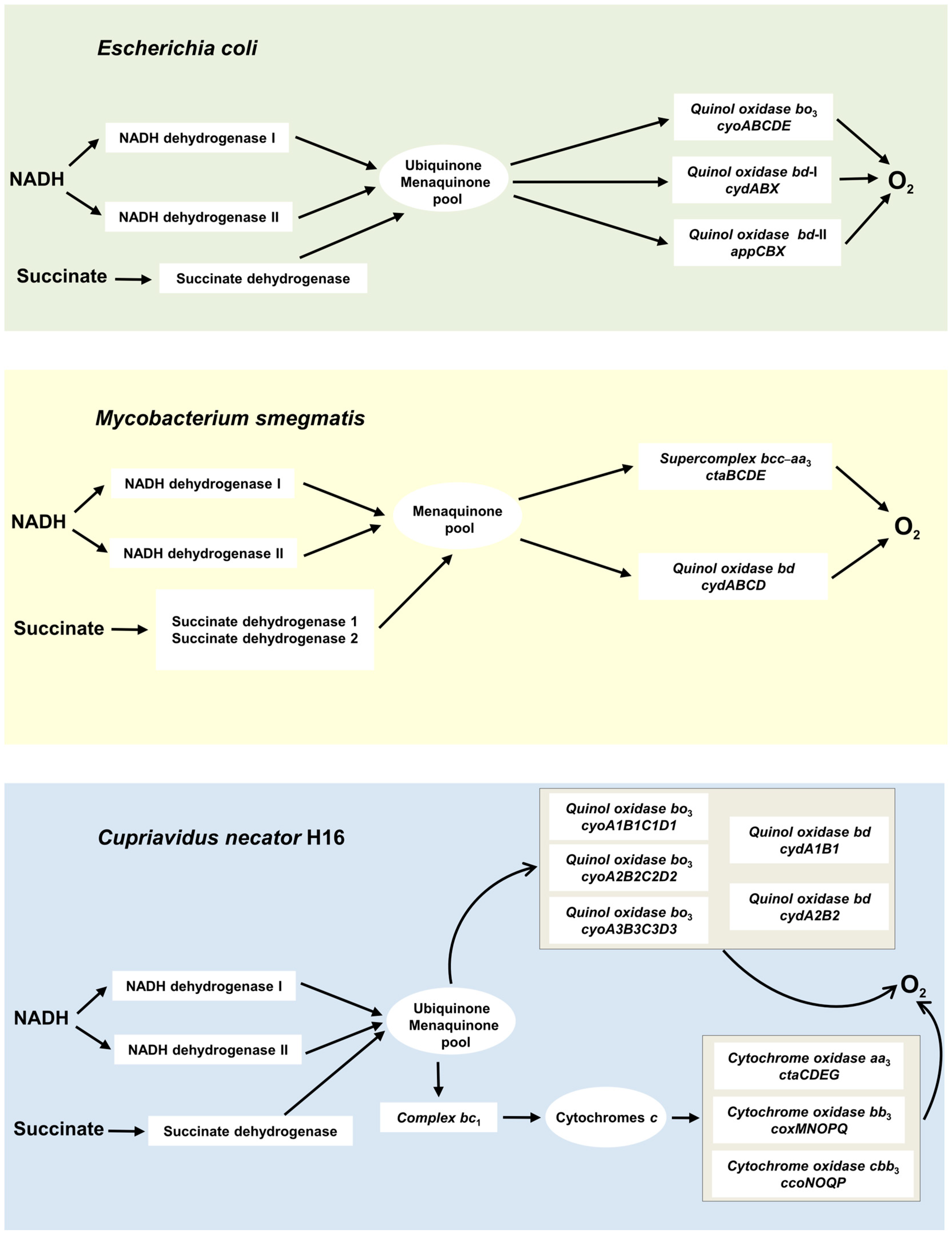
2. Two Superfamilies of Terminal Oxidases

Membrane-bound terminal oxidases are divided into two structurally and evolutionarily unrelated superfamilies: heme–copper oxidases and copper-lacking bd-type oxidases, also called cytochromes bd [50,51,52,53,54,55,56,57,58,59,60,61,62]. The mammalian genome encodes only one respiratory oxidase, the heme–copper aa3-type cytochrome c oxidase [63,64,65,66,67]. In contrast, the aerobic respiratory chains of prokaryotes may contain enzymes from both superfamilies [68,69,70,71,72,73,74,75,76,77,78,79,80,81,82].
A peculiar feature of heme–copper oxidases is the presence of the BNC where molecular oxygen is reduced to water. The BNC comprises two closely located redox-active groups, a high-spin heme (a3, o3, or b3) and a copper ion named CuB. The catalyzed redox reaction is coupled to the PMF generation through the proton-pumping mechanism [83]. The heme–copper oxidases are classified into three families, A, B, and C, based on structural details, primarily the number and type of proton-transfer pathways [84]. The superfamily members can use cytochrome c or quinol as the natural electron donor. Cytochrome c oxidases contain a second copper site called CuA, which is mixed-valence and binuclear, directly accepting electrons from reduced cytochrome c. Quinol oxidases lack CuA. In addition, heme–copper oxidases carry a low-spin heme (a or b) that accepts electrons either from CuA in cytochrome c oxidases or from quinol in quinol oxidases and transfers them to the BNC. The caa3- and cbb3-type oxidases have one or more covalently bound c-type hemes, which serve as additional redox site(s). Heme–copper oxidases can also be associated with other respiratory chain complexes forming supercomplexes [85,86,87,88,89,90].
The bd-type oxidases are encoded by prokaryotic genomes only. All biochemically and structurally characterized cytochromes bd proved to be quinol oxidases. Interestingly, recent phylogenomic analysis suggests that there are bd enzymes which use cytochrome c as an electron donor [51]. The active site, where O2 is reduced to 2H2O, contains a high-spin heme d but no copper [91,92,93,94,95,96,97,98,99]. Cytochromes bd also have two more hemes, a high-spin b595 and a low-spin b558. Heme b558 mediates electron transfer from quinol to hemes b595 and d. The role of heme b595 is still not clear; some data suggest that it could perform some of the functions of CuB [69]. In some cases, heme d can be replaced with a b-type heme [51,100]. The bd-type oxidases produce PMF during the oxygen-reduction reaction but do not pump protons [101,102]. Cytochromes bd usually display a very high affinity for O2 [103,104]. Phylogenomics identified three families and several subfamilies within the bd oxidase superfamily [51]. The earlier classification was based on the size of the hydrophilic region between transmembrane helices 6 and 7 in heme-containing subunit I (often named CydA). This region is a binding domain for quinol oxidation named the Q-loop. Accordingly, cytochromes bd can be divided into two families: L (long Q-loop) and S (short Q-loop) [69,105]. It has to be noted that the latter classification is still commonly used.
The main function of most heme–copper oxidases is energy conservation in the form of PMF. The bd-type oxidases not only produce PMF but perform other essential physiological functions. In particular, they help prokaryotes to adapt to adverse environmental conditions, such as the presence of some antibiotics, hydrogen peroxide, cyanide, nitric oxide, peroxynitrite, sulfide, and ammonia [106,107,108,109,110,111,112,113]. As cytochromes bd are often present in pathogenic bacteria but absent in human mitochondria, they are considered promising protein targets for next-generation antimicrobials [114,115,116,117,118,119,120,121,122,123,124,125,126,127,128].

3. Effect of CO on Bacterial Growth and Aerobic Respiration

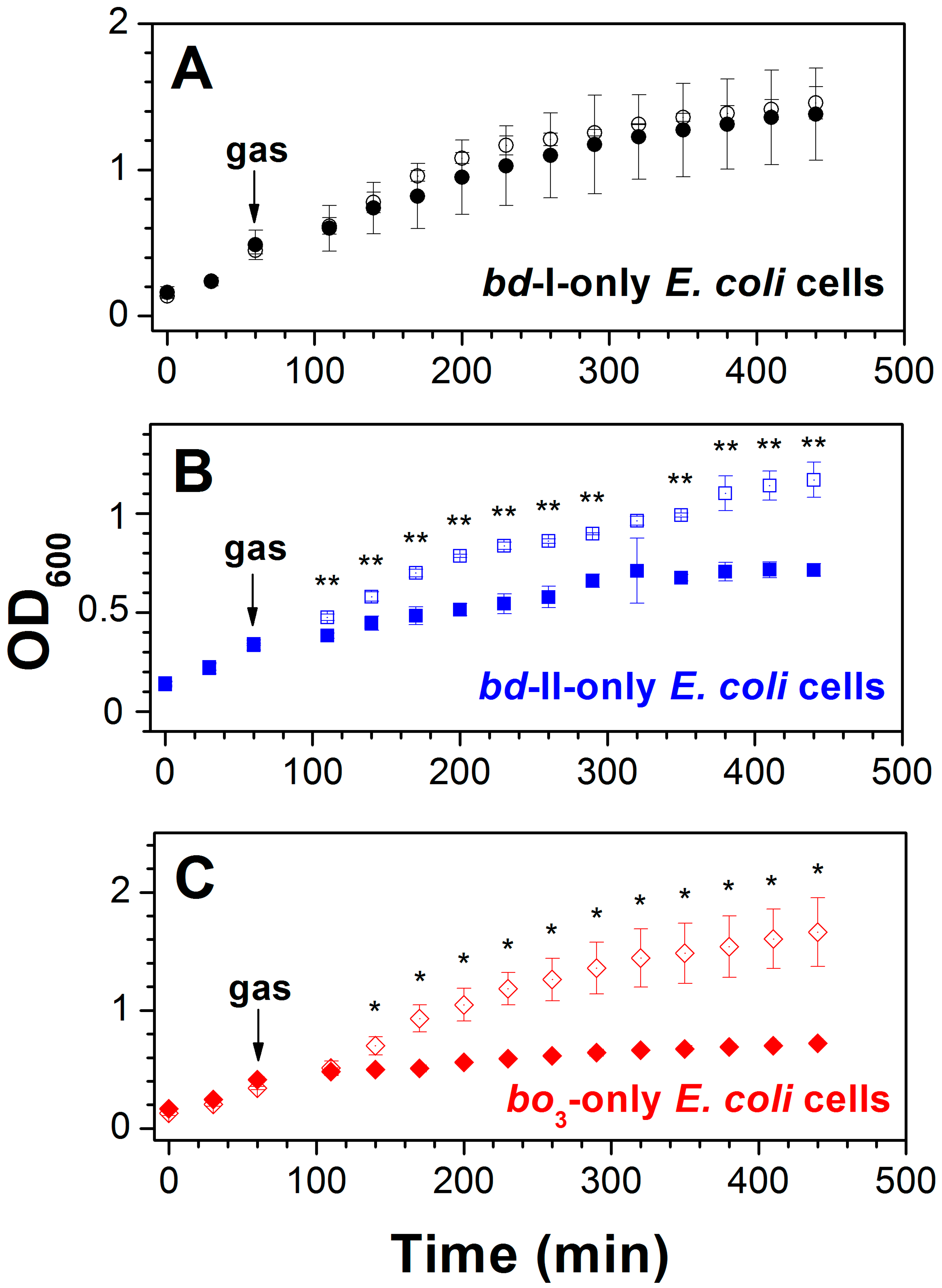
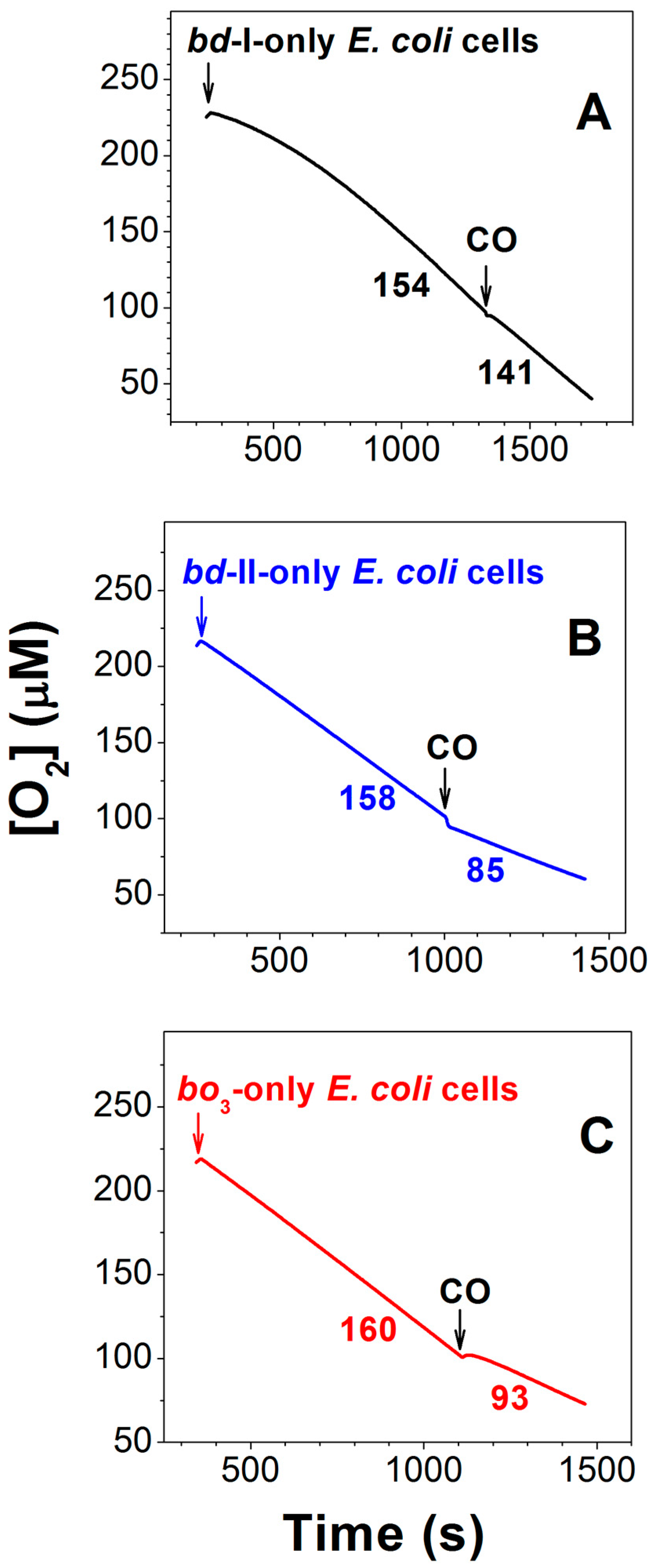
3.1. Effect of CO on E. coli Cell Growth and Aerobic Respiration

The aerobic respiratory chain of E. coli is branched and terminates with three different quinol oxidases, one heme–copper, bo3, and two bd-types, bd-I and bd-II [101] (Figure 6). The bo3, bd-I, and bd-II enzymes are encoded by the cyoABCDE, cydABX, and appCBX operons, respectively. Working with cells and isolated membranes, Nastasi et al. [129,130] found that these oxidases are differently sensitive to CO and consequently can affect E. coli growth and aerobic respiration in the presence of the gas under different conditions. The impact of CO on bacterial growth was examined by using three different mutant strains [130]. Each mutant strain expresses only one quinol oxidase, either bd-I or bd-II, or bo3. Cell growth of the mutant strains was observed in the presence of either ~20% CO or ~20% N2 as a control (Figure 7). The CO addition to the strain possessing bd-I as the sole quinol oxidase has little effect on cell growth (Figure 7A). In contrast, the growth of bacterial cells containing either bd-II or bo3 as the only oxidase was severely impaired following the addition of CO, as compared to the control with N2 (Figure 7B,C). Therefore, we can conclude that cytochrome bd-I promotes E. coli growth in the presence of CO, whereas neither cytochrome bd-II nor cytochrome bo3 helps growing bacteria to tolerate CO.
The effect of CO on the aerobic respiration of bacterial cells of the three mutant strains was also studied. The respiration was sustained by endogenous respiratory substrates. For this reason, the addition of an exogenous electron donor was not needed. As shown in Figure 8, the inhibition of O2 consumption of bd-I-only cells by 96.3 μM CO added at 100 μM O2 is small (Panel A), whereas it is much stronger in the case of bd-II-only and bo3-only cells (panels B and C). The CO inhibition of cell respiration of the three mutants was studied in detail. CO titration experiments were performed at four different O2 concentrations: 50, 100, 150, and 200 μM [129,130]. At all oxygen concentrations tested, the quinol oxidase bd-I in cell cultures turned out to be significantly more resistant to CO than the quinol oxidase bd-II or the quinol oxidase bo3. For instance, at 200 μM O2, the maximum inhibition percentage at 196.3 μM CO (the maximum [CO] added) in the case of bd-I-only cells is 9.7 ± 4.9%, whereas the respective values for bd-II-only and bo3-only cells appeared to be 47.0 ± 6.0% and 39.7 ± 11.5% [130]. It is worth noting that the degree of CO inhibition of each E. coli mutant decreases with increasing [O2] [130], suggesting competitive inhibition. In other words, in either E. coli quinol oxidase, CO competes with O2 for binding to the active site under steady-state conditions.
Since the inhibition of O2 consumption of bd-II-only and bo3-only mutant cells by CO was significant, it was possible to determine the apparent half-maximal inhibitory concentrations (IC50) for CO added at different [O2] [129,130]. The IC50 values obtained allowed us to estimate Ki values for CO, which turned out to be 2.5 ± 0.2 µM and 8.4 ± 0.7 µM for bd-II-only and bo3-only E. coli cells, respectively [130].
The effect of CO on the rate of O2 consumption of wild-type E. coli cells was also studied [130]. In agreement with [131,132], Forte et al. [133] showed that at an early growth phase (low OD600) when [O2] is high, cytochrome bo3 is predominantly expressed in wild-type E. coli. However, when the bacterial culture reaches high OD600 and growth conditions become oxygen-limited, there is a prevalent expression of a bd-type oxidase. Consistently, Nastasi et al. [130] reported that aerobic respiration of wild-type cells harvested at high OD600 (high bd-type cytochrome contents) and low OD600 (high bo3-type cytochrome contents) displays low and high sensitivity to CO, respectively.
In addition, Nastasi et al. [130] examined CO inhibition of O2 consumption of membranes isolated from E. coli mutant cells. The results appeared to be very similar to those observed with mutant cells. O2 consumption of the bd-I-containing membranes is relatively resistant to inhibition by CO, while the same reaction catalyzed by both bd-II- and bo3-containing membranes is strongly inhibited by the gas.
Previously, the transcriptomic analysis of wild-type E. coli exposed to CO [134] showed that under aerobic conditions the expression of the cyoABCDE operon decreases five to ten times, while the expression of the cydABX operon increases fourfold. The expression of the appCBX operon under the same conditions changes slightly. This is consistent with the data reported by Nastasi et al. [129,130]. Indeed, it sounds logical that after the addition of CO to the wild-type E. coli cells, the CO-sensitive cytochrome bo3 is downregulated, whereas the CO-insensitive cytochrome bd-I is upregulated. Cytochrome bd-II is not expressed under aerobic conditions, and its upregulation is not required as it is sensitive to the gas.
Thus, the membrane-bound quinol oxidase bd-I apparently endows E. coli with CO resistance, allowing for aerobic growth and respiration in the presence of the gas at toxic concentrations. It has to be noted that this finding contradicts earlier work [135] showing that O2 consumption by the isolated bd-I enzyme is sensitive to inhibition by CO. As discussed in [130], the inconsistency likely originated from the difference in the cytochrome bd-I environment, detergent micelles in [135] versus a natural lipid bilayer in [129,130].

3.2. Effect of CO on M. smegmatis Cell Growth and Aerobic Respiration

The branched aerobic respiratory chain of M. smegmatis ends with the cytochrome bcc-aa3 supercomplex, composed of cytochrome bcc and the aa3-type heme–copper cytochrome c oxidase, and the bd-type quinol oxidase [136,137,138] (Figure 6). It has to be noted that M. smegmatis also contains Mo,Cu-CODH to use atmospheric CO as a supplemental energy source. The expression of Mo,Cu-CODH is upregulated when preferred organic energy sources are exhausted. This enhances the long-term survival of the actinobacterium during organic carbon starvation [139]. It is worth noting that CO addition results in enhanced O2 consumption in M. smegmatis [139]. Electrons derived from CO oxidation by Mo,Cu-CODH may be donated to either of the two terminal oxidases of M. smegmatis, thereby coupling Mo,Cu-CODH to the aerobic respiratory chain [140].
Bayly et al. conducted a detailed study of the effect of CO on the growth and aerobic respiration of wild-type and mutant strains of M. smegmatis [140]. While studying the growth of the wild-type cells in the presence of 20% CO, they found that M. smegmatis is initially inhibited by the gas but grows normally after adapting to CO. Proteomic analysis showed that the CydA and CydB subunits of the bd-type quinol oxidase are significantly induced (24- and 4.8-fold) in response to growth in CO, whereas the levels of the bcc-aa3 supercomplex are unaffected. The data on the growth of M. smegmatis strains with genetic deletions of the bcc-aa3 supercomplex (ΔqcrCAB) and cytochrome bdcydAB) in the presence of 20% CO or 20% N2 (as a control) were consistent with those with the wild-type strain. It was shown that the growth rate of the ΔqcrCAB strain does not differ in the presence or absence of CO, whereas the ΔcydAB strain grows significantly slower in CO than in N2 [140]. Interestingly, the proteomic analysis showed that in the absence of CO, the ΔqcrCAB mutant markedly increases synthesis of CydA (52-fold) and CydB (9.6-fold) as compared to the wild-type, probably to compensate for the loss of the bcc-aa3 supercomplex. To validate the growth phenotypes associated with the ΔqcrCAB and ΔcydAB mutants, Bayly et al. used a CRISPR interference (CRISPRi) system to independently repress the expression of qcrC and cydA in a wild-type background strain. It turned out that the growth of qcrC and cydA knockdown strains in the presence and absence of CO is very similar to that observed for the corresponding knockout strains [140]. Thus, the bd enzyme is induced in response to CO and is required for adaptation to growth in CO.
In order to determine the sensitivity of the two different oxidases to CO, Bayly et al. studied amperometrically O2 consumption in wild-type, ΔqcrCAB, and ΔcydAB strains by treating respiring cells with a CO-saturated buffer. In the ΔcydAB mutant, complete inhibition of O2 consumption after CO addition was detected. Inhibition of the wild-type strain appeared to be substantial but less than that for the ΔcydAB mutant. Inhibition of the ΔqcrCAB mutant by CO was not significant [140]. To validate these findings, the authors also repeated the experiments with the qcrC and cydA knockdown strains. It turned out that inhibition of O2 consumption by CO in these cultures was identical to that observed for their knockout equivalents. Thus, in M. smegmatis cell cultures, the O2 reductase activity of cytochrome bd is resistant to CO, whereas the bcc-aa3 supercomplex is strongly inhibited by the gas [140].

3.3. Two bd-Type Terminal Oxidases of C. necator H16 Are Differently Sensitive to CO

The extremely branched aerobic respiratory chain of C. necator H16 is terminated with eight terminal oxidases, three heme–copper cytochrome oxidases, aa3, bb3, and cbb3, and five quinol oxidases which include three heme–copper bo3-type and two bd-type ones [141,142] (Figure 6). The two cytochromes bd are encoded by cydA1B1 and cydA2B2 operons [141].
C. necator H16 can grow heterotrophically using fructose, N-acetylglucosamine, gluconate, and fatty acids as electron donors and carbon sources, and O2, nitrate, nitrite, or dimethyl sulfoxide as electron acceptors. Under chemolithoautotrophic growth conditions, the bacterium can use CO2 as a carbon source, H2 as an electron donor, and O2 as an electron acceptor. Therefore, C. necator H16 can be used as a biocatalyst to produce valuable bioproducts, including polymers and potentially chemicals and fuels [143]. For bioproduct production, the bacterium could utilize synthesis gas, an energy-rich feedstock for microbial fermentation. However, the synthesis gas contains not only CO2 and H2 but also high levels of CO, which the wild-type is unable to metabolize [144].
Wickham-Smith [144] decided to increase the CO resistance of C. necator H16 through adaptive laboratory evolution. To achieve this, the bacterium was continually subcultured in the presence of CO under different growth conditions. Ten individual cultures that evolved heterotrophically with fructose showed a clear growth advantage over the wild-type strain. A mutation detected in all evolved isolates was a single point mutation upstream of the cydA2B2 operon. When the mutation was engineered into the parental H16 strain, it enabled faster growth in the presence of CO. The mutation was shown to increase the cydA2B2 operon expression in C. necator H16, possibly by increasing the promoter strength or by enhancing or inhibiting transcription factor binding [144]. The upregulation of the cydA2B2 transcription appeared to increase CO tolerance under heterotrophic conditions. Notably, expression of cydA2B2, but not cydA1B1, enables cell growth in the presence of CO. Deletion of cydA2B2 had a detrimental effect on CO resistance, and plasmid-based expression of cydA1B1 did not improve CO tolerance [144]. Thus, the data indicate that a bd-type quinol oxidase encoded by cydA2B2 is intrinsically more resistant to CO than other terminal oxidases, including a second cytochrome bd encoded by cydA1B1 [144].

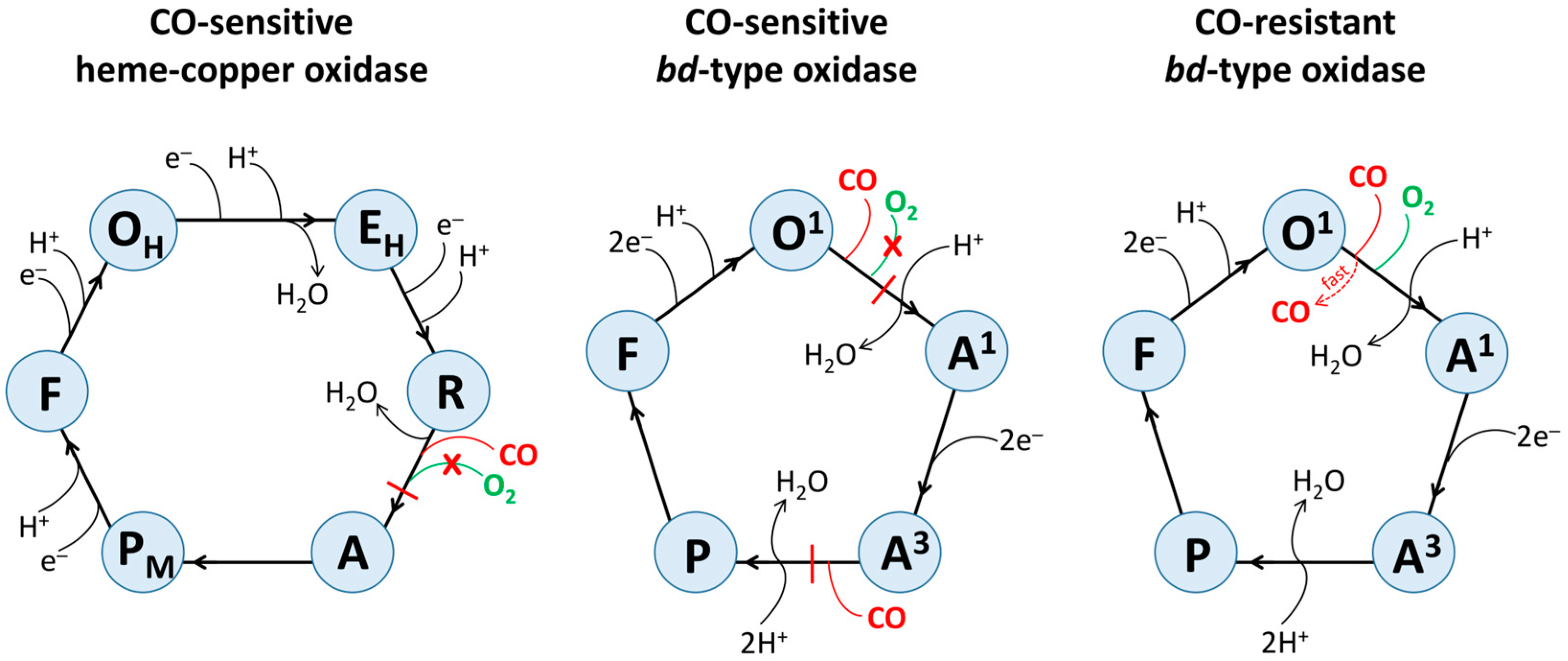
4. Possible Mechanisms Underlying Inhibitory Effects of CO on Different Terminal Oxidases

All heme–copper terminal oxidases studied to date appear to be CO-sensitive [4,5,129,130,140]. In contrast, some copper-lacking bd-type oxidases are CO-resistant, while others seem to be CO-sensitive [129,130,140,144]. The exact molecular mechanisms underlying the inhibitory effects of the gas on different oxidases remain undefined, but it is clear that CO inhibition is strictly competitive with respect to O2. CO directly competes with O2 for binding to the enzyme’s catalytic active site. Consequently, in the case of CO-resistant cytochromes bd, CO binding to the heme d active site should be outcompeted by O2. For CO-sensitive oxidases, the situation is reversed: O2 binding to the catalytic active site (the o3/CuB BNC in cytochrome bo3, the a3/CuB BNC in cytochrome aa3, or the heme d active site in cytochrome bd-II) should be outcompeted by CO.
The difference in the affinity of the enzymes for diatomic gaseous molecules may stem from differences in the structural organization of the active sites and their specific environments, including the chemical structure and geometry of the proximal ligand of the heme as well as its distal amino acid residues [145]. Distal residues can stabilize the bound ligand by weak interactions, such as hydrogen bonds, van der Waals interactions, electrostatic effects, and hydrophobic effects, or on the contrary, cause its destabilization due to steric constraints [145,146].
Regarding the big difference in the susceptibility of different oxidases to CO, it should be noted that the rate of CO dissociation from the high-spin heme in the active site of CO-resistant cytochrome bd-I is ~260-fold higher than that measured for CO-sensitive bovine cytochrome c oxidase (6 s−1 [147] vs. 0.023 s−1 [148] for the fully reduced enzymes). This is probably one of the main reasons for the difference in CO sensitivity of the enzymes. Indeed, the extremely high CO off-rate would result in a prompt restoration of aerobic respiration supported by the bd-I oxidase.
To understand why two oxidases of the same type (bd) located in the same bacterium differ in CO sensitivity, it makes sense to look at the structural differences between them. For bd oxidases encoded by cydA1B1 and cydA2B2 in C. necator H16, this cannot be carried out at this stage. They must first be isolated, purified, and then structurally and functionally characterized. However, structures of E. coli cytochromes bd-I and bd-II were reported [92,93,96,97], and some structural differences can be noticed that might affect the sensitivity of the enzymes to CO. (i) The enzymes differ in the number of subunits. Cytochrome bd-I contains four subunits, CydA, CydB, CydX, CydY, while cytochrome bd-II is composed of three subunits, AppC, AppB, AppX. Interestingly, in the bd-I oxidase, CydY shields heme b595, which is in the high-spin pentacoordinate state, from the lipid bilayer interface. This shielding prevents potential ligands like CO from accessing heme b595 and, therefore, does not allow them to bind to it [92,93]. Indeed, MCD spectroscopy showed that in the bd-I enzyme, heme b595 is resistant to the binding of some ligands [149,150]. In the CydY-lacking bd-II [96,97], direct access of small-molecule ligands to b595 is not hampered, and CO might bind to the ferrous heme in the steady state. This would slow down electron transfer from heme b558 to heme d, which occurs via heme b595, resulting in inhibition of the bd-II enzyme. (ii) There are data showing that in the bd-I and bd-II oxidases, different amino acid residues could serve as the heme d axial ligand [92,96,97]. If true, this could also contribute to the difference in CO sensitivity between the two cytochromes. (iii) Cytochrome bd-I is in the monomeric form, whereas cytochrome bd-II incorporated into amphipols is mainly a dimer [96]. (iv) In E. coli, each of the two bd-type oxidases can associate with other respiratory enzymes, forming supercomplexes. However, for both cytochromes, the composition of such supercomplexes differs. Cytochrome bd-I cooperates with formate dehydrogenase and cytochrome bo3, whereas cytochrome bd-II assembles into a supercomplex together with succinate dehydrogenase [86].
Figure 9 shows possible molecular mechanisms of CO inhibition of the catalytic activity of different terminal oxidases. The catalytic cycle of a heme–copper oxidase includes such intermediates as OH, EH, R, A, PM, and F [55,65,83]. The sequential transfer of two electrons to OH (BNC, consisting of a high-spin heme and CuB, is fully oxidized) leads to sequential formation of EH (BNC is one-electron reduced) and R (BNC is fully reduced). R binds O2 to yield A, the primary diatomic oxygen adduct. Then, the O–O bond is cleaved, and A is converted into PM (its structure is mentioned in the Section 1). The transfer of the third electron to BNC re-reduces the tyrosyl radical in PM, forming F. The transfer of the fourth electron to BNC regenerates OH and completes the cycle. It is worth mentioning that in the A-family heme–copper oxidases, all transitions except R to A and A to PM are electrogenic and coupled to the transfer of a pumped proton.
It is most likely that in a CO-sensitive heme–copper oxidase, CO binds to Fe2+ of a high-spin heme in BNC in the R intermediate producing the Fe2+–CO complex (Figure 9). This prevents the binding of O2 to the CO-bound heme in BNC, thereby inhibiting the entire catalytic reaction.
The catalytic cycle of a bd-type oxidase includes such intermediates as O1, A1, A3, P, and F [102,113] (Figure 9). In O1, heme b558 is reduced, whereas hemes b595 and d are oxidized. In the O1-to-A1 transition, heme d accepts an electron from heme b558 and binds O2. In the A1-to-A3 transition, two electrons from a quinol molecule reduce hemes b558 and b595. In the next A3-to-P transition, heme b595 is oxidized. P is either a true peroxide complex of heme d [151] or a ferryl form of heme d, in which the O–O bond has been cleaved, with π-cation radical on the porphyrin ring [152] or their mixture [102]. Then in the P-to-F transition, a non-radical form of the ferryl complex of heme d is formed, accompanied by the oxidation of heme b558. The transfer of two electrons from another quinol molecule to F regenerates O1 and completes the cycle. The P-to-F and F-to-O1 transitions were shown to be electrogenic [101,151,153].
It can be suggested that in a CO-sensitive bd-type oxidase, the interaction of the gas with O1 stabilizes the electron on heme d, resulting in the d2+–CO complex formation. This prevents the binding of O2 to the CO-ligated heme d2+ and eventually inhibits the enzyme activity (Figure 9). It is also possible that CO binds to heme b5952+ in A3, yielding the b5952+–CO complex. This would stabilize heme b595 in the ferrous state, preventing it from quickly donating an electron to O2 bound to heme d2+ to carry out a concerted four-electron reduction of O2 to 2H2O.
In a CO-resistant bd-type oxidase, CO also reacts with O1, producing the d2+–CO complex. However, in contrast to the CO-sensitive cytochrome bd, in this case, thanks to the very high off-rate [147], CO does not bind with high affinity to heme d2+ and is rapidly ejected from the active site (Figure 9). This results in CO having no significant effect on the catalytic activity of the enzyme. Another factor that possibly contributes to the enzyme’s resistance to CO is the lack of substantial binding of heme b5952+ with the ligand [149,150].

5. Concluding Remarks

The mechanisms that prokaryotes use to resist CO are still unclear. CO-insensitive bd-type terminal oxidases of prokaryotic respiratory chains likely contribute significantly to these mechanisms. For this reason, when using CO-based antimicrobial medicines, it is important to first determine the CO sensitivity of the terminal oxidases on which the disease-causing pathogenic microorganism relies. If a CO-resistant terminal oxidase is found, CO-based therapy alone will be ineffective and should be combined with selective cytochrome bd inhibitors to completely block energy production in the pathogen. In addition, these oxidases may provide a basis for biotechnological applications in which an increased bacterial resistance to CO is needed.

Author Contributions

Conceptualization, V.B.B. and E.F.; writing and editing, V.B.B. and E.F.; supervision, V.B.B. and E.F.; funding acquisition, V.B.B. All authors have read and agreed to the published version of the manuscript.

Funding

This work was supported by the Russian Science Foundation (project No. 24-24-00006, https://rscf.ru/en/project/24-24-00006/) to V.B.B. (accessed on 20 February 2025).

Institutional Review Board Statement

Not applicable.

Informed Consent Statement

Not applicable.

Data Availability Statement

Data sharing is not applicable.

Conflicts of Interest

The authors declare no conflicts of interest.

Abbreviations

BNCbinuclear center
COcarbon monoxide
CODHCO dehydrogenase
CORMcarbon monoxide-releasing molecule
ECHenergy-converting hydrogenase
IC50half-maximal inhibitory concentration
Kiinhibition constant
ODoptical density
PMFproton motive force

References

  1. Delvau, N.; Elens, L.; Penaloza, A.; Liistro, G.; Thys, F.; Roy, P.M.; Gianello, P.; Hantson, P. Carboxyhemoglobin half-life toxicokinetic profiles during and after normobaric oxygen therapy: On a swine model. Toxicol. Rep. 2024, 12, 271–279. [Google Scholar] [CrossRef] [PubMed]
  2. Abramov, A.Y.; Myers, I.; Angelova, P.R. Carbon Monoxide: A Pleiotropic Redox Regulator of Life and Death. Antioxidants 2024, 13, 1121. [Google Scholar] [CrossRef] [PubMed]
  3. Ishigami, I.; Zatsepin, N.A.; Hikita, M.; Conrad, C.E.; Nelson, G.; Coe, J.D.; Basu, S.; Grant, T.D.; Seaberg, M.H.; Sierra, R.G.; et al. Crystal structure of CO-bound cytochrome c oxidase determined by serial femtosecond X-ray crystallography at room temperature. Proc. Natl. Acad. Sci. USA 2017, 114, 8011–8016. [Google Scholar] [CrossRef]
  4. Petersen, L.C. The effect of inhibitors on the oxygen kinetics of cytochrome c oxidase. Biochim. Biophys. Acta 1977, 460, 299–307. [Google Scholar] [CrossRef] [PubMed]
  5. Mason, M.G.; Nicholls, P.; Wilson, M.T.; Cooper, C.E. Nitric oxide inhibition of respiration involves both competitive (heme) and noncompetitive (copper) binding to cytochrome c oxidase. Proc. Natl. Acad. Sci. USA 2006, 103, 708–713. [Google Scholar] [CrossRef]
  6. Wang, T.; Zhang, Y. Mechanisms and therapeutic targets of carbon monoxide poisoning: A focus on reactive oxygen species. Chem. Biol. Interact. 2024, 403, 111223. [Google Scholar] [CrossRef]
  7. Katsnelson, A. The Good Side of Carbon Monoxide. ACS Cent. Sci. 2019, 5, 1632–1635. [Google Scholar] [CrossRef]
  8. Wareham, L.K.; Southam, H.M.; Poole, R.K. Do nitric oxide, carbon monoxide and hydrogen sulfide really qualify as ‘gasotransmitters’ in bacteria? Biochem. Soc. Trans. 2018, 46, 1107–1118. [Google Scholar] [CrossRef]
  9. Yuan, Z.; De La Cruz, L.K.; Yang, X.; Wang, B. Carbon monoxide signaling: Examining its engagement with various molecular targets in the context of binding affinity, concentration, and biologic response. Pharmacol. Rev. 2022, 74, 823–873. [Google Scholar] [CrossRef]
  10. Makino, R.; Obata, Y.; Tsubaki, M.; Iizuka, T.; Hamajima, Y.; Kato-Yamada, Y.; Mashima, K.; Shiro, Y. Mechanistic Insights into the Activation of Soluble Guanylate Cyclase by Carbon Monoxide: A Multistep Mechanism Proposed for the BAY 41-2272 Induced Formation of 5-Coordinate CO-Heme. Biochemistry 2018, 57, 1620–1631. [Google Scholar] [CrossRef]
  11. Kabe, Y.; Yamamoto, T.; Kajimura, M.; Sugiura, Y.; Koike, I.; Ohmura, M.; Nakamura, T.; Tokumoto, Y.; Tsugawa, H.; Handa, H.; et al. Cystathionine beta-synthase and PGRMC1 as CO sensors. Free Radic. Biol. Med. 2016, 99, 333–344. [Google Scholar] [CrossRef] [PubMed]
  12. Uchida, T.; Sato, E.; Sato, A.; Sagami, I.; Shimizu, T.; Kitagawa, T. CO-dependent activity-controlling mechanism of heme-containing CO-sensor protein, neuronal PAS domain protein 2. J. Biol. Chem. 2005, 280, 21358–21368. [Google Scholar] [CrossRef]
  13. Kapetanaki, S.M.; Burton, M.J.; Basran, J.; Uragami, C.; Moody, P.C.E.; Mitcheson, J.S.; Schmid, R.; Davies, N.W.; Dorlet, P.; Vos, M.H.; et al. A mechanism for CO regulation of ion channels. Nat. Commun. 2018, 9, 907. [Google Scholar] [CrossRef] [PubMed]
  14. Schelvis, J.P.M.; Chen, Z.; Messina, M.A.; Catalano, J. Effect of CO binding to P450 BM3 F393 mutants on electron density distribution in the heme cofactor. J. Inorg. Biochem. 2024, 259, 112660. [Google Scholar] [CrossRef] [PubMed]
  15. Nielsen, V.G.; Losada, P.A. Direct Inhibitory Effects of Carbon Monoxide on Six Venoms Containing Fibrinogenolytic Metalloproteinases. Basic Clin. Pharmacol. Toxicol. 2017, 120, 207–212. [Google Scholar] [CrossRef]
  16. Bae, H.; Kim, T.; Lim, I. Carbon monoxide activates large-conductance calcium-activated potassium channels of human cardiac fibroblasts through various mechanisms. Korean J. Physiol. Pharmacol. 2021, 25, 227–237. [Google Scholar] [CrossRef]
  17. Klemz, R.; Reischl, S.; Wallach, T.; Witte, N.; Jurchott, K.; Klemz, S.; Lang, V.; Lorenzen, S.; Knauer, M.; Heidenreich, S.; et al. Reciprocal regulation of carbon monoxide metabolism and the circadian clock. Nat. Struct. Mol. Biol. 2017, 24, 15–22. [Google Scholar] [CrossRef]
  18. Peng, Y.J.; Zhang, X.; Gridina, A.; Chupikova, I.; McCormick, D.L.; Thomas, R.J.; Scammell, T.E.; Kim, G.; Vasavda, C.; Nanduri, J.; et al. Complementary roles of gasotransmitters CO and H2S in sleep apnea. Proc. Natl. Acad. Sci. USA 2017, 114, 1413–1418. [Google Scholar] [CrossRef]
  19. Rahman, F.U.; Park, D.R.; Joe, Y.; Jang, K.Y.; Chung, H.T.; Kim, U.H. Critical Roles of Carbon Monoxide and Nitric Oxide in Ca2+ Signaling for Insulin Secretion in Pancreatic Islets. Antioxid. Redox Signal. 2019, 30, 560–576. [Google Scholar] [CrossRef]
  20. Hopper, C.P.; De La Cruz, L.K.; Lyles, K.V.; Wareham, L.K.; Gilbert, J.A.; Eichenbaum, Z.; Magierowski, M.; Poole, R.K.; Wollborn, J.; Wang, B. Role of carbon monoxide in host-gut microbiome communication. Chem. Rev. 2020, 120, 13273–13311. [Google Scholar] [CrossRef]
  21. Wang, M.; Liao, W. Carbon Monoxide as a Signaling Molecule in Plants. Front. Plant Sci. 2016, 7, 572. [Google Scholar] [CrossRef]
  22. Otterbein, L.E.; May, A.; Chin, B.Y. Carbon monoxide increases macrophage bacterial clearance through Toll-like receptor (TLR)4 expression. Cell. Mol. Biol. 2005, 51, 433–440. [Google Scholar] [PubMed]
  23. Wegiel, B.; Larsen, R.; Gallo, D.; Chin, B.Y.; Harris, C.; Mannam, P.; Kaczmarek, E.; Lee, P.J.; Zuckerbraun, B.S.; Flavell, R.; et al. Macrophages sense and kill bacteria through carbon monoxide-dependent inflammasome activation. J. Clin. Investig. 2014, 124, 4926–4940. [Google Scholar] [CrossRef]
  24. Chung, S.W.; Liu, X.; Macias, A.A.; Baron, R.M.; Perrella, M.A. Heme oxygenase-1-derived carbon monoxide enhances the host defense response to microbial sepsis in mice. J. Clin. Investig. 2008, 118, 239–247. [Google Scholar] [CrossRef]
  25. Lee, S.; Lee, S.J.; Coronata, A.A.; Fredenburgh, L.E.; Chung, S.W.; Perrella, M.A.; Nakahira, K.; Ryter, S.W.; Choi, A.M. Carbon monoxide confers protection in sepsis by enhancing beclin 1-dependent autophagy and phagocytosis. Antioxid. Redox Signal. 2014, 20, 432–442. [Google Scholar] [CrossRef] [PubMed]
  26. Hwang, N.; Ghanta, S.; Li, Q.; Lamattina, A.M.; Murzin, E.; Lederer, J.A.; El-Chemaly, S.; Chung, S.W.; Liu, X.; Perrella, M.A. Carbon monoxide-induced autophagy enhances human mesenchymal stromal cell function via paracrine actions in murine polymicrobial sepsis. Mol. Ther. 2024, 32, 2232–2247. [Google Scholar] [CrossRef]
  27. Ismailova, A.; Kuter, D.; Bohle, D.S.; Butler, I.S. An Overview of the Potential Therapeutic Applications of CO-Releasing Molecules. Bioinorg. Chem. Appl. 2018, 2018, 8547364. [Google Scholar] [CrossRef]
  28. Davidge, K.S.; Sanguinetti, G.; Yee, C.H.; Cox, A.G.; McLeod, C.W.; Monk, C.E.; Mann, B.E.; Motterlini, R.; Poole, R.K. Carbon monoxide-releasing antibacterial molecules target respiration and global transcriptional regulators. J. Biol. Chem. 2009, 284, 4516–4524. [Google Scholar] [CrossRef]
  29. Tavares, A.F.; Parente, M.R.; Justino, M.C.; Oleastro, M.; Nobre, L.S.; Saraiva, L.M. The bactericidal activity of carbon monoxide-releasing molecules against Helicobacter pylori. PLoS ONE 2013, 8, e83157. [Google Scholar] [CrossRef]
  30. Wareham, L.K.; McLean, S.; Begg, R.; Rana, N.; Ali, S.; Kendall, J.J.; Sanguinetti, G.; Mann, B.E.; Poole, R.K. The broad-spectrum antimicrobial potential of [Mn(CO)4(S2CNMe(CH2CO2H))], a water-soluble CO-releasing molecule (CORM-401): Intracellular accumulation, transcriptomic and statistical analyses, and membrane polarization. Antioxid. Redox Signal. 2018, 28, 1286–1308. [Google Scholar] [CrossRef]
  31. Mansour, A.M.; Khaled, R.M.; Ferraro, G.; Shehab, O.R.; Merlino, A. Metal-based carbon monoxide releasing molecules with promising cytotoxic properties. Dalton Trans. 2024, 53, 9612–9656. [Google Scholar] [CrossRef]
  32. Hanson, M.G.; Ambre, R.; Joshi, R.; Amidon, J.D.; Snow, J.B.; Lawless, V.C.; Worrell, B.T. Visible Light Triggerable CO Releasing Micelles. J. Am. Chem. Soc. 2024, 146, 35029–35034. [Google Scholar] [CrossRef] [PubMed]
  33. Yang, G.G.; Liu, W.; Ke, C.; Zhao, Y.Q.; Xu, X. A “turn-on” red cyclometalated iridium (III) complex for long-term tracking the diffusion of CORM-2 in cells and zebrafish. Anal. Chim. Acta 2024, 1288, 342153. [Google Scholar] [CrossRef] [PubMed]
  34. Li, M.; Cheng, J.; He, R.; Chen, K.; Zhang, J.; Liu, X.; Hu, J.; Lu, Y. Red light-induced localized release of carbon monoxide for alleviating postoperative cognitive dysfunction. Biomaterials 2025, 312, 122744. [Google Scholar] [CrossRef]
  35. Choi, Y.K.; Maki, T.; Liang, A.C.; Hayakawa, K.; Koh, S.H.; Kim, Y.M.; Whalen, M.J.; Seo, J.H.; Lok, J.; Gelman, I.H.; et al. A-kinase anchor protein 12 promotes oligodendrogenesis and cognitive recovery in carbon monoxide therapy for traumatic brain injury. J. Cereb. Blood Flow Metab. 2025, 271678X251314371. [Google Scholar] [CrossRef]
  36. Southam, H.M.; Smith, T.W.; Lyon, R.L.; Liao, C.; Trevitt, C.R.; Middlemiss, L.A.; Cox, F.L.; Chapman, J.A.; El-Khamisy, S.F.; Hippler, M.; et al. A thiol-reactive Ru(II) ion, not CO release, underlies the potent antimicrobial and cytotoxic properties of CO-releasing molecule-3. Redox Biol. 2018, 18, 114–123. [Google Scholar] [CrossRef] [PubMed]
  37. Zhuang, P.; Yang, W.; Zhang, Y.; Chen, Y.; Ding, T.; Chen, Y.; Wang, F.; Rosenholm, J.; Li, Y.; Zhang, H.; et al. In situ generating CO gas for destroying bacterial biofilms. Nano Today 2024, 56, 102296. [Google Scholar] [CrossRef]
  38. Sugishima, M.; Wada, K.; Fukuyama, K. Recent Advances in the Understanding of the Reaction Chemistries of the Heme Catabolizing Enzymes HO and BVR Based on High Resolution Protein Structures. Curr. Med. Chem. 2020, 27, 3499–3518. [Google Scholar] [CrossRef]
  39. Dent, M.R.; Weaver, B.R.; Roberts, M.G.; Burstyn, J.N. Carbon monoxide-sensing transcription factors: Regulators of microbial carbon monoxide oxidation pathway gene expression. J. Bacteriol. 2023, 205, e0033222. [Google Scholar] [CrossRef]
  40. Fukuyama, Y.; Inoue, M.; Omae, K.; Yoshida, T.; Sako, Y. Anaerobic and hydrogenogenic carbon monoxide-oxidizing prokaryotes: Versatile microbial conversion of a toxic gas into an available energy. Adv. Appl. Microbiol. 2020, 110, 99–148. [Google Scholar] [CrossRef]
  41. Bahrle, R.; Bohnke, S.; Englhard, J.; Bachmann, J.; Perner, M. Current status of carbon monoxide dehydrogenases (CODH) and their potential for electrochemical applications. Bioresour. Bioprocess. 2023, 10, 84. [Google Scholar] [CrossRef]
  42. Nicholls, P.; Chanady, G.A. Interactions of cytochrome aa3 with oxygen and carbon monoxide. The role of the 607 nm complex. Biochim. Biophys. Acta 1981, 634, 256–265. [Google Scholar] [CrossRef]
  43. Siletsky, S.; Kaulen, A.D.; Konstantinov, A.A. Resolution of electrogenic steps couples to conversion of cytochrome c oxidase from the peroxy to the ferryl-oxo state. Biochemistry 1999, 38, 4853–4861. [Google Scholar] [CrossRef] [PubMed]
  44. Yu, M.A.; Egawa, T.; Shinzawa-Itoh, K.; Yoshikawa, S.; Guallar, V.; Yeh, S.R.; Rousseau, D.L.; Gerfen, G.J. Two tyrosyl radicals stabilize high oxidation states in cytochrome c oxidase for efficient energy conservation and proton translocation. J. Am. Chem. Soc. 2012, 134, 4753–4761. [Google Scholar] [CrossRef]
  45. Tomkova, A.; Cizmar, E.; Jancura, D.; Fabian, M. High stability of the radical at the catalytic center of cytochrome c oxidase. Arch. Biochem. Biophys. 2025, 764, 110271. [Google Scholar] [CrossRef]
  46. Yoshikawa, S.; Shinzawa-Itoh, K.; Nakashima, R.; Yaono, R.; Yamashita, E.; Inoue, N.; Yao, M.; Fei, M.J.; Libeu, C.P.; Mizushima, T.; et al. Redox-coupled crystal structural changes in bovine heart cytochrome c oxidase. Science 1998, 280, 1723–1729. [Google Scholar] [CrossRef] [PubMed]
  47. Roberts, G.P.; Youn, H.; Kerby, R.L. CO-sensing mechanisms. Microbiol. Mol. Biol. Rev. 2004, 68, 453–473. [Google Scholar] [CrossRef]
  48. Mendes, S.S.; Miranda, V.; Saraiva, L.M. Hydrogen sulfide and carbon monoxide tolerance in bacteria. Antioxidants 2021, 10, 729. [Google Scholar] [CrossRef]
  49. Vos, M.H.; Salman, M.; Liebl, U. Early processes in heme-based CO-sensing proteins. Front. Mol. Biosci. 2022, 9, 1046412. [Google Scholar] [CrossRef]
  50. Sousa, F.L.; Alves, R.J.; Ribeiro, M.A.; Pereira-Leal, J.B.; Teixeira, M.; Pereira, M.M. The superfamily of heme-copper oxygen reductases: Types and evolutionary considerations. Biochim. Biophys. Acta 2012, 1817, 629–637. [Google Scholar] [CrossRef]
  51. Murali, R.; Gennis, R.B.; Hemp, J. Evolution of the cytochrome bd oxygen reductase superfamily and the function of CydAA’ in Archaea. ISME J. 2021, 15, 3534–3548. [Google Scholar] [CrossRef]
  52. Murali, R.; Hemp, J.; Gennis, R.B. Evolution of quinol oxidation within the heme-copper oxidoreductase superfamily. Biochim. Biophys. Acta Bioenerg. 2022, 1863, 148907. [Google Scholar] [CrossRef] [PubMed]
  53. van der Velden, T.T.; Kayastha, K.; Waterham, C.Y.J.; Brunle, S.; Jeuken, L.J.C. Menaquinone-specific turnover by M. tuberculosis cytochrome bd is redox regulated by the Q-loop disulfide bond. J. Biol. Chem. 2025, 301, 108094. [Google Scholar] [CrossRef] [PubMed]
  54. Gonzalez-Montalvo, M.A.; Sorescu, J.M.; Baltes, G.; Juarez, O.; Tuz, K. The respiratory chain of Klebsiella aerogenes in urine-like conditions: Critical roles of NDH-2 and bd-terminal oxidases. Front. Microbiol. 2024, 15, 1479714. [Google Scholar] [CrossRef]
  55. Blomberg, M.R.A.; Adelroth, P. Reduction of molecular oxygen in flavodiiron proteins—Catalytic mechanism and comparison to heme-copper oxidases. J. Inorg. Biochem. 2024, 255, 112534. [Google Scholar] [CrossRef] [PubMed]
  56. Janczak, M.; Vilhjalmsdottir, J.; Adelroth, P. Proton transfer in cytochrome bd-I from E. coli involves Asp-105 in CydB. Biochim. Biophys. Acta Bioenerg. 2024, 1865, 149489. [Google Scholar] [CrossRef]
  57. Yi, Y.; Wang, G.; Zhang, W.; Yu, S.; Fei, J.; An, T.; Yi, J.; Li, F.; Huang, T.; Yang, J.; et al. Mitochondrial-cytochrome c oxidase II promotes glutaminolysis to sustain tumor cell survival upon glucose deprivation. Nat. Commun. 2025, 16, 212. [Google Scholar] [CrossRef]
  58. Liang, Y.; Liu, X.; Chang, H.; Yap, J.; Sun, W.; Gao, H. Inhibitory effects of nitrite and sulfite/peroxymonosulfate on bacteria are mediated respectively through respiration and intracellular GSH homeostasis. Microbiol. Res. 2025, 290, 127962. [Google Scholar] [CrossRef]
  59. Smirnova, I.; Wu, F.; Brzezinski, P. Stimulation of cytochrome c oxidase activity by detergents. Biochim. Biophys. Acta Bioenerg. 2025, 1866, 149509. [Google Scholar] [CrossRef]
  60. Chobert, S.C.; Roger-Margueritat, M.; Flandrin, L.; Berraies, S.; Lefevre, C.T.; Pelosi, L.; Junier, I.; Varoquaux, N.; Pierrel, F.; Abby, S.S. Dynamic quinone repertoire accompanied the diversification of energy metabolism in Pseudomonadota. Isme J. 2025, 19, wrae253. [Google Scholar] [CrossRef]
  61. Srivastav, S.; Biswas, A.; Anand, A. Interplay of niche and respiratory network in shaping bacterial colonization. J. Biol. Chem. 2025, 301, 108052. [Google Scholar] [CrossRef] [PubMed]
  62. Inskeep, W.P.; Jay, Z.J.; McKay, L.J.; Dlakic, M. Respiratory processes of early-evolved hyperthermophiles in sulfidic and low-oxygen geothermal microbial communities. Nat. Commun. 2025, 16, 277. [Google Scholar] [CrossRef]
  63. Vogt, S.; Ramzan, R.; Grossman, L.I.; Singh, K.K.; Ferguson-Miller, S.; Yoshikawa, S.; Lee, I.; Huttemann, M. Mitochondrial respiration is controlled by allostery, subunit composition and phosphorylation sites of cytochrome c oxidase: A trailblazer’s tale—Bernhard Kadenbach. Mitochondrion 2021, 60, 228–233. [Google Scholar] [CrossRef]
  64. Capitanio, G.; Papa, F.; Papa, S. The allosteric protein interactions in the proton-motive function of mammalian redox enzymes of the respiratory chain. Biochimie 2021, 189, 1–12. [Google Scholar] [CrossRef]
  65. Wikstrom, M.; Gennis, R.B.; Rich, P.R. Structures of the intermediates in the catalytic cycle of mitochondrial cytochrome c oxidase. Biochim. Biophys. Acta. Bioenerg. 2023, 1864, 148933. [Google Scholar] [CrossRef] [PubMed]
  66. Shimada, A.; Tsukihara, T.; Yoshikawa, S. Recent progress in experimental studies on the catalytic mechanism of cytochrome c oxidase. Front. Chem. 2023, 11, 1108190. [Google Scholar] [CrossRef]
  67. Rousseau, D.L.; Ishigami, I.; Yeh, S.R. Structural and functional mechanisms of cytochrome c oxidase. J. Inorg. Biochem. 2025, 262, 112730. [Google Scholar] [CrossRef] [PubMed]
  68. Poole, R.K.; Cook, G.M. Redundancy of aerobic respiratory chains in bacteria? Routes, reasons and regulation. Adv. Microb. Physiol. 2000, 43, 165–224. [Google Scholar] [CrossRef]
  69. Borisov, V.B.; Gennis, R.B.; Hemp, J.; Verkhovsky, M.I. The cytochrome bd respiratory oxygen reductases. Biochim. Biophys. Acta 2011, 1807, 1398–1413. [Google Scholar] [CrossRef]
  70. Kaila, V.R.I.; Wikstrom, M. Architecture of bacterial respiratory chains. Nat. Rev. Microbiol. 2021, 19, 319–330. [Google Scholar] [CrossRef]
  71. Li, J.; Han, L.; Vallese, F.; Ding, Z.; Choi, S.K.; Hong, S.; Luo, Y.; Liu, B.; Chan, C.K.; Tajkhorshid, E.; et al. Cryo-EM structures of Escherichia coli cytochrome bo3 reveal bound phospholipids and ubiquinone-8 in a dynamic substrate binding site. Proc. Natl. Acad. Sci. USA 2021, 118, e2106750118. [Google Scholar] [CrossRef]
  72. Siletsky, S.A.; Soulimane, T.; Belevich, I.; Gennis, R.B.; Wikstrom, M. Specific inhibition of proton pumping by the T315V mutation in the K channel of cytochrome ba3 from Thermus thermophilus. Biochim. Biophys. Acta Bioenerg. 2021, 1862, 148450. [Google Scholar] [CrossRef]
  73. Hederstedt, L. Diversity of cytochrome c oxidase assembly proteins in bacteria. Microorganisms 2022, 10, 926. [Google Scholar] [CrossRef]
  74. Makarchuk, I.; Gerasimova, T.; Kagi, J.; Wohlwend, D.; Melin, F.; Friedrich, T.; Hellwig, P. Mutating the environment of heme b595 of E. coli cytochrome bd-I oxidase shifts its redox potential by 200 mV without inactivating the enzyme. Bioelectrochemistry 2023, 151, 108379. [Google Scholar] [CrossRef] [PubMed]
  75. Makarchuk, I.; Kagi, J.; Gerasimova, T.; Wohlwend, D.; Friedrich, T.; Melin, F.; Hellwig, P. pH-dependent kinetics of NO release from E. coli bd-I and bd-II oxidase reveals involvement of Asp/Glu58(B). Biochim. Biophys. Acta Bioenerg. 2023, 1864, 148952. [Google Scholar] [CrossRef]
  76. Gao, Y.; Zhang, Y.; Hakke, S.; Mohren, R.; Sijbers, L.; Peters, P.J.; Ravelli, R.B.G. Cryo-EM structure of cytochrome bo(3) quinol oxidase assembled in peptidiscs reveals an “open” conformation for potential ubiquinone-8 release. Biochim. Biophys. Acta Bioenerg. 2024, 1865, 149045. [Google Scholar] [CrossRef]
  77. Deutschmann, S.; Tauber, S.T.; Rimle, L.; Biner, O.; Schori, M.; Stanic, A.M.; von Ballmoos, C. Modulating Liposome Surface Charge for Maximized ATP Regeneration in Synthetic Nanovesicles. ACS Synth. Biol. 2024, 13, 4061–4073. [Google Scholar] [CrossRef] [PubMed]
  78. Patil, A.V.; Shirsath, A.M.; Anand, A. Dioxygen reductase heterogeneity is crucial for robust aerobic growth physiology of Escherichia coli. iScience 2024, 27, 111498. [Google Scholar] [CrossRef]
  79. Rauch, J.; Kurscheidt, K.; Shen, K.W.; Andrei, A.; Daum, N.; Ozturk, Y.; Melin, F.; Layer, G.; Hellwig, P.; Daldal, F.; et al. The small membrane protein CcoS is involved in cofactor insertion into the cbb(3)-type cytochrome c oxidase. Biochim. Biophys. Acta Bioenerg. 2025, 1866, 149524. [Google Scholar] [CrossRef]
  80. Siddeeque, R.; Heger, L.; Kagi, J.; Friedrich, T.; Melin, F.; Hellwig, P. Interplay of acidic residues in the proton channel of E. coli cytochrome bd-I oxidase to promote oxygen reduction and NO release. Biochim. Biophys. Acta Bioenerg. 2025, 1866, 149537. [Google Scholar] [CrossRef]
  81. Paszti, S.; Biner, O.; Liu, Y.; Bolli, K.; Jeggli, S.D.; Pessi, G.; Eberl, L. Insights into the diverse roles of the terminal oxidases in Burkholderia cenocepacia H111. Sci. Rep. 2025, 15, 2390. [Google Scholar] [CrossRef] [PubMed]
  82. Batista, B.B.; de Lima, V.M.; Will, W.R.; Fang, F.C.; da Silva Neto, J.F. A cytochrome bd repressed by a MarR family regulator confers resistance to metals, nitric oxide, sulfide, and cyanide in Chromobacterium violaceum. Appl. Environ. Microbiol. 2025, 91, e02360-24. [Google Scholar] [CrossRef] [PubMed]
  83. Siletsky, S.A. Investigation of the Mechanism of Membrane Potential Generation by Heme-Copper Respiratory Oxidases in a Real Time Mode. Biochemistry 2023, 88, 1513–1527. [Google Scholar] [CrossRef]
  84. Pereira, M.M.; Teixeira, M. Proton pathways, ligand binding and dynamics of the catalytic site in haem-copper oxygen reductases: A comparison between the three families. Biochim. Biophys. Acta 2004, 1655, 340–346. [Google Scholar] [CrossRef]
  85. Letts, J.A.; Fiedorczuk, K.; Sazanov, L.A. The architecture of respiratory supercomplexes. Nature 2016, 537, 644–648. [Google Scholar] [CrossRef]
  86. Melo, A.M.; Teixeira, M. Supramolecular organization of bacterial aerobic respiratory chains: From cells and back. Biochim. Biophys. Acta 2016, 1857, 190–197. [Google Scholar] [CrossRef]
  87. Moe, A.; Dimogkioka, A.R.; Rapaport, D.; Ojemyr, L.N.; Brzezinski, P. Structure and function of the S. pombe III-IV-cyt c supercomplex. Proc. Natl. Acad. Sci. USA 2023, 120, e2307697120. [Google Scholar] [CrossRef] [PubMed]
  88. Di Trani, J.M.; Gheorghita, A.A.; Turner, M.; Brzezinski, P.; Adelroth, P.; Vahidi, S.; Howell, P.L.; Rubinstein, J.L. Structure of the bc1-cbb3 respiratory supercomplex from Pseudomonas aeruginosa. Proc. Natl. Acad. Sci. USA 2023, 120, e2307093120. [Google Scholar] [CrossRef] [PubMed]
  89. Lobez, A.P.; Wu, F.; Di Trani, J.M.; Rubinstein, J.L.; Oliveberg, M.; Brzezinski, P.; Moe, A. Electron transfer in the respiratory chain at low salinity. Nat. Commun. 2024, 15, 8241. [Google Scholar] [CrossRef]
  90. Riepl, D.; Gamiz-Hernandez, A.P.; Kovalova, T.; Krol, S.M.; Mader, S.L.; Sjostrand, D.; Hogbom, M.; Brzezinski, P.; Kaila, V.R.I. Long-range charge transfer mechanism of the III(2)IV(2) mycobacterial supercomplex. Nat. Commun. 2024, 15, 5276. [Google Scholar] [CrossRef]
  91. Safarian, S.; Rajendran, C.; Muller, H.; Preu, J.; Langer, J.D.; Ovchinnikov, S.; Hirose, T.; Kusumoto, T.; Sakamoto, J.; Michel, H. Structure of a bd oxidase indicates similar mechanisms for membrane-integrated oxygen reductases. Science 2016, 352, 583–586. [Google Scholar] [CrossRef] [PubMed]
  92. Thesseling, A.; Rasmussen, T.; Burschel, S.; Wohlwend, D.; Kagi, J.; Muller, R.; Bottcher, B.; Friedrich, T. Homologous bd oxidases share the same architecture but differ in mechanism. Nat. Commun. 2019, 10, 5138. [Google Scholar] [CrossRef] [PubMed]
  93. Safarian, S.; Hahn, A.; Mills, D.J.; Radloff, M.; Eisinger, M.L.; Nikolaev, A.; Meier-Credo, J.; Melin, F.; Miyoshi, H.; Gennis, R.B.; et al. Active site rearrangement and structural divergence in prokaryotic respiratory oxidases. Science 2019, 366, 100–104. [Google Scholar] [CrossRef]
  94. Wang, W.; Gao, Y.; Tang, Y.; Zhou, X.; Lai, Y.; Zhou, S.; Zhang, Y.; Yang, X.; Liu, F.; Guddat, L.W.; et al. Cryo-EM structure of mycobacterial cytochrome bd reveals two oxygen access channels. Nat. Commun. 2021, 12, 4621. [Google Scholar] [CrossRef]
  95. Safarian, S.; Opel-Reading, H.K.; Wu, D.; Mehdipour, A.R.; Hards, K.; Harold, L.K.; Radloff, M.; Stewart, I.; Welsch, S.; Hummer, G.; et al. The cryo-EM structure of the bd oxidase from M. tuberculosis reveals a unique structural framework and enables rational drug design to combat TB. Nat. Commun. 2021, 12, 5236. [Google Scholar] [CrossRef]
  96. Grauel, A.; Kagi, J.; Rasmussen, T.; Makarchuk, I.; Oppermann, S.; Moumbock, A.F.A.; Wohlwend, D.; Muller, R.; Melin, F.; Gunther, S.; et al. Structure of Escherichia coli cytochrome bd-II type oxidase with bound aurachin D. Nat. Commun. 2021, 12, 6498. [Google Scholar] [CrossRef]
  97. Grund, T.N.; Radloff, M.; Wu, D.; Goojani, H.G.; Witte, L.F.; Josting, W.; Buschmann, S.; Muller, H.; Elamri, I.; Welsch, S.; et al. Mechanistic and structural diversity between cytochrome bd isoforms of Escherichia coli. Proc. Natl. Acad. Sci. USA 2021, 118, e2114013118. [Google Scholar] [CrossRef]
  98. Friedrich, T.; Wohlwend, D.; Borisov, V.B. Recent advances in structural studies of cytochrome bd and its potential application as a drug target. Int. J. Mol. Sci. 2022, 23, 3166. [Google Scholar] [CrossRef] [PubMed]
  99. Grund, T.N.; Kabashima, Y.; Kusumoto, T.; Wu, D.; Welsch, S.; Sakamoto, J.; Michel, H.; Safarian, S. The cryoEM structure of cytochrome bd from C. glutamicum provides novel insights into structural properties of actinobacterial terminal oxidases. Front. Chem. 2023, 10, 1085463. [Google Scholar] [CrossRef]
  100. Nastasi, M.R.; Caruso, L.; Giordano, F.; Mellini, M.; Rampioni, G.; Giuffre, A.; Forte, E. Cyanide insensitive oxidase confers hydrogen sulfide and nitric oxide tolerance to Pseudomonas aeruginosa aerobic respiration. Antioxidants 2024, 13, 383. [Google Scholar] [CrossRef]
  101. Borisov, V.B.; Murali, R.; Verkhovskaya, M.L.; Bloch, D.A.; Han, H.; Gennis, R.B.; Verkhovsky, M.I. Aerobic respiratory chain of Escherichia coli is not allowed to work in fully uncoupled mode. Proc. Natl. Acad. Sci. USA 2011, 108, 17320–17324. [Google Scholar] [CrossRef]
  102. Borisov, V.B. Generation of membrane potential by cytochrome bd. Biochemistry 2023, 88, 1504–1512. [Google Scholar] [CrossRef] [PubMed]
  103. D’mello, R.; Hill, S.; Poole, R.K. The cytochrome bd quinol oxidase in Escherichia coli has an extremely high oxygen affinity and two-oxygen-binding haems: Implicaitons for regulation of activity in vivo by oxygen inihibition. Microbiology 1996, 142, 755–763. [Google Scholar] [CrossRef] [PubMed]
  104. Belevich, I.; Borisov, V.B.; Konstantinov, A.A.; Verkhovsky, M.I. Oxygenated complex of cytochrome bd from Escherichia coli: Stability and photolability. FEBS Lett. 2005, 579, 4567–4570. [Google Scholar] [CrossRef]
  105. Arutyunyan, A.M.; Sakamoto, J.; Inadome, M.; Kabashima, Y.; Borisov, V.B. Optical and magneto-optical activity of cytochrome bd from Geobacillus thermodenitrificans. Biochim. Biophys. Acta 2012, 1817, 2087–2094. [Google Scholar] [CrossRef] [PubMed]
  106. Forte, E.; Borisov, V.B.; Vicente, J.B.; Giuffre, A. Cytochrome bd and gaseous ligands in bacterial physiology. Adv. Microb. Physiol. 2017, 71, 171–234. [Google Scholar] [CrossRef]
  107. Borisov, V.B.; Siletsky, S.A.; Paiardini, A.; Hoogewijs, D.; Forte, E.; Giuffre, A.; Poole, R.K. Bacterial oxidases of the cytochrome bd family: Redox enzymes of unique structure, function and utility as drug targets. Antioxid. Redox Signal. 2021, 34, 1280–1318. [Google Scholar] [CrossRef]
  108. Borisov, V.B.; Forte, E. Terminal oxidase cytochrome bd protects bacteria against hydrogen sulfide toxicity. Biochemistry 2021, 86, 22–32. [Google Scholar] [CrossRef]
  109. Borisov, V.B.; Siletsky, S.A.; Nastasi, M.R.; Forte, E. ROS defense systems and terminal oxidases in bacteria. Antioxidants 2021, 10, 839. [Google Scholar] [CrossRef]
  110. Borisov, V.B.; Forte, E. Impact of hydrogen sulfide on mitochondrial and bacterial bioenergetics. Int. J. Mol. Sci. 2021, 22, 12688. [Google Scholar] [CrossRef]
  111. Seregina, T.A.; Lobanov, K.V.; Shakulov, R.S.; Mironov, A.S. Inactivation of terminal oxidase bd-I leads to supersensitivity of E. coli to quinolone and beta-lactam antibiotics. Mol. Biol. 2022, 56, 619–627. [Google Scholar] [CrossRef]
  112. Borisov, V.B.; Forte, E. Bioenergetics and reactive nitrogen species in bacteria. Int. J. Mol. Sci. 2022, 23, 7321. [Google Scholar] [CrossRef] [PubMed]
  113. Borisov, V.B.; Giardina, G.; Pistoia, G.; Forte, E. Cytochrome bd-type oxidases and environmental stressors in microbial physiology. Adv. Microb. Physiol. 2025, in press. [Google Scholar] [CrossRef]
  114. Lee, B.S.; Hards, K.; Engelhart, C.A.; Hasenoehrl, E.J.; Kalia, N.P.; Mackenzie, J.S.; Sviriaeva, E.; Chong, S.M.S.; Manimekalai, M.S.S.; Koh, V.H.; et al. Dual inhibition of the terminal oxidases eradicates antibiotic-tolerant Mycobacterium tuberculosis. EMBO Mol. Med. 2021, 13, e13207. [Google Scholar] [CrossRef] [PubMed]
  115. Radloff, M.; Elamri, I.; Grund, T.N.; Witte, L.F.; Hohmann, K.F.; Nakagaki, S.; Goojani, H.G.; Nasiri, H.; Hideto, M.; Bald, D.; et al. Short-chain aurachin D derivatives are selective inhibitors of E. coli cytochrome bd-I and bd-II oxidases. Sci. Rep. 2021, 11, 23852. [Google Scholar] [CrossRef]
  116. Makarchuk, I.; Nikolaev, A.; Thesseling, A.; Dejon, L.; Lamberty, D.; Stief, L.; Speicher, A.; Friedrich, T.; Hellwig, P.; Nasiri, H.R.; et al. Identification and optimization of quinolone-based inhibitors against cytochrome bd oxidase using an electrochemical assay. Electrochim. Acta 2021, 381, 138293. [Google Scholar] [CrossRef]
  117. Harikishore, A.; Chong, S.S.M.; Ragunathan, P.; Bates, R.W.; Gruber, G. Targeting the menaquinol binding loop of mycobacterial cytochrome bd oxidase. Mol. Divers. 2021, 25, 517–524. [Google Scholar] [CrossRef]
  118. Hopfner, S.M.; Lee, B.S.; Kalia, N.P.; Miller, M.J.; Pethe, K.; Moraski, G.C. Structure guided generation of thieno[3,2-d]pyrimidin-4-amine Mycobacterium tuberculosis bd oxidase inhibitors. RSC Med. Chem. 2021, 12, 73–77. [Google Scholar] [CrossRef]
  119. Anand, P.; Akhter, Y. A review on enzyme complexes of electron transport chain from Mycobacterium tuberculosis as promising drug targets. Int. J. Biol. Macromol. 2022, 212, 474–494. [Google Scholar] [CrossRef]
  120. Hards, K.; Cheung, C.Y.; Waller, N.; Adolph, C.; Keighley, L.; Tee, Z.S.; Harold, L.K.; Menorca, A.; Bujaroski, R.S.; Buckley, B.J.; et al. An amiloride derivative is active against the F1Fo-ATP synthase and cytochrome bd oxidase of Mycobacterium tuberculosis. Commun. Biol. 2022, 5, 166. [Google Scholar] [CrossRef]
  121. Harikishore, A.; Mathiyazakan, V.; Pethe, K.; Gruber, G. Novel targets and inhibitors of the Mycobacterium tuberculosis cytochrome bd oxidase to foster anti-tuberculosis drug discovery. Expert Opin. Drug Discov. 2023, 18, 917–927. [Google Scholar] [CrossRef] [PubMed]
  122. Capela, R.; Felix, R.; Clariano, M.; Nunes, D.; Perry, M.J.; Lopes, F. Target Identification in Anti-Tuberculosis Drug Discovery. Int. J. Mol. Sci. 2023, 24, 10482. [Google Scholar] [CrossRef] [PubMed]
  123. Jeffreys, L.N.; Ardrey, A.; Hafiz, T.A.; Dyer, L.A.; Warman, A.J.; Mosallam, N.; Nixon, G.L.; Fisher, N.E.; Hong, W.D.; Leung, S.C.; et al. Identification of 2-aryl-quinolone inhibitors of cytochrome bd and chemical validation of combination strategies for respiratory inhibitors against Mycobacterium tuberculosis. ACS Infect. Dis. 2023, 9, 221–238. [Google Scholar] [CrossRef]
  124. Kagi, J.; Sloan, W.; Schimpf, J.; Nasiri, H.R.; Lashley, D.; Friedrich, T. Exploring ND-011992, a quinazoline-type inhibitor targeting quinone reductases and quinol oxidases. Sci. Rep. 2023, 13, 12226. [Google Scholar] [CrossRef]
  125. Zhou, Y.; Shao, M.; Wang, W.; Cheung, C.Y.; Wu, Y.; Yu, H.; Hu, X.; Cook, G.M.; Gong, H.; Lu, X. Discovery of 1-hydroxy-2-methylquinolin-4(1H)-one derivatives as new cytochrome bd oxidase inhibitors for tuberculosis therapy. Eur. J. Med. Chem. 2023, 245, 114896. [Google Scholar] [CrossRef]
  126. Henry, S.A.; Webster, C.M.; Shaw, L.N.; Torres, N.J.; Jobson, M.E.; Totzke, B.C.; Jackson, J.K.; McGreig, J.E.; Wass, M.N.; Robinson, G.K.; et al. Steroid drugs inhibit bacterial respiratory oxidases and are lethal toward methicillin-resistant Staphylococcus aureus. J. Infect. Dis. 2024, 230, e149–e158. [Google Scholar] [CrossRef] [PubMed]
  127. Saha, P.; Das, S.; Indurthi, H.K.; Kumar, R.; Roy, A.; Kalia, N.P.; Sharma, D.K. Cytochrome bd oxidase: An emerging anti-tubercular drug target. RSC Med. Chem. 2024, 15, 769–787. [Google Scholar] [CrossRef]
  128. Seitz, C.; Ahn, S.H.; Wei, H.; Kyte, M.; Cook, G.M.; Krause, K.L.; McCammon, J.A. Targeting Tuberculosis: Novel Scaffolds for Inhibiting Cytochrome bd Oxidase. J. Chem. Inf. Model. 2024, 64, 5232–5241. [Google Scholar] [CrossRef]
  129. Nastasi, M.R.; Borisov, V.B.; Forte, E. The terminal oxidase cytochrome bd-I confers carbon monoxide resistance to Escherichia coli cells. J. Inorg. Biochem. 2023, 247, 112341. [Google Scholar] [CrossRef]
  130. Nastasi, M.R.; Borisov, V.B.; Forte, E. Membrane-bound redox enzyme cytochrome bd-I promotes carbon monoxide-resistant Escherichia coli growth and respiration. Int. J. Mol. Sci. 2024, 25, 1277. [Google Scholar] [CrossRef]
  131. Alexeeva, S.; Hellingwerf, K.; Teixeira de Mattos, M.J. Quantitative assessment of oxygen availability: Perceived aerobiosis and its effect on flux distribution in the respiratory chain of Escherichia coli. J. Bacteriol. 2002, 184, 1402–1406. [Google Scholar] [CrossRef] [PubMed]
  132. Rolfe, M.D.; Ter Beek, A.; Graham, A.I.; Trotter, E.W.; Asif, H.M.; Sanguinetti, G.; de Mattos, J.T.; Poole, R.K.; Green, J. Transcript profiling and inference of Escherichia coli K-12 ArcA activity across the range of physiologically relevant oxygen concentrations. J. Biol. Chem. 2011, 286, 10147–10154. [Google Scholar] [CrossRef] [PubMed]
  133. Forte, E.; Borisov, V.B.; Falabella, M.; Colaco, H.G.; Tinajero-Trejo, M.; Poole, R.K.; Vicente, J.B.; Sarti, P.; Giuffre, A. The terminal oxidase cytochrome bd promotes sulfide-resistant bacterial respiration and growth. Sci. Rep. 2016, 6, 23788. [Google Scholar] [CrossRef]
  134. Wareham, L.K.; Begg, R.; Jesse, H.E.; Van Beilen, J.W.; Ali, S.; Svistunenko, D.; McLean, S.; Hellingwerf, K.J.; Sanguinetti, G.; Poole, R.K. Carbon monoxide gas is not inert, but global, in its consequences for bacterial gene expression, iron acquisition, and antibiotic resistance. Antioxid. Redox Signal. 2016, 24, 1013–1028. [Google Scholar] [CrossRef]
  135. Forte, E.; Borisov, V.B.; Siletsky, S.A.; Petrosino, M.; Giuffre, A. In the respiratory chain of Escherichia coli cytochromes bd-I and bd-II are more sensitive to carbon monoxide inhibition than cytochrome bo3. Biochim. Biophys. Acta Bioenerg. 2019, 1860, 148088. [Google Scholar] [CrossRef]
  136. Cook, G.M.; Hards, K.; Vilcheze, C.; Hartman, T.; Berney, M. Energetics of respiration and oxidative phosphorylation in mycobacteria. Microbiol. Spectr. 2014, 2, MGM2-0015-2013. [Google Scholar] [CrossRef]
  137. Liang, Y.; Plourde, A.; Bueler, S.A.; Liu, J.; Brzezinski, P.; Vahidi, S.; Rubinstein, J.L. Structure of mycobacterial respiratory complex I. Proc. Natl. Acad. Sci. USA 2023, 120, e2214949120. [Google Scholar] [CrossRef]
  138. Kalia, N.P.; Singh, S.; Hards, K.; Cheung, C.Y.; Sviriaeva, E.; Banaei-Esfahani, A.; Aebersold, R.; Berney, M.; Cook, G.M.; Pethe, K.M. Tuberculosis relies on trace oxygen to maintain energy homeostasis and survive in hypoxic environments. Cell Rep. 2023, 42, 112444. [Google Scholar] [CrossRef]
  139. Cordero, P.R.F.; Bayly, K.; Man Leung, P.; Huang, C.; Islam, Z.F.; Schittenhelm, R.B.; King, G.M.; Greening, C. Atmospheric carbon monoxide oxidation is a widespread mechanism supporting microbial survival. ISME J. 2019, 13, 2868–2881. [Google Scholar] [CrossRef]
  140. Bayly, K.; Cordero, P.R.F.; Kropp, A.; Huang, C.; Schittenhelm, R.B.; Grinter, R.; Greening, C. Mycobacteria tolerate carbon monoxide by remodeling their respiratory chain. mSystems 2021, 6, e01292-01220. [Google Scholar] [CrossRef]
  141. Cramm, R. Genomic view of energy metabolism in Ralstonia eutropha H16. J. Mol. Microbiol. Biotechnol. 2009, 16, 38–52. [Google Scholar] [CrossRef] [PubMed]
  142. Jahn, M.; Crang, N.; Gynna, A.H.; Kabova, D.; Frielingsdorf, S.; Lenz, O.; Charpentier, E.; Hudson, E.P. The energy metabolism of Cupriavidus necator in different trophic conditions. Appl. Environ. Microbiol. 2024, 90, e0074824. [Google Scholar] [CrossRef] [PubMed]
  143. Windhorst, C.; Gescher, J. Efficient biochemical production of acetoin from carbon dioxide using Cupriavidus necator H16. Biotechnol. Biofuels 2019, 12, 163. [Google Scholar] [CrossRef]
  144. Wickham-Smith, C.; Malys, N.; Winzer, K. Improving carbon monoxide tolerance of Cupriavidus necator H16 through adaptive laboratory evolution. Front. Bioeng. Biotechnol. 2023, 11, 1178536. [Google Scholar] [CrossRef] [PubMed]
  145. Tsai, A.L.; Berka, V.; Martin, E.; Olson, J.S. A “sliding scale rule” for selectivity among NO, CO, and O2 by heme protein sensors. Biochemistry 2012, 51, 172–186. [Google Scholar] [CrossRef]
  146. Bartlett, G.J.; Newberry, R.W.; VanVeller, B.; Raines, R.T.; Woolfson, D.N. Interplay of hydrogen bonds and n→π* interactions in proteins. J. Am. Chem. Soc. 2013, 135, 18682–18688. [Google Scholar] [CrossRef]
  147. Borisov, V.B.; Forte, E.; Sarti, P.; Brunori, M.; Konstantinov, A.A.; Giuffre, A. Redox control of fast ligand dissociation from Escherichia coli cytochrome bd. Biochem. Biophys. Res. Commun. 2007, 355, 97–102. [Google Scholar] [CrossRef]
  148. Gibson, Q.; Greenwood, C. Reactions of cytochrome oxidase with oxygen and carbon monoxide. Biochem. J. 1963, 86, 541–554. [Google Scholar] [CrossRef]
  149. Borisov, V.; Arutyunyan, A.M.; Osborne, J.P.; Gennis, R.B.; Konstantinov, A.A. Magnetic circular dichroism used to examine the interaction of Escherichia coli cytochrome bd with ligands. Biochemistry 1999, 38, 740–750. [Google Scholar] [CrossRef]
  150. Borisov, V.B.; Arutyunyan, A.M. The fully reduced terminal oxidase bd-I isolated from Escherichia coli binds cyanide. J. Inorg. Biochem. 2024, 259, 112653. [Google Scholar] [CrossRef]
  151. Belevich, I.; Borisov, V.B.; Verkhovsky, M.I. Discovery of the true peroxy intermediate in the catalytic cycle of terminal oxidases by real-time measurement. J. Biol. Chem. 2007, 282, 28514–28519. [Google Scholar] [CrossRef] [PubMed]
  152. Paulus, A.; Rossius, S.G.; Dijk, M.; de Vries, S. Oxoferryl-porphyrin radical catalytic intermediate in cytochrome bd oxidases protects cells from formation of reactive oxygen species. J. Biol. Chem. 2012, 287, 8830–8838. [Google Scholar] [CrossRef] [PubMed]
  153. Belevich, I.; Borisov, V.B.; Zhang, J.; Yang, K.; Konstantinov, A.A.; Gennis, R.B.; Verkhovsky, M.I. Time-resolved electrometric and optical studies on cytochrome bd suggest a mechanism of electron-proton coupling in the di-heme active site. Proc. Natl. Acad. Sci. USA 2005, 102, 3657–3662. [Google Scholar] [CrossRef] [PubMed]
Figure 1. Proposed mechanisms for CO poisoning. The most notable targets of CO include hemoglobin (PDB 1GZX), myoglobin (PDB 3RGK), neuroglobin (PDB 1OJ6), and cytochrome c oxidase (PDB 1V54).
Ijms 26 02809 g001
Figure 2. Structures of metal-based CO-releasing molecules and metal-free organic CO prodrugs. Reprinted with permission from Hopper et al. [20]. Copyright © 2020 American Chemical Society.
Ijms 26 02809 g002
Figure 3. Endogenous CO is a byproduct in the reaction catalyzed by heme oxygenase (HO), in which heme (iron-protoporphyrin IX) is cleaved to produce an open-chain tetrapyrrole, biliverdin IXα, via three steps. Biliverdin IXα is then reduced to bilirubin IXα by biliverdin reductase (BVR). In photosynthetic organisms, biliverdin IXα can be reduced by ferredoxin-dependent bilin reductases (FDBR) to produce pigments, such as phycocyanobilin, phycoerythrobilin, phytochromobilin, and phycourobilin. Reprinted from Sugishima et al. [38] under the terms of the Creative Commons Attribution 4.0 International Public License.
Ijms 26 02809 g003
Figure 4. CO oxidation catalyzed by anaerobic CO dehydrogenases, Ni,Fe-CODHs, (PDB 3B52) and aerobic CO dehydrogenases. Mo,Cu-CODHs, (PDB 1N63) fuel different metabolic processes in prokaryotes.
Ijms 26 02809 g004
Figure 5. Possible mechanism of CO oxidation catalyzed by beef heart cytochrome c oxidase (PDB 1V54). Shown are catalytic intermediates (O, R, PM) and the structure of the binuclear a3/CuB center for each intermediate.
Ijms 26 02809 g005
Figure 6. Aerobic respiratory chains of Escherichia coli, Mycobacterium smegmatis, and Cupriavidus necator H16. Arrows indicate direction of electron flow from NADH to O2 via the respiratory complexes. Operons, which encode the terminal oxidases, are also shown. Dehydrogenases that use electron donors other than NADH or succinate are not shown for simplicity.
Ijms 26 02809 g006
Figure 7. Effect of CO on E. coli cell growth. Cell growth of E. coli mutant strains expressing either bd-I (A) or bd-II (B) or bo3 (C) as the sole quinol oxidase was monitored in the presence of either ~20% CO (‘closed symbols’) or ~20% N2 (‘open symbols’). The arrow shows the time (60 min) at which cells were subjected to the gas-flushing treatment for 30 s. Values represent the mean (n = 3) ± standard deviation. Asterisks denote statistically significant differences between CO- and N2-treated cells (*, p < 0,05; **, p < 0,01; t-test). Reprinted from Nastasi et al. [130] under the terms of the Creative Commons Attribution 4.0 International Public License.
Ijms 26 02809 g007
Figure 8. Effect of CO on O2 consumption by cell suspensions of E. coli mutant strains expressing either bd-I (A) or bd-II (B) or bo3 (C) as the only quinol oxidase. Shown are typical experimental traces. A total of 96.3 μM CO was added at [O2] = 100 μM. O2 consumption rates (nM O2/s) measured before and after addition of CO are shown adjacent to each trace. The arrows denote respective additions of bacterial cells or CO. Reprinted from Nastasi et al. [130] under the terms of the Creative Commons Attribution 4.0 International Public License.
Ijms 26 02809 g008
Figure 9. Possible molecular mechanisms for inhibitory effects of CO on the catalytic cycle of different terminal oxidases. Only chemical protons are shown. Pumped protons for heme–copper oxidase are not shown for clarity.
Ijms 26 02809 g009
Disclaimer/Publisher’s Note: The statements, opinions and data contained in all publications are solely those of the individual author(s) and contributor(s) and not of MDPI and/or the editor(s). MDPI and/or the editor(s) disclaim responsibility for any injury to people or property resulting from any ideas, methods, instructions or products referred to in the content.

