Article

Pacificimonas pallium sp. nov., an Isolated Bacterium from the Mantle of Pacific Oyster Crassostrea gigas in Germany, and Prediction of One-Carbon Metabolism

1
Microbial Strain Collection (MISG), Helmholtz Centre for Infection Research (HZI), 38124 Braunschweig, Germany
2
Research Unit for Clean Technology, National Research and Innovation Agency (BRIN), Bandung 40135, Indonesia
3
Central Facility for Microscopy, Helmholtz Centre for Infection Research (HZI), 38124 Braunschweig, Germany
4
Institute for Chemistry and Biology of the Marine Environment, Oldenburg University, 26129 Oldenburg, Germany
*
Author to whom correspondence should be addressed.
Diversity 2022, 14(3), 181; https://doi.org/10.3390/d14030181
Submission received: 11 February 2022 / Revised: 25 February 2022 / Accepted: 25 February 2022 / Published: 28 February 2022

Abstract

:
A yellow bacterium from marine agar, strain WHA3T, was isolated from the mantel of the Pacific oysters Crassostrea gigas in the Wilhelmshaven Sea in northern Germany. Based on the 16S rRNA gene sequence, strain WHA3T had a high similarity to Pacificimonas flava JLT2015T (95.80%) and 94.79% to Pacificimonas aurantium JLT2012T. Furthermore, the dDDH and ANI value analysis between WHA3T and other closest type strains were lower than 70% and 95%, respectively. The percentage of conserved proteins (POCP) and the average amino acid identity (AAI) value against Pacificimonas flava JLT2015T and Pacificimonas aurantium JLT2012T represented in the ranges of higher than 50% and 60%, respectively. Strain WHA3T contained ubiquinone-10 (Q-10) as the predominant quinone, and the major fatty acids were C16:1 ω7c and C18:1 ω7c. Granules of polyhydroxyalkanoates (PHAs) were absent. The main polar lipids were diphosphatidylglycerol, phosphatidylethanolamine, phosphatidylglycerol, several sphingoglycolipids, an unknown phospholipid, an unknown glycolipid, and an unknown polar lipid. The polyamines contained spermidine and spermine. The DNA G + C content of strain WHA3T was 61.69%. An analysis of the whole-genome sequence in the frame of genome mining strain WHA3T predicted the presence of genomes for one-carbon metabolism, TonB-dependent transporters, vitamin B12 transporter, iron siderophore receptor protein, and other genes, some of which play important roles against restricted nutrient sources. The extract of strain WHA3T moderately inhibited the growth of Candida albicans DSM 1665. The polyphasic taxonomic analysis results suggested that strain WHA3T could be separated from its closest type strains. Strain WHA3T represents a novel species in the genus Pacificimonas, for which we propose the name Pacificimonas pallium sp. nov., with the type strain WHA3T (= DSM 111825T = NCCB 100832T).

1. Introduction

Pacificimonas, a genus in the family Sphingomonadaceae [1] (class Sphingomonadida), was first reported by Liu et al. [2]. There are currently two species in this genus, Pacificimonas flava [2] and Pacificimonas aurantium [3]. All representatives of the family Sphingomonadaceae have an oligotrophic lifestyle, marked by slow growth rates in the nutrient-scarce marine environment [4]. Oligotrophic bacteria, which are key participants in the global cycling of carbon, nitrogen, and other biochemical reactions, generate bacterial biomass [5]. A variety of phenotypic characteristics are shared by members of Sphingomonadaceae, such as small cell size, yellow colony appearance, prevalent quinone types (Q-10), and spermidine as main polyamine trends [3,6,7]. Marine dissolved organic carbon (DOC) has a wide range of C1 and methylated compounds that might be used as C1 oxidation substrates. Many new bacteria have been discovered from oligotrophic oceans due to the application of more modern methods, and novel metabolic pathways have been known from these microbes [3,8,9]. TonB-dependent transporters (TBDTs) are outer membrane proteins found in Gram-negative bacteria to bind and transport restricted resources, such siderophore (ferric chelates), as well as vitamin B12, nickel complexes, and carbohydrates, from nutrient-limiting conditions. Thus, the prediction of TBDT distribution and function is critical for a better understanding of absorption and its effects on nutrient cycling in the environment [10]. The transport mechanism needs energy in the form of proton motive force, which is transferred to the outer membrane by a combination of three inner membrane proteins called TonB-ExbB-ExbD. In recent years many new structures of TBDTs have been solved in various states, resulting in a complete picture of siderophore selectivity and binding, signal transduction across the outer membrane, and interaction with TonB-ExbB-ExbD [11].
The Pacificimonas flava genome has been reported to contain a great number of TonB-dependent transporter genes. The bacteria are able to adapt effectively because of the existence of these transporters, which help the bacteria to obtain adequate resources from their habitats [10]. In this article, we identify a novel strain (WHA3T) with polyphasic taxonomy from the genus Pacificimonas isolated from a wild oyster mantel.

2. Materials and Methods

2.1. Isolation

In December 2019, following the collection of wild oysters from the coastal site of Wilhelmshaven, situated in northern Germany (Latitude: 53.5131 Longitude: 08.14714), the WHA3T strain was isolated by the dilution plate method from the mantel of the Pacific oyster Crassostrea gigas. Preliminary isolation was conducted utilizing the artificial seawater medium (ASW) supplemented with vitamin and antifungal agent (ATI Coral Ocean salt (39 g/L), agar (15 g/L), biotin (vitamin B7, 2 mg/L), nicotinic acid (20 mg/L), thiamine (vitamin B1, 10 mg/L), 4-aminobenzoic acid (10 mg/L), pantothenic acid (5 mg/L), pyridoxamine (vitamin B6, 50 mg/L), cyanocobalamin (vitamin B12, 20 mg/L), and cycloheximide (100 mg/L), pH 7.3) and the incubation was conducted for 12 days at 30 °C. The yellow slime colonies were selected and transferred to Bacto marine agar (MA, Difco 2216) where they were purified by streaking over the same medium sequentially. The strain WHA3T was isolated and stored at −80 °C as a bacterial suspension for long-term preservation.

2.2. Morphological, Physiological, and Biochemical Studies

A light microscope (Zeiss Axio Scope A1 microscope- HZI, Braunschweig, Germany) was used to examine cell growth and morphology from a marine broth medium after 2 days of incubation at 30 °C. To prepare for electron microscopy observation, cells were grown for 2 days at 30 °C in MB (marine broth) medium, fixed with aldehydes (final concentrations: 5% formaldehyde and 2% glutaraldehyde), dehydrated in a gradient series of acetone, critical point dried, and coated with gold-palladium as previously described [12]. At different magnifications, images were obtained using a Zeiss Merlin field emission scanning electron microscope (FESEM- HZI, Braunschweig, Germany) with a 25:75% Everhart–Thornley SE detector and an Inlens-SEM detector. A morphology comparison of strain WHA3T with the only two species of this genus, Pacificimonas flava DSM 107612T and Pacificimonas aurantium DSM 107782T, was carried out on plates containing MB, YTSS agar, CSY-3, YED,1.5LBM, TSA, TCBS (thiosulphate/citrate/bile salts/sucrose agar), SSM+T (synthetic Suter medium with tyrosine), SSM-T (synthetic Suter medium without tyrosine), YEA (yeast extract agar), ASG (artificial sea glutamine) with cycloheximide, and 216L marine medium agar after 9 days of incubation at 30 °C. The growth in various temperatures (4, 15, 20, 25, 30, 35, 40, and 45 °C) and pH conditions (pH 5, 6, 7, 8, 9, 10, and 11) was assessed on MB. To find out the colors of colonies and the diffusible pigments, the RAL-code was used (https://www.ralfarben.de (accessed on 14 September 2020)). The sodium chloride tolerance of WHA3T was evaluated using some NaCl (w/v) concentrations: 0%, 2.5%, 5.0%, 7.5%, 10%, 15%, 25%, and 30% following the method of Kutzner [13]. The growth of bacteria on sole carbon and nitrogen sources was determined using Microlog GENIII plates (Biolog) according to Rüger’s method [14]. Sudan black B staining 3% (w/v in 70% ethanol) was used to detect granules of polyhydroxyalkanoates (PHAs) [15]. An antibiotic susceptibility test was performed on MA medium for 48 h with the following antibiotics: polymyxin (50 µg/mL), gentamycin (10 µg/mL), oxytetracycline (10 µg/mL), ampicillin (10 µg/mL), chloramphenicol (30 µg/mL), spectinomycin (50 µg/mL), kanamycin (30 µg/mL), cephalosporin (50 µg/mL), fusidic acid (50 µg/mL), bacitracin (50 µg/mL), thiostrepton (50 µg/mL), trimethoprim (50 µg/mL), erythromycin (15 µg/mL), and tetracycline (50 µg/mL).

2.3. 16S rRNA Gene Analysis

Genomic DNA was extracted using the Invisorb Spin Plant Mini Kit according to the manufacturer’s instructions (Stratec molecular, Germany). Following the DNA extraction, the 16S rRNA gene sequence was amplified with the PCR method using two primers: F27 (5′AGAGTTTGATCMTGGCTCAG3′) and R1492 (5′TACGGYTACCTTGTTACGACTT-3′) [16]. The purified PCR product was sequenced on an Applied Biosystems 3730XL automated sequencer (ABI). BioEdit software was used to modify and assemble the sequence (version 7.0.5.3) [17]. The almost complete 16S rRNA gene sequence of strain WHA3T (1293 bp) was submitted in GenBank under the accession number MW888980. The closest strains of strain WHA3T were identified based on 16S rRNA gene sequence similarity using the EZBioCloud database (https://www.ezbiocloud.net/ accessed on 12 October 2021) [18]. The GGDC web service (http://ggdc.dsmz.de/ accessed on 15 November 2021) was employed for identifying phylogenetic relationships between strain WH24T and its closely related strains based on the 16S rRNA gene sequence [19]. A single-gene adaption of the DSMZ phylogenomics algorithm was used to analyze the sequence [20], and a pairwise sequence similarity evaluation was carried out according to Meier-Kolthoff et al. [21]. Sequence alignments were determined using MUSCLE [22]. Maximum likelihood (ML) and maximum parsimony (MP) trees were estimated using the randomized axelerated maximum likelihood (RAxML) [23] and TNT (tree analysis using new technology) [24] algorithms, respectively. Rapid bootstrapping was carried out using the autoMRE (extended majority rule) bootstrapping criterion for the ML analysis [25]. For the MP analysis, 1000 bootstrapping replicates, tree bisection, reconnection branch switching, and ten random sequence addition repetitions were used. The sequences were analyzed using the X2 tests as implemented in PAUP* (phylogenetic analysis using parsimony*) [26].

2.4. Chemotaxonomy

Bacterial biomass was collected after 2 days (for polyamine analysis) and 7 days from a rotating shaker in a 250 mL flask containing 100 mL of MB medium (160 rpm, 30 °C). The freeze-dried biomass was used for the chemotaxonomic study. Polyamine was extracted according to Scherer and Kneifel’s method (Scherer and Kneifel 1983). Isoprenoid quinone extraction was carried out based on Minnikin’s method [27] and combined with the analysis using high-performance liquid chromatography coupled with diode-array detection and mass spectrometry (HPLC-DAD-MS) based on the method described by Risdian [28], with some modification. The column was a Waters ACQUITY UPLC BEH C18 column (2.1 × 50 mm, 1.7 m), and the isocratic condition with two different solvents was 35% of solvent A (isopropanol with 1% water and 0.1% formic acid) and 65% of solvent B (acetonitrile with 0.1% formic acid) with the flow rate of 0.3 mL/min. Fatty acid extraction and methylation were carried out in line with Sasser’s procedure [29] and were investigated utilizing an Agilent 6890N gas chromatography system with a Macherey Nagel Optima 5 column (5% phenyl, 95% dimethylpolysiloxane; 50 m length; 0.32 mm inner diameter; 0.25 m film thickness) equipped with an FID (flame ionization detector). To identify particular fatty acid methyl esters, their retention times were compared to our in-house database.

2.5. Whole-Genome Analysis

The whole-genome sequencing was performed using Illumina’s next-generation sequencing technology MiSeq 600 cycle v3, and de novo genome assembly was performed using Unicycler [30]. ContEst16S was used to determine the purity of the genome (https://www.ezbiocloud.net/tools/contest16s accessed on 7 July 2021) [31]. Genome annotation was performed using NCBI Prokaryotic Genome Annotation Pipeline (PGAP) [32]. The prediction of secondary metabolite gene clusters from the genomic data was carried out using antiSMASH server (https://antismash.secondarymetabolites.org accessed on 29 September 2021) [33,34]. A phylogenomic tree was generated using the Type (Strain) Genome Server (TYGS) (https://tygs.dsmz.de accessed on 24 September 2021). The analysis was also carried out with the recent methodological updates and features [35]. The NCBI databases were used to obtain whole-genome sequences of Pacificimonas flava JLT2015T (DSM 107612T) and Pacificimonas aurantium JLT2012T (DSM 107782T). All comparisons were made using Genome BLAST Distance Phylogeny (GBDP) for phylogenomic inference, and correct intergenomic distances were computed with an algorithm and a distance formula d5 for ‘trimming’. A total of one hundred distance replicates were computed for each participant. A phylogenomic analysis was also performed based on the entire proteomic data for generating a better resolved phylogeny [19]. To compute digital DDH (dDDH) values and confidence intervals, the Genome-to-Genome Distance Calculator (GGDC 2.1) was employed using the recommended parameters (GGDC 2.1) [19]. The obtained intergenomic distances with branch support were used to construct a balanced minimum evolution tree using FASTME 2.1.6.1, which included postprocessing for subtree pruning and regrafting (SPR) [36]. A total of 100 pseudo-bootstrap replications were used to calculate the tree’s branch support.
A genome-based phylogenetic analysis using the KBase database was also performed using the Insert Genome Into Species Tree v2.2.0 tool [37]. The process, including genome annotation with Prokka v1.14.5 [38] and similarity calculation, was based on 49 COG (clusters of orthologous groups) domains of core universal genes. The genomic data from strain WHA3T and all closely related strains were put into a curated multiple sequence alignment (MSA) for each COG family and concatenated. The inferred phylogenetic tree was built by FastTree2 using a maximum-likelihood approach [39]. The average nucleotide identity (ANI), genome size, and guanine and cytosine (G + C) content were determined based on the whole-genome data using the OrthoANIu method (https://www.ezbiocloud.net/tools/ani accessed on 20 November 2021) [40]. To prove the classification of strain WHA3T in the appropriate genus, the average amino acid identity (AAI) value (http://enve-omics.ce.gatech.edu/aai/index accessed on 20 November 2021) and the percentage of conserved proteins (POCP) value (https://github.com/2015qyliang/POCP accessed on 20 November 2021) were calculated [41]. Specific genomes involved in C1 metabolism were described by Sun et al. [9]. The tetrahydrofolate (THF)-linked oxidation pathway, which oxidizes C1 units to CO2, releasing energy in the form of reduced nucleotides and ATP, is crucial to the process of C1 and methylated compound oxidation. The RAST algorithm v1.073 from the KBase database https://narrative.kbase.us/genes (accessed on 24 February 2022) (The genome features were functionally annotated using the following algorithms: Kmers V2; Kmers V1; and protein similarity [42]) was applied to genome mining and annotated the following genes: MetF, folD, and fhs gene (catalyze the early steps in the methyl THF-linked oxidation process) [9]; the genes fdhF, fdsB, and fdhD, which encode formate dehydrogenase (FDH) subunits (catalyzing the pathway’s last step, formate oxidation to CO2); the genes mobA and moeA, which produce proteins that synthesize a molybdenum cofactor, which is necessary for the action of most bacterial molybdoenzymes, such as FDH [9,43,44]; the gene that encodes the glycine cleavage system T-protein (GcvT), which is a component of the glycine cleavage multi-enzyme complex (GCV) found in most bacteria and mitochondria [45] ( GCV catalyzes glycine breakdown to produce 5, 10-methylene-THF, CO2, and NH3 [46]); the S-formyl-glutathione hydrolase (FGH) group gene (These proteins catalyze the glutathione (GSH)-dependent pathway that converts formaldehyde to CO2 in Paracoccus denitrificans [9]); genes related to the function of sarcosine oxidase subunit beta (soxB) for methylamine oxidation [9] and the TonB-dependent receptor and iron siderophore receptor proteins; and some iron biomarker genes, such as bfr (iron storage); exbB and fur (regulation); fbpC and futA (Fe3+ uptake); feoB, yfeA, yfeB, and yfeC (Fe2+ uptake); fecA (siderophore uptake); and isiA, sodA, and sodB (protection against oxidative stress) [47].
isiA gene encodes the iron-stress chlorophyll-binding protein and has previously been suggested as an iron biomarker gene [48]. However, the prevalence of isiA is significantly higher in open ocean sites as opposed to coastal sites [47]. Polysaccharide genes were predicted and analyzed in WHA3T as the major constituent of the naturally occurring marine high-molecular-weight DOM [49]. Additionally, RAST (rapid annotation using subsystem technology) (https://rast.nmpdr.org accessed on 16 June 2021) was also used to reconstruct metabolic pathways from the draft genome assembly [50]. The draft genome of strain WHA3T was submitted to NCBI/GenBank with the accession number JAGSPA000000000.

2.6. Secondary Metabolite Production and Antimicrobial Activity

The strain WHA3T was grown for 5 days at 30 °C in 250 mL Erlenmeyer flasks containing 100 mL MB medium with 2% (v/v) XAD-2 polymeric resin (160 revolutions per minute). The secondary metabolites were extracted from the XAD-2 using acetone according to Pira et al. [51]. A rotary evaporator was used to dry the extract at a temperature of 40 °C. After being diluted in one milliliter of methanol, the extract was tested for antimicrobial activity against a range of microorganisms: Escherichia coli wild type BW25113T, Escherichia coli acrB JW25113T, Pseudomonas aeruginosa DSM 19882T, Staphylococcus aureus Newman, Citrobacter freundii DSM 30039T, Acinetobacter baumannii DSM 30008T, Bacillus subtilis DSM 10T, Mycobacterium smegmatis ATCC 700,084T, Mucor hiemalis DSM 2656T, Wickerhamomyces anomalus DSM 6766T, and Candida albicans DSM 1665T. The antimicrobial test was conducted using the serial dilution method in 96-well microplates described by Khosravi Babadi et al. [52].

2.7. Chromatogram and Mass Analysis for Extraction of WHA3T

The extract of strain WHA3T was analyzed using an Agilent 1260 series HPLC-DAD system equipped with a MaXis ESI–TOF (time of flight) mass spectrometer (Bruker Daltonics, Bremen, Germany). The separation of the compounds was performed using a column C18 Acquity UPLC BEH (Ultra Performance Liquid Chromatography Ethylene Bridged Hybrid, Waters) and the gradient system with two mobile phases (solvent A: H2O + 0.1% formic acid; solvent B: ACN + 0.1% formic acid). The gradient condition was 5% B (0.5 min), 5–100% B (0.5–20 min), and 100% B (20–25 min), and the flow rate was 0.6 mL/min (40 °C) (Primahana et al., 2021). The chromatogram and mass analysis were carried out using Compass DataAnalysis version 4.4 (Bruker Daltonics). Major peaks (cut-off intensity of 30%) were chosen from the retention time of 1.5–20 min in the base peak chromatogram (BPC). The Dictionary of Natural Products database (DNP on USB, version 30.1, CRC Press, Taylor & Francis, Boca Raton, FL, USA) was used to determine the compounds based on the accurate mass with ±0.01 Da.

3. Result and Discussion

3.1. Morphological, Physiological, and Biochemical Results

The strain WHA3T was Gram-negative, motile by gliding, rod-shaped, aerobic, with no spore form, and devoid of flagella; a cell size measurement demonstrated a diameter of 0.3–0.4 µm in width and 0.7–2.2 µm in length (Figure 1).
On CSY-3, 1.5LBM, and MB, the highest growth for WHA3T was observed. There was no growth or very restricted growth on YEA, 216L, ASG + Cycloheximide, YTSS, TSA, SSM-T, SSM + T, and TCBS. On YED, intermediate growth was observed. The colony’s form was a concave curve with undamaged margins. The growth range on MB was observed at 15–40 °C and in the pH range of 5–11, with the optimum growth at 30 °C and a pH of 5–9. The growth range for NaCl was 2.5–5% and was optimum with 2.5% (w/v) NaCl. The strain WHA3T had catalase and oxidase enzyme function. Sudan black B did not indicate any preference for PHA granules. The colors of the colonies for WHA3T, P. falava DSM 107612T, and P. aurantium DSM 107782T are shown in Table 1.
The results of the biochemical property-based Api ZYM (Table 2), Api Coryne, Api 20E, and GENIII microplate tests indicated that strain WHA3T exhibited high activity or was positive for some biochemical properties, such as phosphatase alkaline, esterase (C4), esterase lipase (C8), leucine arylamidase, trypsin, α-chymotrypsin, reduction to N2 gas, acetoacetic acid utilization, propionic acid utilization, acetic acid utilization, formic acid utilization, sodium butyrate, potassium tellurite, lithium chloride, β-hydroxy-D, L-butyric acid, L-alanine, L-serin, and D-aspartic acid. The isolate WHA3T had negative results for gelatinase, α-galactosidase, β-galactosidase, β-glucuronidase, α-glucosidase, β-glucosidase, N-acetyl-beta-glucosaminidase, α-mannosidase, α-fucosidase, nitrate reduction, pyrazinamidase, pyrrolidonyl arylamidase, Tween 40, D-fructose, D-raffinose, D-trehalose, D-Turanose, α -keto glutaric acid, L-glutamic acid, urease, glucose fermentation, ribose fermentation, xylose fermentation, mannitol fermentation, maltose fermentation, lactose fermentation, sucrose fermentation, glycogen fermentation, O-nitrophenyl-ß-D-galactopyranoside, utilization of citrate, production of hydrogen sulphide, tryptophan deaminase, indole, detection of acetoin (acetyl methylcarbinol), fermentation of mannose, fermentation of inositol, fermentation of sorbitol, fermentation of rhamnose, fermentation of melibiose, fermentation of amygdalin, fermentation of arabinose, and was absent of NO2 production (Table 3). Strain WHA3T was sensitive to gentamycin, oxytetracycline, chloramphenicol, kanamycin, fusidic acid, thiostrepton, spectinomycin, trimethoprim, and erythromycin, whereas it was resistant to polymyxin, ampicillin, cephalosporin, bacitracin, and tetracyclin, as shown in Table 3. The extract of strain WHA3T showed no inhibition against the most tested microbes except Candida albicans DSM 1665T, which represented 1.67–0.42% (moderate activity) for antimicrobial activity in the MIC value (%).
The difference of characteristics of strain WHA3T and the closely related type strains are shown in Table 3.

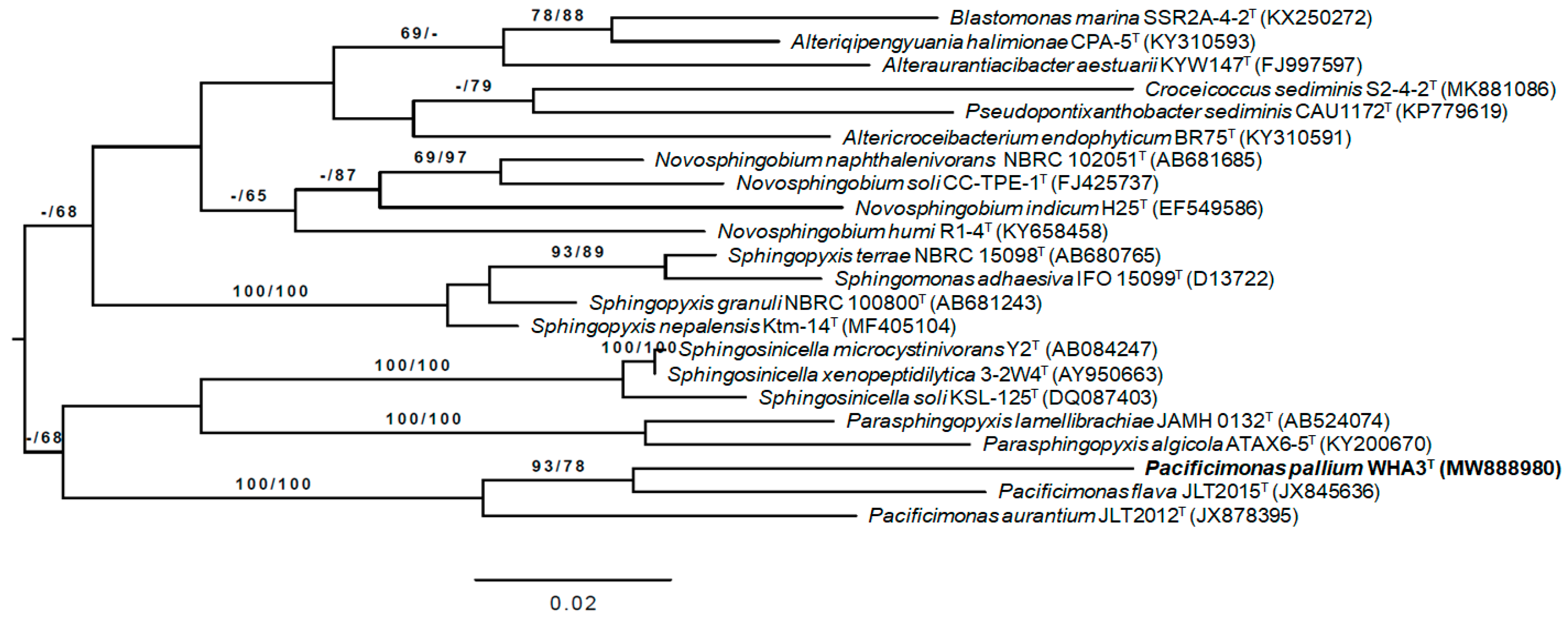
3.2. 16S rRNA Gene Analysis

According to the EZBioCloud server’s results, strain WHA3T was most closely related to the following strain types: 95.80% to Pacificimonas flava JLT2015T and 94.79% to Pacificimonas aurantium JLT2012T. A phylogenetic tree based on the 16S rRNA gene sequences of strain WHA3T strain WHA3T and its closely related type strains is shown in Figure 2. It shows that strain WHA3T formed a very high supported cluster with Pacificimonas species (P. flava JLT2015T and P. aurantium JLT2012T). Moreover, strain WHA3T was located in the well-supported branch together with P. flava JLT2015T.

3.3. Chemotaxonomic Characterization

The major cellular fatty acids of WHA3T were C16:1ω7c and C18:1ω7c (Table 4). The fatty acid C18:1ω7c was also detected as one of the major fatty acids in P. flava DSM 107612T and P. aurantium DSM 107782T. However, C16:1ω7c was not detected in P. aurantium DSM 107782T, and C16:02-OH was only found in WHA3T. Therefore, from the fatty acid content, WHA3T can be distinguished from P. flava DSM 107612T and P. aurantium DSM 107782T.
The dominant polar lipids of strain WHA3T were diphosphatidylglycerol, phosphatidylethanolamine, phosphatidylglycerol, several sphingoglycolipids, an unknown phospholipid, an unknown glycolipid, and an unknown polar lipid. Ubiquinone-10 (Q-10) was found to be the major respiratory quinone in WHA3T. Sphingoglycolipids and Q-10 were also detected in the type strains of Pacificimonas from the previous study [2,3].

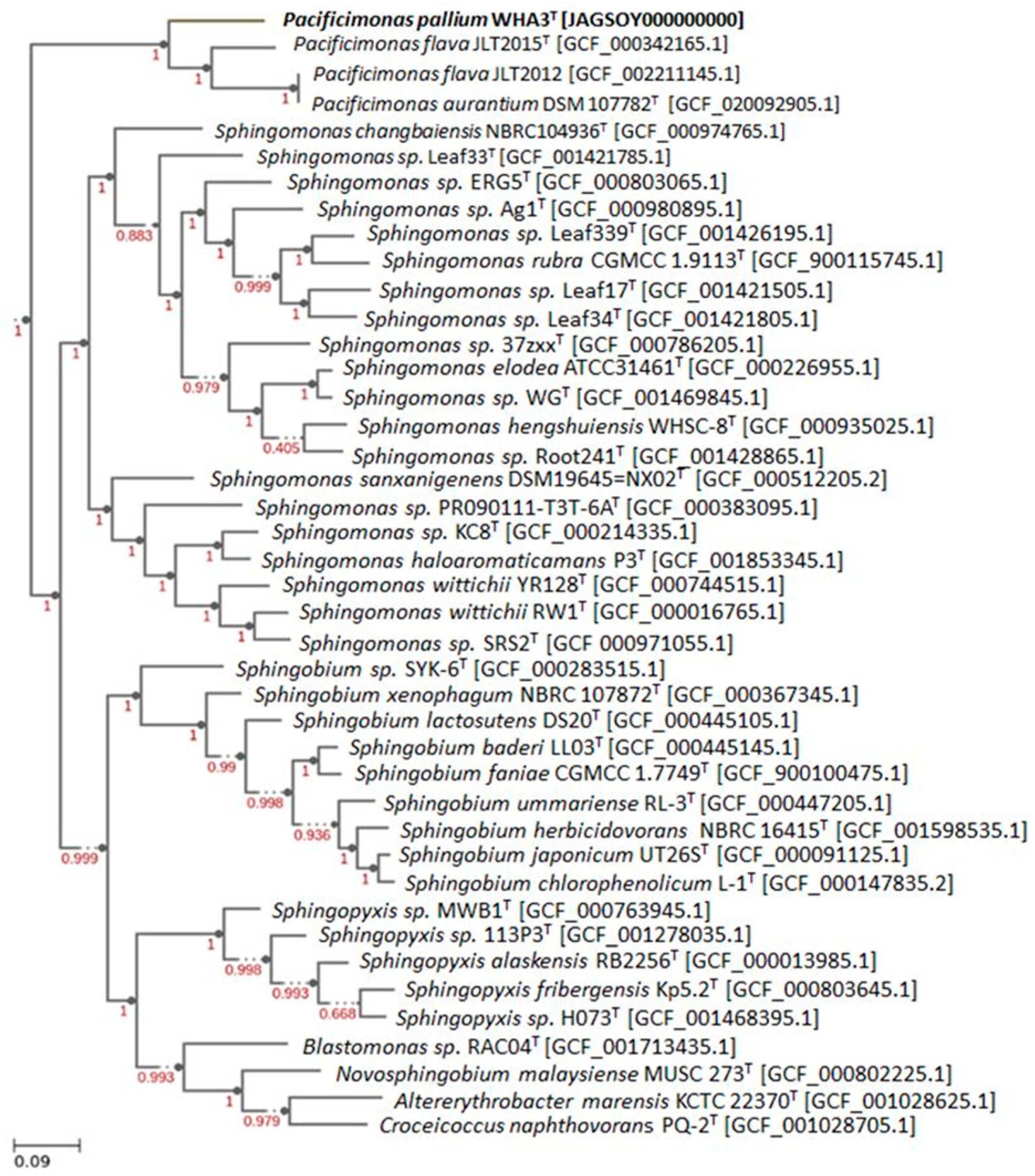
3.4. Genomic Characteristics and Phylogenomic Analysis

In the whole-genome data of strain WHA3T, only one 16S rRNA gene sequence was identified, indicating that other organisms did not contaminate the genomic data. The draft assembled genome sequence of strain WHA3T comprised the following: 3,017,344 bp with a G + C content of 61.69% (GenBank accession No. JAGSPA000000000). The genome included 2859 genes comprising 2794 protein-coding genes, 44 tRNA genes, 3 rRNA genes, and 3 non-coding RNA.
The phylogenomic tree (Figure S1) shows that strain WHA3T falls in a cluster, although it was not well supported with Parasphingopyxis lamellibrachiae DSM 26725T and Henriciella pelagia CGMCC 1.15928T, instead of with the other Pacificimonas species. Moreover, P. flava DSM 107612T and P. aurantium DSM 107782T were not closely related to the resulting phylogenomic tree. Therefore, the further study is based on the whole-proteome-based GBDP distances resulting from a phylogenetic tree with a very high average branch support of 94.1%, which is more reliable than the phylogenomic tree result (Figure 3). The strain WHA3T was in the same clade, with the very high support score, with other Pacificimonas species.
In the whole-proteome-based phylogenetic tree, strain WHA3T was located in a very highly supported clade together with P. flava DSM 107612T and P. aurantium DSM 107782T. Another phylogenomic tree result based on 49 COG (clusters of orthologous groups) also shows that strain WHA3T formed a clade with other Pacificimonas species with a very high support value (Figure 4).
Further study using genomic data to determine the genus classification was performed using the average amino acid identity (AAI) [53] value and the percentage of conserved proteins (POCP) value. The AAI values between the genomes of strain WHA3T and Pacificimonas flava DSM 107612T, Pacificimonas aurantium DSM 107782T, Parasphingopyxis lamellibrachiae DSM 26725T, and Henriciella pelagia CGMCC 1.15928T were 66.00%, 64.48%, 54.47%, and 46.38%, respectively. In contrast, the POCP values between the genomes of strain WHA3T and Pacificimonas flava DSM 107612T, Pacificimonas aurantium DSM 107782T, Parasphingopyxis lamellibrachiae DSM 26725T, and Henriciella pelagia CGMCC 1.15928T were 60.36%, 60.55%, 49.96%, and 33.01%, respectively (Table 5). It is suggested that the strains can be classified in the same genus if the AAI and the POCP values are higher than 60% and 50%, respectively [41,53]. Therefore, strain WHA3T could be determined to be a member of the genus Pacificimonas instead of Parasphingopyxis or Henriciella.
Furthermore, all of the type strains exhibited an ANI value less than the species cut-off value of 95% and dDDH scores less than the threshold value of 70%, as shown in Table 6 [54], suggesting that strain WHA3T can be separated from the other Pacificimonas species.
The genomes involved in C1 (one-carbon) metabolism for WHA3T are reported in Table 7.
The genes related to TonB-dependent transporters (TBDTs) and iron siderophore receptor protein in Pacificimonas pallium WHA3T and the other closest type strains are reported in Table 8.
The iron biomarker genes for Pacificimonas pallium WHA3T were related to the FbpC, fur, FeoB, and sodB genes; the vitamin B12 transporter reported for btuB; the biopolymer transport protein reported for exbD; and a variety of polysaccharides, including lptB, kpsT, lptA, lptF, lptG, kpsS, and kpsM, were observed (Table 9).
Additionally, gene annotation using RAST analysis (https://rast.nmpdr.org) predicted 2934 coding sequences in the genome of WHA3T. A dominant fraction of the subsystem feature was amino acids and derivatives (243), carbohydrates (146), protein metabolism (128), and fatty acids, lipids, and isoprenoids (100) (Figure S2, Table S1). Other genes, such as ones having a role for stress response (34), virulence, disease, and defense (28), metabolism of aromatic compounds (10), and motility and chemotaxis (5), were also detected. For the stress response genes, the major percentage was for oxidative stress, whereas in the virulence, disease, and defense genes, the genes for resistance to antibiotics and toxic compounds were the dominant ones, which are needed for the strain’s survival.
Secondary metabolite prediction based on the antiSMASH program suggested that there were only two detected secondary metabolite regions: PKS type 3 and terpene. There was an unknown cluster for PKS type 3, while the terpene region was 50% similar to carotenoid (Figure S3).

3.5. Chromatogram and Mass Analysis for Extraction of WHA3T

Ten major peaks were detected in the base peak chromatogram (BPC) of the extract of strain WHA3T. All of them were located in 1.5–7 min, suggesting that their characteristics are relatively polar. The detected ion masses ranged from 197.1276 Da to 1122.5594 Da (Figure 5, Table S2).

4. Conclusions

According to the results from this polyphasic study, strain WHA3T is a novel species that belongs to the genus Pacificimonas. Therefore, we suggest Pacificimonas pallium sp. nov. as the name for this newly discovered bacterium. We also predict that methylovory was possible in strain WHA3T. Based on these data, we hypothesize that WHA3T may have a role in the C1 oxidation pathways in the marine carbon cycle. The presence of genomes for TonB-dependent transporters (TBDTs) as outer membrane proteins could emphasize the capability of WHA3T to use restricted nutrient sources as well as vitamin B12 transporter and iron siderophore receptor protein. Chromatogram and mass analysis determined that there is no information about the compounds isolated from Pacificimonas bacteria in the Dictionary of Natural Products database. The majority of the detected masses were identified as many hits from the database. Only three masses were indicated as less than five hits, with one of them only showing one hit. Given that none of these hits were isolated from Pacificimonas bacterial strains, further study is needed to isolate the compounds, elucidate their structures, and reveal their bioactivities.

Description of Pacificimonas pallium sp. nov.

Pacificimonas pallium (pal.li.um is N. genitive palliī or pallī refers to the strain’s isolation from the mantel in the pacific oyster Crassostrea gigas). Optimal growth was observed at 30 °C, pH 5–9 and 2.5% (w/v) NaCl. This species is Gram-negative, motile by gliding, rod-shaped, aerobic, with no spore form, and without flagellum (Figure 1). Orange-colored colonies and concave curves with undamaged margins are significant. The optimum growth was observed on On CSY-3, 1.5LBM, and MB. The strain WHA3T has oxidase and catalase enzyme function. There is no PHA granulate. Cell size averages approximately 0.3–0.4 µm in width and 0.7–2.2 µm in length (Figure 1). Analysis for Api ZYM, Api Coryne, Api 20E, and GENIII microplate tests indicated that strain WHA3T exhibits high activity or is positive for alkaline, esterase (C4), esterase lipase (C8), leucine arylamidase, trypsin, α-chymotrypsin, reduction to N2gas, utilization of acetoacetic acid, utilization of propionic acid, utilization of acetic acid, utilization of formic acid, sodium butyrate, potassium tellurite, lithium chloride, β-hydroxy-D, L-butyric acid, L-alanine, L-serin, and D-aspartic acid. The strain WHA3T is sensitive against gentamycin (10 µg/mL), oxytetracycline (10 µg/mL), chloramphenicol (30 µg/mL), kanamycin (30 µg/mL), fusidic acid (50 µg/mL), thiostrepton (50 µg/mL), spectinomycin (50 µg/mL), trimethoprim (50 µg/mL), and erythromycin (15 µg/mL). The polyamine pattern is dominated by spermidine and spermine. The major cellular fatty acids are C16:1ω7c and C18:1ω7c. The dominant polar lipids are diphosphatidylglycerol (DFG), phosphatidylethanolamine (PE), phosphatidylglycerol (PG), several sphingoglycolipids (SGL), an unknown phospholipid (PL), an unknown glycolipid (GL), and an unknown polar lipid (L) (Figure S4). The major quinone is ubiquinone-10 (Q-10).
The type strain is WHA3T (= DSM 111825T = NCCB 100832 T), isolated from the mantle of the pacific oyster Crassostrea gigas collected from the Whilmshaven, Germany. The DNA G + C content is 61.69%. The genome size is 3,017,344  bp with 2794 coding sequences, 44 tRNA genes, and 3 rRNA operons. The complete genome and the 16S rRNA sequence of strain WHA3T were deposited in the NCBI GenBank with accession numbers JAGSPA000000000 and MW888980, respectively.

Supplementary Materials

The following are available online at https://www.mdpi.com/article/10.3390/d14030181/s1, Figure S1: Phylogenomic tree inferred from GBDP distances based on genomic data between strain WHA3T and the closely related type strains based on Type (Strain) Genome Server (TYGS) (https://tygs.dsmz.de/ accessed on 24 September 2021). The nodes are GBDP pseudo-bootstrap support values > 60% from 100 replications, with an average branch support of 50.8%, Figure S2: Subsystem category distribution of strain WHA3T based on the RAST annotation server (https://rast.nmpdr.org/ accessed on 16 June 2021), Figure S3: Prediction of secondary metabolite gene clusters of strain WHA3T using the antiSMASH server (https://antismash.secondarymetabolites.org/ accessed on 29 September 2021). Figure S4: Two -dimensional thin-layer chromatogram of polar lipids extracted from strain WHA3T. Table S1: RAST analysis result (https://rast.nmpdr.org accessed on 16 June 2021) of the draft genome of strain Pacificimonas pallium WHA3T. Table S2: Peak analysis of the base peak chromatogram (BPC) of the extract of strain WHA3T.

Author Contributions

Conceptualization, investigation, data analyses, writing—original draft preparation, H.P.; writing—review and editing, data analyses, C.R.; electron microscopy, M.M.; pacific oyster sample preparation and technical support, P.J.S.; project supervision and writing—review and editing J.W., C.R. and H.P. contributed equally to this work. All authors have read and agreed to the published version of the manuscript.

Funding

This work was funded by a scholarship awarded by Konrad Adenauer Stiftung (KAS) in Germany https://www.kas.de/en/home (accessed on 24 February 2022).

Institutional Review Board Statement

Not applicable.

Data Availability Statement

The GenBank accession number for the 16S rRNA gene sequence of strain WHA3T is MW888980. The draft genome of strain WHA3T and Pacificimonas aurantium DSM 107782T were deposited in the NCBI/GenBank under the accession number JAGSPA000000000 and JAGSGB000000000, respectively.

Acknowledgments

The authors appreciate the excellent effort of Stephanie Schulz, Klaus Peter Conrad, Birte Trunkwalter, and Wera Collisi for technical assistance, Ina Schleicher for electron microscopy sample preparation, and Aileen Gollasch for recording the HRESIMS data. Special thanks to Jolanta Lulla for medium preparation.

Conflicts of Interest

The authors declare no conflict of interest.

References

  1. Kosako, Y.; Yabuuchi, E.; Naka, T.; Fujiwara, N.; Kobayashi, K. Proposal of Sphingomonadaceae fam. nov., consisting of Sphingomonas Yabuuchi et al. 1990, Erythrobacter Shiba and Shimidu 1982, Erythromicrobium Yurkov et al. 1994, Porphyrobacter Fuerst et al. 1993, Zymomonas Kluyver and van Niel 1936, and Sandaracinobacter Yurkov et al. 1997, with the type genus Sphingomonas Yabuuchi et al. 1990. Microbiol. Immunol. 2000, 44, 563–575. [Google Scholar] [CrossRef]
  2. Liu, K.; Li, S.; Jiao, N.; Tang, K. Pacificamonas flava gen. nov., sp. nov., a novel member of the family Sphingomonadaceae isolated from the Southeastern Pacific. Curr. Microbiol. 2014, 69, 96–101. [Google Scholar] [CrossRef]
  3. Li, S.; Zhou, W.; Lin, D.; Tang, K.; Jiao, N. Pacificimonas aurantium sp. nov., Isolated from the Seawater of the Pacific Ocean. Curr. Microbiol. 2016, 72, 752–757. [Google Scholar] [CrossRef]
  4. Lauro, F.M.; McDougald, D.; Thomas, T.; Williams, T.J.; Egan, S.; Rice, S.; DeMaere, M.Z.; Ting, L.; Ertan, H.; Johnson, J.; et al. The genomic basis of trophic strategy in marine bacteria. Proc. Natl. Acad. Sci. USA 2009, 106, 15527–15533. [Google Scholar] [CrossRef] [Green Version]
  5. Sowell, S.M.; Wilhelm, L.J.; Norbeck, A.D.; Lipton, M.S.; Nicora, C.D.; Barofsky, D.F.; Carlson, C.A.; Smith, R.D.; Giovanonni, S.J. Transport functions dominate the SAR11 metaproteome at low-nutrient extremes in the Sargasso Sea. ISME J. 2009, 3, 93–105. [Google Scholar] [CrossRef] [Green Version]
  6. Takeuchi, M.; Hamana, K.; Hiraishi, A. Proposal of the genus Sphingomonas sensu stricto and three new genera, Sphingobium, Novosphingobium and Sphingopyxis, on the basis of phylogenetic and chemotaxonomic analyses. Int. J. Syst. Evol. Microbiol. 2001, 51, 1405–1417. [Google Scholar] [CrossRef]
  7. Maruyama, T.; Park, H.D.; Ozawa, K.; Tanaka, Y.; Sumino, T.; Hamana, K.; Hiraishi, A.; Kato, K. Sphingosinicella microcystinivorans gen. nov., sp. nov., a microcystin-degrading bacterium. Int. J. Syst. Evol. Microbiol. 2006, 56, 85–89. [Google Scholar] [CrossRef]
  8. Stingl, U.; Desiderio, R.A.; Cho, J.-C.; Vergin, K.L.; Giovannoni, S.J. The SAR92 Clade: An abundant coastal clade of culturable marine bacteria possessing proteorhodopsin. Appl. Env. Microbiol. 2007, 73, 2290–2296. [Google Scholar] [CrossRef] [Green Version]
  9. Sun, J.; Steindler, L.; Thrash, J.C.; Halsey, K.H.; Smith, D.P.; Carter, A.E.; Landry, Z.C.; Giovannoni, S.J. One carbon metabolism in SAR11 pelagic marine bacteria. PLoS ONE 2011, 6, e23973. [Google Scholar] [CrossRef] [Green Version]
  10. Tang, K.; Jiao, N.; Liu, K.; Zhang, Y.; Li, S. Distribution and functions of TonB-dependent transporters in marine bacteria and environments: Implications for dissolved organic matter utilization. PLoS ONE 2012, 7, e41204. [Google Scholar] [CrossRef] [Green Version]
  11. Noinaj, N.; Guillier, M.; Barnard, T.J.; Buchanan, S.K. TonB-dependent transporters: Regulation, structure, and function. Annu. Rev. Microbiol. 2010, 64, 43–60. [Google Scholar] [CrossRef] [Green Version]
  12. Landwehr, W.; Kampfer, P.; Glaeser, S.P.; Ruckert, C.; Kalinowski, J.; Blom, J.; Goesmann, A.; Mack, M.; Schumann, P.; Atasayar, E.; et al. Taxonomic analyses of members of the Streptomyces cinnabarinus cluster, description of Streptomyces cinnabarigriseus sp. nov. and Streptomyces davaonensis sp. nov. Int. J. Syst. Evol. Microbiol. 2018, 68, 382–393. [Google Scholar] [CrossRef]
  13. Kutzner, H.J. The Family Streptomycetaceae; Springer: Berlin/Heidelberg, Germany, 1981. [Google Scholar]
  14. Rüger, H.J.; Krambeck, H.J.J.S.; Microbiology, A. Evaluation of the BIOLOG substrate metabolism system for classification of marine bacteria. Syst. Appl. Microbiol. 1994, 17, 281–288. [Google Scholar] [CrossRef]
  15. Legat, A.; Gruber, C.; Zangger, K.; Wanner, G.; Stan-Lotter, H. Identification of polyhydroxyalkanoates in Halococcus and other haloarchaeal species. Appl. Microbiol. Biotechnol. 2010, 87, 1119–1127. [Google Scholar] [CrossRef] [Green Version]
  16. Chaiya, L.; Matsumoto, A.; Wink, J.; Inahashi, Y.; Risdian, C.; Pathom-aree, W.; Lumyong, S. Amycolatopsis eburnea sp. nov., an actinomycete associated with arbuscular mycorrhizal fungal spores. Int. J. Syst. Evol. Microbiol. 2019, 69, 3603–3608. [Google Scholar] [CrossRef]
  17. Hall, T.A. BIOEDIT: A user- friendly biological sequence alignment editor and analysis for windows 95/98/ NT. Nucleic Acids Symp. Ser. 1999, 41, 95–98. [Google Scholar]
  18. Yoon, S.H.; Ha, S.M.; Kwon, S.; Lim, J.; Kim, Y.; Seo, H.; Chun, J. Introducing EzBioCloud: A taxonomically united database of 16S rRNA gene sequences and whole-genome assemblies. Int. J. Syst. Evol. 2017, 67, 1613–1617. [Google Scholar] [CrossRef]
  19. Meier-Kolthoff, J.P.; Auch, A.F.; Klenk, H.-P.; Göker, M. Genome sequence-based species delimitation with confidence intervals and improved distance functions. BMC Bioinform. 2013, 14, 60. [Google Scholar] [CrossRef] [Green Version]
  20. Meier-Kolthoff, J.P.; Hahnke, R.L.; Petersen, J.; Scheuner, C.; Michael, V.; Fiebig, A.; Rohde, C.; Rohde, M.; Fartmann, B.; Goodwin, L.A.; et al. Complete genome sequence of DSM 30083T, the type strain (U5/41T) of Escherichia coli, and a proposal for delineating subspecies in microbial taxonomy. Stand. Genom. Sci. 2014, 9, 2. [Google Scholar] [CrossRef] [Green Version]
  21. Meier-Kolthoff, J.P.; Goker, M.; Sproer, C.; Klenk, H.P. When should a DDH experiment be mandatory in microbial taxonomy? Arch. Microbiol. 2013, 195, 413–418. [Google Scholar] [CrossRef]
  22. Edgar, R.C. Muscle: Multiple sequence alignment with high accuracy and high throughput. Nucleic. Acids Res. 2004, 32, 1792–1797. [Google Scholar] [CrossRef] [Green Version]
  23. Stamatakis, A. RAxML version 8: A tool for phylogenetic analysis and post-analysis of large phylogenies. Bioinformatics 2014, 30, 1312–1313. [Google Scholar] [CrossRef]
  24. Goloboff, P.A.; Farris, J.S.; Nixon, K.C. TNT, a free program for phylogenetic analysis. Cladistics-Int. J. Willi Hennig Soc. 2008, 24, 774–786. [Google Scholar] [CrossRef]
  25. Pattengale, N.D.; Alipour, M.; Bininda-Emonds, O.R.; Moret, B.M.; Stamatakis, A. How many bootstrap replicates are necessary? J. Comput. Biol. 2010, 17, 337–354. [Google Scholar] [CrossRef]
  26. Swofford, D. PAUP*. Phylogenetic Analysis Using Parsimony (*and Other Methods); Sinauer Associates: Sunderland, MA, USA, 2002; Volume 4. [Google Scholar]
  27. Minnikin, D.E.; O’Donnell, A.G.; Goodfellow, M.; Alderson, G.; Athalye, M.; Schaal, A.; Parlett, J.H. An integrated procedure for the extraction of bacterial isoprenoid quinones and polar lipids. J. Microbiol. Methods 1984, 2, 233–241. [Google Scholar] [CrossRef]
  28. Risdian, C.; Landwehr, W.; Rohde, M.; Schumann, P.; Hahnke, R.L.; Sproer, C.; Bunk, B.; Kampfer, P.; Schupp, P.J.; Wink, J. Streptomyces bathyalis sp. nov., an actinobacterium isolated from the sponge in a deep sea. Antonie Van Leeuwenhoek 2021, 114, 425–435. [Google Scholar] [CrossRef]
  29. Sasser, M. Identification of bacteria by gas chromatography of cellular fatty acids. USFCC Newsl. 1990, 20, 1–6. [Google Scholar]
  30. Wick, R.R.; Judd, L.M.; Gorrie, C.L.; Holt, K.E. Unicycler: Resolving bacterial genome assemblies from short and long sequencing reads. PLoS Comput. Biol. 2017, 13, e1005595. [Google Scholar] [CrossRef] [Green Version]
  31. Lee, I.; Chalita, M.; Ha, S.-M.; Na, S.-I.; Yoon, S.-H.; Chun, J. ContEst16S: An algorithm that identifies contaminated prokaryotic genomes using 16S RNA gene sequences. Int. J. Syst. Evol. Microbiol. 2017, 67, 2053–2057. [Google Scholar] [CrossRef]
  32. Tatusova, T.; DiCuccio, M.; Badretdin, A.; Chetvernin, V.; Nawrocki, E.P.; Zaslavsky, L.; Lomsadze, A.; Pruitt, K.D.; Borodovsky, M.; Ostell, J. NCBI prokaryotic genome annotation pipeline. Nucleic. Acids Res. 2016, 44, 6614–6624. [Google Scholar] [CrossRef]
  33. Medema, M.H.; Blin, K.; Cimermancic, P.; de Jager, V.; Zakrzewski, P.; Fischbach, M.A.; Weber, T.; Takano, E.; Breitling, R. antiSMASH: Rapid identification, annotation and analysis of secondary metabolite biosynthesis gene clusters in bacterial and fungal genome sequences. Nucleic. Acids Res. 2011, 39, W339–W346. [Google Scholar] [CrossRef]
  34. Blin, K.; Shaw, S.; Steinke, K.; Villebro, R.; Ziemert, N.; Lee, S.Y.; Medema, M.H.; Weber, T. antiSMASH 5.0: Updates to the secondary metabolite genome mining pipeline. Nucleic. Acids Res. 2019, 47, W81–W87. [Google Scholar] [CrossRef] [PubMed] [Green Version]
  35. Meier-Kolthoff, J.P.; Carbasse, J.S.; Peinado-Olarte, R.L.; Goker, M. TYGS and LPSN: A database tandem for fast and reliable genome-based classification and nomenclature of prokaryotes. Nucleic. Acids Res. 2021, 50, D801–D807. [Google Scholar] [CrossRef] [PubMed]
  36. Lefort, V.; Desper, R.; Gascuel, O. FastME 2.0: A comprehensive, accurate, and fast distance-based phylogeny inference program. Mol. Biol. Evol. 2015, 32, 2798–2800. [Google Scholar] [CrossRef] [PubMed] [Green Version]
  37. Arkin, A.P.; Cottingham, R.W.; Henry, C.S.; Harris, N.L.; Stevens, R.L.; Maslov, S.; Dehal, P.; Ware, D.; Perez, F.; Canon, S.; et al. KBase: The United States Department of Energy Systems Biology Knowledgebase. Nat. Biotechnol. 2018, 36, 566–569. [Google Scholar] [CrossRef] [Green Version]
  38. Seemann, T. Prokka: Rapid prokaryotic genome annotation. Bioinformatics 2014, 30, 2068–2069. [Google Scholar] [CrossRef]
  39. Price, M.N.; Dehal, P.S.; Arkin, A.P. FastTree 2—Approximately maximum-likelihood trees for large alignments. PLoS ONE 2010, 5, e9490. [Google Scholar] [CrossRef]
  40. Yoon, S.-H.; Ha, S.-m.; Lim, J.; Kwon, S.; Chun, J. A large-scale evaluation of algorithms to calculate average nucleotide identity. Antonie Van Leeuwenhoek 2017, 110, 1281–1286. [Google Scholar] [CrossRef]
  41. Qin, Q.L.; Xie, B.B.; Zhang, X.Y.; Chen, X.L.; Zhou, B.C.; Zhou, J.; Oren, A.; Zhang, Y.Z. A proposed genus boundary for the prokaryotes based on genomic insights. J. Bacteriol. 2014, 196, 2210–2215. [Google Scholar] [CrossRef] [Green Version]
  42. Ateba, T.P.; Alayande, K.A.; Mwanza, M. Feces metagenomes and metagenome-assembled genome sequences from two separate dogs (Canis lupus familiaris) with multiple diarrheal episodes. Microbiol. Resour. Announc. 2020, 9, e01065-20. [Google Scholar] [CrossRef]
  43. Guse, A.; Stevenson, C.E.; Kuper, J.; Buchanan, G.; Schwarz, G.; Giordano, G.; Magalon, A.; Mendel, R.R.; Lawson, D.M.; Palmer, T. Biochemical and structural analysis of the molybdenum cofactor biosynthesis protein MobA. J. Biol. Chem. 2003, 278, 25302–25307. [Google Scholar] [CrossRef] [PubMed] [Green Version]
  44. Wang, W.; Zhang, W.; Lu, J.; Yang, Y.; Chiao, J.; Zhao, G.; Jiang, W. MoeA, an enzyme in the molybdopterin synthesis pathway, is required for rifamycin SV production in Amycolatopsis mediterranei U32. Appl. Microbiol. Biotechnol. 2002, 60, 139–146. [Google Scholar] [CrossRef] [PubMed]
  45. Scott, D.A.; Hickerson, S.M.; Vickers, T.J.; Beverley, S.M. The role of the mitochondrial glycine cleavage complex in the metabolism and virulence of the protozoan parasite Leishmania major. J. Biol. Chem. 2008, 283, 155–165. [Google Scholar] [CrossRef] [PubMed] [Green Version]
  46. Dworkin, M. The Prokaryotes: Vol. 1: Symbiotic Associations, Biotechnology, Applied Microbiology; Springer Science & Business Media: Berlin/Heidelberg, Germany, 2006. [Google Scholar]
  47. Toulza, E.; Tagliabue, A.; Blain, S.; Piganeau, G. Analysis of the global ocean sampling (GOS) project for trends in iron uptake by surface ocean microbes. PLoS ONE 2012, 7, e30931. [Google Scholar] [CrossRef] [PubMed]
  48. Bibby, T.S.; Zhang, Y.; Chen, M. Biogeography of photosynthetic light-harvesting genes in marine phytoplankton. PLoS ONE 2009, 4, e4601. [Google Scholar] [CrossRef] [Green Version]
  49. McCarthy, M.; Hedges, J.; Benner, R.J.M.C. Major biochemical composition of dissolved high molecular weight organic matter in seawater. Mar. Chem. 1996, 55, 281–297. [Google Scholar] [CrossRef]
  50. Aziz, R.K.; Bartels, D.; Best, A.A.; DeJongh, M.; Disz, T.; Edwards, R.A.; Formsma, K.; Gerdes, S.; Glass, E.M.; Kubal, M.; et al. The RAST Server: Rapid Annotations using Subsystems Technology. BMC Genom. 2008, 9, 75. [Google Scholar] [CrossRef] [Green Version]
  51. Pira, H.; Risdian, C.; Kämpfer, P.; Müsken, M.; Schupp, P.J.; Wink, J. Zooshikella harenae sp. nov., Isolated from Pacific Oyster Crassostrea gigas, and Establishment of Zooshikella ganghwensis subsp. marina subsp. nov. and Zooshikella ganghwensis subsp. ganghwensis subsp. nov. Diversity 2021, 13, 641. [Google Scholar] [CrossRef]
  52. Khosravi Babadi, Z.; Ebrahimipour, G.; Wink, J.; Narmani, A.; Risdian, C. Isolation and identification of Streptomyces sp. Act4Zk, a good producer of Staurosporine and some derivatives. SfAM 2021, 72, 206–218. [Google Scholar] [CrossRef]
  53. Rodriguez, R.L.M.; Konstantinidis, K.J.M.M. Bypassing cultivation to identify bacterial species: Culture-independent genomic approaches identify credibly distinct clusters, avoid cultivation bias, and provide true insights into microbial species. Microbe 2014, 9, 111–118. [Google Scholar] [CrossRef]
  54. Chun, J.; Oren, A.; Ventosa, A.; Christensen, H.; Arahal, D.R.; da Costa, M.S.; Rooney, A.P.; Yi, H.; Xu, X.-W.; De Meyer, S.; et al. Proposed minimal standards for the use of genome data for the taxonomy of prokaryotes. Int. J. Syst. Evol. Microbiol. 2018, 68, 461–466. [Google Scholar] [CrossRef] [PubMed]
Figure 1. Scanning electron microscopy image of strain WHA3T.
Diversity 14 00181 g001
Figure 2. ML tree of strain WHA3T and its closely related type strains inferred under the GTR+GAMMA model and rooted by midpoint-rooting. The numbers above the branches are support values (above 60%) from ML (left) and MP (right) bootstrapping. The ML and MP bootstrapping average supports were 69.63% and 79.05%, respectively.
Diversity 14 00181 g002
Figure 3. Phylogenomic tree inferred from the whole-proteome-based GBDP distances between strain WHA3T and the closely related type strains based on Type (Strain) Genome Server (TYGS) (https://tygs.dsmz.de/ accessed on 11 November 2021). The numbers at the nodes are GBDP pseudo-bootstrap support values >60% from 100 replications, with an average branch support of 94.9%.
Diversity 14 00181 g003
Figure 4. Phylogenomic tree showing relationships between strain WHA3T and the closely related type strains based on the Insert Genome Into Species Tree v2.2.0 tool using 49 COG (clusters of orthologous groups) domains of core universal genes.
Diversity 14 00181 g004
Figure 5. The base peak chromatogram (BPC) of the extract of strain WHA3T.
Diversity 14 00181 g005
Table 1. Colony color comparison of the three strains, WHA3T, Pacificimonas flava DSM 107612T, and Pacificimonas aurantium DSM 107782T, in various culture media according to Ralcolor code.
Culture MediaPacificimonas pallium
WHA3T
Pacificimonas flava
DSM 107612T
Pacificimonas aurantium
DSM 107782T
MB 514fRAL2003 Pastel orangeRAL1037 Sun yellowRAL1032 Broom yellow
CSY-3RAL2008 Bright red orangeRAL1002 Sand yellowRAL1002 Sand yellow
1.5 LBMRAL2003 Pastel orangeRAL1033 Dahlia yellowRAL1037 Sun yellow
YEDRAL2000 Yellow orangeRAL1002 Sand yellowRAL1005 Honey yellow
TSANGRAL2000 Yellow orangeNG
SSM + TNGRAL1011 Brown beigeNG
SSM-TNGRAL1002 Sand yellowNG
YTSSRAL1017 Saffron yellowRAL1003 Signal yellowRAL1003 Signal yellow
ASG + CycloheximideNGNGRAL1015 Light ivory
YEANGRAL1023 Traffic yellowNG
NG: No growth or very restricted; RAL codes available at (https://www.ralfarben.de accessed on 14 September 2020).
Table 2. Different enzymes produced by strain WHA3T detected by the ApiZym system.
Enzyme ObservationEnzymeObservation
Phosphatase alkaline++Naphtol-AS-BI-phosphohydrolase(+)
Esterase (C4)+α-galactosidase-
Esterase lipase (C8)+β-galactosidase-
Lipase (C14)(+)β-glucurunidase-
Leucin arylamidase++α-glucosidase-
Valine arylamidase(+)β-glucosidase-
Cystine arylamidase(+)N-acetyl-beta- glucosaminidase-
Trypsin+α-mannosidase-
Chymotrypsin++α-fucosidase-
Phosphatase acid(+)
++ strong; + good; (+) weak; - no activity.
Table 3. Comparison of phenotypic characteristics that distinguish strain WHA3T from the most closely related type strains. Strains: 1, Pacificimonas pallium WHA3T; 2, Pacificimonas flava DSM 107612T; 3, Pacificimonas aurantium DSM 107782T.
Characteristics123
Color of colonyorangeorangeorange
Temperature range for growth (°C)15–4020–30 *10–40 †
pH spectrum for growth5–115–12 *5–10 †
NaCl optimum for growth (%)2.5–50.5–7 *0.5–4 †
Trypsin++(+)
α-Chymotrypsin++(+)-
Acid phosphatase(+)++(+)
Naphthol-AS-BI-phosphohydrolase(+)+(+)
N-acetyl-β-glucosaminidase-(+)(+)
gelatine (hydrolysis)-++
Oxidation of (Biolog GN2)
Acetic acid++ *- †
Methyl pyruvate-+ *- †
D-galactose-ND *+ †
D-raffinose-- *+ †
D-trehalose-- *+ †
D-Turanose-- *+ †
α -keto glutaric acid-- *+ †
L-alanine+- *+ †
L-glutamic acid-- *+ †
L-aspartic acid-- *+ †
γ-amino butyric acid-ND *+ †
Dextrin-+ *+ †
D-fructose-+ *+ †
α-D-glucose(+)+ *+ †
Tween 40-+ *+ †
Susceptibility to
Ampicillin-- *+ †
Gentamycin+- *+ †
Kanamycin+- *+ †
Polar lipidsDPG-PE-PG-SGL-PL-GL-LDPG-PE-PG-SGL-LDPG-PE-PG-SGL †
Major fatty acidC18:1ω7c
C16:1ω7c
C18:1ω7c
C14:02-OH
C18:1ω7c
C14:02-OH
Unknown fatty acid
Contigs113212
No. of proteins279429083104
rRNA343
tRNA444545
No. of Genes285930383185
Other RNA 344
Pseudogenes157729
G + C content (%)61.6964.2 *65.5 †
Total sequence length (Mbp)3.013.253.34
++ strong; + good; (+) weak; - no activity; ND no data * Data from Liu et al. [2] † Data from Li et al. [3] Diphosphatidylglycerol (DPG), phosphatidylcholine (PC), phosphatidylinositol (PI), phosphatidylglycerol (PG), phosphatidylethanolamine (PE), sphingoglycolipid (SGL), unidentified phospholipids (PL), unidentified glycolipid (GL), and unidentified polar lipid (L).
Table 4. The fatty acid content of strain WHA3T and its closest Pacificimonas type strains.
Pacificimonas
pallium WHA3T
Pacificimonas flava
DSM 107612T
Pacificimonas
aurantium DSM 107782T
Fatty acid
C14:0--5.0--
C15:0--2.2--
C16:04.29.19.8
C17:0----5.2
C19:0--2.48.3
anteiso-C15:0--5.7--
anteiso-C17:0--1.9--
iso-C16:0--4.1--
C16:1ω7c20.55.8--
C17:1ω6c5.14.05.3
C18:1ω7c50.626.922.0
C14:02-OH8.422.827.6
C16:02-OH3.6----
cyclo-C19:0d8,91.35.74.0
Unknown fatty acid6.34.417.8
--: Not detected.
Table 5. Genome relatedness between the strain WHA3T, the genus of Pacificimonas, and other closet type strains according to the average amino acid identity (AAI) value and the percentage of conserved proteins (POCP) value. 1, Pacificimonas flava DSM 107612T (JACHGC010000001); 2, Pacificimonas aurantium DSM 107782T (JAGSGB000000000); 3, Parasphingopyxis lamellibrachiae DSM 26725T (NZ_QRDP00000000), 4; Henriciella pelagia CGMCC 1.15928T (GCA_014644035.1).
Strain1234
POCP
(%)
AAI
(%)
POCP
(%)
AAI
(%)
POCP
(%)
AAI
(%)
POCP
(%)
AAI
(%)
Pacificimonas pallium WHA3T
(JAGSPA000000000)
60.3666.0060.5564.4849.9654.4733.0146.38
Pacificimonas flava
DSM 107612T (JACHGC010000001)
10010063.3969.2246.9255.0232.1246.42
Pacificimonas aurantium DSM 107782T (JAGSGB000000000)63.3969.2210010047.2054.5833.6346.85
POCP less than 50% is a different (or new) genus, and more than 50% (bold) is the same genus. AAI less than 60% is a different (or new) genus, and more than 60% (bold) is the same genus.
Table 6. ANI and dDDH values of the strain Pacificimonas sp. WHA3T and its closely related type strains. 1, Pacificimonas aurantium DSM 107782T (JAGSGB000000000); 2, Pacificimonas flava DSM 107612T (JACHGC010000001); 3, Parasphingopyxis lamellibrachiae DSM 26725T(NZ_QRDP00000000); 4, Henriciella pelagia CGMCC 1.15928T (GCA_014644035.1).
Strain1234
Ortho
ANIu (%)
dDDH (%)Ortho
ANIu (%)
dDDH (%)Ortho
ANIu (%)
dDDH (%)Ortho
ANIu (%)
dDDH (%)
Pacificimonas pallium WHA3T (JAGSPA000000000)71.9519.2072.2518.4068.8419.0066.7317.90
Pacificimonas flava
DSM 107612T (JACHGC010000001)
74.3620.3010010069.1719.2067.1516.30
Pacificimonas aurantium DSM 107782T (JAGSGB000000000)10010074.3620.3069.1318.9067.0518.30
OrthoANIu values ≥ 95% and dDDH values > 70% are demonstrated.
Table 7. Distribution of genes involved in C1 metabolism in 1, Pacificimonas pallium WHA3T; 2, Pacificimonas flava DSM 107612T; 3, Pacificimonas aurantium DSM 107782T genomes using https://narrative.kbase.us/ accessed on 24 February 2022.
Genes for C1 Oxidation and Methylotrophy123
THF-linked oxidation
Methylene -THF methylenetetrahydrofolate reductase metF+--
Bifunctional methylene-THF dehydrogenase-methenyl-THF cyclohydrolase folD+--
AMTs
Glycine system cleavage T-protein gcvT+--
Glycine cleavage system H protein gcvH+--
GSH-dependent pathway
S-formyl-glutathione hydrolase (FGH) fghA+--
methylamine oxidation
Sarcosine oxidase subunit beta soxB+--
Table 8. Distribution of genes involved in TonB-dependent transporters (TBDTs) in 1, Pacificimonas pallium WHA3T; 2, Pacificimonas flava DSM 107612T; 3, Pacificimonas aurantium DSM 107782T genomes using https://narrative.kbase.us/ accessed on 24 February 2022.
Genes TonB-Dependent Transporters (TBDTs)123
btuB_1
btuB_3
btuB_4
btuB_6
btuB_8
btuB_9
btuB_10
btuB_12
btuB_13
btuB_14
btuB_15
btuB_17
btuB_19
btuB_20
btuB_21
btuB_23
btuBbtuB
fyuA_1
fyuA_2
TonBTonB
putative TonB-dependent receptor BfrD+--
TonB-dependent hemin and ferrichrome receptor hemR+--
TonB-dependent receptor and iron siderophore receptor protein btuB_7+--
+: Presence of gene.
Table 9. Distribution of genes involved iron biomarker, vitamin B12 uptake, biopolymer transport protein, and polysaccharides proteins in 1, Pacificimonas pallium WHA3T; 2, Pacificimonas flava DSM 107612T; 3, Pacificimonas aurantium DSM 107782T genomes using https://narrative.kbase.us/ accessed on 24 February 2022.
123
Vitamin B12 transporter btuB+--
Outer membrane vitamin B12 receptor btuB+++
Biopolymer transport protein exbD_1
exbD_2
exbD_3
exbDexbD
Fe(3+) ions import ATP-binding protein FbpC+--
Ferric uptake regulation protein fur+++
Fe(2+) transporter FeoB+++
Superoxide dismutase [Fe] sodB+--
Lipopolysaccharide ABC transporter, ATP-binding protein lptB+++
Capsular polysaccharide ABC transporter, ATP-binding protein kpsT++-
Lipopolysaccharide export system protein lptA+-+
Lipopolysaccharide export system permease protein lptF-lptG+++
Capsular polysaccharide export system protein kpsS++-
Capsular polysaccharide ABC transporter, permease protein kpsM++-
+: Presence of gene.
Publisher’s Note: MDPI stays neutral with regard to jurisdictional claims in published maps and institutional affiliations.

