Article

The Natural Infection of Freshwater Snails with the Avian Air Sac Fluke, Cyclocoelum mutabile (Trematoda: Cyclocoelidae), in Brazil

by
Jordana Costa Alves de Assis
and
Hudson Alves Pinto
*
Laboratório de Biologia de Trematoda, Instituto de Ciências Biológicas, Universidade Federal de Minas Gerais, Av. Pres. Antônio Carlos, 6627-Pampulha, Belo Horizonte 31270-901, MG, Brazil
*
Author to whom correspondence should be addressed.
Diversity 2024, 16(7), 422; https://doi.org/10.3390/d16070422
Submission received: 4 June 2024 / Revised: 11 July 2024 / Accepted: 12 July 2024 / Published: 19 July 2024
(This article belongs to the Special Issue Diversity of Wildlife Pathogens)

Abstract

:
Trematodes of the family Cyclocoelidae are parasites mainly of the respiratory system of birds and present a cosmopolitan distribution. Although infection with these flukes can result in pathological changes and even bird death, information on their life cycles is scarce and almost entirely based on experimental infection data. Thus, the generation of knowledge on the mollusks that act as natural intermediate hosts of cyclocoelids is necessary and can aid control measures against these air sac trematodes. In the present study, gastropod mollusks collected in an urban stream from Belo Horizonte, Minas Gerais, Brazil, were subjected to the compression technique for the detection of non-emerging larval trematodes. Tailless cercariae with confluent ceca were found in 8/30 (26.7%) specimens of Biomphalaria glabrata and 3/33 (9.1%) specimen of Physella acuta. Samples of the cercariae were subjected to morphological characterization and genetic study (28S, Cox-1, and Nad-1). For comparative purposes, adult trematodes previously collected in the air sac of a common gallinule (Gallinula galeata) found dead in another waterbody from the same region were also characterized. The molecular sequences obtained revealed a high degree of similarity (100% in 28S, 99.2% in Cox-1, and 99.5% in Nad-1) between larval stages found in mollusks and adult parasites found in G. galeata and morphologically identified as Cyclocoelum mutabile. The conspecificity with this widely distributed cyclocoelid was also corroborated by phylogenetic analysis and comparison with isolates of this species previously characterized in Peru and the Czech Republic (99.4–100% and 96.7–97.0% of similarity in Nad-1, respectively). Thus, the integrative analysis carried out in the present work enabled us to identify C. mutabile in mollusks in South America for the first time. The finding of B. glabrata and P. acuta as new intermediate hosts corroborates the importance of freshwater gastropods in the transmission of C. mutabile, as well as the low specificity to the mollusk group, as previously characterized through experimental studies.

1. Introduction

Species of the family Cyclocoelidae Stossich, 1902, comprise a diverse group of avian trematodes with worldwide distribution, represented by 6 subfamilies, 23 genera, and about 130 species currently considered valid [1,2,3,4]. Infection with these helminths can be associated with pathological changes in the respiratory and cardiovascular systems, in addition to liver damage caused during the migration of immature stages in the early infection [5,6,7,8]. The more speciose genus is Cyclocoelum Brandes, 1892, currently with 11 valid species described infecting mainly the air sacs of birds in different parts of the world [1,2,3,9,10,11]. Although reports of the infection of birds with these trematodes are not uncommon, information on the epidemiology of cyclocoelosis is scarce, especially regarding data on the mollusks that act as intermediate hosts.
Studies carried out a few decades ago in the Northern Hemisphere revealed that species of Cyclocoelum require, in addition to birds as definitive hosts, gastropod mollusks (physids, lymnaeids, and planorbids) as obligatory intermediate hosts [5,12,13,14,15,16]. In such invertebrates, rediae produce cercariae, which became encysted as metacercariae in the same mollusk. The emergence of the larval stage from the mollusk to the external environment is not verified in the life cycle of cyclocoelids. The infection of the birds occurs through the ingestion of mollusks harboring metacercariae [14,15,17]. Most information available on the life cycles of Cyclocoelum spp. was produced by experimental or semi-experimental infection studies using laboratory-reared mollusks that were exposed to miracidia obtained from eggs recovered from adult parasites [12,13,14,15]. Considering natural infection, lymnaeids and planorbids mollusks were found naturally with these trematodes in Europe [16,18,19]. However, in some of these works, the parasite identification was based exclusively on the morphology of cercariae, which raises doubts about the real identity of the species.
The scarcity of data on the transmission of cyclocoelids under natural conditions can be related to the difficulties of linking larval stages with the respective adult parasites. In the past, this challenging task could be achieved only by the classical experimental infection approach [18]. However, in the last two decades, molecular tools have been used in studies of species of air sac trematodes from different parts of the world [4,7,8,9,10,11,20,21]. The growing generation and availability of genetic data for these parasites reduce the barriers involved in identifying larval cyclocoelids found in mollusks.
In the present study, non-emergent larval trematodes found in naturally infected mollusks from Brazil were characterized by morphological and molecular approaches. Adult cyclocoelids found in a common gallinule from the same region were identified as Cyclocoelum mutabile (Zeder, 1800) and molecularly linked to the cercariae found in the mollusks. In addition to finding new natural intermediate hosts for this widely distributed species, new genetic and phylogenetic information are presented and discussed.

2. Materials and Methods

2.1. Parasites

A malacological collection was performed in an urban stream (19°52′07.8″ S and 43°57′11.9″ W) located in Belo Horizonte, Minas Gerais, southeastern Brazil, in April 2021. The collected mollusks were sent to the laboratory, transferred individually to polystyrene containers with tap water, and subjected to a routine artificial photostimulation test to detect the emergence of larval trematodes. After this exam, the gastropods were transferred to a Petri dish, pressed between glass slides, and dissected under a stereomicroscope to search for non-emergent larval stages, as recommended by Assis et al. [21]. The taxonomic identification of the mollusks was based on morphological traits according to Paraense [22] and Paraense and Pointier [23].
Adult trematodes obtained during the necropsy of a juvenile specimen of Gallinula galeata (Lichtenstein, 1818) (Gruiformes: Rallidae) found naturally dead in an urban lake located in the same city (19°47′06.20″ S and 43°57′11.41″ W; 11 km far from the stream of the malacological study) in April 2019 were also evaluated. The worms recovered were rinsed in saline solution (0.85% NaCl), counted, placed between glass sides, killed in water at 70 °C, and fixed in 10% formalin. The study of this vertebrate was performed with permission from the Brazilian Institute of Environment and Renewable Natural Resources (IBAMA, Biodiversity Authorization, and Information System—SISBIO permit number 52870-1) and with authorization of the Ethics Committee in Animal Experimentation of the Universidade Federal de Minas Gerais (CEUA-UFMG, protocol 254–2018).

2.2. Morphological Study

The larval stages (cercariae, metacercariae, and rediae) obtained after compression of the mollusks were collected with the aid of a micropipette, wet-mounted between a glass slide and a coverslip, and observed alive under an optical microscope. Preparations using vital stains (0.05% Neutral red or Nile blue sulfate) were also evaluated. A subsample of cercariae was also killed with hot water, fixed in 10% formalin, and analyzed under an optical microscope. The adult worms obtained from the bird were stained with alum acetocarmine, dehydrated in ethanol series, diaphanized in beechwood creosote, and mounted as permanent slides with Canada balsam. Larval stages were photographed with a Leica ICC50 HD® camera attached to a Leica DM500® microscope. The captured images were analyzed using Leica Application Suite® software (LAZ EZ) version 2.0 and were edited using the PowerPoint® and Photoshop® programs. Photographs of the adults in toto were taken using a digital compact camera coupled to a Leica EZ4® stereomicroscope. Measurements obtained for larval and adult stages were compared with data reported by different authors [1,2,3,10,11,12,13,14,16,24,25]. Samples of the parasites studied were deposited at the Collection of Trematodes of UFMG (UFMG-TRE 138-139).

2.3. Genetic Study

Samples of rediae and cercariae obtained from mollusks and adult parasites from G. galeata were fixed in 95% ethanol for genetic study. DNA extractions of larval stages were performed with the Mini QIAamp Qiagen®, while for the adult, we used the Wizard® Genomic DNA Purification kit (Promega, Fitchburg, WI, USA). For both kits, the methodological steps were as recommended by the manufacturers. The concentration of extracted DNA was determined in a microvolume spectrophotometer (NanoDrop® Lite). We amplified by PCR partial regions of the genes 28S (primers Dig12 and 1500R in Tkach et al. [26]), Cox-1 (primers JB3 and COI-R Trema in Miura et al. [27]), and Nad-1 (primers NDJ1 and NDJ2A in Morgan and Blair [28]). A fragment of the barcode region of the Cox-1 (primers Dice1 and Dice 11 in Van Steenkiste et al. [29]) was also generated for the adult stage. The PCR conditions were previously described by the authors of the primers mentioned above. Sequencing, generation of contigs, alignment construction, and phylogenetic analysis were carried out as previously described in Assis et al. [21]. Information on the sequences used in the alignment construction is available in the Supplementary Materials Table S1. For phylogenetic inferences, the CIPRES computational analysis platform (phylo.org) was used [30]. The final phylogenetic trees were edited in PowerPoint® version Microsoft 365 and Photoshop CS5® version 12.0.4. Molecular sequences generated in this study were deposited in the GenBank [accession numbers PP970333-PP970334 (28S), PP971685-PP971688 (Cox-1), and PP974311-PP974313 (Nad-1)].

3. Results

3.1. Parasitological Data

Larval stages morphologically compatible with those known for species of cyclocoelids (Figure 1) were found in 8/30 (26.7%) specimens of Biomphalaria glabrata (Say, 1828) (Gastropoda: Planorbidae) and 3/33 (9.1%) specimens of Physela acuta (Draparnaud, 1805) (Gastropoda: Physidae). Three mature rediae were found in B. glabrata, while only cercariae and metacercariae were recovered in P. acuta. Regarding the adult stage, 24 worms were found in the air sacs, pulmonary pleura, and visceral surface of the specimen of G. galeata evaluated (Figure 2). The morphological study of these worms resulted in the identification of C. mutabile (Figure 3). The molecular data obtained enable us to link the larval stages found in mollusks with this species, as shown in the following sections.

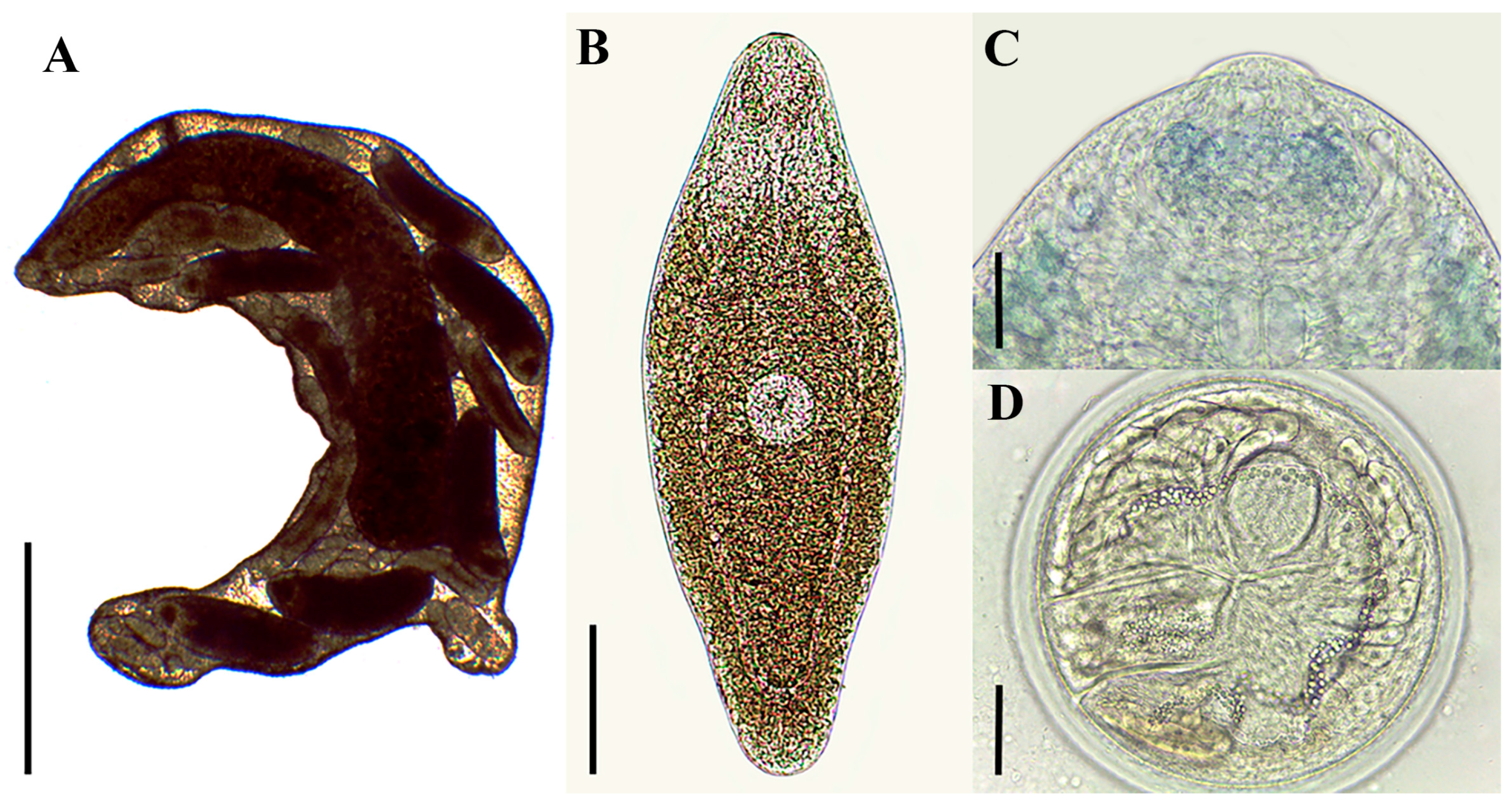
3.2. Morphological Characterization

Mature rediae (n = 3 from B. glabrata) (Figure 1A, measurements in Table 1): In vivo very active. Body robust and elongated, with two locomotor appendages in the posterior region. Mouth at the anterior end. Pharynx small, globose, and muscular. Cecum long and wide, with brown contents, occupying the region between the half and final third of the body. Cercariae at different stages of maturation present, at least 20 in number. Germ masses visualized in only one redia, located in the anterior portion of the body.
Cercariae (n = 15 from B. glabrata, 10 from P. acuta) (Figure 1B, measurements in Table 1): Body elongated, oval, dorsoventrally flattened, brown in color. Cystogenic glands distributed throughout the body. Tail absent. A bulge is visible at the posterior end only in immature specimens. Anterior organ inconspicous. Anterior organ subterminal, oval. Ventral sucker present, developed, rounded, located in the middle third of the body. Prepharynx short. Pharynx muscular and rounded (Figure 1C). Ceca smooth, dividing into two branches that join at the posterior end of the body.
Metacercariae (n = 10 from B. glabrata, 10 from P. acuta) (Figure 1D, measurements in Table 1): Spherical, brown, with a thick wall formed by 3 layers.
Adults (n = 10) (Figure 2 and Figure 3, measurements in Table 2): In vivo, the trematodes had a pink color, with little movement (Figure 2). After mounting, body elongated, lanceolate, with smooth tegument (Figure 3A). Oral sucker absent. Pre-pharynx short. Pharynx developed, muscular. Esophagus short. Cecum smooth, joint near the posterior end of the body, forming a cyclocoel. Genital pore prepharyngeal (Figure 3B). Cirrus sac piriform, small, located laterally to the pharynx, above the cecal bifurcation. Seminal vesicle present. Two testes, round to oval, located in the posterior quarter of the body, intercecal, oblique, forming a triangle with the ovary. Ovary small, spherical, intertesticular, on the right side of the body. Mehlis’ gland developed, posterior to ovary. Vitellaria bilateral, follicular, cecal, and extracecal, extending from anterior second half of body to posterior fourth of body, non-confluent. Vitelline reservoir present, tubular, ventral to Mehlis’ gland. Uterus with sinuous loops, filling most of the intercecal region, projecting from the anterior border of the posterior testis to the anterior quarter of the body. Egg elliptical to oval, operculated, golden in color, mature, with occulate miracidium presenting a redia inside (Figure 3C,D).

Remarks

The larval stages characterized in our study present morphological and biological characteristics compatible with those described for representative species of the family Cyclocoelidae, i.e., large rediae with two locomotor appendages in the posterior region of the body produce tailless cercariae with ceca confluent at the posterior region, which encyst as spherical thick-walled metacercariae [13,14,17,21,31]. The cercariae evaluated in this study present a ventral sucker in the middle region of the body, a morphological trait compatible with larvae known for species of Cyclocoelum [12,14,16] and absent in species of other genera, such as Typhlocoelum cucumerinum (Rudolphi, 1809) [21,32], Ophthalmophagus sp. [33], and Morishitium dollfusi (Timon-David, 1950) (originally described as Pseudohyptiasmus dollfusi) [34]. The presence of the ventral sucker was also reported in an unidentified larva described as Cercaria acaudata Ruiz, 1952, found in B. glabrata from the same locality of Brazil [35]. However, the cercariae of C. mutabile evaluated in this study was larger. It is important to note that there are divergences in the size of the cercariae identified as C. mutabile in the consulted literature, which was here interpreted as an effect of the fixation process. In our study, the measures of alive or killed in hot water larvae were quite similar to each other, and the larvae were studied under the same conditions [12]. On the other hand, larvae fixed with cold fixative are smaller (practically half the size) [12]. Cercariae fixed in hot 10% formalin and identified as C. mutabile [16] were smaller than those evaluated in our study. Regarding rediae recovered from B. glabrata, they had measures similar to those associated with C. acaudata in Brazil [35] and those recovered from mollusks from Russia [12]. However, they are larger than those found in the USA [14]. The metacercariae found in the present study were slightly larger than those described for C. mutabile in other studies [12,14].
Regarding the adult stage, all diagnostic characteristics known for C. mutabile, including the absence of oral sucker, prepharyngeal genital pore, intercecal uterine loops, cecal and extracecal vitellaria, with branches that do not converge at the posterior end of the body [1,2,3,10,11] were observed in the specimens found in G. galeata from this study. Moreover, the measures obtained are compatible with those described by the authors listed above, as shown in Table 2.

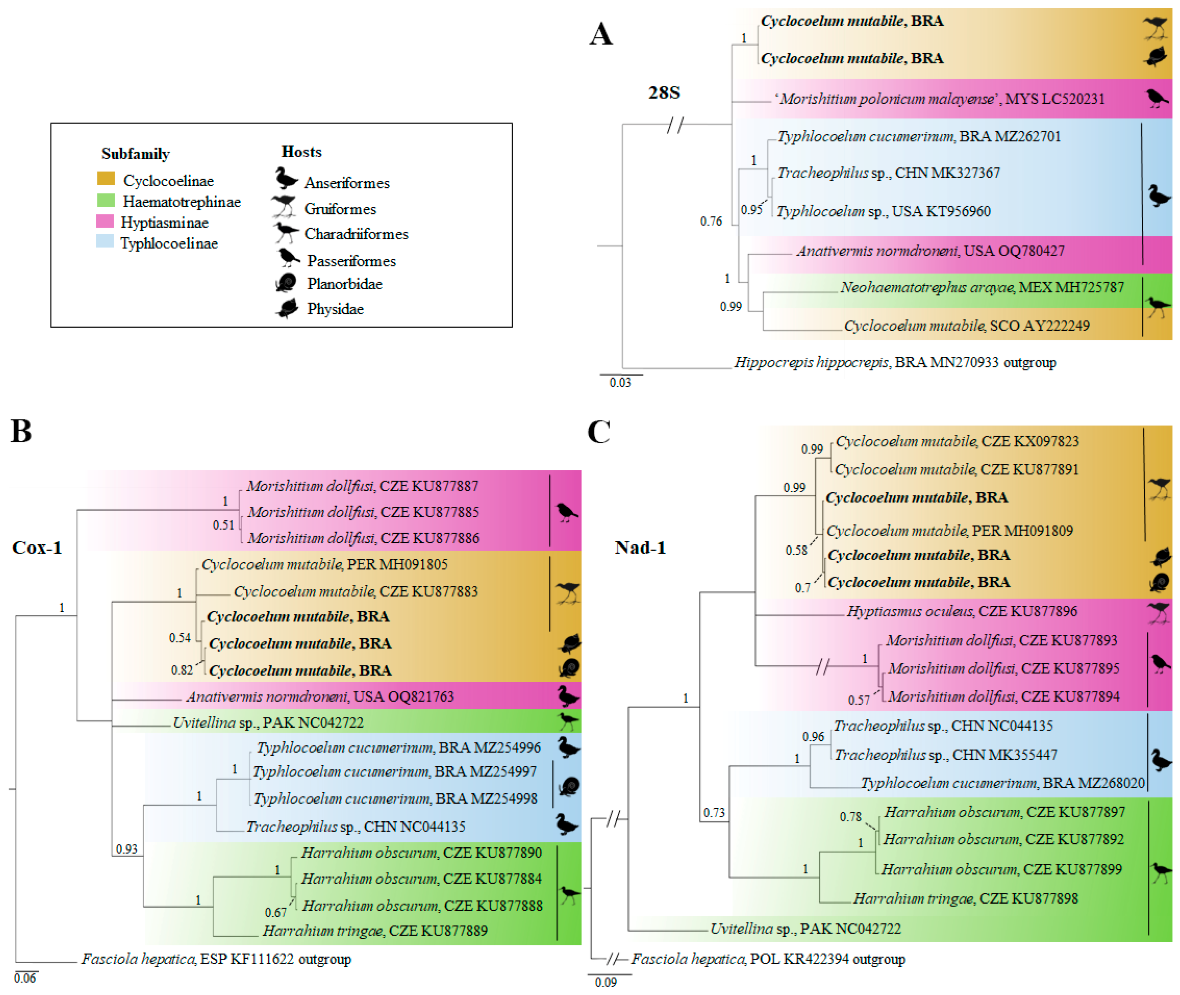
3.3. Genetic Characterization

The analysis of the sequences 28S (1173 bp) revealed 100% similarity between the larval stages from mollusks and the adult from G. galeata. Our Brazilian isolates of C. mutabile presented low similarity (92.7%) to one isolated from Scotland (AY222249, Olson et al., 2003) [36]. The similarities between our C. mutabile and other species of cyclocoelids from different subfamilies (Haemototrephinae, Hyptiasminae, Typhlocoelinae) included in the analysis were relatively low (92.7–98.9%). In the phylogeny, C. mutabile of the present study formed a distinct clade from the members of the other subfamilies. Curiously, the Scotland isolate of C. mutabile grouped with Neohaematotrephus arayae Zamparo, Brooks, Causey, and Rodriguez, 2003 in a well-supported clade (0.99), but the similarity value between these two species was low (91.4%) (Figure 4A).
In the analysis of a fragment of the Cox-1 gene (768 bp, pos-barcode region), the larval stages found in B. glabrata and P. acuta had 100% similarity. Moreover, they were 99.2% similar to the adult specimen from G. galeata from the present study. The new sequences generated were also compared with shorter sequences available to other isolates of C. mutabile from the Czech Republic (KU877883) and Peru (MH091805-07) [9,11]. For this porpoise, an alignment with 306 bp was evaluated. As a result, the Brazilian isolates of C. mutabile were >99% similar to isolates of this species found in G. galeata (considered as Gallinula chloropus (Linnaeus, 1758)) in Peru (MH091805-07) and 94.4–95.2% in relation to worms found in Fulica atra (Linnaeus, 1758) from the Czech Republic (KU877883). Regarding the other seven species of the family Cyclocoelidae included in the analysis, similarity values were lower than 88.5%. In the phylogeny, the Brazilian isolate of C. mutabile formed a well-supported clade with the isolates of this species from Peru and the Czech Republic commented above (Figure 4B). Regarding the barcode region of Cox-1, a fragment of 586 bp was here generated for the first time to C. mutabile, which presented with low similarity (81.94%) with Uvitelina sp. (NC042722) [37], the only cyclocoelid species with data available for this same region.
Sequences of the Nad-1 gene (437 bp) were generated for the developmental stages of C. mutabile in the present study. Larval stages from mollusks were identical and 99.5% similar to adults found in G. galeata. Moreover, these Brazilian isolates were 99.4–100% and 96.6–97% similar to isolates of C. mutabile from Peru (MH091808 and MH091809) and the Czech Republic (KU877891 and KX097823), respectively. In relation to other cyclocoelids included in the analysis, the similarity values were lower (75.8–83.0%). In the phylogenetic analysis, the isolates of C. mutabile from South America and Europe were also grouped in a well-supported clade (Figure 4C). The two sequences from the European continent were grouped in a well-supported subclade. The pairwise comparison data generated for this study are available in Supplementary Material Table S2.

3.4. Taxonomic Summary

Cyclocoelum mutabile (Zeder, 1800)
Locality: Belo Horizonte, Minas Gerais, Brazil
New natural intermediate hosts: Biomphalaria glabrata (Planorbidae) and Physela acuta (Physidae)
Prevalence of infection: 8/30 (26.7%) in B. glabrata and 3/33 (9.7) in P. acuta.
Definitive host: Gallinula galeata (Gruiformes: Rallidae)
Intensity of infection: 24

4. Discussion

Cyclocoelum mutabile is an air sac trematode with wide geographic distribution and has been reported to infect about 45 species of aquatic or semi-aquatic birds from different parts of the Americas, Europe, and Asia [1,2,3,9,10,11,24,25]. Although information on the occurrence of this species in the definitive host is not uncommon, data on the mollusks involved in its transmission are scarce. Experimental infection studies carried out decades ago in the Northern Hemisphere reveal the susceptibility of different freshwater gastropods to C. mutabile. Among the susceptible experimental hosts to this cyclocoelid are lymnaeids (Ladislavella elodes (Say, 1821)), physids (Physa sp., Physella gyrina (Say, 1821), and Physa jennessi Dall, 1919) and planorbids (Planorbella trivolvis (Say, 1817), Promenetus exacuous (Say, 1821), Gyraulus circumstriatus (Tryon, 1866), and Armiger crista (Linnaeus, 1758)) [12,13,14,15,16,38]. Information on finding mollusks naturally infected with C. mutabile is limited to the works by Ginetsinskaya and Saakova [18] and Korol and Stenko [16] in Eurasia. In the Americas, to the best of our knowledge, this is the first report of mollusks naturally infected with C. mutabile. Moreover, we included species of the genus Biomphalaria in the roll of susceptible hosts for this trematode, which can be considered a piece of relevant epidemiological information considering the diversity and wide distribution of these planorbids. In fact, similar larvae were found in B. glabrata in Brazil and described as Cercaria acaudata [35] or identified as cercariae of the type Cercariaeum [39], which can be conspecific with C. mutabile.
The use of the integrative taxonomy approach in this study made it possible to identify the larval stages of C. mutabile. However, some challenges had to be overcome in interpreting our results. They were a consequence of methodological aspects involved in studying larval stages of trematodes, the complex taxonomy of cyclocoelids, and the divergent information available in the literature. Regarding the morphological study of larval stages, the condition of the sample (alive or fixed) can result in significant differences in the measurements, a fact already stated by Ginetsinskaia [12]. In our study, the size of the larvae measured alive or after being killed in hot water and fixed in formalin were quite similar to each other and with data originally obtained from larvae alive studied by this author. Ginetsinskaia [12] also showed that larvae of C. mutabile fixed, probably with cold fixative, were smaller than alive ones. On the other hand, cercariae found in Lymnaea spp. and Planorbis planorbis in the Crimean Peninsula and fixed in 10% hot formalin were identified as C. mutabile [16], even being smaller than those described to the species under the same condition. Moreover, larvae identified as Cyclocoelidae gen. sp. in the same study [16] had measurements similar to the cercariae of C. mutabile of previous studies. However, the rediae of this unknown species were smaller [16], suggesting these isolates belong to different species. A curious fact verified in the analysis of the literature is that Yamaguti [13] showed data for two species, Cyclocoelum microstomum (Creplin, 1829) and C. mutabile, but reproduced the measures of those alive or fixed cercariae, respectively, studied in Russia [12]. The origin of this is difficult to elucidate, but analysis of the Russian work left no doubt that it is C. mutabile, in the original work as C. microstomum, but these species are considered synonymous [1,2]. Given the complexity resulting from the type of material analyzed, the use of heat-killed and fixed cercariae is recommended for morphometric analysis of larval stages of cyclocoelids.
Over time, some taxonomic revisions related to species of the family Cyclocoelidae became available, and the list of synonyms of C. mutabile is very expressive [1,2]. To add complexity to this scenario, Feizullaev [40] proposed that C. mutabile and C. microstomum be reconsidered as distinct, which was followed by McLaughlin [14] during the experimental study of the biology of C. mutabile in Canada. However, the figures represented in the report by Feizullaev [40] reveal identification mistakes, given that the species considered as C. mutabile cannot be assigned to the genus Cyclocoelum, but belong probably to Morishitium. On the other hand, the morphology of the specimens identified as C. microstomum by Feizullaev [40] is not distinguished from C. mutabile. From the morphological point of view, the trematodes recovered from G. galeata in our study are compatible with C. mutabile, presenting all the differential traits associated with this species [1,2,9,10,11]. Our specimens also present measures similar to those described for the species by Fernandes [24], who reported C. mutabile in the same avian host for the first time in Brazil. They are slightly smaller than those found in F. atra in the Czech Republic [9] and in Tringa semipalmata Gmelin, 1789, in Mexico [10] and larger than specimens obtained in G. galeata (considered as G. chloropus) from Peru [11].
Our study generated new sequences for C. mutabile, which are the first ones obtained for larval stages found in mollusks. Genetic data obtained reveal high similarity values (>99% for all genetic markers, indicating conspecificity) between larval stages found in B. glabrata and P. acuta and adult specimens identified as C. mutabile found in the air sacs of G. galeata. Again, some findings call our attention during the comparison with data available for comparison, deserving some comments. In the analysis of the 28S dataset, a very high divergence between our C. mutabile and an isolate of this species from Scotland [36] was observed. Furthermore, in the phylogenetic analysis, the Scotland isolate did not group with the isolates of the C. mutabile from our study but with the Mexican isolate of N. arayae [10], a representative species of the subfamily Haematotrephinae. However, the high divergence between the cyclocoelid from Scotland and the Mexican isolate of N. arayae led us to infer that the first belongs to another genus of cyclocoelid that was not yet sequenced. Thus, we recommend that the 28S data of the present study be associated with C. mutabile, and that the taxonomic identity of the Scotland cyclocoelid of Olson et al. [36] be re-evaluated. In relation to the mitochondrial markers, similarities values of the order 97% were verified in Nad-1, supporting that the isolates from South America (Brazil and Peru) are conspecific with isolates of C. mutabile obtained in F. atra from the Czech Republic [6], supporting the transcontinental distribution of this cyclocoelid species. Curiously, the similarity found for the Cox-1 in comparison with these same isolates was relatively low (94.4%). We consider that few sequences of cyclocoelids are available to these trematodes, and the possibility of the occurrence of cryptic speciation in cyclocoelids cannot be ruled out.
Taken together, data herein presented for C. mutabile obtained in Brazil reveal the contribution of the integrative taxonomy to enable the elucidation of the natural hosts involved in the life cycle of species of Cyclocoelidae. However, the amount of information available for larval stages of trematodes of this family is still very small or practically non-existent if we consider parasite detection in mollusks that are naturally infected. Moreover, an increase in the generation of sequence for this group is necessary to advance our knowledge of its phylogenetic interrelationships inside the family and in relation to other members of the superfamily Echinostomatoidea [41]. In this sense, new efforts to advance the generation of basic knowledge must be continuously encouraged, which can be achieved by searching for the non-emergent intramolluscan stages in the potential hosts and the generation of adult-based sequences to subside the link between these different developmental stages. Only the generation of different sorts of biological data will enable understanding of issues such as the real diversity, phylogeography, cryptic speciation, and evolution involving avian air sac trematodes.

Supplementary Materials

The following supporting information can be downloaded at: https://www.mdpi.com/article/10.3390/d16070422/s1, Table S1: Information on the sequences used in the alignment construction. Table S2: Pairwise comparison based on 28S, Cox-1, and Nad-1 among members of the Cyclocoelidae included in the genetic analysis.

Author Contributions

Conceptualization, H.A.P. and J.C.A.d.A.; methodology, H.A.P. and J.C.A.d.A.; formal analysis, J.C.A.d.A.; resources H.A.P., writing—original draft preparation, H.A.P. and J.C.A.d.A.; writing—review and editing, H.A.P. and J.C.A.d.A.; supervision, H.A.P.; funding acquisition, H.A.P.; project administration, H.A.P. All authors have read and agreed to the published version of the manuscript.

Funding

J.C.A.d.A. was supported by Coordination for the Improvement of Higher Education (CAPES, Ph.D scholarship). H.A.P. was supported by the National Council for Scientific and Technological (CNPq, research scholarship, process 303754/2021-4).

Institutional Review Board Statement

The animal study protocol was approved by the Ethics Committee in Animal Experimentation of the Universidade Federal de Minas Gerais (CEUA-UFMG, protocol 254–2018).

Data Availability Statement

Data is contained within the article or supplementary material. New genetic sequences generated for this study are available on GenBank.

Acknowledgments

We thank the Coordination for the Improvement of Higher Education (CAPES, Brazil) for the Ph.D. scholarship to JCAA and the National Council for Scientific and Technological (CNPq, Brazil) for the research scholarship to HAP. Many thanks are also due to Kirill Galaktionov, Russian Academy of Sciences, for supporting us with the Russian literature.

Conflicts of Interest

The authors declare no conflicts of interest.

References

  1. Dubois, G. Revision des Cyclocoelidae Kossack 1911. Rev. Suisse Zool. 1959, 66, 67–147. [Google Scholar] [CrossRef]
  2. Kanev, I.; Radev, V.; Fried, B. family Cyclocoelidae Stossich, 1902. In Keys to the Trematoda; Gibson, D.I., Jones, A., Bray, R.A., Eds.; CAB International and The Natural History Museum: London, UK, 2002; Volume 1, pp. 131–145. [Google Scholar]
  3. Dronen, N.O.; Blend, C.K. Updated keys to the genera in the subfamilies of Cyclocoelidae Stossich, 1902, including a reconsideration of species assignments, species keys and the proposal of a new genus in Szidatitreminae Dronen, 2007. Zootaxa. 2015, 4053, 1–100. [Google Scholar] [CrossRef]
  4. Dutton, H.R.; Bullard, S.A.; Kelly, A.M. New genus and species of Cyclocoelidae Stossich, 1902 (Platyhelminthes: Digenea) infecting the nasopharyngeal cavity of Canada Goose, Branta canadensis (Anseriformes: Anatidae) from Western Alabama. J. Parasitol. 2023, 190, 349–356. [Google Scholar] [CrossRef] [PubMed]
  5. McLaughlin, J.D. The migratory route of Cyclocoelum mutabile (Zeder) (Trematoda: Cyclocoelidae) in the American coot, Fulica americana (Gm.). Can. J. Zool. 1977, 55, 274–279. [Google Scholar] [CrossRef]
  6. Branton, S.L.; Deaton, J.W.; Gerlach, H.; Ruff, M.D. Cyclocoelum mutabile infection and aortic rupture in an American coot (Fulica americana). Avian Dis. 1985, 29, 246–249. [Google Scholar] [CrossRef]
  7. Galosi, L.; Heneberg, P.; Rossi, G.; Sitko, J.; Magi, G.E.; Perrucci, S. Air sac trematodes: Morishitium polonicum as a newly identified cause of death in the common blackbird (Turdus merula). Int. J. Parasitol. Parasites Wildl. 2019, 9, 74–79. [Google Scholar] [CrossRef]
  8. Diaz, E.A.; Donoso, G.; Mosquera, J.D.; Ramírez-Villacís, D.X.; Gonzáles, G.; Zapata, S.; Cisneros-Heredia, D.F. Death by massive air sac fluke (Trematoda: Bothriogaster variolaris) infection in a free-ranging snail kite (Rostrhamus sociabilis). Int. J. Parasitol. 2022, 19, 155–160. [Google Scholar] [CrossRef]
  9. Sitko, J.; Bizos, J.; Heneberg, P. Central European parasitic flatworms of the Cyclocoelidae Stossich, 1902 (Trematoda: Plagiorchiida): Molecular and comparative morphological analysis suggests the reclassification of Cyclocoelum obscurum (Leidy, 1887) into the Harrahium Witenberg, 1926. Parasitology 2017, 144, 368–383. [Google Scholar] [CrossRef] [PubMed]
  10. López-Jiménez, A.; García-Varela, M.; Hernández-Orts, J.S. Review of five species of cyclocoelids (Digenea: Cyclocoelidae) from aquatic birds in Mexico with notes on their interspecific variation. Syst. Parasitol. 2018, 95, 921–942. [Google Scholar] [CrossRef]
  11. Gomez-Puerta, L.A.; Salas, M.Y.; Lopez-Urbina, M.T.; Gonzalez, A.E. Diagnóstico morfológico y molecular de Cyclocoelum mutabile (Trematoda: Cyclocoelidae) en el Perú. Rev. Peru. Biol. 2018, 25, 315–320. [Google Scholar] [CrossRef]
  12. Ginetsinskaya, T.A. Life cycle and biology of the developmental stages of Cyclocoelum microstomum (Trematoda). Uchenye Zap. Leningr. Univ. Ser. Biol. 1954, 172, 90–112. (In Russian) [Google Scholar]
  13. Yamaguti, S. A Synoptical Review of Life Histories of Digenetic Trematodes of Vertebrates; Keigaku Publishing: Tokyo, Japan, 1975. [Google Scholar]
  14. McLaughlin, J.D. Experimental studies on the life cycle of Cyclocoelum mutabile (Zeder) (Trematoda: Cyclocoelidae). Can. J. Zool. 1976, 54, 48–54. [Google Scholar] [CrossRef] [PubMed]
  15. McLaughlin, J.D. The biology of Cyclocoelum mutabile (Trematoda) infections in American coots. Proc. Helminthol. Soc. Wash. 1986, 53, 177–181. [Google Scholar]
  16. Korol, E.; Stenko, P. Larvae of trematodes of the family Cyclocoelidae (Trematoda, Digenea) in Crimean Mollusks. Vestn. Zool. 2005, 18, 192–194. (In Ukrainian) [Google Scholar]
  17. Galaktionov, K.V.; Dobrovolskij, A.A. The Biology and Evolution of Trematodes; Kluwer Academic Publishers: Dordrecht, The Netherlands; Boston, MA, USA; London, UK, 2003. [Google Scholar]
  18. Ginetsinskaya, T.A.; Saakova, E.O. Migration of trematodes of the family Cyclocoelidae Koss in the body of the final host. Dokl. Akad. Nauk. SSSR. 1952, 85, 1432–1436. (In Russian) [Google Scholar]
  19. Ẑdárská, Z. Larvální stadia motolic z vodních plů na území ČSSR. Česk. Parasitol. 1963, 10, 207262. [Google Scholar]
  20. Urabe, M.; Hashim, N.E.N.; Uni, S.; Takachi, I.; Halim, M.R.A.; Marzuki, M.E.; Udin, A.S.M.; Zainuri, N.A.; Omar, H.; Agatsuma, T.; et al. Description and molecular characteristics of Morishitium polonicum malayense Urabe, Nor Hashim & Uni, n. subsp. (Trematoda: Cyclocoelidae) from the Asian glossy starling, Aplonis panayensis strigata (Passeriformes: Sturnidae) in Peninsular Malaysia. Parasitol. Int. 2020, 76, 102074. [Google Scholar] [CrossRef]
  21. Assis, J.C.A.; López-Hernández, D.; Favoretto, S.; Medeiros, L.B.; Melo, A.L.; Martins, N.R.S.; Pinto, H.A. Identification of the avian tracheal trematode Typhlocoelum cucumerinum (Trematoda: Cyclocoelidae) in a host-parasite-environment system: Diagnosis, life cycle and molecular phylogeny. Parasitology 2021, 148, 1383–1391. [Google Scholar] [CrossRef] [PubMed]
  22. Paraense, W.L. Planorbídeos hospedeiros intermediários do Schistosoma mansoni. In Esquistossomose Mansoni; Cunha, A.S., Ed.; Ed USP: São Paulo, Brasil, 1970; pp. 13–30. [Google Scholar]
  23. Paraense, W.L.; Pointier, J.P. Physa acuta Draparnaud, 1805 (Gastropoda: Physidae): A study of topotypic specimens. Mem Inst Oswaldo Cruz. 2003, 98, 513–517. [Google Scholar] [CrossRef]
  24. Fernandes, B.M.M. Sobre as espécies brasileiras da família Cyclocoelidae Kossak, 1911 (Trematoda, Cyclocoelidae). Mem. Inst. Oswaldo Cruz. 1976, 74, 289–294. [Google Scholar] [CrossRef]
  25. Fernandes, B.M.M.; Justo, M.C.N.; Cárdenas, M.Q.; Cohen, S. South American Trematodes Parasites of Birds and Mammals; Fundação Oswaldo Cruz: Rio de Janeiro, Brazil, 2015. [Google Scholar]
  26. Tkach, V.V.; Littlewood, T.J.; Olson, P.D.; Kinsella, M.; Swiderski, Z. Molecular phylogenetic analysis of the Microphalloidea Ward, 1901 (Trematoda: Digenea). Syst. Parasitol. 2003, 56, 1–15. [Google Scholar] [CrossRef] [PubMed]
  27. Miura, O.; Kuris, A.M.; Torchin, M.E.; Hechinger, R.F.; Dunham, E.J.; Chiba, S. Molecular-genetic analyses reveal cryptic species of trematodes in the intertidal gastropod, Batillaria cumingi (Crosse). Int. J. Parasitol. 2005, 35, 793–801. [Google Scholar] [CrossRef] [PubMed]
  28. Morgan, J.A.T.; Blair, D. Relative merits of nuclear ribosomal internal transcribed spacers and mitochondrial CO1 and ND1 genes for distinguishing among Echinostoma species (Trematoda). Parasitology 1998, 116, 289–297. [Google Scholar] [CrossRef] [PubMed]
  29. Van Steenkiste, N.; Locke, S.A.; Castelin, M.; Marcogliese, D.J.; Abbott, C.L. New primers for DNA barcoding of digeneans and cestodes (Platyhelminthes). Mol. Ecol. Resour. 2015, 15, 945–952. [Google Scholar] [CrossRef] [PubMed]
  30. Miller, M.A.; Pfeiffer, W.; Schwartz, T. Creating the CIPRES science gateway for inference of phylogenetic trees. In Proceedings of the Gateway Computing Environments Workshop (GCE), New Orleans, LA, USA, 14 November 2010; pp. 1–8. [Google Scholar] [CrossRef]
  31. Stunkard, H.W. The life history of Typhlocoelum cymbium (Diesing, 1850) Kossack. 1911 (Trematoda, Cyclocoelidae). A contribution to the phylogeny of the monostomes. Bull. Soc. Zool. Fr. 1934, 59, 447–466. [Google Scholar]
  32. Schafranski, N.L.; Freitas, M.G.; Costa, J.O. Ciclo biológico de Typhlocoelum cucumerinum (Rudolphi, 1809) (Trematoda: Cyclocoelidae). Rev. Bras. Biol. 1975, 35, 519–526. [Google Scholar]
  33. Taft, S.J.; Heard, R.W. Aspects of the larval development of Ophthalmophagus sp. (Trematoda: Cyclocoelidae). J. Parasitol. 1978, 64, 597–600. [Google Scholar] [CrossRef]
  34. Timon-David, T. Cycle évolutif d’un Trématode Cyclocoeidé: Pseudhyptiasmus dollfusi Timon-David 1950. Recherches expérimentales. Ann. Parasitol. Hum. Comp. 1955, 30, 43–61. [Google Scholar] [CrossRef] [PubMed]
  35. Ruiz, J.M. Contribuição ao estudo de formas larvárias de trematódeos brasileiros. 3—Fauna de Belo Horizonte e Jaboticatubas, Estado de Minas Gerais. Mem. Inst. Butantan. 1952, 24, 45–62. [Google Scholar]
  36. Olson, P.D.; Cribb, T.H.; Tkach, V.V.; Bray, R.A.; Littlewood, D.T. Phylogeny and classification of the Digenea (Platyhelminthes: Trematoda). Int. J. Parasitol. 2003, 33, 735–755. [Google Scholar] [CrossRef]
  37. Suleman, M.S.K.; Heneberg, P.; Zhou, C.Y.; Muhammad, N.; Zhu, X.Q.; Ma, J. Characterization of the complete mitochondrial genome of Uvitellina sp., representative of the family Cyclocoelidae and phylogenetic implications. Parasitol Res. 2019, 118, 2203–2211. [Google Scholar] [CrossRef] [PubMed]
  38. McKindsey, C.W.; McLaughlin, J.D. Field studies on the transmission and survival of Cyclocoelum mutabile (Digenea) infections in natural snail populations in Southern Manitoba, Canada. J. Parasitol. 1995, 81, 520–525. [Google Scholar] [CrossRef] [PubMed]
  39. Ohlweiler, F.P.; Eduardo, J.M.; Takahashi, F.Y.; Crein, G.A.; Luca, L.R.; Oliveira, R.C. Larvas de trematódeos associadas a moluscos de água doce em municípios da Região Metropolitana de São Paulo, Estado de São Paulo, Brasil. Rev. Pan-Amaz Saúde 2013, 4, 37–48. [Google Scholar] [CrossRef]
  40. Feizullaev, A. On specific characters and distinct status of Cyclocoelum mutabile (Zeder, 1800) and Cyclocoelum microstomum (Creplin, 1829) (Trematoda, Cyclocoelidae). Parazitologiya 1970, 4, 39–42. (In Russian) [Google Scholar]
  41. Tkach, V.V.; Kudlai, O.; Kostadinova, A. Molecular phylogeny and systematics of the Echinostomatoidea Looss, 1899 (Platyhelminthes: Digenea). Int. J. Parasitol. 2016, 46, 171–185. [Google Scholar] [CrossRef]
Figure 1. Larval stages of the Cyclocoelum mutabile found in Biomphalaria glabrata and Physella acuta from Brazil. (A) Redia with developing cercariae. (B) Detail of cercaria. (C) Anterior end of the cercaria with the anterior organ and pharynx. (D) Metacercaria. Scale bars: (A)—500 µm, (B)—150 µm, (C,D)—50 µm.
Diversity 16 00422 g001
Figure 2. Air sac flukes in a juvenile specimen of the common gallinule (Gallinula galeata) from Brazil. Scale bar: 1 cm.
Diversity 16 00422 g002
Figure 3. Cyclocoelum mutabile found in Gallinula galeata from Brazil. (A) Specimen in toto. (B) Detail of the anterior end, with the pre-pharyngeal genital pore, oral opening, and pharynx. (C,D) Mature egg with occulate miracidium presenting a developed redia inside. Scale bars: (A)—2 mm, (B)—500 m, (C,D)—50 μm.
Diversity 16 00422 g003
Figure 4. Phylogenetic relationships between the Cyclocoelum mutabile found in mollusks (Biomphalaria glabrata and Physella acuta) and common gallinule (Gallinula galeata) from Brazil and selected members of the family Cyclocoelidae, inferred from partial sequences of the genes (A) 28S (1173 bp, evolutive model: GTR + G), (B) Cox-1 (306 bp, evolutive model: TN93 + G) and (C) Nad-1 (437 bp, evolutive model: HKY + G). The phylogenetic trees were generated by Bayesian Inference, and nodal supports are indicated by posterior probability. New sequences from the current study are shown in bold. Scale bars indicate the number of substitutions per site.
Diversity 16 00422 g004
Table 1. Measurements of rediae, cercariae, and metacercariae of Cyclocoelum mutabile found in Biomphalaria glabrata and Physella acuta from Brazil and data reported for this species by other authors. Data in micrometer. Abbreviations: N = number, L = length, W = width, D = diameter.
Host Biomphalaria glabrataPhysella acutaMollusk species 1Mollusk species 2Mollusk species 3
Locality BrazilBrazilRussiaCanadaCrimea
Reference Present studyPresent studyGinetsinskaia [12]McLaughlin [14]Korol and Stenko [16]
Condition AliveHot killed
and Fixed
AliveFixedAliveAliveHot killed
Mature rediae N3------
BodyL4649 ± 1105--2000–30003500–4000700–2030-
(3578–6171)
W1079 ± 183)---620--
(906–1000)
PharynxL138 ± 60 (86–221)--160---
W117 ± 29 (88–157)--180---
Cercariae N151510----
BodyL685 ± 122661 ± 96669 ± 229340–370625–764520–550318
(490–936)(504–794)(509–922)
W271 ± 49 (170–367)198 ± 54 (144–378)307 ± 30 (238–344)90–100208–222166–180143
AnteriorL100 ± 22 (70–159)85 ± 7 (73–94)96 ± 14 (80–109)6583–8798–11755
organW85 ± 16 (66–119)72 ± 8 (65–89)89 ± 9 (82–98)6583–87 57
PharynxL38 ± 6 (30–48)27 ± 0.6 (27–28)40 ± 3 (36–44)-41–4941–4618
W41 ± 8 (30–53)26 ± 3 (23–31)45 ± 2 (43–47)-41–4939–4218
Ventral L75 ± 7 (67–97)69 ± 9 (58–89)76 ± 7 (69–838)3475–8368–8347
suckerW74 ± 6 (67–89)68 ± 9 (60–92)78 ± 7 (71–85)3775–8368–8342
Metacercariae N101010----
D317 ± 35 (241–358)384 ± 11 (359–400)338 ± 25 (287–368)180–230300250–270-
1 Ampullaceana balthica (= Lymnaea ovata), Planorbis planorbis. 2 Planorbella trivolvis, Gyralus circumstriatus, Promenetus exacuous, Armiger crista, Physella gyrina, and Ladislavella elodes. 3 Radix auricularia, Ampullaceana fontinalis, Planorbis planorbis.
Table 2. Morphometric data for Cyclocoelum mutabile recovered from Gallinula galeata from Brazil and data reported from this species by different authors. Data in micrometer unless otherwise indicated. C: Length. L: Width. N: Number.
Hosts Galinulla galeataG. galeataFulica atraG. galeataTringa semipalmata
Locality BrazilBrazilCzech RepublicPeruMexico
Reference Present studyFernandesSitko Gomez-Puerta López-Jiménez
[24]et al. [9] et al. [11]et al. [10]
N1073047
Body (mm)L15.2 ± 5.6 (13–18.5)13.6–18.919.3 ± 3.1 (15–28.3)12.2 ± 2.7 (10.2–13.2)17.5–20.5
W4.5 ± 0.6 (3.5–5.0)4–65.2 ± 0.9 (3.8–7.4)4.5 ± 0.9 (3.8–4.8)3.2–4.4
Oral openingL328 ± 34 (283–354)-615 ± 209 (373–1286)614 ± 13 (580–642)-
W470 ± 67 (425–567)-789 ± 255 (289–1286)723 ± 49 (601–822)-
PrepharynxL342 ± 77 (284–447)430–510--192
PharynxL695 ± 68 (528–764)790–820855 ± 130 (596–1118)741 ± 15 (710–771)234–323
W783 ± 100 (586–878)1090–1240935 ± 153 (596–1237)759 ± 20 (724–805)237–326
Cirrus sacL1170 ± 80 (1100–1300)900–940-901 ± 6 (886–912)818–1035
Anterior testesL675 ± 154 (425–1009)580–9701095 ± 366 (328–1857)831 ± 40 (745–935)1078–2082
W770 ± 154 (531–1027)580–9701098 ± 395 (373–1857)889 ± 50 (779–1008)1169–1974
Posterior testesL653 ± 130 (443–835)460–640940 ± 357 (373–1771)803 ± 41 (685–873)962–1668
W755 ± 140 (514–992)780–1160940 ± 263 (447–1571)837 ± 79 (670–986)1165–1983
OvaryL379 ± 54 (286–457)300–490557 ± 76 (429–771)416 ± 10 (402–445)374–465
W400 ± 60 (286–514)310–450527 ± 87 (328–771)485 ± 36 (430–589)408–532
Egg (N = 50)L109 ± 4 (104–114)75–102111 ± 5 (104–116)116 ± 1 (112–118)130–146
W64 ± 2 (60–67)47–7560 ± 4 (52–64)57 ± 1 (54–60)77–88
Disclaimer/Publisher’s Note: The statements, opinions and data contained in all publications are solely those of the individual author(s) and contributor(s) and not of MDPI and/or the editor(s). MDPI and/or the editor(s) disclaim responsibility for any injury to people or property resulting from any ideas, methods, instructions or products referred to in the content.

