Article

Effects of Rhythm Step Training on Foot and Lower Limb Balance in Children and Adolescents with Flat Feet: A Radiographic Analysis

1
Sports Rehabilitation Center, Ain Hospital, 372, Gyeongin-ro, Michuhol-gu, Incheon 22148, Republic of Korea
2
Nowon Samsung Orthopedics, 456 Nohae-ro, Nowon-gu, Seoul 01762, Republic of Korea
3
Department of Global Sport Studies, Korea University, 2511 Sejong-ro, Jochiwon-eup, Sejong 30019, Republic of Korea
4
Department of Sports and Exercise Medicine, Biomedical Science, Korea University, 2511 Sejong-ro, Jochiwon-eup, Sejong 30019, Republic of Korea
5
Department of Physical Therapy, Gim Cheon University, 214 Daehak-ro, Gimcheon-si 39528, Gyeongsangbuk-do, Republic of Korea
6
Lotte Healthcare, 300 Olympic-ro, Songpa-gu, Seoul 05551, Republic of Korea
*
Author to whom correspondence should be addressed.
Medicina 2024, 60(9), 1420; https://doi.org/10.3390/medicina60091420
Submission received: 31 July 2024 / Revised: 26 August 2024 / Accepted: 27 August 2024 / Published: 30 August 2024
(This article belongs to the Section Sports Medicine and Sports Traumatology)

Abstract

:
Background and Objectives: Owing to the recent reports regarding the efficacy of rhythm step training (RST) in lower limb muscle development and motor skill enhancement, this study aimed to evaluate the effects of RST on foot and lower limb balance in children and adolescents diagnosed with flat feet using radiographic analysis. Materials and Methods: A total of 160 children and adolescents diagnosed with flat feet from a hospital in Seoul were randomly assigned to the general flat feet training (GFFT) (n = 80) or RST (n = 80) group. Patients in both groups exercised for 50 min once a week for 12 weeks. Key variables, such as quadriceps angle (Q-angle), calcaneal pitch angle (CPA), calcaneal–first metatarsal angle (CFMA), and navicular–cuboid overlap ratio (OR) were measured before and after the intervention. Results: Significant improvements in Q-angle (p < 0.001), CPA (p < 0.001), CFMA (p < 0.001), and navicular–cuboid OR (p < 0.001) were observed in the RST group compared to the GFFT group. RST was found to be more effective in normalizing the biomechanical function of the calcaneus and improving lower limb function. Conclusions: RST significantly enhances foot and lower limb balance in children and adolescents with flat feet, suggesting its potential use as an effective intervention for this population. The study did not specifically analyze the effects of various components of rhythm training, such as music, exercise intensity, and frequency, on the outcomes. Further research is needed to determine how each of these elements individually influences the results.

1. Introduction

Deformities of the knee joint are common issues among growing children and can lead to severe physical problems if not properly assessed and treated. Typically, normal knee development progresses from a varus to valgus alignment, which stabilizes around the age of 6 years [1,2]. During this developmental process, weakness and imbalance in muscles, tendons, and ligaments, as well as excessive weight, can cause knee instability [3]. Knee instability, particularly when it persists or worsens during growth, can lead to alterations in gait mechanics, such as increased internal rotation of the tibia and valgus alignment of the knee. These changes result in an uneven distribution of weight across the foot, particularly increasing weight-bearing on the medial side. This increased medial pressure can induce pronation of the ankle joint, which in turn exacerbates the development of flat feet by flattening the medial longitudinal arch. As a result, these biomechanical changes can contribute to limitations in physical activity and motor performance in children [4,5]. Moreover, these issues are often difficult to correct naturally during growth, necessitating active intervention to prevent further musculoskeletal complications [6,7].
The foot and ankle are essential for shock absorption, support, and balance during walking [8,9]. Flexible flat feet, a common alignment disorder, disrupts lower limb biomechanics, leading to pain, disability, and potential pronation distortion syndrome [10,11,12,13,14]. Joint flexibility indicates physical stiffness and fatigue, influencing movement and bone growth during development [15,16]. The rising number of children and adolescents seeking treatment for flat feet, from 4030 in 2010 to 13,862 in 2023, highlights ongoing issues with foot and lower limb balance [17].
Previous interventions aimed at correcting lower limb deformities have included specific exercises to induce muscle growth [18]. However, these exercises can sometimes lead to imbalances or new deformities [19,20]. In contrast, jump exercises have shown promise in activating the medial longitudinal arch muscles and improving the flat feet [21]. Incorporating music into step training can enhance physical and cognitive functions, making exercises more engaging and effective [22,23]. Recent studies have highlighted the benefits of rhythm step training (RST) in improving lower limb muscle development and motor skills [24,25]. This study acknowledges that existing research has suggested that Rhythm Step Training (RST) is effective in enhancing physical development and motor skills in children and adolescents. However, there is a significant lack of studies evaluating not only the potential benefits but also the specific impacts of RST on normalizing biomechanical functions. Therefore, this study aims to analyze the effects of RST on muscle development and balance in the feet and lower limbs of children and adolescents with flat feet and to explore the potential for improving lower limb deformities. In particular, this study will focus on how combining Rhythm Step Training with music can reduce the monotony of simple exercise and simultaneously enhance physical functions, investigating whether this approach can yield better results compared to traditional treatment methods.

2. Materials and Methods
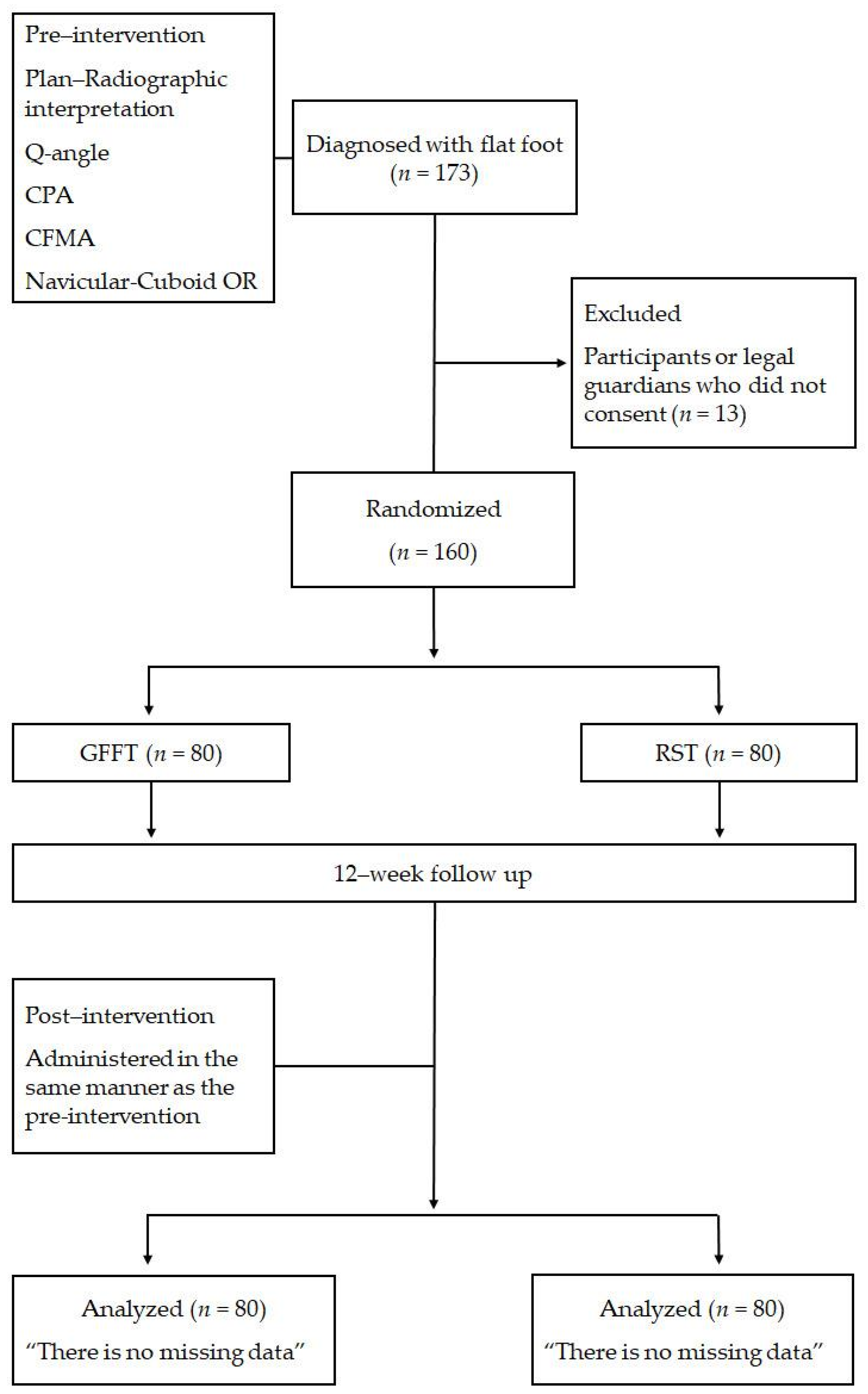
2.1. Participants

This study was approved by the Research Ethics Committee at Gim Cheon University (approval number: GU-202303-HRa-08-P, approval date: 18 July 2023). Participants were recruited at Nowon Samsung Orthopedics in Seoul, Republic of Korea, where a pediatric orthopedic surgeon selected 173 patients diagnosed with flat feet, aged 6 to 13 years, as potential participants. In this study, participants were selected from patients who visited the hospital during the research period. To ensure uniformity in the training program, only one trainer was assigned to guide all participants throughout the study. Of these, 13 individuals were excluded because they or their guardians did not provide written informed consent, resulting in a final sample of 160 participants. None of the final 160 participants met any further exclusion criteria, and all satisfied the study’s inclusion criteria. Participants were randomly allocated to either the general flat feet training (GFFT) (n = 80) or RST (n = 80) group. We opted for the F test within Analysis of Variance (repeated measures) as our analytical approach. The study sample size was calculated using G*Power (ver. 3.1.9.7, Germany), with a significance level (α) of 0.05, a power (1 − β) of 0.85, and an effect size (f) of 0.24. During the study, a dropout rate of approximately 7.5% was observed [(13/173) × 100], resulting in the final sample size of 160 participants. Prior to the experiment, all participants and their guardians were fully informed about the purpose, content, and procedures of the study, and written consent was obtained. The physical characteristics of the study participants are presented in Table 1.

2.2. Procedures

The experimental procedures of this study are shown in Figure 1.

2.3. Experimental Design and Measurements

This study aimed to compare the effects of GFFT and RST on bilateral foot and lower limb balance. Changes in the quadriceps angle (Q-angle) were assessed using a scanogram, a type of radiographic image, while the degree of flat feet deformity was determined by measuring the calcaneal pitch angle (CPA), calcaneal–first metatarsal angle (CFMA), and navicular-cuboid overlap ratio (OR) through lateral view radiographs of the foot. To ensure accuracy and minimize potential sources of error, all radiographic measurements were consistently performed by a single pediatric orthopedic specialist, thereby maintaining measurement uniformity throughout the study.

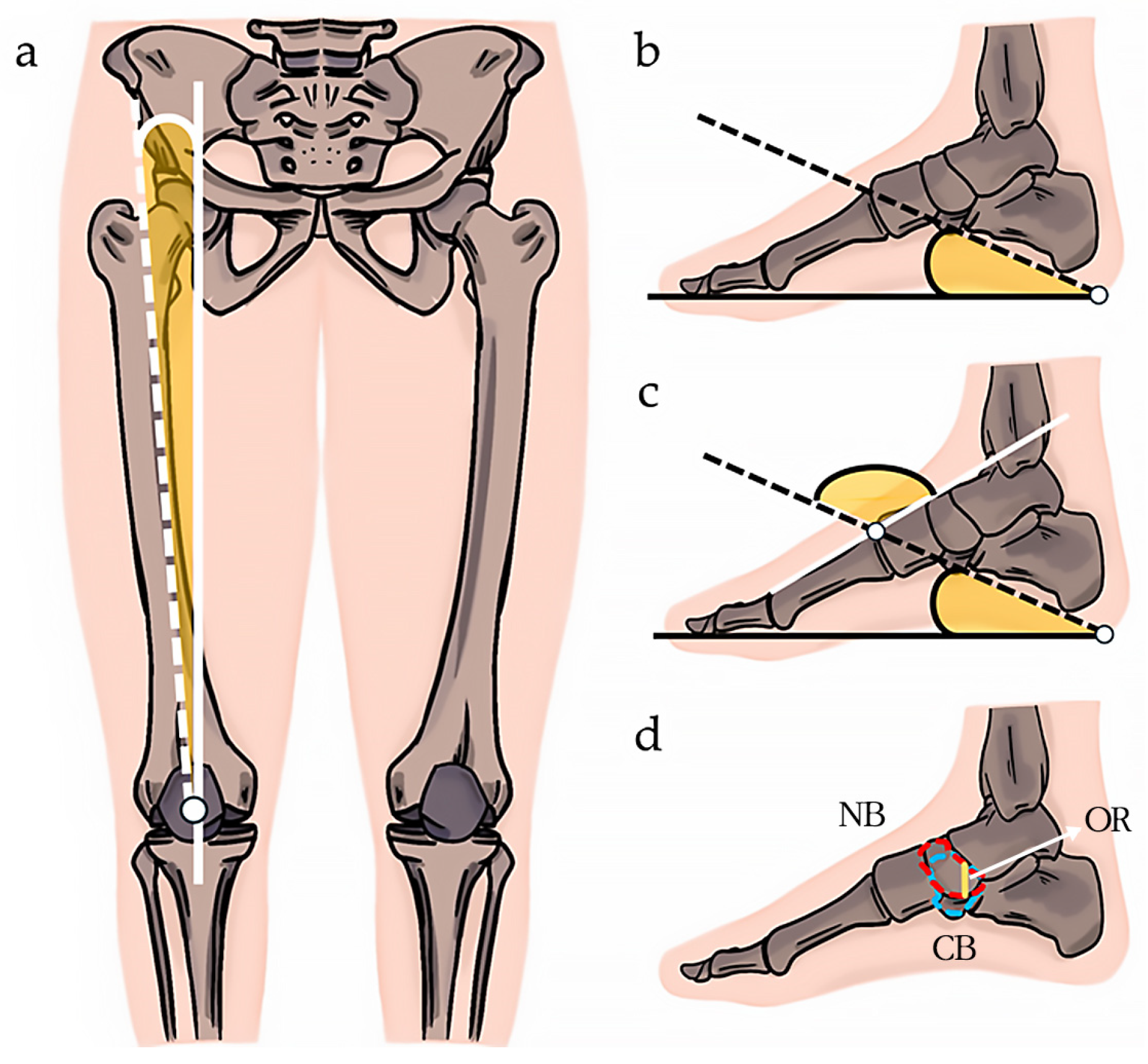
2.4. Q-Angle Measurement

The Q-angle, or quadriceps angle, is a key indicator of lower limb alignment, particularly in relation to flat feet. An increased Q-angle often results from foot adduction, femoral internal rotation, and tibial internal rotation, which can lead to knee malalignment and contribute to flat feet development. Clinically, Q-angle measurement is crucial for diagnosing and managing flat feet and related conditions, such as knee instability.
In this study, the Q-angle was measured as the angle between the line connecting the patella’s center to the tibial tubercle and the line connecting the patella to the anterior superior iliac spine [26] (Figure 2a). This provides insights into lower limb alignment and its impact on foot and knee biomechanics.

2.5. CPA Measurement

The calcaneal pitch angle (CPA) is a clinically significant measure in diagnosing flat feet, as it helps assess the alignment and structure of the foot’s arch. A lower CPA typically indicates a flattened arch, which is characteristic of a flat feet. In this study, the CPA was measured by assessing the angle formed by the lines connecting the most prominent lower part of the calcaneus to the rounded end of the calcaneus and the big toe [27] (Figure 2b).

2.6. CFMA Measurement

CFMA measurement is also used for flat feet identification. In this study, the CFMA was measured by assessing the angle at the intersection of the straight line along the first metatarsal and the upper line of the CPA [28] (Figure 2c).

2.7. Navicular–Cuboid OR Measurement

The navicular–cuboid OR is important for assessing the alignment and structural relationships between the navicular and cuboid bones, which are critical in maintaining the medial longitudinal arch of the foot. In the context of flat feet, an abnormal OR can indicate a collapse of the arch, leading to biomechanical imbalances in the lower limb. In this study, the navicular–cuboid OR was measured as the ratio of the length of the navicular bone to the length of the overlap between the navicular and cuboid bones [29] (Figure 2d).

2.8. GFFT and RST Exercise Programs

Based on the exercise prescription guidelines of the American College of Sports Medicine [30], we conducted GFFT and RST once a week for 50 min over a 12-week period. The exercise program consisted of a 5 min warm-up, a 40 min main exercise, and a 5 min cool-down.
GFFT was developed by building on the research of [31], with specific modifications to better address the needs of children with flat feet. These modifications included incorporating balance exercises that focused on improving proprioception and lower limb alignment, which are crucial for managing flat feet. Additionally, exercises were adjusted to be age-appropriate and to progressively challenge the participants’ stability and coordination.
RST was structured by modifying and refining the program outlined by [24]. These modifications were designed to ensure the exercises were safe and effective for pediatric participants, with an emphasis on rhythm and timing to improve motor coordination and balance. The exercise intensity was gradually increased over the course of the intervention period to ensure the stability of the pediatric participants, reaching a level of “somewhat hard” on the Rating of Perceived Exertion (RPE) scale.
In the RST program, participants were required to perform jump steps in synchronization with a predetermined beat and rhythm, set at 120 beats per minute (BPM). To maintain exercise intensity, participants were instructed to avoid stepping on the lines and to continue jumping without stopping, even if they could not follow the movements accurately. This ensured that the exercise remained challenging and effective, promoting endurance and rhythm consistency. Table 2 presents the details regarding the exercise programs.

2.9. Statistical Analysis

Data were analyzed using IBM SPSS Statistics version 22.0 (SPSS Corp., Chicago, CA, USA). Means and standard deviations were calculated for all measurement variables in both groups. A two-way mixed analysis of variance (ANOVA) was performed to analyze the two independent variables, time (pre-intervention and post-intervention) and group (RST and GFFT), during the 12-week training period and to analyze the interaction effect between the two independent variables. Post hoc tests for the interaction effect included paired-sample t-tests for pre- and post-differences within groups and independent-sample t-tests for differences between groups. The statistical significance level for all analyses was set at p < 0.05.

3. Results

The pre- and post-intervention changes in the Q-angle, CPA, CFMA, and navicular–cuboid OR for the RST and GFFT groups are presented in Table 3 and Figure 3.

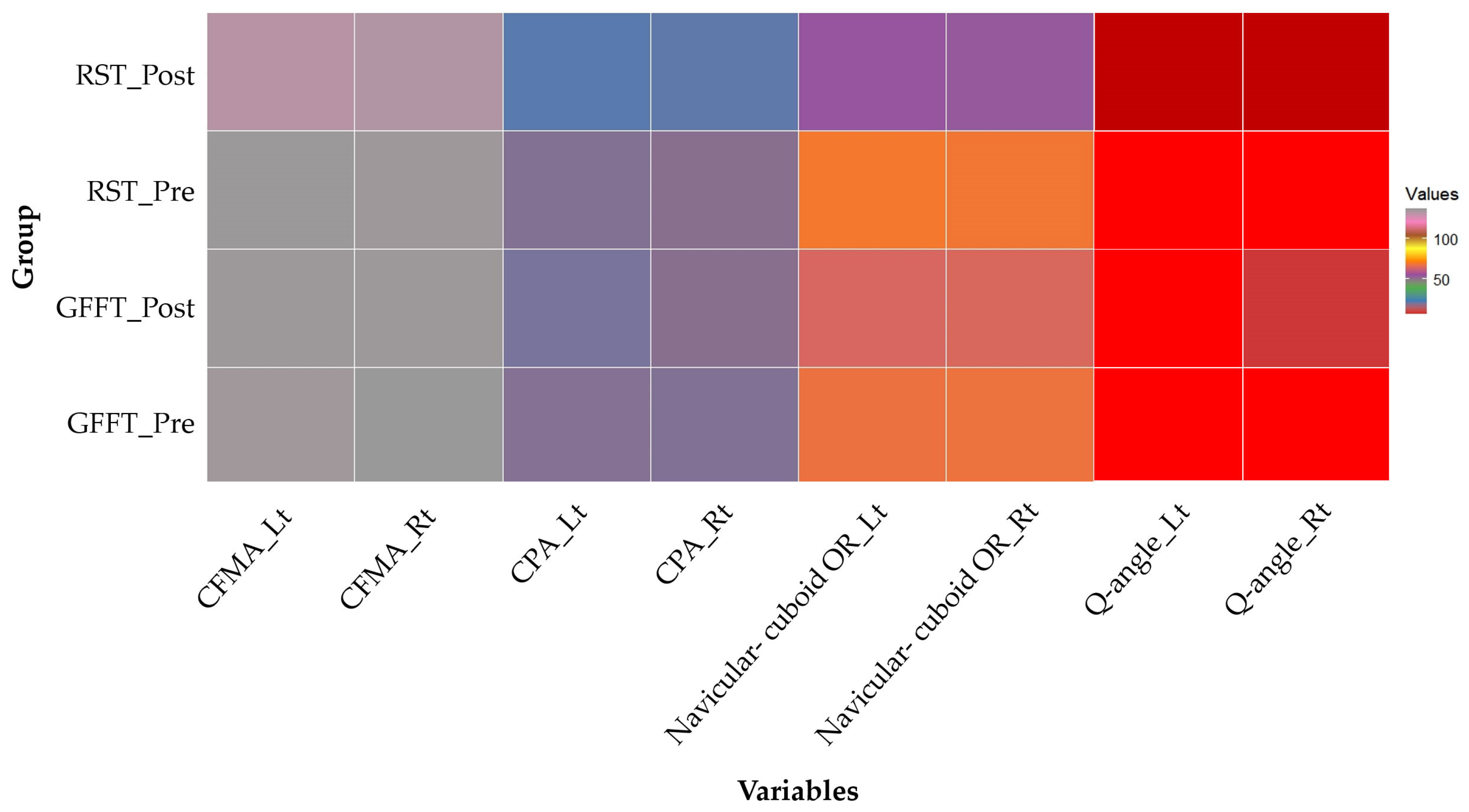
3.1. Q-Angle Changes

After the 12-week exercise intervention, the Q-angle showed significant differences in terms of the time of measurement (p < 0.001), groups (p < 0.001), and the interaction between the time of measurement and groups (p < 0.001) in both left and right foot. Post hoc analysis of the interaction effect revealed no significant within-group difference in the GFFT group for the left foot (p = 0.634), while a significant difference was observed in the RST group (p < 0.001). For the right foot, significant within-group differences were observed in both GFFT (p < 0.05) and RST (p < 0.001) groups, with a greater Q-angle improvement in the RST group. Furthermore, significant between-group differences were observed for both the left and right foot (p < 0.001).

3.2. CPA Changes

After the 12-week exercise intervention, the CPA demonstrated significant differences in terms of the time of measurement (p < 0.001), groups (p < 0.05), and the interaction between the time of measurement and groups (p < 0.005) in the left foot. Similarly, the differences were also significant in terms of the time of measurement (p < 0.001), groups (p < 0.05), and the interaction between the time of measurement and groups (p < 0.001) in the right foot. Post-hoc analysis of the interaction effect revealed significant within-group differences in both the GFFT (p < 0.005) and RST (p < 0.001) groups for the left foot, with greater increases in the RST group. Moreover, for the right foot, significant within-group differences were observed in both the GFFT (p < 0.05) and RST (p < 0.001) groups, with greater increases in the RST group. Significant between-group differences were also observed for both the left and right foot (p < 0.001).

3.3. CFMA Changes

After the 12-week exercise intervention, in the left foot, the CFMA showed significant differences in terms of the time of measurement (p < 0.05) and the interaction between the time of measurement and groups (p < 0.005), but not between groups. Post hoc analysis of the interaction effect revealed no significant within-group difference in the GFFT group (p = 0.672). However, a significant within-group difference was observed in the RST group (p < 0.001) as well as significant between-group differences (p < 0.001). In the right foot, the CFMA showed significant differences in terms of the time of measurement (p < 0.05), groups (p < 0.001), and the interaction between the time of measurement and groups (p < 0.05). Post hoc analysis of the interaction effect revealed that no significant within-group difference in the GFFT group (p = 0.291), although a significant within-group difference was observed in the RST group (p < 0.001) as well as significant between-group differences (p < 0.001).

3.4. Navicular–Cuboid OR Changes

After the 12-week exercise intervention, the navicular–cuboid OR demonstrated significant differences in terms of the time of measurement (p < 0.001), groups (p < 0.05), and the interaction between the time of measurement and groups (p < 0.001) in the left foot. Similarly, in the right foot, significant differences were observed in terms of the time of measurement (p < 0.005), groups (p < 0.001), and the interaction between the time of measurement and groups (p < 0.001). Post hoc analysis of the interaction effect revealed significant within-group differences in both the GFFT (p < 0.05) and RST (p < 0.001) groups for the left and right foot, with greater decreases in the RST group. Significant between-group differences were also observed (p < 0.001).

4. Discussion

This study aimed to investigate the calcaneal biomechanical function normalization and lower limb functional activity improvement in children and adolescents with flat feet who underwent RST.

4.1. Q-Angle Changes

In this study, the Q-angle was measured to identify functional knee issues and observe changes before and after the exercise intervention. The Q-angle is an important indicator for lower limb alignment assessment. It varies with age, gender, and growth status, and the normal values for children and adolescents aged 6 to 13 years range between 10° and 16° [32,33,34,35,36]. Participants in this study had Q-angles outside the normal range of less than 10 degrees and the Q-angle changed as a result of the exercise intervention. The participants in this study had Q-angles outside this normal range, and the exercise intervention triggered Q-angle changes. In the left foot, RST increased the Q-angle from 6.97 ± 1.52° to 8.22 ± 1.39°, whereas GFFT elicited little Q-angle changes, from 6.60 ± 1.53° to 6.53 ± 1.17°. In the right foot, RST increased the Q-angle from 6.81 ± 1.37° to 8.12 ± 1.26°, whereas GFFT decreased the Q-angle from 6.94 ± 1.45° to 6.48 ± 1.45°. This suggests that RST can improve lower limb alignment and reduce the negative effects of flat feet. Previous studies have indicated that the Q-angle can be influenced by muscle imbalances, particularly between the knee extensors and flexors [37], and gluteal muscle strength, which provides stability between the pelvis and femur, thereby controlling lower limb alignment [38]. Additionally, shortened calf muscles or increased stiffness can reduce the Q-angle [39]. Training involving jumping and bounding has been shown to positively affect lower limb muscle structure and joint mechanics [40], and such exercises can strengthen the quadriceps and other supporting muscles, thereby reducing the excessive Q-angle and improving overall knee function [32]. In this study, the greater improvement in the Q-angle observed with RST compared to GFFT may be attributed to the more substantial increases in quadriceps and gluteal muscle strength achieved through the RST training.

4.2. CPA Changes

In this study, changes in the CPA, an important indicator for structural foot assessment, were observed before and after the exercise intervention. Generally, the normal CPA range for adults is reportedly between 17° and 32° [41]. However, the anatomical foot structure, which is undergoing development in children and adolescents, can differ from that of adults, based on gender, genetic factors, and physical activity levels. Previous studies have defined flat feet in children and adolescents using a CPA < 20° [42,43] or <23° [44]. In this study, participant CPA values were approximately 19°, indicating the presence of flat feet. Following the 12-week exercise intervention, RST significantly increased the CPA for both feet. For the left foot, RST increased the CPA from 19.03 ± 3.48° to 21.62 ± 2.59°, whereas GFFT showed a smaller CPA increase, from 18.93 ± 1.53° to 19.85 ± 3.31°. For the right foot, RST increased the CPA from 18.55 ± 3.16° to 21.30 ± 3.56°, whereas GFFT decreased the CPA from 19.15 ± 3.45° to 18.54 ± 3.01°. Previous studies have indicated that jumping and plyometric training can maintain the foot arch by strengthening key muscles, such as the tibialis posterior, flexor digitorum longus, flexor hallucis longus, gastrocnemius, and soleus [45]. Neuromuscular control enhancement can also increase foot stability, preventing foot arch collapse [9]. In one study, jump exercises were found to be effective in foot arch improvement by strengthening the support muscles [46]. In this study, RST demonstrated a greater improvement in CPA compared to GFFT. This may be due to the RST regimen incorporating exercises similar to plyometric training, as suggested by previous research [45]. These exercises likely enhanced intrinsic foot muscle strength and neuromuscular control, which are critical for maintaining the foot arch, thereby resulting in a more significant improvement in CPA.

4.3. CFMA Changes

In this study, the CFMA was crucial in flat feet deformity evaluation. Research has indicated that abnormal CFMA changes can occur owing to structural foot alterations caused by fractures of the first metatarsal, muscle imbalances, and muscle weakness [47]. Particularly, the weakening of muscles that support the foot arch, such as the tibialis posterior, is relevant [48]. The normal CFMA ranges for men and women are 128.1–136.1° and 129.3–137.4°, respectively [49]. After the 12-week exercise intervention, RST showed significant CFMA improvements for the left foot in terms of interaction effects, although there were no significant between-group differences. Post hoc analysis revealed significant improvements after RST compared with after GFFT. For the right foot, significant changes were observed in all measures. Previous studies have shown that intrinsic foot muscle strengthening exercises can improve the foot arch [45], thereby significantly impacting dynamic posture and balance [50]. Jumping and landing exercises can strengthen intrinsic foot muscles, such as the plantar fascia, abductor hallucis, and flexor hallucis brevis [51]. This study suggests that RST, which includes jumping and landing exercises as indicated in previous research [50], was more effective than GFFT in improving CFMA. This effectiveness is attributed to the strengthening of intrinsic foot muscles and enhancements in dynamic posture facilitated by these exercises

4.4. Navicular–Cuboid OR Changes

This study used the navicular–cuboid OR, an important measure for foot structure evaluation which indicates the stability and support of the medial arch of the foot [27]. Navicular–cuboid ORs vary between children, adolescents, and adults. Generally, the normal range for children and adolescents is slightly higher than that for adults owing to ongoing foot development [27,52,53]. The normal navicular to cuboid OR range in children is 47 ± 14% [54]. After the 12-week exercise intervention, RST significantly decreased the ratio for both feet. For the left foot, RST decreased the ratio from 69.48 ± 13.61 to 54.61 ± 13.88, whereas GFFT elicited a smaller ratio decrease, from 67.81 ± 11.70 to 64.17 ± 11.66. For the right foot, RST and GFFT decreased the ratio from 69.02 ± 5.81 to 53.84 ± 12.57 and from 68.00 ± 12.89 to 64.59 ± 10.53 respectively. Previous studies have suggested that an increased navicular–cuboid OR can result from congenital structural abnormalities, foot trauma, weakness of muscles and ligaments around the foot and ankle, or prolonged standing or walking in improper footwear [52,55,56,57,58]. Jump rope exercises performed at a steady rhythm can help strengthen the intrinsic foot muscles that support the arch [46], thereby maintaining proper navicular and cuboid bone alignment [59]. In this study, RST demonstrated greater efficacy in strengthening intrinsic foot muscles compared to GFFT, likely due to the inclusion of exercises performed with a consistent rhythm, such as jump rope, as suggested in previous research [46]. This rhythmic exercise approach may have contributed to better maintenance of proper alignment of the fibular and cuboid bones, thereby leading to improvements in the navicular–cuboid OR.
This study has some limitations. First, the sample size was relatively small, and a long-term effect was not evaluated. Second, it was difficult to maintain the same exercise intensity and frequency for all participants. Future research should gather more data and examine the applicability of RST across different age groups and conditions to confirm its effectiveness in flat feet treatment.

5. Conclusions

This study aimed to evaluate the effectiveness of RST in improving lower limb function in children and adolescents with flat feet. After a 12-week intervention period, RST resulted in a significant reduction in the Q-angle in both the left and right feet, a change not observed in the GFFT group. Additionally, both groups showed an increase in CPA, with a more pronounced improvement in the RST group. RST also led to significant improvements in CFMA, and both groups experienced a decrease in navicular–cuboid OR, with a more notable reduction in the RST group. These findings suggest that RST is a promising approach for addressing biomechanical imbalances caused by flat feet.
For future research, it would be beneficial to measure outcomes such as sustained improvements over the long term and the prevention of flat feet recurrence. Additionally, the challenge of maintaining consistent exercise intensity and frequency should be addressed, perhaps by exploring strategies such as personalized training programs or the use of technology to monitor and adjust exercise parameters. Expanding research to explore the applicability of RST across different age groups and other conditions, such as other types of foot deformities, would also provide valuable insights. These recommendations could guide further studies in optimizing and broadening the use of RST as an effective intervention.

Author Contributions

Conceptualization, J.-M.P., M.-K.K. and K.-B.M.; methodology, J.-M.P., M.-K.K., K.-B.M., K.-R.H. and J.-H.L.; validation, B.-C.M., B.-C.C. and M.-S.K.; formal analysis, J.-M.P., K.-B.M., B.-C.M. and B.-C.C.; investigation, J.-M.P., K.-B.M., K.-R.H., J.-H.L. and H.-H.K.; data curation, J.-M.P., K.-B.M., M.-S.K. and M.-J.C.; writing—original draft preparation, J.-M.P., M.-K.K., K.-B.M. and H.-H.K.; writing—review and editing, J.-M.P. and K.-B.M.; supervision, M.-K.K. and K.-B.M. All authors have read and agreed to the published version of the manuscript.

Funding

This research received no external funding.

Institutional Review Board Statement

The study was conducted in accordance with the Declaration of Helsinki and was approved by the Research Ethics Committee of Gim Cheon University (approval number GU-202303-HRa-08-P, approval date 18 July 2023).

Informed Consent Statement

Informed consent was obtained from the study participants and their guardians.

Data Availability Statement

Study data are included within the article.

Conflicts of Interest

The authors declare no conflicts of interest.

References

  1. Saini, U.C.; Bali, K.; Sheth, B.; Gahlot, N.; Gahlot, A. Normal development of the knee angle in healthy Indian children: A clinical study of 215 children. J. Child. Orthop. 2010, 4, 579–586. [Google Scholar] [CrossRef]
  2. Cheng, J.C.; Chan, P.S.; Chiang, S.C.; Hui, P.W. Angular and rotational profile of the lower limb in 2630 Chinese children. J. Pediatr. Orthop. 1991, 11, 154–161. [Google Scholar] [CrossRef] [PubMed]
  3. Pantano, K.J.; White, S.C.; Gilchrist, L.A.; Leddy, J. Differences in peak knee valgus angles between individuals with high and low Q-angles during a single limb squat. Clin. Biomech. 2005, 20, 966–972. [Google Scholar] [CrossRef]
  4. Jafarnezhadgero, A.A.; Shad, M.M.; Majlesi, M.; Granacher, U. A comparison of running kinetics in children with and without genu varus: A cross sectional study. PLoS ONE 2017, 12, e0185057. [Google Scholar] [CrossRef]
  5. Mosca, V.S. Flexible flat foot in children and adolescents. J. Child. Orthop. 2010, 4, 107–121. [Google Scholar] [CrossRef] [PubMed]
  6. Carr, J.B.; Yang, S.; Lather, L.A. Pediatric pes planus: A state-of-the-art review. Pediatrics 2016, 137, e20151230. [Google Scholar] [CrossRef]
  7. Evans, A.M.; Rome, K. A Cochrane review of the evidence for non-surgical interventions for flexible pediatric flat feet. Eur. J. Phys. Rehabil. Med. 2011, 47, 69–89. [Google Scholar] [PubMed]
  8. Angin, S.; Demirbuken. Comparative Kinesiology of the Human Body, 1st ed.; Elsevier: Amsterdam, The Netherlands, 2020; pp. 411–439. [Google Scholar]
  9. Mckeon, P.O.; Hertel, J.; Bramble, D.; Davis, I. The foot core system: A new paradigm for understanding intrinsic foot muscle function. Br. J. Sports Med. 2015, 49, 290. [Google Scholar] [CrossRef]
  10. Golchini, A.; Rahnama, N. The effect of systematic corrective exercises on the ankle proprioception in people with functional pronation distortion syndrome: A randomized controlled clinical trial study. J. Res. Rehabil. Sci. 2019, 14, 366–374. [Google Scholar]
  11. Najafi, M.; Shojaedin, S.; Hadadnezhad, M.; Barati, A.H. Effect of eight-week corrective exercises on the activity of involved muscles in ankle balance strategies in girls with lower limb pronation syndrome. Sci. J. Rehabil. Med. 2018, 7, 77–87. [Google Scholar]
  12. Sahrmann, S.; Azevedo, D.C.; Dillen, V.L. Diagnosis and treatment of movement system impairment syndromes. Braz. J. Phys. Ther. 2017, 21, 391–399. [Google Scholar] [CrossRef] [PubMed]
  13. Ryu, S.M.; Lee, T.K.; Lee, S.H. Prevalence of flatfoot among young korean males and the correlation among flatfoot angles measured in weight-bearing lateral radiographs. Medicine 2022, 101, e29720. [Google Scholar] [CrossRef] [PubMed]
  14. Toullec, E. Adult flatfoot. Orthop. Traumatol. Surg. Res. 2015, 101, S11–S17. [Google Scholar] [CrossRef] [PubMed]
  15. Anderson, B.; Burke, E.R. Scientific, medical, and practical aspects of stretching. Clin. Sports Med. 1991, 10, 63–86. [Google Scholar] [CrossRef] [PubMed]
  16. Yu, H.S.; Kim, J.J.; Kim, H.W.; Lewis, M.P.; Wall, I. Impact of mechanical stretch on the cell behaviors of bone and surrounding tissues. J. Tissue Eng. 2016, 13, 2041731415618342. [Google Scholar] [CrossRef]
  17. Health Insurance Review & Assessment Service. HIRA Bigdata Open Portal. The Number of Patients Who Visited the Hospital for Flat Feet. Available online: https://opendata.hira.or.kr/op/opc/olap3thDsInfoTab3.do (accessed on 16 January 2024).
  18. Markowicz, M.; Skrobot, W.; Łabuć, A.; Poszytek, P.; Orlikowska, A.; Perzanowska, E.; Krasowska, K.; Drewek, K.; Kaczor, J.J. The rehabilitation program improves balance control in children with excessive body weight and flat feet by activating the intrinsic muscles of the foot: A preliminary study. J. Clin. Med. 2023, 12, 3364. [Google Scholar] [CrossRef]
  19. Lipnowski, S.; Leblanc, C.M. Healthy active living: Physical activity guidelines for children and adolescents. Paediatr. Child Health 2012, 17, 209–212. [Google Scholar] [CrossRef]
  20. Almeida-Neto, P.F.; Oliveira, V.M.M.; Matos, D.G.; Santos, I.K.D.; Baxter-Jones, A.; Pinto, V.C.M.; Cesário, T.M.; Aidar, F.J.; Dantas, P.M.S.; Cabral, B.G.A.T. Factors related to lower limb performance in children and adolescents aged 7 to 17 years: A systematic review with meta-analysis. PLoS ONE 2021, 16, e0258144. [Google Scholar] [CrossRef]
  21. Arima, S.; Maeda, N.; Tsutsumi, S.; Ishihara, H.; Ikuta, Y.; Ushio, K.; Fujishita, H.; Kobayashi, T.; Komiya, M.; Morikawa, M.; et al. Association of the intrinsic foot muscles and plantar fascia with repetitive rebound jumping and jump landing in adolescent athletes: An ultrasound-based study. Gait Posture 2023, 99, 146–151. [Google Scholar] [CrossRef]
  22. Park, K.S.; Williams, D.M.; Etnier, J.L. Exploring the use of music to promote physical activity: From the viewpoint of psychological hedonims. Front. Psychol. 2023, 14, 1021825. [Google Scholar] [CrossRef]
  23. Tabei, K.I.; Satoh, M.; Ogawa, J.I.; Tokita, T.; Nakaguchi, N.; Nakao, K.; Kida, H.; Tomimoto, H. Physical exercise with music reduces gray and white matter loss in the frontal cortex of elderly people: The mihama-kiho scan project. Front. Aging Neurosci. 2017, 9, 174. [Google Scholar] [CrossRef]
  24. Park, S.K.; Jee, Y.S. Effects of Rhythm step training on physical and cognitive functions in adolescents: A prospective randomized controlled trial. Healthcare 2022, 10, 712. [Google Scholar] [CrossRef] [PubMed]
  25. Park, Y.S.; Koh, K.; Yang, J.S.; Shim, J.K. Efficacy of rhythmic exercise and walking exercise in older adults’ exercise participation rates and physical function outcomes. Geriatr. Gerontol. Int. 2017, 17, 2311–2318. [Google Scholar] [CrossRef] [PubMed]
  26. Wu, C.C.; Yeow, K.M.; Yeow, Y.J. Imaging approaches for accurate determination of the quadriceps angle. Orthop. Surg. 2020, 12, 1270–1276. [Google Scholar] [CrossRef] [PubMed]
  27. Lee, K.M.; Chung, C.Y.; Park, M.S.; Lee, S.H.; Cho, J.H.; Choi, I.H. Reliability and validity of radiographic measurements in hind foot varus and valgus. J. Bone Jt. Surg. Am. 2010, 92, 2319–2327. [Google Scholar] [CrossRef] [PubMed]
  28. Abd-Elmonem, A.M.; El-Negamy, E.H.; Mahran, M.A.; Ramadan, A.T. Clinical and radiological outcomes of corrective exercises and neuromuscular electrical stimulation in children with flexible flat feet: A randomized controlled trial. Gait Posture 2021, 88, 297–303. [Google Scholar] [CrossRef]
  29. Koo, S.B.; Chun, S.H.; Lee, K.M.; Cho, B.C.; Koo, Y.J.; Kang, D.W.; Park, M.S. Sex differences in pedobarographic findings and relationship between radiographic and pedobarographic measurements in young healthy adults. Clin. Orthop. Surg. 2018, 10, 216–224. [Google Scholar] [CrossRef]
  30. Garber, C.E.; Blissmer, B.; Deschenes, M.R.; Franklin, B.A.; Lamonte, M.J.; Lee, I.; Nieman, D.C.; Swain, D. American college of sports medicine position stand. Quantity and quality of exercise for developing and maintaining cardiorespiratory, musculoskeletal, and neuromotor fitness in apparently healthy adults: Guidance for prescribing exercise. Med. Sci. Sports Exerc. 2011, 43, 1334–1359. [Google Scholar] [CrossRef]
  31. Sivachandiran, S.; Kumar, G.V. Effect of corrective exercises programme among athletes with flat feet on foot alignment factors. Int. J. Sports Health Phys. Educ. 2016, 3, 193–196. [Google Scholar]
  32. Sharma, R.; Vaibhav, V.; Meshram, R.; Singh, B.; Khorwal, G. A Systematic review on quadriceps angle in relation to knee abnormalities. Cureus 2023, 15, e34355. [Google Scholar] [CrossRef]
  33. Çankaya, T.; Dursun, Ö.; Davazlı, B.; Toprak, H.; Çankaya, H.; Alkan, B. Assessment of quadriceps angle in children aged between 2 and 8 years. Turk. Arch. Pediatr. 2020, 55, 124–130. [Google Scholar]
  34. Khasawneh, R.R.; Allouh, M.Z.; Abu-El-Rub, E. Measurement of the quadriceps (Q) angle with respect to various body parameters in young Arab population. PLoS ONE 2019, 14, e0218387. [Google Scholar] [CrossRef]
  35. Bhalara, A.; Talsaniya, D.; Nikita, G.N. Q Angle in children population aged between 7 to 12 years. Int. J. Health Sci. Res. 2013, 3, 57–64. [Google Scholar]
  36. Rajeev, C.; Mudasir, M.; Ammar, A.; Deepak, K.; Sangita, C. Effect of various parameters on quadriceps angle in adult Indian population. J. Clin. Orthop. Trauma 2019, 10, 149–154. [Google Scholar]
  37. Wilkosz, P.; Kabacinski, J.; Mackala, K.; Murawa, M.; Ostarello, J.; Rzepnicka, A.; Szczesny, L.; Fryzowicz, A.; Maczynski, J.; Dworak, L.B. Isokinetic and Isometric Assessment of the knee joint extensors and flexors of professional volleyball players. Int. J. Environ. Res. Public Health 2021, 18, 6780. [Google Scholar] [CrossRef]
  38. Zhang, Y.; Su, Q.; Zhang, Y.; Ge, H.; Wei, W.; Cheng, B. Multivariate analysis of the relationship between gluteal muscle contracture and coxa valga. BMC Musculoskelet. Disord. 2021, 22, 561. [Google Scholar] [CrossRef] [PubMed]
  39. Nienke, E.L.; Sita, M.A.B.; Marienke, M. Factors associated with patellofemoral pain syndrome: A systematic review. Br. J. Sports Med. 2013, 47, 193–206. [Google Scholar]
  40. Ramírez-delaCruz, M.; Bravo-Sánchez, A.; Esteban-García, P.; Jiménez, F.; Abián-Vicén, J. Effects of plyometric training on lower body muscle architecture, tendon structure, stiffness and physical performance: A systematic review and meta–analysis. Sports Med. Open 2022, 8, 40. [Google Scholar] [CrossRef]
  41. Lamm, B.M.; Stasko, P.A.; Gesheff, M.G.; Bhave, A. Normal foot and ankle radiographic angles, measurements, and reference points. J. Foot Ankle Surg. 2016, 55, 991–998. [Google Scholar] [CrossRef]
  42. Bok, S.K.; Lee, H.K.; Kim, B.O.; Ahn, S.Y.; Song, Y.S.; Park, I. The effect of different foot orthosis inverted angles on plantar pressure in children with flexible flat feet. PLoS ONE 2016, 11, e0159831. [Google Scholar] [CrossRef]
  43. Youn, K.-J.; Ahn, S.-Y.; Kim, B.-O.; Park, I.-S.; Bok, S.-K. Long-term effect of rigid foot orthosis in children older than six years with flexible flat foot. Ann. Rehabil. Med. 2019, 43, 224–229. [Google Scholar] [CrossRef] [PubMed]
  44. Pauk, J.; Ihnatouski, M.; Najafi, B. Assessing plantar pressure distribution in children with flat foot arch: Application of the Clarke angle. J. Am. Podiatr. Med. Assoc. 2014, 104, 622–632. [Google Scholar] [CrossRef]
  45. Tourillon, R.; Gojanovic, B.; Fourchet, F. How to evaluate and improve foot strength in athletes: An update. Front. Sports Act. Living 2019, 11, 46. [Google Scholar] [CrossRef] [PubMed]
  46. Kamalakannan, M.; Priya, S.S.; Swetha, V. Efficacy of Jumping rope for young age students in relation with bilateral flat foot. Biomedicine 2020, 40, 236–240. [Google Scholar]
  47. Buddecke, D.E.; Polk, M.A.; Barp, E.A. Metatarsal fractures. Clin. Podiatr. Med. Surg. 2010, 27, 601–624. [Google Scholar] [CrossRef] [PubMed]
  48. Neville, C.; Flemister, A.S.; Houck, J.R. Deep posterior compartment strength and foot kinematics in subjects with stage II posterior tibial tendon dysfunction. Foot Ankle Int. 2010, 31, 302–328. [Google Scholar] [CrossRef] [PubMed]
  49. Lee, H.; Ishikawa, H.; Shibuya, T.; Takai, C.; Nomura, Y.; Kobayashi, D.; Abe, A. Changes in radiographic findings and plantar pressure distribution following forefoot reconstructive surgery for patients with rheumatoid arthritis. Mod. Rheumatol. 2020, 30, 967–974. [Google Scholar] [CrossRef]
  50. Okamura, K.; Fukuda, K.; Oki, S.; Ono, T.; Tanaka, S.; Kanai, S. Effects of plantar intrinsic foot muscle strengthening exercise on static and dynamic foot kinematics: A pilot randomized controlled single-blind trial in individuals with pes planus. Gait Posture 2020, 75, 40–45. [Google Scholar] [CrossRef]
  51. Morikawa, M.; Maeda, N.; Komiya, M.; Hirota, A.; Mizuta, R.; Kobayashi, T.; Kaneda, K.; Nishikawa, Y.; Urabe, Y. Contribution of plantar fascia and intrinsic foot muscles in a single-leg drop landing and repetitive rebound jumps: An ultrasound-based study. Int. J. Environ. Res. Public Health 2021, 18, 4511. [Google Scholar] [CrossRef]
  52. Squibb, M.; Sheerin, K.; Francis, P. Measurement of the developing foot in shod and barefoot paediatric populations: A narrative review. Children 2022, 9, 750. [Google Scholar] [CrossRef]
  53. Riddick, R.; Farris, D.J.; Kelly, L.A. The foot is more than a spring: Human foot muscles perform work to adapt to the energetic requirements of locomotion. J. R. Soc. Interface 2019, 16, 20180680. [Google Scholar] [CrossRef] [PubMed]
  54. Kim, H.W.; Park, K.B.; Kwak, Y.H.; Jin, S.; Park, H. Radiographic assessment of foot alignment in juvenile hallux valgus and its relationship to flatfoot. Foot. Ankle. Int. 2019, 40, 1079–1086. [Google Scholar] [CrossRef]
  55. Inarejos Clemente, E.J.; Gómez, M.P.A.; March, J.C.; Restrepo, R. Ankle and foot deformities in children. Semin. Musculoskelet. Radiol. 2023, 27, 367–377. [Google Scholar] [CrossRef]
  56. Cowley, E.; Marsden, J. The effects of prolonged running on foot posture: A repeated measures study of half marathon runners using the foot posture index and navicular height. J. Foot Ankle Res. 2013, 6, 20. [Google Scholar] [CrossRef] [PubMed]
  57. Jonely, H.; Brismée, J.M.; Sizer, P.S., Jr.; James, C.R. Relationships between clinical measures of static foot posture and plantar pressure during static standing and walking. Clin. Biomech. 2011, 26, 873–879. [Google Scholar] [CrossRef] [PubMed]
  58. Banu, U.; Emin, U.E.; Eda, A. Effects of short foot exercises on foot posture, pain, disability and plantar pressure in pes planus. J. Sport Rehabil. 2020, 29, 436–440. [Google Scholar]
  59. Haun, C.; Brown, C.N.; Hannigan, K.; Johnson, S.T. The effects of the short foot exercise on navicular drop: A critically appraised topic. J. Sport Rehabil. 2020, 30, 152–157. [Google Scholar] [CrossRef]
Figure 1. Allocation of participants (flow diagram of the unified criteria for trial reporting). Q-angle: quadriceps angle; CPA: calcaneal pitch angle; CFMA: calcaneal–first metatarsal angle; OR: overlap ratio; GFFT: general flat feet training; RST: rhythm step training.
Medicina 60 01420 g001
Figure 2. (a) Q-angle: the Q-angle is the angle formed by the vertical line drawn upward from the center of the patella and the white dotted line with the anterior superior iliac spine. The white line represents a vertical line upward from the center of the patella, and the white dotted line measures the angle with the anterior superior iliac spine. (b) CPA: the black line measures the angle from the rounded end of the calcaneus to the big toe, and the black dotted line measures the angle from the lower protruding part of the calcaneus. (c) CFMA: the CFMA is the angle formed at the intersection of the straight line along the first metatarsal and the upper line of the CPA. To measure the CFMA, calculate the angle between the white line, which starts at the first metatarsal and extends toward the tibial direction, and the black dotted line of the CPA, which starts at the calcaneus. (d) The navicular–cuboid OR is the ratio of the length of the navicular bone (NB, red) to the length of the overlap between the NB and the cuboid bone (CB, blue). CFMA: calcaneal–first metatarsal angle; CPA: calcaneal pitch angle; OR: overlap ratio.
Medicina 60 01420 g002
Figure 3. Heatmap visually comparing the values of each variable using colors. Darker hues represent greater values. Q-angle: a significant increase in angle after training is clearly visible in the RST group. CPA: angles increase dramatically following RST, but do not change considerably after GFFT. CFMA: a slight decrease is observed in the RST group, with little changes in the GFFT group. Navicular–cuboid OR: while the ratio declined greatly in the RST group, it also reduced in the GFFT group, albeit less dramatically.
Medicina 60 01420 g003
Table 1. Participant characteristics.
SexGFFT (n = 80)RST (n = 80)
MFMF
Variables 40403743
Age (year)8.40 ± 1.847.88 ± 1.548.14 ± 1.958.60 ± 2.18
Height (cm)125.93 ± 10.22122.18 ± 9.95126.54 ± 12.90127.44 ± 14.32
Weight (kg)30.30 ± 9.8725.10 ± 6.0828.86 ± 8.5228.00 ± 7.00
BMI (kg/m2)15.43 ± 2.5116.38 ± 2.5217.84 ± 3.5717.03 ± 2.67
Data are presented as the mean ± standard deviation. BMI: body mass index; M: male; F: female; GFFT: general flat feet training; RST: rhythm step training.
Table 2. GFFT and RST exercise programs.
IntensityRPEExercise
GFFTRST
Warm-up1–12 weeks
30 reps × 3 sets
6–9Foam rolling exercise
Main
Exercise
1–2 weeks
(5 s × 10 reps) × 3 sets
10–12Toe exercise
Calf raise
Hip cell
Hip bridge
Toe exercise
Full squat
Calf raise
Cat and dog core
Jump
3–7 weeks
30 reps × 3 sets
Squat
Lunge
Balance
Forward and backward
Jump step (mat and trek)
Side jump step (mat and trek)
Cross step (mat and trek)
Total jump step (mat and trek)
8–12 weeks
30 reps × 3 sets
Step box up and down
Step box side up and down
Step box jumping
One leg forward step
Back jump step
Turn 180 jump step
Cool-down1–12 weeks
30 reps × 3 sets
6–9Achilles tendon stretching and foam rolling exercise
RPE: rating of perceived exertion; GFFT: general flat feet training; RST: rhythm step training; Reps: repetitions.
Table 3. Changes in Q-angle, CPA, CFMA, and navicular–cuboid OR.
VariablesGroupPre
Intervention
Post
Intervention
Fp
Lower limbsQ-angleLtGFFT6.60 ± 1.536.53 ± 1.17G31.8760.000 ***
T20.9730.000 ***
RST6.97 ± 1.528.22 ± 1.39 ###†††
G × T26.5380.000 ***
RtGFFT6.94 ± 1.456.48 ± 1.45 G17.7310.000 ***
T11.5340.000 ***
RST6.81 ± 1.378.12 ± 1.26 ###†††
G × T49.5820.000 ***
ArchCPALtGFFT18.93 ± 1.5319.85 ± 3.31 ††G4.8440.029 *
T50.3270.000 ***
RST19.03 ± 3.4821.62 ± 2.59 ###†††
G × T11.3940.001 **
RtGFFT19.15 ± 3.4518.54 ± 3.01 G5.6420.019 *
T17.6170.000 ***
RST18.55 ± 3.1621.30 ± 3.56 ###†††
G × T43.6230.000 ***
CFMALtGFFT135.56 ± 12.20136.18 ± 5.42G3.0610.082
T6.2030.014 *
RST136.51 ± 6.24131.86 ± 5.90 ###†††
G × T10.6460.001 **
RtGFFT136.80 ± 6.05136.19 ± 5.53G8.7330.004 **
T15.0620.000 ***
RST135.77 ± 5.81132.78 ± 4.88 ###†††
G × T6.6020.011 *
Navicular–cuboid ORLtGFFT67.81 ± 11.7064.17 ± 11.66 G5.5690.020 *
T67.2960.000 ***
RST69.48 ± 13.6154.61 ± 13.88 ###†††
G × T24.7650.000 ***
RtGFFT68.00 ± 12.8964.59 ± 10.53 G8.7170.004 **
T84.4570.000 ***
RST69.02 ± 5.8153.84 ± 12.57 ###†††
G × T33.9170.000 ***
Data are presented as the mean ± standard deviation. Q-angle: quadriceps angle; CPA: calcaneal pitch angle; CFMA: calcaneal–first metatarsal angle; OR: overlap ratio; GFFT: general flat feet training; RST: rhythm step training; G: group; T: time; G × T: group × time; Lt: left foot; Rt: right foot. ### p < 0.001: significant difference between two groups.  p < 0.05, †† p < 0.005, ††† p < 0.001: significant difference between pre- and post-test. * p < 0.05, ** p < 0.005, *** p < 0.001: significant main effect or interaction.
Disclaimer/Publisher’s Note: The statements, opinions and data contained in all publications are solely those of the individual author(s) and contributor(s) and not of MDPI and/or the editor(s). MDPI and/or the editor(s) disclaim responsibility for any injury to people or property resulting from any ideas, methods, instructions or products referred to in the content.

