
Structural and Magnetic Characterization of Mechanically Alloyed (Fe2O3)1−x(Al2O3)x Solid Solutions via Pulsed Neutron Powder Diffraction

 , , , and
Abstract
1. Introduction
2. Materials and Methods
3. Results and Discussion
3.1. Temperature Dependences of the Structure of (Fe2O3)0.5(Al2O3)0.5
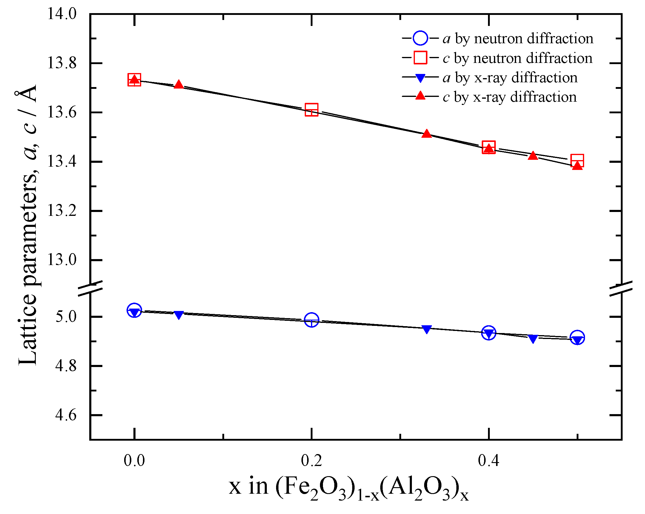
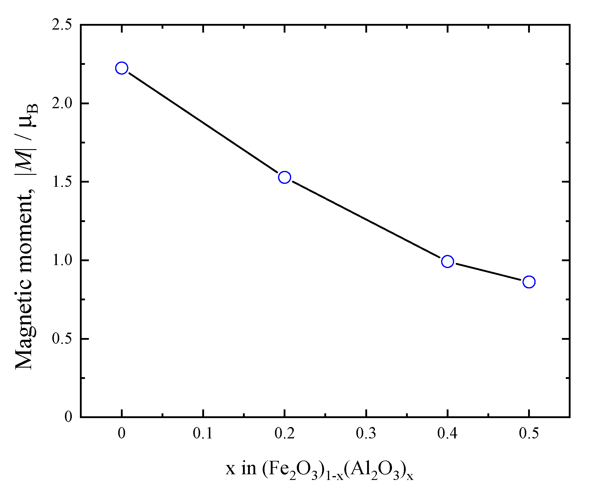
3.2. Compositional Dependence of the Structure of (Fe2O3)1−x(Al2O3)x
4. Conclusions
Author Contributions
Funding
Institutional Review Board Statement
Informed Consent Statement
Data Availability Statement
Acknowledgments
Conflicts of Interest
References
- Scrosati, B.; Hassoun, J.; Sun, Y.-K. Lithium-ion batteries. A look into the future. Energy Environ. Sci. 2011, 4, 3287. [Google Scholar] [CrossRef]
- Reddy, M.V.; Yu, T.; Sow, C.H.; Shen, Z.X.; Lim, C.T.; Rao, G.V.S.; Chowdari, B.V.R. α-Fe2O3 nanoflakes as an anode material for Li-ion batteries. Adv. Funct. Mater. 2007, 17, 2792–2799. [Google Scholar] [CrossRef]
- Roy, P.; Srivastava, S.K. Nanostructured anode materials for lithium-ion batteries. J. Mater. Chem. A 2014, 3, 2454–2484. [Google Scholar] [CrossRef]
- Jiang, Y.; Zhang, D.; Li, Y.; Yuan, T.; Bahlawane, N.; Liang, C.; Sun, W.; Lu, Y.; Yan, M. Amorphous Fe2O3 as a high-capacity, high-rate and long-life anode material for lithium-ion batteries. Nano Energy 2013, 4, 23–30. [Google Scholar] [CrossRef]
- Wu, N.; Shi, Y.R.; Ma, C.; Zhang, X.; Zhou, J.-M.; Wei, Y.; Liu, H.; Yan, Y.; Liu, H.-T. High performance nano-α-Fe2O3 electrode materials synthesized by facile and green approaches for lithium-ion batteries. Mater. Lett. 2018, 238, 155–158. [Google Scholar] [CrossRef]
- Muan, A. On the stability of the phase Fe2O3–Al2O3. Am. J. Sci. 1958, 256, 413–422. [Google Scholar] [CrossRef]
- Muan, A.; Gee, C.L. Phase equilibrium studies in the system iron Fe2O3–Al2O3 in air and at 1 atm. O2 pressure. J. Am. Ceram. Soc. 1956, 39, 207–214. [Google Scholar] [CrossRef]
- Popović, S.; Ristić, M.; Musić, S. Formation of solid solutions in the system Fe2O3–Al2O3. Mater. Lett. 1995, 23, 139–142. [Google Scholar] [CrossRef]
- Polli, A.D.; Lange, F.F.; Levi, C.G.; Mayer, J. Crystallization Behavior and Microstructure Evolution of (Al, Fe)2O3 Synthesized from Liquid Precursors. J. Am. Ceram. Soc. 1996, 79, 1745–1755. [Google Scholar] [CrossRef]
- Jiang, J.Z.; Mørup, S.; Linderoth, S. Formation of 25 Mol % Fe2O3–Al2O3 Solid Solution by High-Energy Ball Milling. Mater. Sci. Forum 1996, 225–227, 489–496. [Google Scholar] [CrossRef]
- Mahmoud, M.H.; Taha, T.A. FTIR and Mössbauer Spectroscopy investigations of Ag/FexAl2−xO3 nanocomposites. J. Electron. Mater. 2019, 48, 7396–7403. [Google Scholar] [CrossRef]
- Takai, S.; Sawada, E.; Harada, J.; Park, S.; Oda, M.; Esaki, S.; Nishijima, M.; Yoshie, T.; Yabutsuka, T.; Yao, T. Synthesis and anode properties of corundum-type structured (Fe2O3)1–x (Al2O3)x solid solutions in the whole compositional range. Solid State Ionics 2017, 313, 1–6. [Google Scholar] [CrossRef]
- Hubbard, C.R.; McCarthy, G.J. JCPDS-International Centre for Diffraction Data. Acta Crystallogr. Sect. A 1981, 37, C344. [Google Scholar] [CrossRef]
- Nakaishi, H.; Yabutsuka, T.; Yao, T.; Kitao, S.; Seto, M.; Chen, W.-J.; Shimonishi, Y.; Yoshida, S.; Takai, S. Homogeneous solid-solution formation in Fe2O3–Al2O3 system observed by TEM, XAFS, and Mössbauer spectroscopy. Mater. Chem. Phys. 2023, 303, 127764. [Google Scholar] [CrossRef]
- Luo, D.; Yabutsuka, T.; Yao, T.; Kitani, S.; Kawaji, H.; Takai, S. Low-temperature heat-capacities of corundum-type structured (Fe2O3)1–x (Al2O3)x solid solutions with x = 0.25, 0.50 and 0.75. J. Therm. Anal. Calorim. 2024. [Google Scholar] [CrossRef]
- Chase, M.W., Jr. JANAF Thermodynamical tables. J. Phy. Chem. Ref. Data 1985, 14. [Google Scholar] [CrossRef]
- Adachi, K.; Sato, K.; Matsui, M.; Mitani, S. Neutron diffraction investigations of (CoMn)1−xFex. J. Phys. Soc. Jpn. 1973, 35, 426–433. [Google Scholar] [CrossRef]
- Andersson, M.; Rapp, Ö.; Tellgren, R. Neutron diffraction studies of interatomic distances in the Y1−2xCaxThxBa2Cu3O7−α System. Solid State Commun. 1992, 81, 425–429. [Google Scholar] [CrossRef]
- Harrison, R.J. Neutron diffraction of magnetic materials. Rev. Mineral Geochem. 2006, 63, 113–143. [Google Scholar] [CrossRef]
- Yonemura, M.; Mori, K.; Kamiyama, T.; Fukunaga, T.; Torii, S.; Nagao, M.; Ishikawa, Y.; Onodera, Y.; Adipranoto, D.S.; Arai, H.; et al. Development of SPICA, new dedicated neutron powder diffractometer for battery studies. J. Phys. Conf. Ser. 2014, 502, 012053. [Google Scholar] [CrossRef]
- Oishi, R.; Yonemura, M.; Nishimaki, Y.; Torii, S.; Hoshikawa, A.; Ishigaki, T.; Morishima, T.; Mori, K.; Kamiyama, T. Rietveld analysis software for J-PARC. Nucl. Instrum. Methods Phys. Res. Sect. A Accel. Spectrometers Detect. Assoc. Equip. 2008, 600, 94–96. [Google Scholar] [CrossRef]
- Shull, C.G.; Strauser, W.A.; Wollan, E.O. Neutron diffraction by paramagnetic and antiferromagnetic substances. Phys. Rev. 1951, 83, 333–345. [Google Scholar] [CrossRef]
- Momma, K.; Izumi, F. VESTA: A three-dimensional visualization system for electronic and structural analysis. J. Appl. Crystallogr. 2008, 41, 653–658. [Google Scholar] [CrossRef]
- Kraushofer, F.; Jakub, Z.; Bichler, M.; Hulva, J.; Drmota, P.; Weinold, M.; Schmid, M.; Setvin, M.; Diebold, U.; Blaha, P.; et al. Atomic-Scale structure of the hematite α-Fe2O3(11̅02) “R-CUT” surface. J. Phys. Chem. C 2017, 122, 1657–1669. [Google Scholar] [CrossRef] [PubMed]
- Lucht, M.; Lerche, M.; Wille, H.-c.; Shvyd’ko, Y.V.; Rüter, H.D.; Gerdau, E.; Becker, P. Precise measurement of the lattice parameters of α-Al2O3 in the temperature range 4.5–250 K using the Mössbauer wavelength standard. J. Appl. Crystallogr. 2003, 36, 1075–1081. [Google Scholar] [CrossRef]
- Tsyshevsky, R.; Zverev, A.; Mitrofanov, A.; Rashkeev, S.; Kuklja, M. Photochemistry of the α-Al2O3-PETN interface. Molecules 2016, 21, 289. [Google Scholar] [CrossRef]
- Ruffa, A.R. Temperature dependence of the elastic shear moduli of the cubic metals. Phys. Rev. B Solid State 1977, 16, 2504–2514. [Google Scholar] [CrossRef]
- Shannon, R.D. Revised effective ionic radii and systematic studies of interatomic distances in halides and chalcogenides. Acta Crystallogr. Sect. A 1976, 32, 751–767. [Google Scholar] [CrossRef]
- Brown, I.D.; Shannon, R.D. Empirical bond-strength–bond-length curves for oxides. Acta Crystallogr. Sect. A 1973, 29, 266–282. [Google Scholar] [CrossRef]
- Gallego, S.V.; Perez-Mato, J.M.; Elcoro, L.; Tasci, E.S.; Hanson, R.M.; Momma, K.; Aroyo, M.I.; Madariaga, G. MAGNDATA: Towards a database of magnetic structures. I. The commensurate case. J. Appl. Crystallogr. 2016, 49, 1750–1776. [Google Scholar] [CrossRef]
- Sun, X.; Zheng, C.-M.; Zhang, F.-X.; Yang, Y.; Yu, A.-M.; Guan, N.-J. Size-Controlled Synthesis of Magnetite (Fe3O4) Nanoparticles Coated with Glucose and Gluconic Acid from a Single Fe(III) Precursor by a Sucrose Bifunctional Hydrothermal Method. J. Phys. Chem. C 2009, 113, 16002–16008. [Google Scholar] [CrossRef]
- Naik, R.; Kumar, A.N.; Shanbhag, V.; Nagaswarupa, H.P.; Boddula, R.; Al-Kahtani, A.A.; Kumar, K.D. Energy Storage, Sensors, Photocatalytic Applications of Green Synthesized ZnO: Fe3+ Nanomaterials. Chem. Phys. Impact. 2023, 7, 100387. [Google Scholar] [CrossRef]
- Issa, B.; Obaidat, I.; Albiss, B.; Haik, Y. Magnetic nanoparticles: Surface effects and properties related to biomedicine applications. Int. J. Mol. Sci. 2013, 14, 21266–21305. [Google Scholar] [CrossRef] [PubMed]
- Keshavarz, S.; Kvashnin, Y.O.; Rodrigues, D.C.M.; Pereiro, M.; Di Marco, I.; Autieri, C.; Nordström, L.; Solovyev, I.V.; Sanyal, B.; Eriksson, O. Publisher’s Note: Exchange interactions of CaMnO3 in the bulk and at the surface [Phys. Rev. B 95, 115120 (2017)]. Phys. Rev. B 2018, 97, 239901. [Google Scholar] [CrossRef]


| Temperature (K) | Atom | Site | g | x | y | z | Biso (Å2) |
|---|---|---|---|---|---|---|---|
| 4 | Fe/Al | 12c | 1 | 0 | 0 | 0.14729(2) | 0.810(1) |
| O | 18e | 1 | 0.31477(5) | 0 | 0.25 | 0.939(1) | |
| a = 4.91009(4) Å, c = 13.39012(17) Å, Rwp = 2.4458% | |||||||
| 20 | Fe/Al | 12c | 1 | 0 | 0 | 0.14620(2) | 0.772(3) |
| O | 18e | 1 | 0.30777(6) | 0 | 0.25 | 0.763(4) | |
| a = 4.90967(4) Å, c = 13.39059(16) Å, Rwp = 2.3661% | |||||||
| 100 | Fe/Al | 12c | 1 | 0 | 0 | 0.14731(3) | 0.830(4) |
| O | 18e | 1 | 0.31149(8) | 0 | 0.25 | 0.832(4) | |
| a = 4.91040(5) Å, c = 13.3916(2) Å, Rwp = 2.5226% | |||||||
| 200 | Fe/Al | 12c | 1 | 0 | 0 | 0.14667(2) | 0.776(4) |
| O | 18e | 1 | 0.30910(7) | 0 | 0.25 | 0.743(3) | |
| a = 4.91233(4) Å, c = 13.3960(2) Å, Rwp = 2.0989% | |||||||
| R.T. | Fe/Al | 12c | 1 | 0 | 0 | 0.14738(2) | 0.858(4) |
| O | 18e | 1 | 0.30858(6) | 0 | 0.25 | 0.745(3) | |
| a = 4.91514(3) Å, c = 13.4041(2) Å, Rwp = 2.4458% | |||||||
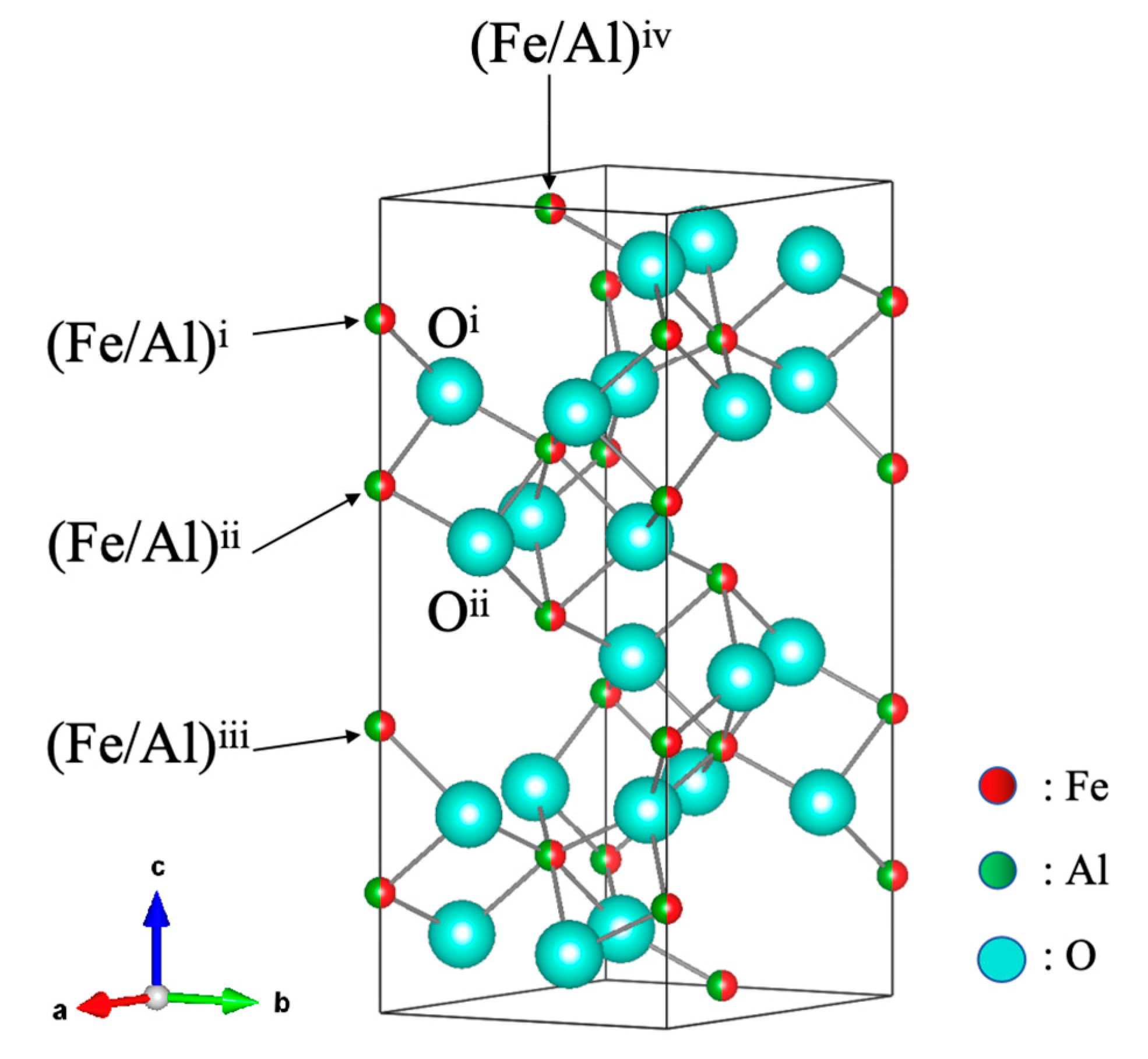
| Temperature (K) | (Fe/Al)i–Oi (Å) | (Fe/Al)ii–Oii (Å) | (Fe/Al)i–(Fe/Al)ii (Å) | (Fe/Al)ii–(Fe/Al)iii (Å) | (Fe/Al)i–(Fe/Al)iv (Å) |
|---|---|---|---|---|---|
| 4 | 2.0689(3) | 1.8894(2) | 2.7506(5) | 3.9445(5) | 3.3121(3) |
| 20 | 2.0531(3) | 1.8995(2) | 2.7800(5) | 3.9153(5) | 3.2969(3) |
| 100 | 2.0568(4) | 1.8974(3) | 2.7504(9) | 3.9454(9) | 3.3126(5) |
| 200 | 2.0533(3) | 1.9009(2) | 2.7687(6) | 3.9293(6) | 3.3049(3) |
| R.T. | 2.0476(3) | 1.9069(2) | 2.7511(6) | 3.9510(6) | 3.3168(3) |
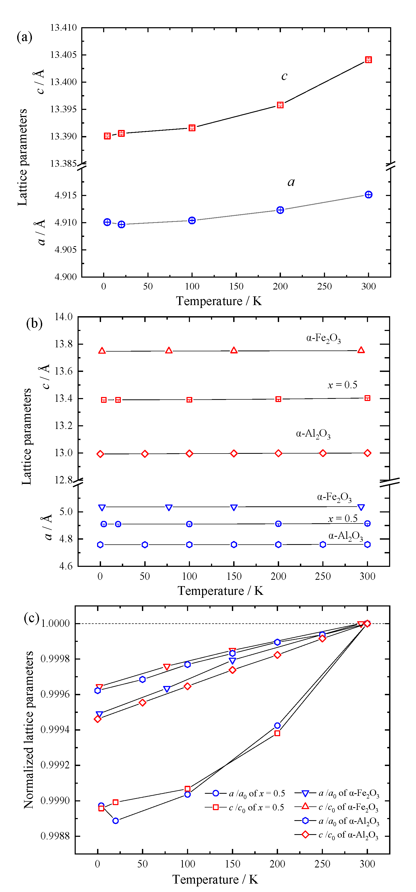
| Temperature (K) | a (10−6 K−1) | c (10−6 K−1) | |
|---|---|---|---|
| (Fe2O3)0.5(Al2O3)0.5 | 4–100 | 0.66(2) | 1.15(3) |
| 200–300 | 5.76(2) | 6.19(5) | |
| α-Fe2O3 [24] | 2–77 | 1.90 | 1.53 |
| 150–293 | 1.44 | 1.05 | |
| α-Al2O3 [25,26] | 2–100 | 1.50 | 1.89 |
| 200–300 | 1.05 | 1.77 |
| Composition | Atom | Site | g | x | y | z | Biso (Å2) |
|---|---|---|---|---|---|---|---|
| x = 0 | Fe/Al | 12c | 1 | 0 | 0 | 0.145556(8) | 0.937(1) |
| O | 18e | 1 | 0.30992(4) | 0 | 0.25 | 0.610(2) | |
| a = b = 5.025556(17) Å, c = 13.73302(9) Å, Rwp = 3.6910% | |||||||
| x = 0.2 | Fe/Al | 12c | 1 | 0 | 0 | 0.14723(1) | 0.870(1) |
| O | 18e | 1 | 0.30862(4) | 0 | 0.25 | 0.636(2) | |
| a = b = 4.98646(2) Å, c = 13.61093(10) Å, Rwp = 2.6605% | |||||||
| x = 0.4 | Fe/Al | 12c | 1 | 0 | 0 | 0.146591(13) | 0.915(2) |
| O | 18e | 1 | 0.31000(5) | 0 | 0.25 | 0.774(3) | |
| a = b = 4.93405(3) Å, c = 13.45815(14) Å, Rwp = 2.0553% | |||||||
| x = 0.5 | Fe/Al | 12c | 1 | 0 | 0 | 0.14738(2) | 0.858(4) |
| O | 18e | 1 | 0.30858(6) | 0 | 0.25 | 0.745(3) | |
| a = b = 4.91514(3) Å, c = 13.4041(2) Å, Rwp = 2.4458% | |||||||
| x-Value | (Fe/Al)i–Oi (Å) | (Fe/Al)ii–Oii (Å) | (Fe/Al)i–(Fe/Al)ii (Å) | (Fe/Al)ii–(Fe/Al)iii (Å) | (Fe/Al)i–(Fe/Al)iv (Å) |
|---|---|---|---|---|---|
| 0 | 2.1174(2) | 1.9358(1) | 2.8687(3) | 3.9978(3) | 3.3674(1) |
| 0.2 | 2.0942(2) | 1.9240(1) | 2.8163(3) | 3.9891(3) | 3.3539(1) |
| 0.4 | 2.0679(3) | 1.9059(2) | 2.7834(4) | 3.9457(4) | 3.3187(2) |
| 0.5 | 2.0476(3) | 1.9069(2) | 2.7511(6) | 3.9510(6) | 3.3168(2) |
Disclaimer/Publisher’s Note: The statements, opinions and data contained in all publications are solely those of the individual author(s) and contributor(s) and not of MDPI and/or the editor(s). MDPI and/or the editor(s) disclaim responsibility for any injury to people or property resulting from any ideas, methods, instructions or products referred to in the content. |
© 2025 by the authors. Licensee MDPI, Basel, Switzerland. This article is an open access article distributed under the terms and conditions of the Creative Commons Attribution (CC BY) license (https://creativecommons.org/licenses/by/4.0/).
Share and Cite
Luo, D.; Nakaishi, H.; Yabutsuka, T.; Saito, T.; Kamiyama, T.; Hagihala, M.; Takai, S. Structural and Magnetic Characterization of Mechanically Alloyed (Fe2O3)1−x(Al2O3)x Solid Solutions via Pulsed Neutron Powder Diffraction. Materials 2025, 18, 1911. https://doi.org/10.3390/ma18091911
Luo D, Nakaishi H, Yabutsuka T, Saito T, Kamiyama T, Hagihala M, Takai S. Structural and Magnetic Characterization of Mechanically Alloyed (Fe2O3)1−x(Al2O3)x Solid Solutions via Pulsed Neutron Powder Diffraction. Materials. 2025; 18(9):1911. https://doi.org/10.3390/ma18091911
Chicago/Turabian StyleLuo, Dong, Hayato Nakaishi, Takeshi Yabutsuka, Takashi Saito, Takashi Kamiyama, Masato Hagihala, and Shigeomi Takai. 2025. "Structural and Magnetic Characterization of Mechanically Alloyed (Fe2O3)1−x(Al2O3)x Solid Solutions via Pulsed Neutron Powder Diffraction" Materials 18, no. 9: 1911. https://doi.org/10.3390/ma18091911
APA StyleLuo, D., Nakaishi, H., Yabutsuka, T., Saito, T., Kamiyama, T., Hagihala, M., & Takai, S. (2025). Structural and Magnetic Characterization of Mechanically Alloyed (Fe2O3)1−x(Al2O3)x Solid Solutions via Pulsed Neutron Powder Diffraction. Materials, 18(9), 1911. https://doi.org/10.3390/ma18091911