Review

The Integration of Advanced Drug Delivery Systems into Conventional Adjuvant Therapies for Peri-Implantitis Treatment

by
Iria Seoane-Viaño
1,2,*,
Mariola Seoane-Gigirey
1,
Carlos Bendicho-Lavilla
1,2,
Luz M. Gigirey
3,
Francisco J. Otero-Espinar
1,2 and
Santiago Seoane-Trigo
4,*
1
Department of Pharmacology, Pharmacy and Pharmaceutical Technology, Faculty of Pharmacy, and Institute of Materials (iMATUS), University of Santiago de Compostela (USC), 15782 Santiago de Compostela, Spain
2
Paraquasil Group (GI-2109), Health Research Institute of Santiago de Compostela (IDIS), 15706 Santiago de Compostela, Spain
3
Department of Applied Physics, Faculty of Optics and Optometry, University of Santiago de Compostela (USC), 15782 Santiago de Compostela, Spain
4
Ph. Dr. Adult Comprehensive Dentistry, University of Santiago de Compostela (USC), 15782 Santiago de Compostela, Spain
*
Authors to whom correspondence should be addressed.
Pharmaceutics 2024, 16(6), 769; https://doi.org/10.3390/pharmaceutics16060769
Submission received: 7 April 2024 / Revised: 31 May 2024 / Accepted: 3 June 2024 / Published: 5 June 2024
(This article belongs to the Special Issue Advanced Materials Science and Technology in Drug Delivery)

Abstract

:
Despite the high success rates of dental implants, peri-implantitis is currently the most common complication in dental implantology. Peri-implantitis has an inflammatory nature, it is associated with the accumulation of plaque in the peri-implant tissues, and its evolution can be progressive depending on various factors, comorbidities, and poor oral health. Prophylaxis and different treatment methods have been widely discussed in recent decades, and surgical and non-surgical techniques present both advantages and disadvantages. In this work, a literature review of different studies on the application of adjuvant treatments, such as local and systemic antibiotics and antiseptic treatments, was conducted. Positive outcomes have been found in the short (up to one year after treatment) and long term (up to ten years after treatment) with combined therapies. However, there is still a need to explore new therapies based on the use of advanced drug delivery systems for the effective treatment of peri-implantitis in the long term and without relapses. Hence, micro- and nanoparticles, implants, and injectable hydrogels, among others, should be considered in future peri-implantitis treatment with the aim of enhancing overall therapy outcomes.

1. Introduction

Dental implantology has become a routine procedure in the realm of prosthetic rehabilitation. Following implant placement and subsequent healing of the area, the damaged tissues undergo restructuring to facilitate the integration of the implant. Osseointegration implies the establishment of a direct, structural, and functional connection between the newly formed bone and the implant, thereby endowing the implant with the capacity to withstand normal oral physiological forces and adapt fully to the oral environment.
Numerous studies have reported a high prevalence of peri-implantitis. A meta-analysis review from 2015 including 15 articles from 11 studies reported a prevalence of peri-implant mucositis and peri-implantitis ranging from 19 to 65% and from 1 to 47%, respectively [1]. Another systematic review from 2012 [2] indicated that the prevalence of peri-implantitis may be in the order of 10% for implants and 20% for patients from 5 to 10 years after implant placement. A more recent cross-sectional study from 2019 [3] involving patients with more than 1 year of follow-up after loading showed a prevalence of peri-implant mucositis and peri-implantitis of 38.8% and 35%, respectively.
Peri-implantitis therapy aims to control infection, reduce inflammation, and promote tissue regeneration around dental implants. Conventional treatment of this pathology typically involves mechanical debridement to remove biofilm from the implant surface. Chemical decontamination using various antimicrobial agents is applied locally to disinfect the implant surface, alongside chemical decontamination using various antimicrobial agents applied locally to disinfect the implant surface and surrounding tissues. In cases where non-surgical approaches prove insufficient, systemic or local antibiotics may be administered to supplement mechanical and chemical debridement. Surgical interventions are reserved for severe peri-implantitis cases. The implementation of advanced drug delivery systems, such as micro- and nanoparticles, implants, and injectable hydrogels, holds promise for enhancing therapy outcomes by increasing the bioavailability or residence time of antibiotics and antimicrobial agents in the implant surroundings. However, these novel approaches are not yet fully described in the literature.
This literature review aims to offer a comprehensive overview of the adjuvant therapies employed in the non-surgical management of peri-implantitis, with a specific focus on the efficacy of local and systemic treatments. Firstly, a brief outline of the disease’s characteristics and conventional treatments will be presented. Subsequently, the most recent advances in adjuvant therapies will be discussed. Lastly, the most relevant examples of advanced drug delivery systems with high potential for peri-implantitis treatment will be thoroughly explored.

1.1. Clinical Characteristics of Peri-Implantitis

Certain parameters can be employed to aid in the diagnosis and classification of peri-implantitis, including pain, mobility, bleeding on probing, probing depth, suppuration/exudate, and radiographic bone loss [4]. Moreover, two main forms of peri-implant diseases can be distinguished: peri-implant mucositis and peri-implantitis. The 2017 World Workshop of the American Academy of Periodontology (AAP) and the European Federation of Periodontology (FEP), Working Group 4, defined peri-implantitis as “a plaque-associated pathological condition occurring in tissues around dental implants, characterized by inflammation in the peri-implant mucosa and subsequent progressive loss of supporting bone” [5]. On the other hand, peri-implant mucositis manifests as bleeding upon gentle probing (BoP) accompanied by clinical signs of inflammation in the soft tissues, yet without any loss of peri-implant bone (Figure 1).
Peri-implant mucositis is presumed to precede the onset of peri-implantitis, as data from various studies indicate that patients with peri-implant mucositis are more likely to develop peri-implantitis. Since peri-implantitis is a late-stage complication that can ultimately lead to implant loss, effective control of bacterial plaque in patients is of paramount importance since it is closely linked to this type of infection [6]. Several studies have identified various risk factors that can affect patients who have undergone implant placement, including smoking, bone quality, surgical trauma, bacterial contamination in the oral cavity, and the patient’s ability to maintain a harmonious oral microbiome balance. These factors are closely linked to the presence of bacterial plaque [7,8]. Upon the onset of peri-implantitis, the implant becomes exposed to the oral environment, facilitating bacterial colonization. This accumulation of oral microbiota in the form of plaque initiates an inflammatory process, which is one of the primary factors contributing to the development of peri-implantitis [9]. The composition of the microbiota after implant placement is conditioned by the existing microbiota at the time of implantation. Once peri-implantitis develops, a significant portion of the oral microbiota consists of Gram-negative bacteria. Notably, Porphiromonas gingivalis, Treponema denticola, and Tannerella forsythia have been found to be more abundant in peri-implantitis samples [10,11].

1.2. Peri-Implantitis Conventional Non-Surgical Therapies

There are multiple approaches as well as disparity of criteria among clinicians for the treatment of peri-implant diseases, and the specific treatment approach may vary depending on the severity of the disease. Nonetheless, the main objective remains consistent: eliminating the peri-implant plaque layer to halt the infection. Treatment strategies can be categorized as surgical, non-surgical, or a combination of both. Implantology professionals usually resort to a combination of surgical methods, which are widely used and yield rapid results, alongside non-surgical methods to reduce bacterial load within the peri-implant pocket and decontaminate the implant surface [12,13,14]. Practitioners agree that the main objective is the removal of biofilm from the implant surface in an attempt to decelerate disease progression [15].
Surgical approaches, including access flap surgery with or without resective and/or augmentative techniques, have demonstrated positive outcomes in addressing peri-implantitis by removing biofilm and plaque deposits from the implant surface [16]. Access flap surgery effectively decontaminates the implant surface while maintaining the soft tissues surrounding the affected area, whereas resective therapy reduces probing depths around the affected implant. Augmentative techniques attempt to regenerate bone and achieve re-osseointegration.
Non-surgical treatments are designed to eradicate the bacterial load and disinfect the implant surface. These include mechanical debridement of the implant surface using instruments such as steel or titanium curettes, ultrasonic devices, air polishers, and combinations thereof [17,18,19]. Disinfection procedures are also applied to remove plaque bacteria, resolve inflammation, and treat infections [20]. These procedures can take the form of mechanical, chemical, photodynamic, or antibiotic-based approaches [21] (Table 1). However, it should be noted that the improvements achieved with non-surgical treatments for peri-implantitis have been observed to be temporary [22]. Each method has its own advantages and disadvantages, but there is currently no gold-standard treatment [23]. However, most dental professionals concur on the use of oral antiseptics to eliminate bacteria on the implant surface, as they inhibit microbial growth through a non-selective toxicity mechanism [24]. Recommended solutions include chlorhexidine digluconate (0.12% and 0.20%) (a bisbiguanide antiseptic with bactericidal and fungicidal properties), hydrogen peroxide solutions (3% or 5%) (which release oxygen ions and reduce anaerobic bacterial load), povidone iodine, or sodium perborate. Depending on the oral antiseptic employed, some, such as povidone iodine, exhibit greater bactericidal effects than others in the short term. Combining them with mechanical debridement has shown improved results [25,26].
Moreover, antibiotic therapy is often employed to deliver medication into the crevicular fluid, and this therapy is continued alongside mechanical procedures. The most widely used therapy in clinical practice is based on the use of amoxicillin 500 mg and metronidazole 250 mg every 8 h for 7 days or amoxicillin–clavulanic acid 875/125 mg and metronidazole 500 mg every 12 h for 7 days. It is recommended to initiate this treatment one day prior to surgery and continue it post-surgery [27]. Nevertheless, there are insufficient data to support a particular evidence-based antibiotic protocol to treat peri-implantitis using surgical or non-surgical therapy due to the scarcity of published high-quality clinical studies. Systemic antibiotics might be beneficial as an adjunct to surgical treatment in specific patient groups and implants with specific surface characteristics [28].
In a six-year study [29], a combination of local and systemic administration of tetracycline, doxycycline, ciprofloxacin, sulfonamides, and trimethoprim was prescribed. The authors reported a reduction in the inflammatory pocket; however, they also emphasized the need to combine treatment with other techniques, mainly debridement using titanium or Teflon curettes. Unfortunately, the extensive use of systemic antibiotic therapy in dentistry has led to significant antimicrobial resistance. For peri-implantitis, prophylactic use of clindamycin, amoxicillin, doxycycline, and metronidazole resulted in resistance rates of 46.7%, 39.2%, 25%, and 21.7%, respectively [30,31]. In another work [32], the effectiveness of antiseptics in eliminating bacteria deposited in the peri-implant bed and improving the efficacy of systemic treatment was highlighted. The authors concluded that the best results were achieved using 0.12% chlorhexidine combined with timolol, eucalyptol, menthol, and methyl salicylate. Subgingival irrigation with 0.12% chlorhexidine has also demonstrated the ability to eliminate microorganisms for at least 3 months [17].
Table 1. Adjuvant treatments for peri-implantitis.
Treatment TypeEfficacyRef.
Systemic AntibioticsChemical Agents
Sr(OH)2Proven bacterial inhibition (p < 0.001)[33]
Chlorhexidine (C22H30Cl2N10)
H2O2
Proven reduction in anaerobic bacteria[34]
Chlorhexidine (C22H30Cl2N10)
H2O2
PO4H3
Cetylpyridinium chloride (C21H38ClN)
Proven, it allows osseointegration within a short time period[35]
PO4H3Proven efficacy (3 months)[36]
0.12% Chlorhexidine (C22H30Cl2N10)
Timolol (C13H24N4O3S)
Eucalyptol (C10H18O)
Menthol (C10H20O)
Methyl salicylate (C8H8O3)
Proven, better than combined systemic treatment[32]
Clindamycin
Amoxicillin
Doxycycline
Metronidazole
Proven, but bacterial resistance appears[30]
Chlorhexidine (C22H30Cl2N10)
H2O2
Proven reduction in anaerobic bacteria[37]
Chloramine gel (NH2Cl)
Chlorhexidine chips (C22H30Cl2N10)
Proven only short-term clinical efficacy (3 months) (p < 0.001)[38]
Tetracycline Doxycycline Ciprofloxacin
Sulfonamides
Proven, reduction in inflammatory pockets in the area[29]
0.12% Chlorhexidine (C22H30Cl2N10)Proven (3-month period)[17]
Chlorhexidine (C22H30Cl2N10)
Cetylpyridinium chloride (C21H38ClN)
Proven, bacterial reduction but no clinical benefit[39]
The treatment of peri-implant infection via mechanical debridement combined with chloramine gel, diode laser, and chlorhexidine chips has also been analyzed in some studies. After three months of follow-up, there were no statistically significant differences between conventional mechanical debridement (scalers and curettes) and the use of chloramine gel in terms of the measured parameters. However, both treatment groups showed significant clinical improvement [38].
Treatment with disinfectants has also been studied by some authors [34,37] who proposed the combination of different antiseptics at varying concentrations (0.12–0.2% chlorhexidine or 3% hydrogen peroxide). Chlorhexidine acts as a bactericidal and fungicidal agent, while hydrogen peroxide releases oxygen, reducing the presence of anaerobic bacteria. When used together, these disinfectants help maintain a low bacterial load.
Limited evidence exists regarding the use of antibiotic therapy in crevicular fluid. In a systematic review published in 2016 [31], the authors identified only six articles reporting clinical cases with a five-year follow-up. These studies revealed inconsistent results regarding the efficacy of antibiotics, and in some cases, they even contributed to the occurrence of superinfections involving Staphylococcus aureus and Epstein–Barr virus. Consequently, it is advisable to conduct prior and follow-up antibiograms to ensure the selection of the appropriate antibiotic. Another study focusing on the use of antibiotics reviewed ten different studies [40]. Among them, six studies employed tetracycline and doxycycline, three studies used minocycline, one study utilized doxycycline, and the remaining study employed a combination of tetracycline and HCl fibers. The authors of these studies observed a reduction in probing depth and a decrease in suppuration following the respective treatments.
In vitro studies can also shed light on the bacterial composition and its susceptibility to antibiotics commonly used in peri-implantitis cases. One study examined the sensitivity of bacteria commonly associated with mucositis and peri-implantitis to ten different antibiotics in anaerobic cultures at 37 °C [41]. The bacteria analyzed included Aggregatibacter actinomycetemcomitans (Aa), Capnocytophaga ochracea (Co), Eikenella corrodens; Enterococcus faecalis; Enterococcus faecium, Fusobacterium nucleatum; Lactobacillus brevis; Lactobacillus buchneri; Parvimonas micra (Pm), Staphylococcus aureus, Staphylococcus epidermidis, Streptococcus gordonii, Streptococcus mutans, and Streptococcus oralis. The antibiotics administered (at doses ranging from 0.016 to 256 mg/mL) were penicillin G, ampicillin, amoxicillin, ampicillin/sulbactam, amoxicillin/clavulanic acid, minocycline, metronidazole, linezolid, azithromycin, and moxifloxacin.
The results of the study showed that the sample (Aa) exhibited more resistance in 17 out of the 30 antibiograms performed in the co-cultures of Aa-Co, Aa-Pm, and Aa-Co-Pm. Conversely, Co and Pm were more susceptible to antibiotic treatment in 25 out of the 30 tests performed. Several authors have highlighted the increasing difficulty in treating oral infections caused by Gram-negative bacteria due to their antibiotic resistance. Although the incorporation of chlorhexidine as a local treatment improves treatment outcomes, its duration of action in the area is limited, and it may exhibit a certain degree of cytotoxicity by reducing the production of gingival fibroblasts [42].
Another study [33] compares the antimicrobial potential of Sr(OH)2 at different concentrations (100, 10, 1, 1, 0.1, and 0.01 mM) against six bacteria frequently associated with infectious biomaterials: Streptococcus mitis, Staphylococcus epidermidis, Aggregatibacter actinomycetemcomitans, Porphyromonas gingivalis, Escherichia coli, and Fusobacterium nucleatum. The antimicrobial properties were evaluated using Brucella agar diffusion, which revealed significant inhibition (p < 0.001) in the growth of all groups. Regarding biofilm viability, it was found that concentrations of 1 mM and 100 mM exhibited the highest bactericidal activity against S. mitis, S. epidermidis, A. actinomycetemcomitans, E. coli, and P. gingivalis. Specifically, the 100 mM concentration of Sr(OH)2 completely eradicated all bacteria, while the 10 mM concentration resulted in less than 1% viability of A. actinomycetemcomitans, S. mitis, and S. epidermidis strains when compared to the control groups.
The choice of non-surgical treatment for peri-implantitis depends on the characteristics of the implant surface, as it responds differently to specific combination treatments [43,44] (Table 2). Different chemical agents have been used for decontaminating the implant surface, such as 0.2% chlorhexidine, H2O2, H3PO4, and ethylenediaminetetraacetic acid [40]. It is important to always maintain a pH > 3 to prevent corrosion on the implant surface and promote osseointegration [35]. Applying 35% H3PO4 to the implant surface along with debridement has been shown to effectively reduce anaerobic bacterial count and facilitate decontamination [36]. However, after three months, the group treated with H3PO4 did not show significant improvements compared to the control group without treatment.
Similarly, the use of 0.12% chlorhexidine and 0.05% cetylpyridinium chloride (CPC, Perio Aid®) resulted in reduced bacterial growth on the implant surface but did not yield significant clinical benefits in the study group at 12 months [39]. Similar results were reported in a subsequent study that used 0.2% chlorhexidine, concluding that the combination of chlorhexidine and cetylpyridinium chloride effectively reduces the bacterial load compared to debridement alone but does not provide superior therapeutic outcomes.
A recent study also proposed the possibility of using probiotics in the early stage of peri-implantitis when peri-implant mucositis has already developed [45]. The study aimed to evaluate the effectiveness of probiotics in combination with debridement as an alternative therapy. When comparing the probing depth between the test group and the control group, the results showed a minimal difference of only −0.12 mm (p = 0.38). This suggests that, at least in the short term, the use of probiotics did not yield statistically significant results in controlling microbial plaque.
The information presented in this literature review infers that antibiotics and surgical treatment are not always the best treatment options for patients with peri-implantitis. Therefore, there is a need to investigate complementary and alternative therapies as adjuvant treatments for peri-implantitis. When comparing peri-implant mucositis and peri-implantitis [46], it was found that the highest proportions (25%) of Porphyromonas gingivalis, Tannerella forsythia, and Treponema denticola were present in peri-implantitis lesions, followed by mucositis (11%), and the lowest proportion (1%) was observed in oral health samples. These differences were statistically significant with values of p < 0.005. Opportunistic pathogens such as Pseudomonas Aeruginosa, Staphylococcus aureus, fungi (Candida albicans), or viruses, as well as bacteria commonly associated with infections related to implanted medical devices, were also identified. Based on the reviewed literature, it has been concluded that systemic antibiotic treatment combined with local debridement is not highly effective in the long term and may even lead to superinfection induced by opportunistic bacteria [31,47]. However, local treatment with tetracyclines has shown positive effects on assessed clinical and microbiological parameters, although it is not sufficient to effectively control the disease process [48,49,50].
Subgingival irrigation with 0.12% chlorhexidine has been shown to suppress microorganisms for approximately three months [17]. Moreover, treatment approaches combining debridement with the placement of local antibiotics or laser therapy have shown more favorable outcomes in reducing signs of inflammation compared to debridement in conjunction with chlorhexidine use [51].

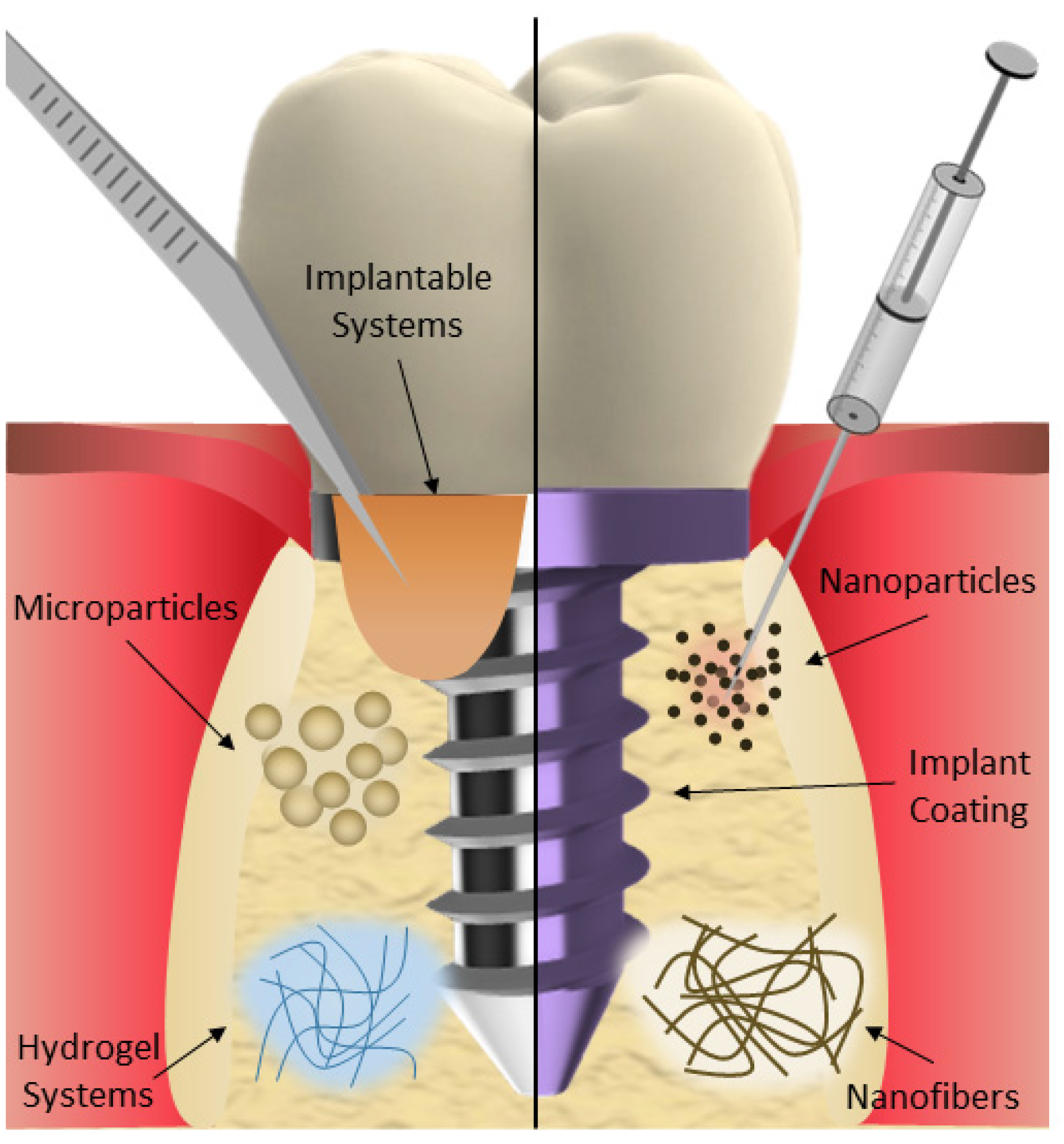
2. Drug Delivery Systems for Peri-Implantitis Management

Conventional forms of local drug therapy, while demonstrating effectiveness in treating peri-implantitis, can be easily washed off, resulting in a shorter duration of action within the oral cavity. On the other hand, systemic drug therapy using antibiotics can lead to issues such as drug resistance, dysbiosis, and systemic side effects. Moreover, the antibacterial effect is compromised and limited by the low amount of the drug that reaches the oral lesion after systemic circulation. Modern drug delivery systems have the potential to provide localized and sustained drug release to the affected area while offering improved biocompatibility [52]. Thus, new biomaterials and drug delivery strategies would open a window of new possibilities for the management of oral peri-implantitis. Micro- and nanoparticles loaded with different antibiotics or antibacterial agents, as well as polymeric fibers, implantable systems, hydrogels, or antibacterial coatings, have shown great potential for application in peri-implantitis (Figure 2). These drug delivery systems can also help with bone regeneration while maintaining their antibacterial effect (Table 3).

2.1. Micro- and Nanoparticles

Calcium phosphate cements are used as injectable and self-setting bone-substituting materials. Despite their functionality, these materials lack osteoinductivity and present poor antibacterial properties. In a study [53] investigating a bone graft substitute for treating peri-implantitis, it was demonstrated that including gelatine microspheres loaded with minocycline hydrochloride into calcium phosphate cements can improve the mechanical properties of the material while promoting osteogenesis and bone formation. Moreover, the local release of the drug exhibited excellent antibacterial activity against prevalent pathogens associated with dental peri-implantitis, including P. gingivalis and F. nucleatum. In another study using minocycline as a drug [54], two different carriers were evaluated for local minocycline delivery in an experimentally induced mucositis model in Beagle dogs. Chitosan-coated alginate carriers exhibited prolonged biodegradable sustainability for the controlled release of minocycline and demonstrated a superior bacteriostatic effect when compared to poly(meth)acrylate-glycerine (PG) microspheres. Another strategy for the local delivery of minocycline is found in the preparation of magnetic drug-loaded osteoinductive Fe3O4/CaCO3 hybrid microspheres [55]. These microspheres incorporate magnetite (Fe3O4) for magnetic targeting, while the addition of cyclodextrins enhances their porosity, resulting in well-defined mesoporous structures. These microspheres also exhibited excellent drug loading efficiency, release properties, magnetic characteristics, and osteoinductive potential.
Icariin, the primary pharmacological component of Herba Epimedium, is a centuries-old traditional herb medicine. There is evidence that icariin may play a role in bone health by stimulating bone formation while simultaneously inhibiting bone resorption. In a study [56], calcium phosphate cement with icariin-loaded gelatine microspheres was prepared. Icariin exhibited antibacterial activity against bacteria associated with peri-implantitis, and the system promoted osteoinductivity, bone formation, and inflammation alleviation. On the other hand, insulin plays a role in bone development and physiology [57]. Thus, the controlled release of insulin for peri-implant bone regeneration in non-diabetic subjects could be beneficial. Poly(lactic-co-glycolic acid (PLGA) microspheres loaded with insulin were developed to release insulin in a controlled manner without causing adverse effects such as hypoglycemia or hyperinsulinemia, which can result from excessive insulin [58]. The bioactivity of the microspheres was tested on human bone marrow mesenchymal stromal cells and rabbit implant models. The PLGA microspheres, prepared using the solvent evaporation method, demonstrated a relatively steady release rate in the first four weeks. This controlled release stimulated the osteogenic differentiation of the stem cells and peri-implant bone regeneration. In another study [59], the impact of a single injection of PLGA microspheres loaded with insulin around the metal implant was evaluated. The results, obtained through histological analysis, micro-CT scans, and biomechanical testing, demonstrated higher peri-implant bone formation and improved osseointegration. Arestin® (OraPharma, Inc., Bridgewater, NJ, USA), a commercially available formulation of minocycline hydrochloride microspheres, was administered locally to patients with a follow-up of 12 months [60]. The findings showed that Arestin® exhibited a more pronounced impact on A. actinomycetemcomitans compared to other pathogens. Additionally, significant reductions in the levels of Tannerella forsythia, P. gingivalis, and Treponema denticola were observed up to day 180. In a study comparing the efficacy of minocycline and chlorhexidine, 95 implants were involved, with 1 mg minocycline microspheres administered to 58 patients and 1% chlorhexidine administered to 37 patients [50]. The treatment was carried out in three phases: immediately post-surgery, at 30 days and 90 days, followed by assessments at 30, 60, 90, 180, and 360 days. The findings indicated that both treatments significantly reduced probing depth and bleeding (p < 0.001) when implemented.
Nanoparticulate systems are also exploited for the treatment of peri-implantitis. In one study, the mechanical, surface roughness, and antibacterial efficacy of photo-sonodynamic therapy using methylene blue-loaded PLGA nanoparticles on dental implants were evaluated [61]. The application of this therapy through PLGA nanoparticles demonstrated significant antibacterial activity against P. gingivalis, without compromising the surfaces or mechanical properties of dental implants. In another study, curcumin was employed as a photosensitizer for antimicrobial photodynamic chemotherapy [62]. To overcome its low solubility and poor bioavailability, curcumin was loaded into polycaprolactone nanoparticles. The study compared the cytotoxicity and photodynamic antimicrobial effects of curcumin-loaded nanoparticles with free curcumin as a photosensitizing compound against planktonic cultures and single- as well as multi-species biofilms. The results revealed that the curcumin-loaded nanoparticles exhibited properties similar to those of free curcumin. Whether nano-encapsulated or not, curcumin demonstrated enhanced antimicrobial activity when activated by blue light, particularly against multi-species biofilms, and exhibited no cytotoxicity.
Omega-3, a polyunsaturated fatty acid, proved to be more effective when administered as a supplement alongside non-surgical periodontal therapy for patients with periodontitis, compared to non-surgical periodontal therapy alone. In a recent study [63], docosahexaenoic acid (DHA) was encapsulated into nanostructured lipid carriers (NLC) and tested for efficacy in vitro and in vivo using a rat peri-implantitis model. The results demonstrated a reduction in alveolar bone resorption along with a decrease in inflammatory mediators, which might be attributed to the gradual release of DHA, leading to the sustained suppression of inflammation in the local microenvironment. Several authors have explored antimicrobial agents with extended action times and evaluated them in vitro. For instance, the in vitro antibacterial effect induced by the agar diffusion of biosynthesized and silver nanoparticles (AgNPs) using endophytic fungi was compared with the antibacterial effect of 0.12% chlorhexidine and ampicillin against P. gingivalis [64]. The results showed the high efficacy of the silver nanoparticles (at a concentration of 1 mM) when inoculated in volumes of 80 μL and 100 μL of AgNP, resulting in inhibition halos of 17.3 and 18 mm, respectively, compared to a 0.2% chlorhexidine halo of 17.8 mm and 19.8 mm for 2.0%, and for ampicillin, a halo of 20.5 mm. Although silver nanoparticles have exhibited superior antibacterial efficacy compared to 0.12% chlorhexidine, there is an ongoing debate regarding their potential cytotoxicity in tissues, as some studies report that they may induce oxidative and endoplasmic stress [65]. However, it should be noted that the amount of silver nanoparticles used is much lower than the threshold for adverse effects, as nanoparticles with dimensions smaller than 10 nm and at concentrations below 6.25 mg/mL are considered non-cytotoxic [66].
Table 3. Drug delivery systems for the treatment of peri-implantitis.
Type of Drug CarrierDrugEfficacyRef.
Micro- and nanoparticles
Gelatine microspheresMinocyclineAntibacterial activity against P. gingivalis and F. nucleatum and promoted osteogenic differentiation in vitro.[53]
Chitosan-coated alginate microspheresMinocyclineControlled release of the drug and bacteriostatic effects.[54]
Fe3O4/CaCO3 microspheresMinocyclineGreat drug delivery and magnetic targeting properties and osteoinductive potential.[55]
Gelatine microspheresIcariinPromotes bone formation and alleviates inflammation.[56]
PLGA microspheresInsulinStimulation of the osteogenic differentiation of the stem cells and peri-implant bone regeneration.[58]
PLGA microspheresInsulinHigher peri-implant bone formation and improved osseointegration.[59]
PLGA microspheres (Arestin®)MinocyclineReduction in total bacteria loading, with a higher impact on A. actinomycetemcomitans.[60]
PLGA nanoparticlesMethylene blueAntibacterial activity against P. gingivalis without deteriorating the surfaces and compromising the mechanical properties of dental implants.[61]
PCL nanoparticlesCurcuminAntimicrobial and antibiofilm properties, with better effects when associated with blue light.[62]
Nanostructured lipid carriers (NLC)Docosahexaenoic acid (DHA)Enhances the anti-inflammatory bioavailability of DHA, preventing the activation of certain inflammatory pathways.[63]
Silver nanoparticlesSilver nanoparticlesAntibacterial efficacy against P. gingivalis.[64]
Nanofibers
Nanocrystals in nanofibersCurcuminIncreased bioavailability of the drug and enhanced release properties.[67]
PLA nanofibersMetronidazoleImproved sustained drug release and in vitro antibacterial effect.[68]
PCL nanofibersOxytetracycline HClImproved sustained drug release and in vitro antibacterial effect.[69]
PLA nanofibersQuercetinIn vitro antibacterial activity and pH-dependent drug release.[70]
Electrospun membranesResveratrolIn vitro antibacterial activity and pH-dependent drug release.[71]
Implantable systems
PerioChip®Clorhexidine digluconateBetter treatment outcomes than using adjuvant therapies alone[72,73,74,75]
Titania nanotube arrays implantSilver nanoparticlesBiocompatible to osteoblasts, osteoinductive properties, and strong antimicrobial properties in vitro.[76]
ChipSilver nanoparticlesSignificant antimicrobial activity against P. aeruginosa.[77]
PLGA extrudatesMinocycline and doxycyclinIn vitro controlled release of drugs over 42 days.[78]
Hydrogels
Atridox® gel systemDoxycycline hyclateFavorable clinical outcomes when used in combination with conventional therapies.[79,80]
Ozonized gelOzoneBetter outcome than chlorhexidine on specific clinical periodontal parameters.[81]
Alginate hydrogelGingival and human bone marrow mesenchymal stem cellsPromising outcomes for bone tissue engineering with in vitro antimicrobial properties.[82]
Gelatine hydrogelAntimicrobial peptideAntimicrobial activity against P. gingivalis. Ability to support the growth of autologous bone in mice.[83]
Chitosan hydrogelTannic acidAntibacterial efficacy against P. gingivalis and F. nucleatum.[84]
Hyaluronic acid-chitosan hydrogelDexamethasoneSustained release of the drug. In vitro inhibition of S. aureus and E. coli and downregulation of the expression levels of several inflammation factors.[85]
Thermosensitive micellar hydrogelIbuprofen and basic fibroblast growth factorIn vitro proliferation and adhesion of human gingival fibroblasts while inhibiting inflammation.[86]
Thermo-reversible hydrogelDoxycycline and/or lipoxin A4Decreases the subgingival bacterial load and specific pro-inflammatory markers.[87]
Redox gelNitroxide radicalsReduction in oxidative damage in a rat peri-implantitis model, providing protection against bone resorption and loss of bone density.[88]
Coatings
Alcoholic solutionChlorhexidine gluconateEffective control of bacterial loading in the peri-implant tissue, with the ability to influence the quality of the microbiota.[89]
SuspensionTotarolEfficient contact killing and inhibition effects on S. gordonii.[90]
Multilayer coatingTetracyclineBurst release under neutral and acidic conditions, showing robust antibacterial efficacy against P. gingivalis.[91]
Polymeric solutionCiprofloxacineShort-term antibacterial effect.[92]
Abutment coatingTannic acid, cerium, and minocyclineEffective isolation of the immune microenvironment from pathogen invasion.[93]
PLGA coatingDoxycylineDrug release faster at acidic pHs.[94]
Niosomes thin filmsMinocyclineControlled drug release for up to 7 days.[95]
Porous polymeric coatingsN-halamineAntibacterial effectiveness persisted for an extended period in vitro, in animal models, and in the human oral cavity.[96]
Bioactive glassStrontiumStimulation of osteoblasts and inhibition of osteoclast activities in vitro.[97]

2.2. Nanofibers

Nanofibers, a subgroup of nanomaterials, possess two external dimensions at the nanoscale (≤100 nm), with the third dimension significantly larger. Their large surface area, adjustable porosity, and diverse material options position them as excellent candidates for achieving sustained drug release in the peri-implant area. The loading of nanofibers with anti-inflammatory compounds has been explored. For instance, curcumin, known for its anti-inflammatory and antimicrobial properties, was incorporated into nanocrystals and subsequently loaded into nanofibers to enhance its bioavailability [67]. Ex vivo mucosal deposition studies revealed a 10-fold increase in the system’s capacity to deposit curcumin, and in vitro release profiles indicated a significantly higher dissolution rate, reaching approximately 100% of the drug released by day 40. As shown in the previous example, polymeric nanofibers prepared via electrospinning represent a common strategy to increase the bioavailability of the drug. Additional examples include the fabrication of poly(l-lactide-co-d,l-lactide) (PLA) or polycaprolactone (PCL) fibers incorporating metronidazole [68] or oxytetracycline hydrochloride and zinc oxide [69]. In both cases, the polymeric fibers exhibited enhanced sustained release compared to conventional fibers, demonstrating their potential as effective drug delivery systems in local periodontitis treatment.
Natural flavonoids, such as quercetin [70] and resveratrol [71], have also been included in PLA nanofibers and membranes for their antibiofilm and anti-inflammatory effects, offering an antibiotic alternative. These studies resulted in the creation of effective pH-sensitive drug delivery systems, releasing the active compounds when the pH decreases due to oral bacterial infections. Notably, these systems demonstrated in vitro antibacterial activity against P. aeruginosa and S. mutans.

2.3. Implantable Systems

Implantable drug-loaded systems present an alternative approach for the treatment of periodontal diseases, as they offer the potential for a sustained release of the therapeutic drugs directly into the peri-implant pocket over an extended period. PerioChip® (Dexcel Pharma, Or-Akiva, Israel) is a biodegradable gelatine matrix containing 2.5 mg of chlorhexidine digluconate. It releases 40% of chlorhexidine within the initial 2 h, with the remaining amount released over the course of the one-week treatment period. Promising results have been observed with the use of PerioChip® as an adjuvant therapy alongside scaling and root planning, showing its efficacy when employed in conjunction with these procedures rather than relying on them alone [73]. In another study, there was a nearly significant improvement (p = 0.07) observed in a group treated with chlorhexidine chips compared to a group treated with a hydrolyzed gelatine matrix [72]. Both treatments were administered at 2, 4, 6, 8, 12, and 18 weeks post-procedure, and the authors concluded that the use of chlorhexidine devices along with debridement substantially improved the peri-implantitis affected areas. Subsequent studies using PerioChip® have shown the same promising results [74,75]. Nevertheless, while still available in some markets, its use has significantly declined due to the development of more effective and less invasive treatments. Some treatments, like Arestin®, are generally considered more effective than PerioChip®.
Titania nanotube array implants loaded with pH-dependent silver nanoparticles were also evaluated in a study [76]. During bacterial infections, the pH level around the peri-implant surface can drop as low as pH 5.5. This decrease serves as a trigger, enhancing the release of nanoparticles from the implant. These nanoparticles, in turn, elevate antimicrobial activities against bacteria, as well as promoting osteoblast differentiation and proliferation. Pseudomonas aeruginosa, a major respiratory pathogen linked to dental implant failure, was the focus of a study [77] aiming to fabricate a mucoadhesive silver nanoparticle-based local drug delivery chip and evaluate its effectiveness. The positive results obtained highlight the potential utility of the chip as a complementary tool to mechanical debridement in treating peri-implantitis. Hot melt extrusion is a well-established process in the pharmaceutical industry for crafting filaments and extrudates with adapted dimensions and shapes. This technology was used to fabricate PLGA extrudates loaded with minocycline and doxycycline, aiming for the controlled release of the antibiotics within the peri-implant pocket [78]. These extrudates were capable of releasing the drug over an extended period of 42 days, offering prolonged antibacterial activity compared to commercial products.

2.4. Hydrogel Systems

Hydrogels are hydrated polymers that showcase significant therapeutic versatility. These biomaterials create a robust, cross-linked network of either natural or synthetic molecules, enabling the storage of drugs within their internal spaces. Atridox® (Atrix Laboratories, Red Bank, NJ, USA) is a commercially available syringeable gel system that includes doxycycline. Clinical studies, including those combining it with autogenous bone graft [80] or scaling and root planning [79] for peri-implantitis treatment, have reported yielding more favorable outcomes when Atridox® is used as an adjuvant to conventional treatment. Antimicrobial bioadhesives represent an interesting approach to preventing bacterial growth while facilitating tissue regeneration in the treatment of peri-implantitis. In another clinical study [81], the effectiveness of an ozonized gel was assessed in comparison with chlorhexidine following a domiciliary oral hygiene protocol over a 6-month period. Both chlorhexidine and ozone emerged as valuable adjuncts for the in-office and at-home treatment of peri-implant mucositis. Notably, the ozonized hydrogel exhibited superior efficacy in specific clinical peri-implant indexes, highlighting its potential as a promising adjuvant therapy.
Mesenchymal stem cells present an advantageous therapeutic option for various applications, including bone tissue engineering for peri-implantitis treatment. In a study [82], gingival mesenchymal stem cells (GMSCs) or human bone marrow mesenchymal stem cells (hBMMSCs) were encapsulated in silver-lactate-loaded alginate hydrogel microspheres. This injectable and biodegradable system, enriched with antimicrobial properties from silver-lactate, effectively delivered the encapsulated GMSCs. These cells differentiated into osteogenic tissue, exhibiting significant potential as a synergistic system for bone regeneration and antimicrobial activity. In a different study [83], an alternative approach based on the creation of a light-activated hydrogel precursor, derived from gelatine and loaded with an antimicrobial peptide, was evaluated. This hydrogel can be crosslinked using commercially available dental curing systems, forming a hydrogel that adheres to both gingiva and dental implants/bone. The formulation showed notable antimicrobial activity against P. gingivalis. Moreover, it demonstrated the ability to support the growth of autologous bone after sealing calvarial bone defects in mice. Another approach consisted of the preparation of a gallic acid-grafted chitosan hydrogel containing tannic acid miniaturized particles [84]. This formulation offers antibacterial and antioxidant properties. Upon exposure to near-infrared irradiation, the hydrogel showed gradual improvements in its antibacterial efficacy against P. gingivalis and F. nucleatum, representing a promising approach for the prophylaxis and management of peri-implantitis.
Hydrogels loaded with anti-inflammatory drugs have also been prepared for peri-implantitis treatment. In a study [85], a hyaluronic acid-chitosan composite hydrogel system loaded with dexamethasone was developed and tested in mice. The results showed that the prepared hydrogels achieved the sustained release of the drug. In vitro antibacterial tests revealed that the hydrogels could inhibit methicillin-resistant S. aureus and E. coli. Additionally, they downregulated the expression levels of several inflammation factors. In another study [86], ibuprofen and basic fibroblast growth factor were encapsulated in a thermosensitive micellar hydrogel. This system demonstrated injectability at room temperature, transforming into a hydrogel in situ at body temperature. The hydrogel enabled the controlled release of the drug and prolonged the half-life of the basic fibroblast growth factor, facilitating the in vitro proliferation and adhesion of human gingival fibroblasts while inhibiting inflammation. This mechanism helps prevent local inflammation and subsequent early bone loss.
Doxycycline and lipoxin A4, known for their antimicrobial and anti-inflammatory properties, were also incorporated into thermo-reversible polyisocyanopeptide hydrogels [87]. These formulations underwent in vitro characterization and were then tested in dogs with naturally occurring periodontitis. The outcomes revealed that the hydrogels effectively decreased the subgingival bacterial load and specific pro-inflammatory markers. The production of reactive oxygen species (ROS) is also related to the implant surgery process and there is no established treatment. The antioxidant effect of a redox injectable hydrogel [88] was investigated in both a rat model of alveolar bone resorption and in vitro. Treatment with the redox hydrogel demonstrated a reduction in oxidative damage in a rat peri-implantitis model, providing protection against bone resorption and loss of bone density. The results also highlighted the antioxidative effect of this approach in peri-implantitis.

2.5. Coatings

The development of anti-bioadhesion coatings and antimicrobial-releasing coatings or coating surfaces with antimicrobial agents are some of the approaches used to combat infections and enhance normal cell/tissue attachment to implant surfaces. In a clinical study [89], the effectiveness of an antibacterial coating applied to the internal chamber of the implant was assessed. This coating involved applying an alcoholic solution containing polysiloxane oligomers and 1% chlorhexidine gluconate to the implant. The investigation results demonstrated that the coated implant effectively controlled bacterial loading in the peri-implant tissue. Moreover, it exhibited the ability to influence the quality of the microbiota, particularly affecting species implicated in the pathogenesis of peri-implantitis. In another study [90], a natural antibacterial agent, totarol, was employed as a coating on experimental implant surfaces. The interaction between totarol and the oral primary colonizer S. gordonii was investigated in vitro. The results revealed that totarol coatings exhibited efficient contact killing and inhibition effects on S. gordonii. Although the bactericidal effect weakened after 12 days of salivary incubation, the anti-adhesion and inhibition effects on biofilm development persisted even after 24 days of salivary exposure. Another approach involved the development of drug-release coatings, such as the one developed in a study [91] based on layer-by-layer deposited poly(acrylic acid) and poly-l-lysine coatings on titanium. Tetracycline exhibited an initial burst release under neutral and acidic conditions, showing robust antibacterial efficacy against P. gingivalis. In another work [92], ciprofloxacin-enriched coatings were tested for their short-term antibacterial effect, also showing positive results.
The abutment of implants serves as the transmucosal element and acts as a pathway for pathogen invasion. Recognising this vulnerability, abutments need to be functionalized to reinforce the gingival barrier. In a study [93], a mussel-bioinspired coating for implant abutments was developed, incorporating tannic acid, cerium, and minocycline. This coating harnesses exogenous antioxidation from the inherent properties of cerium and tannic acid, along with endogenous antioxidation through the maintenance of mitochondrial homeostasis and the promotion of antioxidases. Consequently, the coating effectively isolates the immune microenvironment from pathogen invasion. Another strategy involved the use of doxycycline on dental implants with titanium nanotube surfaces at different pHs [94]. The coatings exhibited biocompatibility and sustained drug release over a 30-day period, with doxycycline release occurring more rapidly at acidic pH levels. Minocycline-loaded niosomes were coated on dental implants in another work [95], and the results showed that minocycline release from the coated implant could be controlled for up to 7 days, resulting in the inhibition of P. gingivalis.
In a study [96], a potential drawback of coatings on titanium surfaces—the rapid decrease in antibacterial efficacy—was addressed by applying N-halamine polymeric coatings. These coatings offered long-lasting renewable antibacterial efficacy, demonstrating good stability and biocompatibility on titanium surfaces. The antibacterial effectiveness persisted for an extended period (12~16 weeks) in vitro, in animal models, and even in the human oral cavity. Additionally, the coating could regain its antibacterial ability after consumption through facile rechlorination, highlighting a valuable concept of renewable antibacterial coatings for dental implants.
Furthermore, some studies describe the antimicrobial activity of Sr-coated titanium disks at different concentrations against S. aureus and E. coli, as well as the antibacterial potential of Sr against A. actinomycetemcomitans and P. gingivalis [97,98]. These studies yielded statistically significant results, indicating the effectiveness of Sr in inhibiting bacterial growth. However, the wide range of concentrations examined did not allow the authors to infer what minimum amount of Sr is effective. Nevertheless, it can be concluded that concentrations within the range of 4–10 mM show potential effectiveness in combating bacterial pathogens.

3. Discussion and Future Perspectives

Dental implants are the gold standard for replacing missing teeth, but they have led to an increase in peri-implant diseases among patients [99]. Among the available treatment options, resective treatment aims to eliminate causative factors and maintain optimal peri-implant conditions by cleaning implant surfaces, facilitating subsequent regeneration and restoration of the periodontal unit. The ultimate goal in managing peri-implantitis should be disease eradication along with the restoration of healthy hard and soft peri-implant tissues [29]. Surgical interventions, such as access flap surgery and resective therapy, demonstrate positive outcomes by effectively reducing probing depths and enhancing clinical parameters. Non-surgical methods, including mechanical debridement and disinfection procedures, play a crucial role in diminishing bacterial load, albeit with temporary effects. Oral antiseptics like chlorhexidine have found consensus among dental professionals, inhibiting microbial growth through non-selective toxicity.
The widespread use of antibiotics, though common practice, raises concerns about antimicrobial resistance. Local and systemic administration of antibiotics shows mixed results, necessitating caution due to potential superinfections. The exploration of alternative agents, such as Sr(OH)2, has exhibited promising inhibitory effects against bacteria commonly associated with infectious biomaterials. On the other hand, probiotics, while showing minimal short-term impact, emerge as a potential adjunct in the early stages of peri-implantitis. However, microbial composition differences between peri-implant mucositis and peri-implantitis highlight the need for tailored treatments.
The development and application of advanced drug delivery systems, including nano- and microparticles, implants, nanofibers, and injectable hydrogels in peri-implantitis treatment, offer new possibilities to enhance the effectiveness of conventional non-surgical treatments. Although most of these therapies are still in the preclinical phase, undergoing testing in animals, and some are already in clinical trials, there are commercially available examples, such as PerioChip®, showing promising results in clinical practice. These positive results highlight the potential benefits of integrating drug delivery systems into peri-implantitis management alongside existing adjuvant therapies, aiming to enhance their overall efficacy.

4. Conclusions

Peri-implantitis stands as one of the most frequent complications encountered in dental clinical practice. In view of the information extracted from the available literature, the outcome of this review suggests the following. Relying solely on systemic antibiotic therapy proves ineffective for long-term peri-implantitis treatment. Combining these systemic treatments with a surgical approach like debridement becomes essential. Local application of antibiotics in the crevicular pocket enhances both clinical and microbiological outcomes when paired with surgical techniques like debridement. The combination of systemic and topical antibiotic therapy shows potential for improving mucositis and peri-implantitis conditions. Advanced drug delivery systems, with their antimicrobial properties and excellent biocompatibility, emerge as an alternative for local peri-implantitis treatment. These systems, including polymeric coatings, nanoparticles, and hydrogels, not only inhibit bacterial biofilm formation but also aid in bone regeneration and sustained local drug release. Additionally, polymeric coatings can serve as preventive measures, inhibiting bacterial biofilm formation. These coatings can be functionalized to release antibiotics locally, providing a sustained antimicrobial effect and reducing the risk of peri-implantitis.

Author Contributions

Conceptualization, M.S.-G., F.J.O.-E. and S.S.-T., writing—original draft preparation, I.S.-V., M.S.-G., F.J.O.-E. and S.S.-T.; writing—review and editing, I.S.-V., C.B.-L., L.M.G. and F.J.O.-E.; visualization, I.S.-V., M.S.-G. and C.B.-L.; supervision, F.J.O.-E. and S.S.-T.; project administration, I.S.-V., F.J.O.-E. and S.S.-T. All authors have read and agreed to the published version of the manuscript.

Funding

This work was supported by the Axencia Galega de Innovación, GAIN, Xunta de Galicia, Spain (Grupo de Referencia Competitiva ED431C2021/26).

Acknowledgments

I.S.-V. acknowledges Consellería de Cultura, Educación e Universidade for her post-doctoral fellowship (Xunta de Galicia, Spain; ED481B-2021-019).

Conflicts of Interest

The funders had no role in the design of the study; in the collection, analysis, or interpretation of the data; in the writing of the manuscript; or in the decision to publish the results.

References

  1. Derks, J.; Tomasi, C. Peri-implant health and disease. A systematic review of current epidemiology. J. Clin. Periodontol. 2015, 42 (Suppl. S16), S158–S171. [Google Scholar] [CrossRef] [PubMed]
  2. Mombelli, A.; Müller, N.; Cionca, N. The epidemiology of peri-implantitis. Clin. Oral Implant. Res. 2012, 23 (Suppl. S6), 67–76. [Google Scholar] [CrossRef]
  3. Vignoletti, F.; Di Domenico, G.L.; Di Martino, M.; Montero, E.; de Sanctis, M. Prevalence and risk indicators of peri-implantitis in a sample of university-based dental patients in Italy: A cross-sectional study. J. Clin. Periodontol. 2019, 46, 597–605. [Google Scholar] [CrossRef] [PubMed]
  4. Ramanauskaite, A.; Juodzbalys, G. Diagnostic Principles of Peri-Implantitis: A Systematic Review and Guidelines for Peri-Implantitis Diagnosis Proposal. J. Oral Maxillofac. Res. 2016, 7, e8. [Google Scholar] [CrossRef] [PubMed]
  5. Berglundh, T.; Armitage, G.; Araujo, M.G.; Avila-Ortiz, G.; Blanco, J.; Camargo, P.M.; Chen, S.; Cochran, D.; Derks, J.; Figuero, E.; et al. Peri-implant diseases and conditions: Consensus report of workgroup 4 of the 2017 World Workshop on the Classification of Periodontal and Peri-Implant Diseases and Conditions. J. Clin. Periodontol. 2018, 45 (Suppl. S20), S286–S291. [Google Scholar] [CrossRef] [PubMed]
  6. Serino, G.; Ström, C. Peri-implantitis in partially edentulous patients: Association with inadequate plaque control. Clin. Oral Implant. Res. 2009, 20, 169–174. [Google Scholar] [CrossRef] [PubMed]
  7. Sánchez-Gárces, M.A.; Gay-Escoda, C. Periimplantitis. Med. Oral Patol. Oral Y Cir. Bucal 2004, 9, 69–74, 63–69. [Google Scholar]
  8. Roccuzzo, M.; Mirra, D.; Pittoni, D.; Ramieri, G.; Roccuzzo, A. Reconstructive treatment of peri-implantitis infrabony defects of various configurations: 5-year survival and success. Clin. Oral Implant. Res. 2021, 32, 1209–1217. [Google Scholar] [CrossRef]
  9. Salvi, G.E.; Stähli, A.; Imber, J.-C.; Sculean, A.; Roccuzzo, A. Physiopathology of peri-implant diseases. Clin. Implant Dent. Relat. Res. 2023, 25, 629–639. [Google Scholar] [CrossRef]
  10. Alves, C.H.; Russi, K.L.; Rocha, N.C.; Bastos, F.; Darrieux, M.; Parisotto, T.M.; Girardello, R. Host-microbiome interactions regarding peri-implantitis and dental implant loss. J. Transl. Med. 2022, 20, 425. [Google Scholar] [CrossRef]
  11. Schmid, E.; Eick, S.; Sculean, A.; Salvi, G.E. Peri-Implant Diseases: Characteristics of the Microbiota and of the Host Response in Humans—A Narrative Review. Monogr. Oral Sci. 2021, 29, 98–104. [Google Scholar] [CrossRef] [PubMed]
  12. Butera, A.; Maiorani, C.; Gallo, S.; Pascadopoli, M.; Venugopal, A.; Marya, A.; Scribante, A. Evaluation of Adjuvant Systems in Non-Surgical Peri-Implant Treatment: A Literature Review. Healthcare 2022, 10, 886. [Google Scholar] [CrossRef] [PubMed]
  13. Øen, M.; Leknes, K.N.; Lund, B.; Bunæs, D.F. The efficacy of systemic antibiotics as an adjunct to surgical treatment of peri-implantitis: A systematic review. BMC Oral Health 2021, 21, 666. [Google Scholar] [CrossRef] [PubMed]
  14. Toledano, M.; Osorio, M.T.; Vallecillo-Rivas, M.; Toledano-Osorio, M.; Rodríguez-Archilla, A.; Toledano, R.; Osorio, R. Efficacy of local antibiotic therapy in the treatment of peri-implantitis: A systematic review and meta-analysis. J. Dent. 2021, 113, 103790. [Google Scholar] [CrossRef] [PubMed]
  15. Parlar, A.; Bosshardt, D.D.; Cetiner, D.; Schafroth, D.; Unsal, B.; Haytaç, C.; Lang, N.P. Effects of decontamination and implant surface characteristics on re-osseointegration following treatment of peri-implantitis. Clin. Oral Implant. Res. 2009, 20, 391–399. [Google Scholar] [CrossRef] [PubMed]
  16. Schwarz, F.; Jepsen, S.; Obreja, K.; Galarraga-Vinueza, M.E.; Ramanauskaite, A. Surgical therapy of peri-implantitis. Periodontol. 2000 2022, 88, 145–181. [Google Scholar] [CrossRef] [PubMed]
  17. Valderrama, P.; Wilson, T.G., Jr. Detoxification of implant surfaces affected by peri-implant disease: An overview of surgical methods. Int. J. Dent. 2013, 2013, 740680. [Google Scholar] [CrossRef] [PubMed]
  18. Chala, M.; Anagnostaki, E.; Mylona, V.; Chalas, A.; Parker, S.; Lynch, E. Adjunctive Use of Lasers in Peri-Implant Mucositis and Peri-Implantitis Treatment: A Systematic Review. Dent. J. 2020, 8, 68. [Google Scholar] [CrossRef] [PubMed]
  19. Hentenaar, D.F.M.; De Waal, Y.C.M.; Stewart, R.E.; Van Winkelhoff, A.J.; Meijer, H.J.A.; Raghoebar, G.M. Erythritol airpolishing in the non-surgical treatment of peri-implantitis: A randomized controlled trial. Clin. Oral Implant. Res. 2021, 32, 840–852. [Google Scholar] [CrossRef]
  20. Ramanauskaite, A.; Schwarz, F.; Cafferata, E.A.; Sahrmann, P. Photo/mechanical and physical implant surface decontamination approaches in conjunction with surgical peri-implantitis treatment: A systematic review. J. Clin. Periodontol. 2023, 50, 317–335. [Google Scholar] [CrossRef]
  21. Meyle, J. Mechanical, chemical and laser treatments of the implant surface in the presence of marginal bone loss around implants. Eur. J. Oral Implantol. 2012, 5, S71–S81. [Google Scholar]
  22. Roccuzzo, A.; De Ry, S.P.; Sculean, A.; Roccuzzo, M.; Salvi, G.E. Current Approaches for the Non-surgical Management of Peri-implant Diseases. Curr. Oral Health Rep. 2020, 7, 274–282. [Google Scholar] [CrossRef]
  23. Passarelli, P.C.; Netti, A.; Lopez, M.A.; Giaquinto, E.F.; De Rosa, G.; Aureli, G.; Bodnarenko, A.; Papi, P.; Starzyńska, A.; Pompa, G.; et al. Local/Topical Antibiotics for Peri-Implantitis Treatment: A Systematic Review. Antibiotics 2021, 10, 1298. [Google Scholar] [CrossRef]
  24. Figuero, E.; Graziani, F.; Sanz, I.; Herrera, D.; Sanz, M. Management of peri-implant mucositis and peri-implantitis. Periodontol. 2000 2014, 66, 255–273. [Google Scholar] [CrossRef] [PubMed]
  25. Şahiner, A.; Halat, E.; Alğın Yapar, E. Comparison of bactericidal and fungicidal efficacy of antiseptic formulations according to EN 13727 and EN 13624 standards. Turk. J. Med. Sci. 2019, 49, 1564–1567. [Google Scholar] [CrossRef]
  26. Menezes, K.M.; Fernandes-Costa, A.N.; Silva-Neto, R.D.; Calderon, P.S.; Gurgel, B.C. Efficacy of 0.12% Chlorhexidine Gluconate for Non-Surgical Treatment of Peri-Implant Mucositis. J. Periodontol. 2016, 87, 1305–1313. [Google Scholar] [CrossRef] [PubMed]
  27. Boccia, G.; Di Spirito, F.; D’Ambrosio, F.; Di Palo, M.P.; Giordano, F.; Amato, M. Local and Systemic Antibiotics in Peri-Implantitis Management: An Umbrella Review. Antibiotics 2023, 12, 114. [Google Scholar] [CrossRef]
  28. Baus-Domínguez, M.; Bakkali, S.; Hermida-Cabrera, P.; Serrera-Figallo, M.A.; Gutiérrez-Pérez, J.L.; Torres-Lagares, D. A Systematic Review and Meta-Analysis of Systemic and Local Antibiotic Therapy in the Surgical Treatment of Peri-Implantitis. Antibiotics 2023, 12, 1223. [Google Scholar] [CrossRef]
  29. Smeets, R.; Henningsen, A.; Jung, O.; Heiland, M.; Hammächer, C.; Stein, J.M. Definition, etiology, prevention and treatment of peri-implantitis—A review. Head Face Med. 2014, 10, 34. [Google Scholar] [CrossRef]
  30. Rams, T.E.; Degener, J.E.; van Winkelhoff, A.J. Antibiotic resistance in human chronic periodontitis microbiota. J. Periodontol. 2014, 85, 160–169. [Google Scholar] [CrossRef]
  31. Verdugo, F.; Laksmana, T.; Uribarri, A. Systemic antibiotics and the risk of superinfection in peri-implantitis. Arch. Oral Biol. 2016, 64, 39–50. [Google Scholar] [CrossRef] [PubMed]
  32. Pedrazzi, V.; Escobar, E.C.; Cortelli, J.R.; Haas, A.N.; Andrade, A.K.; Pannuti, C.M.; Almeida, E.R.; Costa, F.O.; Cortelli, S.C.; Rode Sde, M. Antimicrobial mouthrinse use as an adjunct method in peri-implant biofilm control. Braz. Oral Res. 2014, 28, 1–9. [Google Scholar] [CrossRef] [PubMed]
  33. Alshammari, H.; Neilands, J.; Svensäter, G.; Stavropoulos, A. Antimicrobial Potential of Strontium Hydroxide on Bacteria Associated with Peri-Implantitis. Antibiotics 2021, 10, 150. [Google Scholar] [CrossRef] [PubMed]
  34. Dostie, S.; Alkadi, L.T.; Owen, G.; Bi, J.; Shen, Y.; Haapasalo, M.; Larjava, H.S. Chemotherapeutic decontamination of dental implants colonized by mature multispecies oral biofilm. J. Clin. Periodontol. 2017, 44, 403–409. [Google Scholar] [CrossRef]
  35. Garaicoa-Pazmino, C.; Sinjab, K.; Wang, H.-L. Current Protocols for the Treatment of Peri-implantitis. Curr. Oral Health Rep. 2019, 6, 209–217. [Google Scholar] [CrossRef]
  36. Hentenaar, D.F.M.; De Waal, Y.C.M.; Strooker, H.; Meijer, H.J.A.; Van Winkelhoff, A.J.; Raghoebar, G.M. Implant decontamination with phosphoric acid during surgical peri-implantitis treatment: A RCT. Int. J. Implant Dent. 2017, 3, 33. [Google Scholar] [CrossRef]
  37. Rismanchian, M.; Nosouhian, S.; Shahabouee, M.; Davoudi, A.; Nourbakhshian, F. Effect of conventional and contemporary disinfectant techniques on three peri-implantitis associated microbiotas. Am. J. Dent. 2017, 30, 23–26. [Google Scholar]
  38. Roos-Jansåker, A.M.; Almhöjd, U.S.; Jansson, H. Treatment of peri-implantitis: Clinical outcome of chloramine as an adjunctive to non-surgical therapy, a randomized clinical trial. Clin. Oral Implant. Res. 2017, 28, 43–48. [Google Scholar] [CrossRef] [PubMed]
  39. de Waal, Y.C.; Raghoebar, G.M.; Huddleston Slater, J.J.; Meijer, H.J.; Winkel, E.G.; van Winkelhoff, A.J. Implant decontamination during surgical peri-implantitis treatment: A randomized, double-blind, placebo-controlled trial. J. Clin. Periodontol. 2013, 40, 186–195. [Google Scholar] [CrossRef] [PubMed]
  40. Javed, F.; Alghamdi, A.S.; Ahmed, A.; Mikami, T.; Ahmed, H.B.; Tenenbaum, H.C. Clinical efficacy of antibiotics in the treatment of peri-implantitis. Int. Dent. J. 2013, 63, 169–176. [Google Scholar] [CrossRef]
  41. Mouratidou, A.; Karbach, J.; d’Hoedt, B.; Al-Nawas, B. Antibiotic susceptibility of cocultures in polymicrobial infections such as peri-implantitis or periodontitis: An in vitro model. J. Periodontol. 2011, 82, 1360–1366. [Google Scholar] [CrossRef] [PubMed]
  42. Almazin, S.M.; Dziak, R.; Andreana, S.; Ciancio, S.G. The effect of doxycycline hyclate, chlorhexidine gluconate, and minocycline hydrochloride on osteoblastic proliferation and differentiation in vitro. J. Periodontol. 2009, 80, 999–1005. [Google Scholar] [CrossRef]
  43. Kubasiewicz-Ross, P.; Fleischer, M.; Pitułaj, A.; Hadzik, J.; Nawrot-Hadzik, I.; Bortkiewicz, O.; Dominiak, M.; Jurczyszyn, K. Evaluation of the three methods of bacterial decontamination on implants with three different surfaces. Adv. Clin. Exp. Med. Off. Organ Wroc. Med. Univ. 2020, 29, 177–182. [Google Scholar] [CrossRef] [PubMed]
  44. Stavropoulos, A.; Bertl, K.; Winning, L.; Polyzois, I. What is the influence of implant surface characteristics and/or implant material on the incidence and progression of peri-implantitis? A systematic literature review. Clin. Oral Implant. Res. 2021, 32, 203–229. [Google Scholar] [CrossRef] [PubMed]
  45. Zhao, R.; Hu, H.; Wang, Y.; Lai, W.; Jian, F. Efficacy of Probiotics as Adjunctive Therapy to Nonsurgical Treatment of Peri-Implant Mucositis: A Systematic Review and Meta-Analysis. Front. Pharmacol. 2020, 11, 541752. [Google Scholar] [CrossRef] [PubMed]
  46. Máximo, M.B.; de Mendonça, A.C.; Renata Santos, V.; Figueiredo, L.C.; Feres, M.; Duarte, P.M. Short-term clinical and microbiological evaluations of peri-implant diseases before and after mechanical anti-infective therapies. Clin. Oral Implant. Res. 2009, 20, 99–108. [Google Scholar] [CrossRef] [PubMed]
  47. Shibli, J.A.; Ferrari, D.S.; Siroma, R.S.; Figueiredo, L.C.; Faveri, M.; Feres, M. Microbiological and clinical effects of adjunctive systemic metronidazole and amoxicillin in the non-surgical treatment of peri-implantitis: 1 year follow-up. Braz. Oral Res. 2019, 33, e080. [Google Scholar] [CrossRef] [PubMed]
  48. Schwarz, F.; Schmucker, A.; Becker, J. Efficacy of alternative or adjunctive measures to conventional treatment of peri-implant mucositis and peri-implantitis: A systematic review and meta-analysis. Int. J. Implant Dent. 2015, 1, 22. [Google Scholar] [CrossRef] [PubMed]
  49. Salvi, G.E.; Persson, G.R.; Heitz-Mayfield, L.J.; Frei, M.; Lang, N.P. Adjunctive local antibiotic therapy in the treatment of peri-implantitis II: Clinical and radiographic outcomes. Clin. Oral Implant. Res. 2007, 18, 281–285. [Google Scholar] [CrossRef]
  50. Renvert, S.; Lessem, J.; Dahlén, G.; Renvert, H.; Lindahl, C. Mechanical and repeated antimicrobial therapy using a local drug delivery system in the treatment of peri-implantitis: A randomized clinical trial. J. Periodontol. 2008, 79, 836–844. [Google Scholar] [CrossRef]
  51. Muthukuru, M.; Zainvi, A.; Esplugues, E.O.; Flemmig, T.F. Non-surgical therapy for the management of peri-implantitis: A systematic review. Clin. Oral Implant. Res. 2012, 23 (Suppl. S6), 77–83. [Google Scholar] [CrossRef] [PubMed]
  52. Liang, J.; Peng, X.; Zhou, X.; Zou, J.; Cheng, L. Emerging Applications of Drug Delivery Systems in Oral Infectious Diseases Prevention and Treatment. Molecules 2020, 25, 516. [Google Scholar] [CrossRef]
  53. Liu, N.; Huang, S.; Guo, F.; Wang, D.; Zuo, Y.; Li, F.; Liu, C. Calcium phosphate cement with minocycline hydrochloride-loaded gelatine microspheres for peri-implantitis treatment. J. Dent. 2023, 136, 104624. [Google Scholar] [CrossRef]
  54. Yoon, S.W.; Kim, M.J.; Paeng, K.W.; Yu, K.A.; Lee, C.K.; Song, Y.W.; Cha, J.K.; Sanz, M.; Jung, U.W. Locally Applied Slow-Release of Minocycline Microspheres in the Treatment of Peri-Implant Mucositis: An Experimental In Vivo Study. Pharmaceutics 2020, 12, 668. [Google Scholar] [CrossRef] [PubMed]
  55. Xue, J.; Li, X.; Li, Q.; Lyu, J.; Wang, W.; Zhuang, L.; Xu, Y. Magnetic drug-loaded osteoinductive Fe3O4/CaCO3 hybrid microspheres system: Efficient for sustained release of antibiotics. J. Phys. D Appl. Phys. 2020, 53, 245401. [Google Scholar] [CrossRef]
  56. Liu, N.; Huang, S.; Guo, F.; Zhai, S.; Wang, D.; Li, F.; Liu, C. Calcium phosphate cement with icariin-loaded gelatin microspheres as a local drug delivery system for bone regeneration. Biomed. Eng. Online 2022, 21, 89. [Google Scholar] [CrossRef] [PubMed]
  57. Ferron, M.; Wei, J.; Yoshizawa, T.; Del Fattore, A.; DePinho, R.A.; Teti, A.; Ducy, P.; Karsenty, G. Insulin signaling in osteoblasts integrates bone remodeling and energy metabolism. Cell 2010, 142, 296–308. [Google Scholar] [CrossRef]
  58. Wang, X.; Qi, F.; Xing, H.; Zhang, X.; Lu, C.; Zheng, J.; Ren, X. Uniform-sized insulin-loaded PLGA microspheres for improved early-stage peri-implant bone regeneration. Drug Deliv. 2019, 26, 1178–1190. [Google Scholar] [CrossRef]
  59. Wang, X.; Wang, L.; Qi, F.; Zhao, J. The effect of a single injection of uniform-sized insulin-loaded PLGA microspheres on peri-implant bone formation. RSC Adv. 2018, 8, 40417–40425. [Google Scholar] [CrossRef]
  60. Persson, G.R.; Salvi, G.E.; Heitz-Mayfield, L.J.A.; Lang, N.P. Antimicrobial therapy using a local drug delivery system (Arestin®) in the treatment of peri-implantitis. I: Microbiological outcomes. Clin. Oral Implant. Res. 2006, 17, 386–393. [Google Scholar] [CrossRef]
  61. Aldegheishem, A.; Alharthi, R.; Al-Qahtani, Y.M.; Soliman, M.; Mostafa, M.S.; Mohsin, S.F.; Eldwakhly, E. Mechanical and antibacterial efficacy of photo-sonodynamic therapy via methylene blue-loaded nanoparticles over dental implants for treating peri-implantitis. Photodiagnosis Photodyn. Ther. 2022, 40, 103188. [Google Scholar] [CrossRef] [PubMed]
  62. Tonon, C.C.; Panariello, B.; Chorilli, M.; Spolidorio, D.M.P.; Duarte, S. Effect of curcumin-loaded photoactivatable polymeric nanoparticle on peri-implantitis-related biofilm. Photodiagnosis Photodyn. Ther. 2022, 40, 103150. [Google Scholar] [CrossRef] [PubMed]
  63. Li, Z.; Yin, Z.; Li, B.; He, J.; Liu, Y.; Zhang, N.; Li, X.; Cai, Q.; Meng, W. Docosahexaenoic Acid-Loaded Nanostructured Lipid Carriers for the Treatment of Peri-Implantitis in Rats. Int. J. Mol. Sci. 2023, 24, 1872. [Google Scholar] [CrossRef] [PubMed]
  64. Halkai, K.R.; Mudda, J.A.; Shivanna, V.; Rathod, V.; Halkai, R.S. Biosynthesis, Characterization and Antibacterial Efficacy of Silver Nanoparticles Derived from Endophytic Fungi against P. gingivalis. J. Clin. Diagn. Res. JCDR 2017, 11, zc92–zc96. [Google Scholar] [CrossRef] [PubMed]
  65. Bouwmeester, H.; Poortman, J.; Peters, R.J.; Wijma, E.; Kramer, E.; Makama, S.; Puspitaninganindita, K.; Marvin, H.J.; Peijnenburg, A.A.; Hendriksen, P.J. Characterization of translocation of silver nanoparticles and effects on whole-genome gene expression using an in vitro intestinal epithelium coculture model. ACS Nano 2011, 5, 4091–4103. [Google Scholar] [CrossRef] [PubMed]
  66. Ferdous, Z.; Nemmar, A. Health Impact of Silver Nanoparticles: A Review of the Biodistribution and Toxicity Following Various Routes of Exposure. Int. J. Mol. Sci. 2020, 21, 2375. [Google Scholar] [CrossRef] [PubMed]
  67. Zhang, C.; Jahan, S.A.; Zhang, J.; Bianchi, M.B.; Volpe-Zanutto, F.; Baviskar, S.M.; Rodriguez-Abetxuko, A.; Mishra, D.; Magee, E.; Gilmore, B.F.; et al. Curcumin nanocrystals-in-nanofibres as a promising platform for the management of periodontal disease. Int. J. Pharm. 2023, 648, 123585. [Google Scholar] [CrossRef]
  68. Reise, M.; Kranz, S.; Guellmar, A.; Wyrwa, R.; Rosenbaum, T.; Weisser, J.; Jurke, A.; Schnabelrauch, M.; Heyder, M.; Watts, D.C.; et al. Coaxial electrospun nanofibers as drug delivery system for local treatment of periodontitis. Dent. Mater. 2023, 39, 132–139. [Google Scholar] [CrossRef]
  69. Dias, A.M.; da Silva, F.G.; Monteiro, A.P.F.; Pinzón-García, A.D.; Sinisterra, R.D.; Cortés, M.E. Polycaprolactone nanofibers loaded oxytetracycline hydrochloride and zinc oxide for treatment of periodontal disease. Mater. Sci. Eng. C Mater. Biol. Appl. 2019, 103, 109798. [Google Scholar] [CrossRef] [PubMed]
  70. Di Cristo, F.; Valentino, A.; De Luca, I.; Peluso, G.; Bonadies, I.; Calarco, A.; Di Salle, A. PLA Nanofibers for Microenvironmental-Responsive Quercetin Release in Local Periodontal Treatment. Molecules 2022, 27, 2205. [Google Scholar] [CrossRef]
  71. Bonadies, I.; Di Cristo, F.; Valentino, A.; Peluso, G.; Calarco, A.; Di Salle, A. pH-Responsive Resveratrol-Loaded Electrospun Membranes for the Prevention of Implant-Associated Infections. Nanomaterials 2020, 10, 1175. [Google Scholar] [CrossRef] [PubMed]
  72. Machtei, E.E.; Frankenthal, S.; Levi, G.; Elimelech, R.; Shoshani, E.; Rosenfeld, O.; Tagger-Green, N.; Shlomi, B. Treatment of peri-implantitis using multiple applications of chlorhexidine chips: A double-blind, randomized multi-centre clinical trial. J. Clin. Periodontol. 2012, 39, 1198–1205. [Google Scholar] [CrossRef] [PubMed]
  73. Gupta, R.; Singh, P.; Dev, Y.; Sardana, S.; Rathee, K.; Sethi, M. Effectiveness of Controlled Release Chlorhexidine Chip as an Adjunctive to Scaling and Root Planning for the Treatment of Chronic Periodontitis. J. Contemp. Dent. Pract. 2019, 20, 1402–1405. [Google Scholar] [PubMed]
  74. Sahrmann, P.; Bettschart, C.; Wiedemeier, D.B.; Al-Majid, A.; Attin, T.; Schmidlin, P.R. Treatment of Peri-Implant Mucositis with Repeated Application of Chlorhexidine Chips or Gel during Supportive Therapy—A Randomized Clinical Trial. Dent. J. 2019, 7, 115. [Google Scholar] [CrossRef] [PubMed]
  75. Machtei, E.E.; Romanos, G.; Kang, P.; Travan, S.; Schmidt, S.; Papathanasiou, E.; Tatarakis, N.; Tandlich, M.; Liberman, L.H.; Horwitz, J.; et al. Repeated delivery of chlorhexidine chips for the treatment of peri-implantitis: A multicenter, randomized, comparative clinical trial. J. Periodontol. 2021, 92, 11–20. [Google Scholar] [CrossRef]
  76. Dong, Y.; Ye, H.; Liu, Y.; Xu, L.; Wu, Z.; Hu, X.; Ma, J.; Pathak, J.L.; Liu, J.; Wu, G. pH dependent silver nanoparticles releasing titanium implant: A novel therapeutic approach to control peri-implant infection. Colloids Surf. B Biointerfaces 2017, 158, 127–136. [Google Scholar] [CrossRef] [PubMed]
  77. Dhingra, K.; Dinda, A.K.; Kottarath, S.K.; Chaudhari, P.K.; Verma, F. Mucoadhesive silver nanoparticle-based local drug delivery system for peri-implantitis management in COVID-19 era. Part 1: Antimicrobial and safety in-vitro analysis. J. Oral Biol. Craniofacial Res. 2022, 12, 177–181. [Google Scholar] [CrossRef] [PubMed]
  78. Kirchberg, M.; Eick, S.; Buchholz, M.; Kiesow, A.; Sarembe, S.; Mäder, K. Extrudates of lipophilic tetracycline complexes: A new option for periodontitis therapy. Int. J. Pharm. 2019, 572, 118794. [Google Scholar] [CrossRef] [PubMed]
  79. Javali, M.A.; Vandana, K.L. A comparative evaluation of atrigel delivery system (10% doxycycline hyclate) Atridox with scaling and root planing and combination therapy in treatment of periodontitis: A clinical study. J. Indian Soc. Periodontol. 2012, 16, 43–48. [Google Scholar] [CrossRef]
  80. Büchter, A.; Kleinheinz, J.; Meyer, U.; Joos, U. Treatment of severe peri-implant bone loss using autogenous bone and a bioabsorbable polymer that delivered doxycycline (Atridox). Br. J. Oral Maxillofac. Surg. 2004, 42, 454–456. [Google Scholar] [CrossRef]
  81. Butera, A.; Pascadopoli, M.; Gallo, S.; Pérez-Albacete Martínez, C.; Maté Sánchez de Val, J.E.; Parisi, L.; Gariboldi, A.; Scribante, A. Ozonized Hydrogels vs. 1% Chlorhexidine Gel for the Clinical and Domiciliary Management of Peri-Implant Mucositis: A Randomized Clinical Trial. J. Clin. Med. 2023, 12, 1464. [Google Scholar] [CrossRef] [PubMed]
  82. Diniz, I.M.A.; Chen, C.; Ansari, S.; Zadeh, H.H.; Moshaverinia, M.; Chee, D.; Marques, M.M.; Shi, S.; Moshaverinia, A. Gingival Mesenchymal Stem Cell (GMSC) Delivery System Based on RGD-Coupled Alginate Hydrogel with Antimicrobial Properties: A Novel Treatment Modality for Peri-Implantitis. J. Prosthodont. 2016, 25, 105–115. [Google Scholar] [CrossRef] [PubMed]
  83. Sani, E.S.; Lara, R.P.; Aldawood, Z.; Bassir, S.H.; Nguyen, D.; Kantarci, A.; Intini, G.; Annabi, N. An Antimicrobial Dental Light Curable Bioadhesive Hydrogel for Treatment of Peri-Implant Diseases. Matter 2019, 1, 926–944. [Google Scholar] [CrossRef] [PubMed]
  84. Shen, L.; Hu, J.; Yuan, Y.; Wang, X.; Jiang, Q. Photothermal-promoted multi-functional gallic acid grafted chitosan hydrogel containing tannic acid miniaturized particles for peri-implantitis. Int. J. Biol. Macromol. 2023, 253, 127366. [Google Scholar] [CrossRef] [PubMed]
  85. Zhou, Z.; Zhang, Q.; Wang, Y. Preparation and characterization of antibacterial and anti-inflammatory hyaluronic acid-chitosan-dexamethasone hydrogels for peri-implantitis repair. J. Biomater. Appl. 2022, 36, 1141–1150. [Google Scholar] [CrossRef] [PubMed]
  86. Chen, W.; Zhi, M.; Feng, Z.; Gao, P.; Yuan, Y.; Zhang, C.; Wang, Y.; Dong, A. Sustained co-delivery of ibuprofen and basic fibroblast growth factor by thermosensitive nanoparticle hydrogel as early local treatment of peri-implantitis. Int. J. Nanomed. 2019, 14, 1347–1358. [Google Scholar] [CrossRef]
  87. Wang, B.; Booij-Vrieling, H.E.; Bronkhorst, E.M.; Shao, J.; Kouwer, P.H.J.; Jansen, J.A.; Walboomers, X.F.; Yang, F. Antimicrobial and anti-inflammatory thermo-reversible hydrogel for periodontal delivery. Acta Biomater. 2020, 116, 259–267. [Google Scholar] [CrossRef]
  88. Ozawa, R.; Saita, M.; Sakaue, S.; Okada, R.; Sato, T.; Kawamata, R.; Sakurai, T.; Hamada, N.; Kimoto, K.; Nagasaki, Y. Redox injectable gel protects osteoblastic function against oxidative stress and suppresses alveolar bone loss in a rat peri-implantitis model. Acta Biomater. 2020, 110, 82–94. [Google Scholar] [CrossRef]
  89. Carinci, F.; Lauritano, D.; Bignozzi, C.A.; Pazzi, D.; Candotto, V.; Santos de Oliveira, P.; Scarano, A. A New Strategy Against Peri-Implantitis: Antibacterial Internal Coating. Int. J. Mol. Sci. 2019, 20, 3897. [Google Scholar] [CrossRef]
  90. Xu, Z.; Krajewski, S.; Weindl, T.; Loeffler, R.; Li, P.; Han, X.; Geis-Gerstorfer, J.; Wendel, H.-P.; Scheideler, L.; Rupp, F. Application of totarol as natural antibacterial coating on dental implants for prevention of peri-implantitis. Mater. Sci. Eng. C 2020, 110, 110701. [Google Scholar] [CrossRef]
  91. de Avila, E.D.; Castro, A.G.B.; Tagit, O.; Krom, B.P.; Löwik, D.; van Well, A.A.; Bannenberg, L.J.; Vergani, C.E.; van den Beucken, J.J.J.P. Anti-bacterial efficacy via drug-delivery system from layer-by-layer coating for percutaneous dental implant components. Appl. Surf. Sci. 2019, 488, 194–204. [Google Scholar] [CrossRef]
  92. Jaworska, J.; Jelonek, K.; Jaworska-Kik, M.; Musiał-Kulik, M.; Marcinkowski, A.; Szewczenko, J.; Kajzer, W.; Pastusiak, M.; Kasperczyk, J. Development of antibacterial, ciprofloxacin-eluting biodegradable coatings on Ti6Al7Nb implants to prevent peri-implant infections. J. Biomed. Mater. Res. Part A 2020, 108, 1006–1015. [Google Scholar] [CrossRef] [PubMed]
  93. Li, D.; Tan, X.; Zheng, L.; Tang, H.; Hu, S.; Zhai, Q.; Jing, X.; Liang, P.; Zhang, Y.; He, Q.; et al. A Dual-Antioxidative Coating on Transmucosal Component of Implant to Repair Connective Tissue Barrier for Treatment of Peri-Implantitis. Adv. Healthc. Mater. 2023, 12, 2301733. [Google Scholar] [CrossRef] [PubMed]
  94. Alécio, A.B.W.; Ferreira, C.F.; Babu, J.; Shokuhfar, T.; Jo, S.; Magini, R.; Garcia-Godoy, F. Doxycycline Release of Dental Implants with Nanotube Surface, Coated with Poly Lactic-Co-Glycolic Acid for Extended pH-controlled Drug Delivery. J. Oral Implantol. 2019, 45, 267–273. [Google Scholar] [CrossRef] [PubMed]
  95. Wongsuwan, N.; Dwivedi, A.; Tancharoen, S.; Nasongkla, N. Development of dental implant coating with minocycline-loaded niosome for antibacterial application. J. Drug Deliv. Sci. Tec. 2020, 56, 101555. [Google Scholar] [CrossRef]
  96. Wu, S.; Xu, J.; Zou, L.; Luo, S.; Yao, R.; Zheng, B.; Liang, G.; Wu, D.; Li, Y. Long-lasting renewable antibacterial porous polymeric coatings enable titanium biomaterials to prevent and treat peri-implant infection. Nat. Commun. 2021, 12, 3303. [Google Scholar] [CrossRef] [PubMed]
  97. Liu, J.; Rawlinson, S.C.; Hill, R.G.; Fortune, F. Strontium-substituted bioactive glasses in vitro osteogenic and antibacterial effects. Dent. Mater. 2016, 32, 412–422. [Google Scholar] [CrossRef]
  98. Zhou, J.; Wang, X.; Zhao, L. Antibacterial, angiogenic, and osteogenic activities of Ca, P, Co, F, and Sr compound doped titania coatings with different Sr content. Sci. Rep. 2019, 9, 14203. [Google Scholar] [CrossRef] [PubMed]
  99. Mahato, N.; Wu, X.; Wang, L. Management of peri-implantitis: A systematic review, 2010–2015. SpringerPlus 2016, 5, 105. [Google Scholar] [CrossRef]
Figure 1. (A) Peri-implant health, (B) peri-implant mucositis, and (C) peri-implantitis.
Pharmaceutics 16 00769 g001
Figure 2. Drug delivery systems for peri-implantitis management: microparticles, nanoparticles, nanofibers, implantable systems, hydrogels, and coatings.
Pharmaceutics 16 00769 g002
Table 2. Chemical and antimicrobial agents used in the decontamination of the implant surface.
MaterialAdvantagesDisadvantages
ChlorhexidineNoneNo co-adjuvant effects
Chemical agents (H2O2, H3PO4, EDTA, etc.)ControversyCorrosion with pH < 3
Systemic antibioticsLimited evidence
Local antibioticsLimited evidence
Disclaimer/Publisher’s Note: The statements, opinions and data contained in all publications are solely those of the individual author(s) and contributor(s) and not of MDPI and/or the editor(s). MDPI and/or the editor(s) disclaim responsibility for any injury to people or property resulting from any ideas, methods, instructions or products referred to in the content.

