Article

Synthesis and Characterizations of Fe-Doped NiO Nanoparticles and Their Potential Photocatalytic Dye Degradation Activities

1
Research Scholar (Reg No. 19213012132032), Department of Physics and Research Center, Annai Velankani College, Tholayavattam 629157, Affiliated to Manonmaniam Sundaranar University, Abishekapatti, Tirunelveli 627012, Tamil Nadu, India
2
Department of Physics and Research Center, Annai Velankani College, Manonmaniam Sundaranar University, Tirunelveli 627012, India
3
Chemistry Department, College of Science, King Saud University, Riyadh 11451, Saudi Arabia
4
Institute of Pharmaceutical Research, GLA University Mathura, Chaumuhan 281406, India
5
Department of Intelligent Mechatronics Engineering, Sejong University, Seoul 05006, Republic of Korea
6
Department of Electrical Engineering, Ming Chi University of Technology, New Taipei City 640243, Taiwan
*
Authors to whom correspondence should be addressed.
Sustainability 2023, 15(19), 14552; https://doi.org/10.3390/su151914552
Submission received: 10 September 2023 / Revised: 30 September 2023 / Accepted: 5 October 2023 / Published: 7 October 2023

Abstract

:
Recently, the preparation of smart multifunctional hybrid nanoparticles has captured significant interest in versatile areas, including medicine, environment, and food, due to their enhanced physicochemical properties. The present study focuses on the synthesis of Fe-doped NiO nanoparticles by the coprecipitation method using the sources of nickel (II) acetate tetrahydrate and iron (III) nitrate nonahydrate. The prepared Fe-doped NiO nanoparticles are characterized by X-ray diffraction, Fourier transform infrared spectroscopy, UV–visible spectroscopy, field-emission scanning electron microscopy, transmission electron microscopy, and X-ray photon spectroscopic analysis. The XRD results clearly confirm the face-centered cubic structure and polycrystalline nature of the synthesized Fe-NiO nanoparticles. The Tauc plot analysis revealed that the bandgap energy of the Fe-doped NiO nanoparticles decreased with the increasing concentration of the Fe dopant from 2% to 8%. The XPS analysis of the samples exhibited the existence of elements, including Fe, Ni, and O, with the absence of any surplus compounds. The FE-SEM and TEM analyses proved the formation of nanostructured Fe-NiO with few spherical and mostly unevenly shaped particles. Further, the photocatalytic efficiency of the prepared Fe-doped NiO nanoparticles were identified by using the cationic dye rhodamine B (Rh-B). The photocatalytic results proved the 8% of Fe doped with NiO nanoparticles achieved 99% of Rh-B degradation within 40 min of visible-light irradiation. Hence, the results of the present study exemplified the Fe-doped NiO nanoparticles have acted as a noticeable photocatalyst to degrade the Rh-B dye.

1. Introduction

Water scarcity and pollution are serious issues that have a global impact on people’s health and well-being. The lack of clean and safe drinking water, as well as polluted water sources, has resulted in the emergence of various waterborne diseases [1,2,3]. According to the World Health Organization (WHO), the lack of access to safe drinking water is a major global concern, affecting an estimated worldwide population of 2.2 billion. Furthermore, approximately 4.2 billion people do not have access to well-managed sanitation services [4,5].
Various water-remediation methods have been developed to address the water crisis and pollution. These approaches can be broadly classified as chemical, physical, or biological. Chemical approaches use chemical reagents such as chlorine and ozone to treat water, whereas physical approaches use physical processes such as filtration and sedimentation [6,7]. Microorganisms are used in biological approaches to degrade pollutants in water.
Photocatalytic dye degradation has emerged as a promising technique for the removal of organic contaminants from water [8,9,10]. In this technique, the photocatalyst degrades or breaks down organic dye molecules in water under the influence of light [11]. Photocatalysis involves the absorption of light energy by the catalyst, which results in the formation of reactive oxygen species (ROS), such as hydroxyl radicals (OH•) [12,13]. These ROS can react with and break down organic contaminants found in water [14,15].
Rhodamine B (Rh-B) is a common dye used in a variety of industrial processes, including textile dyeing, printing, and paper manufacturing [16]. It is a water-soluble cationic dye that can be easily absorbed and is toxic to aquatic organisms [17]. However, there are some drawbacks to the photocatalytic dye degradation of Rh-B, such as a low photocatalytic efficiency and the need of a long reaction time. Titanium dioxide (TiO2) and zinc oxide (ZnO) nanoparticles have been extensively studied for their photocatalytic activity in the degradation of organic pollutants in water [18,19,20]. These metal oxides, however, have also shown some limitations, including the requirement for UV light to activate their photocatalytic activity and the rapid recombination of electron–hole pairs, which reduces their photocatalytic efficiency [21]. Because of its stability, low cost, and excellent physiochemical properties, NiO is a widely studied metal-oxide semiconductor for photocatalytic applications [22]. However, the efficiency of NiO photocatalytic degradation is frequently limited by its wide bandgap, low surface area, and electron–hole pair recombination rate [23]. As a result, doping with transition metals is frequently used to improve the photocatalytic efficacy of NiO. Several transition-metal dopants, including Cu, Mg, Cd, Zn, Co, and Mn, have been investigated for their ability to improve the photocatalytic activity of NiO [24,25,26,27,28,29]. Among these dopants, however, Fe has been found to be the most effective in improving the NiO photocatalytic performance. Fe doping can reduce the bandgap energy of NiO, resulting in better light absorption in the visible range. The presence of Fe can also act as an electron trap, preventing electron recombination with holes, and thus increasing the efficiency of photocatalytic reactions.
Sol–gel, hydrothermal, microwave-assisted, and coprecipitation methods have all been developed to produce NiO nanoparticles [30]. The coprecipitation method has received a lot of attention because of its simplicity, low cost, and high yield [31]. The coprecipitation method involves the simultaneous precipitation of metal ions in an alkaline solution while maintaining constant stirring, resulting in the formation of metal-hydroxide nanoparticles, which can then be calcined to produce metal-oxide nanoparticles. The coprecipitation method has several advantages over other methods, including the ability to control nanoparticle size and morphology by adjusting the reaction conditions, such as the pH, temperature, and stirring rate [32,33,34,35]. Furthermore, the coprecipitation method can yield nanoparticles with a narrow size distribution that are highly crystalline and uniform.
The goal of this research is to create Fe-NiO nanoparticles and test their photocatalytic ability to break down Rh-B dye. The growing global threat of antibiotic-resistant bacterial infections necessitates the investigation of alternative antibacterial agents, and nanoparticles have emerged as a promising avenue in this pursuit due to their unique physicochemical properties. The Fe-NiO nanoparticles have received a lot of attention for their remarkable antibacterial activity against different pathogens.
In this context, it is critical to investigate the antibacterial efficacy and mechanisms of action of Fe-NiO nanoparticles in order to assess their potential as novel antimicrobial agents. Our work elucidating the antibacterial properties of Fe-NiO nanoparticles adds to the expanding body of knowledge on nanomaterial-based antibacterial agents and paves the way for novel approaches to combating bacterial infections, especially in the face of antibiotic resistance, thus improving public health outcomes. The study aims to investigate effect of Fe concentrations on NiO nanoparticles; their photocatalytic activity was also elucidated.

2. Materials and Methods

2.1. Materials

The chemicals nickel (II) acetate tetrahydrate, ferric nitrate nonahydrate, NaOH, ammonia solution, rhodamine B, and other analytical grade chemicals, were purchased from HiMedia (purity = 99%), India. Double-distilled water was used throughout the reaction processes.

2.2. Synthesis of Fe-Doped NiO Nanoparticles

A solution of aqueous nickel acetate with a concentration of 0.3 M was prepared and continuously stirred. Then, ammonia (NH3) was dropwise added to the solution while stirring. In another solution of 50 mL distilled water, sodium hydroxide (NaOH) at a concentration of 0.3 M was dissolved and slowly added to the previous solution until the pH reached 12. The resulting solution was further stirred and heated at 80 °C for 6 h, resulting in the formation of a blueish-green-colored precipitate. This precipitate was washed multiple times with distilled water and ethanol to remove byproducts. The resulting solid was then dried in a hot-air oven at 100 °C for 24 h. Subsequently, the dried sample was powdered and calcined at 400 °C for 2 h to complete the decomposition of Ni(OH)2 to NiO, resulting in the formation of black-colored NiO nanoparticles.
The Fe-NiO nanoparticles were synthesized by slowly adding 3 mL of ammonia to a stirred solution of 0.3 M nickel acetate in water. The solution was treated with various molar concentrations of ferric nitrate nonahydrate (2%, 5%, and 8%), followed by a dropwise addition of 0.3 M NaOH solution in 50 mL of distilled water until the pH reached 12. The resulting mixture was heated at 80 °C for 6 h while stirred, producing a blueish-green precipitate. To remove any byproducts, the precipitate was washed several times with distilled water and ethanol. The solution was then dried in a hot-air oven at 100 °C for 24 h, and the resulting powder was calcined at 400 °C for 2 h to decompose Ni(OH)2 into black-colored Fe-NiO nanoparticles. The synthesis protocol used in the present study was followed by Rana (2023), with minor modifications [35].

2.3. Characterization of Fe-NiO Nanoparticles

Various analytical techniques were used to investigate the properties of the materials. The crystalline structure and phase of the materials were identified using a PANalytical X’Pert Pro X-ray diffractometer. FT-IR Perkin Elmer spectroscopy was used to identify the chemical interactions and functional groups present in the materials. UV–visible spectroscopy (UV-2600 Shimadzu, Kyato, Japan) was used to analyze the optical properties of the materials. The morphology and elemental composition of the samples were analyzed using FESEM with EDX (Carl Zeiss, Jena, Germany) and TEM (Titan, Hillsboro, OR, USA). The compounds’ bonding and binding properties were investigated using X-ray photoelectron spectroscopy (XPS) on a PHI 5000 VersaProbe III instrument from the Chanhassen, MN United States.

2.4. Photocatalytic Dye Degradation

A photocatalytic experiment was carried out to assess the ability of NiO and Fe-NiO nanoparticles to facilitate the degradation of rhodamine-B (Rh-B) dye in the existence of visible light (power = 150 W). Synthesized nanoparticles, 10 mg in weight, were added to a 100 mL volume of Rh-B dye solution (initial concentration of 40 mg/L) in this experiment. After stirring the solution in the dark until it reached the adsorption–desorption equilibrium, it was exposed to visible light (at 400 nm) produced by a Xenon lamp for 30 min. At 10 min intervals, 3 mL of the irradiated solution was extracted for the UV spectroscopy absorbance measurement. Before measuring the absorbance, the samples were disconnected from the toxic pollutants by centrifugation at 10,000 rpm [33,34]. The dye’s degradation efficiency was then calculated using Equation (1).
Dye degradation (%) = (Co − Ct)/Co × 100
where Co represents the primary concentration of the dye and Ct represents the dynamic concentration of the dye at each 10 min interval [35].
Scavenging analysis was used in this experiment to determine the active species that was driving the photocatalytic activity. The present study attempted to confirm the existence of superoxides, free radicals, and holes in the photocatalytic dye degradation process. Three scavengers, triethanolamine (TEOA), p-benzoquinone (BQ), and isopropyl alcohol (IPA), were used in the procedure at a concentration of 1 mmol/L to accomplish this. UV absorbance readings were used to gauge how effectively reactive species were produced.

3. Results and Discussion
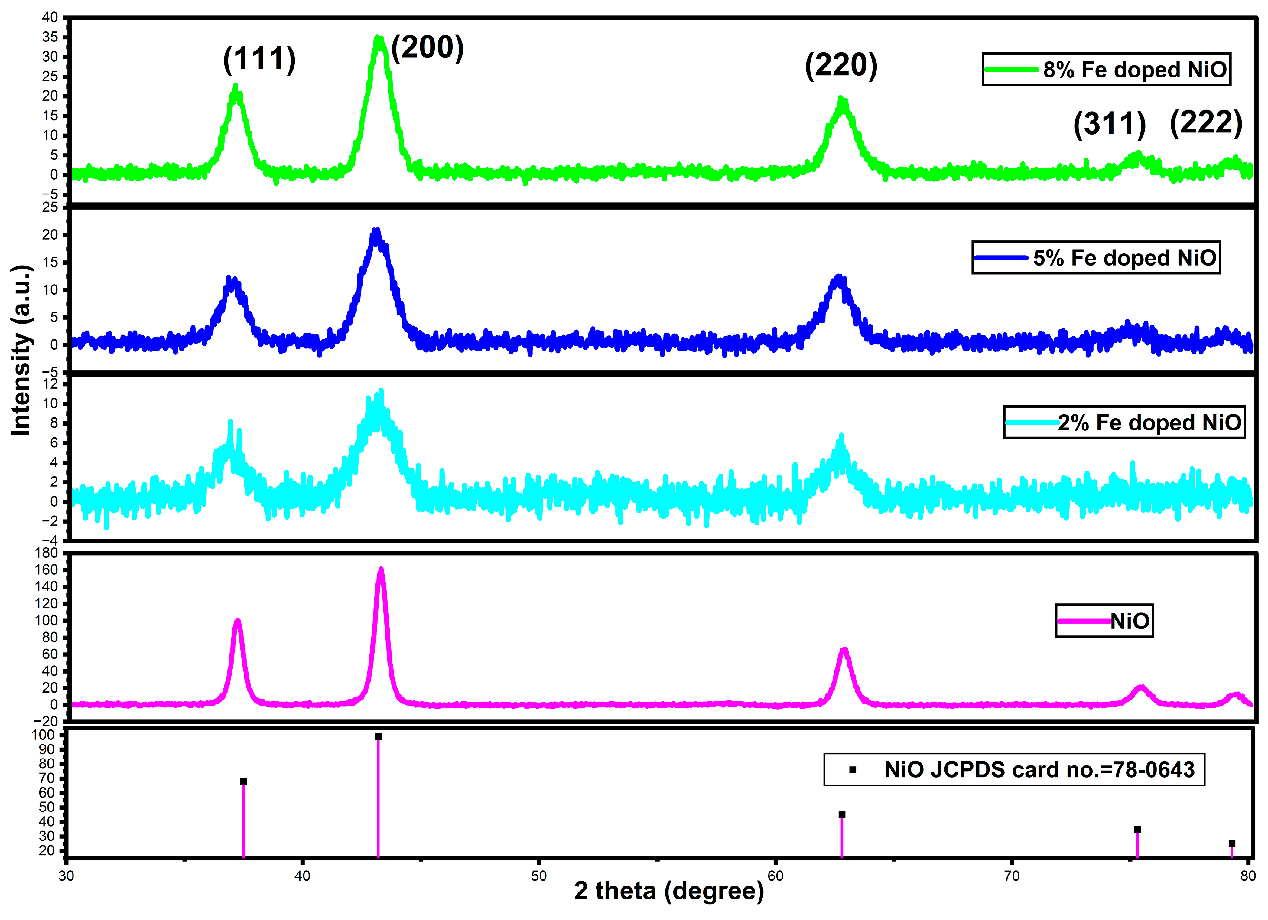
3.1. XRD Analysis

Figure 1 exhibits the X-ray diffractive pattern of different concentrations of NiO and Fe-doped NiO nanoparticles. Pure NiO nanoparticles exhibited diffraction patterns at 2θ = 37, 43, 62, and 75, equivalent to the (111), (200), (220), (311), and (222) planes, respectively. These observed patterns matched perfectly with the standard JCPDS (78-0643) spectrum and previous reports. Peaks were observed in the synthesized Fe-NiO nanoparticles at two-theta angles of 36.6, 42.8, 61.7, and 74.7 degrees, which agree with the (111), (200), (220), and (311) planes, respectively. The broadening of the X-ray diffraction (XRD) peaks observed in the presence of Fe doping in materials such as NiO can be attributed to a variety of reasons, even when the average size of the synthesized nanoparticles does not vary significantly. The introduction of strain, dislocations, and microstrain generated by Fe ions modify the NiO lattice structure. Furthermore, the presence of lattice defects, stacking faults, and twin boundaries caused by Fe doping might contribute to peak broadening. Changes in the chemical composition and distribution of Fe ions within the lattice can also result in peak broadening [35,36]. Moreover, nanoscale surface disorder and differences in crystallite size distribution within the material can influence the XRD pattern by causing scattering variations. These peaks corresponded to the standard reference pattern of JCPDS (78-0643) and previous studies [37,38]. In addition, the size of the synthesized particles of 38 nm, 34 nm, 30 nm, and 27 nm for pure NiO, 2%, 5%, and 8% Fe-doped NiO nanoparticles were measured by the following Scherrer formula.
D = kλ/βCOSθ (nm)
where the D denotes the average size of the crystallite, k is the constant (0.9), λ is the wavelength of the X-ray, β denotes the full width of the half maximum (FWHM), and θ represents the diffraction angle.
High concentrations of Fe doping to the NiO lattice decreased the crystallite sizes [39]. The peak intensities of the diffraction patterns are increased with the increasing concentration of Fe due to the integration of smaller-sized Fe into the crystal lattice of NiO, which results in lattice distortion, strain, and a reduction in crystallite size. The integration of Fe ions into the NiO lattice may affect the crystalline nature and also the size of the synthesized particles. The intensity of the synthesized Fe-NiO nanoparticles (8%) decreased compared to 2% and 5% Fe due to the increasing doping concentration of Fe. Additionally, no other peaks were observed except the standard NiO peaks, which also indicated the uniform dispersion of Fe ions in the NiO lattice.

3.2. FTIR Analysis

Figure 2 shows the FTIR spectra of various concentrations NiO and Fe (2%, 5%, and 8%) doped with NiO nanoparticles. The pure NiO nanoparticles exhibited bands at 3427 cm−1, 1620 cm−1, 1381 cm−1, and 560 cm−1. The wide peak at 3440 cm−1 and the narrow peak are responsible for the OH stretching on the material surfaces. The bands at 1620 and 1381 cm−1 represent the primary and secondary stretching of C-O and C-H bending, which are related to the reduction of metal ions and the association of lattice oxygen from the synthesized nanoparticles [40,41]. The metal–O bond of the Ni-O band vibrations were observed at 560 cm−1. Fe dopants increased the energy of NiO nanoparticles and induced the electrons which were located at the NiO surfaces. The Fe-NiO nanoparticles’ OH-stretching vibrations were found in the bands 3428–3450 cm−1 [40]. In the Fe-NiO nanoparticles, the bands at 2901–2921 cm−1 represent the CH-stretching and the bands at 1574–1626 cm−1 represent the stretching vibrations of the C=O group [40]. The minor peaks at around 2350 cm−1 indicate the presence of carbon-group association with the inorganic metal compounds [40,41,42]. Furthermore, the peaks at 1097–1118 cm−1 [40,41,42] revealed lattice oxygen with organic-compound C-O stretching. There were no peaks observed in the 2% and 5% Fe-doped NiO nanoparticles at the range of 500–600 cm−1 due to the least integration of Fe ions into the NiO crystal lattice. However, there was the appearance of a weak peak in the 8% Fe-doped NiO nanoparticles, which signifies the Fe-O stretching vibrations and proves the integration of the Fe ions in the NiO lattice. The peaks appearing at 673 cm−1 in the 8% Fe-NiO nanoparticles indicate the Ni-O stretching vibrations. Obviously, the appearance of new peaks with the increased concentration of Fe-NiO (8%) also reveals the inclusion of Fe ions on the lattice of the NiO. The observation of weaker peaks at the locations from 1000 to 450 cm−1 in the 2% and 5% Fe-doped NiO nanoparticles indicates the Ni-O-Ni and Ni-OH vibrations. The stretching vibrations of Fe on the Ni-O group [43,44] were identified in the FTIR spectra of 2%, 5%, and 8% Fe-NiO by bands at 869–673 cm−1, respectively. Metal-doped metal–oxygen bonding causes electron shrinkage on the surface, which can stimulate radical production and increase the degradation potentials of the nanoparticles. According to these findings, the integration of Fe in NiO nanoparticles resulted in the formation of various new functional groups, which may have improved their photocatalytic properties.

3.3. FESEM Analysis

FESEM was used to investigate the effect of pure NiO, and the Fe doping concentration on the morphological properties of NiO nanoparticles are presented in Figure 3. The results showed that the Fe doping concentration had a significant influence on the morphology of the nanoparticles, with the 8% Fe-NiO nanoparticles exhibiting a regular spherical morphology and a relatively uniform size distribution, whereas the 2% and 5% Fe-NiO nanoparticles exhibited an irregular morphology with a nonuniform size distribution. Pure NiO nanoparticles exhibited the irregular morphology with uneven atomic distribution on the surfaces. The observed morphological changes were attributed to a change in crystal lattice structure caused by Fe doping. The regular morphology of the 8% Fe-NiO nanoparticles was attributed to the Fe dopant stabilizing the crystal structure, resulting in a more ordered and uniform morphology [45].
The irregular morphology of the 2% and 5% Fe-NiO nanoparticles, on the other hand, was attributed to the disruption of the crystal lattice structure by the Fe dopant, resulting in a more disordered and nonuniform morphology [46]. These findings suggest that the Fe doping concentration should be carefully optimized for specific applications, as it has a significant impact on the morphology of Fe-NiO nanoparticles.
To find the elemental nature and distribution, energy-dispersive X-ray spectroscopy (EDX) was performed. The synthesized NiO and Fe3+-substituted NiO nanoparticles exhibited Fe, Ni, and O elements in the EDX spectra, as shown in Figure 3e–h. The EDX spectra confirmed the formation of NiO and Fe3+-substituted NiO nanoparticles, and their elemental composition. These findings can be helpful in understanding the fundamental properties and potential applications of synthesized NiO and Fe3+-substituted NiO nanoparticles.

3.4. TEM with SAED Analysis
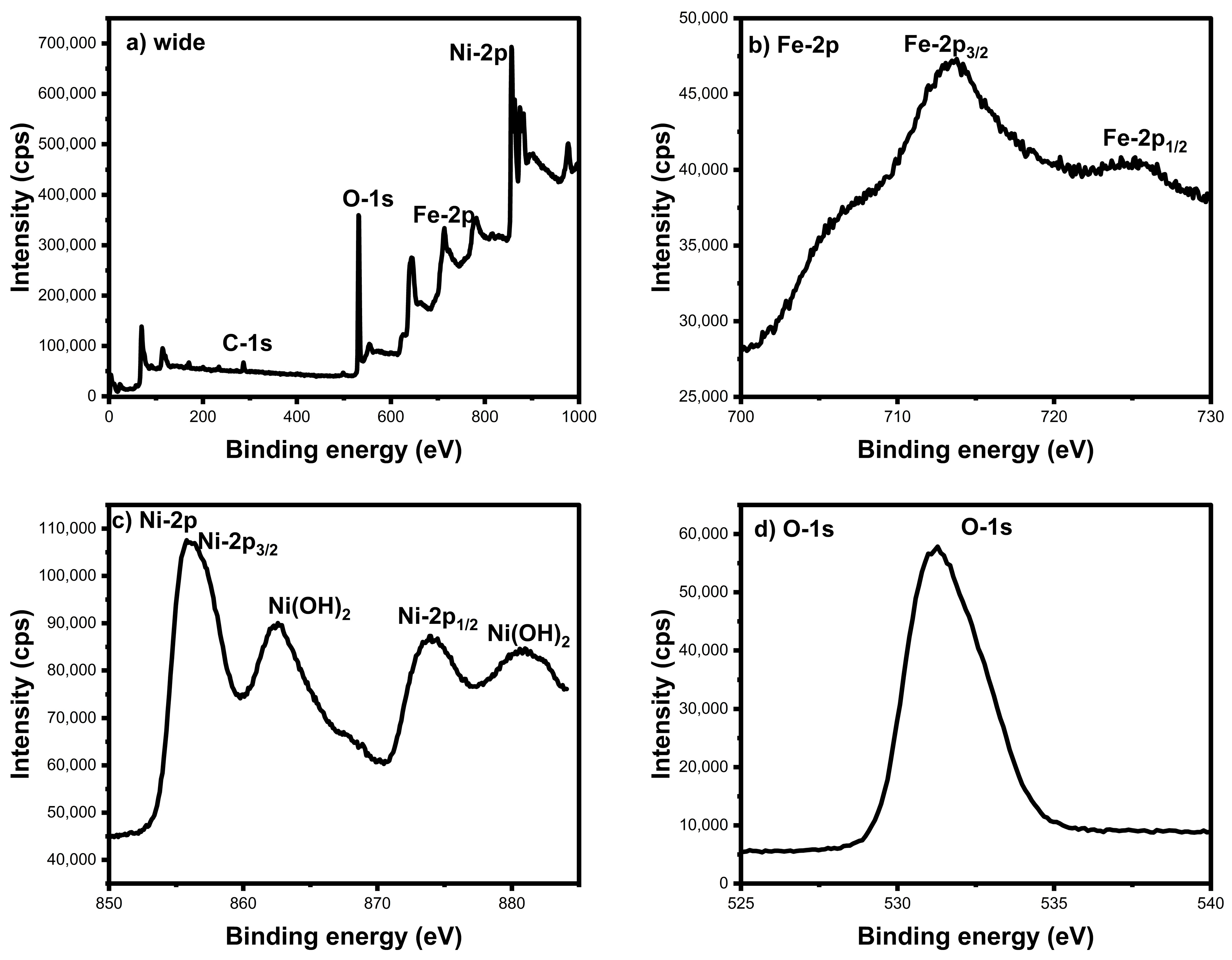
Transmission electron microscopy (TEM) is a powerful imaging technique that allows us to study the morphology and structure of materials at the nanoscale (as illustrated in Figure 4a,b). TEM can provide information regarding the crystal structure and orientation of nanoparticles when combined with selected-area electron diffraction (SAED). TEM imaging is particularly useful for studying iron-doped nickel-oxide nanoparticles, which have a spherical morphology and a high surface-area-to-volume ratio, making them highly reactive and useful in a variety of applications, such as catalysis, drug delivery, and sensing [47]. The spherical shape of nanoparticles is typically determined by the synthesis method used to prepare them.
The crystal structure of the nanoparticles, which is commonly a spinel structure, is confirmed by using SAED. The SAED (Figure 4b) diffraction spots (111), (200), and (220) provide information about the crystal orientation, grain size, and defects within the nanoparticles [48]. The observation of dark spots in the TEM images may confirm the high density of Fe particles in the NiO lattice. In the SAED pattern, the appearance of bright spots proved the synthesized NiO nanoparticles were crystalline, and the few high bright spots also revealed the integration of Fe ions in the NiO nanoparticles. The TEM and SAED analysis of the iron-doped nickel-oxide nanoparticles can reveal important details about their morphology, crystal structure, and orientation, which can help us better understand their properties and optimize their performance in a variety of applications.

3.5. XPS Analysis

The chemical properties of Fe-NiO nanoparticles were investigated using X-ray photoelectron spectroscopy (XPS), and their spectra are shown in Figure 5a–d. Figure 5a–d depicts the broad spectrum, while Figure 5a reveals the core metals Ni and Fe, as well as peaks for Ni-2p, Fe-2p, and O-1s. The peaks at 713 eV (2p3/2) and 723 eV (2p1/2) in the Fe-2p spectrum indicated that the Fe3+ ions were successfully doped into the NiO lattice [49]. The double valency of the Fe-2p state was reoriented with NiO lattices, and it was constructed by the NiO nanophase with the Fe substitution. The Ni-2p spectrum confirmed the successful synthesis of the NiO nanoparticles, as it showed two major peaks at 857 eV (2p3/2) and 874 eV (2p1/2), which are typical of the Ni2+ oxidation state in NiO [49].
Due to the nanoparticles’ exposure to ambient conditions during the XPS measurements, a small peak at 532 eV in the O 1s region indicated the formation of Ni(OH)2 [49,50,51]. The presence of Fe3+ ions in the lattice of the Fe-NiO nanoparticles may have contributed to their enhanced photocatalytic activity in the degradation of the Rh-B dye [50,51]. The XPS analysis sheds light on the surface chemistry and electronic properties of Fe-NiO nanoparticles, which can help researchers better understand their photocatalytic activity and potential applications in environmental remediation.

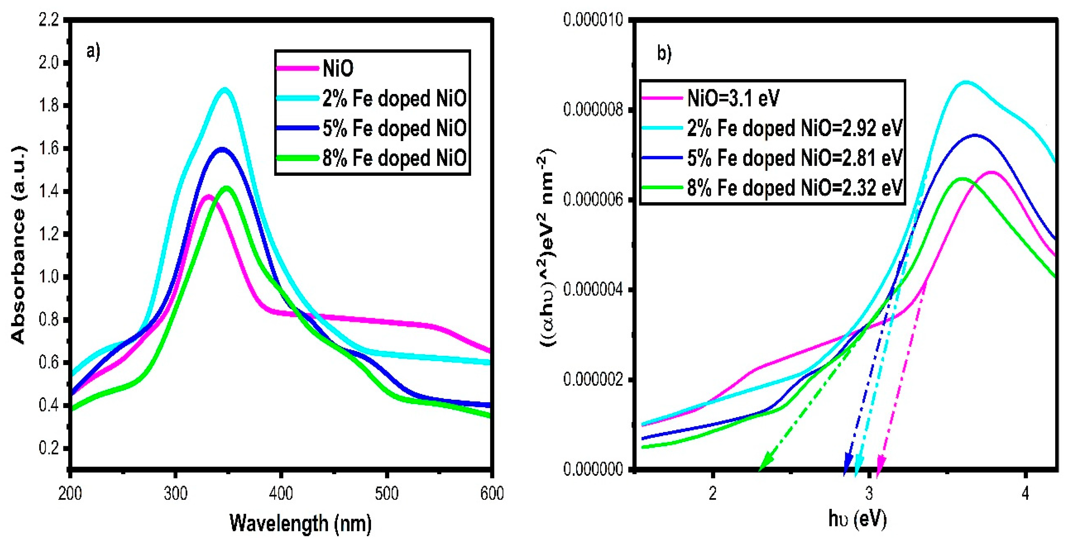
3.6. UV–Visible Analysis

Figure 6a,b shows the UV–visible and Tauc plot spectra of the synthesized NiO and Fe-NiO nanoparticles with doping concentrations of 2%, 5%, and 8%. The optical absorption of pure NiO nanoparticles was observed at 285 nm, indicating high energy transitions in the UV region of the spectrum. The peaks of the Fe-NiO nanoparticles were exhibited at 300–350 nm and their absorbance decreased as the concentration of Fe doping increased [52].
The bandgap energy was calculated using the Tauc plot method and was found to be 3.1 eV, 2.92 eV, 2.81 eV, and 2.32 eV for pure NiO, 2%, 5%, and 8% Fe-NiO, respectively. The decrease in the bandgap energy and the increase in the visible-region absorbance can be attributed to the higher concentration of Fe ions in the NiO lattice, which introduces the high-energy electron occupations on the nanostructure surfaces. Fe dopants deduced more defects and impurities, which act as recombination centers for electron–hole pairs, enhancing the photocatalytic activity [52,53,54,55,56,57,58,59,60]. Nonetheless, the bandgap energy of Fe-NiO is still within the visible-light range, making it suitable for photocatalytic applications when exposed to visible light. The observed trend in bandgap energy with the Fe doping concentration suggests that, among the investigated samples, the 8% Fe-NiO nanoparticles are the most promising candidate for photocatalytic dye degradation.

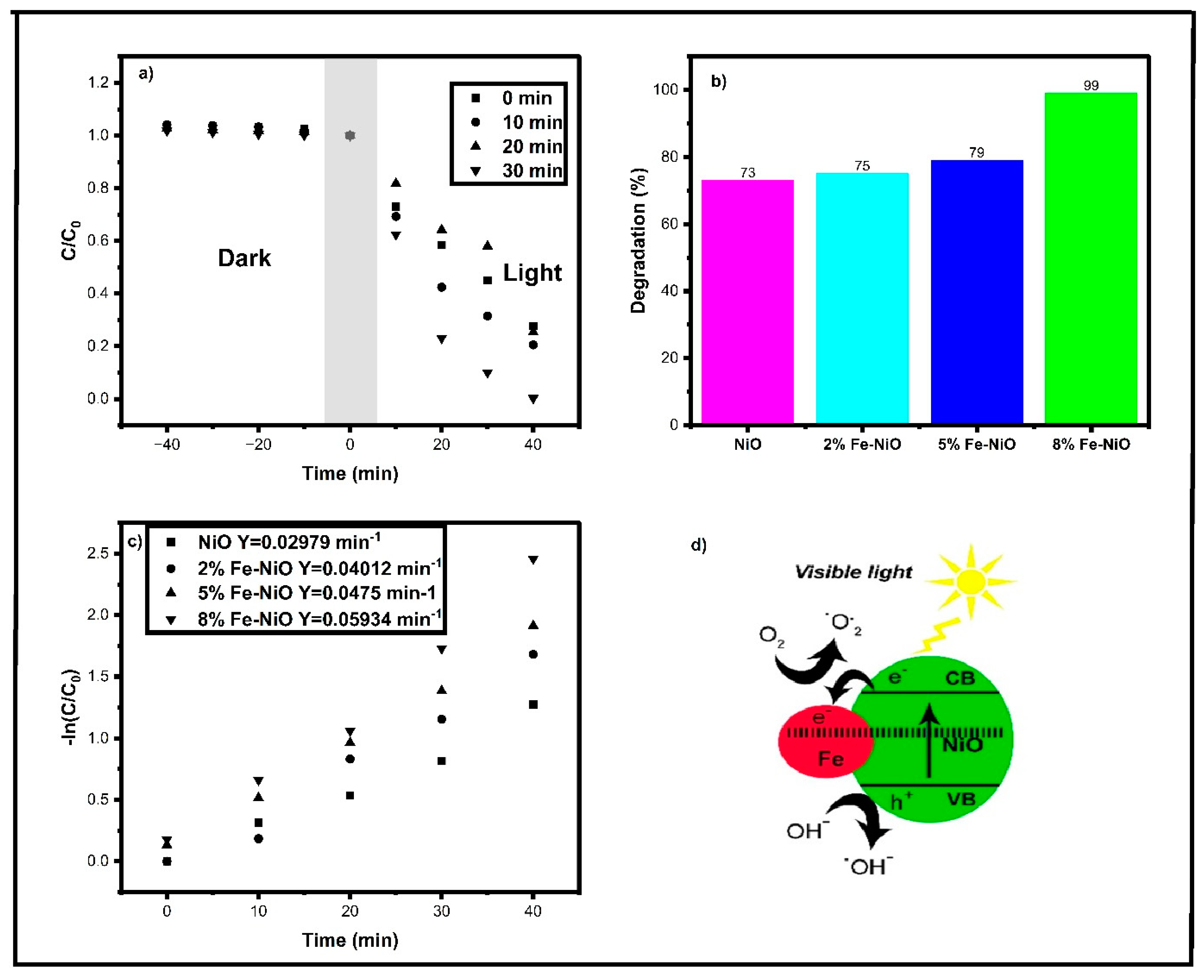
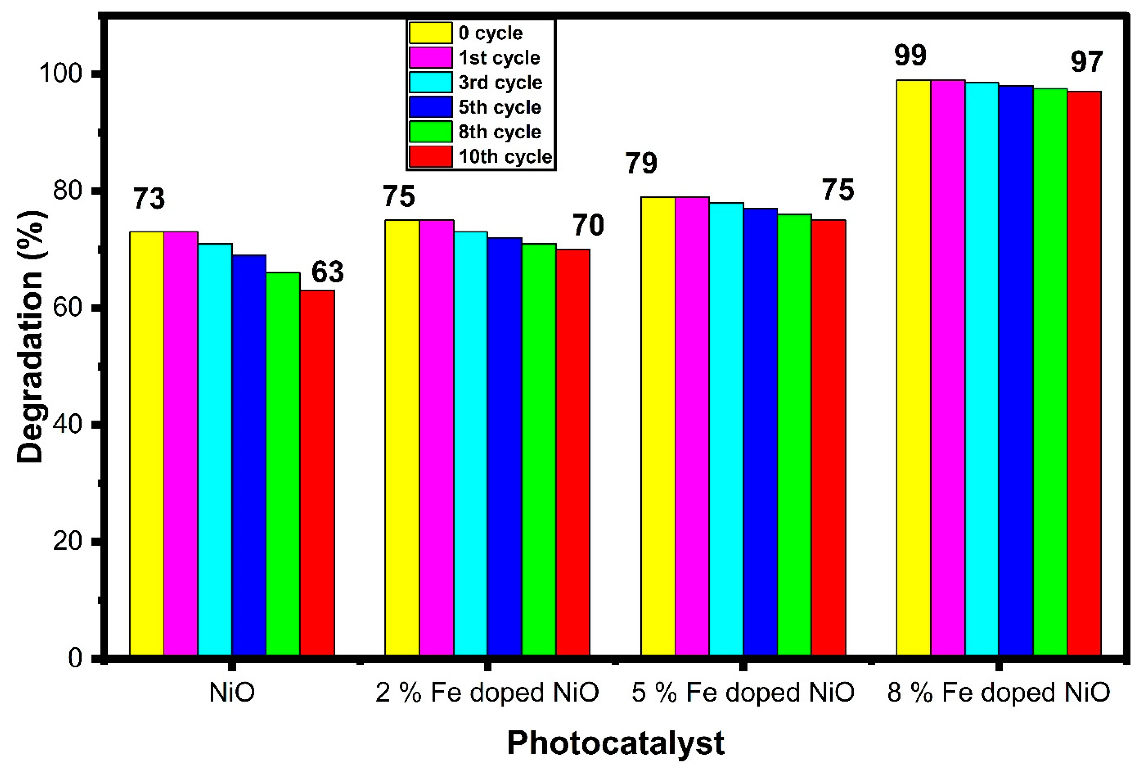
3.7. Photocatalytic Dye Degradation Activity

The photocatalytic activity of nickel-oxide and iron-doped nickel-oxide nanoparticles (Fe-NiO) with 2%, 5%, and 8% doping concentrations against rhodamine B (Rh-B) dye was investigated using visible-light irradiation (Figure 7). Fe-doped NiO nanoparticles can be used in several ways to improve their ability to break down dyes photocatalytically, both in the dark and in the light. To increase active sites, the surface area of the nanoparticles was increased using specialized calcination or mesoporous support materials. The bandgap engineering was investigated to optimize the light-absorption properties of the nanoparticles, which are supported by the incorporation of light-absorbing materials and cocatalysts that improve electron–hole pair formation and separation. A suitable visible-light source was employed with the appropriate wavelength and intensity, and precise agitation techniques were used to ensure uniform dispersion [61,62,63]. Because of its high catalytic activity, stability, and low cost, Fe-NiO nanoparticles are a promising material for catalytic applications. Photocatalysis is an effective and powerful technique for reducing toxic substances in the environment. While metal-oxide nanoparticles are traditional photocatalysts, their shortcomings can be addressed by adding dopants. The metal doping of metal-oxide nanoparticles is highly applicable in water-remediation processes due to their ability to trap metals and drift electrons on the outer surfaces. The utilization of Rh-B dye is widespread in color-related sectors, such as food, textile, and leather industries, due to its cationic nature. Due to its nonbiodegradable nature, it is a highly noxious and carcinogenic substance [61,62,63,64].
In the context of rhodamine B degradation, Fe-NiO nanoparticles can act as a photocatalyst, producing reactive species such as hydroxyl radicals (•OH) upon light exposure. The following steps are involved in the rhodamine B degradation mechanism (Figure 7d) using Fe-NiO:
  • Absorption of light: Fe-NiO absorbs photons with energy greater than its bandgap, which promotes electrons from the valence band to the conduction band.
  • Generation of electron–hole pairs: The excited electrons and holes can migrate to the surface of the Fe-NiO nanoparticles and generate electron–hole pairs, which can participate in the following reactions:
  • Formation of reactive species: The holes can react with water molecules or hydroxyl ions (OH) adsorbed on the surface of Fe-NiO nanoparticles to produce hydroxyl radicals:
h+ + H2O → •OH + H+
h+ + OH → •OH
2.
Oxidation of rhodamine B: The hydroxyl radicals can attack and break down the rhodamine B molecule, leading to its degradation into smaller, less toxic compounds:
•OH + rhodamine B → Degradation products [65,66,67,68,69,70,71,72,73,74].
The photocatalytic degradation mechanism of rhodamine B (Rh-B) dye using NiO and Fe-doped NiO (Fe:NiO) nanoparticles under visible-light irradiation consists of several steps. Firstly, both NiO and Fe:NiO nanoparticles absorb visible light, with the Fe dopants extending the absorption into the visible range. This absorption generates electron–hole pairs in the nanoparticles. These photogenerated electron–hole pairs then initiate a series of redox reactions with adsorbed Rh-B dye molecules on the nanoparticle surfaces. Electrons typically reduce the dye molecules, breaking them down into less-colored compounds, while holes can oxidize water or hydroxide ions, forming highly reactive hydroxyl radicals (•OH). These hydroxyl radicals attack and break the chemical bonds of the adsorbed Rh-B dye molecules, leading to dye degradation into simpler and less-colored products. The process ultimately results in the release of nontoxic compounds or mineralization products, with the photocatalyst’s surface being continuously regenerated as photogenerated electrons and holes participate in redox reactions throughout the degradation process. The presence of Fe dopants enhances both light absorption and charge separation, enhancing the overall photocatalytic efficiency [71,72,73,74].
The durability and efficiency of the photocatalytic material are used to select industrial-scale photocatalysts. Figure 8 depicts the study’s evaluation of the recycling efficiency of pure NiO and Fe-NiO nanoparticles over ten cycles. Degradation losses can occur due to a variety of factors, including (i) surface usage through dry and wash processes, (ii) coverage of active sites by targeted materials, and (iii) secondary-product blockage of the entire surface and pores. Nonetheless, the results indicate that Fe-NiO nanoparticles exhibit high stability and minimal degradation. Because of their high reusability and low catalyst wastage, Fe-NiO nanoparticles have the potential to be effective photocatalysts for pollutant degradation. Fe-NiO nanoparticles are promising candidates for practical applications in environmental remediation and wastewater treatment due to their high stability and recyclability, effectively combating pollutants with minimal degradation over multiple cycles.
The production of holes, superoxides, and free radicals, which are crucial for photocatalytic activity, is what determines how well the degradation performs. The findings of the scavenger analysis are shown in Figure 9. The readings obtained after mitigating holes, superoxides, and hydroxide suppression were compared to the outstanding degrading efficiency of 99% displayed by the pure catalyst. Based on the illustration’s depiction, the degradation efficacy orders are as follows: TEOA > BQ > IPA for the suppression of holes > superoxides > hydroxide, respectively. Predominantly, TEOA performs better in terms of degrading activity than BQ and IPA. The catalyst surface’s pores made it easier for dye molecules to separate, changing them from poisonous to nontoxic substances.
The effectiveness of various metal-oxide and metal-doped metal-oxide nanoparticles was compared to that of synthesized Fe-doped NiO nanoparticles for the degradation of rhodamine B (Rh-B) dye under visible-light irradiation, as shown in Table 1. Metals were strategically added to metal-oxide nanoparticles in order to efficiently overcome the limitations of the photocatalytic activity of the metal-oxide nanoparticles. Metal doping or the decorating of metal-oxide nanoparticles resulted in a decrease in the bandgap energy and particle size, improving the nanoparticles’ ability to absorb light in the visible spectrum [75,76,77,78]. The effectiveness of the targeted dye molecules’ breakdown was subsequently improved by this change. Particularly noteworthy were the impacts of noble metal decorating, doping, or coating, which increased the surface area of the nanoparticles and aided in the production of plasmonic-resonance-driven “hot electrons”. These hot electrons were crucial in improving the catalytic activity of the nanoparticles, outpacing other metal-decorating techniques. The results of this investigation showed that, when added to the NiO surface, Fe-doped NiO nanoparticles produced catalytic activity that was more efficient overall than metal decorations and noble metal decorations [75,76,77,78,79,80,81,82]. The results demonstrate the enhanced photocatalytic capabilities of Fe-doped NiO nanoparticles for the degradation of Rh-B dye in visible light. The addition of iron (Fe) as a dopant into the nickel oxide (NiO) lattice displayed outstanding catalytic activity. By overcoming the inherent constraints of metal-oxide nanoparticles, this method created new opportunities for improving their photocatalytic performance. The practical ramifications of this research would also be further highlighted by emphasizing the significance of these discoveries in the context of environmental remediation, water treatment, and sustainable-energy applications.

4. Conclusions

The coprecipitation method was used for the synthesis of NiO and Fe-NiO nanoparticles, and their structural and optical properties were investigated using various techniques. The X-ray diffraction (XRD) analysis revealed the formation of a crystalline cubic NiO structure, with no additional Fe-dopant peaks. The FTIR spectra revealed the presence of Fe-O and Ni-O bonds, while the UV–visible spectra revealed a decrease in visible absorbance and an increase in the bandgap energy of the Fe-NiO nanoparticles. The regular morphological shapes of the Fe-NiO nanoparticles were confirmed from FESEM and images from the TEM revealed a spherical morphology. The presence of Fe, Ni, and O elements in the samples was confirmed by X-ray photoelectron spectroscopy (XPS). The results of the Rh-B dye degradation showed that the Fe-NiO nanoparticles have higher photocatalytic activity, with 99% degradation achieved for the 8% Fe-NiO. The obtained findings suggest that Fe-NiO nanoparticles have the potential to be effectively used as photocatalysts for the degradation of organic dyes, as evidenced by their promising photocatalytic activity. These results indicate that Fe-NiO nanoparticles may be a viable option for use in environmental remediation and other related applications.

Author Contributions

Conceptualization, S.M. and M.R.S.; methodology, S.M. and W.-C.L.; software, S.M., J.K.G. and W.-C.L.; validation, S.A. and W.-C.L.; formal analysis, J.J., S.M.W., J.K.G., M.R.S. and W.-C.L.; investigation, S.M.W., J.K.G. and S.A.; resources, J.J., S.A., M.R.S. and W.-C.L.; data curation, S.M. and J.K.G.; writing—original draft, S.M.; writing—review & editing, S.M. and J.J.; visualization, J.J. and S.M.W.; supervision, J.J. and W.-C.L.; project administration, S.M.W., J.K.G., S.A. and M.R.S.; funding acquisition, S.M.W., J.K.G., S.A., M.R.S. and W.-C.L. All authors have read and agreed to the published version of the manuscript.

Funding

The authors are grateful to the Researchers Supporting Project Number (RSP2023R326), King Saud University, Riyadh, Saudi Arabia.

Institutional Review Board Statement

Not applicable.

Informed Consent Statement

Not applicable.

Data Availability Statement

All research data associated to the manuscript.

Acknowledgments

The authors are grateful to the Researchers Supporting Project Number (RSP2023R326), King Saud University, Riyadh, Saudi Arabia. Minisha thanks the Department of Physics, Annai Velankani College, Tholayavattam-629157 and Manonmaniam Sundaranar University, Abishekapatti, Tirunelveli-627012, Tamil Nadu, India.

Conflicts of Interest

The authors declare that there are no conflict of interest.

References

  1. Jha, S.; Gaur, R.; Shahabuddin, S.; Tyagi, I. Biochar as sustainable alternative and green adsorbent for the remediation of noxious pollutants: A comprehensive review. Toxics 2023, 11, 117. [Google Scholar] [CrossRef] [PubMed]
  2. du Plessis, A. South Africa’s Impending Water Crises: Transforming Water Crises into Opportunities and the Way Forward. In South Africa’s Water Predicament: Freshwater’s Unceasing Decline; Springer International Publishing: Cham, Switzerland, 2023; pp. 143–170. [Google Scholar]
  3. Ranganathan, S.; Rajendran, S.; Ponce, L.C. Chemically Modified Carbon Nanotubes for Pollutants Adsorption. In Chemically Modified Carbon Nanotubes for Commercial Applications; Wiley: Hoboken, NJ, USA, 2023; pp. 129–150. [Google Scholar]
  4. Tazoe, H. Water quality monitoring. Anal. Sci. 2023, 39, 1–3. [Google Scholar] [CrossRef] [PubMed]
  5. Kiwanuka, M.; Mutanda, H.E.; Niyomukiza, J.B.; Nakasagga, E. Assessment of suitability of drinking water from the springs in Urban slums of Kampala. Environ. Chall. 2023, 10, 100667. [Google Scholar] [CrossRef]
  6. Verma, R.; Mishra, S.R.; Gadore, V.; Ahmaruzzaman, M. Hydroxyapatite-based composites: Excellent materials for environmental remediation and biomedical applications. Adv. Colloid Interface Sci. 2023, 315, 102890. [Google Scholar] [CrossRef]
  7. Khan, H.; Hussain, S.; Zahoor, R.; Arshad, M.; Umar, M.; Marwat, M.A.; Khan, A.; Khan, J.R.; Haleem, M.A. Novel modeling and optimization framework for Navy Blue adsorption onto eco-friendly magnetic geopolymer composite. Environ. Res. 2023, 216, 114346. [Google Scholar] [CrossRef] [PubMed]
  8. Vezzone, M.; Cesar, R.; Serrano, A.; Lourenço, R.; Rodrigues, A.P.; Castilhos, Z.; dos Anjos, R.M.; Polivanov, H. Mercury Contamination in Sediments and Fish from an Urban Tropical Estuary: Ecological and Human Health Risks. Water Air Soil Pollut. 2023, 234, 72. [Google Scholar] [CrossRef]
  9. Maleki, F.; Ghaemi, A.; Mir Mohamad Sadeghi, G. Synthesis and characterization of waste Styrofoam hypercrosslinked polymer as an adsorbent for CO2 capture. Environ. Prog. Sustain. Energy 2023, 42, e13954. [Google Scholar] [CrossRef]
  10. Larsen, J.J.; Lévy, L.; Asif, M.R. Removal of Powerline Noise in Geophysical Datasets with a Scientific Machine-Learning Based Approach. IEEE Trans. Geosci. Remote Sens. 2022, 60, 5923410. [Google Scholar] [CrossRef]
  11. Wang, Q.; Zhao, Y.; Zhang, Z.; Liao, S.; Deng, Y.; Wang, X.; Ye, Q.; Wang, K. Hydrothermal preparation of Sn3O4/TiO2 nanotube arrays as effective photocatalysts for boosting photocatalytic dye degradation and hydrogen production. Ceram. Int. 2023, 49, 5977–5985. [Google Scholar] [CrossRef]
  12. Wang, D.; Li, Y.; Wen, L.; Xi, J.; Liu, P.; Hansen, T.W.; Li, P. Ni-Pd-incorporated Fe3O4 yolk-shelled nanospheres as efficient magnetically recyclable catalysts for reduction of n-containing unsaturated compounds. Catalysts 2023, 13, 190. [Google Scholar] [CrossRef]
  13. Shakil, M.; Inayat, U.; Tanveer, M.; Nabi, G.; Gillani, S.S.A.; Rafique, M.; Tariq, N.H.; Shah, A.; Mahmood, A. NiO and Ag–Cd co-doped NiO nanoparticles: Study of photocatalytic degradation of rhodamine B dye for wastewater treatment. Int. J. Environ. Sci. Technol. 2023, 20, 2021–2036. [Google Scholar] [CrossRef]
  14. Wen, L.; Wang, D.; Xi, J.; Tian, F.; Liu, P.; Bai, Z.W. Heterometal modified Fe3O4 hollow nanospheres as efficient catalysts for organic transformations. J. Catal. 2022, 413, 779–785. [Google Scholar] [CrossRef]
  15. Zhang, Y.; Huang, J.; Dong, Z.; Zhan, Y.; Xi, J.; Xiao, J.; Huang, S.; Tian, F. Pd–Fe bimetallic nanoparticles anchored on N-doped carbon-modified graphene for efficient catalytic organic reactions. Carbon Lett. 2023, 33, 77–87. [Google Scholar] [CrossRef]
  16. Guo, T.; Fan, X.; Jiang, X.; Qi, Y.; Du, J.; Zhang, A.; Wang, H. Engineering shape of BiOClnanosheets with improved visible-light response for superior photocatalytic degradation of Rhodamine B. J. Alloys Compd. 2023, 948, 169586. [Google Scholar] [CrossRef]
  17. Cheng, H.; Li, X.; Huang, C.; Zhu, J.; Wang, P.; Cao, H.; Feng, C.; Ling, D.; Liu, H.; Cheng, M. Accelerated Fe (III)/Fe (II) cycle for rapid elimination of Rhodamine B by a novel Mo2C co-catalytic Fe2+/H2O2 system. J. Clean. Prod. 2023, 393, 136354. [Google Scholar] [CrossRef]
  18. Tanji, K.; El Mrabet, I.; Fahoul, Y.; Jellal, I.; Benjelloun, M.; Belghiti, M.; El Hajam, M.; Naciri, Y.; El Gaidoumi, A.; El Bali, B.; et al. Epigrammatic progress on the photocatalytic properties of ZnO and TiO2 based hydroxyapatite@ photocatlyst toward organic molecules photodegradation: A review. J. Water Process Eng. 2023, 53, 103682. [Google Scholar] [CrossRef]
  19. Lv, H.; Liu, Y.; Zhao, P.; Bai, Y.; Cui, W.; Shen, S.; Liu, Y.; Wang, Z.; Yu, D.G. Insight into the superior piezophotocatalytic performance of BaTiO3//ZnO Janus nanofibrous heterostructures in the treatment of multi-pollutants from water. Appl. Catal. B Environ. 2023, 330, 122623. [Google Scholar] [CrossRef]
  20. Nowak, E.; Chłopocka, E.; Szybowicz, M. ZnO and ZnO-Based Materials as Active Layer in Resistive Random-Access Memory (RRAM). Crystals 2023, 13, 416. [Google Scholar] [CrossRef]
  21. Ameen, F.; Mostafazadeh, R.; Hamidian, Y.; Erk, N.; Sanati, A.L.; Karaman, C.; Ayati, A. Modeling of adsorptive removal of azithromycin from aquatic media by CoFe2O4@ NiO anchored microalgae-derived nitrogen-doped porous activated carbon adsorbent and colorimetric quantifying of azithromycin in pharmaceutical products. Chemosphere 2023, 329, 138635. [Google Scholar] [CrossRef]
  22. Deng, Y.; Yang, Z.; Xu, T.; Jiang, H.; Ng, K.W.; Liao, C.; Danni, S.; Yanli, P.; Zimin, C.; Gang, W.; et al. Band alignment and electrical properties of NiO/β-Ga2O3 heterojunctions with different β-Ga2O3 orientations. Appl. Surf. Sci. 2023, 622, 156917. [Google Scholar] [CrossRef]
  23. Dejam, L.; Sabbaghzadeh, J.; Ghaderi, A.; Solaymani, S.; Matos, R.S.; Țălu, Ș.; da Fonseca Filho, H.D.; Sari, A.H.; Kiani, H.; Shayegan, A.H.S.; et al. Advanced nano-texture, optical bandgap, and Urbach energy analysis of NiO/Si heterojunctions. Sci. Rep. 2023, 13, 6518. [Google Scholar] [CrossRef] [PubMed]
  24. Chen, S.C.; Kuo, T.Y.; Lin, Y.C.; Lin, H.C. Preparation and properties of p-type transparent conductive Cu-doped NiO films. Thin Solid Films 2011, 519, 4944–4947. [Google Scholar] [CrossRef]
  25. Amor, M.B.; Boukhachem, A.; Boubaker, K.; Amlouk, M. Structural, optical and electrical studies on Mg-doped NiO thin films for sensitivity applications. Mater. Sci. Semicond. Process. 2014, 27, 994–1006. [Google Scholar] [CrossRef]
  26. Amor, M.B.; Boukhachem, A.; Labidi, A.; Boubaker, K.; Amlouk, M. Physical investigations on Cd doped NiO thin films along with ethanol sensing at relatively low temperature. J. Alloys Compd. 2017, 693, 490–499. [Google Scholar] [CrossRef]
  27. Al Boukhari, J.; Zeidan, L.; Khalaf, A.; Awad, R. Synthesis, characterization, optical and magnetic properties of pure and Mn, Fe and Zn doped NiO nanoparticles. Chem. Phys. 2019, 516, 116–124. [Google Scholar] [CrossRef]
  28. Zhang, J.H.; Cai, G.F.; Zhou, D.; Tang, H.; Wang, X.L.; Gu, C.D.; Tu, J.P. Co-doped NiOnanoflake array films with enhanced electrochromic properties. J. Mater. Chem. C 2014, 2, 7013–7021. [Google Scholar] [CrossRef]
  29. Layek, S.; Verma, H.C. Room temperature ferromagnetism in Mn-doped NiO nanoparticles. J. Magn. Magn. Mater. 2016, 397, 73–78. [Google Scholar] [CrossRef]
  30. Peng, S.; Wang, C.; Xie, J.; Sun, S. Synthesis and stabilization of monodisperse Fe nanoparticles. J. Am. Chem. Soc. 2006, 128, 10676–10677. [Google Scholar] [CrossRef]
  31. Guo, J.; Wang, R.; Tjiu, W.W.; Pan, J.; Liu, T. Synthesis of Fe nanoparticles@ graphene composites for environmental applications. J. Hazard. Mater. 2012, 225, 63–73. [Google Scholar] [CrossRef]
  32. Pereira, C.; Pereira, A.M.; Fernandes, C.; Rocha, M.; Mendes, R.; Fernández-García, M.P.; Guedes, A.; Tavares, P.B.; Grenèche, J.-M.; Araújo, J.P.; et al. Superparamagnetic MFe2O4 (M = Fe, Co, Mn) nanoparticles: Tuning the particle size and magnetic properties through a novel one-step coprecipitation route. Chem. Mater. 2012, 24, 1496–1504. [Google Scholar] [CrossRef]
  33. Chelliah, P.; Wabaidur, S.M.; Sharma, H.P.; Jweeg, M.J.; Majdi, H.S.; Al Kubaisy, M.M.R.; Iqbal, A.; Lai, W.-C. Green Synthesis and Characterizations of Cobalt Oxide Nanoparticles and Their Coherent Photocatalytic and Antibacterial Investigations. Water 2023, 15, 910. [Google Scholar] [CrossRef]
  34. Chelliah, P.; Wabaidur, S.M.; Sharma, H.P.; Majdi, H.S.; Smait, D.A.; Najm, M.A.; Iqbal, A.; Lai, W.-C. Photocatalytic Organic Contaminant Degradation of Green Synthesized ZrO2 NPs and Their Antibacterial Activities. Separations 2023, 10, 156. [Google Scholar] [CrossRef]
  35. Gurugubelli, T.R.; Babu, B.; Kim, J.; Yoo, K. Hydrothermal synthesis of Fe-doped ZnAl2O4 nanosheets: Bandgap engineering and room temperature ferromagnetism. Chem. Pap. 2021, 75, 6407–6416. [Google Scholar] [CrossRef]
  36. Rao, G.T.; Babu, B.; Ravikumar, R.V.S.S.N.; Shim, J.; Reddy, C.V. Structural and optical properties of Fe-doped SnO2 quantum dots. Mater. Res. Express 2017, 4, 125021. [Google Scholar]
  37. Rana, P.S. Structural, optical, electrical, and photocatalytic application of NiFe2O4@ NiOnanocomposites for methylene blue dye. Ceram. Int. 2023, 49, 13520–13530. [Google Scholar]
  38. Vivek, P.; Sivakumar, R.; Esakki, E.S.; Deivanayaki, S. Fabrication of NiO/RGO nanocomposite for enhancing photocatalytic performance through degradation of RhB. J. Phys. Chem. Solids 2023, 176, 111255. [Google Scholar] [CrossRef]
  39. Motene, K.; Mahlaule-Glory, L.M.; Ngoepe, N.M.; Mathipa, M.M.; Hintsho-Mbita, N.C. Photocatalytic degradation of dyes and removal of bacteria using biosynthesised flowerlike NiO nanoparticles. Int. J. Environ. Anal. Chem. 2023, 103, 1107–1122. [Google Scholar] [CrossRef]
  40. Majeed, K.; Ambreen, J.; Khan, S.A.; Muhammad, S.; Shah, A.A.; Bhatti, M.A.; Batool, S.S.; Farooq, M.; Shah Bukhari, S.N.U.; Chandio, A.D.; et al. Effective Removal of Methylene Blue by Mn3O4/NiO Nanocomposite under Visible Light. Separations 2023, 10, 200. [Google Scholar] [CrossRef]
  41. Mandal, R.K.; Mondal, A.S.; Ghosh, S.; Halder, A.; Majumder, T.P. Synthesis, characterisation and optical studies of CdO-NiO NCs for comparative dye degradation study between two hazardous dyes Congo red and Rose Bengal. Results Chem. 2023, 5, 100810. [Google Scholar] [CrossRef]
  42. Huang, B.; Tong, X.; Zhang, X.; Feng, Q.; Rumyantseva, M.N.; Prakash, J.; Li, X. MXene/NiO Composites for Chemiresistive-Type Room Temperature Formaldehyde Sensor. Chemosensors 2023, 11, 258. [Google Scholar] [CrossRef]
  43. Bhat, M.A.; Rana, P.; Mir, F.A.; Pathak, D. A brief study on exploration of Ni doped PrFeO3 perovskite as multifunctional material. J. Mater. Sci. Mater. Electron. 2023, 34, 269. [Google Scholar] [CrossRef]
  44. Du, Y.; Li, Z.; Liu, H.; Qiao, S.; Chen, Y.; Zhu, Z.; Tang, Y.; Liu, C. Scalable oxygen-assisted-Fe2+ etching approach towards amorphous/crystalline structure Fe-Ni2P nanoarray for efficient water splitting. J. Alloys Compd. 2023, 936, 168073. [Google Scholar] [CrossRef]
  45. Kachhadiya, D.D.; Murthy, Z.V.P. Preparation and characterization of ZIF-8 and ZIF-67 engineered PVDF mixed-matrix membranes: Stability enhancement in pervaporation study. Environ. Sci. Water Res. Technol. 2023, 9, 1502–1517. [Google Scholar] [CrossRef]
  46. Ali, S.M.; Kassim, H.; Amer, M.S. Nanoarchitectonics of tin telluride: A novel pseudocapacitive material for energy storage application. Mater. Chem. Phys. 2023, 301, 127698. [Google Scholar] [CrossRef]
  47. Singh, S.; Kumar, P.; Patidar, R.; Srivastava, V.C.; Lo, S.L.; Štangar, U.L. Catalytic oxidation of Bisphenol A with Co3+ rich spinel Co3O4: Performance evaluation with peroxymonosulfate activation and mineralization mechanism. J. Environ. Chem. Eng. 2023, 11, 110023. [Google Scholar] [CrossRef]
  48. Wei, Z.; Qiao, H.; Yang, H.; Zhang, C.; Yan, X. Characterization of NiO nanoparticles by anodic arc plasma method. J. Alloys Compd. 2009, 479, 855–858. [Google Scholar] [CrossRef]
  49. Qiao, H.; Wei, Z.; Yang, H.; Zhu, L.; Yan, X. Preparation and characterization of NiO nanoparticles by anodic arc plasma method. J. Nanomater. 2009, 2009, 795928. [Google Scholar] [CrossRef]
  50. Pebley, A.C.; Decolvenaere, E.; Pollock, T.M.; Gordon, M.J. Oxygen evolution on Fe-doped NiOelectrocatalysts deposited via microplasma. Nanoscale 2017, 9, 15070–15082. [Google Scholar] [CrossRef]
  51. Liu, S.; Jia, J.; Wang, J.; Liu, S.; Wang, X.; Song, H.; Hu, X. Synthesis of Fe-doped NiOnanofibers using electrospinning method and their ferromagnetic properties. J. Magn. Magn. Mater. 2012, 324, 2070–2074. [Google Scholar] [CrossRef]
  52. Bhoye, M.; Pansambal, S.; Basnet, P.; Lin, K.Y.A.; Gutierrez-Mercado, K.Y.; Pérez-Larios, A.; Chauhan, A.; Oza, R.; Ghotekar, S. Eco-Friendly Synthesis of Ni/NiO Nanoparticles Using Gymnemasylvestre Leaves Extract for Antifungal Activity. J. Compos. Sci. 2023, 7, 105. [Google Scholar] [CrossRef]
  53. Rad, L.R.; Anbia, M.; Vatanpour, V. Adsorption and Photocatalytic Degradation of Fluoxetine Using TiO2-Supported-Clinoptilolite, NaX and MIL-101 (Fe) Metal Organic Framework. J. Inorg. Organomet. Polym. Mater. 2023, 33, 2154–2171. [Google Scholar] [CrossRef]
  54. Ramasamy, N.; NagarasampattiPalani, K.; Mathew, A.; Natesan, B. Development of chitosan@ Fe2O3/rGO/Bi2S3 as a new eco-friendly photocatalyst for enhancing the catalytic stability and superior degradation of organic pollutants. Res. Chem. Intermed. 2023, 49, 2603–2624. [Google Scholar] [CrossRef]
  55. Mala, N.A.; Dar, M.A.; Rather, M.U.D.; Reshi, B.A.; Sivakumar, S.; Batoo, K.M.; Ahmad, Z. Supercapacitor and magnetic properties of NiO and manganese-doped NiO nanoparticles synthesized by chemical precipitation method. J. Mater. Sci. Mater. Electron. 2023, 34, 505. [Google Scholar] [CrossRef]
  56. Sharma, K.R.; Negi, N.S. Significant improvement in the structural, microstructural, and room-temperature magnetic properties of Fe-doped NiO nanoparticles prepared by the solution combustion method. J. Mater. Sci. Mater. Electron. 2022, 33, 22518–22540. [Google Scholar] [CrossRef]
  57. Zhao, T.; Zhang, H.; Zhang, H.H.; Yi, D.Q.; Zhu, J.F.; Qin, Y. Facile synthesis of NiFe2O4/Ni3Fe/LAS core-shell-like composites with improved impedance matching and microwave absorption properties. J. Phys. Chem. Solids 2023, 176, 111241. [Google Scholar] [CrossRef]
  58. Faramawy, A.; Elsayed, H.; Scian, C.; Mattei, G. Structural, optical, magnetic and electrical properties of sputtered ZnO and ZnO: Fe thin films: The role of deposition power. Ceramics 2022, 5, 1128–1153. [Google Scholar] [CrossRef]
  59. Che, H.; Huso, J.; Morrison, J.L.; Thapa, D.; Huso, M.; Yeh, W.J.; Tarun, M.C.; McCluskey, M.D.; Bergman, L. Optical properties of ZnO-alloyed nanocrystalline films. J. Nanomater. 2012, 2012, 7. [Google Scholar] [CrossRef]
  60. Marotti, R.E.; Guerra, D.N.; Bello, C.; Machado, G.; Dalchiele, E.A. Bandgap energy tuning of electrochemically grown ZnO thin films by thickness and electrodeposition potential. Sol. Energy Mater. Sol. Cells 2004, 82, 85–103. [Google Scholar] [CrossRef]
  61. Reddy, N.R.; Reddy, P.M.; Jyothi, N.; Kumar, A.S.; Jung, J.H.; Joo, S.W. Versatile TiO2 bandgap modification with metal, non-metal, noble metal, carbon material, and semiconductor for the photoelectrochemical water splitting and photocatalytic dye degradation performance. J. Alloys Compd. 2023, 935, 167713. [Google Scholar] [CrossRef]
  62. Nguyen, N.T.T.; Nguyen, L.M.; Nguyen, T.T.T.; Nguyen, N.H.; Nguyen, D.H.; Nguyen, D.T.C.; Van Tran, T. Green synthesis of ZnFe2O4@ ZnOnanocomposites using Chrysanthemum spp. floral waste for photocatalytic dye degradation. J. Environ. Manag. 2023, 326, 116746. [Google Scholar] [CrossRef]
  63. Beni, F.A.; Gholami, A.; Ayati, A.; Shahrak, M.N.; Sillanpää, M. UV-switchable phosphotungstic acid sandwiched between ZIF-8 and Au nanoparticles to improve simultaneous adsorption and UV light photocatalysis toward tetracycline degradation. Microporous Mesoporous Mater. 2020, 303, 110275. [Google Scholar] [CrossRef]
  64. Chahar, D.; Kumar, D.; Thakur, P.; Thakur, A. Visible light induced photocatalytic degradation of methylene blue dye by using Mg doped Co-Zn nanoferrites. Mater. Res. Bull. 2023, 162, 112205. [Google Scholar] [CrossRef]
  65. Moura, K.O.; Lima, R.J.S.; Coelho, A.A.; Souza-Junior, E.A.; Duque, J.G.S.; Meneses, C.T. Tuning the surface anisotropy in Fe-doped NiO nanoparticles. Nanoscale 2014, 6, 352–357. [Google Scholar] [CrossRef] [PubMed]
  66. Abbas, H.; Nadeem, K.; Hassan, A.; Rahman, S.; Krenn, H. Enhanced photocatalytic activity of ferromagnetic Fe-doped NiO nanoparticles. Optik 2020, 202, 163637. [Google Scholar] [CrossRef]
  67. Ramesh, M. N and Fe doped NiO nanoparticles for enhanced photocatalytic degradation of azo dye methylene blue in the presence of visible light. SN Appl. Sci. 2021, 3, 817. [Google Scholar] [CrossRef]
  68. Khatri, A.; Rana, P.S. Visible light assisted photocatalysis of Methylene Blue and Rose Bengal dyes by iron doped NiO nanoparticles prepared via chemical co-precipitation. Phys. B Condens. Matter 2020, 579, 411905. [Google Scholar] [CrossRef]
  69. Sankar, S.; Sharma, S.K.; An, N.; Lee, H.; Kim, D.Y.; Im, Y.B.; Cho, Y.D.; Ganesh, R.S.; Ponnusamy, S.; Raji, P.; et al. Photocatalytic properties of Mn-doped NiO spherical nanoparticles synthesized from sol-gel method. Optik 2016, 127, 10727–10734. [Google Scholar] [CrossRef]
  70. Sudha, S.; Ramprasath, R.; Cholan, S.; Gokul, B.; Sridhar, S.; Ali, H.E.; Shkir, M. Enhanced triethylamine gas sensing and photocatalytic performance of Sn doped NiO (SNO) nanoparticles. Inorg. Chem. Commun. 2022, 136, 109104. [Google Scholar] [CrossRef]
  71. Zeng, L.; Peng, T.; Sun, H.; Yang, J.; Li, Y.; Qin, Y. Fe-doped LaNi1-xFexO3 perovskite oxides for enhanced visible-light-driven photocatalytic activity. J. Solid State Chem. 2021, 297, 122033. [Google Scholar] [CrossRef]
  72. Bawazeer, T.M.; Alsoufi, M.S.; Shkir, M.; Al-Shehri, B.M.; Hamdy, M.S. Excellent improvement in photocatalytic nature of ZnO nanoparticles via Fe doping content. Inorg. Chem. Commun. 2021, 130, 108668. [Google Scholar] [CrossRef]
  73. Mahendran, V.; Gogate, P.R. Degradation of Acid Scarlet 3R dye using oxidation strategies involving photocatalysis based on Fe doped TiO2 photocatalyst, ultrasound and hydrogen peroxide. Sep. Purif. Technol. 2021, 274, 119011. [Google Scholar] [CrossRef]
  74. Vijayaraghavan, T.; Althaf, R.; Babu, P.; Parida, K.M.; Vadivel, S.; Ashok, A.M. Visible light active LaFeO3 nanoperovskite-RGO-NiO composite for efficient H2 evolution by photocatalytic water splitting and textile dye degradation. J. Environ. Chem. Eng. 2021, 9, 104675. [Google Scholar] [CrossRef]
  75. Kahsay, M.H. Synthesis and characterization of ZnO nanoparticles using aqueous extract of Becium grandiflorum for antimicrobial activity and adsorption of methylene blue. Appl. Water Sci. 2021, 11, 45. [Google Scholar] [CrossRef]
  76. Selvi, S.; Rajendran, R.; Barathi, D.; Jayamani, N. Facile Synthesis of CeO2/CoWO4 Hybrid Nanocomposites for High Photocatalytic Performance and Investigation of Antimicrobial Activity. J. Electron. Mater. 2021, 50, 2890–2902. [Google Scholar] [CrossRef]
  77. Sierra-Fernandez, A.; De la Rosa-García, S.C.; Gomez-Villalba, L.S.; Gómez-Cornelio, S.; Rabanal, M.E.; Fort, R.; Quintana, P. Synthesis, Photocatalytic, and Antifungal Properties of MgO, ZnO and Zn/Mg Oxide Nanoparticles for the Protection of Calcareous Stone Heritage. ACS Appl. Mater. Interfaces 2017, 9, 24873–24886. [Google Scholar] [CrossRef] [PubMed]
  78. Balakrishnan, G.; Velavan, R.; Batoo, K.M.; Raslan, E.H. Microstructure, optical and photocatalytic properties of MgO nanoparticles. Results Phys. 2020, 16, 103013. [Google Scholar] [CrossRef]
  79. Shahid, M.; Farrukh, M.A.; Umar, A.A.; Khaleeq-ur-Rahman, M. Solvent controlled synthesis of CaO-MgO nanocomposites and their application in the photodegradation of organic pollutants of industrial waste. Russ. J. Phys. Chem. A 2014, 88, 836–844. [Google Scholar] [CrossRef]
  80. George, S.E.; George, M.; Alex, J.; Joy, L.K.; Aravind, A.; Sajan, D.; Thakur, A.; Hussain, S.; Vinitha, G. Nonlinear optical and photocatalytic dye degradation of Co doped CeO2 nanostructures synthesized through a modified combustion technique. Ceram. Int. 2020, 46, 13932–13940. [Google Scholar] [CrossRef]
  81. Wang, H.; Li, G.; Fakhri, A. Fabrication and structural of the Ag2S-MgO/graphene oxide nanocomposites with high photocatalysis and antimicrobial activities. J. Photochem. Photobiol. B Biol. 2020, 207, 111882. [Google Scholar] [CrossRef]
  82. Monazzam, P.; Kisomi, B.F. Co/TiO2 nanoparticles: Preparation, characterization and its application for photocatalytic degradation of methylene blue. Desalination Water Treat. 2017, 63, 283–292. [Google Scholar]
Figure 1. X-ray diffraction pattern of synthesized pure and Fe-doped NiO nanoparticles.
Sustainability 15 14552 g001
Figure 2. FTIR spectra of synthesized pure and Fe-NiO nanoparticles.
Sustainability 15 14552 g002
Figure 3. FESEM with EDX (ah) images of synthesized pure and Fe-doped NiO nanoparticles.
Sustainability 15 14552 g003
Figure 4. TEM image (a) and SAED pattern (b) of synthesized 8% of Fe-NiO nanoparticles.
Sustainability 15 14552 g004
Figure 5. XPS (a) wide, (b) Fe, (c) Ni, and (d) O spectra of synthesized 8% of Fe-NiO nanoparticles.
Sustainability 15 14552 g005
Figure 6. UV–visible absorbance spectra (a) and bandgap graph (b) of synthesized pure Fe-doped NiO nanoparticles.
Sustainability 15 14552 g006
Figure 7. Photocatalytic dye degradation C/Co spectra (a), degradation efficiency (b), kinetic spectra (c), and mechanism (d) of synthesized pure and Fe-doped NiO nanoparticles.
Sustainability 15 14552 g007
Figure 8. Recycle study of Fe-NiO nanoparticles.
Sustainability 15 14552 g008
Figure 9. Quenching study of Fe-NiO nanoparticles.
Sustainability 15 14552 g009
Table 1. Photocatalytic dye degradation comparison table on different metal-oxide and metal-doped metal-oxide nanoparticles.
S.NoNanoparticlesSourceTime (min)Experimental ConditionsDegradation (%)References
1ZnOUV light2003 mL suspension/UV Batch reactor69[75]
2CeO2Visible light10520 ppm dye/10 mg/300 W Xe/λ = above 420 nm43[76]
3MgOUV light603 mg/50 mL/20 W Halogen/λ = 250 nm38[77]
4MgOUV light120125 W/λ = 365 nm (HEBER MODELHVAR-MP400)75[78]
6CaO-MgOUV light10010 ppm/1 mg44[79]
7Co-CeO2Sunlight18010 ppm/10 mg29[80]
8Ag2O/MgO/GOVisible light6050 mL/100 mg 400 W/λ = above 420 nm65[81]
9Co/TiO2UV light6210 mg/100 mL/400 W Kr lamp150[82]
10NiOVisible light40100 mL/10 mg 150 W/λ = above 400 nm73Present work
112%Fe-NiOVisible light40100 mL/10 mg 150 W/λ = above 400 nm75Present work
125%Fe-NiOVisible light40100 mL/10 mg 150 W/λ = above 400 nm79Present work
138%Fe-NiOVisible light40100 mL/10 mg 150 W/λ = above 400 nm99Present work
Disclaimer/Publisher’s Note: The statements, opinions and data contained in all publications are solely those of the individual author(s) and contributor(s) and not of MDPI and/or the editor(s). MDPI and/or the editor(s) disclaim responsibility for any injury to people or property resulting from any ideas, methods, instructions or products referred to in the content.

