Review

Effects of GLP-1 Analogues and Agonists on the Gut Microbiota: A Systematic Review

by
Krzysztof Ksawery Gofron
1,*,
Andrzej Wasilewski
2 and
Sylwia Małgorzewicz
3,4
1
Student Scientific Circle at Department of Clinical Nutrition, Medical University of Gdańsk, Marii Skłodowskiej-Curie 3a, 80-210 Gdańsk, Poland
2
Student Scientific Association of Medical Chemistry and Immunochemistry, Wroclaw Medical University, Marii Skłodowskiej-Curie 48/50, 59-369 Wroclaw, Poland
3
Department of Clinical Nutrition, Medical University of Gdańsk, Marii Skłodowskiej-Curie 3a, 80-210 Gdańsk, Poland
4
Department of Nephrology, Transplantology, and Internal Medicine, Medical University of Gdańsk, Marii Skłodowskiej-Curie 3a, 80-210 Gdańsk, Poland
*
Author to whom correspondence should be addressed.
Nutrients 2025, 17(8), 1303; https://doi.org/10.3390/nu17081303
Submission received: 6 March 2025 / Revised: 1 April 2025 / Accepted: 3 April 2025 / Published: 9 April 2025
(This article belongs to the Special Issue The Role of Diet and Medication in Shaping Gut Microbiota in Disease)

Abstract

:
Background: GLP-1 analogues are a relatively new class of medications that form the cornerstone of diabetes treatment. They possess invaluable glucose-lowering properties without hypoglycemic effects as well as strong cardioprotective effects. The gut microbiome has become the focus of numerous studies, demonstrating its influence not only on the gut but also on the overall well-being of the entire body. However, the effects of GLP-1 analogs on gut microbiota remain uncertain. Scope of review: Our systematic review (based on PRISMA guidelines) aimed to gather knowledge on the effects of GLP-1 analogue medications on the composition, richness, and abundance of gut microbiota in both animal and human models. Conclusions: Thirty-eight studies were included in this systematic review. GLP-1 analogues have demonstrated a notable impact on the composition, richness, and diversity of gut microbiota. We can conclude, following the obtained research results of our study, that liraglutide promotes the growth of beneficial genera relevant for beneficial metabolic functions. Exenatide and exendin-4 administration showed various effects on the microbiome composition in animal and human studies. In animal models, it increased genera associated with improved metabolism; however, in human models, genera linked to better metabolic functions and escalated inflammation increased. Following dulaglutide administration, increases in Bacteroides, Akkermansia, and Ruminococcus, genera connected to an improved metabolic model, were significant. Finally, varied results were obtained after semaglutide treatment, in which A. muciniphila, known for its positive metabolic functions, increased; however, microbial diversity decreased. Semaglutide treatment provided various results indicating many confounding factors in semaglutide’s impact on the gut microbiota. Results varied due to dissimilarities in the studied populations and the duration of the studies. Further research is essential to confirm these findings and to better recognize their implications for the clinical outcomes of patients.

Graphical Abstract

1. Introduction

GLP-1 analogues and GLP-1 receptor agonists are relatively new drugs that are now gaining popularity. In addition to their main effect of treating type 2 diabetes mellitus (T2D), they are recommended for treating chronic kidney disease (CKD) co-morbid to T2D and lowering the risk of cardiovascular events in at-risk groups [1]. Guidelines from the American Diabetes Association recommend the use of GLP-1RA regardless of metformin intake and in patients who need to reduce weight—especially those prone to hypoglycemia [2]. A particularly important effect and recommendation is the use of these drugs in patients at risk of hypoglycemia, as this condition is a significant and potentially fatal consequence of treating diabetes with drugs such as insulins or sulfonylurea derivatives, which are now going out of use [3]. It is important to emphasize the fact that the indications do not include only T2D but are registered and play an important role in the treatment of obesity as an independent disease entity [4,5]. Thus, demonstrating a preventive effect against the potential consequences, complications, and properties of obesity disease and metabolic syndrome [6]. Given the totality of the cited gains and indications, GLP-1 analogues and their receptor agonists represent an important role in the modern treatment of T2D and obesity.
The current view of the pathophysiology of T2D points to its compound nature, listing numerous factors as important components of the whole disease. Several groups of factors can be singled out, such as genetic [7], environmental [8], and dietary factors [9]. However, there are reports on the important role of the gut microbiome as a factor influencing the development, course, and progression of the disease [10]. The diversity of the microbiome in T2D can be affected both in terms of the number of reciprocal ratios of the different bacterial species that make up the intestinal microenvironment and in terms of quantity; observations as to the number of specific species indicate significance in both increasing and decreasing the number of individual individuals. In the reports cited, a reduction in the number of types such as Bifidobacterium, Bacteroides, Faecalibacterium, Akkermansia, and Roseburia stands out [10]. On the other hand, an increase in the number of types is postulated: Ruminococcus, Fusobacterium, and Blautia [10].
Demonstrating the important roles of GLP-1 analogs and GLP-1 receptor agonists in the modern therapy of type 2 diabetes and obesity, and given their complex pathophysiology, including potential changes in the gut microbiome environment, we undertook to examine the current state of knowledge on the effects of the cited drugs on the state of the gut microbiome. Moreover, the validity of the conducted research can be indirectly inferred by looking at analogous reviews made on a similar drug—metformin, where the relevance of conducting research on the effects of pharmacotherapy discernible in the disturbances of the qualitative and quantitative state of the intestinal microbiome was emphasized [11].

2. Materials and Methods

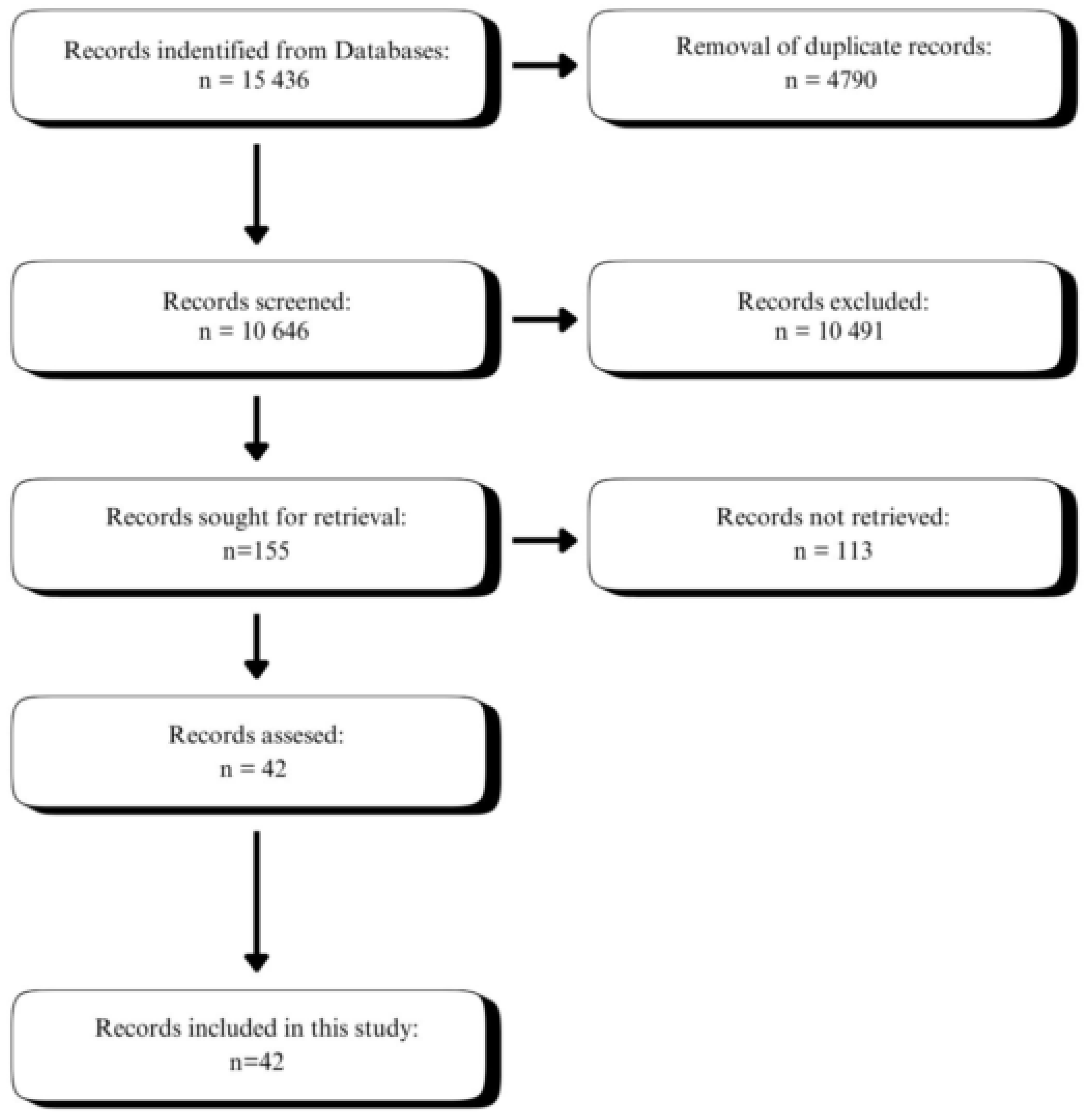
2.1. Literature Search

Our search focused on the impact of GLP-1 agonists and analogues on the gut microbiome of both animals and humans, including those healthy and with accompanying conditions like obesity, polycystic ovarian syndrome, prediabetes, or diabetes. Our systematic search was conducted of PubMed and SCOPUS databases on 7 December 2024. The search strategy was shown in Figure 1. Initially, we searched the databases with the GLP-1 analogues, GLP-1 agonists, and GLP-1/GIP analogue. These keywords were then associated with microbiome, microbiota, bacteria, prebiotic, and probiotic. Additionally, we manually inspected the reference list to check for any publications of interest. We focused our search on observational studies and clinical trials, which is recommended for systematic reviews.

2.2. Study Selection Criteria

We followed the preferred reporting items for systematic reviews and meta-analyses (PRISMA) guidelines to ensure that our review was transparent as well as reliable [12]. We included only original studies that provide data on gut microbiota after GLP-1 analogue treatment, where the microbiota was analyzed using fecal samples. Additionally, we focused our search only on publications written in English.

2.3. Study Selection

We screened the abstracts of all articles to determine their eligibility. We have rejected all studies that showed the impact of the microbiome on the treatment outcome and not the impact of GLP-1 analogues on microbiota. Furthermore, we rejected the studies that did not state which type of GLP-1 analogue was used. With the help of an experienced librarian, we conducted a search of the Medline (from 2004 to 2025) and Cochrane Library (from 2004 to 2025) databases using terms such as GLP-1, combined with the term “microbiota”. In the next step, we included only records written in English and related to humans and mice and rats. In the last step, we excluded studies from the basic sciences or those containing duplicate data. The entire literature search procedure is detailed in Figure 1. The relevance and eligibility of retrieved records were considered by two authors independently. All discrepancies were resolved by consensus between the authors.

2.4. Data Collection Process

The data collection process was carried out using the Microsoft Excel program. This process was carried out by one author, while another reviewed it for reliability. The file included data about study characteristics, treatment, and participant information, and microbiome shift outcomes. All discrepancies were resolved by consensus between the authors.

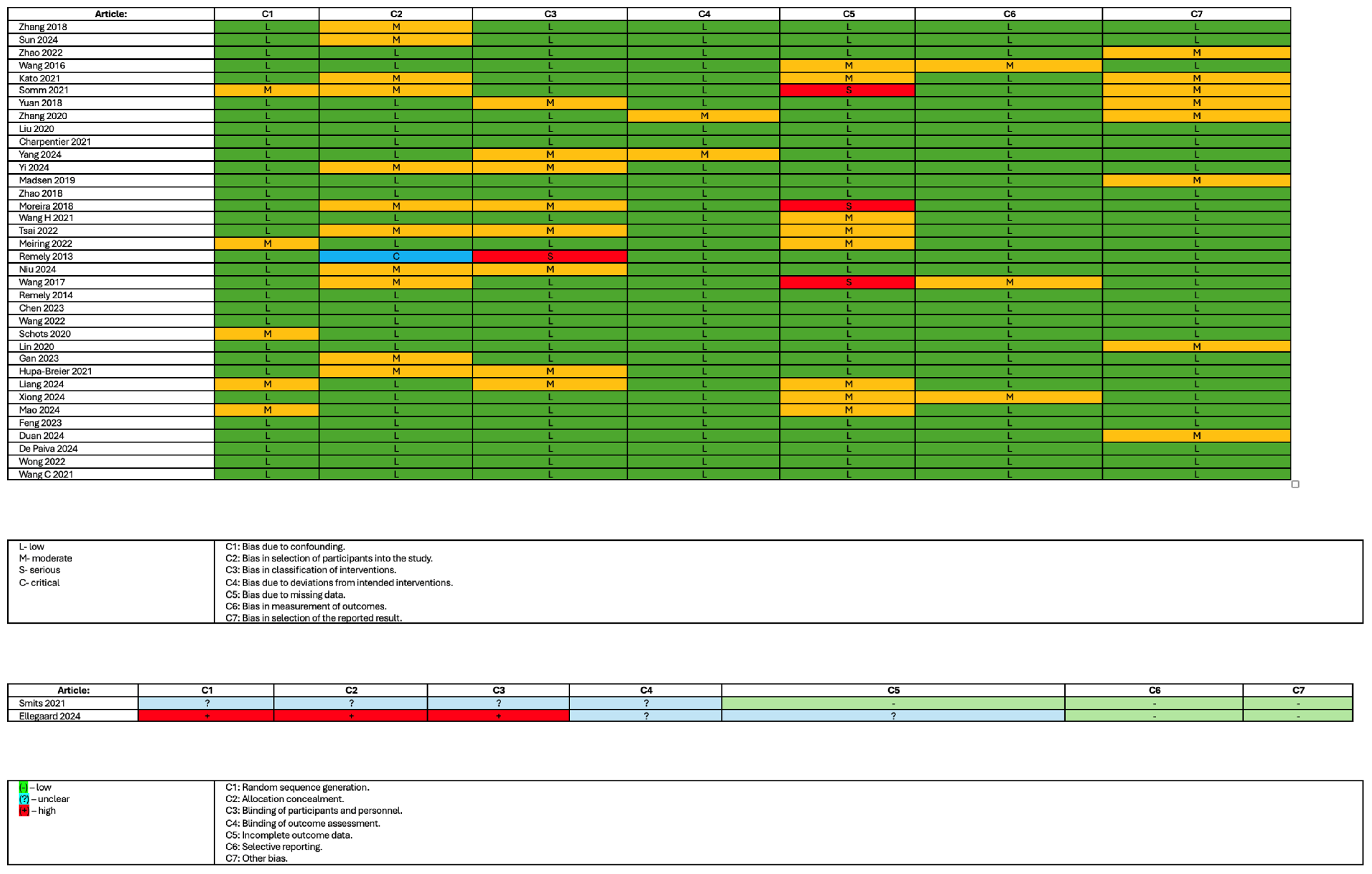
2.5. Study Risk of Bias Assessment

Risk of bias was evaluated by the usage of Robins-I [13] and RoB2 [14] tools. This process was carried out by one author, while another reviewed it for reliability. All discrepancies were resolved by consensus between the authors.

2.6. Assessment Outcomes

Our primary goal in this systematic review was to investigate and analyze any changes at the phylum, genus, and species levels due to GLP-1 analogue administration. Furthermore, our secondary goal was to determine any variations in microbial richness and diversity due to GLP-1 agonist usage.

3. Results

3.1. Reviewed Studies

After screening, 155 records were identified, and after evaluation, we have retrieved 38 studies. In the final analysis, 9 human and 29 animal studies were included (Figure 1). Most of the studies included were performed in or after 2021. The studies varied in participants and used drugs as well as treatment duration. Out of 38 included studies, 28 were experimental studies (73.5%), 6 were longitudinal studies (15.5%), 2 were randomized trials (5%), 1 was a non-randomized trial (3%), and 1 was a spin-off study (3%).

3.2. Methods of Analysis for Gut Microbiota Composition in Studies

In this systematic review we have found that in all of the included studies fecal samples were analyzed to assess the microbial composition. 16S rRNA gene sequencing was the most commonly chosen method by researchers. Among various 16S rRNA gene sequencing methods, the V3–V4 regions were dominant, as shown in Table 1.

3.3. The Risk of Bias

In the studies included in this systematic review, one study had a high risk of bias in random sequence generation, allocation concealment, and blinding of participants and personnel while having an unclear risk of bias in blinding of outcome assessment and incomplete outcome data. One study had shown an unclear risk in four domains. Five studies had shown a serious risk of bias concern in one domain each, with one showing a crucial risk of bias in the selection of participants into the study with low or moderate risk in other domains. The risk of bias assessment is shown in Figure 2.

3.4. Changes in Bacterial Composition Resulting from GLP-1 Analogue Treatment

3.4.1. Changes in Bacterial Genera and Species Induced by Liraglutide Treatment in the Animal Models

Among the 38 chosen studies, 27 focused on liraglutide. Of these, 8 were performed on human subjects, while 19 were carried out on animals (mice and rats). Animal studies clearly indicated that liraglutide influences the microbiome both on the phylum and genus, and at the species level as presented in Table 2.
The Bacteroidota phylum exhibited an increase in Alistipes [15,16] and Butyricimonas [17,18], both in two studies. The genus Bacteroides increased in five studies [16,19,20,21,22] and decreased in three [18,23,24]. Norank_F_Bacteroidales_S24-7_Group [23] and Barnesiella [24] both showed an increase in one study, while Parabacteroides increased in two studies [15,17] and decreased in one [23]. Prevotella_9 [15,18] decreased in two studies.
In the Bacillota phylum, Lactobacillus increased in six studies with no reported decrease [16,17,18,22,24,25]. Allobaculum increased in four studies [17,18,20,22], and one study has reported its decrease [26]. Clostridium showed an increase in three studies [21,26,27], while two studies showed a decrease in its levels [18,27]. Oscillospira [17,26], Blautia [18,22], Ruminococcus [16,25], and Turicibacter [16,18] increased in two studies, while Ruminococcus [15,16,19,28] and Turicibacter [20] decreased in four and one, respectively. Staphylococcus [16], Faecalibaculum [16], Candidatus Arthromitus [18], and Marvinbryantia [18] decreased in one, while Anaerotruncus [23,27] and Roseburia [18,23] decreased in two. Flavonifractor [15,24] and Lachnospiraceae_UCG-010 [15,23] both increased and decreased in one study. Lachnoclostridium [15], Cellulosilyticum [15], Peptoniphilus [15], and Anaerostipes [18] increased in one study while Christensenellaceae_R-7_group [15], Oscillibacter [23], Ruminoclostridium_6 [15], and Ruminiclostridium_9 [23] decreased in one. Romboutsia [23], Sarcina [26], SMB53 [26], and 02d06 [26] all increased once, while uncultured_f__Peptococcaceae [23] and Lachnospiraceae_NK4A136_group [23] decreased once.
In the Pseudomonadota phylum, Desulfovibrio abundance increased in two studies [15,18] and decreased in one [23], while Helicobacter was reported to increase in one study [16] and decrease in two [25,29]. The genera Escherichia and Shigella both increased in one study [24], while Desulfobacterota [25] and Klebsiella [24] decreased in one. Sutterella [17], Oxalobacter [15], and Sphingomonas [15] all increased in one study.
In the Actinomycetota phylum, Bifidobacteria increased in two studies [15,21]. Enterorhabdus [15], Gardnerella [15], and Johnsonella [29] increased, and Candidatus Saccharimonas [23] decreased in one study.
In the Verrumicrobiota phylum, Akkermansia [16,23,27,29,30] increased in five studies, and none of the studies reported its decrease after liraglutide treatment in animals.
Finally, in the Fusobacteria phylum, one study reported an increase in the genus Sneathia [15].
At the species level in the Bacillota phylum, Lactobacillus reuteri [16], Lactobacillus johnsonii [16], Anaerotruncus sp. G3 [27], Anaerofustis_stercorihominis [15], Lactobacillus_mucosae [15], Ruminococcus_gravus [15], and Flavonifractor_plautii [15] each increased in one study. In the Pseudomonadota phylum, increases were reported for E. coli [19], Helicobacter typhi [16], Burkholderiales bacterium YL45 [27], Brevundimonas vesicularis [15], and Moraxella_osloensis [15], each in a single study. In the Bacteroidota phylum, Bacteroides_acidifaciens increased in one study [15]. Akkermansia muciniphila, representing the phylum Verrucomicrobiota, increased in 4 studies [17,27,29,30].

3.4.2. Changes in Bacterial Genera and Species Induced by Liraglutide Treatment in Humans

Eight studies addressed the topic of gut microbiota in humans undergoing liraglutide treatment. Five studies reported no alterations in gut microbiota genera or species after treatment [31,32,33,34,35]. The results are shown in Table 3.
In the Bacillota phylum, Lactococcus [36] and an unknown genus in the family Christensenellaceae [37] were observed to increase in one study. Conversely, Blautia [36], Dialister [36], and Megasphaera [36] were shown to decrease in one study.
In the Bacteroidota phylum, only the genus Alistipes [38] was reported to decrease in one study. In the Pseudomonadota phylum, one study reported a decrease in Sutterella level [37]. In the Verrucomicrobiota phylum, Akkermansia increased in one study [37].
At the species level, Faecalibacterium prausnitzii [38] and Peptostreptococcus anaerobius [38], both belonging to the Bacillota phylum, increased in one study each. Bacteroides vulgatus [38], representing the Bacteroidota phylum, and Akkermansia muciniphila [38], representing the Verrucomicrobiota phylum, were both shown to increase in one study.

3.4.3. Changes in Bacterial Genera and Species Induced by Exenatide or Exendin-4 in the Animal Model

Eight studies examined the effect of exenatide or exendin-4 on the composition of gut microbiota. Five studies investigated this impact using animal models. The results of our assessment are summarized in Table 4.
In the Bacteroidota phylum, a decrease in Bacteroides [24] was observed in one study, whereas increases in Barnesiella [24] and Odoribacter [24] levels were reported in one study each. In the Verrucomicrobiota phylum, an increase in Akkermansia was documented in one study [39]. Within the Actinomycetota phylum, Enterorhabdus [39] decreased in one study.
In the Bacillota phylum, Streptococcus [39,41] decreased across two studies. Additionally, Romboutsia [39], Weissella [39], Marvinbryantia [39], Enterococcus [39], Flavonifractor [24], and Lactococcus [41] each decreased in one study. Ruminococcus increased in one study [41]. Lactobacillus increased [24] in one and decreased in another [41].
At the species level in the Bacillota phylum, Lactobacillus distasonis, Lactobacillus intestinalis, and Lactobacillus reuteri increases were documented in one study [42]. Moreover, the Bacteroidota phylum showed an increase in Alistipes finegoldii, Bacteroides acidifaciens, Bacteroides caccae, Parabacteroides distasonis, Parabacteroides goldsteinii, and Parabacteroides merdae, all in one study [42]. Finally, in the Verrucomicrobiota phylum, an increase in Akkermansia muciniphila was noted in one study [42].

3.4.4. Changes in Bacterial Genera and Species Induced by Exenatide in Humans

Three studies reported changes over the exenatide treatment in the microbiome. The results are summarized in Table 5.
In the Bacillota phylum, Coprococcus increased in one study [43] while Anaerotignum, Lactococcus, Flavonifractor, and Oscillospira decreased in the same study [43]. In the Bacteroidota phylum, it was reported that only Prevotella increased once [43]. In the Actinomycetota phylum, Bifidobacterium increased in one study [43].
At the species level, an increase in Bacteroides uniformis [43] and Phocaeicola vulgatus [43], belonging to the Bacteroidota phylum, has been seen in one study each. In the Bacillota phylum, Hungatella hathewayi, Lachnospira eligens, Roseburia hominis, and Roseburia intestinalis increased in one study [43]. In the Verrucomicrobiota phylum, Akkermansia muciniphila increased in one study [38].

3.4.5. Changes in Bacterial Genera and Species Induced by Dulaglutide Treatment in an Animal Model

Only one study reported changes in the intestinal microbiota over dulaglutide treatment in animals. This study outcome is shown in Table 6.
In the Bacillota phylum, dulaglutide treatment was associated with an increase in Aerococcus, Clostridium sensu stricto, Clostridium XlVb, and Enterococcus in one study. Moreover, increases in Ruminococcus, Pseudoflavonifractor, Lachnospiracea_ incertae_sedis, and Oscillibacter were reported while Coprobacillus, Macrococcus, Roseburia, and Streptococcus showed decreases, all in one study [44].
After Dulaglutide treatment, it was reported that in the Bacteroidota phylum, Bacteroides, Parabacteroides, Barnesiella, and Alistipes all increased in one study [44].
In the Pseudomonadota phylum, increases in Escherichia, Shigella, and the genus Parasutterella were observed in one study [44]. In the Verrucomicrobiota phylum, Akkermansia also increased in one study [44].
At the species level, this study did not report any changes.

3.4.6. Changes in Bacterial Genera and Species Induced by Dulaglutide Treatment in Humans

Only one study showed changes in the intestinal microbiota over dulaglutide treatment in humans. This study outcome is shown in Table 7.
In the Bacteroidota phylum, Bacteroides was noted to increase while Prevotella both increased and decreased over the study period [45].
In the Bacillota phylum, it was reported that Lactobacillus increased while Blautia and Ruminococcus showed a decrease [45].
Within the Actinomycetota phylum, Bifidobacterium both increased and decreased over the study period [45].
At the species level, the study did not report any changes.

3.4.7. Changes in Bacterial Genera and Species Induced by Semaglutide Treatment in the Animal Model

Six studies reporting changes in the gut microbiota after semaglutide treatment were included in this review. We found no studies researching the impact of semaglutide treatment on the gut microbiome in humans. The outcome of our assessment is shown in Table 8.
In the Bacteroidota phylum, Bacteroides [46], Alistipes [47], and Alloprevotella [47] increased in one study while Odoribacter decreased to the same extent [48].
In the Bacillota phylum, Allobaculum [49], Blautia [50], Enterococcus [51], and Clostridia _UCG_014 [48] were demonstrated to increase in one study each, while Ligilactobacillus [47], Lachnospiraceae_NK4A136_group [49], and unclassified_f_Oscillospiraceae [49] showed a decrease in one study. Dubosiella increased in two studies [49,51] and decreased in one [48], while Lactobacillus increased [48] and decreased in one study [47]. The Romboutsia genus was reported to be decreased in two studies [48,51].
In the Pseudomonadota phylum, it was noted that Escherichia [51], Shigella [51], and Helicobacter [46] were shown to increase in one study.
In the Verrucomicrobiota phylum, Akkermansia was observed to increase in two studies [48,49].
In the Actinomycetota phylum, Coriobacteriaceae UCG-002 was reported to show an increase in two studies [48,51].
At the species level in the Bacteroidota phylum, Alistipes muris [46] increased and Muribaculaceae bacterium Isolate-037 [46] decreased in one study each. Bacteroides acidifaciens increased in one study and decreased in another [46,49]. In the Bacillota phylum, Blautia coccoides [50] increased and Lepagella muris [46] decreased in one study.

3.5. Effects of GLP-1 Analogues on Gut Microbiota Diversity

3.5.1. Effects of Liraglutide Treatment on Gut Microbiota Diversity

In high-fat diet-fed Wang C et al. [52] reported an increase in microbiota diversity by ACE, Chao, Simpson, Shannon, and Sobs indices. All of the indices, except the Simpson index, which remained stable, have shown an increase after both low and high dose liraglutide treatment (200 and 400 µg/kg/d) when compared to HFD-fed mice without treatment. This showed an increase in microbial diversity with liraglutide treatment, especially observed with low-dose liraglutide application. Moreover, Zhao L et al. [17] also reported that Chao1, Shannon’s, and Simpson’s indices increased with liraglutide usage when compared to HFD-fed mice. Furthermore, the Sobs index used by Moreira GV et al. [29] to investigate the changes in phylogenetic microbial diversity showed an increase after liraglutide treatment. Madsen MSA et al. [27] stated that the richness in microbiota tended to increase after liraglutide usage, but it was not statistically significant. Finally, Charpentier J et al. [24] reported no statistically significant changes in Shannon’s index, indicating no change in microbial diversity.
When examining streptozotocin-induced diabetic mice, Yang Q et al. [25] reported no changes in the diversity of the gut microbiota after checking the Chao1 index. Conversely, Liu Q et al. [23] declared a significant decrease in alpha diversity even though the Sobs index was not affected in diabetic mice. Wang L et al. [18] presented a decrease in diversity based on Shannon’s and Simpson’s indices in normoglycemic mice, while in hyperglycemic mice, it was restored to a normal level after treatment.
In other mouse studies, Somm E et al. [20] did not report any changes in microbial diversity in MCD-fed mice. In dehydroepiandrosterone-induced polycystic ovary syndrome (PCOS) mice, Xiong C et al. [46] reported no change in beta diversity evaluated with principal coordinate analysis (PCoA). Wang H et al. [30] in the study with lipid-induced hepatic stenosis mice reported that Chao1 and Shannon’s indices increased and did not show significant change, respectively.
Three studies investigated diabetic rats. Zhang Q et al. [15] stated a decreased alpha diversity using Chao1 and Shannon’s indices and lowered beta diversity, evaluated by PCoA. A decrease in total bacterial number was also shown by Yuan X et al. [21] with 16S-based PCR and DEEP analysis. 16S rRNA V3–V4 region sequencing performed by Zhao L et al. [28] showed a significant decrease in Chao and Shannon indices, indicating a lowered microbiota richness.
In HFD-fed rats, Yi B et al. [26] found no change in Chao1 and ACE indices; however, they found a change in the Simpson index, which was upregulated with treatment. Zhang N et al. [22] reported no changes in diversity with Simpson and Shannon indices, while they showed a decrease in microbial richness with Chao1 and ACE indices.
Out of eight human studies investigating the impact of GLP-1 analogues, seven focus on patients with type 2 diabetes mellitus. Remely M et al. [34] found no differences in microbiome diversity in T2DM over the 4-month intervention. Meiring S et al. [33] reported no difference in either alpha or beta diversity of T2DM participants over a 3-month study period. Same results about alpha and beta diversities were reported by Smits MM et al. [31] with Shannon’s index, PCoA, and multilevel principal component analysis (PCA). To the same conclusion about alpha diversity came Tsai CY et al. [32]; on the other hand, they found statistically significant changes in beta diversity when comparing GLP-1 agonist responders and non-responders with PCoA. In the combined treatment of liraglutide and metformin, over the 42-day study period, Wang Z et al. [37] found no diversity differences to participants who used only metformin treatment. Moreover, Niu X et al. [36] tested Shannon, Simpson, and Chao1 indices between the control, metformin, and metformin + liraglutide groups and came to the conclusion that liraglutide partially improved microbiota diversity, seen by a change in Shannon’s and Simpson’s indices, but the Chao1 index still pointed to a higher diversity in the control group. Furthermore, although liraglutide lowered beta diversity, it changed it to be more similar to healthy individuals. Finally, Remely M et al. [38] stated no differences in microbial abundance after liraglutide intervention.
The last out of 8 human studies that looked into the impact of liraglutide treatment focused on patients with bile acid diarrhea, in which Ellegaard AM et al. [35] found no significant differences in microbial diversity with both Chao1 and Shannon’s indices.

3.5.2. Effects of Exenatide/Exentin-4 Treatment on Gut Microbiota Diversity

During the study conducted on high-fat diet (HFD)-fed mice, Charpentier J et al. [24], using a Shannon index of taxonomy, did not find any changes in microbial diversity. Similar results in HFD-fed mice have been obtained by Schots PC et al. [41]; they did not observe any statistically important changes in alpha diversity of the microbiome. Finally, Lin K et al. [42] also reported no difference in alpha diversity using abundance-based coverage estimator (ACE) but found an increase in beta diversity.
Chen Y et al. [39] investigated the impact of exenatide on gut microbiota in diabetic mice, using Shannon and Simpson indices. Neither of the indices showed any statistically significant difference while exenatide affected the beta diversity.
In patients with both obesity and polycystic ovarian syndrome (PCOS), Gan J et al. [43] announced that metformin plus exenatide therapy hugely impacted intestinal microbiota composition, which was investigated by PCoA.
Remely M et al. [34], in a study performed on humans with type 2 diabetes, reported no changes in microbial abundance.

3.5.3. Effects of Dulaglutide Treatment on Gut Microbiota Diversity

Hupa-Breier KL et al. [44] examined the non-diabetic mouse model of NASH. Both Shannon and Simpson indices were increased after dulaglutide treatment, indicating increased alpha diversity. They also reported an increase in beta diversity when compared to the mice treated with saline vehicle.
In type 2 diabetic humans, Liang L et al. [45] found no changes in microbiota diversity and abundance after 1 week of dulaglutide treatment but reported changes in Chao1 and ACE, whose levels were lower than before treatment, with stable Simpson and Shannon indices. This led to a conclusion that dulaglutide treatment impacts the abundance but not the diversity of microbiota.

3.5.4. Effects of Semaglutide Treatment on Gut Microbiota Diversity

In Type 2 diabetes mellitus mice, de Paiva IHR et al. [50] reported changes in beta diversity but also in species diversity and abundance. The same result regarding beta diversity using PCoA was received by Mao T et al. [47]. Moreover, they obtained an increase in alpha diversity after semaglutide treatment using 16S rRNA sequencing.
Duan X et al. [49] reported changes in Shannon, Chao, ACE, and Sobs indices in HFD-fed mice. In this study, semaglutide upregulated the microbial diversity. On the other hand, it did not affect the Simpson index. On the contrary, Feng J et al. [48], after examining Chao1, Shannon, Simpson, and ACE indices, found an increase only in the ACE index. They also reported a significant change in bacterial diversity with PCoA plots.
Finally, Xiong C et al. [46] tested the Dehydroepiandrosterone-Induced Polycystic Ovary Syndrome Mice. Both ACE and Shannon indices were decreased after semaglutide treatment, indicating a downregulation of semaglutide on microbiota richness and diversity.

3.6. Effects of GLP-1 Receptor Agonists on Metabolic Syndrome

Semaglutide, liraglutide, exenatide, or dulaglutide, which belong to the GLP-1 receptor agonists, are broadly used in type II diabetes mellitus and obesity treatment. They treat these illnesses by increasing insulin secretion as a result of food intake. Moreover, they suppress the glucagon release and gastric emptying, which finally leads to better glycemic control and body mass reduction. More and more studies suggest that GLP-1 agonists effectiveness in mitigating metabolic syndrome can be a result of microbiome transformation. It has been shown that beneficial bacteria such as Akkermansia muciniphila or Bacteroides, associated with better metabolic homeostasis, have thrived after GLP-1 receptor agonist administration. They also negatively correlate with the inflammatory state of the intestines. Furthermore, microbial modulation by GLP-1 agonists can result in an increase in short chain fatty acids. Butyrate, propionate, and acetate improve insulin sensitivity, which is crucial in glucose metabolism. GLP-1RAs can also reduce the abundance of Bacillota highly associated with obesity and insulin resistance. Apart from Bacillota, they can also reduce lipopolysaccharide-producing bacteria, resulting in a lower level of LPS, which can explain the positive effect of GLP-1RAs in metabolic syndrome. Decreased inflammatory state can also protect pancreatic β-cells. Moreover, it can be beneficial in inflammatory bowel diseases such as Crohn’s disease or ulcerative colitis.

4. Discussion

GLP-1 was discovered in 1984 by Svetlana Mosjov. The first GLP-1 agonist (exenatide) was approved in 2005, and the popularity of this group of drugs has only grown ever since. Thanks to better glycemic control, weight loss, and cardiovascular benefits, GLP-1 agonists are now considered to be a standard in T2D therapy. It is known that the microbiome plays a vital role in human health [53,54,55]. Therefore, we performed a systematic review to analyze the current knowledge on the impact of GLP-1 analogues on the intestinal microbiome.
Our findings of this analysis supply precious insights into the interplay between pharmacological interventions and gut microbiota. We provide insights into the impact of GLP-1 analogues on the microbiome diversity and composition. The complexity of the gut microbiome is highlighted by the differences across studies, which vary in treatment methods, bacterial taxa, as well as conditions of the experiments.
Liraglutide, which was the most studied GLP-1 receptor agonist, treatment changes showed significant alternations in microbiome composition. We showed increased abundances of Alistipes and Butyricimonas genera from the Bacteroidota phylum. Moreover, in the Bacillota phylum, Lactobacillus and Allobaculum increased. From these results we can conclude that liraglutide administration promotes the growth of genera relevant to beneficial metabolic functions. It is worth noticing that the species Akkermansia muciniphila increased, as aligned with improved intestinal barrier integrity and metabolic health [10]. The change towards a healthier microbiome profile is also shown by a shift in Ruminococcus and Turicibacter, known for their involvement in intestinal inflammation [56,57].
Exenatide and exendin-4 administration showed various effects on the microbiome composition in animal and human studies. In animal studies it was reported that Akkermansia, Barnesiella, and Ruminococcus genera all increased upon exenatide or exendin-4 treatment, while the genera associated with dysbiosis, such as Streptococcus and Marvinbryantia, decreased [58,59]. Human studies obtained fewer changes. Coprococcus and Bifidobacterium genera, associated with improved metabolic and inflammatory profiles, increased. This may suggest a potentially differential impact of exenatide due to host-specific factors.
Dulaglutide effects were studied not as extensively as other GLP-1 analogues; however, they showed a promising trend. In animal studies it was reported that dulaglutide was significantly associated with increases in Bacteroides, Akkermansia, and Ruminococcus, genera connected to an improved metabolic model. Changes in humans after dulaglutide treatment demonstrated limited changes. Worth noticing was an increase in Lactobacillus genera. This suggests a potentially positive impact of dulaglutide on the gut microbiome.
Semaglutide demonstrated significant alterations in the microbiome in animals. An increase in Alistipes, Alloprevotella, and Akkermansia genera could not go without notice, as they associated with a healthier metabolic model. An increase in A. muciphila known for its positive metabolic effects on human health support semaglutide’s potential as a therapeutic. On the other hand, some studies showed results suggesting decreased diversity indices. This may indicate that semaglutide’s effects vary over the metabolic state, diet and concomitant diseases.
Many studies reported the impact of microbiota on inflammation and insulin resistance [60,61]. The Akkermansia genus was increased after all GLP-1 analogue administrations. The genus includes the species Akkermansia muciniphila, which is associated with lower cardiovascular risk, improved weight management, and intestinal health [62]. Moreover, the Faecalibacterium and Lactobacillus genus were increased in several studies, and they contain several species with a positive impact on human health. We want to highlight the Lactobacillus reuteri species, which was elevated after liraglutide and exenatide administration. L. reuteri positively impacts GLP-1 secretion, intestinal barrier, and anti-inflammatory functions [63,64,65]. Furthermore, Faecalibacterium prausnitzii levels were also increased after liraglutide treatment. It is a known butyrate producer with anti-inflammatory effects [66]. This leads to a conclusion that microbial changes may impact T2D pathogenesis, as well as treatment outcomes.
Accumulating evidence indicates that glucagon-like peptide-1 receptor agonists (GLP-1RAs) may induce compositional shifts in the gut microbiota, though significant heterogeneity across studies complicates interpretation. Variability in study design (e.g., randomized controlled trials vs. observational cohorts), intervention duration (8–52 weeks), and analytical methodologies (16S rRNA sequencing vs. metagenomic sequencing) contributes to conflicting reports, such as inconsistent increases in Akkermansia muciniphila or ambiguous changes in microbial alpha diversity. Furthermore, confounding variables—including dietary intake, concomitant antidiabetic medications (e.g., metformin), and lifestyle factors—are frequently unaccounted for, obscuring the direct contribution of GLP-1RAs to observed microbiota alterations.
Preclinical models propose mechanistic pathways linking GLP-1RA-induced microbiota changes to metabolic improvements, such as enhanced short-chain fatty acid production or attenuation of endotoxemia. However, human data remain largely associative, with correlations between specific microbial taxa (e.g., Roseburia, Faecalibacterium) and metabolic parameters (e.g., HbA1c reduction, weight loss) failing to establish causality. The predominance of short-term, uncontrolled studies further limits insight into whether microbial shifts persist or drive clinically meaningful outcomes.
To address these gaps, future investigations should employ longitudinal cohort studies with standardized microbiota profiling, rigorous adjustment for confounders, and integration of multi-omics approaches (metagenomics, metabolomics) to delineate host-microbe interactions. Until such evidence is available, assertions of clinically significant microbiota modulation by GLP-1RAs should be circumspect, acknowledging that observed changes may represent secondary effects of metabolic improvement rather than primary therapeutic mechanisms. Current findings underscore the need for cautious interpretation, framing gut microbiota modulation as a plausible but unproven contributor to the pleiotropic effects of GLP-1RA therapy.
We need to highlight the importance of further studies, including human in vivo studies. Out of 38 studies, 9 investigated the impact of GLP-1 analogues in humans, while 29 were conducted on animals. The number of studies concerning liraglutide, exenatide or exentin-4, dulaglutide, and semaglutide in animals was nineteen, eight, one, and six, respectively. The results obtained in the animal model should be confirmed in further studies in humans. The number of studies regarding liraglutide, exenatide, and dulaglutide in humans was eight, three, and one, in order. To better understand the impact of GLP-1 receptor agonists on the microbiome and their application in T2D treatment, larger sample sizes are required.
Finally, in our systematic review we provide many examples of GLP-1 analogues’ impact on the microbiota. However, further studies on bacterial involvement in T2D are crucial to explain the impact of bacteria and to investigate potential therapeutics.

5. Limitations

The aim of this publication is to provide a comprehensive evaluation of the impact of GLP-1 agonists on the gut microbiome. However, we need to keep in mind various limitations. Firstly, the included studies showed a high diversity in the number and concomitant diseases of participants, the GLP-1 analogue dosage, sequencing methods, as well as the duration of drug administration. Moreover, the risk of bias assessment revealed varying levels across studies. While it was minimized in most studies, some still showed a higher risk of bias, which may have altered the overall conclusions. This analysis focused on changes at the bacterial genus and species levels, which is suboptimal, as it overlooks potential discrepancies at the strain level. Furthermore, many studies have had confounding factors that could have altered the results, such as the type of disease, treatment duration, and multi-drug therapy. Most of the studies were published in or after 2021, and the duration of GLP-1 usage was relatively short. This may hinder our ability to understand the long-term effects of using GLP-1 analogues. Finally, we need to underline that the bacterial taxonomy is very variable. This may lead to many inaccuracies. All of the above-said limitations could have altered obtained results, which limits the ability to acquire unambiguous conclusions.

6. Summary and Conclusions

In our systematic review, based on the PRISMA guidelines, we focused on gathering the knowledge of the effects of GLP-1 analogue medications on gut microbiota richness, composition, and abundance in both animals and humans. Our primary goal was to investigate the changes of bacteria following GLP-1 analogue administration on the phylum, genus, and species level. Moreover, as a secondary goal, we determined any alternations in richness and diversity. We can conclude, following the obtained research results of our study, that liraglutide promotes the growth of beneficial genera relevant for beneficial metabolic functions. Exenatide and exendin-4 administration showed various effects on the microbiome composition in animal and human studies. In animal models, it increased genera associated with improved metabolism; however, in human models, genera linked to better metabolic functions and escalated inflammation increased. Following dulaglutide administration, increases in Bacteroides, Akkermansia, and Ruminococcus, genera connected to an improved metabolic model, were significant. Finally, varied results were obtained after semaglutide treatment, in which A. muciniphila, known for its positive metabolic functions, increased; however, microbial diversity decreased.
The effect of GLP-1 analogues on the gut microbiota may have a positive impact on the long-term treatment of diabetes, preventing complications of this disease and maintaining a proper body weight. Further research is essential to confirm the above findings and to better recognize their implications for the clinical outcomes of patients.

Author Contributions

K.K.G. and S.M. conceptualized and designed the study. K.K.G. and A.W. conducted the literature search and data extraction. K.K.G. and A.W. analyzed and interpreted the data. K.K.G. and S.M. drafted the manuscript, and all authors provided critical feedback and revisions. During the preparation of this work, the authors used ChatGPT-4 in order to improve the language. After using this tool/service, the authors reviewed and edited the content as needed and take full responsibility for the content of the publication. All authors have read and agreed to the published version of the manuscript.

Funding

This study was funded by the Medical University of Gdańsk NIP 584-09-55-985.

Conflicts of Interest

The authors declare no conflicts of interest.

Abbreviations

GLP-1—glucagon-like peptide-1, GLP-1RAs—glucagon-like peptide-1 receptor agonists, PRISMA—preferred reporting items for systematic reviews and meta-analyses, T2D—type 2 diabetes, CKD—chronic kidney disease, HFD—high fat diet, NASH—non-alcoholic steatohepatitis.

References

  1. Li, S.; Vandvik, P.O.; Lytvyn, L.; Guyatt, G.H.; Palmer, S.C.; Rodriguez-Gutierrez, R.; Foroutan, F.; Agoritsas, T.; Siemieniuk, R.A.C.; Walsh, M.; et al. SGLT-2 inhibitors or GLP-1 receptor agonists for adults with type 2 diabetes: A clinical practice guideline. BMJ 2021, 373, n1091, Erratum in BMJ 2022, 377, o1080. https://doi.org/10.1136/bmj.o1080. [Google Scholar] [CrossRef] [PubMed]
  2. ElSayed, N.A.; Aleppo, G.; Aroda, V.R.; Bannuru, R.R.; Brown, F.M.; Bruemmer, D.; Collins, B.S.; Cusi, K.; Das, S.R.; Gibbons, C.H.; et al. Introduction and Methodology: Standards of Care in Diabetes-2023. Diabetes Care 2023, 46 (Suppl. 1), S1–S4. [Google Scholar] [CrossRef] [PubMed] [PubMed Central]
  3. International Hypoglycaemia Study Group. Hypoglycaemia, cardiovascular disease, and mortality in diabetes: Epidemiology, pathogenesis, and management. Lancet Diabetes Endocrinol. 2019, 7, 385–396, Erratum in Lancet Diabetes Endocrinol. 2019, 7, e18. https://doi.org/10.1016/S2213-8587(19)30139-1. [Google Scholar] [CrossRef] [PubMed]
  4. SAXENDA® (Liraglutide) Injection 3 mg Medication Guide. Available online: https://www.novo-pi.com/saxenda.pdf#guide (accessed on 5 April 2025).
  5. Wegovy® (Semaglutide) Injection 2.4 mg Medication Guide. Available online: https://www.novo-pi.com/wegovy.pdf#guide (accessed on 5 April 2025).
  6. Xu, D.; Nair, A.; Sigston, C.; Ho, C.; Li, J.; Yang, D.; Liao, X.; Chen, W.; Kuang, M.; Li, Y.; et al. Potential Roles of Glucagon-Like Peptide 1 Receptor Agonists (GLP-1 RAs) in Nondiabetic Populations. Cardiovasc. Ther. 2022, 2022, 6820377. [Google Scholar] [CrossRef] [PubMed] [PubMed Central]
  7. Morris, A.P.; Voight, B.F.; Teslovich, T.M.; Ferreira, T.; Segrè, A.V.; Steinthorsdottir, V.; Strawbridge, R.J.; Khan, H.; Grallert, H.; Mahajan, A.; et al. Large-scale association analysis provides insights into the genetic architecture and pathophysiology of type 2 diabetes. Nat. Genet. 2012, 44, 981–990. [Google Scholar]
  8. Dietrich, S.; Jacobs, S.; Zheng, J.S.; Meidtner, K.; Schwingshackl, L.; Schulze, M.B. Gene-lifestyle interaction on risk of type 2 diabetes: A systematic review. Obes. Rev. 2019, 20, 1557–1571. [Google Scholar] [CrossRef] [PubMed] [PubMed Central]
  9. Mehta, A.; Singh, S.; Saeed, A.; Mahtta, D.; Bittner, V.A.; Sperling, L.S.; Virani, S.S. Pathophysiological Mechanisms Underlying Excess Risk for Diabetes and Cardiovascular Disease in South Asians: The Perfect Storm. Curr. Diabetes Rev. 2021, 17, e070320183447. [Google Scholar] [CrossRef] [PubMed]
  10. Gurung, M.; Li, Z.; You, H.; Rodrigues, R.; Jump, D.B.; Morgun, A.; Shulzhenko, N. Role of gut microbiota in type 2 diabetes pathophysiology. EBioMedicine 2020, 51, 102590. [Google Scholar] [CrossRef] [PubMed] [PubMed Central]
  11. Petakh, P.; Kamyshna, I.; Kamyshnyi, A. Effects of metformin on the gut microbiota: A systematic review. Mol. Metab. 2023, 77, 101805. [Google Scholar] [CrossRef] [PubMed] [PubMed Central]
  12. Page, M.J.; McKenzie, J.E.; Bossuyt, P.M.; Boutron, I.; Hoffmann, T.C.; Mulrow, C.D.; Shamseer, L.; Tetzlaff, J.M.; Akl, E.A.; Brennan, S.E.; et al. The PRISMA 2020 statement: An updated guideline for reporting systematic reviews. BMJ 2021, 372, 71. [Google Scholar] [CrossRef] [PubMed] [PubMed Central]
  13. Sterne, J.A.; Hernán, M.A.; Reeves, B.C.; Savović, J.; Berkman, N.D.; Viswanathan, M.; Henry, D.; Altman, D.G.; Ansari, M.T.; Boutron, I.; et al. ROBINS-I: A tool for assessing risk of bias in non-randomised studies of interventions. BMJ 2016, 355, i4919. [Google Scholar] [CrossRef] [PubMed] [PubMed Central]
  14. Sterne, J.A.C.; Savović, J.; Page, M.J.; Elbers, R.G.; Blencowe, N.S.; Boutron, I.; Cates, C.J.; Cheng, H.Y.; Corbett, M.S.; Eldridge, S.M.; et al. RoB 2: A revised tool for assessing risk of bias in randomised trials. BMJ 2019, 366, l4898. [Google Scholar] [CrossRef] [PubMed]
  15. Zhang, Q.; Xiao, X.; Zheng, J.; Li, M.; Yu, M.; Ping, F.; Wang, T.; Wang, X. Featured article: Structure moderation of gut microbiota in liraglutide-treated diabetic male rats. Exp. Biol. Med. 2017, 243, 34–44. [Google Scholar] [CrossRef] [PubMed] [PubMed Central]
  16. Sun, H.; Shu, J.; Tang, J.; Li, Y.; Qiu, J.; Ding, Z.; Xuan, B.; Chen, M.; Gan, C.; Lin, J.; et al. GLP-1 receptor agonists alleviate colonic inflammation by modulating intestinal microbiota and the function of group 3 innate lymphoid cells. Immunology 2024, 172, 451–468. [Google Scholar] [CrossRef] [PubMed]
  17. Zhao, L.; Qiu, Y.; Zhang, P.; Wu, X.; Zhao, Z.; Deng, X.; Yang, L.; Wang, D.; Yuan, G. Gut microbiota mediates positive effects of liraglutide on dyslipidemia in mice fed a high-fat diet. Front. Nutr. 2022, 9, 1048693. [Google Scholar] [CrossRef] [PubMed] [PubMed Central]
  18. Wang, L.; Li, P.; Tang, Z.; Yan, X.; Feng, B. Structural modulation of the gut microbiota and the relationship with body weight: Compared evaluation of liraglutide and saxagliptin treatment. Sci. Rep. 2016, 6, 33251. [Google Scholar] [CrossRef] [PubMed] [PubMed Central]
  19. Kato, S.; Sato, T.; Fujita, H.; Kawatani, M.; Yamada, Y. Effects of GLP-1 receptor agonist on changes in the gut bacterium and the underlying mechanisms. Sci. Rep. 2021, 11, 9167. [Google Scholar] [CrossRef] [PubMed] [PubMed Central]
  20. Somm, E.; Montandon, S.A.; Loizides-Mangold, U.; Gaïa, N.; Lazarevic, V.; De Vito, C.; Perroud, E.; Bochaton-Piallat, M.L.; Dibner, C.; Schrenzel, J.; et al. The GLP-1R agonist liraglutide limits hepatic lipotoxicity and inflammatory response in mice fed a methionine-choline deficient diet. Transl. Res. 2021, 227, 75–88. [Google Scholar] [CrossRef] [PubMed]
  21. Yuan, X.; Ni, H.; Chen, X.; Feng, X.; Wu, Q.; Chen, J. Identification of therapeutic effect of glucagon-like peptide 1 in the treatment of STZ-induced diabetes mellitus in rats by restoring the balance of intestinal flora. J. Cell. Biochem. 2018, 119, 10067–10074. [Google Scholar] [CrossRef] [PubMed]
  22. Zhang, N.; Tao, J.; Gao, L.; Bi, Y.; Li, P.; Wang, H.; Zhu, D.; Feng, W. Liraglutide Attenuates Nonalcoholic Fatty Liver Disease by Modulating Gut Microbiota in Rats Administered a High-Fat Diet. BioMed Res. Int. 2020, 2020, 2947549. [Google Scholar] [CrossRef] [PubMed] [PubMed Central]
  23. Liu, Q.; Cai, B.Y.; Zhu, L.X.; Xin, X.; Wang, X.; An, Z.M.; Li, S.; Hu, Y.Y.; Feng, Q. Liraglutide modulates gut microbiome and attenuates nonalcoholic fatty liver in db/db mice. Life Sci. 2020, 261, 118457. [Google Scholar] [CrossRef] [PubMed]
  24. Charpentier, J.; Briand, F.; Lelouvier, B.; Servant, F.; Azalbert, V.; Puel, A.; Christensen, J.E.; Waget, A.; Branchereau, M.; Garret, C.; et al. Liraglutide targets the gut microbiota and the intestinal immune system to regulate insulin secretion. Acta Diabetol. 2021, 58, 881–897. [Google Scholar] [CrossRef] [PubMed]
  25. Yang, Q.; Deng, L.; Feng, C.; Wen, J. Comparing the effects of empagliflozin and liraglutide on lipid metabolism and intestinal microflora in diabetic mice. PeerJ 2024, 12, e17055. [Google Scholar] [CrossRef] [PubMed] [PubMed Central]
  26. Yi, B.; Su, K.; Cai, Y.L.; Chen, X.L.; Bao, Y.; Wen, Z.Y. Liraglutide ameliorates diabetic kidney disease by modulating gut microbiota and L-5-Oxoproline. Eur. J. Pharmacol. 2024, 983, 176905. [Google Scholar] [CrossRef] [PubMed]
  27. Madsen, M.S.A.; Holm, J.B.; Pallejà, A.; Wismann, P.; Fabricius, K.; Rigbolt, K.; Mikkelsen, M.; Sommer, M.; Jelsing, J.; Nielsen, H.B.; et al. Metabolic and gut microbiome changes following GLP-1 or dual GLP-1/GLP-2 receptor agonist treatment in diet-induced obese mice. Sci. Rep. 2019, 9, 15582. [Google Scholar] [CrossRef] [PubMed] [PubMed Central]
  28. Zhao, L.; Chen, Y.; Xia, F.; Abudukerimu, B.; Zhang, W.; Guo, Y.; Wang, N.; Lu, Y. A Glucagon-Like Peptide-1 Receptor Agonist Lowers Weight by Modulating the Structure of Gut Microbiota. Front. Endocrinol. 2018, 9, 233. [Google Scholar] [CrossRef] [PubMed] [PubMed Central]
  29. Moreira, G.V.; Azevedo, F.F.; Ribeiro, L.M.; Santos, A.; Guadagnini, D.; Gama, P.; Liberti, E.A.; Saad, M.; Carvalho, C. Liraglutide modulates gut microbiota and reduces NAFLD in obese mice. J. Nutr. Biochem. 2018, 62, 143–154. [Google Scholar] [CrossRef] [PubMed]
  30. Wang, H.; Wang, L.; Li, Y.; Luo, S.; Ye, J.; Lu, Z.; Li, X.; Lu, H. The HIF-2α/PPARα pathway is essential for liraglutide-alleviated, lipid-induced hepatic steatosis. Biomed. Pharmacother. 2021, 140, 111778. [Google Scholar] [CrossRef] [PubMed]
  31. Smits, M.M.; Fluitman, K.S.; Herrema, H.; Davids, M.; Kramer, M.H.H.; Groen, A.K.; Belzer, C.; de Vos, W.M.; Cahen, D.L.; Nieuwdorp, M.; et al. Liraglutide and sitagliptin have no effect on intestinal microbiota composition: A 12-week randomized placebo-controlled trial in adults with type 2 diabetes. Diabetes Metab. 2021, 47, 101223. [Google Scholar] [CrossRef] [PubMed]
  32. Tsai, C.Y.; Lu, H.C.; Chou, Y.H.; Liu, P.Y.; Chen, H.Y.; Huang, M.C.; Lin, C.H.; Tsai, C.N. Gut Microbial Signatures for Glycemic Responses of GLP-1 Receptor Agonists in Type 2 Diabetic Patients: A Pilot Study. Front. Endocrinol. 2022, 12, 814770. [Google Scholar] [CrossRef] [PubMed] [PubMed Central]
  33. Meiring, S.; van Baar, A.C.G.; Sørensen, N.; Holleman, F.; Soeters, M.R.; Nieuwdorp, M.; Bergman, J.J.G.H.M. A Changed Gut Microbiota Diversity Is Associated With Metabolic Improvements After Duodenal Mucosal Resurfacing With Glucagon-Like-Peptide-1 Receptor Agonist in Type 2 Diabetes in a Pilot Study. Front. Clin. Diabetes Healthc. 2022, 3, 856661. [Google Scholar] [CrossRef] [PubMed] [PubMed Central]
  34. Remely, M.; Aumueller, E.; Merold, C.; Dworzak, S.; Hippe, B.; Zanner, J.; Pointner, A.; Brath, H.; Haslberger, A.G. Effects of short chain fatty acid producing bacteria on epigenetic regulation of FFAR3 in type 2 diabetes and obesity. Gene 2014, 537, 85–92. [Google Scholar] [CrossRef] [PubMed]
  35. Ellegaard, A.M.; Kårhus, M.L.; Krych, L.; Sonne, D.P.; Forman, J.L.; Hansen, S.H.; Dragsted, L.O.; Nielsen, D.S.; Knop, F.K. Liraglutide and Colesevelam Change Serum and Fecal Bile Acid Levels in a Randomized Trial With Patients With Bile Acid Diarrhea. Clin. Transl. Gastroenterol. 2024, 15, e00772. [Google Scholar] [CrossRef] [PubMed] [PubMed Central]
  36. Niu, X.; Lu, P.; Huang, L.; Sun, Y.; Jin, M.; Liu, J.; Li, X. The effect of metformin combined with liraglutide on gut microbiota of Chinese patients with type 2 diabetes. Int. Microbiol. 2023, 27, 265–276. [Google Scholar] [CrossRef] [PubMed]
  37. Wang, Z.; Saha, S.; Van Horn, S.; Thomas, E.; Traini, C.; Sathe, G.; Rajpal, D.K.; Brown, J.R. Gut microbiome differences between metformin- and liraglutide-treated T2DM subjects. Endocrinol. Diabetes Metab. 2017, 1, e00009. [Google Scholar] [CrossRef] [PubMed] [PubMed Central]
  38. Remely, M.; Hippe, B.; Zanner, J.; Aumueller, E.; Brath, H.; Haslberger, A.G. Gut Microbiota of Obese, Type 2 Diabetic Individuals is Enriched in Faecalibacterium prausnitzii, Akkermansia muciniphila and Peptostreptococcus anaerobius after Weight Loss. Endocr. Metab. Immune Disord.-Drug Targets (Former. Curr. Drug Targets-Immune Endocr. Metab. Disord.) 2016, 16, 99–106. [Google Scholar] [CrossRef] [PubMed]
  39. Chen, Y.; Shu, A.; Jiang, M.; Jiang, J.; Du, Q.; Chen, T.; Shaw, C.; Chai, W.; Chao, T.; Li, X.; et al. Exenatide improves hypogonadism and attenuates inflammation in diabetic mice by modulating gut microbiota. Int. Immunopharmacol. 2023, 120, 110339. [Google Scholar] [CrossRef] [PubMed]
  40. Wang, X.L.; Chen, W.J.; Jin, R.; Xu, X.; Wei, J.; Huang, H.; Tang, Y.H.; Zou, C.W.; Chen, T.T. Engineered probiotics Clostridium butyricum-pMTL007-GLP-1 improves blood pressure via producing GLP-1 and modulating gut microbiota in spontaneous hypertension rat models. Microb. Biotechnol. 2022, 16, 799–812. [Google Scholar] [CrossRef] [PubMed] [PubMed Central]
  41. Schots, P.C.; Jansen, K.M.; Mrazek, J.; Pedersen, A.M.; Olsen, R.L.; Larsen, T.S. Obesity-induced alterations in the gut microbiome in female mice fed a high-fat diet are antagonized by dietary supplementation with a novel, wax ester-rich, marine oil. Nutr. Res. 2020, 83, 94–107. [Google Scholar] [CrossRef] [PubMed]
  42. Lin, K.; Dong, C.; Zhao, B.; Zhou, B.; Yang, L. Glucagon-like peptide-1 receptor agonist regulates fat browning by altering the gut microbiota and ceramide metabolism. Medcomm 2023, 4, e416. [Google Scholar] [CrossRef] [PubMed] [PubMed Central]
  43. Gan, J.; Chen, J.; Ma, R.L.; Deng, Y.; Ding, X.S.; Zhu, S.Y.; Sun, A.J. Metagenomics study on taxonomic and functional change of gut microbiota in patients with obesity with PCOS treated with exenatide combination with metformin or metformin alone. Gynecol. Endocrinol. 2023, 39, 2219342. [Google Scholar] [CrossRef] [PubMed]
  44. Hupa-Breier, K.L.; Dywicki, J.; Hartleben, B.; Wellhöner, F.; Heidrich, B.; Taubert, R.; Mederacke, Y.E.; Lieber, M.; Iordanidis, K.; Manns, M.P.; et al. Dulaglutide Alone and in Combination with Empagliflozin Attenuate Inflammatory Pathways and Microbiome Dysbiosis in a Non-Diabetic Mouse Model of NASH. Biomedicines 2021, 9, 353. [Google Scholar] [CrossRef] [PubMed] [PubMed Central]
  45. Liang, L.; Su, X.; Guan, Y.; Wu, B.; Zhang, X.; Nian, X. Correlation between intestinal flora and GLP-1 receptor agonist dulaglutide in type 2 diabetes mellitus treatment-A preliminary longitudinal study. iScience 2024, 27, 109784. [Google Scholar] [CrossRef] [PubMed] [PubMed Central]
  46. Xiong, C.; Wu, J.; Ma, Y.; Li, N.; Wang, X.; Li, Y.; Ding, X. Effects of Glucagon-Like Peptide-1 Receptor Agonists on Gut Microbiota in Dehydroepiandrosterone-Induced Polycystic Ovary Syndrome Mice: Compared Evaluation of Liraglutide and Semaglutide Intervention. Diabetes Metab. Syndr. Obes. Targets Ther. 2024, ume 17, 865–880. [Google Scholar] [CrossRef] [PubMed] [PubMed Central]
  47. Mao, T.; Zhang, C.; Yang, S.; Bi, Y.; Li, M.; Yu, J. Semaglutide alters gut microbiota and improves NAFLD in db/db mice. Biochem. Biophys. Res. Commun. 2024, 710, 149882. [Google Scholar] [CrossRef] [PubMed]
  48. Feng, J.; Teng, Z.; Yang, Y.; Liu, J.; Chen, S. Effects of semaglutide on gut microbiota, cognitive function and inflammation in obese mice. PeerJ 2024, 12, e17891. [Google Scholar] [CrossRef] [PubMed] [PubMed Central]
  49. Duan, X.; Zhang, L.; Liao, Y.; Lin, Z.; Guo, C.; Luo, S.; Wang, F.; Zou, Z.; Zeng, Z.; Chen, C.; et al. Semaglutide alleviates gut microbiota dysbiosis induced by a high-fat diet. Eur. J. Pharmacol. 2024, 969, 176440. [Google Scholar] [CrossRef] [PubMed]
  50. de Paiva, I.H.R.; da Silva, R.S.; Mendonça, I.P.; de Souza, J.R.B.; Peixoto, C.A. Semaglutide Attenuates Anxious and Depressive-Like Behaviors and Reverses the Cognitive Impairment in a Type 2 Diabetes Mellitus Mouse Model Via the Microbiota-Gut-Brain Axis. J. Neuroimmune Pharmacol. 2024, 19, 36. [Google Scholar] [CrossRef] [PubMed]
  51. Wong, C.K.; Yusta, B.; Koehler, J.A.; Baggio, L.L.; McLean, B.A.; Matthews, D.; Seeley, R.J.; Drucker, D.J. Divergent roles for the gut intraepithelial lymphocyte GLP-1R in control of metabolism, microbiota, and T cell-induced inflammation. Cell Metab. 2022, 34, 1514–1531.e7. [Google Scholar] [CrossRef] [PubMed]
  52. Wang, C.; Hu, H.J.; Dong, Q.Q.; Huang, R.; Zhao, W.; Song, Y.J.; Li, Z.Y.; Wang, N.; Zhang, T.C.; Luo, X.G. Enhancing bile tolerance of Lactobacilli is involved in the hypolipidemic effects of liraglutide. Biosci. Biotechnol. Biochem. 2021, 85, 1395–1404. [Google Scholar] [CrossRef] [PubMed]
  53. Asadifard, E.; Hokmabadi, M.; Hashemi, M.; Bereimipour, A. Linking gut microbiota dysbiosis to molecular pathways in Alzheimer’s disease. Brain Res. 2024, 1845, 149242. [Google Scholar] [CrossRef] [PubMed]
  54. Yang, C.; Camargo Tavares, L.; Lee, H.C.; Steele, J.R.; Ribeiro, R.V.; Beale, A.L.; Yiallourou, S.; Carrington, M.J.; Kaye, D.M.; Head, G.A.; et al. Faecal metaproteomics analysis reveals a high cardiovascular risk profile across healthy individuals and heart failure patients. Gut Microbes 2024, 17, 2441356. [Google Scholar] [CrossRef] [PubMed]
  55. Wang, W.; Wang, F.; Li, Y.; Shi, Y.; Wang, X.; Chen, X.; Zheng, W.; Hsing, J.C.; Lu, Y.; Wu, Y.S.; et al. Distinct Gut Microbiota Profiles in Normal Weight Obesity and Their Association With Cardiometabolic Diseases: Results From Two Independent Cohort Studies. J. Cachexia Sarcopenia Muscle 2024, 16, e13644. [Google Scholar] [CrossRef] [PubMed] [PubMed Central]
  56. Misera, A.; Kaczmarczyk, M.; Łoniewski, I.; Liśkiewicz, P.; Podsiadło, K.; Misiak, B.; Skonieczna-Żydecka, K.; Samochowiec, J. Comparative analysis of gut microbiota in major depressive disorder and schizophrenia during hospitalisation—The case-control, post hoc study. Psychoneuroendocrinology 2024, 171, 107208. [Google Scholar] [CrossRef] [PubMed]
  57. Bellando-Randone, S.; Russo, E.; Di Gloria, L.; Lepri, G.; Baldi, S.; Fioretto, B.S.; Romano, E.; Ghezzi, G.; Bertorello, S.; El Aoufy, K.; et al. Gut microbiota in very early systemic sclerosis: The first case-control taxonomic and functional characterisation highlighting an altered butyric acid profile. RMD Open 2024, 10, e004647. [Google Scholar] [CrossRef] [PubMed] [PubMed Central]
  58. Liu, Y.; Chen, Z.; Li, C.; Sun, T.; Luo, X.; Jiang, B.; Liu, M.; Wang, Q.; Li, T.; Cao, J.; et al. Associations between changes in the gut microbiota and liver cirrhosis: A systematic review and meta-analysis. BMC Gastroenterol. 2025, 25, 16. [Google Scholar] [CrossRef] [PubMed] [PubMed Central]
  59. Wu, J.; Ou, G.; Wang, S.; Chen, Y.; Xu, L.; Deng, L.; Xu, H.; Chen, X. The predictive, preventive, and personalized medicine of depression: Gut microbiota and inflammation. EPMA J. 2024, 15, 587–598. [Google Scholar] [CrossRef] [PubMed] [PubMed Central]
  60. Adeshirlarijaney, A.; Gewirtz, A.T. Considering gut microbiota in treatment of type 2 diabetes mellitus. Gut Microbes 2020, 11, 253–264. [Google Scholar] [CrossRef]
  61. Cheng, Y.; Lin, S.; Cao, Z.; Yu, R.; Fan, Y.; Chen, J. The role of chronic low-grade inflammation in the development of sarcopenia: Advances in molecular mechanisms. Int. Immunopharmacol. 2025, 147, 114056. [Google Scholar] [CrossRef] [PubMed]
  62. Gofron, K.; Berezowski, A.; Gofron, M.; Borówka, M.; Dziedzic, M.; Kazimierczak, W.; Kwiatkowski, M.; Gofron, M.; Nowaczyk, Z.; Małgorzewicz, S. Akkermansia muciniphila—Impact on the cardiovascular risk, the intestine inflammation and obesity. Acta Biochim. Pol. 2024, 71, 13550. [Google Scholar] [CrossRef] [PubMed] [PubMed Central]
  63. Wang, J.X.; Chang, S.Y.; Jin, Z.Y.; Li, D.; Zhu, J.; Luo, Z.B.; Han, S.Z.; Kang, J.D.; Quan, L.H. Lactobacillus reuteri-Enriched Eicosatrienoic Acid Regulates Glucose Homeostasis by Promoting GLP-1 Secretion to Protect Intestinal Barrier Integrity. J. Agric. Food Chem. 2024, 73, 393–408. [Google Scholar] [CrossRef] [PubMed]
  64. Bonab, S.F.; Tahmasebi, S.; Ghafouri-Fard, S.; Eslami, S. Preventive impact of probiotic supplements on heart injury and inflammatory indices in a rat model of myocardial infarction: Histopathological and gene expression evaluation. APMIS 2024, 133, e13479. [Google Scholar] [CrossRef] [PubMed]
  65. Chi, Y.Y.; Xiang, J.Y.; Li, H.M.; Shi, H.Y.; Ning, K.; Shi, C.; Xiang, H.; Xie, Q. Schisandra chinensis polysaccharide prevents alcohol-associated liver disease in mice by modulating the gut microbiota-tryptophan metabolism-AHR pathway axis. Int. J. Biol. Macromol. 2024, 282, 136843. [Google Scholar] [CrossRef] [PubMed]
  66. Miquel, S.; Martín, R.; Rossi, O.; Bermúdez-Humarán, L.G.; Chatel, J.M.; Sokol, H.; Thomas, M.; Wells, J.M.; Langella, P. Faecalibacterium prausnitzii and human intestinal health. Curr. Opin. Microbiol. 2013, 16, 255–261. [Google Scholar] [CrossRef] [PubMed]
Figure 1. Search strategy.
Nutrients 17 01303 g001
Figure 2. Risk of bias assessment [15,16,17,18,19,20,21,22,23,24,25,26,27,28,29,30,31,32,33,34,35,36,37,38,39,40,41,42,43,44,45,46,47,48,49,50,51,52].
Nutrients 17 01303 g002
Table 1. Description of the included studies.
No.ArticleType of StudyDescription of the Experiment and Study Groups/Description of the StudyMethod of AnalysisConclusion
1Zhang 2018 [15]ExperimentalDiabetic male rats16S rRNA V3-V4In liraglutide-treated diabetic male rats, short-chain fatty acid (SCFA)-producing bacteria, including Bacteroides, Lachnospiraceae, and the probiotic Bifidobacterium, were selectively enhanced. Lactobacillus showed a negative correlation with fasting blood glucose.
2Sun 2024 [16]ExperimentalWild-type and Rag1−/−, Rag2−/− i, Rorcgfp/gfp mice16S rRNA V3-V4Liraglutide administration significantly alleviates the DSS-induced colitis symptoms and ameliorates histological damage.
3Zhao 2022 [17]ExperimentalC57BL/6 mice16S rDNA V3-V4Correlation analysis showed that TC and LDL were linked to harmful bacteria and inversely related to beneficial ones. Liraglutide improves dyslipidemia in HFD-fed mice by modulating gut microbiota, especially Akkermansia, suggesting this as a key mechanism of its effect.
4Wang 2016 [18]ExperimentalApoE−/− mice on C57BL/6 background16s rDNA V1-V3The GLP-1 receptor agonist liraglutide modulates gut microbiota composition, promoting a lean-associated profile that aligns with its weight-loss effects.
5Kato 2021 [19]Experimental Mice16S/18S rRNAIncreased levels of caseinolytic protease B, a component of Escherichia coli, and norepinephrine were observed in the cecum.
6Somm 2021 [20]ExperimentalMice fed a regular diet or a methionine-choline deficient (MCD) diet.16S rDNA V3-V4Liraglutide altered gut microbiota composition affected by the MCD diet, restoring normal Bacteroides levels and shifting Erysipelotrichaceae from Allobaculum to Turicibacter.
7Yuan 2018 [21]ExperimentalSTZ-induced Sprague-Dawley rats 16S rRNA V3GLP-1, by restoring the intestinal flora dysbiosis, can moderate STZ-induced DM treatment.
8Zhang 2020 [22]ExperimentalMale rats fed a high-fat diet. After 12 weeks, two rats 16s rDNA V4-V5Liraglutide significantly modified the structure and composition of HFD-disrupted gut microbiota in comparison to rats on a normal diet.
9Liu 2020 [23]Experimental Male db/db mice16S rRNA V3-V4Bacteria like Parabacteroides, Oscillibacter, and Prevotellaceae_UCG-001 produce anti-inflammatory SCFAs and influence T cell differentiation. In the LG group, bacteria such as Anaerotruncus and Lachnospiraceae were reduced and correlated with ALT and AST. Oscillibacter and Klebsiella correlated with ALT, while Desulfovibrio and Bacteroides correlated with AST.
10Charpentier 2021 [24]ExperimentalDiet-induced dysmetabolic mice 16S rRNAData showed that GLP-1 receptor agonists, like liraglutide, regulate insulin secretion by simultaneously modulating intestinal immunity and bacterial ecology.
11Yang 2024 [25]ExperimentalMouse diabetic model16S rRNAEmpagliflozin reduced the Firmicutes/Bacteroidota ratio, while liraglutide had no effect. Both decreased Helicobacter and increased Lactobacillus. Empagliflozin also raised Muribaculaceae, Muribaculum, Olsenella, and Odoribacter, whereas liraglutide increased Ruminococcus.
12Yi 2024 [26]ExperimentalDiabetic kidney disease rats16S rRNA V3-V4The gut microbiota-metabolites-kidney axis, particularly Clostridium-5-OP-ELD, may serve as a potential target of Liraglutide in DKD.
13Madsen 2019 [27]ExperimentalDiet-induced obese mice.16S rRNA V3-V4DIO mice showed similar gut bacterial changes after liraglutide and GUB09-145 treatment, with shifts in low-abundant species and metabolic pathways.
14Zhao 2018 [28]ExperimentalWistar and Goto–Kakizaki (GK) rats16S rDNA V3-V4Liraglutide prevents weight gain by altering gut microbiota, reducing diversity, increasing the Firmicutes/Bacteroidetes ratio, and promoting lean-associated microbial profiles in obesity and T2DM.
15Moreira 2018 [29]ExperimentalC57BL/6J mice16S rRNA V3-V4Liraglutide modified gut microbiota by reducing Proteobacteria and increasing Akkermansia muciniphila, contributing to weight loss, microbiota balance, and NAFLD improvement.
16Wang H 2021 [30]ExperimentalHIF-2α heterozygous knockout (HIF-2α+/−) and wild-type (WT) littermate mice16S rRNA V3-V4HIF-2α is essential for the effect of liraglutide in promoting the increased abundance of Akkermansia muciniphila.
17Smits 2021 [31]Randomized trial Fifty-one adults (humans) with type 2 diabetes16S rRNA V3-V4Liraglutide and sitagliptin improve glucose metabolism, body weight, and bile acids as add-ons to metformin or sulphonylureas, independent of changes in intestinal microbiota.
18Tsai 2022 [32]Longitudinal studyFifty-two adults16S rRNABacteroides dorei, Lachnoclostridium sp., and Mitsuokella multacida were significant after adjusting for baseline glycohemoglobin and C-peptide concentrations, two clinical confounders.
19Meiring 2022 [33]spin-off studyFecal samples from 16 patients were collected for Illumina shotgun sequencing at baseline and three months post-DMR.shotgun sequencingGut microbiota diversity changes correlate with HbA1c, PDFF, and metabolic improvements after DMR and GLP-1 receptor agonist treatment in type 2 diabetes.
20Remely 2013 [34]Longitudinal studyFourteen obese individuals (OC) with no established insulin resistance and twenty-four insulin-dependent type 2 diabetes (D) patients16S rRNAGut microbiota in obesity and type 2 diabetes may influence FFAR epigenetics, affecting satiety and hunger. Butyrate supports regenerative medicine by promoting epigenetic remodeling and gene expression.
21Ellegaard 2024 [35]Randomized trial Fifty-two adults16S rRNANeither treatment resulted in changes to the composition of fecal microbiota.
22Niu 2024 [36]Longitudinal studyAll 32 participants in the pre-diabetic/overweight control group (NCP) later transitioned to the untreated T2DM group (UNT) upon diagnosis.16S rRNA V4MET and MET + LRG treatment significantly altered gut microbiota from T2DM diagnosis, with MET + LRG showing distinct changes, suggesting LRG’s additive effect.
23Wang 2017 [37]Longitudinal study 37 patients with T2DM16S rRNA V4The study suggests that altering the gut microbiome may play a key role in the mechanisms of action of metformin and liraglutide.
24Remely 2014 [38]Longitudinal studyT2DM patients16S rRNAGut microbiota composition varies between groups with GLP-1 analogue therapy and that with no therapy.
25Chen 2023 [39]ExperimentalC57BL/6J mice16S rDNA V3-V4Exenatide reduced pathogenic bacteria, increased Akkermansia, improved testosterone, and lowered inflammation. Fecal bacteria from the Exe group reduced harmful microbes and protected against testicular damage in diabetic mice.
26Wang 2022 [40]ExperimentalHypertensive rats (SHR). SHR and Wistar rats.16S rRNA V3-V4The treatments corrected dysbiosis in SHR rats by reducing Porphyromonadaceae and increasing Lactobacillus. GLP-1 improved blood pressure and cardiomegaly by restoring the gut microbiome and reducing ventricular hypertrophy.
27Schots 2020 [41]Experimental57bl/6J mice16s rDNA V4-V5Exenatide prevented the HFD-induced increase in Lactococcus and caused a decrease in the abundance of Streptococcus compared to the HFD
group.
28Lin 2020 [42]ExperimentalMice16S/18S rRNALactobacillus reuteri increased in bacterial counts, while ceramide levels in mouse serum decreased, with a negative correlation between L. reuteri and ceramide. The decrease in ceramide was linked to overexpression of alkaline ceramidase 2 (Acer2). L. reuteri may work synergistically with GLP-1 RA for therapeutic benefits.
29Gan 2023 [43]Non-randomized controlled trialObese patients with polycystic ovary syndrome (PCOS)Paired-end sequencingBoth groups had high levels of Firmicutes, Bacteroidetes, Uroviricota, Actinobacteria, and Proteobacteria. Post-treatment, probiotics like Phocaeicola and Anaerobutyricum increased. Clostridium, Fusobacterium, and Oxalobacter dominated in the post-MF group, while Lactococcus garvieae, Clostridium perfringens, and Coprococcus sp. AF16_5 were dominant in the post-COM group.
30Hupa-Breier 2021 [44]ExperimentalC57BL/6HanJ Ztm. micereal-time PCRThe study highlights dulaglutide’s significant anti-inflammatory effects, especially when combined with empagliflozin, by modulating pro-inflammatory immune responses and microbiome dysbiosis.
31Liang 2024 [45]Longitudinal study Patients with newly diagnosed T2DM with no GLP-1RA history.16S rRNANo significant changes in gut microbiota after 1 week of dulaglutide treatment in newly diagnosed T2DM patients. After 48 weeks, notable changes occurred, including a significant reduction in microbial abundance.
32Xiong 2024 [46]ExperimentalDehydroepiandrosterone (DHEA)-induced PCOS miceshotgun sequencingThe greater efficacy in weight loss compared with liraglutide observed after semaglutide intervention was positively related with Helicobacter.
33Mao 2024 [47]Experimentaldb/m and db/db mice16S rRNASignificant changes in Alloprevotella, Alistipes, Ligilactobacillus, and Lactobacillus were observed between the db/db control group and the semaglutide treatment group. LDA scores showed increased Alloprevotella and Alistipes, while Ligilactobacillus and Lactobacillus decreased after semaglutide treatment.
34Feng 2023 [48]ExperimentalC57BL/6J mice16S rRNA V3-V4Semaglutide may influence gut microbiota composition and structure, impacting cognitive function and inflammation.
35Duan 2024 [49]ExperimentalMice fed a high-fat diet16S rRNASemaglutide could alter gut microbiota dysbiosis, which may explain the anti-obesity effects of semaglutide.
36De Paiva 2024 [50]ExperimentalC57BL/6 mice16S rRNASemaglutide may treat depression and anxiety by reducing hippocampal neuroinflammation and promoting neurogenesis through the insulin/GLP-1 pathway and gut microbiota modulation.
37Wong 2022 [51]Experimental Rag2−/−;Il2rg−/− mice and Glp1rTcell−/− mice 16S rRNA V3-V4The intestinal intraepithelial lymphocyte GLP-1R is required for actions of GLP-1 on gut microbiota and for selective restraint of local and systemic T cell-induced, but not lipopolysaccharide-induced, inflammation.
38Wang C 2021 [52]ExperimentalMice with a high-fat diet16S rRNALiraglutide increases Lactobacillaceae abundance in hyperlipidemic mice, enhances LAB bile tolerance by upregulating bile salt hydrolases, and liraglutide-sensitive LAB lysates directly downregulate HMGCR.
Table 2. Impact of liraglutide treatment on gut microbiota in animals.
PhylumGenusIncreasedDecreased
BacteroidotaBacteroides53
BacteroidotaAlistipes20
BacteroidotaParabacteroides21
BacteroidotaButyricimonas20
BacteroidotaPrevotella_902
BacteroidotaNorank_F_Bacteroidales_S24-7_Group10
BacteroidotaBarnesiella10
BacillotaRuminicoccus24
BacillotaLactobacillus60
BacillotaTuricibacter21
BacillotaStaphylococcus01
BacillotaFaecalibaculum01
BacillotaAllobaculum41
BacillotaOscillospira20
BacillotaClostridium32
BacillotaAnaerotruncus02
BacillotaAnaerostipes10
BacillotaBlautia20
BacillotaCandidatus Arthromitus01
BacillotaRoseburia02
BacillotaMarvinbryantia01
BacillotaFlavonifractor11
BacillotaLachnoclostridium10
BacillotaCellulosilyticum10
BacillotaChristensenellaceae_R-7_group01
BacillotaLachnospiraceae_UCG-01011
BacillotaPeptoniphilus10
BacillotaRuminoclostridium_602
BacillotaRomboutsia10
BacillotaLachnospiraceae_NK4A136_group01
BacillotaOscillibacter01
BacillotaRuminiclostridium_901
Bacillotauncultured_f__Peptococcaceae01
BacillotaSarcina10
BacillotaSMB5310
Bacillota02d0610
VerrucomicrobiaAkkermansia50
PseudomonadotaHelicobacter12
PseudomonadotaDesulfobacterota01
PseudomonadotaSutterella10
PseudomonadotaDesulfovibrio21
PseudomonadotaOxalobacter10
PseudomonadotaSphingomonas10
PseudomonadotaKlebsiella01
PseudomonadotaEscherichia10
PseudomonadotaShigella10
ActinomycetotaBifidobacteria20
ActinomycetotaEnterorhabdus10
ActinomycetotaGardnerella10
ActinomycetotaJohnsonella10
ActinomycetotaCandidatus_Saccharimonas01
FusobacteriaSneathia10
Table 3. Impact of liraglutide treatment on gut microbiota in humans.
PhylumGenusIncreasedDecreased
BacteroidotaAlistipes01
BacillotaLactococcus10
BacillotaBlautia01
BacillotaDialister01
BacillotaMegasphaera01
Bacillotaunknown genus in the family Christensenellaceae10
VerrucomicrobiaAkkermansia10
PseudomonadotaSutterella01
Table 4. Impact of exenatide or exendin-4 treatment on gut microbiota in animals.
PhylumGenusIncreasedDecreased
BacteroidotaOdoribacter10
BacteroidotaBacteroides01
BacteroidotaBarnesiella10
BacillotaLactobacillus11
BacillotaRomboutsia01
BacillotaStreptococcus02
BacillotaWeissella01
BacillotaMarvinbryantia01
BacillotaEnterococcus01
BacillotaLactococcus01
BacillotaRuminococcus10
BacillotaFlavonifactor01
VerrucomicrobiaAkkermansia10
ActinomycetotaEnterorhabdus01
Table 5. Impact of exenatide treatment on gut microbiota in humans.
PhylumGenusIncreasedDecreased
ActinomycetotaBifidobacterium10
BacteroidotaPrevotella10
BacillotaOscillospira01
BacillotaFlavonifractor01
BacillotaLactococcus01
BacillotaAnaerotignum01
BacillotaCoprococcus10
Table 6. Impact of dulaglutide treatment on gut microbiota in animals.
PhylumGenusIncreasedDecreased
BacteroidotaAlistipes10
BacteroidotaBacteroides10
BacteroidotaBarnesiella10
BacteroidotaParabacteroides10
BacillotaAerococcus10
BacillotaClostridium sensu stricto10
BacillotaClostridium XlVb10
BacillotaCoprobacillus01
BacillotaEnterococcus10
BacillotaLachnospiracea_ incertae_sedis10
BacillotaMacrococcus01
BacillotaOscillibacter10
BacillotaPseudoflavonifractor10
BacillotaRoseburia01
BacillotaRuminococcus10
BacillotaStreptococcus01
PseudomonadotaEscherichia/Shigella10
PseudomonadotaParasutterella10
VerrucomicrobiaAkkermansia10
Table 7. Impact of dulaglutide treatment on gut microbiota in humans.
PhylumGenusIncreasedDecreased
BacteroidotaBacteroides10
BacteroidotaPrevotella11
BacillotaBlautia01
BacillotaLactobacillus10
BacillotaRuminococcus01
ActinomycetotaBifidobacterium11
Table 8. Impact of semaglutide treatment on gut microbiota in animals.
PhylumGenusIncreasedDecreased
BacteroidotaBacteriodes10
BacteroidotaOdoribacter01
BacteroidotaMuribaculaceae10
BacteroidotaAlloprevotella10
BacteroidotaAlistpes10
BacillotaBlautia10
BacillotaDubosiella21
BacillotaRomboutsia02
BacillotaEnterococcus10
BacillotaLactobacillus11
BacillotaClostridia _UCG_01410
BacillotaLigilactobacillus01
BacillotaAllobaculum10
BacillotaLachnospiraceae_NK4A136_group01
Bacillotaunclassified_f_Oscillospiraceae01
PseudomonadotaEscherichia-Shigella10
PseudomonadotaHelicobacter10
ActinomycetotaCoriobacteriaceae UCG-00220
VerrucomicrobiaAkkermansia20
Disclaimer/Publisher’s Note: The statements, opinions and data contained in all publications are solely those of the individual author(s) and contributor(s) and not of MDPI and/or the editor(s). MDPI and/or the editor(s) disclaim responsibility for any injury to people or property resulting from any ideas, methods, instructions or products referred to in the content.

