Article

Interruptins Extracted from Cyclosorus terminans Protect Gut Pathologies Induced by High-Fat Diet in Rats

by
Chanisa Thonusin
1,2,3,
Kanokphong Suparan
2,3,
Chanon Kunasol
2,3,
Nopphakhun Lungruammit
4,
Wichwara Nawara
2,3,
Busarin Arunsak
2,3,
Sasiwan Kerdphoo
2,3,
Aphisek Kongkaew
5,
Sujinda Songtrai
6,
Hiranya Pintana
2,3,
Chayodom Maneechote
2,3,
Wasana Pratchayasakul
1,2,3,
Sireewan Kaewsuwan
7,8,
Nipon Chattipakorn
1,2,3 and
Siriporn C. Chattipakorn
2,3,9,*
1
Cardiac Electrophysiology Unit, Department of Physiology, Faculty of Medicine, Chiang Mai University, Chiang Mai 50200, Thailand
2
Cardiac Electrophysiology Research and Training Center, Faculty of Medicine, Chiang Mai University, Chiang Mai 50200, Thailand
3
Center of Excellence in Cardiac Electrophysiology Research, Chiang Mai University, Chiang Mai 50200, Thailand
4
Faculty of Dentistry, Phayao University, Phayao 56000, Thailand
5
Research Administration Section, Faculty of Medicine, Chiang Mai University, Chiang Mai 50200, Thailand
6
Faculty of Medical Technology, Rangsit University, Pathumthani 12000, Thailand
7
Department of Pharmacognosy and Pharmaceutical Botany, Faculty of Pharmaceutical Sciences, Prince of Songkhla University, Songkhla 90110, Thailand
8
Phytomedicine and Pharmaceutical Biotechnology Excellence Center, Faculty of Pharmaceutical Sciences, Prince of Songkhla University, Songkhla 90110, Thailand
9
Department of Oral Biology and Diagnostic Sciences, Faculty of Dentistry, Chiang Mai University, Chiang Mai 50200, Thailand
*
Author to whom correspondence should be addressed.
Nutrients 2025, 17(8), 1387; https://doi.org/10.3390/nu17081387
Submission received: 22 March 2025 / Revised: 16 April 2025 / Accepted: 19 April 2025 / Published: 20 April 2025
(This article belongs to the Section Lipids)

Abstract

:
Background/Objectives: The fern “Cyclosorus terminans” (C. terminans) or “Maiden Fern” contains interruptin A and interruptin B. This plant could attenuate obesity, insulin resistance, and fatty liver in rats fed a high-fat/calorie diet. However, the benefits of C. terminans to the gut remain unknown. We investigated the protective effect of C. terminans extract against gut dysfunction in rats exposed to a high-fat/calorie diet. Methods: Male Wistar rats were assigned to receive either (1) a normal diet treated with vehicle, (2) a high-fat/calorie diet treated with vehicle, (3) a high-fat/calorie diet treated with 100 mg per kg per day (mg·kg−1·day−1) of C. terminans extract, or (4) a high-fat/calorie diet treated with 200 mg·kg−1·day−1 of C. terminans extract. The rats were euthanized after 12 weeks of treatment to enable feces and colon tissue collection. Results: Both 100 and 200 mg·kg−1·day−1 of C. terminans extract reduced body weight (−10.49%; p = 0.030 and −10.54%; p = 0.037, respectively) and ameliorated gut inflammation, gut barrier disruption, changes in short-chain fatty acid levels, and gut dysbiosis caused by high-fat/calorie diet. Conclusions: C. terminans extract attenuated an increase in body weight and exerted prophylactic effects against gut pathologies induced by high-fat/calorie diet.

1. Introduction

A strong association between obesity and gut microbiota has been widely established [1,2]. Previous studies have shown that obesity and obesity-related diseases are characterized by alterations in gut microbiota composition, known as “gut dysbiosis” [3,4,5]. In detail, it has been widely observed that obesity attributable to overconsumption of a high-fat/calorie diet led to increased inflammation of adipocytes, which resulted in the release of cytokines and chemokines into the circulation, known as “systemic inflammation” [6,7], which is a pathological process caused by systemic damage after a chronic inflammatory disease condition [8]. This consequently leads to gut inflammation and gut barrier disruption, finally contributing to gut dysbiosis and the changes in host-gut microbial cometabolite levels [9,10,11]. However, it was also found that gut dysbiosis played a critical role in the pathogenesis of obesity [12,13,14]. Indeed, gut dysbiosis-induced obesity was mediated by the changes in host-gut microbial cometabolite levels, resulting in a disruption of energy homeostasis, and consequently decreased energy expenditure [15,16]. Gut dysbiosis promoted gut inflammation and impaired gut barrier integrity and subsequently led to systemic inflammation [17,18,19,20,21,22]. Ultimately, it was found that the systemic inflammation is mechanistically linked to the pathophysiology of obesity [23]. According to all the mechanisms mentioned above, it is highly possible that any prophylactic interventions that help maintain the normal physiology of the gut can protect against high-fat/calorie diet-induced obesity and gut pathologies.
Currently, natural products are commonly applied as a therapeutic option for obesity-related diseases [24]. Our product of interest is “Cyclosorus terminans (J. Sm. ex Hook.) K.H. Shing” (C. terminans), which is more well known as “Maiden Fern”. This plant is widely consumed in Thailand as a vegetable [25]. Interestingly, plants from the genus Cyclosorus, including C. interruptus and C. acuminatus, have been traditionally used. In fact, C. interruptus has been used for the treatment of burn, cough, and malaria [26,27], while C. acuminatus has been used for the treatment of inflammation, edema, and circulation stasis [28]. For these reasons, we expected that C. terminans, which is more commonly found in our country, Thailand, also exerts beneficial effects and can be used as a complementary therapeutic agent. The bioactive agents in C. terminans include interruptins A and B [29]. A prior in vitro study showed that C. terminans extract exerted an anti-diabetic property [29]. In addition, an in vivo study demonstrated that treatment with either 100 or 200 mg·kg−1·day−1 of C. terminans extract for two weeks reduced body weight, improved insulin sensitivity, increased HDL cholesterol level, and attenuated triglyceride accumulation in liver tissues of rats exposed to a high-fat/calorie diet [25]. Nonetheless, the effect of C. terminans as a protection against high-fat/calorie diet-induced gut dysfunction remains unknown.
After studying all the earlier research, we determined the prophylactic effects of C. terminans extract on high-fat/calorie diet-induced obesity and gut pathologies. In other words, we examined the connection between gut dysfunction and early intervention, which is important for the prevention of gastrointestinal disorders in obese condition. The indicators of gut function investigated in this study included gut inflammation, gut barrier integrity, host-gut microbial cometabolite levels, and the composition of gut microbiota. We hypothesized that C. terminans could protect against gut dysfunction in obese condition.

2. Materials and Methods
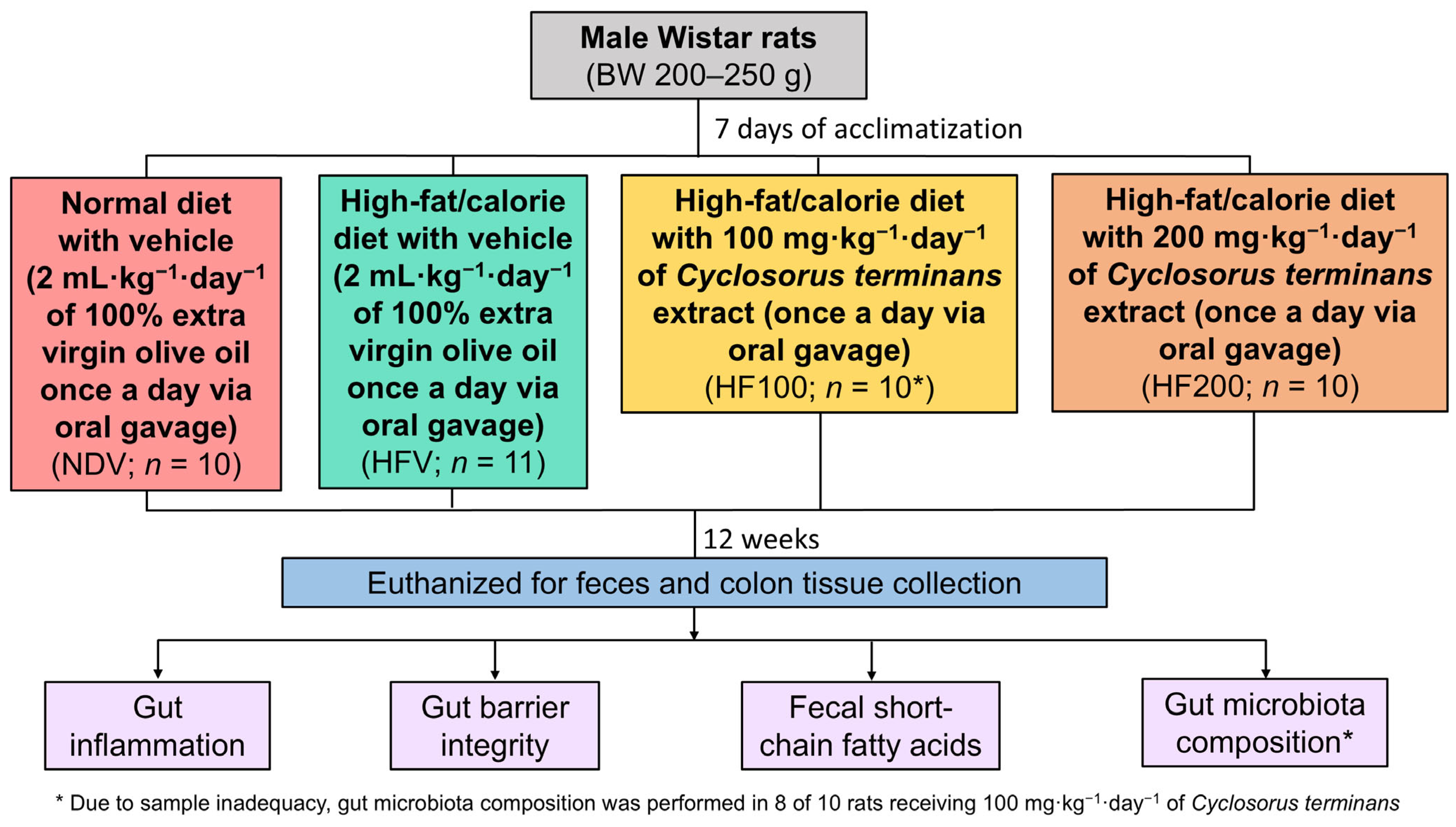
2.1. Study Protocol

The protocol is shown in Figure 1. All experiments were approved by the Laboratory Animal Center, Chiang Mai University Animal Care and Use Committee, Chiang Mai University (approval number: 2565/RT-0016, approval date: 27 April 2022). Male Wistar rats with a body weight of 200–250 g were bought from Nomura Siam International, Bangkok, Thailand.
After 1 week of an acclimatization period, the rats were assigned to receive either (1) a normal diet treated with vehicle (NDV), (2) a high-fat/calorie diet treated with vehicle (HFV), (3) a high-fat/calorie diet treated with 100 mg·kg−1·day−1 of C. terminans extract (HF100), or (4) a high-fat/calorie diet treated with vehicle treated with 200 mg·kg−1·day−1 of C. terminans extract (HF200). The randomization was conducted using an animal randomization tool “https://acmeresearchlabs.in/animal-randomization-tool/ (accessed on 5 January 2022)” by ACME Research Solutions (New Delhi, India). The compositions of the diets were described in a previous study [30]. Since C. terminans extract can be completely dissolved in oil, the crude extract was dissolved in extra virgin olive oil before administering to the rats. The feeding volume of C. terminans extract was 2 mL·kg−1·day−1. Therefore, the vehicle used in this study was 2 mL·kg−1·day−1 of 100% extra virgin olive oil. Both vehicle and C. terminans extract were administered once a day via oral gavage. The doses of C. terminans extract were chosen based upon previous findings from our group, in which 100 and 200 mg· kg−1·day−1 of C. terminans attenuated obesity and fatty liver in obese rats [25]. The diets and treatment were continued for 12 weeks in each group.
At the end of treatment, the rats were sacrificed. Feces were collected for the quantification of short-chain fatty acid levels and the identification of gut microbiota composition. Colon tissues were obtained for the evaluation of gut inflammation and gut barrier integrity.
The blinding procedures were conducted during the experiments. In fact, people who treated the rats were not involved in the processes of euthanasia, molecular studies, and data analyses. In addition, the groups of rats were concealed from people who performed euthanasia, molecular studies, and data analyses.

2.2. Plant Extraction

Aerial parts of C. terminans were obtained. A voucher specimen (no. SKP 2080320001) was stored. The extraction method was detailed in a previous study [25]. The extract was evaluated for the amount of interruptins A (5.45% weight by weight) and B (1.41% weight by weight) by a high-performance liquid chromatography method [31]. From this method, the amount of a non-active ingredient—interruptin C (4.53% weight by weight)—was also determined.

2.3. Sample Size Calculation

According to a prior study showing that an extract from a plant Emblica officinalis could change gut microbial abundance and the related metabolites in obese mice [32], we used the acetic acid level to estimate the sample size. The previous findings yielded the effect size value of 0.67. Along with an alpha error probability of 0.05 and power of 0.80, the G*Power version 3.1.9.7 (Universität Kiel, Kiel, Germany) demonstrated the total sample size of 32 (8 per group).

2.4. The Expression of mRNA Analyses to Evaluate Gut Inflammation

The levels of mRNA expression of inflammatory-related genes including TNF-α, IL-1β, and IL-6 were evaluated using a quantitative polymerase chain reaction, as detailed in a prior study [33]. All primer pairs are shown in Table S1.

2.5. Protein Expression Analyses to Evaluate Gut Barrier Integrity

Occludin and claudins play roles in the maintenance of epithelial barrier [34]. Hence, the expressions of occludin and claudin-5 proteins in colonic tissues were used as tight junction markers to determine gut barrier integrity. The protein expression analyses were performed using Western blot, which has been detailed in a previous study [35].

2.6. Quantification of Fecal Short-Chain Fatty Acids

The levels of fecal short-chain fatty acids, including acetic acid, propionic acid, butyric acid, and valeric acid were quantified using single quadrupole gas chromatography mass spectrometry, as detailed in a prior study [36]. This method has been specifically validated for the fecal samples used.

2.7. Identification of Gut Microbiota Composition

A genomic DNA isolation kit (QIAGEN, Hilden, Germany) was used to obtain bacterial DNA. The 250 mg of feces was extracted in accordance with the manufacturer’s instructions. Then, V3–V4 amplification and paired-end sequencing were performed and analyzed, as described in a prior study [36]. The taxonomy was annotated using a Naïve Bayes classifier implemented in Scikit-learn from the SILVA database version 138 (Leibniz Institute DSMZ-German Collection of Microorganisms and Cell Cultures GmbH, Braunschweig, Germany) [37,38].

2.8. Statistical Analyses

Alpha diversity and beta diversity between groups were compared using the Kruskal–Wallis test and permutational multivariate analysis of variance, respectively. The linear discriminant analysis effect size was performed to identify the taxa that were most likely to explain the differences in the gut microbiota composition between groups. The metrics for beta diversity included (1) Bray–Curtis, which examines the abundances of microbes that are shared between two samples, and the abundances of microbes found in each sample, (2) Jaccard, which determines the presence of microbes in one or both samples, (3) unweighted UniFrac, which defines the fraction of branch lengths between all gut microbes in two samples that show differences between the samples, and (4) weighted UniFrac, which resembles unweighted UniFrac but also takes the abundances of microbes into account [39]. All microbiome analyses were generated using R version 4.3.1 (the R Foundation for Statistical Computing Platform, Vienna, Austria). For the non-microbiome data, a parametric one-way analysis of variance followed by Fisher’s least significant difference method was conducted to detect the differences between groups using GraphPad Prism version 8 (GraphPad Software, San Diego, CA, USA). Spearman’s rank correlation coefficient was performed to examine the correlations between gut microbiota composition and other parameters using R version 4.3.1 (the R Foundation for Statistical Computing Platform, Vienna, Austria).

3. Results

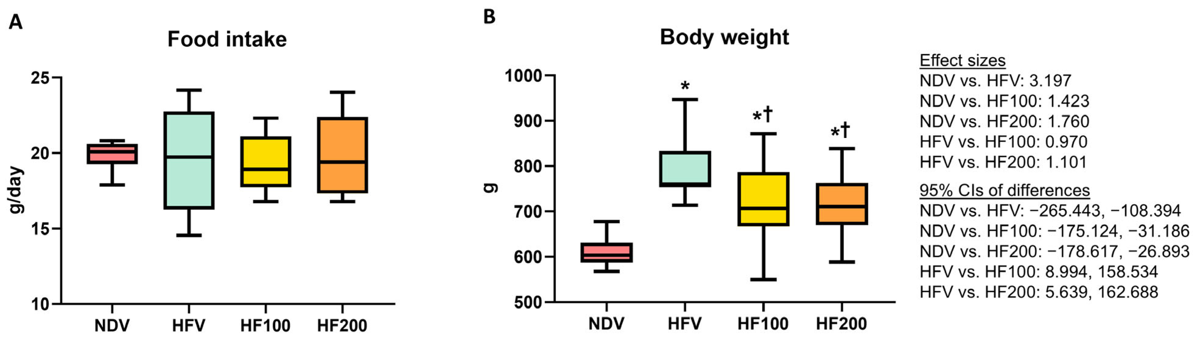
3.1. C. terminans Extract Attenuated an Increase in Body Weight in Rats Exposed to High-Fat/Calorie Diet

The side effects of the vehicle and C. terminans extract were not observed in our rats. The amount of food intake was no different between groups (Figure 2A). Following 12 weeks of high-fat/calorie diet consumption, the HFV group exhibited increased body weight, when compared to the NDV group (Figure 2B). The HF100 and HF200 groups had lower body weight than the HFV group, but the levels remained higher than that of the NDV group (Figure 2B). All of these findings indicated that C. terminans extract could ameliorate high-fat/calorie diet-induced increased body weight.

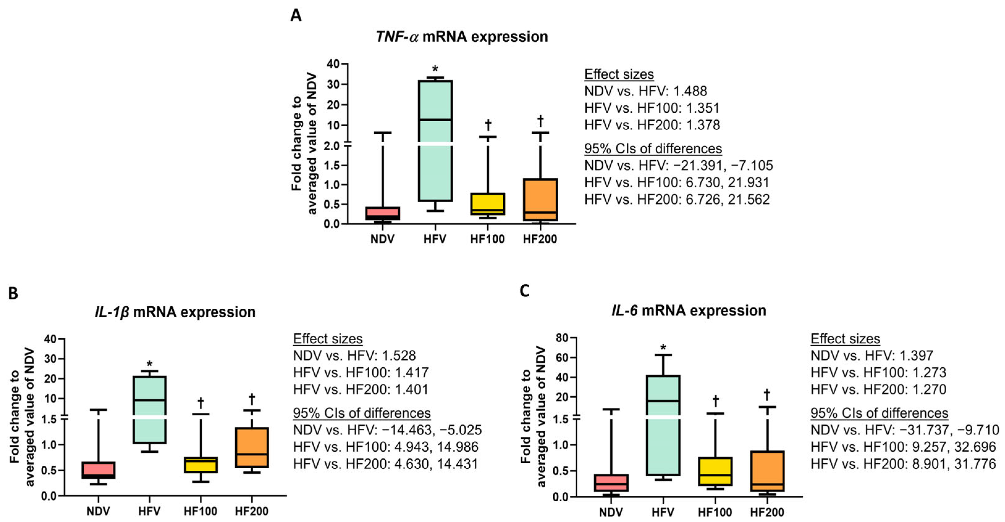
3.2. C. terminans Extract Exerted a Prophylactic Effect Against Gut Inflammation in Rats Exposed to High-Fat/Calorie Diet

The analysis of mRNA expression revealed that TNF-α, IL-1β, and IL-6 levels were higher in the colons of the HFV group, when compared with those of the NDV group (Figure 3A–C), indicating high-fat/calorie diet-induced gut inflammation. The HF100 and HF200 groups had lower TNF-α, IL-1β, and IL-6 levels than those of the HFV group, and these levels did not differ from those of the NDV group (Figure 3A–C). All of these results suggested that C. terminans extract exerted protection against gut inflammation caused by a high-fat/calorie diet.

3.3. C. terminans Extract Exerted a Prophylactic Effect Against Gut Barrier Disruption in Rats Exposed to High-Fat/Calorie Diet

Expressions of occludin and claudin-5 proteins were lower in the colon tissues of the HFV group than those of the NDV group (Figure 4A–C and Figure S1), indicating the high-fat/calorie diet-induced compromised gut barrier integrity. The HF100 and HF200 groups displayed higher levels of the expression of these proteins, when compared to those of the HFV group, and these levels were no different from those of the NDV group (Figure 4A–C and Figure S1). All of these findings suggested that C. terminans extract exerted a protection against gut barrier disruption caused by high-fat/calorie diet.

3.4. C. terminans Extract Exerted a Prophylactic Effect Against the Alterations of Fecal Short-Chain Fatty Acid Levels in Rats Exposed to High-Fat/Calorie Diet

It was demonstrated that the HFV group exhibited greater levels of acetic acid, propionic acid, butyric acid, and valeric acid, when compared with those in the NDV group (Figure 5A–D). The HF100 and HF200 groups had lower levels of all four fecal short-chain fatty acids than those of the HFV group, and these levels did not differ from those of the NDV group (Figure 5A–D). All of these results suggested that C. terminans extract exerted protection against the alterations of host-gut microbial cometabolite levels in rats exposed to high-fat/calorie diet.

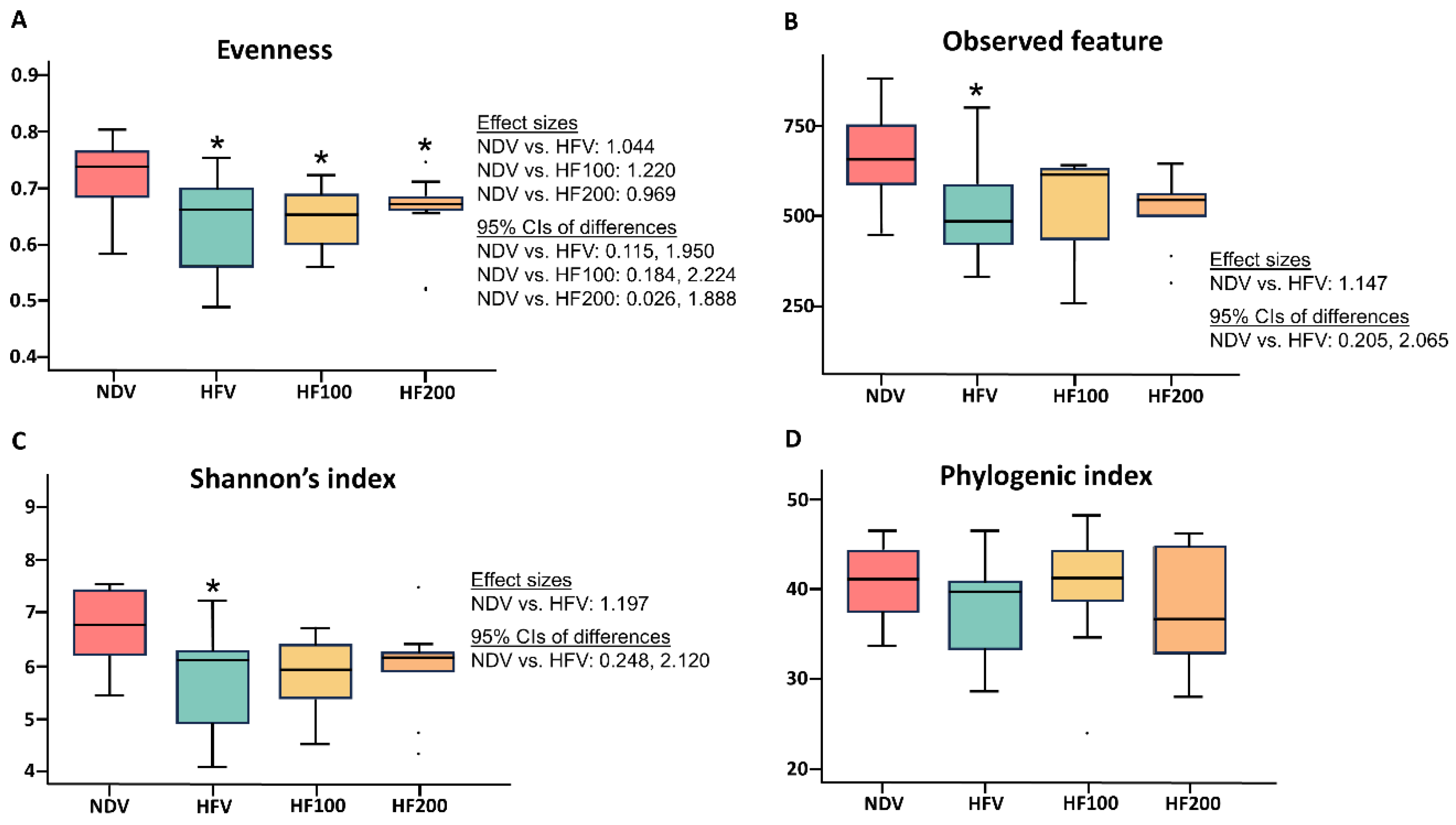
3.5. C. terminans Extract Exerted a Prophylactic Effect Against the Alteration of Alpha Diversity in Rats Exposed to High-Fat/Calorie Diet

Four metrics of alpha diversity were determined. These included (1) evenness, which refers to how similar the abundances of different species are in the community, (2) observed feature, which represents community richness, (3) Shannon’s index, which measures the diversity of species in a community, and (4) phylogenic index, which also identifies the diversity of species, but it is specific to the phylogenetic tree [40]. Compared with those of the NDV group, the HFV group exhibited decreased evenness, observed feature, as well as Shannon’s index of alpha diversity (Figure 6A–C), whereas the phylogenic index was no different between groups (Figure 6D). Both 100 mg·kg−1·day−1 and 200 mg·kg−1·day−1 of C. terminans extract could attenuate the reduction in observed features and Shannon’s index of alpha diversity in rats exposed to high-fat/calorie diet (Figure 6B,C). Nonetheless, neither 100 mg·kg−1·day−1 nor 200 mg·kg−1·day−1 of C. terminans extract improved the evenness of alpha diversity in rats exposed to a high-fat/calorie diet (Figure 6A).

3.6. C. terminans Extract Did Not Improve Beta Diversity in Rats Fed High-Fat/Calorie Diet

Beta diversity is illustrated by principal component analysis, as shown in Figure 7A–D. Each dot represents the gut microbial composition of an individual rat, with lines connecting samples to their respective group centroids (Figure 7A–D). The variance values ranged from 7.82% to 42.15% (Figure 7A–D). Clustering distances between the NDV group and the other groups indicated that the NDV group exerted obvious dissimilarities (Figure 7A–D). Specifically, Bray–Curtis, Jaccard, unweighted UniFrac, and weighted UniFrac differed between the NDV group and the HFV group (Figure 7A–D), indicating high-fat/calorie diet-induced change in beta diversity. However, 100 mg·kg−1·day−1 of C. terminans extract could not alleviate the change in beta diversity induced by a high-fat/calorie diet, as indicated by the overlapping clusters and no difference in any beta diversity metrics between the HFV group and the HF100 group (Figure 7A–D). Interestingly, the clustering distances of Bray–Curtis, Jaccard, and unweighted UniFrac between the HFV group and the HF200 group were revealed, which reached statistical significance (Figure 7A–C). Nevertheless, all these remained different between the NDV group and the HF200 group (Figure 7A–D). All of these results suggested that C. terminans extract did not improve beta diversity in rats exposed to a high-fat/calorie diet. Moreover, 200 mg·kg−1·day−1 of C. terminans extract caused the alteration in beta diversity in rats exposed to high-fat/calorie diet.

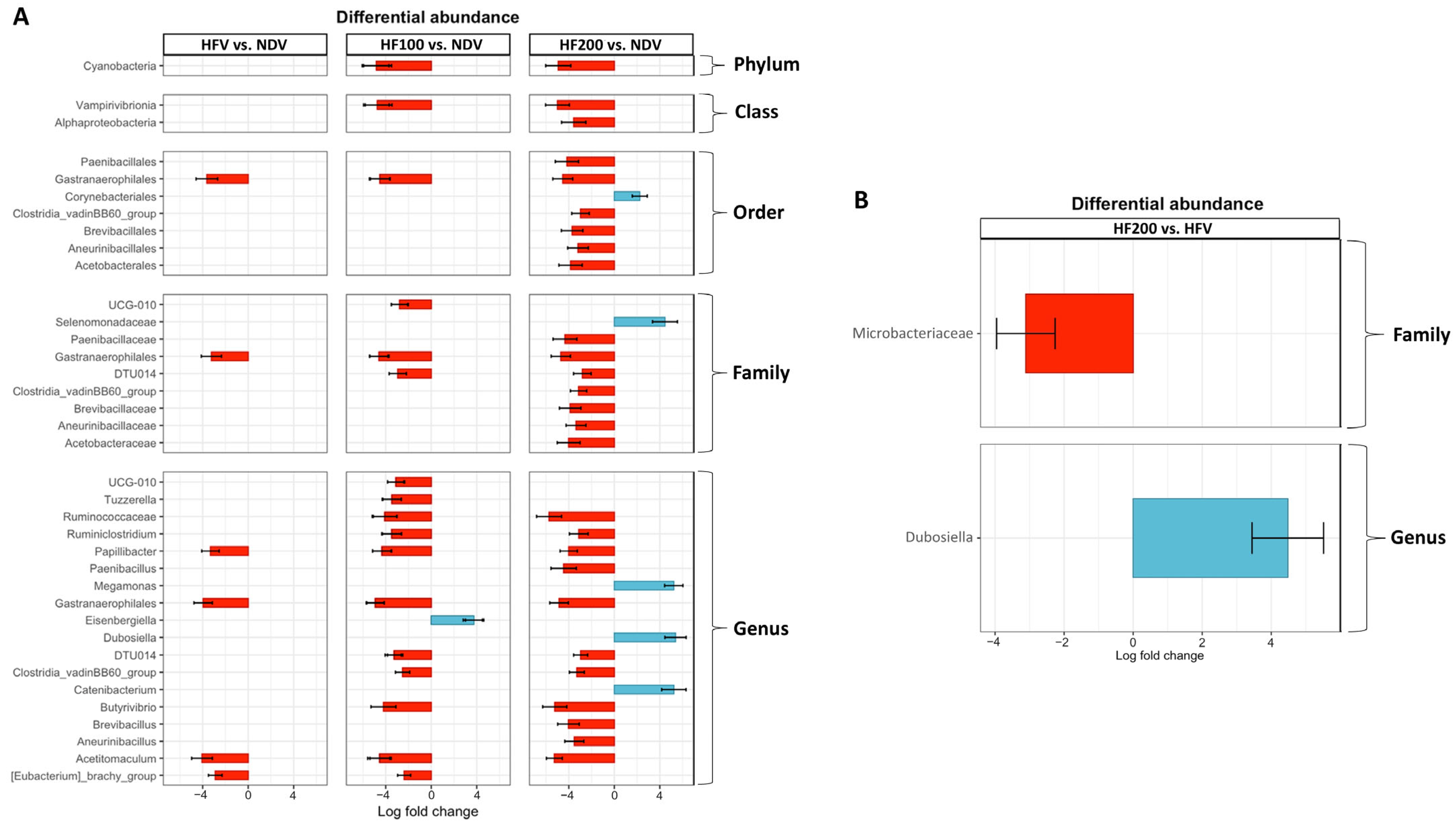
3.7. Differential Abundance of Gut Microbiota Composition

In addition to beta diversity, we evaluated differential gut microbial abundance. The results are shown in Figures S2 and S3. Focusing on the gut microbiomes that were significantly different between groups, we found that the HFV group exhibited increases in the order Gastranaerophilales, family Gastranaerophilales, genus Gastranaerophilales, genus Papillibacter, and genus Acetitomaculum, when compared to those of the NDV group (Figure 7A). In the HF 100 and HF 200 groups, these changes remained present, along with the decreases in several gut microbiomes, specifically genus Tuzzerella, genus Ruminococcaceae, genus Ruminiclostridium, genus Paenibacillus, genus Butyrivibrio, genus Brevibacillus, genus Aneurinibacillus, and genus Acetitomaculum (details are shown in Figure 8A). On the other hand, increases in genus Megamonas, genus Dubosiella, genus Catenibacterium, and genus Eisenbergiella were found in the HF100 and HF200 groups, when compared to those of the NDV group (details are shown in Figure 8A).
When compared to the HFV group, the HF200 group revealed a reduction in family Microbacteriaceae, along with an increase in genus Dubosiella (Figure 8B). Nonetheless, there was no significant difference in gut microbial abundance between the HFV group and the HF100 group, as well as between the HF100 group and the HF200 group.

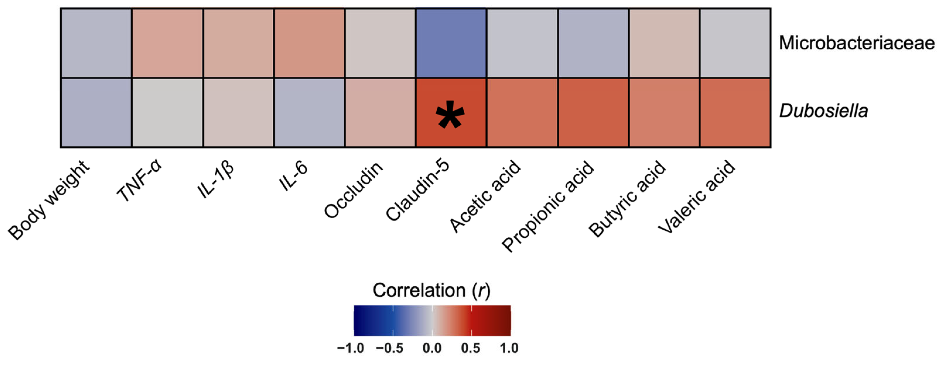
3.8. The Abundance of Genus Dubosiella Was Positively Correlated with Claudin-5 Protein Expression in Colon Tissues

To identify whether the effects of the C. terminans extract on gut microbiota composition were related to body weight and other gut-related outcomes or not, we determined the correlations between gut microbial abundance versus body weight, inflammatory markers, gut barrier integrity markers, and short-chain fatty acid levels. We focused on the abundance of family Microbacteriaceae and genus Dubosiella: the only two microbiomes that were changed in our rats exposed to high-fat/calorie diet after treatment with C. terminans extract. We found a significantly positive correlation between genus Dubosiella and claudin-5 protein expression in the colon tissues of our rats (Figure 9). This result suggested that the improvement in claudin-5 protein expression induced by C. terminans extract was associated with an increase in genus Dubosiella.

4. Discussion

We examined the prophylactic effects of C. terminans extract against high-fat/calorie diet-induced obesity and gut dysfunction. Our vehicle-treated rats were exposed to high-fat/calorie diet to induce obesity, and we expected that obesity subsequently caused gut dysfunction. The results showed that we successfully developed a rat model of obesity with gut dysfunction, as indicated by increased body weight, increased gut inflammation, gut barrier disruption, altered short-chain fatty acid levels, and gut dysbiosis in the HFV group, when compared to those of the NDV group.
The result showed that the early treatment with C. terminans extract ameliorated an increase in body weight in rats exposed to a high-fat/calorie diet. These effects were likely to be mediated by the benefits of interruptins in promoting brown adipogenic differentiation, which were revealed in a prior in vitro study [29]. Mechanistically, brown adipogenic differentiation promotes lipolysis and increases total energy expenditure [41], all of which prevent the development of obesity [42]. Interestingly, we observed that these effects of C. terminans extract were comparable between the doses of 100 mg·kg−1·day−1 and 200 mg−1 kg−1 day−1. These findings are inconsistent with a prior study as they show that 2 weeks of treatment with 200 mg·kg−1·day−1 of C. terminans extract was superior to that of 100 mg·kg−1·day−1 in attenuating increased body weight in rats previously exposed to 12 weeks of high-fat/calorie diet [25]. All of these results highlight the greater benefits of early treatment.
Previous understanding regarding the pathophysiology of obesity-induced gut dysbiosis [9,10,11] has indicated that this prophylactic effect of C. terminans extract in our rats exposed to a high-fat/calorie diet began with protection against gut inflammation, resulting in a protection against gut barrier disruption, ultimately leading to a protection against the changes in alpha diversity. Nevertheless, our results failed to explain whether the protective effect of C. terminans extract on the gut was the direct effect on the gut itself or it was mediated by the benefit of C. terminans extract in protecting against increased body weight. Since gut dysbiosis also played a pivotal role in the development of obesity [12,13,14], our findings could not explain whether the prophylactic effect of C. terminans extract against obesity was mediated by the protective effect of C. terminans extract against the change in alpha diversity. Indeed, the mechanistic link between C. terminans extract, gut dysbiosis, and obesity could not be holistically determined in this study. This may limit the use of C. terminans extract in future clinical practice. For these reasons, future experiments in an animal model of gut dysbiosis-induced obesity or a different time-point study into high-fat/calorie diet consumption with C. terminans extract is required.
In addition to the impact of gut microbiomes on energy homeostasis and the pathophysiology of obesity [12,13,14], a healthy gut exerts a pivotal role in maintaining the physiological function of other organ systems [43,44]. These include the nervous system [45], cardiovascular system [46], and skeletal system [47]. For this reason, we speculated that C. terminans extract also exerts protection against neurological disorders, cardiovascular diseases, and bone remodeling induced by obesity. To verify these benefits of C. terminans extract, future studies evaluating the effect of this extract on the brain, heart, and bone are needed.
Although C. terminans extract could not improve beta diversity in our rats exposed to high-fat/calorie diet, we found that the abundances of some gut microbiomes were significantly altered only in rats exposed to high-fat/calorie diet treated with this extract. For example, a decrease in genus Tuzzerella: a microbiome that is associated with immune dysfunction [48], the reduction in opportunistic pathogens: genus Paenibacillus and genus Brevibacillus [49,50], an increase in genus Dubosiella that is a probiotic [51], and an increase in genus Catenibacterium that is associated with the lower cardiovascular disease risk [52]. All of these findings suggested that C. terminans extract could promote a more favorable gut microbiome profile.
A positive correlation between genus Dubosiella and claudin-5 protein expression in colon tissues was observed in our rats, suggesting an association between this gut microbiome and gut barrier integrity. This was consistent with a prior study demonstrating that treatment with Dubosiella newyorkensis attenuated gut barrier injury in mice with colitis [53]. All of these have highlighted the benefit of Dubosiella in improving gut barrier integrity, both in obesity and colitis.
This study has some limitations. The sample size for gut microbiota analysis across groups was varied, which might affect the validity of our findings. Importantly, our results could not clarify the mechanistic insight into the relationship between C. terminans extract, obesity, and gut dysbiosis. Therefore, a study with an equal sample size across groups and a study in a model of gut dysbiosis-induced obesity or a different time-point study into high-fat/calorie diet consumption with C. terminans extract are needed in the future.

5. Conclusions

The findings of this study revealed the potential for C. terminans extract to alleviate the increase in body weight and protect against gut dysfunction induced by high-fat/calorie diet. Nonetheless, further long-term studies identifying the safety of the use of C. terminans extract and further studies comparing the efficacy between C. terminans extract and other paradigms of gut microbiome modulation in animals are warranted and necessary, prior to the use of C. terminans extract in clinical trials.

Supplementary Materials

The following supporting information can be downloaded at: https://www.mdpi.com/article/10.3390/nu17081387/s1, Table S1: primer pairs used in this study; Figure S1: original Western blots for gut barrier-related proteins; Figure S2: differential abundance of gut microbiota composition (family level); Figure S3: differential abundance of gut microbiota composition (genus level).

Author Contributions

Conceptualization: C.T., S.K. (Sireewan Kaewsuwan), N.C., and S.C.C.; methodology: C.T., K.S., C.K., N.L., W.N., B.A., S.K. (Sasiwan Kerdphoo), A.K., S.S., H.P., W.P., C.M., and S.K. (Sireewan Kaewsuwan); data curation: C.T., C.K., N.L., W.N., B.A., S.K. (Sasiwan Kerdphoo), S.S., H.P., W.P., and S.K. (Sireewan Kaewsuwan); writing—original draft: C.T.; writing—review and editing: C.T., S.K. (Sireewan Kaewsuwan), and S.C.C.; visualization: C.T.; supervision: S.K. (Sireewan Kaewsuwan), N.C., and S.C.C.; project administration: C.T., W.P., S.K. (Sireewan Kaewsuwan), N.C., and S.C.C.; funding acquisition: C.T., H.P., W.P., S.K. (Sireewan Kaewsuwan), N.C., and S.C.C. All authors have read and agreed to the published version of the manuscript.

Funding

The Mid-Career Research Grant from the National Research Council of Thailand (N42A680478 to C.T.), Chiang Mai University Fundamental Fund 2024 (W.P.), the Faculty of Pharmaceutical Sciences, Prince of Songkla University (PSU; PHA6304112S to S.K. (Sireewan Kaewsuwan)), the Research Chair Grant from the National Research Council of Thailand (N42A670594 to N.C.), a Chiang Mai University Center of Excellence Award (N.C.), and the Distinguished Research Professor Grant from the National Research Council of Thailand (N42A660301 to S.C.C.).

Institutional Review Board Statement

This study was conducted in accordance with the Declaration of Helsinki, and approved by the Laboratory Animal Center, Chiang Mai University Animal Care and Use Committee, Chiang Mai University (approval number: 2565/RT-0016 and date of approval 27 April 2022).

Informed Consent Statement

Not applicable.

Data Availability Statement

The datasets generated and/or analyzed during the current study have been deposited in the National Center for Biotechnology Information (NCBI) Sequence Read Archive (SRA) under the BioProject accession number PRJNA1249221, with individual sample accessions ranging from SRA:47883419 to SRA:47883457.

Conflicts of Interest

The authors declare no conflicts of interest.

Abbreviations

The following abbreviations are used in this manuscript:
C. terminansCyclosorus terminans
DNAdeoxyribonucleic acid
HDLhigh-density lipoprotein
HFVhigh-fat/calorie diet treated with vehicle
HF100high-fat/calorie diet treated with 100 mg·kg−1·day−1 of C. terminans extract
HF200high-fat/calorie diet treated with 200 mg·kg−1·day−1 of C. terminans extract
IL-1βinterleukin-1beta
IL-6interleukin-6
mg·kg−1·day−1milligram per kilogram per day
mRNAmessenger ribonucleic acid
NCBINational Center for Biotechnology Information
NDVnormal diet treated with vehicle
SRASequence Read Archive
TNF-αtumor necrosis factor-alpha

References

  1. Davis, C.D. The Gut Microbiome and Its Role in Obesity. Nutr. Today 2016, 51, 167–174. [Google Scholar] [CrossRef] [PubMed]
  2. Breton, J.; Galmiche, M.; Déchelotte, P. Dysbiotic Gut Bacteria in Obesity: An Overview of the Metabolic Mechanisms and Therapeutic Perspectives of Next-Generation Probiotics. Microorganisms 2022, 10, 452. [Google Scholar] [CrossRef] [PubMed]
  3. Jumpertz, R.; Le, D.S.; Turnbaugh, P.J.; Trinidad, C.; Bogardus, C.; Gordon, J.I.; Krakoff, J. Energy-balance studies reveal associations between gut microbes, caloric load, and nutrient absorption in humans. Am. J. Clin. Nutr. 2011, 94, 58–65. [Google Scholar] [CrossRef] [PubMed]
  4. Kalliomäki, M.; Collado, M.C.; Salminen, S.; Isolauri, E. Early differences in fecal microbiota composition in children may predict overweight. Am. J. Clin. Nutr. 2008, 87, 534–538. [Google Scholar] [CrossRef]
  5. Turnbaugh, P.J.; Bäckhed, F.; Fulton, L.; Gordon, J.I. Diet-induced obesity is linked to marked but reversible alterations in the mouse distal gut microbiome. Cell Host Microbe 2008, 3, 213–223. [Google Scholar] [CrossRef]
  6. Ellulu, M.S.; Patimah, I.; Khaza’ai, H.; Rahmat, A.; Abed, Y. Obesity and inflammation: The linking mechanism and the complications. Arch. Med. Sci. AMS 2017, 13, 851–863. [Google Scholar] [CrossRef]
  7. Khanna, D.; Khanna, S.; Khanna, P.; Kahar, P.; Patel, B.M. Obesity: A Chronic Low-Grade Inflammation and Its Markers. Cureus 2022, 14, e22711. [Google Scholar] [CrossRef]
  8. Zotova, N.V.; Chereshnev, V.A.; Gusev, E.Y. Systemic Inflammation: Methodological Approaches to Identification of the Common Pathological Process. PLoS ONE 2016, 11, e0155138. [Google Scholar] [CrossRef]
  9. Kang, G.G.; Trevaskis, N.L.; Murphy, A.J.; Febbraio, M.A. Diet-induced gut dysbiosis and inflammation: Key drivers of obesity-driven NASH. iScience 2023, 26, 105905. [Google Scholar] [CrossRef]
  10. Shahi, S.K.; Ghimire, S.; Lehman, P.; Mangalam, A.K. Obesity induced gut dysbiosis contributes to disease severity in an animal model of multiple sclerosis. Front. Immunol. 2022, 13, 966417. [Google Scholar] [CrossRef]
  11. Bui, T.I.; Gill, A.L.; Mooney, R.A.; Gill, S.R. Modulation of Gut Microbiota Metabolism in Obesity-Related Type 2 Diabetes Reduces Osteomyelitis Severity. Microbiol. Spectr. 2022, 10, e0017022. [Google Scholar] [CrossRef] [PubMed]
  12. Liu, B.N.; Liu, X.T.; Liang, Z.H.; Wang, J.H. Gut microbiota in obesity. World J. Gastroenterol. 2021, 27, 3837–3850. [Google Scholar] [CrossRef]
  13. Cheng, Z.; Zhang, L.; Yang, L.; Chu, H. The critical role of gut microbiota in obesity. Front. Endocrinol. 2022, 13, 1025706. [Google Scholar] [CrossRef]
  14. Amabebe, E.; Robert, F.O.; Agbalalah, T.; Orubu, E.S.F. Microbial dysbiosis-induced obesity: Role of gut microbiota in homoeostasis of energy metabolism. Br. J. Nutr. 2020, 123, 1127–1137. [Google Scholar] [CrossRef]
  15. Turnbaugh, P.J.; Ley, R.E.; Mahowald, M.A.; Magrini, V.; Mardis, E.R.; Gordon, J.I. An obesity-associated gut microbiome with increased capacity for energy harvest. Nature 2006, 444, 1027–1031. [Google Scholar] [CrossRef]
  16. Bäckhed, F.; Ding, H.; Wang, T.; Hooper, L.V.; Koh, G.Y.; Nagy, A.; Semenkovich, C.F.; Gordon, J.I. The gut microbiota as an environmental factor that regulates fat storage. Proc. Natl. Acad. Sci. USA 2004, 101, 15718–15723. [Google Scholar] [CrossRef] [PubMed]
  17. Mishra, S.P.; Wang, B.; Jain, S.; Ding, J.; Rejeski, J.; Furdui, C.M.; Kitzman, D.W.; Taraphder, S.; Brechot, C.; Kumar, A.; et al. A mechanism by which gut microbiota elevates permeability and inflammation in obese/diabetic mice and human gut. Gut 2023, 72, 1848–1865. [Google Scholar] [CrossRef] [PubMed]
  18. Scott, S.A.; Fu, J.; Chang, P.V. Microbial tryptophan metabolites regulate gut barrier function via the aryl hydrocarbon receptor. Proc. Natl. Acad. Sci. USA 2020, 117, 19376–19387. [Google Scholar] [CrossRef]
  19. Kang, Y.; Kang, X.; Yang, H.; Liu, H.; Yang, X.; Liu, Q.; Tian, H.; Xue, Y.; Ren, P.; Kuang, X.; et al. Lactobacillus acidophilus ameliorates obesity in mice through modulation of gut microbiota dysbiosis and intestinal permeability. Pharmacol. Res. 2022, 175, 106020. [Google Scholar] [CrossRef]
  20. Cani, P.D.; Bibiloni, R.; Knauf, C.; Waget, A.; Neyrinck, A.M.; Delzenne, N.M.; Burcelin, R. Changes in gut microbiota control metabolic endotoxemia-induced inflammation in high-fat diet-induced obesity and diabetes in mice. Diabetes 2008, 57, 1470–1481. [Google Scholar] [CrossRef]
  21. Brandsma, E.; Kloosterhuis, N.J.; Koster, M.; Dekker, D.C.; Gijbels, M.J.J.; van der Velden, S.; Ríos-Morales, M.; van Faassen, M.J.R.; Loreti, M.G.; de Bruin, A.; et al. A Proinflammatory Gut Microbiota Increases Systemic Inflammation and Accelerates Atherosclerosis. Circ. Res. 2019, 124, 94–100. [Google Scholar] [CrossRef]
  22. Hefley, S.; Howard, N.; Crasto, C.; Vasylyeva, T. Microbiome Dysbiosis in Obese Adolescents: A Potential Contributor to Inflammation. FASEB J. 2021, 35, 02509. [Google Scholar] [CrossRef]
  23. Artemniak-Wojtowicz, D.; Kucharska, A.M.; Pyrżak, B. Obesity and chronic inflammation crosslinking. Cent. Eur. J. Immunol. 2020, 45, 461–468. [Google Scholar] [CrossRef] [PubMed]
  24. Sun, N.N.; Wu, T.Y.; Chau, C.F. Natural Dietary and Herbal Products in Anti-Obesity Treatment. Molecules 2016, 21, 1351. [Google Scholar] [CrossRef] [PubMed]
  25. Songtrai, S.; Pratchayasakul, W.; Arunsak, B.; Chunchai, T.; Kongkaew, A.; Chattipakorn, N.; Chattipakorn, S.C.; Kaewsuwan, S. Cyclosorus terminans Extract Ameliorates Insulin Resistance and Non-Alcoholic Fatty Liver Disease (NAFLD) in High-Fat Diet (HFD)-Induced Obese Rats. Nutrients 2022, 14, 4895. [Google Scholar] [CrossRef]
  26. Holdsworth, D.; Rali, T. A Survey of Medicinal Plants of the Southern Highlands, Papua New Guinea. Int. J. Crude Drug Res. 1989, 27, 1–8. [Google Scholar] [CrossRef]
  27. Nick, A.; Rali, T.; Sticher, O. Biological screening of traditional medicinal plants from Papua New Guinea. J. Ethnopharmacol. 1995, 49, 147–156. [Google Scholar] [CrossRef]
  28. Chen, J.; Lei, Y.; Liu, Y.; Xiong, C.; Fu, W.; Ruan, J. Extract of Cyclosorus acuminatus attenuates diabetic nephropathy in mice via modifying peroxisome proliferators activated receptor signalling pathway. Food Chem. 2011, 128, 659–666. [Google Scholar] [CrossRef]
  29. Kaewsuwan, S.; Plubrukarn, A.; Utsintong, M.; Kim, S.H.; Jeong, J.H.; Cho, J.G.; Park, S.G.; Sung, J.H. Interruptin B induces brown adipocyte differentiation and glucose consumption in adipose-derived stem cells. Mol. Med. Rep. 2016, 13, 2078–2086. [Google Scholar] [CrossRef]
  30. Thonusin, C.; Apaijai, N.; Jaiwongkam, T.; Kerdphoo, S.; Arunsak, B.; Amput, P.; Palee, S.; Pratchayasakul, W.; Chattipakorn, N.; Chattipakorn, S.C. The comparative effects of high dose atorvastatin and proprotein convertase subtilisin/kexin type 9 inhibitor on the mitochondria of oxidative muscle fibers in obese-insulin resistant female rats. Toxicol. Appl. Pharmacol. 2019, 382, 114741. [Google Scholar] [CrossRef]
  31. Chaiwong, S.; Puttarak, P.; Kaewsuwan, S. Anti propionibacterium acnes activity, HPLC method validation for simultaneous analysis and extraction of coumarins from the fern cyclosorus terminans. Lat. Am. J. Pharm. 2018, 37, 1791–1797. [Google Scholar]
  32. Brochot, A.; Azalbert, V.; Landrier, J.F.; Tourniaire, F.; Serino, M. A Two-Week Treatment with Plant Extracts Changes Gut Microbiota, Caecum Metabolome, and Markers of Lipid Metabolism in ob/ob Mice. Mol. Nutr. Food Res. 2019, 63, e1900403. [Google Scholar] [CrossRef] [PubMed]
  33. Thiennimitr, P.; Yasom, S.; Tunapong, W.; Chunchai, T.; Wanchai, K.; Pongchaidecha, A.; Lungkaphin, A.; Sirilun, S.; Chaiyasut, C.; Chattipakorn, N.; et al. Lactobacillus paracasei HII01, xylooligosaccharides, and synbiotics reduce gut disturbance in obese rats. Nutrition 2018, 54, 40–47. [Google Scholar] [CrossRef]
  34. Chelakkot, C.; Ghim, J.; Ryu, S.H. Mechanisms regulating intestinal barrier integrity and its pathological implications. Exp. Mol. Med. 2018, 50, 1–9. [Google Scholar] [CrossRef] [PubMed]
  35. Suparan, K.; Sriwichaiin, S.; Thonusin, C.; Sripetchwandee, J.; Khuanjing, T.; Maneechote, C.; Nawara, W.; Arunsak, B.; Chattipakorn, N.; Chattipakorn, S.C. Donepezil ameliorates gut barrier disruption in doxorubicin-treated rats. Food Chem. Toxicol. 2024, 189, 114741. [Google Scholar] [CrossRef]
  36. Sriwichaiin, S.; Thiennimitr, P.; Thonusin, C.; Sarichai, P.; Buddhasiri, S.; Kumfu, S.; Nawara, W.; Kittichotirat, W.; Fucharoen, S.; Chattipakorn, N.; et al. Deferiprone has less benefits on gut microbiota and metabolites in high iron-diet induced iron overload thalassemic mice than in iron overload wild-type mice: A preclinical study. Life Sci. 2022, 307, 120871. [Google Scholar] [CrossRef]
  37. Quast, C.; Pruesse, E.; Yilmaz, P.; Gerken, J.; Schweer, T.; Yarza, P.; Peplies, J.; Glöckner, F.O. The SILVA ribosomal RNA gene database project: Improved data processing and web-based tools. Nucleic Acids Res. 2013, 41, D590–D596. [Google Scholar] [CrossRef]
  38. Yilmaz, P.; Parfrey, L.W.; Yarza, P.; Gerken, J.; Pruesse, E.; Quast, C.; Schweer, T.; Peplies, J.; Ludwig, W.; Glöckner, F.O. The SILVA and “All-species Living Tree Project (LTP)” taxonomic frameworks. Nucleic Acids Res. 2014, 42, D643–D648. [Google Scholar] [CrossRef]
  39. Legendre, P.; De Cáceres, M. Beta diversity as the variance of community data: Dissimilarity coefficients and partitioning. Ecol. Lett. 2013, 16, 951–963. [Google Scholar] [CrossRef]
  40. Cassol, I.; Ibañez, M.; Bustamante, J.P. Key features and guidelines for the application of microbial alpha diversity metrics. Sci. Rep. 2025, 15, 622. [Google Scholar] [CrossRef]
  41. Alcalá, M.; Calderon-Dominguez, M.; Serra, D.; Herrero, L.; Viana, M. Mechanisms of Impaired Brown Adipose Tissue Recruitment in Obesity. Front. Physiol. 2019, 10, 94. [Google Scholar] [CrossRef] [PubMed]
  42. Grabner, G.F.; Xie, H.; Schweiger, M.; Zechner, R. Lipolysis: Cellular mechanisms for lipid mobilization from fat stores. Nat. Metab. 2021, 3, 1445–1465. [Google Scholar] [CrossRef] [PubMed]
  43. Ding, R.X.; Goh, W.R.; Wu, R.N.; Yue, X.Q.; Luo, X.; Khine, W.W.T.; Wu, J.R.; Lee, Y.K. Revisit gut microbiota and its impact on human health and disease. J. Food Drug Anal. 2019, 27, 623–631. [Google Scholar] [CrossRef] [PubMed]
  44. Clemente, J.C.; Ursell, L.K.; Parfrey, L.W.; Knight, R. The impact of the gut microbiota on human health: An integrative view. Cell 2012, 148, 1258–1270. [Google Scholar] [CrossRef]
  45. Cryan, J.F.; O’Riordan, K.J.; Cowan, C.S.M.; Sandhu, K.V.; Bastiaanssen, T.F.S.; Boehme, M.; Codagnone, M.G.; Cussotto, S.; Fulling, C.; Golubeva, A.V.; et al. The Microbiota-Gut-Brain Axis. Physiol. Rev. 2019, 99, 1877–2013. [Google Scholar] [CrossRef]
  46. Tang, W.H.; Kitai, T.; Hazen, S.L. Gut Microbiota in Cardiovascular Health and Disease. Circ. Res. 2017, 120, 1183–1196. [Google Scholar] [CrossRef]
  47. Zhang, J.; Lu, Y.; Wang, Y.; Ren, X.; Han, J. The impact of the intestinal microbiome on bone health. Intractable Rare Dis. Res. 2018, 7, 148–155. [Google Scholar] [CrossRef]
  48. Sbierski-Kind, J.; Grenkowitz, S.; Schlickeiser, S.; Sandforth, A.; Friedrich, M.; Kunkel, D.; Glauben, R.; Brachs, S.; Mai, K.; Thürmer, A.; et al. Effects of caloric restriction on the gut microbiome are linked with immune senescence. Microbiome 2022, 10, 57. [Google Scholar] [CrossRef]
  49. Curtis, A.K.; Lamb, C.; Hassan, W.M.; Foxworth, J. Brevibacillus Laterosporus Bacteremia in an Adult. Cureus 2020, 12, e10481. [Google Scholar] [CrossRef]
  50. Szaniawski, M.A.; Spivak, A.M. Recurrent Paenibacillus infection. Oxf. Med. Case Rep. 2019, 2019, omz034. [Google Scholar]
  51. Liu, T.H.; Zhao, L.; Zhang, C.Y.; Li, X.Y.; Wu, T.L.; Dai, Y.Y.; Sheng, Y.Y.; Ren, Y.L.; Xue, Y.Z. Gut microbial evidence chain in high-salt diet exacerbates intestinal aging process. Front. Nutr. 2022, 9, 1046833. [Google Scholar] [CrossRef] [PubMed]
  52. Kelly, T.N.; Bazzano, L.A.; Ajami, N.J.; He, H.; Zhao, J.; Petrosino, J.F.; Correa, A.; He, J. Gut Microbiome Associates With Lifetime Cardiovascular Disease Risk Profile Among Bogalusa Heart Study Participants. Circ. Res. 2016, 119, 956–964. [Google Scholar] [CrossRef] [PubMed]
  53. Zhang, Y.; Tu, S.; Ji, X.; Wu, J.; Meng, J.; Gao, J.; Shao, X.; Shi, S.; Wang, G.; Qiu, J.; et al. Dubosiella newyorkensis modulates immune tolerance in colitis via the L-lysine-activated AhR-IDO1-Kyn pathway. Nat. Commun. 2024, 15, 1333. [Google Scholar] [CrossRef] [PubMed]
Figure 1. The study protocol. Rats were assigned to receive either (1) a normal diet with vehicle, (2) a high-fat/calorie diet with vehicle, (3) a high-fat/calorie diet with 100 mg·kg−1·day−1 of C. terminans extract, or (4) a high-fat/calorie diet with 200 mg·kg−1·day−1 of C. terminans extract. After 12 weeks of treatment, the rats were euthanized for molecular studies.
Nutrients 17 01387 g001
Figure 2. Food intake (A) and body weight (B). NDV: normal diet treated with vehicle (n = 10); HFV: high-fat/calorie diet treated with vehicle (n = 11); HF100: high-fat/calorie diet treated with 100 mg·kg−1·day−1 of C. terminans extract (n = 10); HF200: high-fat/calorie diet treated with 200 mg·kg−1·day−1 of C. terminans extract (n = 10); CIs: confidence intervals. * p < 0.05 when compared with NDV,  p < 0.05 when compared with HFV. The results showed that the HFV group exhibited increased body weight, when compared with the NDV group. The HF100 and HF200 groups had lower body weight than the HFV group, but the levels remained higher than that of the NDV group.
Nutrients 17 01387 g002
Figure 3. mRNA expression of inflammatory-related genes in colon tissues: TNF-α (A), IL-1β (B), and IL-6 (C). NDV: normal diet treated with vehicle (n = 10); HFV: high-fat/calorie diet treated with vehicle (n = 11); HF100: high-fat/calorie diet treated with 100 mg·kg−1·day−1 of C. terminans extract (n = 10); HF200: high-fat/calorie diet treated with 200 mg·kg−1·day−1 of C. terminans extract (n = 10); CIs: confidence intervals. * p < 0.05 when compared with NDV, p < 0.05 when compared with HFV. The results showed that TNF-α, IL-1β, and IL-6 mRNA expressions were higher in colon tissues of the HFV group, indicating high-fat/calorie diet-induced gut inflammation. The HF100 and HF200 groups had lower TNF-α, IL-1β, and IL-6 levels than those of the HFV group, and these levels did not differ from those of the NDV group.
Nutrients 17 01387 g003
Figure 4. Expression of gut barrier proteins in colon tissues: occludin (A), claudin-5 (B), and representative Western blots (C). NDV: normal diet treated with vehicle (n = 10); HFV: high-fat/calorie diet treated with vehicle (n = 11); HF100: high-fat/calorie diet treated with 100 mg·kg−1·day−1 of C. terminans extract (n = 10); HF200: high-fat/calorie diet treated with 200 mg·kg−1·day−1 of C. terminans extract (n = 10); CIs: confidence intervals. * p < 0.05 when compared with NDV, p < 0.05 when compared with HFV. The results showed that expression of occludin and claudin-5 proteins were lower in colon tissues of the HFV group, indicating high-fat/calorie diet-induced gut barrier disruption. The HF100 and HF200 groups displayed higher levels of these proteins in the colon, when compared to those of the HFV group, and these levels were no different from those of the NDV group.
Nutrients 17 01387 g004
Figure 5. Fecal short-chain fatty acid levels: acetic acid (A), propionic acid (B), butyric acid (C), and valeric acid (D). NDV: normal diet treated with vehicle (n = 10); HFV: high-fat/calorie diet treated with vehicle (n = 11); HF100: high-fat/calorie diet treated with 100 mg·kg−1·day−1 of C. terminans extract (n = 10); HF200: high-fat/calorie diet treated with 200 mg·kg−1·day−1 of C. terminans extract (n = 10); CIs: confidence intervals. * p < 0.05 when compared with NDV, p < 0.05 when compared with HFV. The HFV group exhibited greater levels of fecal short-chain fatty acids. The HF100 and HF200 groups had lower levels of fecal short-chain fatty acids than those of the HFV group, and these levels did not differ from those of the NDV group.
Nutrients 17 01387 g005
Figure 6. Alpha diversity: evenness (A), observed feature (B), Shannon’s index (C), and phylogenic index (D). NDV: normal diet treated with vehicle (n = 10); HFV: high-fat/calorie diet treated with vehicle (n = 11); HF100: high-fat/calorie diet treated with 100 mg·kg−1·day−1 of C. terminans extract (n = 10); HF200: high-fat/calorie diet treated with 200 mg·kg−1·day−1 of C. terminans extract (n = 10); CIs: confidence intervals. * p < 0.05 when compared with NDV. Compared to those of the NDV group, the HFV group exhibited decreased evenness, observed features, and Shannon’s index of alpha diversity, whereas phylogenic index was no different between groups. Both 100 mg·kg−1·day−1 and 200 mg·kg−1·day−1 of C. terminans extract could attenuate the reduction in observed features and Shannon’s index of alpha diversity. Nonetheless, neither 100 mg·kg−1·day−1 nor 200 mg·kg−1·day−1 of C. terminans extract improved evenness of alpha diversity.
Nutrients 17 01387 g006
Figure 7. Beta diversity: Bray–Curtis (A), Jaccard (B), unweighted UniFrac (C), and weighted UniFrac (D). NDV: normal diet treated with vehicle (n = 10); HFV: high-fat/calorie diet treated with vehicle (n = 11); HF100: high-fat/calorie diet treated with 100 mg·kg−1·day−1 of C. terminans extract (n = 8); HF200: high-fat/calorie diet treated with 200 mg·kg−1·day−1 of C. terminans extract (n = 10). Each dot represents the gut microbial composition of an individual rat, with lines connecting samples to their respective group centroids. The results showed that the beta diversity metrics differed between the NDV group and the HFV group, indicating high-fat/calorie diet-induced change in beta diversity. Treatment with 100 mg·kg−1·day−1 of C. terminans extract could not alleviate the change in beta diversity. Interestingly, the differences in Bray–Curtis, Jaccard, and unweighted UniFrac were revealed between the HFV group and the HF200 group. However, all these remained different between the NDV group and the HF200 group.
Nutrients 17 01387 g007
Figure 8. Differential gut microbial abundance that showed a significant difference from NDV (A) and HFV (B). NDV: normal diet treated with vehicle (n = 10); HFV: high-fat/calorie diet treated with vehicle (n = 11); HF100: high-fat/calorie diet treated with 100 mg·kg−1·day−1 of C. terminans extract (n = 8); HF200: high-fat/calorie diet treated with 200 mg·kg−1·day−1 of C. terminans extract (n = 10). Data are shown as mean ± standard error of the mean. The HFV group exhibited increases in the order Gastranaerophilales, family Gastranaerophilales, genus Gastranaerophilales, genus Papillibacter, and genus Acetitomaculum, when compared to the NDV group. In the HF100 and HF200 groups, these changes remained present, along with the decreases in several gut microbiomes, specifically genus Tuzzerella, genus Ruminococcaceae, genus Ruminiclostridium, genus Paenibacillus, genus Butyrivibrio, genus Brevibacillus, genus Aneurinibacillus, and genus Acetitomaculum. On the other hand, increases in genus Megamonas, genus Dubosiella, genus Catenibacterium, and genus Eisenbergiella were found in the HF 100 and HF200 groups, when compared to those of the NDV group. When compared with the HFV group, the HF200 group exhibited a reduction in family Microbacteriaceae, along with an increase in genus Dubosiella.
Nutrients 17 01387 g008
Figure 9. Spearman’s rank correlation between family Microbacteriaceae and genus Dubosiella versus body weight and other gut-related parameters. The results were calculated from all four groups of rats (n = 39). * p < 0.05. It was shown that the amount of genus Dubosiella was positively correlated with claudin-5 protein expression in colon tissues.
Nutrients 17 01387 g009
Disclaimer/Publisher’s Note: The statements, opinions and data contained in all publications are solely those of the individual author(s) and contributor(s) and not of MDPI and/or the editor(s). MDPI and/or the editor(s) disclaim responsibility for any injury to people or property resulting from any ideas, methods, instructions or products referred to in the content.

