Simple Summary
Kaposi’s sarcoma (KS) is a rare tumour of uncertain nature. It may be a true cancer or an aggressive viral lesion, which can regress in many patients, and there is a real need for more research on the subject. This systematic review aimed to summarize the available evidence on somatic mutations and clonality within KS to assess whether KS is a neoplasm or not, concluding that knowledge is currently insufficient to determine whether KS is a clonal neoplasm (sarcoma), or simply an aggressive reactive virus-driven lesion.
Abstract
Background: It remains uncertain whether Kaposi sarcoma (KS) is a true neoplasm, in that it regresses after removal of the stimulus to growth (as HHV8) when immunosuppression is reduced. We aimed to summarize the available evidence on somatic mutations and clonality within KS to assess whether KS is a neoplasm or not. Methods: Medline and Web of Science were searched until September 2020 for articles on clonality or mutation in KS. Search strings were supervised by expert librarians, and two researchers independently performed study selection and data extraction. An adapted version of the QUADAS2 tool was used for methodological quality appraisal. Results: Of 3077 identified records, 20 publications reported on relevant outcomes and were eligible for qualitative synthesis. Five studies reported on clonality, 10 studies reported on various mutations, and 5 studies reported on chromosomal aberrations in KS. All studies were descriptive and were judged to have a high risk of bias. There was considerable heterogeneity of results with respect to clonality, mutation and cytogenetic abnormalities as well as in terms of types of lesions and patient characteristics. Conclusions: While KS certainly produces tumours, the knowledge is currently insufficient to determine whether KS is a clonal neoplasm (sarcoma), or simply an aggressive reactive virus-driven lesion.
1. Introduction
Kaposi sarcoma (KS) is an unusual neoplasm, which presents with clinical manifestations that range from those that are slowly progressive and confined to the skin, to an aggressive tumour that invades visceral organs. The clinicopathological characteristics are usually described in terms of the location of the lesions (lymph node, visceral or skin), and the clinical stage of the disease (patch, plaque or nodular). Epidemiologically, KS presents in several forms: (a) a classic form predominantly in older men; (b) an endemic form in young men and children from central Africa; (c) an iatrogenic form secondary to treatment with immunosuppressive drugs, such as steroids; and (d) an epidemic, HIV/AIDS-associated form [1].
KS was first described at the end of the 19th century as mainly occurring in elderly males from Mediterranean regions but was later reported in East and Central Africa with presentation in younger people [1,2]. It was a rare tumour in certain countries, such as North America and Europe, before the rise of HIV/AIDS in the 1980s, when its incidence increased dramatically and this tumour became characteristic as being an AIDS-related malignancy [1,3]. Currently, while its incidence varies greatly in different regions of the world, KS remains a rare disease. Most of the cases seen in Europe, North America and Africa are young children, renal allograft recipients, patients on immunosuppressive therapy and HIV-infected patients [4].
KS became the most common HIV-related “malignancy” worldwide, although HIV alone could not explain all of the cases. Its occurrence in patients on long-term immunosuppressive therapy and the predominance in males and HIV-negative immunocompetent individuals with increased risk for Sexual Transmitted Diseases (STDs) suggested the implication of another transmissible agent. Eventually human herpesvirus-8 (HHV8) was identified as the virus associated with KS in all different groups, including individuals in Eastern Europe, Africa and the United States [5].
Depending on the clinical subtype, age-standardized incidence rates ranged from 0.3 in Eastern Asia to 8.5 ASR per 100,000 in Eastern Africa in 2020 [6]. Various African regions reported an important increase in the incidence of KS over the last decades [7,8], and western countries also described new patterns with more cases in young and older males [4,6]. There are several well-known risk factors that include Human Herpes Virus 8 (HHV8) infection, immune deficiencies (including those caused by treatments), environmental factors related to skin hygiene and/or skin disease and diabetes [1,4]. Improved control of certain risk factors, such as antiviral therapy for HIV infection, has helped control the number of cases of some epidemiological forms in the last two decades.
A neoplasm is usually defined as “an abnormal mass of tissue, the growth of which exceeds and is uncoordinated with that of the normal tissues, and persists in the same excessive manner after cessation of the stimulus which evoked the change” [9]. KS does not meet the second part of this definition, since it does not persist after the inciting virus, HHV8 [10], has been eliminated. This is the usual clinical result of successful anti-HIV treatment in patients with KS and considering the definition, could be taken to mean that KS is not a neoplasm.
Intersecting with this established definition is the concept of clonality, which is not part of the long-accepted definition of a neoplasm and states that cancers evolve by an iterative process of clonal expansion [11]. The dogma that all neoplasms must be monoclonal is widely accepted and used for diagnosis as well as cancer research. Thus, if KS is in fact an oligoclonal proliferation of cells and continues to be accepted as such, then unless the importance of clonality within neoplasms is reassessed, this has consequences for its classification as a sarcoma. Understanding of the clonal origins of tumours is critical not only for a correct classification but also for the development of effective strategies to diagnose, treat and prevent cancer [12].
There are other tumours with similar issues. For example, transient neoplasia is an increasingly used concept, where diseases, such as nodular fasciitis, have a translocation but still spontaneously resolve in many cases [13]. This is also the case for keratoacanthoma, a tumour that is histologically indistinguishable from well-differentiated squamous cell carcinoma and is diagnosed on the basis of length of history but has the ability to resolve spontaneously.
It is likely to be viral in origin; however, there is still a debate about which virus is involved: HPV is the main suspect as in KS. There are other cellular proliferations that are oligoclonal and treated seriously but not regarded as cancers. Infectious mononucleosis [14] is driven by Epstein–Barr virus (EBV) but is certainly not regarded as leukaemia—and it resolves as the immune response to EBV leads to a reduction in viral load [15].
We know that most cancers start as a clonal proliferation from a single cell and acquire mutations with growth/progression, something that was described early by Knudson in his studies of retinoblastoma [16]. However recent research is broadening our knowledge, and many studies suggest that cancer develops as a result of somatic mutation and clonal selection [11,17,18]. The high frequency of cancer-driving mutations in normal tissues sometimes appears to indicate that somatic mutation and clonal selection alone are insufficient to explain cancer development and that other factors must be required to promote carcinogenesis.
KS is currently classified as a neoplasm by the WHO Classification of Tumours (WCT); however, this constitutes a controversial topic that continues to be reviewed periodically. Any future decision on its status will be informed by the best available evidence. The aim of this systematic review is to summarize any available published scientific evidence on KS with regard to somatic mutations and clonality in order to determine whether KS can be defined as a neoplasm or not. The results of this evidence synthesis will serve to inform future decisions of the WCT and detect possible evidence gaps.
2. Materials and Methods
A systematic review was conducted to identify peer-reviewed articles of data on clonality or somatic mutations in KS following a protocol registered with PROSPERO (CRD42018087595). This review was performed following the PRISMA (preferred reporting items for systematic reviews and meta-analyses) guidelines and considering the recently published update of these guidelines in 2020 [19].
2.1. Literature Search and Study Selection
Tailored conceptual strings of relevant keywords and database-specific terms were devised for the major concepts of Kaposi Sarcoma, Clonality and mutations. Conceptual strings were combined with Boolean operators using appropriate MeSH headings and filters to search the Medline and Web of Science databases for peer-reviewed articles published up to 01 September 2020, without date restrictions. The Cochrane library was also consulted, and the reference lists of relevant articles were checked for additional studies. The concepts and keywords used to inform the search strategy are detailed in Table 1, and the full search strategy is presented in Supplementary Materials S1.

Table 1.
Search terms and strategies in MEDLINE and Web of Science.
Studies were excluded if they exclusively focused on: (1) Kaposi Sarcoma Herpes Virus (KSHV) mutations or viral clonality; (2) on cell lines/cultures; (3) describing tumour phenotype, proteins, receptors, vascular markers etc.; (4) mitochondrial genomes; or (5) the epidemiology or aetiology of KS; or if they were not original research, such as editorials, letters, narrative reviews and book chapters. Titles and abstracts were screened by two reviewers independently (I.C. and B.I.I.), and the full text of potentially eligible articles was assessed to decide final inclusion by both of them. The lists of included articles were compared and differences about the selected studies were resolved by consensus.
2.2. Data Extraction
All data were extracted by two researchers independently (B.I.I. and A.L.) into standardized data extraction forms. Discrepancies in data extraction were resolved by discussion and consensus. Relevant information was recorded from the selected publications, including the author(s), publication year, country where the study was performed, baseline population characteristics and demographics, study methodology, methods for clonality or mutation testing, main results on clonality, somatic mutations or chromosomal aberrations (only descriptive outcomes due to the nature of the studies) as well as COI disclosure. We registered frequency measures and ordered the rest of outcomes strategically in order to facilitate interpretation of the results. Data were recorded and compiled using Microsoft Excel.
2.3. Assessment of Risk of Bias of Included Studies
Risk of bias of included individual studies was performed using an ad hoc adapted version of the QUADAS2 tool for the Quality Assessment of Diagnostic Accuracy Studies [20]. This assessment included a general appraisal of the external and internal validity of the selected studies, as well as of the biases relevant to studies focusing on diagnostic determinations, adapted ad hoc to the retrieved study designs. One reviewer (B.I.I.) scored items as low, high, or unclear risk of bias; the results were discussed with the review team and resolved by consensus. The final evaluation of all studies was included into the summary of findings tables after resolving differences through arbitration of a third reviewer (I.A.C.).
2.4. Synthesis of Results
The results were tabulated in Excel sheets and summary of findings tables were drafted to present the main outcomes for each of the included studies. Outcomes were grouped based on their focus: clonality, somatic mutations or chromosomal aberrations. A descriptive analysis was performed for a qualitative synthesis of the results. The performance of a meta-analysis to calculate an adjusted pooled estimate was ruled out because the descriptive nature of the retrieved studies means that a pooled summary of the data would not be useful. Additional important methodological heterogeneity between the studies from the reported outcome measures rendered formal quantitative synthesis inappropriate. Instead, the aim was to narratively assess and combine the studies in order to derive clinically meaningful conclusions about the nature of KS.
3. Results
Of the 3077 records identified and screened through the database searches, 25 publications were eventually deemed as relevant and retrieved for full text inspection and qualitative synthesis (Figure 1). Five of these records were conference abstracts and were therefore excluded for not being peer-reviewed research.
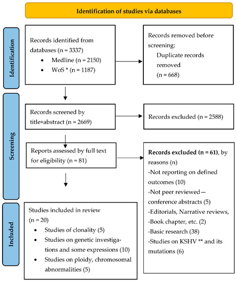
Figure 1.
PRISMA 2020 flow diagram [19] for systematic review on clonality and mutation in Kaposi sarcoma. * Web of Science, ** Kaposi Sarcoma related Herpes Virus research. Copyright statement: this PRISMA diagram contains public sector information licensed under the Open Government Licence v3.0. Adapted From: Moher D, Liberati A, Tetzlaff J, Altman DG, PLOS Medicine (OPEN ACCESS) Page MJ, McKenzie JE, Bossuyt PM, Boutron I, Hoffmann TC, Mulrow CD, et al. The PRISMA 2020 statement: an updated guideline for reporting systematic reviews. PLOS Medicine 2021;18(3):e1003583. doi: 10.1371/journal.pmed.1003583.
Results of the included studies are summarised in Table 2, Table 3 and Table 4 according to the following outcome categories: (case series) reporting outcomes on clonality, reporting on mutations and reporting on chromosomal aberrations. After applying the eligibility criteria, 5 studies reporting on clonality, 10 studies on various mutations and 5 studies reporting on chromosomal aberrations in KS were selected. The majority of publications were traditional case series; however, two of the selected studies aimed applied a different study design.
Tornesello et al. [21] performed a case control study investigating TP53 mutations in classic, epidemic and endemic KS cases, and Cerimele et al. [22] applied a prospective design to a case series obtained from a large cohort study in Sardinia. Nevertheless, only descriptive analysis of the genetic factors was provided by these studies as with the other case series, since the main aim of the studies relied on investigating the epidemiology of endemic cases. Studies provided descriptive results with frequency measures, and only a few studies provided analysis of the differences between groups.
The studies were performed using data from 13 different countries, and eight were international collaborations (Table 2, Table 3 and Table 4). The first study was published in 1984, and the most recent one was from 2015. Together, the studies enrolled 498 cases of KS and 421 controls; 46 cases and 12 controls to investigate clonality, 238 cases and 408 controls to study mutations and 214 cases and 1 control for chromosomal aberration studies. Studies on clonality included only female cases and were of poor reporting quality; often not reporting basic demographic variables, such as age. Additionally, the studies on mutations and chromosomal aberrations often did not provide adequate patient population and sample descriptions, making it difficult to assess the representativeness of the samples (Summary of Findings Table 2, Table 3 and Table 4).
Table 2.
Summary of findings: studies reporting on clonality.
Table 2.
Summary of findings: studies reporting on clonality.
| Included Studies (All Case Series) Reporting Outcomes on Clonality | |||||
|---|---|---|---|---|---|
| Study 1 | Study 2 | Study 3 | Study 4 | Study 5 | |
| Authors | Delabesse et al. [23] | Ding et al. [24] | Gil et al. [25] | Rabkin et al. [26] | Rabkin et al. [27] |
| Year (Country) | 1997 (France) | 2015 (China) | 1998 (USA) | 1995 (USA) | 1997 (Zambia and USA) |
| Sample Description (Ca+Ct) | 7 + 6 ♀ skin biopsies Age not reported. | 14 + 1 ♀ Mean age 48.4 years (range 27–71) | 12 ♀, 4 had multiple biopsies. Mean age 49 years (range 27–89). | 3 ♀ +number Ct not described Age not reported | 10 ♀, 5 lesion samples + 1 Ct for each Median age 26 years (range 20–35). |
| Case Classification | Clinically: 4 Classic KS, 3 AIDS SG: 1 macular, 3 plaque, 3 nodular | Clinically: 6 Classic KS, 8 AIDS SG: 2 macular, 11 plaque, 1 nodular Other: all cases HHV-8 + | Clinically: 2 Classic KS and 10 AIDS (8 advanced disease, 3 history of glucocorticoid use) SG: not reported. | Clinically: all HIV type 1-sero+ SG: all nodular KS lesions | Clinically: all HIV positive (8 serological, 2 clinical diagnosis) SG: multiple nodular KS |
| Study Results | |||||
| Description of Investigation Used | Punch biopsies HP: HES MA: DNA extraction Clonality AS: PCR amplification, HUMARA | HP: Surgical tissues, standard histology MA: IHCS of the primary mcAB. Clonality AS: PCR amplification, HUMARA detection of single-nucleotide polymorphism sites in PGK | HP: Cutaneous tumour biopsies. HES. Review performed by, to DNA results, blinded pathologist. MA: DNA extraction. Clonality AS: Secondary PCR products analysed by electrophoresis + autoradiography | HP: Cutaneous biopsies, HES MA: DNA extraction. Clonality AS: X chromosome inactivation assay (HUMARA) | HP: Cutaneous biopsies, HES MA: DNA extraction. Clonality AS: X chromosome inactivation assay (HUMARA) |
| Main Results | Descriptive: All 7 patients were heterozygous for HUMARA Clonality: All polyclonal pattern of inactivation | Descriptive: 2 Ca failed to amplify HUMARA, 11 analysed HUMARA, 5 KS PGK Clonality: 1Ca+1Ct polyclonal pattern, rest monoclonal Ca with no significant differences between groups (p > 0.05) | Descriptive: 41 different regions from 24 biopsies were studied. Clonality: 5 Ca clonal, 2 Ca inactivate, 7 Ca polyclonal pattern of inactivation, 2 Ca both clonal/polyclonal inactivation | Descriptive: All 3 patients heterozygous androgen receptor Clonality: 2 of 3 Ca monoclonal pattern | Descriptive: 2 Ca excluded (homozygous HUMARA), 8 Ca (40 tumours, 32 studied) Clonality: 23 (85%) tumours had unbalanced methylation (predominance one allele), 6 Ca (23 tumours) concordant methylation (≤0.00001). |
| Conclusions | It is a polyclonal cell proliferation. | Suggest a clonal neoplasm. | Suggest a clonal neoplasm, but polyclonal inactivation pattern observed may be premalignant stage or false negative results. | Suggest a clonal neoplasm (at least in AIDS Ca). | Data indicate monoclonal cancer. |
♀: female, AIDS: AIDS associated Kaposi Sarcoma, Ca: Cases Ct: Controls, Clonality AS: Clonality assay, HES: Haematoxylin and eosin staining, HP: Histopathology, HUMARA: HUMARA gen polymorphism analysis, IHCS: Immunohistochemical staining, KS: Kaposi Sarcoma, MA: Molecular analysis, mcAB: monoclonal antibody, PGK: Phosphoglycerate kinase gene, SG: Staging. When data not reported in the table means the original article did not provide them.
Table 3.
Summary of findings: studies reporting on mutations.
Table 3.
Summary of findings: studies reporting on mutations.
| Included Studies Reporting Outcomes on Mutations | ||||||||||
|---|---|---|---|---|---|---|---|---|---|---|
| Study 1 | Study 2 | Study 3 | Study 4 | Study 5 | Study 6 | Study 7 | Study 8 | Study 9 | Study 10 | |
| Authors | Nicolaides et al. [28] | Kiuru-Kuhlefelt et al. [29] | Cerimele et al. [22] | Tornesello et al. [21] | Huang et al. [30] | Guttman-Yassky et al. [31] | Feller et al. [32] | Cordiali-Fei et al. [33] | Li, Jian J. [34] | Scinicariello et al. [35] |
| Year (Country) | 1994 (USA) | 2000 (Finland + USA) | 1984 (Italy) | 2009 (Italy + Uganda + Greece) | 1993 (USA) | 2012 (USA + Israel) | 2014 (USA) | 2014 (Italy) | 1997 (USA) | 1994 (USA) |
| Study Design | Case series | Case series | Case series | Case control | Case series | Case series | Case series | Case series | Case series | Case series |
| Sample Description (Ca + Ct) | 31 Ca No demographics reported. | 12 Ca 10 ♂, 2 ♀ Age: NR | (65) 22 + 220 49 ♂, 16 ♀ Age: range 57–82 years | 67 + 150 No data disaggregated for ♂♀ Median age: 75 (29.5–47) years | 38 + 10 No demographics reported. | 9 + 4 6 ♂, 7 ♀ Mean age: Ca 69.5 (20–86) years | 24 + 17 21 ♂, 3 ♀ Mean age: Ca 59 (26–86) years | 3 Ca same family 2 ♂, 1 ♀ Age: 63, 64, 40 years | 15 + 5 No demographics reported. | 17 Ca No demographics reported. |
| Case Classification | Clinically: 24 HIV+, 7 HIV− SG: 16 nodular, 6 patch, 9 plaque | Clinically: 6 ♂ HIV+ or AIDS | Clinically: all endemic KS (Sardinia). | Clinically: 33 classic, 2 iatrogenic, 19 epidemic and 13 epidemic Other: all HHV8+ | Clinically: 31 AIDS, 7 classic | Clinically: All classic SG: 4 early, 2 mixed and 3 nodular | Clinically: 6 HIV+, rest unknown SG: 9 patch, 7 plaque, 8 nodular | Clinically: All HIV− Other: all HHV-8+ | Clinically: All AIDS | Clinically: 10 Classic, 7 HIV+ |
| Study Results | ||||||||||
| Description of Investigation Used | GenA: PCR-SSCP (Orita procedure) to detect mutations | GenA: PCR HHV-8 sequence specific primers, aCGH, digital image analysis, FISH, IHCS | GenA: THLA-ABC typing with 162 antisera. HLA-DR and MT typing using 47 antisera. StatA: significance calculated using X’ test+Wolf’s relative risk test. | HP: Cutaneous biopsies GenA: TP53 genotype at codon 72 StatA: Fisher’s exact or X’ test for comparison of Ca/Ct. Student’s t test for age differences | HP: Biopsy/autopsy samples. GenA: R T-PCR, DNA sequencing, ICHS, Southern blot hybridization | HP: Punch biopsies GenA: Gene chip analysis, DNA microarray analysis, IHCS, Immunoflouresence+, obtaining expression profile LEC and BEC gene signature | HP: All sections reviewed by dermatopathologists GenA: FISH, IHCS | Not reported | GenA: RT-PCR + PCR-SSCP in immunoperoxidase stains | GenA: DNA+ PCR amplification, HPV DNA detection, p53 direct sequencing with (ɣ32P) ATP end-labelled primers |
| Main Results | Genetics: 10 Kras overexpression (3 Kras amplification, 7 various mutations Kras exon) | Genetics: 4 recurrent again at 11q13; 4 containingFGF4 and INT2 (expression of FGF4 and INT2 was found in 9 and 3 Ca respectively). | Genetics: No differences in A, B, C antigen frequency (DR5 72.7% Ca, 23.1% Ct, p < 0.0001; DR3 9.1% Ca 53.6% Ct, p < 0.01; DR5 36.4% Ca 18.1% Ct, p not significant) | Genetics: African Ca: PHoZ 50%, PHtZ 31.8%, AHoZ 18.2%, (p = 0.1872) Caucasian Ca: PHoZ 6.7, PHtZ 55.6, AHoZ 37.8%, (p = 0.0567). Stratified by HIV: No significant differences in alleles | Genetics: Int-2 expressed in 21 Ca (55.2%), NASalt in 8. Most variations in int-2 cDNA located in exon 1 (four in exons 2 and 3) | Genetics: Gene expression level markers gradually increased from normal through all KS stages, particularly LEC genes. | Genetics: c-myc amplification in all Ca, IHCS positive for c-Myc in 13 Ca (54%) | Genetics: IL-6 promoter polymorphism G-174C (2 ♂ HtZ, ♀ HoZ) | Genetics: 4 Ca p53 in nuclei+cytoplasm, 5 MDM2 in the nuclei (2 IHCS+ p53, 3 IHCS− p53 protein) | Genetics: 4 Ca (23%) HPV DNA detected (1 AIDS), 5 Ca (24%) p53 HtZ (none in HPV +) |
| Conclusions | Suggests K-ras mutation plays a significant role in KS oncogenesis. | No evidence of HHV-8 integration to genome. | Preliminary evidence of structural chromosome rearrangement. | p53 polymorphism at codon 72 does not represent a RF for KS. | Int-2 expression may play a role in KS oncogenesis. | Suggests local expression of chemokines/growth factors (no clonal exp.) as oncogenesis. | No amplification of the c-myc gene detected. | Suggests that EBV can cause HHV-8 reactivation in predisposed Ca causing KS. | Suggest p53 may be involved in AIDS KS pathogenesis. | Indicate role of HPV to KS pathogenesis and p53 alteration to malignancy progression. |
♂: Male, ♀: female, aCGH: array comparative genomic hybridization, AIDS: AIDS associated Kaposi Sarcoma, Ca: Cases Ct: Controls, FISH: Interphase fluorescence in situ hybridization, GenA: Genetic analysis, HP: Histopathology, PHoZ: Proline homozygous, PHtZ: Proline heterozygous, IHCS: Immunohistochemical staining, KS: Kaposi Sarcoma, NR: Not reported, NASalt: Nucleic acid sequence alterations, PCR-SSCP: Polymerase chain reaction-single-strand conformation polymorphism analysis, RF: Risk factor, R T-PCR: Reverse transcription-PCR, SG: Staging, and StatA: Statistical analysis. When data not reported in the table means the original article did not provide them.
Table 4.
Summary of findings: studies reporting on chromosomal aberrations.
Table 4.
Summary of findings: studies reporting on chromosomal aberrations.
| Included Studies (All Case Series) Reporting Chromosomal Aberrations | |||||
|---|---|---|---|---|---|
| Study 1 | Study 2 | Study 3 | Study 4 | Study 5 | |
| Authors | Kaaya et al. [36] | Kaaya et al. [37] | Bisceglia et al. [38] | Pyakurel et al. [39] | Reizis et al. [40] |
| Year (Country) | 2000 (Sweden + Tanzania) | 1992 (Sweden + Tanzania) | 1991 (UK + Italy) | 2006 (Sweden + Germany + Tanzania) | 1995 (Israel) |
| Sample Description (Ca + Ct) | 32 Ca (12 Ca ploidy analysis) 22 ♂, 10 ♀ Age: range 8–68 years | 20 Ca 17 ♂, 3 ♀ Mean age: 40 (up to 83) years | 96 Ca (66 analysed, 143 biopsies) 69 ♂, 27 ♀ Mean age: 69 (10–89) years | 27 + 1 Mean age: males 37.5 (23–65) years, females not reported | 39 Ca Mean age: Iatrogenic Ca 68 (54–80) years |
| Case Classification | Clinically: 8 endemic, 24 AIDS SG: 24 nodular, 4 patch, 4 plaque Other: 12 Ploidy analysed Ca (6 endemic, 6 AIDS) | Clinically: 10 endemic, 10 AIDS SG: 17 nodular skin, and 3 generalised lymph node lesions | Clinically: 93% sporadic, 6% AIDS, 1% Hepatitis B | Clinically: 9 Endemic, 18 AIDS SG: 18 nodular, 9 patch | Clinically: 31 classic, 8 iatrogenic (steroid induced) |
| Study Results | |||||
| Description of Investigation Used | HP: Surgical tissues Mol.An: HHV-8 DNA PCR, IHCS, Ploidy by DNA flow cytometry, apoptotic cells (TUNEL-assay) | HP: Surgical tissues. Mol.An: IHCS, DNA measurements | HP: slides classified by histological criteria Mol.An: Mitoses count, Flow Cytometry (DNA aneuploidy ≥1 GO/Gl peak modal channel number) | HP: Surgical tissues Mol.An: Ligation-mediated PCR+DNA labelling, aCGH, FISH | HP: tissue blocks Mol.An: Flowcytometry |
| Main Results | Ploidy: All 12 diploid cellular DNA content, and low numbers of cells (1.6–8.9%) in S and G2 phase. Ploidy values similar normal cells in non-involved tissue of the same section. In contrast the malignant cell line (KS Y-1) showed a near triploid, aneuploid DNA content and a moderate proliferation rate (13% cells in S + G2 phase). | Ploidy: 70% cells contained DNA values ≥2.5, but not greater than 5C level. Both clinical types with euploid DNA pattern. | Ploidy: 6 lesions (5.8%) DNA aneuploid with a clustering around a DNA index of 1.5 (range 1.4–1.6). Increasing mitotic counts and S-phase plus G2-phase cells were seen with progression of the phase and pattern of disease. Nodular and spindle cell forms had the highest mitotic counts and S-phase plus G2-phase cells. | Chromosomal results: 20 (87%) Ca only recurrent aberration loss of Y chromosome One patch Ca showed in addition loss of Xq. Nodular showed recurrent copy number changes in chromosomes 16, 17, 21, X, Y, and other random changes. | Ploidy: 28 classic Ca showed a diploid pattern. Of 8 iatrogenic, 7 were aneuploid and 1 diploid. |
| Conclusions | Represents a diploid, probably reactive, cell proliferation, which progressively increases the expression of antiapoptotic factors (cellular and viral). | Corroborates previous suggestions that KS could represent a reactive process, rather than a clonal proliferation. | Suggest a low level of DNA aneuploidy, but flow cytometry does not solve the dilemma of whether KS is a hyperplastic or neoplastic process. | Support the view that KS (in males) develops into a clonal tumour yet initially is a hyperplastic reactive cell proliferation. | Iatrogenic KS mostly aneuploid pattern, classic KS diploid pattern on flow cytometry. |
♂: Male, ♀: female, aCGH: array comparative genomic hybridization, AIDS: AIDS associated Kaposi Sarcoma, Ca: Cases Ct: Controls, FISH: Interphase fluorescence in situ hybridization, Mol.An: Molecular analysis, HP: Histopathology, IHCS: Immunohistochemical staining, and SG: Staging. When data not reported in the table means the original article did not provide them.
Descriptions of clinical and morphological (histopathological) descriptions, such as plaque, nodular and patch stage, were frequently incomplete but still showed an important diversity of case combinations when studying the different outcomes, as follows:
- Clinical and morphological description of clonality studies: 34 (74%) of the 46 cases were HIV associated, 12 (16%) classic KS forms of all 17 (37%) studied nodular lesions, 3 (7%) were plaque lesion samples, and the 26 (57%) remaining cases were from 2 studies [25,27], which did not specify morphological characteristics of the lesions.Clinical and morphological description of mutation studies: in total, 84 (29%) HIV associated cases were included, 146 (52%) classic KS and 2 (1%) cases of iatrogenic KS. For 49 (17%) cases, no clinical information was available since Nicolaides et al. [28], comprising 31 cases, did not describe their clinical characteristics and Feller et al. [32] described only 6 of 18 included cases. All together reported that 20 (7%) were nodular lesions, 27 (10%) plaque lesions and 17 (6%) patch lesions, but these data were missing for 217 (77%) cases since 7 [21,22,29,30,33,34,35] of the 10 included studies did not specify the morphological characteristics of the lesions. Two studies reported separately for HHV8 positivity, Tornesello et al. [21] with all 67 cases reported as positive and Cordiali-Fei et al. [33] detecting in all the three cases of the studied family high titres of anti-HHV-8 (type A virus) antibodies.
- Clinical and morphological description of chromosomal aberrations studies: 58 (27%) of the 214 cases were HIV associated, 147 (69%) classic and 9 (4%) iatrogenic KS presentations. Only 3 studies [36,37,39] reported on morphological stages of these cases summing 3 (2%) plaque, 37 (17%) nodular, 23 (11%) lesion samples and 151 (70%) cases where it was not reported.Outcomes assessing the clonal nature of KS provided by the 20 included studies (Table 2, Table 3 and Table 4) resulted very heterogeneous (Figure 2). For a more comprehensive synthesis of the results, we decided to report retrieved outcomes grouped into the mentioned three categories of studied genetic alterations. However, differences in study aims, outcome definitions, applied methods and reported outcome measures limited seriously our possibility to pool and/or compare the published data. This heterogeneity in reported data was also the reason for ruling out a quantitative synthesis of the retrieved results, and the performance of a meta-analysis was excluded.
 Figure 2. Outcomes of included studies assessing the neoplastic nature of Kaposi sarcoma.
 - Determining clonality: outcomes on clonality in KS samples were mostly determined by monoclonal patterns of gene inactivation or methylation and obtained mixed results. Four case series [24,25,26,27] obtained results suggesting that KS is a clonal neoplasm, while one study obtained results compatible with the description of a polyclonal cell [23]. All studies presented a high risk of bias as assessed by the adapted Quadas-2 tool due to bias inherent to their study design and insufficient reporting.
- Detecting mutations: Ten studies aimed to detect mutations in KS lesions, focusing three on p53 mutations [21,34,35] and seven on other different single mutations (IL-6, c-myc, LEC and BEC gene signatures, FGF3, HLA, FGF4 and KRAS). Each study applied different laboratory technics, determined different outcomes and reported diverse findings. Of these ten studies, three [21,29,32] reported negative results for the investigated mutation, and the other seven obtained outcomes suggested a possible relationship. Of the three studies [21,34,35] focusing on a possible role of p53 mutation in the KS oncogenesis, two [29,35] obtained results that point to a possible implication, and one case control study [21] with a large sample and well-performed statistical analysis failed in obtaining evidence of a possible association. These studies presented a high risk of bias, including the prospective case series [22] and the case control study [21] due to difficulties in assessing the internal and external validity of the studies based on the reported data.
- Demonstrating chromosomal aberrations: Finally, of the five selected studies [36,37,38,39,40] that investigated chromosomal aberrations, two studies [36,38] reported diploidy Reizis et al. [38] (a type of pattern for iatrogenic forms of KS) and aneuploid patterns for the classic form of KS. Another study [40] detected low levels of DNA aneuploidy, and the other two [37,41] reported results compatible with a hyperplastic reactive cell proliferation. Again, a high risk of bias was detected for all included studies due to limitations inherent to the study design and through a lack of detailed reporting of methods. Table 2, Table 3 and Table 4 summarize our findings.
4. Discussion
We performed a systematic review of the peer-reviewed and published scientific literature on genetic alterations assessing the clonal nature of KS. The limited number of retrieved studies and the low-level of evidence of the retrieved studies [42,43], together with the low number of recent publications investigating this topic suggests that this potentially controversial topic has not been studied in depth from a broad, multidisciplinary perspective. Scientific interest in recent years has focused mainly on describing the viral oncogenic role through mechanistic studies [44,45,46,47,48] and relatively little attention has been paid to the analysis of factors influencing clonal growth in KS. However, the identification of such underlying mechanisms and subsequent definitions could lead to improvements in the clinical management of KS patients.
That said, there are several aspects of the pathogenesis of KS that are currently under investigation to determine the relevance of latency-associated nuclear antigen (LANA) and other HHV8 proteins in subverting normal endothelial cell biology to induce proliferation [49,50] and to describe the miRNA-34A effect [51]. Evidence from basic research proposing that HHV8 is an integrating oncovirus that causes amplification and activation of oncogenes needs to be further investigated by the sequencing of early and advanced lesions considering the latent state in which HHV8 has been reported to exist [52] as the clonality of HHV8 integration is sometimes questioned. While many studies have been published reporting genetic alterations in KS samples, few have analysed whether these are driver mutations that might be involved in oncogenesis [53,54].
We retrieved only two studies with an appropriate study design for the investigation of such associations; however, both focused on epidemiological aspects of the disease and did not provide effect measures analysing the relationship of a mutation or clonality with KS. Nicolaides et al. [28] showed KRAS mutation of amplification in substantial numbers of advanced cases in which codons 13, 15, 16, 18 and 31 were implicated. A small number of patients with the TP53 mutation have been found and may be associated with progression [34,35]. TP53 immunohistochemistry has been used, and expression appears to increase with progression [55]. The ability of NGS to examine intra-tumoral heterogeneity, mutation load, copy number variation and temporal heterogeneity makes this a major gap in the current knowledge of KS.
Clonality of the virus can be shown [56,57] but is not direct evidence of clonality of the host cells. Indeed, this may simply represent clonal restriction following polyclonal infection [23]. However, in a large study of 98 patients, Duprez et al. [58] used an HHV-8-fused terminal repeat (TR) to follow up an earlier study [59] to show that most lesions were oligoclonal and concluded that KS was a reactive process in most patients, although many samples were not informative [58].
They found that differences ‘strongly suggest that disseminated lesions represent multiple distinct primary expansions of HHV-8-infected spindle cells originating from different infectious events rather than metastatic proliferations’. It is known that EBV-induced lymphoproliferative disease starts as an oligoclonal proliferation and can lead to monoclonal diffuse large B-cell lymphoma with the adoption of additional genetic mutations; however, the same has not been shown for KS—although it remains a possibility.
The chronic and slow evolution of classic-KS lesions, the partial reversibility of iatrogenic-KS lesions after diminution of immunosuppressive regimens and the complete epidemic-KS regression after anti-retroviral therapy seem more consistent with reactive proliferation [23,26,38,40]. Although there are occasional reports of spontaneous regression of individual lesions (and rarely of all disease) [60]. Dependence on cytokines, such as basic fibroblast growth factor, oncostatin M and interleukin 6, has been shown for the growth of Kaposi’s sarcoma-derived cells in vitro [31]. However, a commonly used cell line (KS Y-1) derived from KS lesions does produce tumours in nude mice [61].
Detailed, and ideally longitudinal, NGS studies with examination of multiples of a range of early (cutaneous patch stage) and late (visceral) lesions in individual patients could provide evidence of clonal selection and progression. Such an approach would evaluate for neoplastic transformation of early lesions.
5. Conclusions
In conclusion, insufficient scientific evidence exists to describe the nature of KS. Current knowledge is insufficient to determine whether KS is a clonal neoplasm, or simply an aggressive reactive virus-driven lesion. Although evidence from mechanistic studies that points towards a reactive virus-driven process that gives rise to a clonal neoplasm is mounting, direct evidence for the pathogenic pathway would allow an evidence-based assessment of KS neoplastic status with all the implications that this may have for clinical practice and patient care. Its current neoplastic classification as a sarcoma is questionable, and regarding it as a ‘Kaposi tumour’ may be more accurate.
Supplementary Materials
The following supporting information can be downloaded at: https://www.mdpi.com/article/10.3390/cancers14051201/s1, Supplementary Materials S1: Detailed search strategy for MEDLINE and WEB OF SCIENCE, Supplementary Materials S2: Standardised data extraction form.
Author Contributions
I.A.C., B.I.I.R., L.U. and A.J.L. developed the study concept and design. B.I.I.R., L.U. and R.W. developed search strategy and performed the search, B.I.I.R., A.J.L. and I.A.C. conducted the study selection and data extraction. B.I.I.R., I.A.C., A.J.L., V.A.W., R.W. and S.A. performed data analysis. B.I.I.R., I.A.C., S.A. and V.A.W. wrote and revised the paper. All authors reviewed the final draft and approved the final paper. All authors have read and agreed to the published version of the manuscript.
Funding
This research received no external funding. The WHO Classification of Tumours is published by IARC on a not-for-profit basis, funded solely by sales of its books and subscription website.
Data Availability Statement
All data generated or analysed during this study are included in this published article and its Supplementary Information Files.
Acknowledgments
We would like to thank Latifa Bouanzi for her expert support with the search strategies and retrieval of articles.
Conflicts of Interest
The authors declare no conflict of interest.
Declarations
The content of this article represents the personal views of the authors and does not represent the views of the authors’ employers and associated institutions. Where authors are identified as personnel of the International Agency for Research on Cancer/World Health Organization, the authors alone are responsible for the views expressed in this article and they do not necessarily represent the decisions, policy or views of the International Agency for Research on Cancer/World Health Organization.
References
- Curtiss, P.; Strazzulla, L.C.; Friedman-Kien, A.E. An Update on Kaposi’s Sarcoma: Epidemiology, Pathogenesis and Treatment. Dermatol. Ther. 2016, 6, 465–470. [Google Scholar] [CrossRef] [PubMed]
- Shiels, R.A. A history of Kaposi’s sarcoma. J. R. Soc. Med. 1986, 79, 532–534. [Google Scholar] [CrossRef] [PubMed]
- Vangipuram, R.; Tyring, S.K. AIDS-Associated Malignancies. Cancer Treat. Res. 2019, 177, 1–21. [Google Scholar] [PubMed]
- Vangipuram, R.; Tyring, S.K. Epidemiology of Kaposi sarcoma: Review and description of the nonepidemic variant. Int. J. Dermatol. 2019, 58, 538–542. [Google Scholar] [CrossRef]
- Mariggio, G.; Koch, S.; Schulz, T.F. Kaposi sarcoma herpesvirus pathogenesis. Philos. Trans. R. Soc. Lond. Ser. B Biol. Sci. 2017, 372, 20160275. [Google Scholar] [CrossRef]
- Ferlay, J.; Colombet, M.; Soerjomataram, I.; Mathers, C.; Parkin, D.M.; Piñeros, M.; Znaor, A.; Bray, F. Estimating the global cancer incidence and mortality in 2018: GLOBOCAN sources and methods. Int. J. Cancer 2019, 144, 1941–1953. [Google Scholar] [CrossRef]
- Etta, E.M.; Alayande, D.P.; Mavhandu-Ramarumo, L.G.; Gachara, G.; Bessong, P.O. HHV-8 Seroprevalence and Genotype Distribution in Africa, 1998(-)2017: A Systematic Review. Viruses 2018, 10, 458. [Google Scholar] [CrossRef]
- Phipps, W.; Ssewankambo, F.; Nguyen, H.; Saracino, M.; Wald, A.; Corey, L.; Orem, J.; Kambugu, A.; Casper, C. Gender differences in clinical presentation and outcomes of epidemic Kaposi sarcoma in Uganda. PLoS ONE 2010, 5, e13936. [Google Scholar] [CrossRef]
- Willis, R.A. Tumor seminar. Tex. State J. Med. 1950, 46, 611–638. [Google Scholar]
- Chang, Y.; Cesarman, E.; Pessin, M.S.; Lee, F.; Culpepper, J.; Knowles, D.M.; Moore, P.S. Identification of herpesvirus-like DNA sequences in AIDS-associated Kaposi’s sarcoma. Science 1994, 266, 1865–1869. [Google Scholar] [CrossRef]
- Greaves, M.; Maley, C.C. Clonal evolution in cancer. Nature 2012, 481, 306–313. [Google Scholar] [CrossRef] [PubMed]
- Parsons, B.L. Multiclonal tumor origin: Evidence and implications. Mutat. Res. Rev. Mutat. Res. 2018, 777, 1–18. [Google Scholar] [CrossRef] [PubMed]
- Erickson-Johnson, M.R.; Chou, M.M.; Evers, B.R.; Roth, C.W.; Seys, A.R.; Jin, L.; Ye, Y.; Lau, A.W.; Wang, X.; Oliveira, A.M. Nodular fasciitis: A novel model of transient neoplasia induced by MYH9-USP6 gene fusion. Lab. Investig. A J. Tech. Methods Pathology. 2011, 91, 1427–1433. [Google Scholar] [CrossRef] [PubMed]
- Callan, M.F.; Steven, N.; Krausa, P.; Wilson, J.D.; Moss, P.A.; Gillespie, G.M.; Bell, J.I.; Rickinson, A.B.; McMichael, A.J. Large clonal expansions of CD8+ T cells in acute infectious mononucleosis. Nat. Med. 1996, 2, 906–911. [Google Scholar] [CrossRef] [PubMed]
- Winter, J.R.; Jackson, C.; Lewis, J.E.; Taylor, G.S.; Thomas, O.G.; Stagg, H.R. Predictors of Epstein-Barr virus serostatus and implications for vaccine policy: A systematic review of the literature. J. Glob. Health. 2020, 10, 010404. [Google Scholar] [CrossRef] [PubMed]
- Knudson, A.G., Jr. Mutation and cancer: Statistical study of retinoblastoma. Proc. Natl. Acad. Sci. USA 1971, 68, 820–823. [Google Scholar] [CrossRef]
- Martincorena, I.; Raine, K.M.; Gerstung, M.; Dawson, K.J.; Haase, K.; Van Loo, P.; Davies, H.; Stratton, M.R.; Campbell, P.J. Universal Patterns of Selection in Cancer and Somatic Tissues. Cell 2017, 171, 1029–1041.e21. [Google Scholar] [CrossRef]
- Martincorena, I. Somatic mutation and clonal expansions in human tissues. Genome Med. 2019, 11, 35. [Google Scholar] [CrossRef]
- Page, M.J.; McKenzie, J.E.; Bossuyt, P.M.; Boutron, I.; Hoffmann, T.C.; Mulrow, C.D.; Shamseer, L.; Tetzlaff, J.M.; Akl, E.A.; Brennan, S.E.; et al. The PRISMA 2020 statement: An updated guideline for reporting systematic reviews. BMJ 2021, 372, n71. [Google Scholar] [CrossRef]
- Whiting, P.F.; Rutjes, A.W.; Westwood, M.E.; Mallett, S.; Deeks, J.J.; Reitsma, J.B.; Leeflang, M.M.; Sterne, J.A.; Bossuyt, P.M. QUADAS-2: A revised tool for the quality assessment of diagnostic accuracy studies. Ann. Intern. Med. 2011, 155, 529–536. [Google Scholar] [CrossRef]
- Tornesello, M.L.; Biryahwaho, B.; Downing, R.; Hatzakis, A.; Alessi, E.; Cusini, M.; Ruocco, V.; Katongole-Mbidde, E.; Buonaguro, L.; Buonaguro, F.M. TP53 codon 72 polymorphism in classic, endemic and epidemic Kaposi’s sarcoma in African and Caucasian patients. Oncology 2009, 77, 328–334. [Google Scholar] [CrossRef] [PubMed]
- Cerimele, D.; Contu, L.; Scappaticci, S.; Cottoni, F. Kaposi’s sarcoma in Sardinia: An epidemiologic and genetic investigation. Ann. N. Y. Acad. Sci. 1984, 437, 216–227. [Google Scholar] [CrossRef] [PubMed]
- Delabesse, E.; Oksenhendler, E.; Lebbe, C.; Verola, O.; Varet, B.; Turhan, A.G. Molecular analysis of clonality in Kaposi’s sarcoma. J. Clin. Pathol. 1997, 50, 664–668. [Google Scholar] [CrossRef] [PubMed][Green Version]
- Yuan, D.; XiuJuan, W.; Yan, Z.; JunQin, L.; Fang, X.; Shirong, Y.; Xiaojing, K.; Yanyan, F.; Weidong, W.; Dong, L.; et al. Use of X-Chromosome Inactivation Pattern to Analyze the Clonality of 14 Female Cases of Kaposi Sarcoma. Med. Sci. Monit. Basic Res. 2015, 21, 116–122. [Google Scholar]
- Gill, P.S.; Tsai, Y.C.; Rao, A.P.; Spruck, C.H.; Zheng, T.; Harrington, W.A.; Cheung, T.; Nathwani, B.; Jones, P.A. Evidence for multiclonality in multicentric Kaposi’s sarcoma. Proc. Natl. Acad. Sci. USA 1998, 95, 8257–8261. [Google Scholar] [CrossRef]
- Rabkin, C.S.; Biggar, R.J.; Coleman, A.; Musaba, E.; Chibwe, G.; Janz, S. Clonality of aids-related kaposis-sarcoma. Aids Res. Hum. Retrovir. 1995, 11, S74. [Google Scholar]
- Rabkin, C.S.; Janz, S.; Lash, A.; Coleman, A.E.; Musaba, E.; Liotta, L.; Biggar, R.J.; Zhuang, Z. Monoclonal origin of multicentric Kaposi’s sarcoma lesions. N. Engl. J. Med. 1997, 336, 988–993. [Google Scholar] [CrossRef]
- Nicolaides, A.; Huang, Y.Q.; Li, J.J.; Zhang, W.G.; Friedman-Kien, A.E. Gene amplification and multiple mutations of the K-ras oncogene in Kaposi’s sarcoma. Anticancer Res. 1994, 14, 921–926. [Google Scholar]
- Kiuru-Kuhlefelt, S.; Sarlomo-Rikala, M.; Larramendy, M.L.; Söderlund, M.; Hedman, K.; Miettinen, M.; Knuutila, S. FGF4 and INT2 oncogenes are amplified and expressed in Kaposi’s sarcoma. Mod. Pathol. 2000, 13, 433–437. [Google Scholar] [CrossRef]
- Huang, Y.Q.; Li, J.J.; Moscatelli, D.; Basilico, C.; Nicolaides, A.; Zhang, W.G.; Poiesz, B.J.; Friedman-Kien, A.E. Expression of int-2 oncogene in Kaposi’s sarcoma lesions. J. Clin. Investig. 1993, 91, 1191–1197. [Google Scholar] [CrossRef]
- Ensoli, B.; Sturzl, M.; Monini, P. Cytokine-mediated growth promotion of Kaposi’s sarcoma and primary effusion lymphoma. Semin. Cancer Biol. 2000, 10, 367–381. [Google Scholar] [CrossRef] [PubMed]
- Feller, K.; Yang, S.; Tung, N.; Lee, J.; Mahalingam, M. c-myc in Kaposi’s sarcoma: Analyses by fluorescent in situ hybridization and immunohistochemistry. J. Eur. Acad. Dermatol. Venereol. 2014, 28, 120–124. [Google Scholar] [CrossRef] [PubMed]
- Cordiali-Fei, P.; Latini, A.; Trento, E.; Zampatti, S.; Ferraresi, V.; Cota, C.; Volpi, S.; D’agosto, G.; Bordignon, V.; Giardina, E.; et al. Familial Kaposi’s Sarcoma in HHV8 infected subjects presenting the G-174C allele of the IL-6 promoter: A possible role for EBV? Eur. J. Dermatol. 2014, 24, 503–504. [Google Scholar] [CrossRef]
- Li, J.J.; Huang, Y.Q.; Cockerell, C.J.; Zhang, W.G.; Nicolaides, A.; Friedman-Kien, A.E. Expression and mutation of the tumor suppressor gene p53 in AIDS-associated Kaposi’s sarcoma. Am. J. Dermatopathol. 1997, 19, 373–378. [Google Scholar] [CrossRef]
- Scinicariello, F.; Dolan, M.J.; Nedelcu, I.; Tyring, S.K.; Hilliard, J.K. Occurrence of human papillomavirus and p53 gene mutations in Kaposi’s sarcoma. Virology 1994, 203, 153–157. [Google Scholar] [CrossRef] [PubMed]
- Kaaya, E.; Castanos-Velez, E.; Heiden, T.; Ekman, M.; Catrina, A.I.; Kitinya, J.; Andersson, L.; Biberfeld, P. Proliferation and apoptosis in the evolution of endemic and acquired immunodeficiency syndrome-related Kaposi’s sarcoma. Med. Oncol. 2000, 17, 325–332. [Google Scholar] [CrossRef]
- Kaaya, E.E.; Parravicini, C.; Sundelin, B.; Mgaya, E.; Kitinya, J.; Lema, L.; Luande, J.; Biberfeld, P. Spindle cell ploidy and proliferation in endemic and epidemic African Kaposi’s sarcoma. Eur. J. Cancer 1992, 28a, 1890–1894. [Google Scholar] [CrossRef]
- Reizis, Z.; Trattner, A.; Katzenelson, V.; David, M.; Rotem, A.; Nativ, O.; Mor, Y. Flow cytometric DNA analysis of classic and steroid-induced Kaposi’s sarcoma. Br. J. Dermatol. 1995, 132, 548–550. [Google Scholar] [CrossRef]
- Pyakurel, P.; Montag, U.; Castaños-Vélez, E.; Kaaya, E.; Christensson, B.; Tönnies, H.; Biberfeld, P.; Heiden, T. CGH of microdissected Kaposi’s sarcoma lesions reveals recurrent loss of chromosome Y in early and additional chromosomal changes in late tumour stages. AIDS 2006, 20, 1805–1812. [Google Scholar] [CrossRef]
- Bisceglia, M.; Bosman, C.; Quirke, P. A histologic and flow cytometric study of Kaposi’s sarcoma. Cancer 1992, 69, 793–798. [Google Scholar] [CrossRef]
- Pyakurel, P.; Pak, F.; Mwakigonja, A.R.; Kaaya, E.; Heiden, T.; Biberfeld, P. Lymphatic and vascular origin of Kaposi’s sarcoma spindle cells during tumor development. Int. J. Cancer 2006, 119, 1262–1267. [Google Scholar] [CrossRef]
- Murad, M.H.; Asi, N.; Alsawas, M.; Alahdab, F. New evidence pyramid. Evid. Based Med. 2016, 21, 125–127. [Google Scholar] [CrossRef] [PubMed]
- Burns, P.B.; Rohrich, R.J.; Chung, K.C. The levels of evidence and their role in evidence-based medicine. Plast Reconstr. Surg. 2011, 128, 305–310. [Google Scholar] [CrossRef] [PubMed]
- Goncales, J.P.; Júnior, J.V.; Lopes, T.R.; Tozetto-Mendoza, T.R.; de Farias Guimarães, D.; de Morais, V.M.; Coêlho, M.R. Association of polymorphisms in NFκB1 promoter and NFκBIA gene with the development of antibodies against HHV-8 in HIV-infected individuals. Virology 2019, 535, 255–260. [Google Scholar] [CrossRef] [PubMed]
- Sbihi, Z.; Dossier, A.; Boutboul, D.; Galicier, L.; Parizot, C.; Emarre, A.; Hoareau, B.; Dupin, N.; Marcelin, A.-G.; Oudin, A.; et al. iNKT and memory B-cell alterations in HHV-8 multicentric Castleman disease. Blood 2017, 129, 855–865. [Google Scholar] [CrossRef] [PubMed]
- Jackson, C.C.; Dickson, M.A.; Sadjadi, M.; Gessain, A.; Abel, L.; Jouanguy, E.; Casanova, J.-L. Kaposi Sarcoma of Childhood: Inborn or Acquired Immunodeficiency to Oncogenic HHV-8. Pediatric Blood Cancer 2016, 63, 392–397. [Google Scholar] [CrossRef] [PubMed]
- Barbarov, I.; Koren-Michowitz, M.; Schiby, G.; Portnoy, O.; Livingstone, D.; Segal, G. Fulminant HHV-8 associated Castleman’s disease in a non-HIV, Kaposi sarcoma patient with borderline hemophagocytic syndrome. IMAJ 2015, 17, 253–255. [Google Scholar]
- Koreishi, A.F.; Saenz, A.J.; Arcila, M.E.; Hedvat, C.; Fleming, S.; Teruya-Feldstein, J. Synchronous Follicular Lymphoma, Kaposi Sarcoma, and Castleman’s Disease in a HIV-Negative Patient With EBV and HHV-8 Coinfection. Int. J. Surg. Pathol. 2011, 19, 685–691. [Google Scholar] [CrossRef]
- Jain, V.; Plaisance-Bonstaff, K.; Sangani, R.; Lanier, C.; Dolce, A.; Hu, J.; Brulois, K.; Haecker, I.; Turner, P.; Renne, R.; et al. A Toolbox for Herpesvirus miRNA Research: Construction of a Complete Set of KSHV miRNA Deletion Mutants. Viruses 2016, 8, 54. [Google Scholar] [CrossRef]
- Katano, H.; Sato, Y.; Sata, T. Expression of p53 and human herpesvirus-8 (HHV-8)-encoded latency-associated nuclear antigen with inhibition of apoptosis in HHV-8-associated malignancies. Cancer 2001, 92, 3076–3084. [Google Scholar] [CrossRef]
- Krause, C.J.; Popp, O.; Thirunarayanan, N.; Dittmar, G.; Lipp, M.; Muller, G. MicroRNA-34a promotes genomic instability by a broad suppression of genome maintenance mechanisms downstream of the oncogene KSHV-vGPCR. Oncotarget 2016, 7, 10414–10432. [Google Scholar] [CrossRef]
- Mui, U.N.; Haley, C.T.; Tyring, S.K. Viral Oncology: Molecular Biology and Pathogenesis. J. Clin. Med. 2017, 6, 111. [Google Scholar] [CrossRef] [PubMed]
- Egashira, S.; Jinnin, M.; Harada, M.; Masuguchi, S.; Fukushima, S.; Ihn, H. Exome sequence analysis of Kaposiform hemangioendothelioma: Identification of putative driver mutations. An. Bras. De Dermatol. 2016, 91, 748–753. [Google Scholar] [CrossRef]
- Naipauer, J.; Salyakina, D.; Journo, G.; Rosario, S.; Williams, S.; Abba, M.; Shamay, M.; Mesri, E.A. High-throughput sequencing analysis of a “hit and run” cell and animal model of KSHV tumorigenesis. PLoS Pathog. 2020, 16, e1008589. [Google Scholar] [CrossRef] [PubMed]
- Noel, J.C.; De Thier, F.; Simonart, T.; Andre, J.; Hermans, P.; Van Vooren, J.P.; Heenen, M. p53 protein overexpression is a common but late event in the pathogenesis of iatrogenic and AIDS-related Kaposi’s sarcoma. Arch. Dermatol. Res. 1997, 289, 660–661. [Google Scholar] [CrossRef] [PubMed]
- Russo, J.J.; Bohenzky, R.A.; Chien, M.-C.; Chen, J.; Yan, M.; Maddalena, D.; Parry, J.P.; Peruzzi, D.; Edelman, I.S.; Chang, Y.; et al. Nucleotide sequence of the Kaposi sarcoma-associated herpesvirus (HHV8). Proc. Natl. Acad. Sci. USA 1996, 93, 14862–14867. [Google Scholar] [CrossRef] [PubMed]
- Juhasz, A.; Remenyik, E.; Szarka, K.; Veress, G.; Hunyadi, J.; Gergely, L. Consistent polymerase chain reaction-single-strand conformation polymorphism pattern of human herpesvirus-8 in the course of classical Kaposi’s sarcoma assumes its clonal origin. J. Med. Virol. 1998, 54, 300–304. [Google Scholar] [CrossRef]
- Duprez, R.; Lacoste, V.; Brière, J.; Couppie, P.; Frances, C.; Sainte-Marie, D.; Kassa-Kelembho, E.; Lando, M.-J.; Oyono, J.-L.E.; Nkegoum, B.; et al. Evidence for a multiclonal origin of multicentric advanced lesions of Kaposi sarcoma. J. Natl. Cancer Inst. 2007, 99, 1086–1094. [Google Scholar] [CrossRef] [PubMed]
- Judde, J.G.; Lacoste, V.; Briere, J.; Kassa-Kelembho, E.; Clyti, E.; Couppié, P.; Buchrieser, C.; Tulliez, M.; Morvan, J.; Gessain, A. Monoclonality or oligoclonality of human herpesvirus 8 terminal repeat sequences in Kaposi’s sarcoma and other diseases. J. Natl. Cancer Inst. 2000, 92, 729–736. [Google Scholar] [CrossRef]
- Brooks, J.J. Kaposi’s sarcoma: A reversible hyperplasia. Lancet 1986, 2, 1309–1311. [Google Scholar] [CrossRef]
- Lunardi-Iskandar, Y.; Gill, P.; Lam, V.H.; Zeman, R.A.; Michaels, F.; Mann, D.L.; Reitz, M.S., Jr.; Kaplan, M.; Berneman, Z.N.; Carter, D.; et al. Isolation and characterization of an immortal neoplastic cell line (KS Y-1) from AIDS-associated Kaposi’s sarcoma. J. Natl. Cancer Inst. 1995, 87, 974–981. [Google Scholar] [CrossRef] [PubMed]


Publisher’s Note: MDPI stays neutral with regard to jurisdictional claims in published maps and institutional affiliations. |
© 2022 by the authors. Licensee MDPI, Basel, Switzerland. This article is an open access article distributed under the terms and conditions of the Creative Commons Attribution (CC BY) license (https://creativecommons.org/licenses/by/4.0/).