Article

Endoscopic Submucosal Dissection (ESD) for the Management of Fibrotic Non-Lifting Colorectal Lesions (NLCLs): Results from a Large Multicenter Retrospective Study

1
Gastroenterology and Gastrointestinal Endoscopy, IRCCS San Raffaele Hospital, 20132 Milan, Italy
2
Gastroenterology and Gastrointestinal Endoscopy, IRCCS Policlinico San Donato, 20097 San Donato Milanese, Italy
3
Faculty of Medicine and Surgery, Vita-Salute San Raffaele University, 20132 Milan, Italy
4
Gastroenterology and Digestive Endoscopy Unit, Arcispedale Santa Maria Nuova di Reggio Emilia, AUSL-IRCCS di Reggio Emilia, 42123 Reggio Emilia, Italy
5
Gastroenterology Unit, IRCCS Azienda Ospedaliero-Universitaria di Bologna, 40138 Bologna, Italy
6
Gastroenterology Unit, Istituti Ospedalieri Bergamaschi, 24046 Bergamo, Italy
7
Department of Medical and Surgical Sciences (DIMEC), University of Bologna, 40100 Bologna, Italy
8
Faculty of Medicine and Surgery, University of Salento, 73100 Lecce, Italy
9
Clinical Effectiveness Research Group, University of Oslo, 0316 Oslo, Norway
10
Unité d’Endoscopie Interventionnelle, Hôpital Privé des Peupliers, 75013 Paris, France
11
Department of Clinical Medicine and Surgery, University of Naples Federico II, 80138 Naples, Italy
*
Author to whom correspondence should be addressed.
These authors contributed equally to this work.
Cancers 2025, 17(7), 1242; https://doi.org/10.3390/cancers17071242
Submission received: 18 January 2025 / Revised: 21 February 2025 / Accepted: 23 February 2025 / Published: 6 April 2025

Simple Summary

The treatment of colorectal lesions, which do not lift easily due to fibrosis caused by previous treatments or biopsies, is a significant challenge for endoscopists. Endoscopic submucosal dissection (ESD) is an advanced method that allows for the complete removal of these lesions in a single procedure, but it can be technically difficult and time-consuming. To address this, a hybrid version of ESD (H-ESD) combines two techniques to make the procedure safer and faster as a backup option. In this study, we analyzed the effectiveness and safety of ESD and H-ESD across two centers to understand their outcomes and identify which factors predict success. Our findings provide valuable insights to improve treatment strategies and reduce the need for surgery in patients with such complex colorectal lesions.

Abstract

Background/Objectives: The management of non-lifting colorectal lesions (NLCLs), often resulting from previous unsuccessful treatments or biopsies, remains challenging due to submucosal fibrosis that prevents adequate lifting. Endoscopic submucosal dissection (ESD) is a viable option for achieving complete resection in such cases. However, when standard ESD is not feasible, conversion to hybrid ESD (H-ESD) has been proposed as a rescue strategy. This study aimed to assess ESD’s feasibility, effectiveness, and safety for NLCLs, including cases requiring conversion to H-ESD, when performed by experienced endoscopists in tertiary referral centers. Methods: In this multicenter retrospective study, data from patients with NLCLs treated by ESD/H-ESD between January 2009 and September 2022 were analyzed. The primary endpoint was the recurrence rate (RR). Secondary endpoints included the adverse event (AE) rate, technical success (TS) rate (en bloc resection regardless of technique), complete resection (CR), curative resection (cR) rates, and surgical intervention rate. Predictors of ESD technical success were identified. Results: In total, 178 patients with NLCLs were included (52 previously biopsied, 126 recurrent after previous resection). ESD was used in 111 (62.4%) and H-ESD in 67 (37.6%) cases. During a median follow-up of 373 days (IQR 540), the overall RR was 3.6%. The overall AE rate was 13.4%, and perforation was the most frequent (8.4%). All AEs were successfully managed endoscopically. The TS rate was 71.9%, significantly higher in previously biopsied lesions compared to recurrent ones (78.8% vs. 55.6%, p = 0.04). On multivariate analysis, rectal location (p < 0.001), F1 fibrosis (p = 0.026), and previously biopsied lesions (p = 0.006) predicted ESD TS without the need for conversion to H-ESD. Conclusions: ESD/H-ESD is feasible and safe for NLCLs when performed by experienced operators, with low RR amenable to endoscopic treatment. Previously biopsied lesions, rectal location, and low fibrosis predict ESD TS.

1. Introduction

The management of non-lifting colorectal lesions (NLCLs) represents a significant challenge in endoscopy [1]. Such lesions, often resulting from previous unsuccessful treatment attempts or biopsies, are difficult to treat due to submucosal fibrosis that prevents adequate lifting [2]. The presence and degree of submucosal fibrosis can significantly impact the feasibility, procedural time, clinical outcomes, and safety profile of the endoscopic treatment [3,4,5]. Various endoscopic techniques have been proposed for the management of NLCLs; however, to date, no standard approach has been established. Endoscopic submucosal dissection (ESD) is a viable option in such cases, owing to its ability to achieve en bloc and curative outcomes in a single procedure [6,7] (Figure 1). Nevertheless, ESD is an advanced technique that necessitate substantial training and expertise [8,9,10]. Moreover, it is associated with a considerable risk of complications and may require longer procedure times [11].
To address the challenges encountered in the management of NLCLs, hybrid-ESD (H-ESD) has been proposed as a rescue therapy [12,13,14,15]. H-ESD combines the principles of conventional ESD and endoscopic mucosal resection (EMR). The procedure involves submucosal injection, followed by knife incision and partial dissection to create a mucosal flap, which is then resected using a snare [16,17]. This approach, also used as rescue therapy, aims to achieve en bloc resection while minimizing fragmentation and reducing the overall procedural time required for complete excision in a single session (Figure 2).
Given these considerations, we conducted a multicenter retrospective study to assess the feasibility, effectiveness, and safety of ESD and H-ESD for the treatment of NLCLs when performed by experienced endoscopists in tertiary referral centers. We aimed to evaluate the role of these techniques in the management of NLCs and to provide insights into their clinical outcomes and associated risks in a real-world setting.

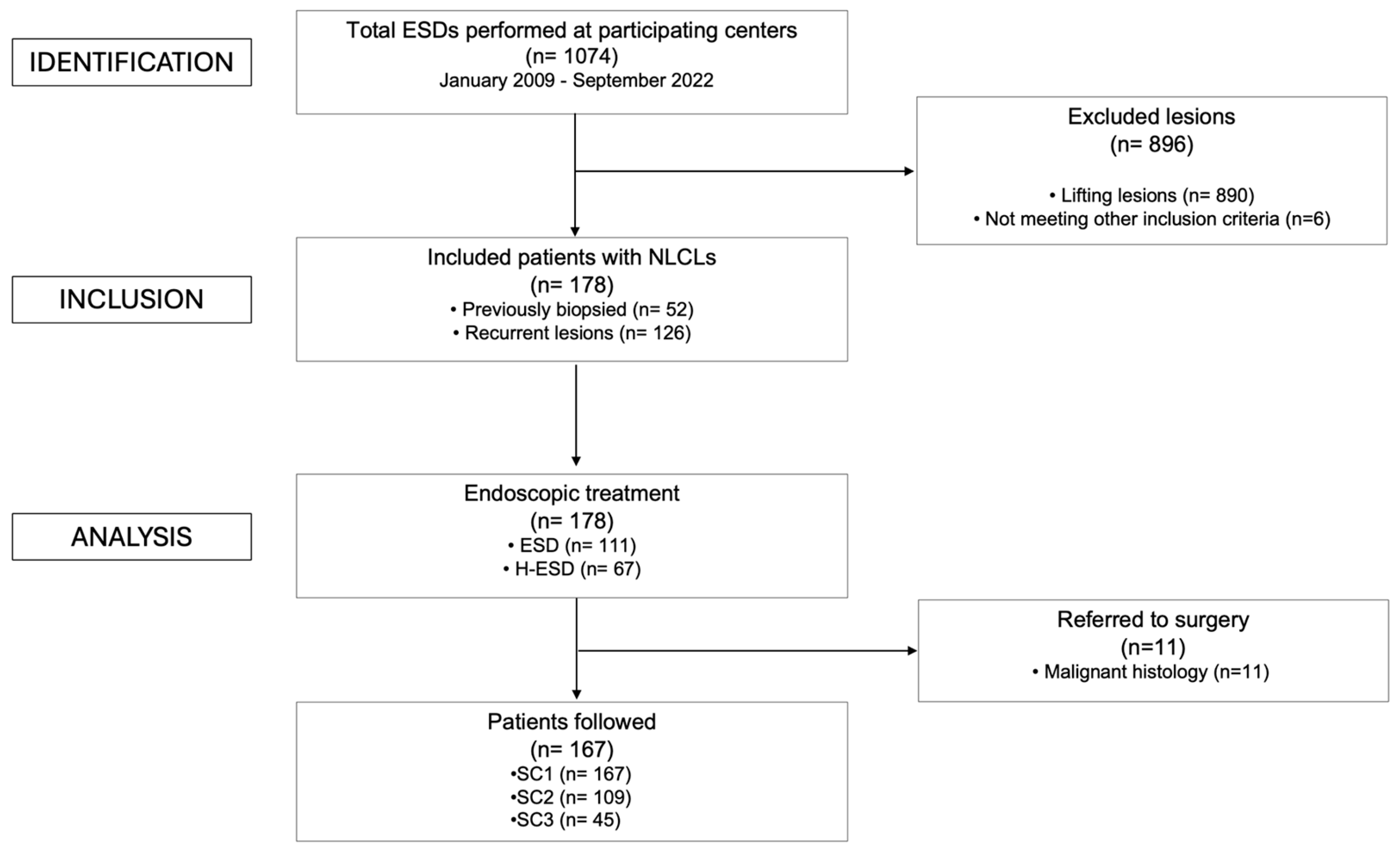
2. Materials and Methods

2.1. Study Population

We performed a retrospective analysis of prospectively collected data of all consecutive patients diagnosed with NLCLs with submucosal fibrosis at IRCCS San Raffaele Hospital in Milan (Italy) and Arcispedale Santa Maria Nuova in Reggio Emilia (Italy) from January 2009 to September 2022. Data were collected into a prospective registry, approved by the local Ethical Committee (Protocol ID: REG_EGDS COLON; Approval Code: 79/12/2022). This observational study was designed and reported following the STROBE (STrengthening the Reporting of OBservational studies in Epidemiology) statement guidelines for reporting observational studies [18]. Inclusion criteria were colorectal lesions presenting the no-lifting sign due to previous endoscopic resection attempts or to previous biopsy sampling, age > 18 years, and lesions endoscopically treatable with ESD according to the most recent guidelines in that period [19,20]. Exclusion criteria were age < 18 years, lesions showing endoscopic features suspected of deep submucosal/muscular invasion or histological report of neoplastic lesion with surgical indication, lesions previously treated with ESD or H-ESD, and lesions unsuitable for ESD indication. The fibrosis degree of lesions was classified during the index procedure according to a semiquantitative classification: F1 or mild, characterized by a white web-like structure in the blue submucosal layer, and F2 or severe, as a white muscular-like structure without a blue transparent layer in the submucosal layer [21].

2.2. Study Endpoint

The primary endpoint of this study was the recurrence rate (RR). Recurrence was defined as any endoscopically visible and histologically confirmed adenomatous tissue at the previous resection site detected during any surveillance colonoscopy (SC). Confirmation was obtained either through biopsies or an examination of the removed specimen if immediate treatment was performed. Secondary endpoints included the technical success rate (TS), defined as en bloc resection with either ESD or H-ESD. Technical failure was defined as the need for piecemeal resection. Additionally, we evaluated the technical success rate specifically for standard ESD, defined as en bloc resection without conversion to H-ESD. This study also assessed the rate of adverse events (AEs), stratified by fibrosis cause and technique used, according to the AGREE classification [22]. Complete resection (CR) was defined as an R0 en bloc resection with histologically tumor-free lateral and deep margins [20]. Curative resection (cR) was defined as an R0 resection with submucosal invasion < 1000 microns from the muscularis mucosae, no lymphatic invasion, no vascular involvement, no budding, and poorly differentiated components [20]. For cases where histopathological reports lacked this information, slides were re-evaluated by an expert gastrointestinal pathologist at each of the two centers. Finally, the rate of surgical intervention at any time during the follow-up period was also included.

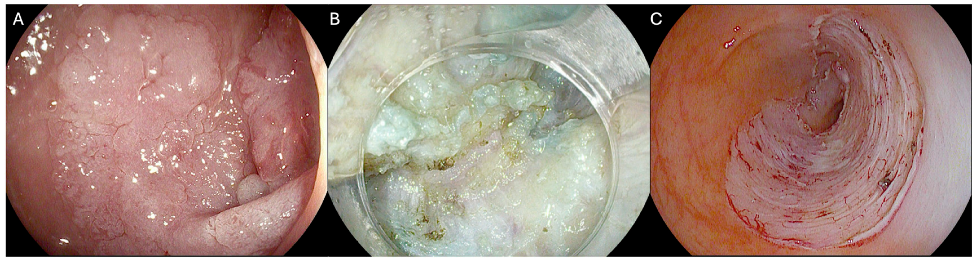
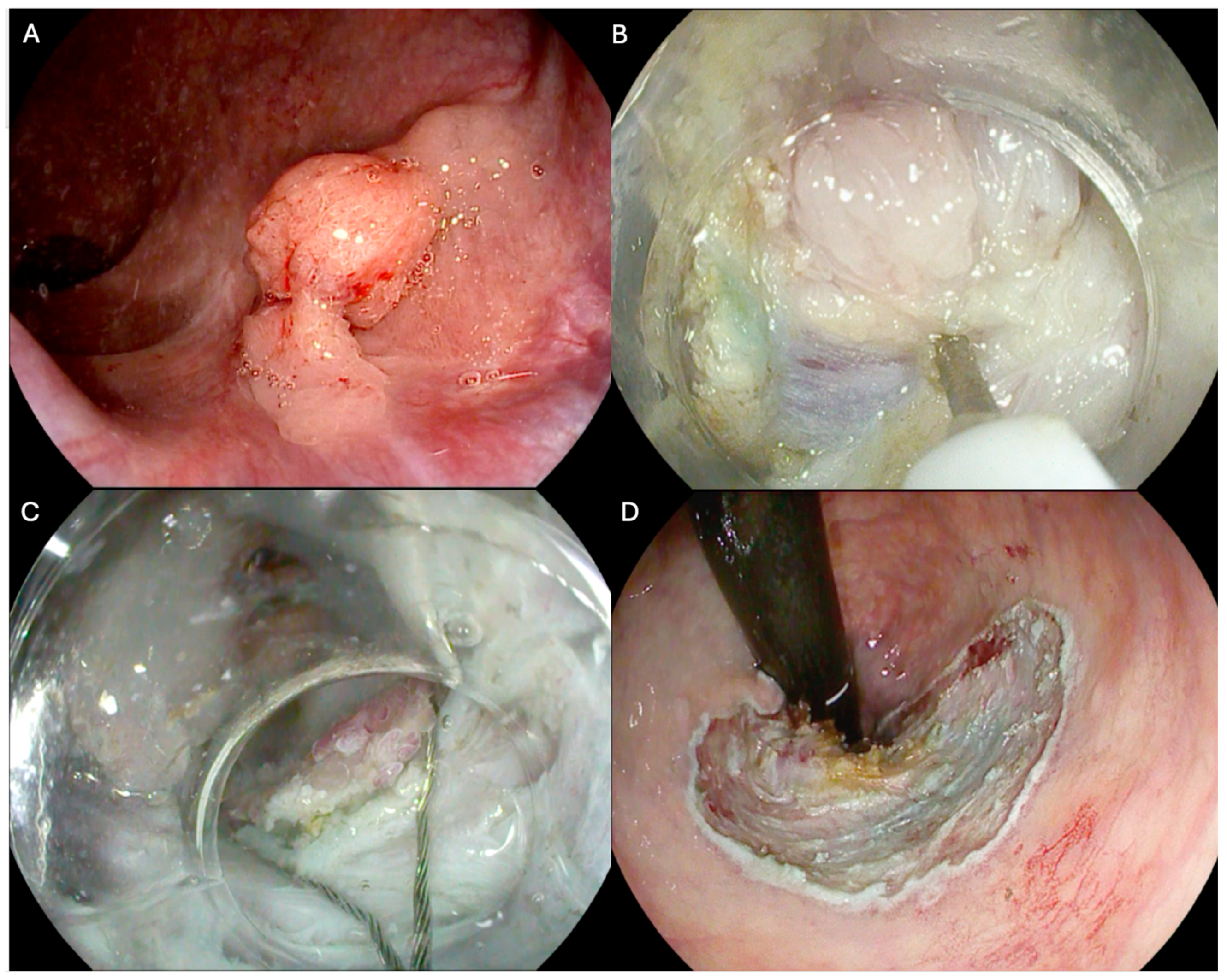
2.3. Endoscopic Procedure

All ESD procedures were conducted under deep sedation with anesthesiologic assistance or general anesthesia, according to local protocol. Different endoscopic systems were used according to the availability of the centers: Olympus (Olympus, Tokyo, Japan); Fujifilm (Fujifilm Tokyo, Japan); Pentax (Hoya Corp., Tokyo, Japan), using standard colonoscopes or gastroscopes for rectal lesions. The scopes were equipped with a distal transparent hood. CO2 insufflation was used in every procedure. All ESD procedures followed the basic principles indicated in ESGE guidelines [19,20]. The dissection strategy (standard, pocket, tunneling) and whether to apply traction systems were left to the discretion of the endoscopist. H-ESD completion involved using different-sized snares as needed. All procedures were performed by expert operators, defined as those having completed at least 100 colorectal ESDs and who regularly receive referrals from other hospitals, performing at least 25 ESDs per year to maintain proficiency throughout the entire study period [23].

2.4. Data Collection

Preprocedural clinical and demographic data included age, sex, and anesthesiologic risk classification according to the American Society of Anaesthesiologists (ASA) Physical Status Classification [24]. The lesions were analyzed based on the cause of fibrosis (recurrence vs. previously biopsied) and the treatment modality (ESD or H-ESD). Intraprocedural data included lesion localization, lesion long-axis and short-axis length (mm), area (cm2) calculated using the ellipse formula (short-axis length × long-axis length × 0.25 × 3.14), lesion morphology classified according to Paris’ endoscopic classification [25], glandular pattern according to Kudo’s classification [26], fibrosis degree, reason for fibrotic development, procedure time (min), and procedure speed (mm2/min), and the type of endoscopic devices used was also recorded. Post-procedure data included histology of the resected specimen, en bloc or piecemeal resection, radicality of the resection, need for surgery, recurrence, and mean follow-up in days and all follow-up SCs. Conversion to H-ESD was performed when, at the operator’s discretion, standard ESD proved unfeasible due to severe fibrosis, unstable position, or difficult access [27]; uncomfortable knife inclination; hemodynamic instability; or perforation. The reason for conversion was registered. Data on AEs were also collected. For any intraprocedural bleeding that required a dedicated endoscopic treatment, considerably lengthening the procedural time was considered; self-limiting or promptly treated bleeds were not recorded as AEs. Delayed bleeding was defined as overt hematochezia or melena arising from the resection site during the 6 h after completion of colorectal ESD. Full-thickness damage of the muscular layer with visualization of intra-abdominal space, mesenteric or mesorectal fat, was considered an intraprocedural perforation. Delayed perforation was defined as the appearance of radiological signs of perforation after colorectal ESD achievement, in an otherwise uneventful procedure. Two reporting systems (Endoxweb, Tesigroup, Italy, and OlyEndo, Unicode, Italy) were implemented in both centers involved in the study since 2019, providing electronic case report forms (CRFs) for patients and requinig mandatory inputs to complete the procedure report.

2.5. Statistical Analysis

Statistical analyses were performed using SPSS 26.0 (SPSS Inc., Chicago, IL, USA). Normality was assessed for the main demographic and descriptive variables using the Kolmogorov–Smirnov normality test, with the generation and evaluation of Q-Q plots assuming a 1-sided p value > 0.05 as an assumption of the normal distribution of the variable, implying the rejection of the null hypothesis. Concomitantly, heteroskedasticity was assessed with the Levene test. Continuous variables were later expressed as mean ± standard deviation (SD) or median and Interquartile Range (IQR) according to the normality test results. Median and range measures were provided for descriptive purposes. Categorical variables were expressed descriptively as numbers and percentages. Categorical parameters were compared using Pearson’s Chi-square test and Fisher’s exact test, along with Bonferroni correction for more than 2 groups within a variable, while continuous variables were compared using Pearson’s or Spearman’s correlation according to the normality distribution, with a Student's t-test or ANOVA test between 2 or more groups for normally distributed data, and with a Mann–Whitney test or Kruskar Wallis test between 2 groups for non-normally distributed data. The main outcomes, the CR, cR, technical success of the ESD procedure (or the “en bloc” resection rate or H-ESD conversion rate), surgery occurrence, and AE rate, were thoroughly calculated, and univariate analysis was performed to spot correlation with the most relevant independent variables. A logistic regression model was used to assess the impact of time on the probability of conversion to H-ESD. Model fit was assessed using the Hosmer and Lemeshow test, and the discriminative ability was evaluated through the area under the ROC curve (AUC). To identify a prediction model for primary and secondary endpoints, multivariate logistic regression models were built with a backward stepwise procedure, checking for multicollinearity (tolerance factor and variance-inflating factor calculation) for continuous independent variables. A p value < 0.05 in the univariate analysis was examined in the multivariate logistic regression models. Odds ratios (ORs) and 95% confidence intervals (CIs) were calculated using a logistic regression analysis. Two-sided p values < 0.05 were considered statistically significant.

3. Results

3.1. Baseline Characteristics

A total of 178 patients with 178 fibrotic NLCLs were included in this study (Figure 3). Fibrosis was due to previous biopsies in 29.2% of cases and to recurrences after previous endoscopic resections in 70.8% of cases. Among the patients included, 111/178 (62.4%) underwent ESD, while 67/178 (37.6%) required conversion to H-ESD. Conversion to H-ESD was mainly performed for severe fibrosis (62.7%) or difficult endoscopic access (i.e., very unstable position and uncomfortable knife inclination that could not be improved through endoscopic maneuvers or by changing patient’s decubitus position) (28.3%) (Supplementary Table S3). The median procedure time was 80 min (IQR, 60) for the overall cohort. The dissection speed was similar between the groups, with a median of 6.1 mm2/min (IQR, 8.1). Low-grade dysplasia (LGD) and high-grade dysplasia (HGD) were observed in 86.0% of cases, adenocarcinoma invading the submucosa (sm2-sm3) was found in 5.6% of cases, while other histologic findings were present in 2.3% of cases. Baseline characteristics are summarized in Table 1 and Supplementary Table S1.

3.2. Primary Endpoint

During a median follow-up of 373 days (IQR 540), the overall RR was 3.3%, with a total of six recurrences observed at the first surveillance colonoscopy (SC1) (median, 190 days; IQR, 240) among 167 patients, after excluding those referred for surgery (Table 2). Recurrent lesions showed a significantly higher rate of F2 fibrosis (68.3% vs. 30.8%, p < 0.01) and larger dimensions compared to previously biopsied, both in terms of the long axis (35 vs. 30 mm, p = 0.006), short axis (25 vs. 20 mm, p = 0.035), and area (7.1 vs. 4.7 cm2, p = 0.011). Recurrence was predominantly found in previously biopsied lesions (5/6 cases) and following H-ESD procedures (4/6 cases). Treatment at SC1 included EMR (2/6 cases), hot-biopsy avulsion (HBA) [28] (2/6 cases), ESD (1/6 cases), and H-ESD (1/6 cases), with a predominance of LGD lesions (83.3%). Importantly, two lesions treated for recurrence at SC1 were found to have recurred at SC2 (median, 390 days; IQR, 405). One of these, initially treated with EMR, was re-treated with ESD, while the other one, initially managed with H-ESD, was re-treated by EMR, due to its small size. At SC3 (median, 957 days; IQR, 953), no residual tissue was found. Late recurrences were equally distributed between biopsied and recurrent lesions and between ESD and H-ESD procedures, all showing LGD. Notably, all recurrences were successfully managed endoscopically without the need for surgical intervention.

3.3. Secondary Endpoints

The TS rate was 71.9% (128/178) for the overall cohort (Table 3). When considering only the traditional ESD approach, the en bloc resection rate was 62.4% (111/178). The TS was significantly higher in previously biopsied lesions compared to recurrent ones (82.7% vs. 67.5%, p = 0.04); conversely, the H-ESD conversion rate was higher in recurrent lesions (44.4% vs. 21.2%, p = 0.004) (Table 3). Severe fibrosis was the most frequent cause of technical failure (62.7%), followed by difficult endoscopic access (28.3%) (Supplementary Table S2). The CR and cR rates were similar between the previously biopsied and previously treated groups (90.7% vs. 93.0%, p = 0.475 for both). When CR was not achieved, the primary factor was deep margin involvement with sm2 histology, observed in 8/9 cases (88.9%). A single specimen exhibited lateral margin involvement, with histological analysis revealing sm1 invasion; this occurred post ESD treatment. The patient was subsequently referred for surgery due to the concomitant presence of lympho-vascular invasion.

3.4. Safety

Overall, the AE rate was 13.5% (24/178), with perforation being the most common complication, occurring in 8.4% (15/178) of cases. Bleeding was observed in 2.8% (5/178) of procedures, and one case reported both perforation and bleeding. All AEs were successfully managed endoscopically, with no cases requiring surgical intervention, as perforations were managed using clips or a clip-looping technique, while bleeding was controlled through mechanical hemostasis or thermal using bipolar forceps (Table 4). Moreover, according to the AGREE classification [22] (Supplementary Table S4), 3.3% (6/178) of patients experienced Grade I AEs, 8.9% (16/178) Grade II, and 1.7% (3/178) Grade III AEs (all Grade IIIa). No Grade IV or V complications were reported (Supplementary Table S5). Importantly, no statistically significant differences were observed in AE rates between the ESD and H-ESD groups (15.3% vs. 10.4%, p = 0.532), or between the groups with prior biopsy and recurrence (11.5% vs. 7.1%, p = 0.197).

3.5. Univariate and Multivariate Analysis

Univariate analysis identified lesion localization in the rectum (OR, 0.10; 95% CI, 0.03–0.28; p < 0.001), the presence of F1 fibrosis (OR, 0.12; 95% CI, 0.03–0.44; p = 0.0017), previously biopsied lesions (OR, 0.33; 95% CI, 0.58–0.86; p = 0.04), and the dimension of the lesion long axis (OR, 0.98; 95% CI, 0.96–0.99; p = 0.032) as factors associated with traditional ESD technical success. In the multivariate analysis, rectal localization (OR, 3.23; 95% CI, 1.65–6.32; p = 0.001), F1 fibrosis grade (OR, 2.16; 95% CI, 1.10–4.26; p = 0.026), and previously biopsied lesions (OR, 0.33, 95% CI, 0.59–0.89; p = 0.006) were found to be independent predictors for avoiding conversion to H-ESD (Table 5). Univariate analysis used to identify predictors of cR and CR did not show any statistically significant correlation (Supplementary Table S3).

4. Discussion

The management of NLCLs remains one of the most challenging scenarios in therapeutic endoscopy. Nonetheless, endoscopic techniques offer a valuable alternative to surgery for a significant number of patients. Our study highlights the effectiveness of the ESD/H-ESD approach, demonstrating favorable outcomes with an RR of 3.6% over a median follow-up of 373 days (IQR 540), despite the complex nature of the treated lesions. Notably, 4/6 recurrences observed during SC1 occurred following H-ESD. While H-ESD may carry a higher recurrence risk, it is crucial to consider its role as a rescue therapy for particularly challenging lesions that often require piecemeal resection. All recurrences were successfully managed with endoscopic re-treatment, and after SC2, no patient required additional treatments. Moreover, no AEs occurred during these re-treatments, highlighting the safety of the approach in experienced hands.
In terms of safety, our study demonstrated an AE rate of 13.5%, which is consistent with the complexity of treating NLCLs. The most common complication was perforation, occurring in 8.4% of cases, while bleeding was observed in 3.4% of procedures. Only 1.7% of patients experienced grade IIIa AEs (according to the AGREE classification), which are the most severe. These findings demonstrate that the ESD/H-ESD approach maintains safety profiles consistent with the literature [29,30]. None of the patients required surgical intervention for AEs; surgery was only indicated when histological evidence of invasive cancer was found. All intraprocedural complications were promptly recognized and managed endoscopically, underscoring the importance of adequate expertise.
The TS rate in our study was 71.9%; while considering only traditional ESD, it was 62.4%. These rates are lower compared to in some studies in the literature, ranging from 80 to 96%, but they must be contextualized within several important factors [7,21,29,30]. First, our study spans from 2009, encompassing the evolution of ESD techniques and devices, including the introduction of the pocket creation method, the traction-assisted ESD, and the saline immersion ESD [31,32,33,34]. Additionally, ESD has a steep learning curve, and the progressive accumulation of operator experience likely contributed to improved success rates over time.
Despite these challenges, our CR rate of 93.0% and the low RR observed during follow-up highlight the effectiveness of ESD/H-ESD in achieving durable, curative outcomes, even when en bloc resection is not achieved.
The temporal logistic regression analysis demonstrated a significant 22.7% annual reduction in the probability of conversion to H-ESD (OR = 0.7727; 95% CI: 0.6811–0.8765; p = 0.0001) (Figure 4). These findings indicate that as experience and technique refinement increased, the need for hybrid conversion progressively decreased. This trend reflects how the refinement of ESD methods, the integration of advanced dissection strategies, and the improvement of endoscopic tools have already enhanced ESD outcomes and represent a pathway toward even better results in the future. The multivariate analysis identified rectal localization, F1 fibrosis grade, and previously biopsied lesions as independent predictors of ESD TS without the need for hybrid conversion. The higher success rate in rectal lesions may be due to easier endoscopic access and stability. The association with F1 fibrosis and previously biopsied lesions is unsurprising, as milder, more localized fibrosis allows for easier submucosal lifting and dissection. These predictors can guide risk stratification and help identify cases where traditional ESD is most likely to succeed.
Japanese multicenter studies have consistently demonstrated superior outcomes in ESD for NLCLs. A large retrospective study reported en bloc and CR rates of 95% and 90%, respectively, with a 6% perforation rate, over a 10-year period, and no local recurrences following cR [35]. Similarly, a prospective study showed a 96.3% en bloc and 83.3% CR rate, with a 7.4% perforation rate and no recurrence over a median follow-up of 60 months, further reinforcing the long-term efficacy of ESD in this setting [30]. This stark contrast highlights the persistent disparity in ESD outcomes between Eastern and Western centers, though AE rates remain comparable. Several factors may contribute to this difference. In Japan, higher case volumes and more structured, uniformly implemented training programs provide endoscopists with greater exposure and a more standardized learning pathway. The higher prevalence of gastric lesions allows trainees to first develop ESD skills on gastric cases before advancing to colorectal procedures. Additionally, the widespread adoption of standardized techniques ensures more consistent outcomes. In contrast, Western centers often face limitations in volume and training uniformity, leading to variability in technique adoption and a prolonged learning curve.
Our multicenter study, comprising 178 cases, represents one of the largest Western series addressing NLCLs through ESD. While Eastern centers report larger series, our cohort size surpasses most European studies and is comparable to recent large series (Table 6). This is particularly relevant given the absence of standardized treatment for NLCLs, where therapeutic strategies are largely dictated by operator expertise, center resources, and referral volumes. Various alternative techniques have been proposed.
Ablative techniques such as forced argon plasma coagulation (APC) and hot-biopsy avulsion have been proposed as potential treatment strategies. They are relatively straightforward to perform; however, they are associated with high RR (10–59%) and critically do not allow for histopathological assessment [30,36]. This limitation is significant, as the inability to assess for potential malignancy can lead to suboptimal treatment.
EMR represents a well-known alternative, yet the challenge of achieving adequate pre-resection lifting remains unresolved [37,38,39,40] and shows an RR up to 30% [37,41,42]. On the other hand, it has demonstrated a solid safety profile, with 6% incidence of bleeding, 2% incidence of intraprocedural perforation, and an even lower rate of delayed perforation [43,44].
Underwater EMR (U-EMR) offers advantages through reduced luminal distension and enhanced tissue capture due to increased mucosal buoyancy, even with fibrosis [45]. Its feasibility is suggested by limited retrospective evidence showing that U-EMR may achieve lower RRs compared to conventional EMR (11% vs. 66%), while maintaining a favorable safety profile [46]. When compared to ESD in a propensity-matched analysis, it demonstrated lower en bloc (73% vs. 100%) and CR rates (41% vs. 81%), with shorter hospital stays and a lower perforation rate, as expected [47].
Another approach, the cold avulsion with adjuvant snare-tip soft coagulation (CAST), offers a technically simple and valid option [48,49]. However, RRs of up to 5.0–18.8% have been described, and AEs are non-negligible, with an intraprocedural perforation rate of up to 5.6% and a need for clip application in almost 24% of procedures [48,49].
Furthermore, owing to their inability to achieve en bloc resection, the previous techniques necessitate close surveillance and potential repeated endoscopic treatments, thereby increasing the number of colonoscopies to which these patients are subjected. This represents a critical point, as the cumulative burden may further affect adherence to follow-up protocols.
On the other hand, endoscopic full-thickness resection (EFTR) allows for the en bloc resection of fibrotic lesions ranging 5–30 mm in size, depending on the tissue’s pliability [50]. However, its efficacy is limited by lesion size, with larger lesions (≥20 mm) demonstrating an R0 resection rate of only 58% [51,52]. Nevertheless, it is associated with an RR of 6.4–13.5%, and its applicability in the right colon is constrained by the device’s size and the consequent reduction in maneuverability [29,51,52,53,54]. As recently demonstrated by Yzet et al., ESD shows a lower RR compared to EFTR (2.1% vs. 11.9%, p = 0.09) even for lesions < 20 mm [29]. However, EFTR exhibits a better safety profile, with a lower rate of AEs (5.1% vs. 11.3%, p = 0.01). This highlights that ESD offers the best outcomes in terms of radicality and recurrence, albeit with a slightly higher risk of complications, while EFTR is indicated for smaller lesions, yields a non-negligible RR, and should be reserved for cases where ESD is not viable.
Our study has several strengths, including a large multicenter cohort with an extended follow-up period. The comprehensive analysis of TS predictors, coupled with the achieved low RR, provides valuable insights for patient selection and procedure planning in this challenging scenario
However, our study is not without limitations. The retrospective design and heterogeneous patient population may introduce potential biases. The lack of a control group with alternative techniques prevents definitive conclusions regarding the role of ESD in the management of NLCLs. Although all procedures were performed by experienced endoscopists, some degree of inter-operator variability cannot be excluded. This variability may also stem from differences in available equipment across centers, and distinct procedural habits, despite operators having comparable training backgrounds. Finally, since all procedures were conducted in high-volume tertiary centers by expert endoscopists, the generalizability of our findings to lower-volume centers or less experienced operators may be limited.
Table 6. Comparison of Western and Eastern studies on the treatment of NLCLs using endoscopic submucosal dissection (ESD).
AuthorYearCountryStudy DesignEndoscopic TechniquesPatients
(n)
Age
(Mean, years)
Sex
(Male, %)
Lesion Size
Median (Range, mm)
Procedural Time (Median, min)En Bloc Resection (%)Adverse Events (%)
Hurlstone et al. [55]2008United KingdomProspective monocentricESD306572NA
(5–45)
6293%16
Spychalski et al. [56]2019PolandRetrospective monocentricESD706555.734.8
(23–46)
77NA12.8
Faller et al. [57]2020FranceRetrospective
multicentric
ESD537050.940
(20–65)
4392.59.4
Yzet et al. [29] *2023FranceRetrospective multicentricESD/eFTR177697635
(5–140)
4594.316.3
Tanaka et al. [35]2021JapanRetrospective multicentricESD102695520
(4–50)
84958.8
Tanaka et al. [58]2024JapanProspective multicentricESD 54706331
(24.5–40)
6596.39.5
Dell’Anna et al.2025ItalyRetrospective multicentricESD/H-ESD1786856.230
(10–120)
8071.913.4
* Data shown in the table refer only to the ESD-treated group of this study. ESD = endoscopic submucosal dissection; NLCL = non-lifting colorectal lesion; eFTR = endoscopic full-thickness resection; H-ESD = hybrid endoscopic submucosal dissection.

5. Conclusions

In conclusion, ESD and H-ESD as rescue therapy demonstrate feasibility, efficacy, and safety in treating NLCLs when performed by experienced endoscopists in tertiary referral centers. This approach yields a 3.6% RR, all amenable to successful endoscopic management in our experience. Factors associated with higher TS rates include previously biopsied lesions rather than recurrencies, rectal localization, and lower grades of fibrosis. These findings further support the potential of ESD as a treatment for these challenging lesions.

Supplementary Materials

The following supporting information can be downloaded at https://www.mdpi.com/article/10.3390/cancers17071242/s1: Table S1. STROBE statement—checklist; Table S2. AGREE classification grading definition; Table S3. Additional baseline characteristics of the included patients with sub-analysis for fibrosis cause; Table S4. Numbers of patients included in each center and endoscopic submucosal dissection (ESD) to hybrid ESD (H-ESD) conversion per center; Table S5. Technical unsuccess causes; Table S6. AGREE classification according to the type of endoscopic procedure; Table S7. Univariate analysis of complete resection (CR) and curative resection (cR) rates. Table S8. Endoscopic submucosal dissection (ESD) and hybrid ESD (H-ESD) conversion across study periods (2009–2022). Figure S1. Scatter plot illustrating the relationship between dissection speed (mm2/min) and the regression standardized predicted value.

Author Contributions

Conceptualization, G.D. (Giuseppe Dell’Anna), E.F. and F.A.; data curation, E.F.; formal analysis, E.F. and A.B.; methodology, G.D. (Giuseppe Dell’Anna) and E.F.; supervision, S.D. and F.A.; writing—original draft, G.D. (Giuseppe Dell’Anna) and E.F.; Writing—review and editing, P.C., G.B., E.V., D.E., S.G., R.S., A.M., S.M., V.A., L.F., A.F., G.D. (Gianfranco Donatelli), S.D. and F.A. All authors have read and agreed to the published version of the manuscript.

Funding

This research received no external funding.

Institutional Review Board Statement

The study was conducted in accordance with the Declaration of Helsinki and approved by the local ethics committee. Protocol ID: REG_EGDS COLON; Approval Code: 79/12/2022.

Informed Consent Statement

Informed consent was obtained from all subjects involved in the study.

Data Availability Statement

The original contributions presented in the study are included in the article, further inquiries can be directed to the corresponding author.

Conflicts of Interest

S.D. has served as a speaker, consultant, and advisory board member for Schering-Plough, AbbVie, Actelion, Alphawasserman, AstraZeneca, Cellerix, Cosmo Pharmaceuticals, Ferring, Genentech, Grunenthal, Johnson and Johnson, Millenium Takeda, MSD, Nikkiso Europe GmbH, Novo Nordisk, Nycomed, Pfizer, Pharmacosmos, UCB Pharma, and Vifor. The other authors declare no conflicts of interest.

References

  1. Ishiguro, A.; Uno, Y.; Ishiguro, Y.; Munakata, A.; Morita, T. Correlation of Lifting versus Non-Lifting and Microscopic Depth of Invasion in Early Colorectal Cancer. Gastrointest. Endosc. 1999, 50, 329–333. [Google Scholar] [CrossRef]
  2. Kobayashi, N.; Saito, Y.; Sano, Y.; Uragami, N.; Michita, T.; Nasu, J.; Matsuda, T.; Fu, K.; Fujii, T.; Fujimori, T.; et al. Determining the Treatment Strategy for Colorectal Neoplastic Lesions: Endoscopic Assessment or the Non-Lifting Sign for Diagnosing Invasion Depth? Endoscopy 2007, 39, 701–705. [Google Scholar] [CrossRef]
  3. Chiba, H.; Tachikawa, J.; Arimoto, J.; Ashikari, K.; Kuwabara, H.; Nakaoka, M.; Goto, T.; Ohata, K.; Nakajima, A. Predictive Factors of Mild and Severe Fibrosis in Colorectal Endoscopic Submucosal Dissection. Dig. Dis. Sci. 2020, 65, 232–242. [Google Scholar] [CrossRef]
  4. Cecinato, P.; Lisotti, A.; Azzolini, F.; Lucarini, M.; Bassi, F.; Fusaroli, P.; Sassatelli, R. Left Colonic Localization, Non-Granular Morphology, and Pit Pattern Independently Predict Submucosal Fibrosis of Naïve Colorectal Neoplasms before Endoscopic Submucosal Dissection. Surg. Endosc. 2023, 37, 3037–3045. [Google Scholar] [CrossRef]
  5. Sferrazza, S.; Maida, M.; Calabrese, G.; Facciorusso, A.; Fuccio, L.; Frazzoni, L.; Maselli, R.; Repici, A.; Di Mitri, R.; Santos-Antunes, J. The Derivation and External Validation of a Fibrosis Risk Model for Colorectal Tumours Undergoing Endoscopic Submucosal Dissection. J. Clin. Med. 2024, 13, 4517. [Google Scholar] [CrossRef]
  6. Azzolini, F.; Biolchini, F.; Sassatelli, R.; Camellini, L.; Decembrino, F.; Iori, V.; Tioli, C.; Sereni, G.; Bedogni, G. Endoscopic Submucosal Dissection for Residual Rectal Polyps Embedded in Tissue Scar: A “Rescue Therapy” to Prevent Surgical Intervention? Endoscopy 2008, 40, E222–E223. [Google Scholar] [CrossRef]
  7. Azzolini, F.; Camellini, L.; Sassatelli, R.; Sereni, G.; Biolchini, F.; Decembrino, F.; De Marco, L.; Iori, V.; Tioli, C.; Cavina, M.; et al. Endoscopic Submucosal Dissection of Scar-Embedded Rectal Polyps: A Prospective Study (Esd in Scar-Embedded Rectal Polyps). Clin. Res. Hepatol. Gastroenterol. 2011, 35, 572–579. [Google Scholar] [CrossRef]
  8. Maselli, R.; Iacopini, F.; Azzolini, F.; Petruzziello, L.; Manno, M.; De Luca, L.; Cecinato, P.; Fiori, G.; Staiano, T.; Rosa Rizzotto, E.; et al. Endoscopic Submucosal Dissection: Italian National Survey on Current Practices, Training and Outcomes. Dig. Liver Dis. 2020, 52, 64–71. [Google Scholar] [CrossRef]
  9. Gupta, N.; Rodríguez-Ruiz, G.; Siddiqui, U.D.; Chapman, C.G.; Donboli, K.; Hart, J.; Xiao, S.-Y.; Waxman, I. Endoscopic Submucosal Dissection for Colorectal Lesions: Outcomes from a United States Experience. Surg. Endosc. 2022, 36, 236–243. [Google Scholar] [CrossRef]
  10. Steinbrück, I.; Faiss, S.; Dumoulin, F.L.; Oyama, T.; Pohl, J.; von Hahn, T.; Schmidt, A.; Allgaier, H.-P. Learning Curve of Endoscopic Submucosal Dissection (ESD) with Prevalence-Based Indication in Unsupervised Western Settings: A Retrospective Multicenter Analysis. Surg. Endosc. 2023, 37, 2574–2586. [Google Scholar] [CrossRef]
  11. Fujiya, M.; Tanaka, K.; Dokoshi, T.; Tominaga, M.; Ueno, N.; Inaba, Y.; Ito, T.; Moriichi, K.; Kohgo, Y. Efficacy and Adverse Events of EMR and Endoscopic Submucosal Dissection for the Treatment of Colon Neoplasms: A Meta-Analysis of Studies Comparing EMR and Endoscopic Submucosal Dissection. Gastrointest. Endosc. 2015, 81, 583–595. [Google Scholar] [CrossRef]
  12. McCarty, T.R.; Bazarbashi, A.N.; Thompson, C.C.; Aihara, H. Hybrid Endoscopic Submucosal Dissection (ESD) Compared with Conventional ESD for Colorectal Lesions: A Systematic Review and Meta-Analysis. Endoscopy 2021, 53, 1048–1058. [Google Scholar] [CrossRef]
  13. Morimoto, S.; Tanaka, H.; Takehara, Y.; Yamamoto, N.; Tanino, F.; Kamigaichi, Y.; Yamashita, K.; Takigawa, H.; Yuge, R.; Urabe, Y.; et al. Hybrid Endoscopic Submucosal Dissection as a Salvage Option for Difficult Colorectal Conventional Endoscopic Submucosal Dissection. Surg. Endosc. 2024, 38, 222–228. [Google Scholar] [CrossRef]
  14. Okamoto, K.; Muguruma, N.; Kagemoto, K.; Mitsui, Y.; Fujimoto, D.; Kitamura, S.; Kimura, T.; Sogabe, M.; Miyamoto, H.; Takayama, T. Efficacy of Hybrid Endoscopic Submucosal Dissection (ESD) as a Rescue Treatment in Difficult Colorectal ESD Cases. Dig. Endosc. 2017, 29, 45–52. [Google Scholar] [CrossRef]
  15. Libânio, D.; Pimentel-Nunes, P.; Bastiaansen, B.; Bisschops, R.; Bourke, M.J.; Deprez, P.H.; Esposito, G.; Lemmers, A.; Leclercq, P.; Maselli, R.; et al. Endoscopic Submucosal Dissection Techniques and Technology: European Society of Gastrointestinal Endoscopy (ESGE) Technical Review. Endoscopy 2023, 55, 361–389. [Google Scholar] [CrossRef]
  16. Chedgy, F.J.Q.; Bhattacharyya, R.; Kandiah, K.; Longcroft-Wheaton, G.; Bhandari, P. Knife-Assisted Snare Resection: A Novel Technique for Resection of Scarred Polyps in the Colon. Endoscopy 2016, 48, 277–280. [Google Scholar] [CrossRef]
  17. Bhattacharyya, R.; Chedgy, F.J.; Kandiah, K.; Longcroft-Wheaton, G.; Bhandari, P. Knife-Assisted Snare Resection (KAR) of Large and Refractory Colonic Polyps at a Western Centre: Feasibility, Safety and Efficacy Study to Guide Future Practice. United Eur. Gastroenterol. J. 2016, 4, 466–473. [Google Scholar] [CrossRef]
  18. Von Elm, E.; Altman, D.G.; Egger, M.; Pocock, S.J.; Gøtzsche, P.C.; Vandenbroucke, J.P. The Strengthening the Reporting of Observational Studies in Epidemiology (STROBE) Statement: Guidelines for Reporting Observational Studies. Lancet 2007, 370, 1453–1457. [Google Scholar] [CrossRef]
  19. Pimentel-Nunes, P.; Dinis-Ribeiro, M.; Ponchon, T.; Repici, A.; Vieth, M.; De Ceglie, A.; Amato, A.; Berr, F.; Bhandari, P.; Bialek, A.; et al. Endoscopic Submucosal Dissection: European Society of Gastrointestinal Endoscopy (ESGE) Guideline. Endoscopy 2015, 47, 829–854. [Google Scholar] [CrossRef]
  20. Pimentel-Nunes, P.; Libânio, D.; Bastiaansen, B.A.J.; Bhandari, P.; Bisschops, R.; Bourke, M.J.; Esposito, G.; Lemmers, A.; Maselli, R.; Messmann, H.; et al. Endoscopic Submucosal Dissection for Superficial Gastrointestinal Lesions: European Society of Gastrointestinal Endoscopy (ESGE) Guideline—Update 2022. Endoscopy 2022, 54, 591–622. [Google Scholar] [CrossRef]
  21. Matsumoto, A.; Tanaka, S.; Oba, S.; Kanao, H.; Oka, S.; Yoshihara, M.; Chayama, K. Outcome of Endoscopic Submucosal Dissection for Colorectal Tumors Accompanied by Fibrosis. Scand. J. Gastroenterol. 2010, 45, 1329–1337. [Google Scholar] [CrossRef] [PubMed]
  22. Nass, K.J.; Zwager, L.W.; van der Vlugt, M.; Dekker, E.; Bossuyt, P.M.M.; Ravindran, S.; Thomas-Gibson, S.; Fockens, P. Novel Classification for Adverse Events in GI Endoscopy: The AGREE Classification. Gastrointest. Endosc. 2022, 95, 1078–1085. [Google Scholar] [CrossRef] [PubMed]
  23. Pimentel-Nunes, P.; Pioche, M.; Albéniz, E.; Berr, F.; Deprez, P.; Ebigbo, A.; Dewint, P.; Haji, A.; Panarese, A.; Weusten, B.L.; et al. Curriculum for Endoscopic Submucosal Dissection Training in Europe: European Society of Gastrointestinal Endoscopy (ESGE) Position Statement. Endoscopy 2019, 51, 980–992. [Google Scholar] [CrossRef] [PubMed]
  24. Horvath, B.; Kloesel, B.; Todd, M.M.; Cole, D.J.; Prielipp, R.C. The Evolution, Current Value, and Future of the American Society of Anesthesiologists Physical Status Classification System. Anesthesiology 2021, 135, 904–919. [Google Scholar] [CrossRef]
  25. Endoscopic Classification Review Group. Update on the Paris Classification of Superficial Neoplastic Lesions in the Digestive Tract. Endoscopy 2005, 37, 570–578. [Google Scholar] [CrossRef]
  26. Kudo, S.; Tamura, S.; Nakajima, T.; Yamano, H.; Kusaka, H.; Watanabe, H. Diagnosis of Colorectal Tumorous Lesions by Magnifying Endoscopy. Gastrointest. Endosc. 1996, 44, 8–14. [Google Scholar] [CrossRef]
  27. Gupta, S.; Miskovic, D.; Bhandari, P.; Dolwani, S.; McKaig, B.; Pullan, R.; Rembacken, B.; Rutter, M.D.; Riley, S.; Valori, R.; et al. The “SMSA” Scoring System for Determining the Complexity of a Polyp. Gut 2011, 60, A129. [Google Scholar] [CrossRef]
  28. Kumar, V.; Broadley, H.; Rex, D.K. Safety and Efficacy of Hot Avulsion as an Adjunct to EMR (with Videos). Gastrointest. Endosc. 2019, 89, 999–1004. [Google Scholar] [CrossRef]
  29. Yzet, C.; Le Baleur, Y.; Albouys, J.; Jacques, J.; Doumbe-Mandengue, P.; Barret, M.; Abou Ali, E.; Schaefer, M.; Chevaux, J.-B.; Leblanc, S.; et al. Use of Endoscopic Submucosal Dissection or Full-Thickness Resection Device to Treat Residual Colorectal Neoplasia after Endoscopic Resection: A Multicenter Historical Cohort Study. Endoscopy 2023, 55, 1002–1009. [Google Scholar] [CrossRef]
  30. Tanaka, H.; Uraoka, T.; Kobayashi, N.; Ohata, K.; Takeuchi, Y.; Chino, A.; Yamada, M.; Tsuji, Y.; Hotta, K.; Harada, K.; et al. Short-term and Long-term Outcomes of Submucosal Dissection for Residual or Recurrent Colorectal Tumors after Endoscopic Resection: Analysis of a Multicenter Prospective Study. Dig. Endosc. 2024, 36, 1003–1011. [Google Scholar] [CrossRef]
  31. Despott, E.J.; Murino, A. Saline-Immersion Therapeutic Endoscopy (SITE): An Evolution of Underwater Endoscopic Lesion Resection. Dig. Liver Dis. 2017, 49, 1376. [Google Scholar] [CrossRef] [PubMed]
  32. Hayashi, Y.; Sunada, K.; Takahashi, H.; Shinhata, H.; Lefor, A.T.; Tanaka, A.; Yamamoto, H. Pocket-Creation Method of Endoscopic Submucosal Dissection to Achieve En Bloc Resection of Giant Colorectal Subpedunculated Neoplastic Lesions. Endoscopy 2014, 46, E421–E422. [Google Scholar] [CrossRef] [PubMed]
  33. Masgnaux, L.-J.; Grimaldi, J.; Rivory, J.; Ponchon, T.; Yzet, C.; Wallenhorst, T.; Lupu, A.; Lafeuille, P.; Legros, R.; Rostain, F.; et al. Endoscopic Submucosal Dissection Assisted by Adaptive Traction: Results of the First 54 Procedures. Endoscopy 2024, 56, 205–211. [Google Scholar] [CrossRef] [PubMed]
  34. Facciorusso, A.; Straus Takahashi, M.; Eyileten Postula, C.; Buccino, V.R.; Muscatiello, N. Efficacy of Hemostatic Powders in Upper Gastrointestinal Bleeding: A Systematic Review and Meta-Analysis. Dig. Liver Dis. 2019, 51, 1633–1640. [Google Scholar] [CrossRef]
  35. Tanaka, H.; Oka, S.; Tanaka, S.; Nagata, S.; Kunihiro, M.; Kuwai, T.; Hiraga, Y.; Mizumoto, T.; Okanobu, H.; Chayama, K. Salvage Endoscopic Submucosal Dissection for Local Residual/Recurrent Colorectal Tumor after Endoscopic Resection: Large Multicenter 10-year Study. Dig. Endosc. 2021, 33, 608–615. [Google Scholar] [CrossRef]
  36. Holmes, I.; Kim, H.G.; Yang, D.-H.; Friedland, S. Avulsion Is Superior to Argon Plasma Coagulation for Treatment of Visible Residual Neoplasia during EMR of Colorectal Polyps (with Videos). Gastrointest. Endosc. 2016, 84, 822–829. [Google Scholar] [CrossRef]
  37. Tsiamoulos, Z.P.; Bourikas, L.A.; Saunders, B.P. Endoscopic Mucosal Ablation: A New Argon Plasma Coagulation/Injection Technique to Assist Complete Resection of Recurrent, Fibrotic Colon Polyps (with Video). Gastrointest. Endosc. 2012, 75, 400–404. [Google Scholar] [CrossRef]
  38. Tate, D.J.; Desomer, L.; Argenziano, M.E.; Mahajan, N.; Sidhu, M.; Vosko, S.; Shahidi, N.; Lee, E.; Williams, S.J.; Burgess, N.G.; et al. Treatment of Adenoma Recurrence after Endoscopic Mucosal Resection. Gut 2023, 72, 1875–1886. [Google Scholar] [CrossRef]
  39. Rashid, M.U.; Khetpal, N.; Zafar, H.; Ali, S.; Idrisov, E.; Du, Y.; Stein, A.; Jain, D.; Hasan, M.K. Colon Mucosal Neoplasia Referred for Endoscopic Mucosal Resection: Recurrence of Adenomas and Prediction of Submucosal Invasion. World J. Gastrointest. Endosc. 2020, 12, 198–211. [Google Scholar] [CrossRef]
  40. Van der Voort, V.R.H.; Moons, L.M.G.; de Graaf, W.; Schrauwen, R.W.M.; Hazen, W.L.; Seerden, T.C.J.; Vleggaar, F.P.; Didden, P. Efficacy and Safety of Cap-Assisted Endoscopic Mucosal Resection for Treatment of Nonlifting Colorectal Polyps. Endoscopy 2022, 54, 509–514. [Google Scholar] [CrossRef]
  41. Shahidi, N.; Vosko, S.; Gupta, S.; van Hattem, W.A.; Sidhu, M.; Tate, D.J.; Williams, S.J.; Lee, E.Y.T.; Burgess, N.; Bourke, M.J. Previously Attempted Large Nonpedunculated Colorectal Polyps Are Effectively Managed by Endoscopic Mucosal Resection. Am. J. Gastroenterol. 2021, 116, 958–966. [Google Scholar] [CrossRef] [PubMed]
  42. Belderbos, T.D.G.; Leenders, M.; Moons, L.M.G.; Siersema, P.D. Local Recurrence after Endoscopic Mucosal Resection of Nonpedunculated Colorectal Lesions: Systematic Review and Meta-Analysis. Endoscopy 2014, 46, 388–402. [Google Scholar] [CrossRef] [PubMed]
  43. Hassan, C.; Repici, A.; Sharma, P.; Correale, L.; Zullo, A.; Bretthauer, M.; Senore, C.; Spada, C.; Bellisario, C.; Bhandari, P.; et al. Efficacy and Safety of Endoscopic Resection of Large Colorectal Polyps: A Systematic Review and Meta-Analysis. Gut 2016, 65, 806–820. [Google Scholar] [CrossRef] [PubMed]
  44. Jacques, J.; Schaefer, M.; Wallenhorst, T.; Rösch, T.; Lépilliez, V.; Chaussade, S.; Rivory, J.; Legros, R.; Chevaux, J.-B.; Leblanc, S.; et al. Endoscopic En Bloc Versus Piecemeal Resection of Large Nonpedunculated Colonic Adenomas: A Randomized Comparative Trial. Ann. Intern. Med. 2024, 177, 29–38. [Google Scholar] [CrossRef]
  45. Burgess, N.G.; Williams, S.J.; Hourigan, L.F.; Brown, G.J.; Zanati, S.A.; Singh, R.; Tam, W.; Butt, J.; Byth, K.; Bourke, M.J. A Management Algorithm Based on Delayed Bleeding After Wide-Field Endoscopic Mucosal Resection of Large Colonic Lesions. Clin. Gastroenterol. Hepatol. 2014, 12, 1525–1533. [Google Scholar] [CrossRef]
  46. Binmoeller, K.F.; Weilert, F.; Shah, J.; Bhat, Y.; Kane, S. “Underwater” EMR without Submucosal Injection for Large Sessile Colorectal Polyps (with Video). Gastrointest. Endosc. 2012, 75, 1086–1091. [Google Scholar] [CrossRef]
  47. Kim, H.G.; Thosani, N.; Banerjee, S.; Chen, A.; Friedland, S. Underwater Endoscopic Mucosal Resection for Recurrences after Previous Piecemeal Resection of Colorectal Polyps (with Video). Gastrointest. Endosc. 2014, 80, 1094–1102. [Google Scholar] [CrossRef]
  48. Ohmori, M.; Yamasaki, Y.; Iwagami, H.; Nakahira, H.; Matsuura, N.; Shichijo, S.; Maekawa, A.; Kanesaka, T.; Yamamoto, S.; Higashino, K.; et al. Propensity Score-matched Analysis of Endoscopic Resection for Recurrent Colorectal Neoplasms: A Pilot Study. J. Gastro Hepatol. 2021, 36, 2568–2574. [Google Scholar] [CrossRef]
  49. Tate, D.J.; Bahin, F.F.; Desomer, L.; Sidhu, M.; Gupta, V.; Bourke, M.J. Cold-Forceps Avulsion with Adjuvant Snare-Tip Soft Coagulation (CAST) Is an Effective and Safe Strategy for the Management of Non-Lifting Large Laterally Spreading Colonic Lesions. Endoscopy 2018, 50, 52–62. [Google Scholar] [CrossRef]
  50. Mandarino, F.V.; O’Sullivan, T.; Gauci, J.L.; Kerrison, C.; Whitfield, A.; Lam, B.; Perananthan, V.; Gupta, S.; Cronin, O.; Medas, R.; et al. Impact of Margin Thermal Ablation after Cold-Forceps Avulsion with Snare-Tip Soft Coagulation for Non-Lifting Large Non-Pedunculated Colorectal Polyps. Endoscopy 2025. [Google Scholar] [CrossRef]
  51. Von Helden, A.; Hildenbrand, R.; Sido, B.; Dumoulin, F.L. Endoscopic Full-Thickness Resection Using an over-the-Scope Device for Treatment of Recurrent/Residual Colorectal Neoplasia: A Single-Center Case Series. BMC Gastroenterol. 2019, 19, 121. [Google Scholar] [CrossRef] [PubMed]
  52. Li, P.; Ma, B.; Gong, S.; Zhang, X.; Li, W. Efficacy and Safety of Endoscopic Full-Thickness Resection in the Colon and Rectum Using an over-the-Scope Device: A Meta-Analysis. Surg. Endosc. 2021, 35, 249–259. [Google Scholar] [CrossRef] [PubMed]
  53. Zwager, L.W.; Bastiaansen, B.A.J.; Bronzwaer, M.E.S.; van der Spek, B.W.; Heine, G.D.N.; Haasnoot, K.J.C.; van der Sluis, H.; Perk, L.E.; Boonstra, J.J.; Rietdijk, S.T.; et al. Endoscopic Full-Thickness Resection (eFTR) of Colorectal Lesions: Results from the Dutch Colorectal eFTR Registry. Endoscopy 2020, 52, 1014–1023. [Google Scholar] [CrossRef] [PubMed]
  54. Andrisani, G.; Soriani, P.; Manno, M.; Pizzicannella, M.; Pugliese, F.; Mutignani, M.; Naspetti, R.; Petruzziello, L.; Iacopini, F.; Grossi, C.; et al. Colo-Rectal Endoscopic Full-Thickness Resection (EFTR) with the over-the-Scope Device (FTRD®): A Multicenter Italian Experience. Dig. Liver Dis. 2019, 51, 375–381. [Google Scholar] [CrossRef]
  55. Barbaro, F.; Papparella, L.G.; Chiappetta, M.F.; Ciuffini, C.; Fukuchi, T.; Hamanaka, J.; Quero, G.; Pecere, S.; Gibiino, G.; Petruzziello, L.; et al. Endoscopic Full-Thickness Resection vs. Endoscopic Submucosal Dissection of Residual/Recurrent Colonic Lesions on Scars: A Retrospective Italian and Japanese Comparative Study. Eur. J. Gastroenterol. Hepatol. 2024, 36, 162–167. [Google Scholar] [CrossRef]
  56. Hurlstone, D.P.; Shorthouse, A.J.; Brown, S.R.; Tiffin, N.; Cross, S.S. Salvage Endoscopic Submucosal Dissection for Residual or Local Recurrent Intraepithelial Neoplasia in the Colorectum: A Prospective Analysis. Color. Dis. 2008, 10, 891–897. [Google Scholar] [CrossRef]
  57. Spychalski, M.; Skulimowski, A.; Nishimura, M.; Dziki, A. Comparison of Endoscopic Submucosal Dissection for Primary and Recurrent Colorectal Lesions: A Single-Center European Study. J. Laparoendosc. Adv. Surg. Tech. 2019, 29, 366–373. [Google Scholar] [CrossRef]
  58. Faller, J.; Jacques, J.; Oung, B.; Legros, R.; Rivory, J.; Subtil, F.; Saurin, J.-C.; Robinson, P.; Ponchon, T.; Pioche, M. Endoscopic Submucosal Dissection with Double Clip and Rubber Band Traction for Residual or Locally Recurrent Colonic Lesions after Previous Endoscopic Mucosal Resection. Endoscopy 2020, 52, 383–388. [Google Scholar] [CrossRef]
Figure 1. Endoscopic images showing (A) recurrent non-lifting colorectal lesion (NLCL) of the sigmoid colon; (B) fibrosis during endoscopic submucosal dissection; and the (C) post-resection site. The copyright of the image belongs to the authors.
Cancers 17 01242 g001
Figure 2. Endoscopic images showing (A) recurrent non-lifting colorectal lesion (NLCL) of the distal rectum; (B) fibrosis during endoscopic submucosal dissection; (C) conversion to hybrid endoscopic submucosal dissection; and the (D) post-resection site. The copyright of the image belongs to the authors.
Cancers 17 01242 g002
Figure 3. Study flow diagram according to the STROBE statement. ESD = endoscopic submucosal dissection; NLCL = non-lifting colorectal lesion; H-ESD = hybrid endoscopic submucosal dissection; SC = surveillance colonoscopy.
Cancers 17 01242 g003
Figure 4. Trend of the predicted probability of conversion to hybrid endoscopic submucosal dissection (H-ESD) over time.
Cancers 17 01242 g004
Table 1. Baseline characteristics of the included patients with sub-analysis for fibrosis cause and technique.
Overall (n = 178)Previously Biopsied (n = 52)Recurrencies (n = 126)p Value ESD (111)H-ESD (67)p Value
Age, mean (SD) (years) *68.4 (11.3)68.6 (11.8)68.3 (11.1)0.87167.5 (10.6)69.9 (12.1)0.161
Sex, (n, %) 0.794 0.842
Female78 (43.8)22 (42.3)56 (44.4)48 (43.2)30 (44.7)
Male100 (56.2)30 (57.7)70 (55.6)63 (56.7)37 (55.2)
ASA (n, %) ** 0.683 0.322
I-II153 (87.4)43 (82.7)110 (87.3)93 (83.8)60 (89.6)
III25 (12.6)9 (17.3)16 (12.7)18 (16.2)7 (10.4)
Localization (n, %) * 0.804 <0.001
Rectum90 (50.6)24 (46.2)66 (52.4)69 (62.1)21 (31.3)
Left colon25 (14.0)7 (13.5)18 (14.3)14 (12.6)11 (16.4)
Transverse colon17 (9.6)5 (9.6)12 (9.5)9 (8.1)8 (11.9)
Right colon46 (25.8)16 (30.8)30 (23.8)19 (17.1)27 (40.3)
Fibrosis degree (n, %) ** 15 (11.9)<0.01 <0.001
F134 (19.1)19 (36.5)111 (88.1)31 (27.9)3 (4.5)
F2144 (66.9)33 (63.5) 80 (72.1)64 (95.5)
Paris morphology (n, %) ** 0.696 0.925
Is/IIa + Is92 (51.7)25 (48.1)67 (47.6)58 (52.3)34 (50.7)
IIa84 (47.2)26 (50.0)58 (46.0)52 (46.8)32 (47.8)
IIb/IIc2 (1.1)1 (1.9)1 (0.8)1 (0.9)1 (1.5)
LST morphology (n, %) ** 0.535 0.508
Granular44 (31.4)15 (37.5)29 (29.0)30 (27.0)14 (20.9)
Non granular41 (29.2)13 (32.5)28 (28.0)23 (20.7)18 (26.9)
Mixed55 (39.2)12 (30.0)43 (43.0)32 (28.8)23 (34.3)
Kudo classification (n, %)
III/IV151 (84.8)35 (67.3)116 (92.1)<0.0189 (80.2)62 (92.5)0.026
Vi27 (15.2)17 (32.7)10 (7.9) 22 (19.8)5 (7.5)
Dimension long axis, mm (median, IQR) ***30 (20)35 (20)30 (20)0.00630 (21)30 (20)0.161
Dimension short axis, mm (median, IQR) ***20 (15)25 (20)20 (15)0.03525 (20)20 (15)0.037
Area, cm2 × 0.25π (median, IQR) ***4.9 (7.1)7.1 (9.1)4.7 (6.3)0.0116.8 (10.1)4.7 (7.1)0.021
Procedure time, min (median, IQR) **80 (60)85 (69)79.5 (65)0.04430 (60)82 (60)0.647
Knife type (n, %) *** 0.271
Hybrid type125 (70.2)29 (55.7)96 (76.1) 79 (71.2)46 (68.7)
Dual type39 (21.9)22 (42.3)17 (13.5)<0.00126 (23.4)13 (19.4)
Hook type14 (7.9)1 (1.9)13 (10.3) 6 (5.4)8 (11.9)
ESD technique (n, %) *** 0.459 0.018
Standard167 (93.8)47 (90.4)120 (95.2)100 (90.1)67(100)
Tunnel/Pocket11 (6.2)5 (9.6)6 (4.8)11 (9.9)0 (0)
Traction (n, %) 0.137
Yes10 (5.6)5 (9.6)5 (4.0)10 (90)0 (0)0.007
No168 (94.4)47 (90.4)121 (96.0)101 (10)67 (100)
Dissection speed, mm2/min (median, IQR) *6.1 (8.1)7.0 (10.8)6.1 (7.3)0.0577.5 (8.8)5.2 (5.2)0.018
ESD = endoscopic mucosal dissection; H-ESD = hybrid endoscopic mucosal dissection. * Normally distributed variables were tested with Student’s t-test. ** Non-significant also at Bonferroni correction. *** Non-normally distributed variables were tested with Mann–Whitney U test.
Table 2. Lesion recurrence at surveillance colonoscopies with outcomes, stratified for fibrosis cause and ESD/H-ESD procedure.
Early RecurrenceLate Recurrence
SC1SC2
RecurrenceTreatmentHistologyRecurrenceTreatmentHistology
Fibrosis cause (n, %)
Previously biopsied1 (16.7)EMR 0LGD 01 (50)EMR 1 (50)LGD 1 (50)
HBA 0HGD 1 (16.7) HBA 0HGD 0
ESD 0Other 0 ESD 0Other 0
H-ESD 1 (16.7) H-ESD
FTR 0 FTR 0
Recurrent5 (83.3)EMR 2 (33.3)LGD 5 (83.3)1 (50)EMR 0LGD 1 (50)
HBA 2 (33.3)HGD 0 HBA 0HGD 0
ESD 1 (16.7)Other 0 ESD 1 (50)Other 0
H-ESD 0 H-ESD 0 (50)
FTR 0 FTR 0
Procedure (n, %)
ESD2 (33.3)EMR 1 (16.7)LGD 2 (33.3)1 (50)EMR 1 (50)LGD 1 (50)
HBA 1 (16.7)HGD 2 (33.3) HBA 0HGD 0
ESD 1 (16.7)Other 0 ESD 0Other 0
H-ESD 1 (16.7) H-ESD 0
FTR 0 FTR 0
H-ESD4 (66.7)EMR 1 (16.7)LGD 2 (33.3)1 (50)EMR 0LGD 1 (50)
HBA 1 (16.7)HGD 0 HBA 0 HGD 0
ESD 0Other 0 ESD 1 (50)Other 0
H-ESD 0 H-ESD 0
FTR 0 FTR 0
Overall (n, %) 6 (100)EMR 2 (33.3)LGD 5 (70)2 (100)EMR 1 (50)LGD 2 (100)
HBA 2 (33.3)HGD 1 (30) HBA 0HGD 0
ESD 1 (16.7)Other 0 ESD 1 (50)Other 0
H-ESD 1 (16.7) H-ESD 0
FTR 0 FTR 0
SC = surveillance colonoscopy; EMR = endoscopic mucosal resection; HBA = hot-biopsy avulsion; ESD = endoscopic mucosal dissection; H-ESD = hybrid endoscopic mucosal dissection; FTR = full-thickness resection; LGD = low-grade dysplasia; HGD = high-grade dysplasia.
Table 3. Outcomes and safety, according to fibrosis cause.
Overall (n = 178)Previously Biopsied (n = 52)Recurrencies (n = 126)p Value
Technical success/en bloc resection (n, %) 0.004
Yes128 (71.9)43 (82.7)85 (67.5)
No60 (28.1)9 (17.3)41 (32.5)
ESD technical success (n,%) 0.04
Yes111 (62.4)41 (78.8)70 (55.6)
No67 (37.6)11 (21.2)56 (44.4)
CR * rate (n, %) 0.475
Yes119 (93.0)39 (90.7)80 (94.1)
No9 (7.0)4 (9.3)5 (5.9)
cR * rate (n, %) 0.475
Yes119 (93.0)39 (90.7)80 (94.1)
No9 (7.0)4 (9.3)5 (5.9)
Histology (n, %) ** 0.277
LGD/HGD153 (86.0)41 (78.9)112 (88.9)
ADK (sm1)11 (6.1)5 (9.6)6 (4.7)
ADK (sm2)10 (5.6)5 (9.6)5 (4.0)
Other4 (2.3)1 (1.9)3 (2.4)
Early adverse events (n, %) ** 0.197
Perforation15 (8.4)6 (11.5)9 (7.1)
Bleeding5 (2.8)0 (0)5 (4.0)
Both1 (0.6)1 (1.9)0 (0)
Late adverse events (n, %) **
Perforation0 (0)0 (0)0 (0)
Bleeding3 (0.017)2 (0.04)1 (0.008)
Surgery (n, %) 0.134
Yes11 (6.1)5 (9.6)6 (4.7)
Histology11 (6.1)5 (9.6)6 (4.7)
Adverse event0 (0)0 (0)0 (0)
Follow-up time, days (median, IQR)373 (540)401 (358)360 (648.2)0.11
ESD = endoscopic mucosal dissection; cR = curative resection; CR = complete resection; LGD = low-grade dysplasia; HGD = high-grade dysplasia; ADK = adenocarcinoma. * The total and the statistics refer to all cases of excluded piecemeal resections, not comprisable in the category of neither CR nor cR. ** Non-significant at Bonferroni correction as well.
Table 4. Histological and safety outcomes of ESD versus H-ESD converted procedures.
ESD (111)H-ESD (67)p Value
Technical success/en bloc resection (n, %)
En Bloc107 (96.4)21 (31.3)<0.001
Piecemeal4 (3.6)46 (68.7)
Fibrosis cause (n, %)
Previously biopsied41 (36.9)11 (16.4)<0.004
Recurrent70 (63.1)56 (83.6)
Histology (n, %) *
LGD/HGD90 (81.1)63 (94.0)0.249
ADK (sm1)9 (8.1)2 (3.0)
ADK (sm2)8 (7.2)2 (3.0)
Other4 (3.6)0 (0.0)
cR ** rate (n, %)
Yes98 (91.9)20 (95.2)0.656
No9 (8.1)1 (4.7)
CR ** rate (n, %)
Yes98 (91.9)20 (95.2)0.656
No9 (8.1)1 (4.7)
Adverse events (n, %) *
Perforation10 (9.9)5 (7.4)0.532
Bleeding4 (5.4)1 (1.4)
Both1 (0.9)0 (0)
Delayed perforation0 (0)0 (0)
Delayed bleeding3 (2.7)0 (0)
Follow-up time, days (median, IQR)376.0 (438.7)366.0 (815.5)0.55
ESD = endoscopic mucosal dissection; H-ESD = hybrid endoscopic mucosal dissection; LGD = low-grade dysplasia; HGD = high-grade dysplasia; ADK = adenocarcinoma; cR = curative resection; CR = complete resection. * Non-significant at Bonferroni correction as well. ** The total and the statistics refer to all cases of excluded piecemeal resections, not comprisable in the category of neither CR nor cR.
Table 5. Predictors of ESD technical success at univariate and multivariate analyses.
UnivariateMultivariate
Unadjusted Odds Ratio (95% CI)p ValueOdds Ratio (95% CI)p Value
Localization (n, %)
Rectum
Left colon
Transverse colon
Right colon
0.10 (0.03–0.28)0.0010.10 (0.03–0.28)<0.001
Fibrosis degree (n, %)
F1
F2
0.12 (0.03–0.44)0.00170.09 (0.02–0.40)0.0003
Fibrosis cause (n, %)
Previously biopsied
Recurrent
0.33 (0.58–0.86)0.0040.33 (0.59–0.89)0.006
Paris morphology (n, %)
Is/IIa-Is
IIa
Other
1.21 (0.80–1.83)0.351
LST morphology (n, %) **
Granular
Non granular
Mixed
0.97 (0.78–1.21)0.822
Kudo classification (n, %)
III/IV
Vi
0.41 (0.17–1.00)0.051
Dimension long axis, mm (median, IQR) ***0.98 (0.96–0.99)0.0321.01 (0.98–1.03)0.397
Dimension short axis, mm (median, IQR) ***0.98 (0.96–1.01)0.125
Area, cm2 × 0.25π (median, IQR) ***0.98 (0.95–1.01)0.199
LST = laterally spreading tumor. ** Total refers to the 85 non-sessile (0-Is) lesions. *** Non-normally distributed variables were tested with the Mann–Whitney U test.
Disclaimer/Publisher’s Note: The statements, opinions and data contained in all publications are solely those of the individual author(s) and contributor(s) and not of MDPI and/or the editor(s). MDPI and/or the editor(s) disclaim responsibility for any injury to people or property resulting from any ideas, methods, instructions or products referred to in the content.

