Article

Synthesis of In-Modified TiO2 Composite Materials from Waste Tobacco Stem Silk and Study of Their Catalytic Performance under Visible Light

1
School of Chemistry and Resources Engineering, Honghe University, Mengzi 661100, China
2
School of Chemical Sciences & Technology, Yunnan University, Kunming 650091, China
3
School of Materials and Energy, Yunnan University, Kunming 650091, China
*
Authors to whom correspondence should be addressed.
These authors contributed equally to this work.
Catalysts 2024, 14(9), 615; https://doi.org/10.3390/catal14090615
Submission received: 26 June 2024 / Revised: 7 September 2024 / Accepted: 11 September 2024 / Published: 12 September 2024
(This article belongs to the Special Issue Cutting-Edge Photocatalysis)

Abstract

:
Titanium dioxide (TiO2) catalysts are primarily utilized under ultraviolet light, and their potential in industrial applications remains largely untapped. To address this issue, our study uses a one-pot impregnation method to prepare a series of In-TiO2/TSS(X) (TSS, Tobacco stem silk. X, the molar ratio of In/Ti) catalysts. Among them, the degradation performance of the In-TiO2/TSS(2.0) material increased from 13.8% for TiO2 to an impressive 92.9%. By establishing a first-order kinetic model, it was determined that the degradation performance of the In-TiO2/TSS(2.0) material surpassed that of TiO2 by a factor of 24. Structural characterization revealed that the introduction of tobacco stem silk templates did not alter the crystal phase of TiO2 and that the main component of the catalyst remained TiO2. Not only that, an O–In structure formed on the surface of the TiO2, leading to a significant increase in the material’s specific surface area. Furthermore, principle tests were conducted, revealing significant enhancements in its light absorption capacity, intensity, and photocurrent density. Through active species trapping experiments, it was observed that, in the photocatalytic degradation process of this catalyst series, holes (h+) played the primary role, while the hydroxyl ion (·OH) and superoxide ion (·O2−) acted as auxiliary species.

Graphical Abstract

1. Introduction

Photocatalytic technology not only enables the conversion of solar energy into chemical energy, but also facilitates various transformations through energy transfer [1,2,3]. When a photocatalyst is exposed to sunlight or illuminating light with a photon energy exceeding its bandgap energy, the semiconducting photocatalyst is excited, causing the generation of electron–hole pairs as its electrons become excited and leave holes behind [4,5,6,7]. Titanium dioxide (TiO2) is an excellent photocatalyst and photoelectrochemical material, with versatile applications in environmental protection, renewable energy development, and biomedical research, among other fields [8,9,10]. Nevertheless, TiO2, as a photocatalyst, exhibits significant drawbacks including a narrow light absorption range, limited visible light responsiveness, a prolonged duration of degradation for pollutants, limited selectivity, and a susceptibility to factors such as light conditions and ambient temperature [11,12,13]. Hence, in order to facilitate the large-scale application of TiO2, research has been conducted that aimed at enhancing the photocatalytic performance of TiO2.
It is well known that doping with transition metals is an effective method for enhancing the photocatalytic performance of TiO2 [14,15,16]. Transition metal doping involves adding small amounts of transition metal elements to a semiconductor material in order to alter its electrical and optical properties, thereby enhancing its performance or regulating its characteristics [17,18]. The use of transition metal doping can change the surface properties of TiO2 photocatalysts. Until now, a variety of transition metals (Co2+, Cu2+, Cr3+, Fe3+ and others) have been utilized for doping TiO2 [19]. Indium (In) is considered an effective metal dopant for improving the activity of TiO2, as the vacant 4d orbitals of indium have the ability to prevent electron–hole recombination [20]. In metal doping has several distinctive characteristics that endow it with the potential for photocatalytic applications: (1) strong plasticity; (2) multiple oxidation states (In0, In+1, In+3); (3) an ability to provide faster and higher electron generation, capture, and migration rates; and (4) low toxicity [21,22]. Therefore, many researchers have explored the use of In metal. Reñones et al. [23] demonstrated the influence of In doping on the activity and selectivity of TiO2 photocatalysis for CO2 conversion. Sobahi et al. [24] introduced In as a dopant through photo-assisted deposition onto the surface of BaSnO3, and they found enhanced photocatalytic activity as a result of In doping. Furthermore, In-based photocatalysts have shown great potential in photocatalytic hydrogen production [25]. The synergistic utilization of In in combination with other metals has also had significant effects on photocatalytic systems. Ding et al. [26] employed a solution combustion method utilizing calcium nitrate and indium nitrate to synthesize CaIn2O4 rods, which had been demonstrated to have exceptional photocatalytic activity. Yu et al. [27] synthesized In and B co-doped TiO2 photocatalysts with enhanced activities. The introduction of In and B significantly increased its capacity to absorb visible light and promoted the effective separation of photoinduced charge carriers. Although the doping of titanium dioxide with In metal has reached a certain level of maturity, the commonly employed ion exchange methods often involve multiple solution exchanges, leading to the structural damage of the catalyst. This drawback hinders the large-scale application of TiO2 photocatalysts.
At present, there is some attention being paid to emerging biotemplate materials derived from biological sources or waste in various research fields. A biotemplate is a method of utilizing the structural and molecular organization inherent within biological entities as templates for the precise modulation of a catalyst’s structure and morphology. Materials prepared through biotemplating can possess unique morphologies, structures, and functionalities, making them highly sought-after in the field of materials science [28,29,30,31]. Biotemplates encompass a diverse array of biological entities, including bacteria, plants, and animals, as well as cellular structures such as proteins and polysaccharides [32]. Ali et al. [33] reported a biotemplated spiral structure. Galloway et al. [34] utilized biotemplated peptides to create ferromagnetic CoPt. Adigun et al. [35] successfully synthesized organometric nanorods using barley stripe mosaic virus (BSMV) as a template. Therefore, it is reasonable to conclude that using a biotemplate method is a good way to control morphology and structure while retaining In metal doping in TiO2, thus reducing the synthesis cost of the material. Furthermore, the synthesized materials exhibit a significant ability to degrade pollutants, making their large-scale application feasible. Furthermore, biocarbon (BC)-based photocatalysts have demonstrated their potential application in the photocatalytic degradation of various organic pollutants. TSS has a complex porous structure that can provide a large amount of surface area and space, which is beneficial to the fixation of metals and TiO2. Hence, to fully exploit the utility of TSS, after employing a biotemplate, partial retention rather than complete removal was adopted. The biotemplate was calcined into biocarbon, thereby forming materials with TSS biocarbon as the substrate [36].
Tetracyclines (TCs) are a widely used antibiotic and mainly used to treat a variety of infections [37]. However, the overuse of tetracyclines has led to increasing environmental concern due to the inability of existent wastewater treatment technologies to treat them [38]. Hence, tetracycline-loaded wastewater is released into streams, rivers, and other watercourses, threatening the viability of many delicate ecosystems. The effective degradation of tetracycline antibiotics through photocatalysis can reduce their residues in the environment, thereby reducing their negative impacts on the ecosystems in water and soil [39]. Tetracycline hydrochloride (TCH) is a type of tetracycline drug, so TCH was used as the degradation substrate to test the performance of our material [40].
The objective of this study is to expand the light absorption range of TiO2 photocatalysts by incorporating In3+. A biotemplate-assisted, facile, hydrothermal method has been employed to fabricate an In-TiO2/TSS(X) series of photocatalysts. Additionally, in order to enhance their performance, an attempt was made to partially retain the biotemplate rather than completely remove it. The biotemplate was transformed into a biocarbon substrate after calcination, with the aim of reinforcing the degradation capability of the material. This presents a feasible pathway for the large-scale development of TiO2.

2. Results and Discussion

2.1. Characterization of Materials

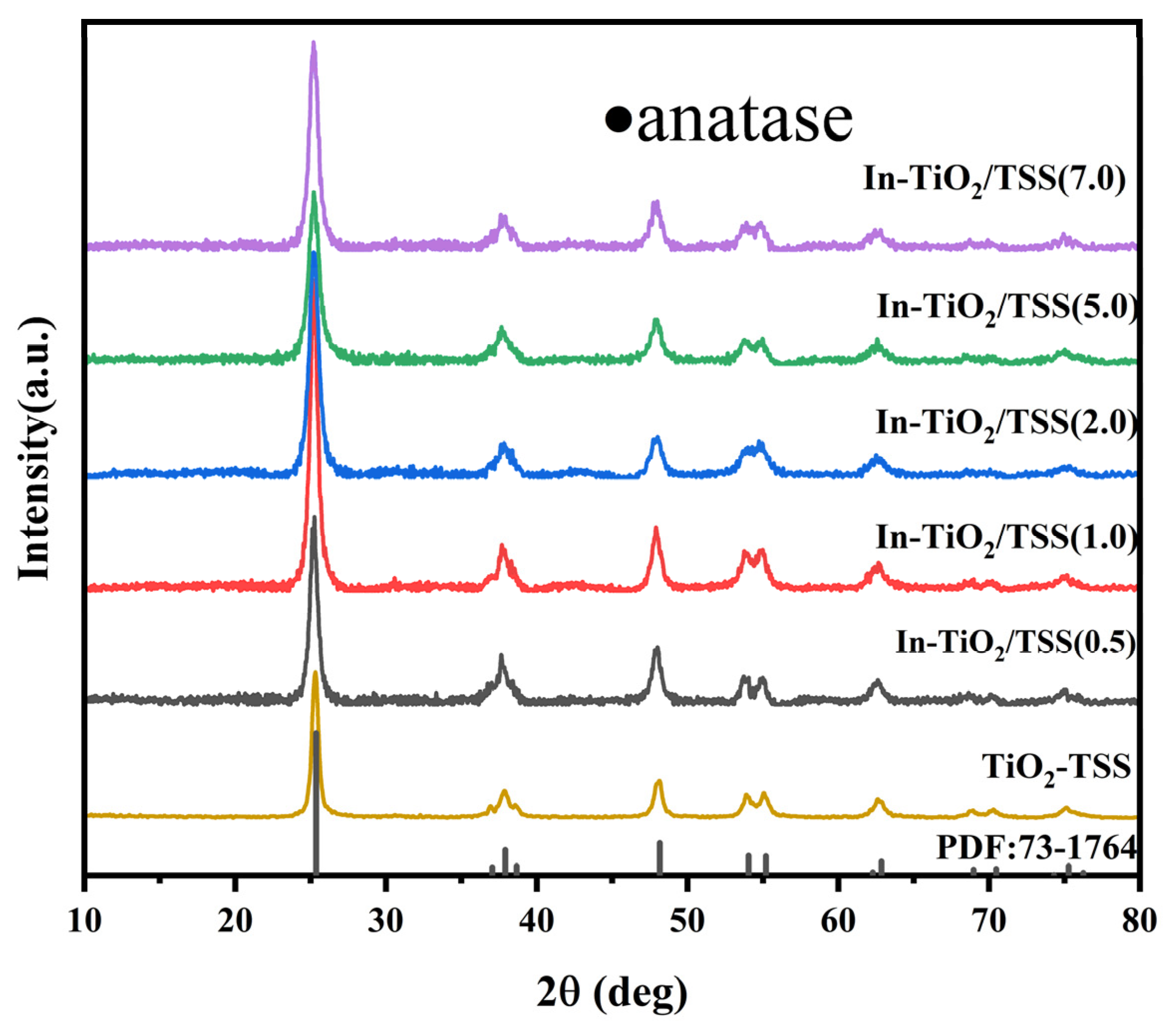
2.1.1. X-ray Diffraction (XRD) Analysis

The XRD patterns of the TiO2-TSS and In-TiO2/TSS(X) materials are presented in Figure 1. The diffraction peaks observed at 2θ values of 25.3°, 37.0°, 37.9°, 38.6°, 48.1°, 54.0°, 55.2°, 62.2°, 62.8°, 68.9°, 70.4°, 74.3°, and 75.2° correspond to the crystal planes (101), (103), (004), (112), (200), (105), (211), (213), (204), (116), (220), (107), and (215), respectively. These diffraction peaks align perfectly with anatase TiO2, as indicated by the standard JCPDS 73-1764 reference card. Consequently, it can be inferred that the synthesized materials predominantly contain the anatase phase of TiO2 and that the TSS template does not alter the crystalline structure of TiO2. Furthermore, an intriguing phenomenon arises from Figure 1, where no characteristic peaks corresponding to In are observed. If the substitution of the Ti4+ ions in TiO2 by In3+ ions were to occur [41], the unit cell volume should consequently increase, as well as a shift towards lower diffraction angles for the characteristic peaks [22]. However, the lattice parameters and unit cell volume, as shown in Table 1, indicate no significant enlargement of the doped crystal lattice. Assuming that metallic In exists within the interstitial sites of the TiO2 lattice, in such a scenario, the metal atoms would occupy a portion of the space within the TiO2 crystalline structure without perceptibly altering its unit cell dimensions. Nonetheless, given that Ti predominantly exists in the form of Ti4+ (which will be later confirmed through XPS, as the ionic radius of Ti4+ (68 pm) is smaller than that of In3+ (81 pm)), it is implausible for In3+ to coexist within the TiO2 unit cell as an interstitial species. Collectively considering these aspects, it can be inferred that In exists as an active substance in the material, likely in the form of a loading or adsorption.

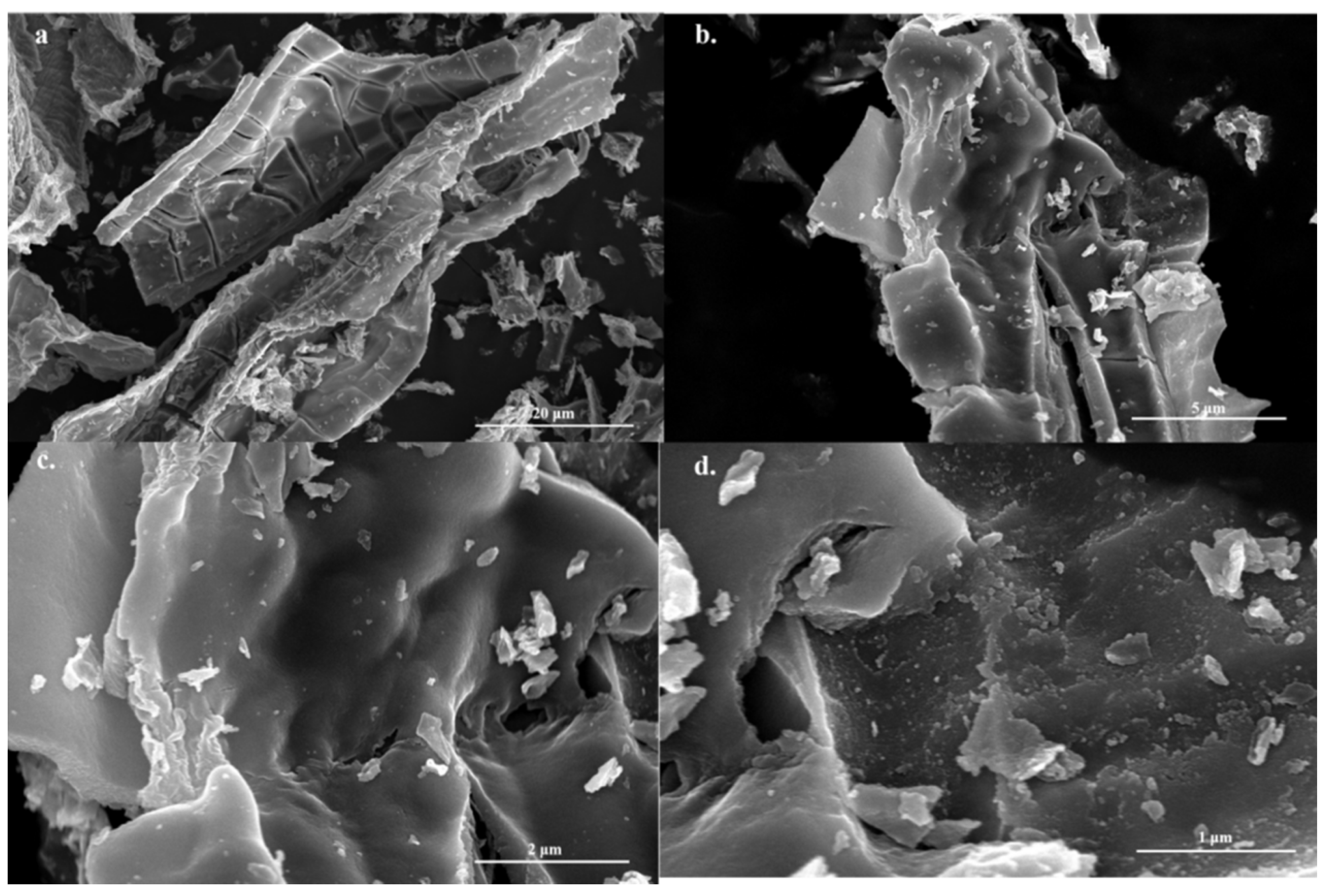
2.1.2. Scanning Electron Microscopy (SEM) Analysis of the Materials

The morphological changes in the In-doped TiO2 material modified with tobacco stem silk templates can be observed through SEM. Figure 2 depicts a high-resolution image of In-TiO2/TSS(2.0). As illustrated in Figure 2a–d, after calcination at 450 °C, the biotemplate was not completely removed from the material, which retained some biocarbon, which still, remarkably, retained the characteristic sheet-like wrinkles of the TSS [42]. There are some irregular particles on the biochar; these particles are TiO2 and In.

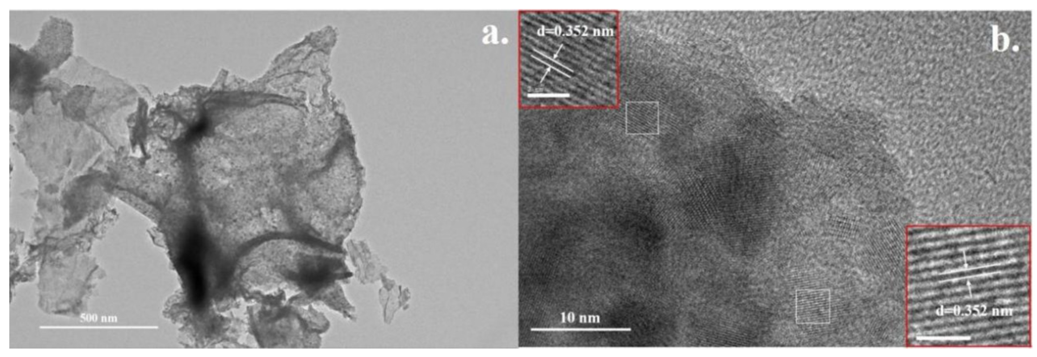
2.1.3. Transmission Electron Microscopy (TEM) Analysis of the Materials

To further validate the structural changes seen, a meticulous examination of the microstructure of In-TiO2/TSS(2.0) was conducted via TEM (JEM-2100, JEOL, Tokyo, Japan). As illustrated in Figure 3a, it can be discerned that the synthesized material impeccably retains the characteristic sheet-like morphology of the tobacco stem silk, thereby corroborating the SEM observations. Furthermore, Figure 3b portrays the lattice diffraction patterns of In-TiO2/TSS(2.0), revealing a distinct diffraction fringe spacing of merely 0.352 nm. Notably, this spacing aligns with the crystal planes characteristic of anatase TiO2. This observation provides further confirmation that the synthesized photocatalyst primarily contains TiO2 as its main constituent, aligning with the results obtained from the XRD characterization.

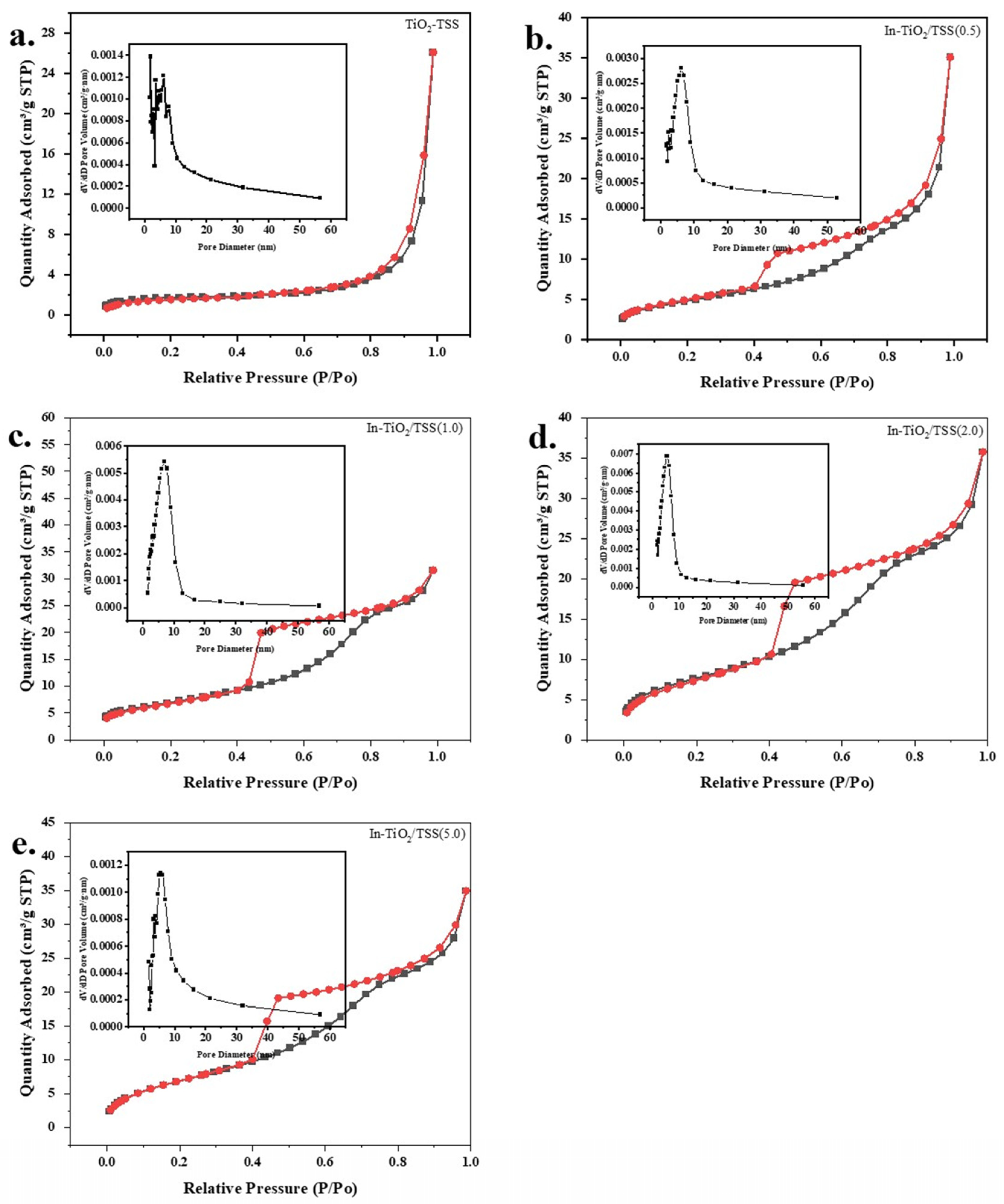
2.1.4. N2 Adsorption–Desorption Analysis of the Materials

To further validate the conclusion that metallic In exists on the surface of the TiO2 either as a supported or adsorbed species, and to ascertain the pore structure of the materials, N2 adsorption–desorption experiments were conducted. As depicted in Figure 4a–e, the N2 adsorption–desorption isotherms of TiO2 and In-TiO2/TSS(X) materials exhibit an irreversible type IV adsorption isotherm. In-TiO2/TSS(1.0), In-TiO2/TSS(2.0), and In-TiO2/TSS(5.0) display distinct saturated adsorption plateaus, indicative of an IUPAC-type H2 curve. The inset graph reveals a prominent accumulation peak in the mesoporous range of 0–10 nm for the synthesized materials. On the other hand, TiO2 and In-TiO2/TSS(0.5) exhibit an IUPAC-type H3 curve, with a similar accumulation peak observed in the 0–10 nm range, suggesting a stacking phenomenon within the pore structures of the materials. When a metal is dispersed or deposited on the carrier surface, it increases the interfacial area between the metal particles and the carrier. As demonstrated in Table 2, through comparison, it can be concluded that the specific surface area of the material first increases and then decreases as its molar ratio increases. In-TiO2/TSS(2.0) has the largest specific surface area, while its pore volume remained constant. The introduction of In results in an increased specific surface area, providing evidence for the presence of metallic In as the active component on the surface of TiO2.

2.2. Exploring the Performance

The results shown in Figure 5a indicate that the materials used without visible light irradiation did not exhibit any degradation effect on TCH, whereas the remaining experiments under visible light demonstrated a degradation performance. It can also be observed that the removal efficiency of TiO2, TiO2-TSS, and In-TiO2 under these conditions was 13.8%, 37.0%, and 34.8%, respectively, indicating a significant difference in performance compared to the In-TiO2/TSS(X) series photocatalysts. Figure 5a also reveals that with the change in In metal concentration from low to high, the removal efficiency showed an initial increase followed by a decrease. The best photocatalytic degradation effect was achieved with In-TiO2/TSS(2.0), reaching a removal rate of 92.9% for 15 mg/L TCH after 90 min of visible light. The removal rates of the In-TiO2/TSS(0.5), In-TiO2/TSS(1.0), and In-TiO2/TSS(2.0) photocatalysts gradually increased to 83.4%, 83.4%, and 92.9%, respectively, while In-TiO2/TSS(5.0) only achieved a removal rate of 75.3%. This validates the observed phenomenon that the degradation rate increases and then decreases with the increase in In concentration. In summary, it was found that TiO2-TSS prepared using tobacco stalk templates alone showed a significant improvement in removal efficiency. Compared to TiO2, the removal rate increased from 13.8% to 37.0%. Furthermore, the addition of In metal further enhanced the photocatalytic performance of the material, increasing the removal rate from 37.0% to 92.9% compared to TiO2-TSS.
To further determine the materials’ performance, a first-order kinetic curve fitting method was employed to obtain the apparent kinetic constant k (min−1) for TCH degradation, with R2 values all exceeding 0.98. As shown in Figure 5b, the k values for TiO2, TiO2-TSS were 0.001 min−1 and 0.004 min−1, respectively. With the increase in In content, the k values of In-TiO2/TSS(0.5), In-TiO2/TSS(1.0), and In-TiO2/TSS(2.0) increased to 0.016 min−1, 0.016 min−1, and 0.024 min−1, respectively. However, the k value for In-TiO2/TSS(5.0) was 0.011 min−1, further indicating that the increase in the In concentration can only improve photocatalytic activity within a certain range. This may be due to the blocking or deactivation of some active sites and the competitive adsorption caused by high concentrations of metal carriers, among other reasons. As can be seen from Table 3, the performance of the materials we prepared reached a high level.
The removal efficiency and kinetic constants of the materials were calculated using Equations (1) and (2). Herein, C0 represents the initial concentration of TCH, C denotes the instantaneous concentration of TCH, and Ce signifies the equilibrium concentration of TCH.
Removal % = (C0 − C)/C0
Ln(Ce/C) = kt

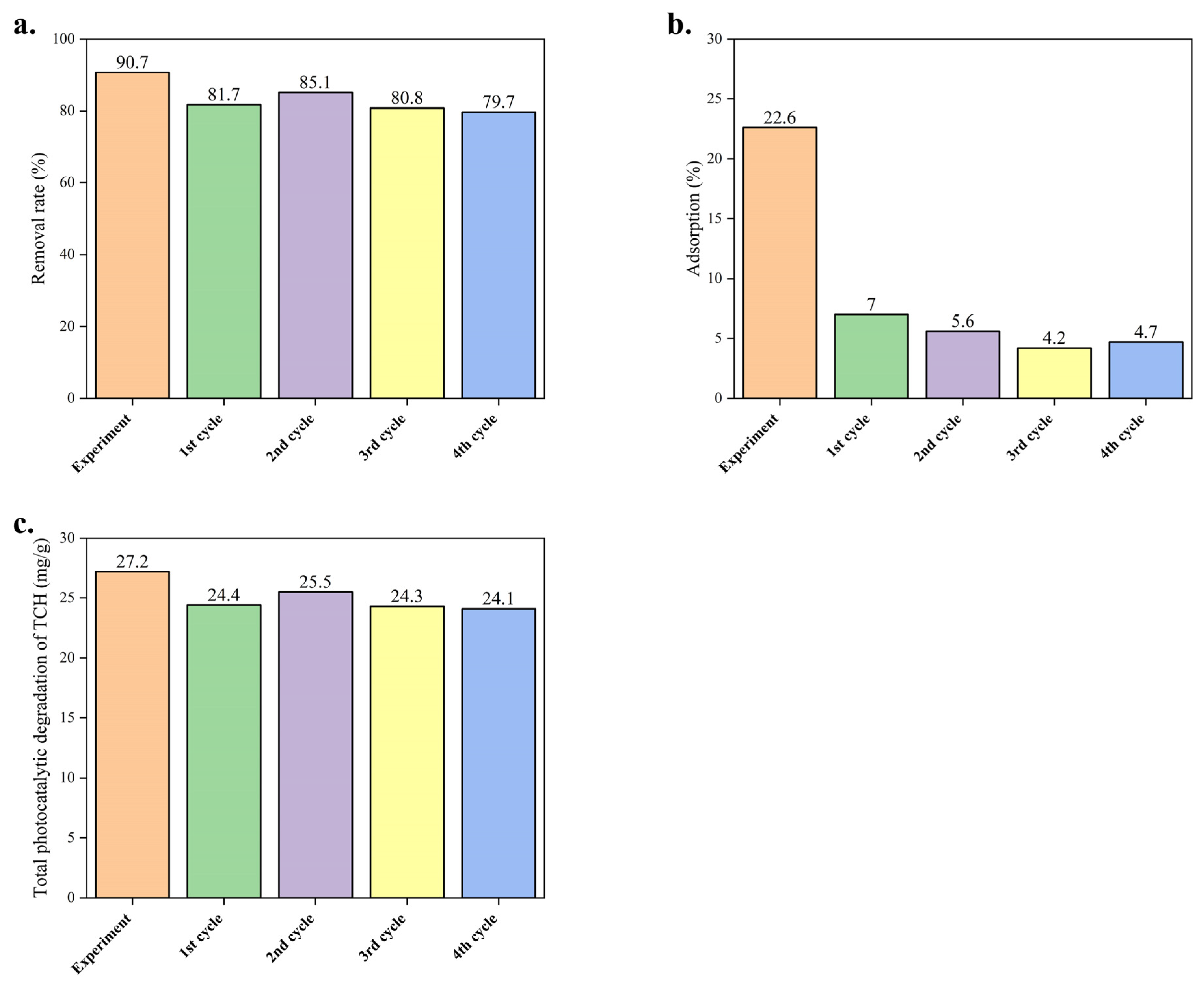
2.3. The Stability of Their Performance

The results of the cycling experiment are illustrated in Figure 6, displaying the removal efficiency, adsorption rate, and total TCH removed after four cycles using In-TiO2/TSS(2.0). Firstly, from Figure 6b, it can be observed that the adsorption rate of the material decreased from an initial 22.6% during the degradation experiment to 4.7% after the fourth cycle. This decline can be attributed to the clogging of the material’s pores by the products and impurities generated during the cycling process. Additionally, most of the active sites on the material surface were occupied by TCH pollutants, limiting the further adsorption of TCH molecules. In other words, the adsorption sites reached a state of near saturation. As for the TCH removal rate, Figure 6a indicates a slight decrease over the course of the cycles, but no significant decline was observed. After calculation and accounting for the decrease in the adsorption rate, the total photodegradation amount showed fluctuation, with values of 27.2 mg/g, 24.4 mg/g, 25.5 mg/g, 24.3 mg/g, and 24.3 mg/g seen. Notably, there was no substantial decrease in adsorption capacity. In conclusion, the material demonstrated favorable stability and reusability, exhibiting a strong resistance to photocorrosion. It can be effectively recycled until reaching its maximum adsorption capacity.

2.4. Mechanistic Investigation of Enhanced Visible-Light Photocatalytic Activity

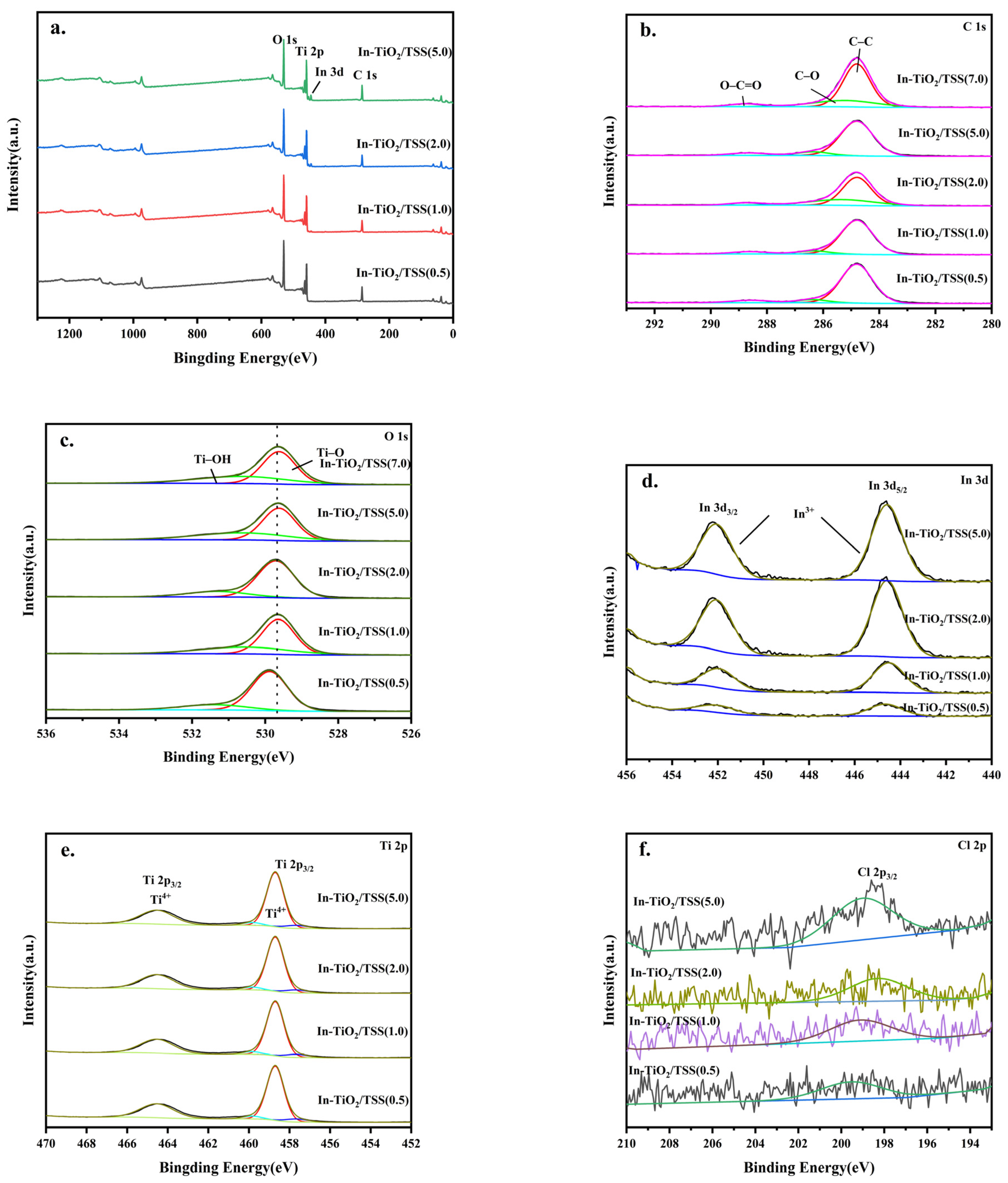
2.4.1. X-ray Photoelectron Spectroscopy (XPS) Analysis of the Materials

XPS analysis can be employed to investigate the elemental composition and surface chemical states of In-TiO2/TSS(X) series photocatalysts. As illustrated in Figure 7a, the In-TiO2/TSS(X) photocatalysts primarily consist of four elements: oxygen (O), titanium (Ti), indium (In), and carbon (C). The high-resolution C 1s spectra depicted in Figure 7b reveal three distinct chemical states of carbon. Through an area comparison, it can be concluded that the contents of Ti and O are similar. At energy positions of 284.8 eV and 284.6 eV, characteristic peaks corresponding to the main chain of carbon and the carbon–oxygen bond (with carbon as the calibration element) were observed, suggesting that these signals emanate from the TSS biocarbon [49]. Additionally, the peak at a binding energy of 288.5 eV is attributed to the characteristic peak of O–C=O, which represents oxidized biocarbon species [50].
The high-resolution O 1s spectra in Figure 7c demonstrate two chemical states of oxygen: lattice oxygen at a binding energy of 529.7 eV and Ti–OH at 531.4 eV [35]. As can be seen from Figure 7d,f, the chemical states of In and Cl are obvious. The binding energy of In 3d5/2 in In-Tio2/TSS(X) ranges from 444.5 eV to 444.69 eV, which is close to In2O3 (444.6 eV), indicating that In is connected to O. Furthermore, Figure 7e exhibits two chemical states for Ti, indicated by the characteristic peaks of Ti 2p3/2 and Ti 2p1/2. This verifies the existence of Ti in the form of Ti4+ [51], thereby corroborating the hypothesis that In3+ ions cannot exist within the TiO2 lattice in an interstitial arrangement.

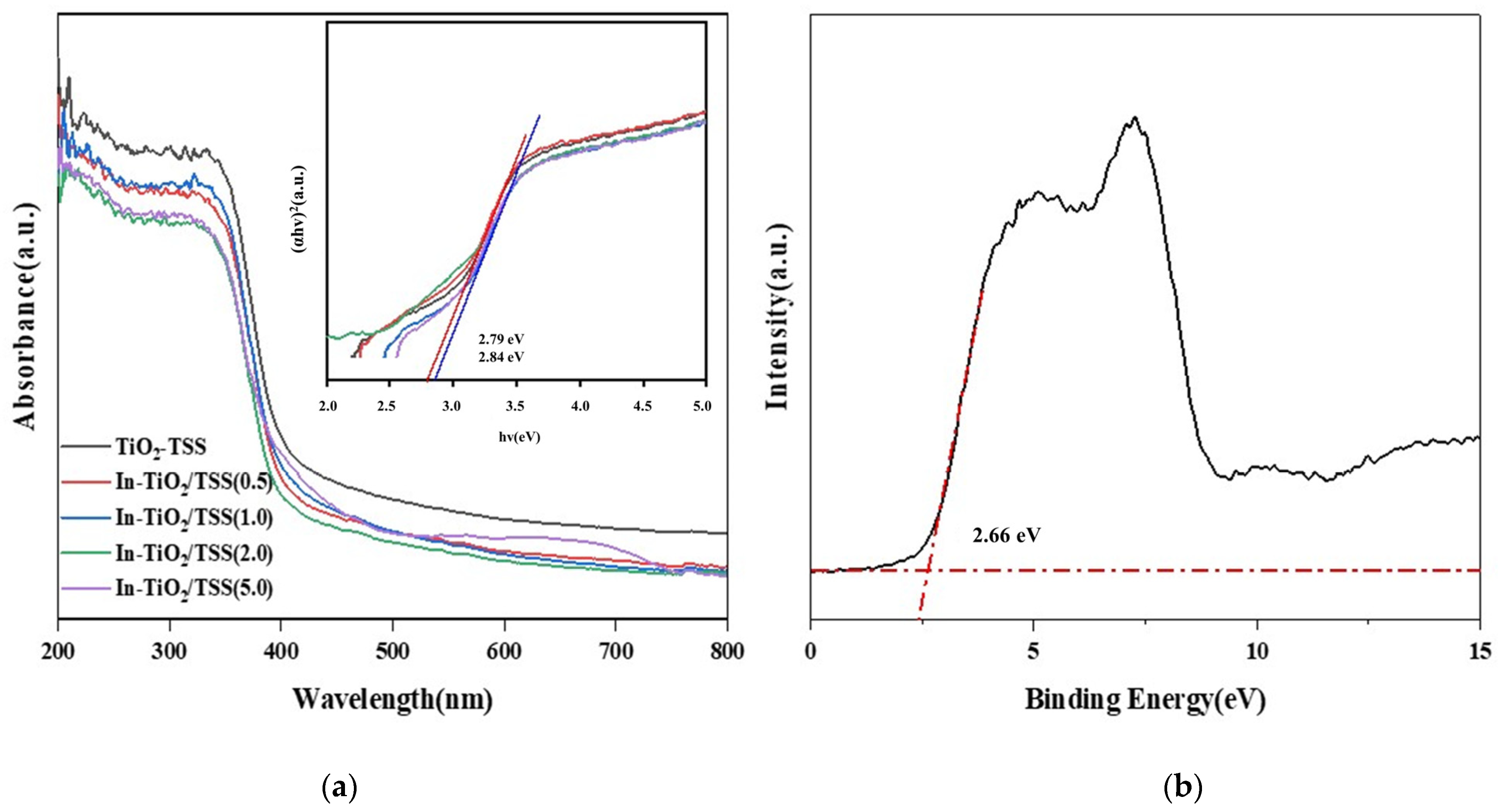
2.4.2. Analysis of Ultraviolet–Visible Diffuse Reflectance Spectroscopy (UV-Vis) and Diffuse Reflectance Spectra

In previous studies, it has been found that photocatalytic efficiency is greatly influenced by the light absorption capacity of a material [52]. During the photocatalytic process, the catalytic reaction involved requires energy provided by light. The higher the absorption efficiency, the more energy there is available for the catalytic reaction, resulting in higher catalytic efficiency [53]. Therefore, enhancing the light absorption capacity can effectively improve the capacity of a material to conduct photocatalytic reactions. To investigate the impact of adding metal In on enhancing the light absorption capacity of TiO2, UV-vis characterization was performed on TiO2-TSS and In-TiO2/TSS(X) materials. As shown in Figure 8a, the light absorption capacity of In-TiO2/TSS(X) photocatalysts gradually increases with increasing In content.
Untreated TiO2 possesses a sizable bandgap width, which is primarily engaged in the absorption of ultraviolet radiation. But, as illustrated in Table 4, the post-treatment In-TiO2/TSS(X) series of materials gradually exhibit a narrowing bandgap, rendering them capable of capturing visible and even near-infrared light, consequently escalating their practical usability. This further elucidates the underlying reasons for the superior catalytic degradation performance of the as-prepared In-TiO2/TSS(X) series materials under visible light irradiation. Figure 8a exhibits a gradual redshift of the absorption band edge, which is attributed to the sensitization caused by TSS biocarbon [44]. Notably, In-TiO2/TSS(2.0) presents a bandgap width of 2.79 eV. As depicted in Figure 8b, the valence band position of In-TiO2/TSS (2.0) was located at 2.66 eV. According to reports in the literature, it is established that the surface state energy level of O–In resides approximately 0.3 eV below the conduction band of TiO2, which is equivalent to −0.1 eV, which is in good agreement with the measured bandgap width of In-TiO2/TSS (2.0).

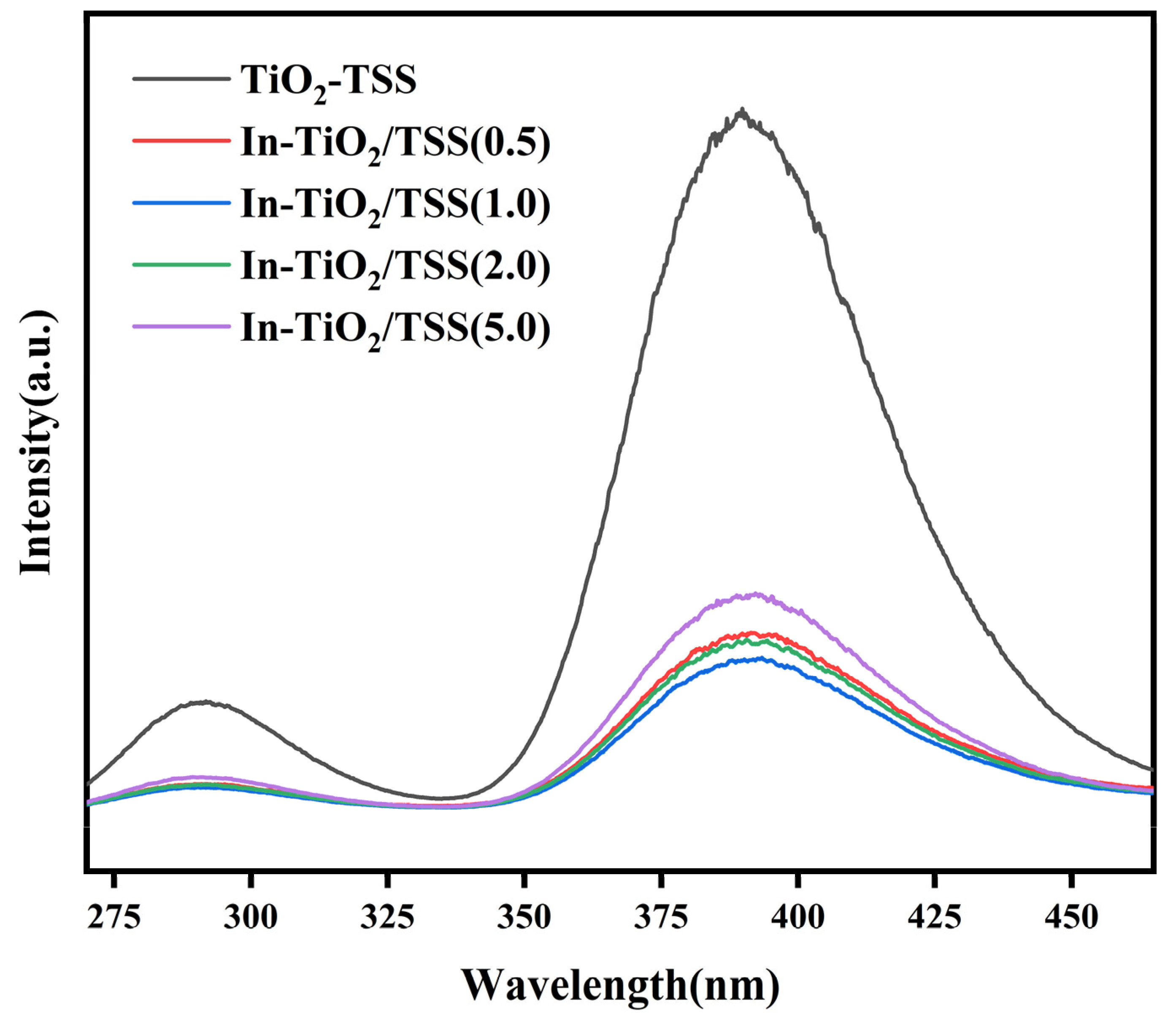
2.4.3. Analysis of Photoluminescence (PL)

Generally speaking, the peak intensity of PL is related to the recombination of electron–hole pairs. A lower intensity indicates a higher degree of separation between the photo-generated electrons and holes. A higher degree of separation can enhance photon emission, prolong the lifetime of charge carriers, enhance their light absorption capacity, and improve their photocatalytic activity [54]. Figure 9 shows the PL intensities of TiO2-TSS and In-TiO2/TSS(X) series materials. Compared to TiO2-TSS, the intensity peak of In-TiO2/TSS(X) series materials is significantly decreased, which could explain the obvious enhancement in its adsorption capacity compared to TiO2-TSS. Meanwhile, it can be observed that among the In-TiO2/TSS(X) series materials, the intensity peak of the material In-TiO2/TSS(1.0) is the lowest, but its difference with the material In-TiO2/TSS(2.0), which exhibits the best photocatalytic degradation performance in practical applications, is not significant. According to the PL analysis, the material In-TiO2/TSS(1.0) is the optimal choice among this series of materials. However, its performance in practical applications does not align with this characterization. We could consider augmenting the concentration of In metal within an appropriate range, leading to an increase in active sites. This would allow In-TiO2/TSS(2.0) to become the premier material among the prepared series, demonstrating its superiority over others in terms of performance.

2.4.4. Electrochemical Analysis

The magnitude and temporal characteristics of a transient photocurrent can serve as an assessment of the separation efficiency of photo-generated charge carriers [55,56]. A higher separation efficiency implies that a majority of the photo-generated charge carriers can escape recombination and actively participate in photocatalytic reactions, hence enhancing the reaction rate. To explore this effect, photovoltaic response tests were conducted on TiO2, TiO2-TSS, In-TiO2/TSS(2.0), and In-TiO2/TSS(5.0). As shown in Figure 10a, an immediate photocurrent was generated upon visible light irradiation, which diminished to its initial level once shielded from light. Among the materials tested, In-TiO2/TSS(2.0) exhibited the highest peak instantaneous photocurrent intensity, indicating its higher photo-generated current density and better photocatalytic performance. This finding is consistent with the results obtained from the photocatalytic degradation experiments and other analyses performed earlier. Additionally, Figure 10b presents a Nyquist plot of TiO2, TiO2-TSS, In-TiO2/TSS(2.0), and In-TiO2/TSS(5.0), where the arcs represent impedance and their radius represents resistance. A smaller radius corresponds to lower charge transfer resistance [57], and In-TiO2/TSS(2.0) exhibited the smallest arc radius, indicating its lower resistance compared to TiO2, TiO2-TSS, and In-TiO2/TSS(5.0). The lower resistance in the photoelectrochemical system suggests a more efficient charge transfer process.

2.4.5. The Study of Active Species Trapping

In order to investigate the primary species that are active during the process of photocatalytic degradation, an experiment was conducted using the In-TiO2/TSS(2.0) material for active species trapping. The experimental results, depicted in Figure 11, revealed a photocatalytic efficiency of 97% in the absence of free radical scavengers. However, with the introduction of t-butanol (tertiary-butyl alcohol, TBA) [58] as a scavenger for the hydroxyl radical (·OH), ethylenediaminetetraacetic acid di-sodium salt (Na2EDTA) [59] as a scavenger for holes (h+), and p-benzoquinone (BQ) [60] as a scavenger for the superoxide radical ·O2, its photocatalytic efficiency decreased to 77%, 31%, and 68%, respectively. This observation supports the notion that h+ functions as the primary active species, while ·OH and ·O2 play auxiliary roles. Remarkably, the introduction of AgNO3 as a capturing agent resulted in a significant enhancement of its photocatalytic efficiency, which reached 100%. This can be attributed to the consumption of e by AgNO3, which, to a certain extent, inhibits the recombination of photo-generated electron–hole pairs. As a consequence, the separation of photo-generated electron–hole pairs is facilitated, effectively promoting the production of the active species h+.
Electron Paramagnetic Resonance (EPR) technology facilitates the investigation of free radicals and other reaction intermediates within photocatalytic reactions. This insight is instrumental in elucidating the underlying mechanisms of photocatalysis, thereby advancing the design and efficacy of catalysts [61,62,63]. In order to ascertain the species that are active during the degradation process of In-TiO2/TSS(2.0), as illustrated in Figure 11b–d, the presence of ·OH, h+, and ·O2 was successfully identified using the EPR technique (instrument: Bruker EMX PLUS (Bruker (Beijing), Beijing, China), center field: 3510.00 G, sweep width: 100.0 G, power: 6.325 mW, power atten: 15.0 dB, frequency mon: 9.823347 GHz, sweep time: 30.00 s, mod amp: 1.000 G, mod freq: 100.00 kHz). In Figure 11c, TEMPO was employed to capture h+, and a distinct signal of TEMPO was detected. However, after 5 min of visible light irradiation, the signal of TEMPO significantly weakened, indicating the generation of holes under light conditions. In Figure 11b,d, the existence of ·OH and ·O2 was effectively verified through DMPO detection [64,65,66], while a weak DMPO-·OH signal and a DMPO-·O2 signal were observed after 5 min of light exposure. These results indicate the generation of ·OH and ·O2 active species under light conditions, and the EPR experiment further confirms that h+ serves as the primary active species, while ·OH and ·O2 play auxiliary roles.

3. Preparation and Structural Characterization of the Material

3.1. Materials

Tobacco stem silk (Kunming Cigarette Factory), glutaraldehyde (C3H8O2, Xilong Chemical, Sichuan, China, 50%), hydrochloric acid (HCl, Chuandong Chemical, Chongqing, China, 36%), ethanol (C2H5OH, Xilong Chemical, Sichuan, China, 99.5%), tetrabutyl orthotitanate (TBOT) (C16H36O4Ti, Adamas, Shanghai, China, 99%), indium chloride tetrahydrate (InCl3·4H2O, Adamas, Shanghai, China, 99%), and TCH (C22H25ClN2O8, Adamas, Shanghai, China, 97%+) were used in this study. All materials were of analytical grade and did not undergo further purification.

3.2. Sample Preparation

Tobacco stem silk underwent a pretreatment protocol prior to being used as a biological template; 5 g of tobacco stem silk was initially immersed in a 5% (w/w) of glutaraldehyde solution for 12 h. After filtering the glutaraldehyde solution, the tobacco stem silk was immersed in a 5% (w/w) HCI solution for a further 12 h. Subsequently, we performed gradient dehydration using 20%, 40%, 60%, 80%, and 100% ethanol. The dehydrated tobacco stem silk was then subjected to a drying period of 12 h in a 90 °C oven to yield the treated tobacco stem silk.
To obtain unmodified TiO2, 5 mL of TBOT was added to a Petri dish and allowed to react under ambient laboratory conditions for 24 h. The residual white solid was then ground and calcined in a muffle furnace (heated at a rate of 2 °C/min and maintained at 450 °C for 2 h) to yield the product.
To obtain In-TiO2, under stirring, 5 g of TBOT and 40 mg of InCl3·4H2O were added to a Petri dish and allowed to react under ambient laboratory conditions for 24 h. The residual white solid was then ground and calcined in a muffle furnace (heated at a rate of 2°C/min and maintained at 450 °C for 2 h) to yield the product.
To obtain the TSS-modified TiO2, 5 mL of TBOT was added to a beaker containing 50 mL of absolute ethanol while stirring, and 2 g of dehydrated tobacco stem, prepared as described above, was added to the stirred TBOT solution. This mixture was allowed to stand (covered with a watch glass) undisturbed under ambient laboratory conditions for 24 h. The clear supernatant was then carefully decanted and poured into a Petri dish, where it was allowed to hydrolyze under ambient laboratory conditions for 24 h. The resulting material was isolated, ground, and calcined in a muffle furnace (2 °C/min, 450 °C, 2 h) to yield TiO2-TSS.
To obtain the indium-modified TiO2 samples, 2 g of treated tobacco stem was added to 50 mL of ethanol. Then, under stirring, 5 g of TBOT and an amount of InCl3·4H2O, calculated to give the desired molar ratio of In:Ti, was added—as an example, when using 5 g of TBOT, for a molar ratio of 0.5% In:Ti, 10 mg InCl3·4H2O was required. As for the TiO2/TSS sample, after allowing the mixture to stand for 24 h, the supernatant was decanted into a Petri dish and allowed to hydrolyze under ambient conditions to yield the crude material, which was isolated, ground, and calcined in a muffle furnace at 450 °C as described previously, with a heating rate of 2 °C/min before being kept for 2 h at 450 °C, to give the desired In-TiO2/TSS(X) product.

3.3. Testing Methods for Materials

The characterization of the materials’ morphology and structure was determined using X-ray diffraction (XRD, TTRⅢ, Rigaku Corporation, Tokyo, Japan), scanning electron microscopy (SEM, Nova NanoSEM 450, FEI Company, Hillsboro, OR, USA), and transmission electron microscopy (TEM, JEM-2100, Japan Electronics Co., Ltd., Tokyo, Japan). Nitrogen gas adsorption–desorption measurements were employed to determine the specific surface area and pore size distribution of AP-NEM. An X-ray photoelectron spectroscopy (XPS, K-Alpha+, Thermo Fisher Scientific, Waltham, MA, USA) analysis was performed to investigate the elemental composition and surface chemical states of In-TiO2/TSS(X) series photocatalysts. Ultraviolet–visible diffuse reflectance spectroscopy (UV-vis, UV-2600, SHIMADZU, Kyoto, Japan) was used to analyze the effectiveness of the metal components. Photoluminescence spectroscopy (PL, F-7000, HITACHI, Tokyo, Japan) was employed to study the recombination of electron–hole pairs. An electrochemical analysis was conducted to evaluate the efficiency of photo-generated charge carrier separation. Additionally, active species capture experiments were carried out to identify the active species.

3.4. Experimental Assessment of Material Degradation Performance

In order to investigate the photocatalytic performance of In-TiO2/TSS(X) series photocatalysts, visible light was used for the photocatalytic degradation of an aqueous solution of TCH with a pollutant concentration of 15 mg/L, with the utilization of a 5-watt LED lamp (emitting wavelengths from 420 to 800 nanometers) to simulate visible light. To test the TCH concentration, 1 mL of the sample was extracted and filtered using a 0.45 μm membrane filter. The obtained filtrate was measured on an Agilent (Santa Clara, CA, USA) C18 column (4.6 × 150 mm, 5 µm) using high-performance liquid chromatography (HPLC, Agilent (Santa Clara, CA, USA) 1260-Infinity). Prior to initiating the photoreaction, a 60 min adsorption phase was conducted in the dark to achieve adsorption–desorption equilibrium. Following this, a 90 min photoreaction was performed, with sampling performed every 15 min. Additionally, to demonstrate the variation in performance, TiO2-TSS performance experiments using a tobacco stem template were conducted, with the blank control group set as TiO2 without any special treatment. Furthermore, to prove the effectiveness of photocatalysis, a group of experiments without visible light irradiation on the material were conducted for comparison. In order to explore whether the degradation rate would increase and then decrease with the increase in In concentration, a series of experiments were carried out by changing the concentration of metal In. Finally, to ensure the attainment of adsorption–desorption equilibrium, a 60 min dark reaction was conducted as a pre-treatment before the start of the visible light experiment.
To investigate the stability of the In-TiO2/TSS(X) materials, a four-cycle experiment was conducted. After each cycle, the materials were recovered through centrifugation and dried before being reused for subsequent cycles.

4. Conclusions

Employing a one-pot impregnation method, with TSS biotemplate assistance, titanium dioxide was anchored onto TSS and, subsequently, indium was introduced and further calcined, resulting in the formation of an In-TiO2/TSS(X) series of photocatalysts with biocarbon as their substrate. This process facilitated the formation of an O–In structure on their surface, consequently augmenting their surface area. Performance-testing experiments were designed to compare the degradation of tetracycline using the synthesized In-TiO2/TSS(X) photocatalysts. The best-performing material, In-TiO2/TSS(2.0), showed an enhancement of the degradation efficiency from 13.8% for TiO2 to 92.9%. Incorporating first-order kinetic simulations, the degradation performance of the In-TiO2/TSS(2.0) material was found to surpass that of TiO2 by an astonishing factor of 24. Furthermore, cyclic stability tests revealed the excellent stability of the material even after four cycles, demonstrating its strong resistance to photocorrosion. To elucidate its photocatalytic degradation mechanism, various mechanistic experiments were conducted, revealing that the introduction of tobacco stem silk templates and In modification altered the UV absorption behavior of TiO2. The In-TiO2/TSS(X) materials exhibited a narrower bandgap, primarily absorbing visible light and thus significantly enhancing their practicality. Furthermore, it was observed that the degree of separation between their photo-generated electrons and holes was high, their light absorption capacity was enhanced, their charge transfer resistance was low, and their photocatalytic efficiency was improved. Moreover, experimental findings confirmed the dominant active species as h+, with ·OH and ·O2 having auxiliary roles.
In summary, conventional TiO2 demonstrates an inadequate degradation performance under visible light. This study unveils the vast possibilities for the utilization of TiO2 under visible light and enhances our understanding of biotemplate-assisted metal ion modification. Compared to conventional ion modification methods, this approach streamlines the experimental procedures required, facilitating the broad application of templates in catalytic technologies and expanding the prospects for the industrial utilization of visible-light photocatalysts.

Author Contributions

J.L. and Y.Z.: Conceptualization, Methodology, Software, and Writing—original draft. J.Z. and A.Z.: Data curation. X.B. and Q.L.: Visualization and Investigation. M.H. and J.W.: Conceptualization and Writing—Reviewing and Editing. All authors have read and agreed to the published version of the manuscript.

Funding

This research was funded by [the Department of Ecology and Environment of Yunnan Province] grant number [202305AM340008], [R&D Project] grant number [2022 No.4], and [the Key Laboratory of Advanced Materials for Wastewater Treatment of Kunming] grant number [2110304]. And The APC was funded by [the Key Laboratory of Advanced Materials for Wastewater Treatment of Kunming] grant number [2110304].

Data Availability Statement

The data that have been used are confidential.

Acknowledgments

This work was supported by a project from the Department of Ecology and Environment of Yunnan Province, the Water Resources Department of Yunnan Province, the Key Laboratory of Advanced Materials for Wastewater Treatment of Kunming, and the Institute of Frontier Technologies in Water Treatment Co., Ltd., Kunming, 650503, China.

Conflicts of Interest

The authors declare that they have no known competing financial interests or personal relationships that could have appeared to influence the work reported in this paper.

References

  1. Wang, C.Q.; Xu, H.; Cheng, T.F.; Tang, S.T.; Zhang, D.Y.; Li, M.C.; Pan, X.L. Affinity-based alleviation of dissolved organic matter (DOM) on tetracycline toxicity to photosynthesis of green algae Chlorella vulgaris: Roles of hydrophilic and hydrophobic DOM. Environ. Sci. Pollut. Res. 2023, 30, 42165–42175. [Google Scholar] [CrossRef] [PubMed]
  2. Chen, Z.; Ou, D.; Gu, G.; Gao, S.; Li, X.; Hu, C.; Liang, X.; Zhang, Y. Removal of tetracycline from water by catalytic photodegradation combined with the microalga Scenedesmus obliquus and the responses of algal photosynthesis and transcription. J. Environ. Manag. 2023, 326, 116693. [Google Scholar] [CrossRef] [PubMed]
  3. Shao, S.C.; Wu, X.W. Microbial degradation of tetracycline in the aquatic environment: A review. Crit. Rev. Biotechnol. 2020, 40, 1010–1018. [Google Scholar] [CrossRef] [PubMed]
  4. Yuan, J.W.; Li, H.; Wang, G.; Zhang, C.; Wang, Y.X.; Yang, L.J.; Li, M.M.; Lu, J.M. Adsorption, isolated electron/hole transport, and confined catalysis coupling to enhance the photocatalytic degradation performance. Appl. Catal. B Environ. 2022, 303, 120892. [Google Scholar] [CrossRef]
  5. Seo, Y.S.; Oh, S.G. Controlling the recombination of electron-hole pairs by changing the shape of ZnO nanorods via sol-gel method using water and their enhanced photocatalytic properties. Korean J. Chem. Eng. 2019, 36, 2118–2124. [Google Scholar] [CrossRef]
  6. Zhou, W.; Fu, H.G. Defect-mediated electron-hole separation in semiconductor photocatalysis. Inorg. Chem. Front. 2018, 5, 1240–1254. [Google Scholar] [CrossRef]
  7. Chen, T.S.; Zhang, Q.X.; Zheng, X.S.; Xie, Z.J.; Zeng, Y.Q.; Chen, P.; Liu, H.J.; Liu, Y.; Lv, W.Y.; Liu, G.G. Photocatalyst with a metal-free electron-hole pair double transfer mechanism for pharmaceutical and personal care product degradation. Environ. Sci. Nano 2019, 6, 3292–3306. [Google Scholar] [CrossRef]
  8. Szkoda, M.; Trzcinski, K.; Nowak, A.P.; Coy, E.; Wicikowski, L.; Lapinski, M.; Siuzdak, K.; Lisowska-Oleksiak, A. Titania nanotubes modified by a pyrolyzed metal-organic framework with zero valent iron centers as a photoanode with enhanced photoelectrochemical, photocatalytical activity and high capacitance. Electrochim. Acta 2018, 278, 13–24. [Google Scholar] [CrossRef]
  9. Song, K.W.; Cui, Y.; Liu, L.; Chen, B.Y.; Hirose, K.; Shahiduzzaman, M.; Umezu, S. Electro-spray deposited TiO2 bilayer films and their recyclable photocatalytic self-cleaning strategy. Sci. Rep. 2022, 12, 1582. [Google Scholar] [CrossRef]
  10. Gagliardi, S.; Rondino, F.; Paoletti, C.; Falconieri, M. On the Morphology of Nanostructured TiO2 for Energy Applications: The Shape of the Ubiquitous Nanomaterial. Nanomaterials 2022, 12, 2608. [Google Scholar] [CrossRef]
  11. Sinha, R.; Ghosal, P.S. A comprehensive appraisal on status and management of remediation of DBPs by TiO2 based-photocatalysts: Insights of technology, performance and energy efficiency. J. Environ. Manag. 2023, 328, 117011. [Google Scholar] [CrossRef] [PubMed]
  12. Pakdel, E.; Daoud, W.A.; Seyedin, S.; Wang, J.F.; Razal, J.M.; Sun, L.; Wang, X.A. Tunable photocatalytic selectivity of TiO2/SiO2 nanocomposites: Effect of silica and isolation approach. Colloid Surf. A—Physicochem. Eng. Asp. 2018, 552, 130–141. [Google Scholar] [CrossRef]
  13. Kanakaraju, D.; Chandrasekaran, A. Recent advances in TiO2/ZnS-based binary and ternary photocatalysts for the degradation of organic pollutants. Sci. Total Environ. 2023, 868, 161525. [Google Scholar] [CrossRef]
  14. Twilton, J.; Le, C.; Zhang, P.; Shaw, M.H.; Evans, R.W.; MacMillan, D.W.C. The merger of transition metal and photocatalysis. Nat. Rev. Chem. 2017, 1, 0052. [Google Scholar] [CrossRef]
  15. Hasan, I.; Rana, A. A review on in SITU green synthesis of titanium dioxide nanoparticles and their photocatalytic activities. Mater. Today Proc. 2023, 81, 916–918. [Google Scholar] [CrossRef]
  16. Hasan, I.; Alharthi, F.A. Caffeine-alginate immobilized CeTiO4 bionanocomposite for efficient photocatalytic degradation of methylene blue. J. Photochem. Photobiol. A Chem. 2022, 433, 114126. [Google Scholar] [CrossRef]
  17. Yang, R.J.; Fan, Y.Y.; Zhang, Y.F.; Mei, L.; Zhu, R.S.; Qin, J.Q.; Hu, J.G.; Chen, Z.X.; Ng, Y.H.; Voiry, D.; et al. 2D Transition Metal Dichalcogenides for Photocatalysis. Angew. Chem.—Int. Ed. 2023, 135, e202218016. [Google Scholar] [CrossRef]
  18. Khurshid, F.; Jeyavelan, M.; Nagarajan, S. Photocatalytic dye degradation by graphene oxide doped transition metal catalysts. Synth. Met. 2021, 278, 116832. [Google Scholar] [CrossRef]
  19. Rauf, M.A.; Meetani, M.A.; Hisaindee, S. An overview on the photocatalytic degradation of azo dyes in the presence of TiO2 doped with selective transition metals. Desalination 2011, 276, 13–27. [Google Scholar] [CrossRef]
  20. Friedmann, D.; Caruso, R.A. Indium Oxides and Related Indium-based Photocatalysts for Water Treatment: Materials Studied, Photocatalytic Performance, and Special Highlights. Sol. RRL 2021, 5, 2100086. [Google Scholar] [CrossRef]
  21. Kurnaravel, V.; Mathew, S.; Bartlett, J.; Pillai, S.C. Photocatalytic hydrogen production using metal doped TiO2: A review of recent advances. Appl. Catal. B Environ. 2019, 244, 1021–1064. [Google Scholar] [CrossRef]
  22. Wang, E.J.; Yang, W.S.; Cao, Y.A. Unique Surface Chemical Species on Indium Doped TiO2 and Their Effect on the Visible Light Photocatalytic Activity. J. Phys. Chem. C 2009, 113, 20912–20917. [Google Scholar] [CrossRef]
  23. Reñones, P.; Fresno, F.; Oropeza, F.E.; Gorni, G.; O’Shea, V.A.D. Structural and electronic insight into the effect of indium doping on the photocatalytic performance of TiO2 for CO2 conversion. J. Mater. Chem. A 2022, 10, 6054–6064. [Google Scholar] [CrossRef]
  24. Sobahi, T.R.; Amin, M.S.; Mohamed, R.M. Enlargement of photocatalytic efficiency of BaSnO3 by indium doping for thiophene degradation. Appl. Nanosci. 2018, 8, 557–565. [Google Scholar] [CrossRef]
  25. Soni, V.; Raizada, P.; Kumar, A.; Hasija, V.; Singal, S.; Singh, P.; Hosseini-Bandegharaei, A.; Thakur, V.K.; Nguyen, V.H. Indium sulfide-based photocatalysts for hydrogen production and water cleaning: A review. Environ. Chem. Lett. 2021, 19, 1065–1095. [Google Scholar] [CrossRef]
  26. Ding, J.J.; Sun, S.; Bao, J.; Luo, Z.L.; Gao, C. Synthesis of CaIn2O4 Rods and Its Photocatalytic Performance under Visible-light Irradiation. Catal. Lett. 2009, 130, 147–153. [Google Scholar] [CrossRef]
  27. Yu, J.G.; Dai, G.P.; Cheng, B. Effect of Crystallization Methods on Morphology and Photocatalytic Activity of Anodized TiO2 Nanotube Array Films. J. Phys. Chem. C 2010, 114, 19378–19385. [Google Scholar] [CrossRef]
  28. Lamm, M.E.; Li, K.; Qian, J.; Wang, L.; Lavoine, N.; Newman, R.; Gardner, D.J.; Li, T.; Hu, L.B.; Ragauskas, A.J.; et al. Recent Advances in Functional Materials through Cellulose Nanofiber Templating. Adv. Mater. 2021, 33, 2005538. [Google Scholar] [CrossRef]
  29. Zhang, Y.; Dong, Y.X.; Zhou, J.H.; Li, X.; Wang, F. Application of Plant Viruses as a Biotemplate for Nanomaterial Fabrication. Molecules 2018, 23, 2311. [Google Scholar] [CrossRef]
  30. Hüttl, R.; Ullrich, F.; Wolf, G.; Kirchner, A.; Löthman, P.; Katzschner, B.; Pompe, W.; Mertig, M. Catalytic Carbon Monoxide Oxidation Using Bio-Templated Platinum Clusters. Catal. Lett. 2009, 132, 383–388. [Google Scholar] [CrossRef]
  31. Zhou, H.; Fan, T.X.; Zhang, D. Biotemplated Materials for Sustainable Energy and Environment: Current Status and Challenges. ChemSusChem 2011, 4, 1344–1387. [Google Scholar] [CrossRef] [PubMed]
  32. Zhao, J.K.; Shao, Q.; Ge, S.S.; Zhang, J.X.; Lin, J.; Cao, D.P.; Wu, S.D.; Dong, M.Y.; Guo, Z.H. Advances in Template Prepared Nano-oxides and Their Applications: Polluted Water Treatment, Energy, Sensing and Biomedical Drug Delivery. Chem. Rec. 2020, 20, 710–729. [Google Scholar] [CrossRef] [PubMed]
  33. Ali, J.; Cheang, U.K.; Darvish, A.; Kim, H.; Kim, M.J. Biotemplated flagellar nanoswimmers. APL Mater. 2017, 5, 116106. [Google Scholar] [CrossRef]
  34. Bereczk-Tompa, É.; Vonderviszt, F.; Horváth, B.; Szalai, I.; Pósfai, M. Biotemplated synthesis of magnetic filaments. Nanoscale 2017, 9, 15062–15069. [Google Scholar] [CrossRef] [PubMed]
  35. Adigun, O.O.; Retzlaff-Roberts, E.L.; Novikova, G.; Wang, L.F.; Kim, B.S.; Ilavsky, J.; Miller, J.T.; Loesch-Fries, L.S.; Harris, M.T. BSMV as a Biotemplate for Palladium Nanomaterial Synthesis. Langmuir 2017, 33, 1716–1724. [Google Scholar] [CrossRef]
  36. Sutar, S.; Otari, S.; Jadhav, J. Biochar based photocatalyst for degradation of organic aqueous waste: A review. Chemosphere 2022, 287, 132200. [Google Scholar] [CrossRef]
  37. Hu, Z.; Wang, Y.; Wang, L.; Wang, Q.; Zhang, Q.; Cui, F.; Jiang, G. Synthesis of S-type heterostructure π-COF for photocatalytic tetracycline degradation. Chem. Eng. J. 2024, 479, 147534. [Google Scholar] [CrossRef]
  38. Amangelsin, Y.; Semenova, Y.; Dadar, M.; Aljofan, M.; Bjorklund, G. The Impact of Tetracycline Pollution on the Aquatic Environment and Removal Strategies. Antibiotics 2023, 12, 440. [Google Scholar] [CrossRef]
  39. Wang, X.; Jia, J.; Wang, Y. Combination of photocatalysis with hydrodynamic cavitation for degradation of tetracycline. Chem. Eng. J. 2017, 315, 274–282. [Google Scholar] [CrossRef]
  40. Wang, B.; Wang, T.; Su, H. Hydrodynamic cavitation (HC) degradation of tetracycline hydrochloride (TC). Sep. Purif. Technol. 2022, 282, 120095. [Google Scholar] [CrossRef]
  41. Lan, Z.J.; Yu, Y.L.; Yan, S.; Wang, E.J.; Yao, J.H.; Cao, Y.A. Synergetic effect of N3−, In3+ and Sn4+ ions in TiO2 towards efficient visible photocatalysis. J. Photochem. Photobiol. A Chem. 2018, 356, 132–137. [Google Scholar] [CrossRef]
  42. Zhang, X.L.; Zhu, X.F.; Duan, J.T.; Li, H.; Xu, L.; Xiong, L.Z.; Luo, Q.; Yang, M.H.; Chang, X. Comparative study on properties of polypropylene-based composites reinforced with tobacco stalk fibers (unmodified/modified) from different parts of tobacco stalk. Ind. Crops Prod. 2023, 201, 116890. [Google Scholar] [CrossRef]
  43. Xu, Y.; Liu, J.; Xie, M.; Jing, L.; Xu, H.; She, X.; Li, H.; Xie, J. Construction of novel CNT/LaVO4 nanostructures for efficient antibiotic photodegradation. Chem. Eng. J. 2019, 357, 487–497. [Google Scholar] [CrossRef]
  44. Li, S.; Hu, S.; Xu, K.; Jiang, W.; Liu, Y.; Leng, Z.; Liu, J. Construction of fiber-shaped silver oxide/tantalum nitride p-n heterojunctions as highly efficient visible-light-driven photocatalysts. J. Colloid Interface Sci. 2017, 504, 561–569. [Google Scholar] [CrossRef]
  45. Jing, H.; Ou, R.; Yu, H.; Zhao, Y.; Lu, Y.; Huo, M.; Huo, H.; Wang, X. Engineering of g-C3N4 nanoparticles/WO3 hollow microspheres photocatalyst with Z-scheme heterostructure for boosting tetracycline hydrochloride degradation. Sep. Purif. Technol. 2021, 255, 117646. [Google Scholar] [CrossRef]
  46. Oseghe, E.O.; Ofomaja, A.E. Facile microwave synthesis of pine cone derived C-doped TiO2 for the photodegradation of tetracycline hydrochloride under visible-LED light. J. Environ. Manag. 2018, 223, 860–867. [Google Scholar] [CrossRef]
  47. Li, C.; Yu, S.; Dong, H.; Liu, C.; Wu, H.; Che, H.; Chen, G. Z-scheme mesoporous photocatalyst constructed by modification of Sn3O4 nanoclusters on g-C3N4 nanosheets with improved photocatalytic performance and mechanism insight. Appl. Catal. B Environ. 2018, 238, 284–293. [Google Scholar] [CrossRef]
  48. Shi, K.P.; Wang, J.; Yin, L.; Xu, Y.; Kong, D.S.; Li, H.X.; Zhang, Y.; He, H.; Yang, S.G.; Ni, L.X.; et al. Photocatalysis Combined with Microalgae to Promote the Degradation and Detoxification of Tetracycline Hydrochloride. Bull. Environ. Contam. Toxicol. 2023, 110, 43. [Google Scholar] [CrossRef] [PubMed]
  49. Chen, J.T.; Zhang, G.A.; Luo, B.M.; Sun, D.F.; Yan, X.B.; Xue, Q.J. Surface amorphization and deoxygenation of graphene oxide paper by Ti ion implantation. Carbon 2011, 49, 3141–3147. [Google Scholar] [CrossRef]
  50. Sadanandam, G.; Lalitha, K.; Kumari, V.D.; Shankar, M.V.; Subrahmanyam, M. Cobalt doped TiO2: A stable and efficient photocatalyst for continuous hydrogen production from glycerol: Water mixtures under solar light irradiation. Int. J. Hydrogen Energy 2013, 38, 9655–9664. [Google Scholar] [CrossRef]
  51. Naseem, S.; Pinchuk, I.V.; Luo, Y.K.; Kawakami, R.K.; Khan, S.; Husain, S.; Khan, W. Epitaxial growth of cobalt doped TiO2 thin films on LaAlO3(100) substrate by molecular beam epitaxy and their opto-magnetic based applications. Appl. Surf. Sci. 2019, 493, 691–702. [Google Scholar] [CrossRef]
  52. Gao, L.K.; Gan, W.T.; Qiu, Z.; Cao, G.L.; Zhan, X.X.; Qiang, T.G.; Li, J. Biomorphic Carbon-Doped TiO2 for Photocatalytic Gas Sensing with Continuous Detection of Persistent Volatile Organic Compounds. ACS Appl. Nano Mater. 2018, 1, 1766–1775. [Google Scholar] [CrossRef]
  53. Wang, Q.Y.; Zhang, Z.M.; Liu, L.; Bai, L.; Bao, R.Y.; Yang, M.B.; Yang, W. Degradable ultrathin high-performance photocatalytic hydrogen generator from porous electrospun composite fiber membrane with enhanced light absorption ability. J. Mater. Chem. A 2021, 9, 10277–10288. [Google Scholar] [CrossRef]
  54. Huang, D.L.; Li, J.; Zeng, G.M.; Xue, W.J.; Chen, S.; Li, Z.H.; Deng, R.; Yang, Y.; Cheng, M. Facile construction of hierarchical flower-like Z-scheme AgBr/Bi2WO6 photocatalysts for effective removal of tetracycline: Degradation pathways and mechanism. Chem. Eng. J. 2019, 375, 15. [Google Scholar] [CrossRef]
  55. Ma, D.D.; Wang, Z.Y.; Shi, J.W.; Zou, Y.J.; Lv, Y.X.; Ji, X.; Li, Z.H.; Cheng, Y.H.; Wang, L.Z. An ultrathin Al2O3 bridging layer between CdS and ZnO boosts photocatalytic hydrogen production. J. Mater. Chem. A 2020, 8, 11031–11042. [Google Scholar] [CrossRef]
  56. Wang, L.M.; Sun, Y.; Zhang, F.Y.; Hu, J.T.; Hu, W.T.; Xie, S.J.; Wang, Y.K.; Feng, J.; Li, Y.B.; Wang, G.Y.; et al. Precisely Constructed Metal Sulfides with Localized Single-Atom Rhodium for Photocatalytic C-H Activation and Direct Methanol Coupling to Ethylene Glycol. Adv. Mater. 2023, 35, 9. [Google Scholar] [CrossRef]
  57. Xing, W.N.; Li, C.M.; Wang, Y.; Han, Z.H.; Hu, Y.D.; Chen, D.H.; Meng, Q.Q.; Chen, G. A novel 2D/2D carbonized poly-(furfural alcohol)/g-C3N4 nanocomposites with enhanced charge carrier separation for photocatalytic H2 evolution. Carbon 2017, 115, 486–492. [Google Scholar] [CrossRef]
  58. Guo, C.R.; Qin, X.; Guo, R.; Lv, Y.; Li, M.R.; Wang, Z.Y.; Li, T.H. Optimization of heterogeneous Fenton-like process with Cu-Fe@CTS as catalyst for degradation of organic matter in leachate concentrate and degradation mechanism research. Waste Manag. 2021, 134, 220–230. [Google Scholar] [CrossRef]
  59. Wang, J.P.; Li, C.Q.; Cong, J.K.; Liu, Z.W.; Zhang, H.Z.; Liang, M.; Gao, J.K.; Wang, S.L.; Yao, J.M. Facile synthesis of nanorod-type graphitic carbon nitride/Fe2O3 composite with enhanced photocatalytic performance. J. Solid State Chem. 2016, 238, 246–251. [Google Scholar] [CrossRef]
  60. Wang, S.J.; Zhao, C.C.; Wang, D.J.; Wang, Y.Q.; Liu, F. •OH initiated heterogeneous oxidation of methyl orange using an Fe- Ce/MCM-41 catalyst. RSC Adv. 2016, 6, 18800–18808. [Google Scholar] [CrossRef]
  61. Al-Madanat, O.; Nunes, B.N.; AlSalka, Y.; Hakki, A.; Curti, M.; Patrocinio, A.O.T.; Bahnemann, D.W. Application of EPR Spectroscopy in TiO2 and Nb2O5 Photocatalysis. Catalysts 2021, 11, 1514. [Google Scholar] [CrossRef]
  62. Zhang, X.; Li, Z.; Zeng, B.; Li, C.; Han, H. EPR study of charge separation associated states and reversibility of surface bound superoxide radicals in SrTiO3 photocatalyst. J. Energy Chem. 2022, 70, 388–393. [Google Scholar] [CrossRef]
  63. Morra, E.; Giamello, E.; Chiesa, M. EPR approaches to heterogeneous catalysis. The chemistry of titanium in heterogeneous catalysts and photocatalysts. J. Magn. Reson. 2017, 280, 89–102. [Google Scholar] [CrossRef] [PubMed]
  64. Deng, X.; Wang, D.D.; Li, H.J.; Jiang, W.; Zhou, T.Y.; Wen, Y.; Yu, B.; Che, G.B.; Wang, L. Boosting interfacial charge separation and photocatalytic activity of 2D/2D g-C3N4/ZnIn2S4S-scheme heterojunction under visible light irradiation. J. Alloys Compd. 2022, 894, 162209. [Google Scholar] [CrossRef]
  65. Li, J.J.; Ding, Y.; Chen, K.Y.; Li, Z.N.; Yang, H.J.; Yue, S.J.; Tang, Y.P.; Wang, Q.Z. δ-FeOOH coupled BiOBr0.5I0.5 for efficient photocatalysis-Fenton synergistic degradation of organic pollutants. J. Alloys Compd. 2022, 903, 163795. [Google Scholar] [CrossRef]
  66. Deng, F.; Zhao, L.N.; Luo, X.B.; Luo, S.L.; Dionysiou, D.D. Highly efficient visible-light photocatalytic performance of Ag/AgIn5S8 for degradation of tetracycline hydrochloride and treatment of real pharmaceutical industry wastewater. Chem. Eng. J. 2018, 333, 423–433. [Google Scholar] [CrossRef]
Figure 1. XRD patterns of TiO2-TSS and In-TiO2/TSS(X).
Catalysts 14 00615 g001
Figure 2. (ad) SEM images of In-TiO2/TSS(2.0).
Catalysts 14 00615 g002
Figure 3. (a) TEM image and (b) HR-TEM image of In-TiO2/TSS(2.0).
Catalysts 14 00615 g003
Figure 4. N2 isothermal adsorption–desorption and embedded pore size distribution of (a) TiO2-TSS and (be) In-TiO2/TSS(X) (TSS, Tobacco stem silk. X, the molar ratio of In/Ti).
Catalysts 14 00615 g004
Figure 5. (a) Removal curves of TCH over TiO2, TiO2-TSS, In-TiO2, and In-TiO2/TSS(X); (b) pseudo-first-order rate constant k (min−1) for TCH’s photocatalytic degradation over TiO2, TiO2-TSS, In-TiO2, and In-TiO2/TSS(X).
Catalysts 14 00615 g005
Figure 6. Plots of (ac) removal rate, adsorption rate, and TCH content of In-TiO2/TSS(2.0) due to visible photocatalytic degradation in TCH recycling experiment.
Catalysts 14 00615 g006
Figure 7. XPS spectra of (a) full spectra, (b) C 1s, (c) O 1s, (d) In 3d, (e) Ti 2p, and (f) Cl 2p of In-TiO2/TSS(X).
Catalysts 14 00615 g007
Figure 8. (a) UV-Vis diffuse reflectance spectra of TiO2-TSS and In-TiO2/TSS(X) and their resulting Tauc plots (inset); (b) valence band spectra of In-TiO2/TSS(2.0).
Catalysts 14 00615 g008
Figure 9. PL spectra of TiO2-TSS and In-TiO2/TSS(X).
Catalysts 14 00615 g009
Figure 10. (a) Transient photocurrent response diagram and (b) Nyquist plot of TiO2, TiO2-TSS, In-TiO2/TSS(2.0), and In-TiO2/TSS(5.0).
Catalysts 14 00615 g010
Figure 11. Plots of the experimental results of (a) the active species for the visible photocatalytic degradation of TCH by In-TiO2/TSS(2.0), (bd) the EPR spectra seen with the addition of DMPO to capture ·OH (b), the addition of TEMPO to capture h+, (c) and the addition of DMPO to capture ·O2 (d).
Catalysts 14 00615 g011
Table 1. The lattice parameters and cell volume of TiO2-TSS and In-TiO2/TSS(X).
SampleCell Parameters (Å)Cell Volume (Å)
a = bc
TiO23.7769.486135.253
TiO2-TSS3.7769.486135.253
In-TiO2/TSS(0.5)3.7839.497135.754
In-TiO2/TSS(1.0)3.7769.486135.253
In-TiO2/TSS(2.0)3.7839.497135.754
In-TiO2/TSS(5.0)3.7769.486135.253
Table 2. The specific surface area and pore structure data of TiO2-TSS and In-TiO2/TSS(X).
CatalystSBET (m2/g)Pore Volume (cm3/g)Pore Size (nm)
TiO2-TSS6.30.0427.0
In-TiO2/TSS(0.5)17.30.059.01
In-TiO2/TSS(1.0)24.90.044.5
In-TiO2/TSS(2.0)28.00.055.2
In-TiO2/TSS(5.0)26.90.055.2
Table 3. Comparison of TCH degradation efficiency of different catalysts.
CatalystConcentration (mg/L)Degradation Efficiency (%)Refs.
CNT/LaVO41084[43]
Ag2O/Ta3N51078.3[44]
C3N4 NP/WO3 NHMs1079.8[45]
C-TiO2570[46]
Sn3O4/g-C3N41072.2[47]
Chlorella sp.1050[48]
TiO2-TSS1537This work
In-TiO2/TSS(2.0)1592.9This work
Table 4. Forbidden band widths of TiO2-TSS and In-TiO2/TSS(X).
PhotocatalystBandgap
(eV)
PhotocatalystBand Gap
(eV)
TiO2-TSS3.08In-TiO2/TSS(2.0)2.79
In-TiO2/TSS(0.5)3.01In-TiO2/TSS(5.0)2.64
In-TiO2/TSS(1.0)2.98
Disclaimer/Publisher’s Note: The statements, opinions and data contained in all publications are solely those of the individual author(s) and contributor(s) and not of MDPI and/or the editor(s). MDPI and/or the editor(s) disclaim responsibility for any injury to people or property resulting from any ideas, methods, instructions or products referred to in the content.

