Review

Oxides and Metal Oxide/Carbon Hybrid Materials for Efficient Photocatalytic Organic Pollutant Removal

by
Abu Saad Ansari
1,*,†,
Griszha Azzahra
2,†,
Fairuz Gianirfan Nugroho
2,
Momin M. Mujtaba
3 and
Abu Talha Aqueel Ahmed
4,*
1
Materials Science & Engineering, Incheon National University, Songdo-dong 119 Academy-ro, Yeonsu-gu, Incheon 22012, Republic of Korea
2
Center of Excellence Applied Nanotechnology, Nano Center Indonesia, Puspiptek, South Tangerang 15314, Indonesia
3
Maharaja Sayajirao Gaikwad Arts, Science and Commerce College Malegaon Camp, Malegaon 423203, India
4
Division of System Semiconductor, Dongguk University, Seoul 04620, Republic of Korea
*
Authors to whom correspondence should be addressed.
These authors contributed equally to this work.
Catalysts 2025, 15(2), 134; https://doi.org/10.3390/catal15020134
Submission received: 20 November 2024 / Revised: 27 January 2025 / Accepted: 28 January 2025 / Published: 1 February 2025

Abstract

:
Pharmaceuticals are increasingly significant contaminants in the environmental ecosystem, prompting the exploration of photocatalysis as a promising method for removing their pollutants. However, the application of semiconductor metal oxides as photocatalysts has been limited by issues such as rapid photocarrier recombination and high band gap energy. One emerging strategy to enhance the photocatalytic performance of metal oxides involves integrating them with carbon dots, which offer advantages including low toxicity, aqueous stability, increased surface area, cost effectiveness, biocompatibility, and chemical inertness. In this study, we conducted a critical review focusing on the nanocomposite development of metal oxide/carbon dots for the photocatalytic removal of pharmaceutical pollutants. Our study highlights that carbon dots can significantly enhance the photocatalytic efficiency of these metal oxides as photocatalytic materials by improving the adsorption of organic pollutants and enhancing light absorption in the visible spectrum. This review aims to provide insights for future research aimed at advancing the development of enhanced photocatalytic metal oxide/carbon dot nanocomposites.

Graphical Abstract

1. Introduction

Water is the Earth’s lifeblood, essential for sustaining ecosystems and supporting all forms of human activity. With the growing global population, the demand for water for human activities has increased. However, this rise in water usage has not been accompanied by improved water quality suitable for human consumption. Access to hygienic and affordable water has been a significant global challenge. Various chemicals have been introduced through industrial discharges and other human activities into the natural environment, polluting water resources. These chemicals include pharmaceuticals, heavy metals, chlorinated pollutants, pesticides, and personal care products that pose high risks to environmental and human health [1]. Among these, pharmaceutical pollutants are prominent. The widespread use of pharmaceuticals, particularly antibiotics, in daily life for public health treatments has become a leading driver of emerging water pollution, posing significant risks to both human health and aquatic ecosystems [2]. The persistent presence of antibiotic residues in water resources can lead to toxic effects such as cancer, severe bleeding, and birth defects, as agriculture, humans, and animals can consume them. Research is ongoing in various fields like catalysis [3,4], optoelectronics [5,6], semiconductors [7], etc., to resolve issues.
To address the biomedical waste issue, numerous efforts have been made to mitigate the pollution risks associated with antibiotic waste using efficient and sustainable technologies. Over the past several years, advanced oxidation processes (AOPs) have garnered significant interest among researchers seeking sustainable solutions. AOPs can degrade antibiotics into smaller substances, reducing their inhibitory effects and enhancing biodegradability. Conventional AOP-based water treatment methodologies, like Fenton oxidation, have limitations such as high operational costs, low removal efficiency, and high energy consumption, which hinder their effectiveness in removing organic pollutants [8]. One alternative AOP is photocatalysis, which focuses on converting light energy into chemical energy. Photocatalysis has gained popularity due to its efficient, effective, economical, and environmentally friendly characteristics [9].
Especially semiconductor-based photocatalysis, which utilizes light-activated semiconductors, represents a promising and environmentally friendly approach for wastewater treatment. The pair of electrons ( e ) and holes ( h + ) are generated within the semiconductor photocatalyst upon exposure to light, which results in the creation of reactive oxygen species, subsequently initiating redox reactions, thereby degrading pharmaceutical contaminants into less harmful components. For example, the successful degradation of the antibiotic levofloxacin was achieved using Bi2O3 underneath the irradiation of visible light, with a degradation efficiency of 57% [10]. Mukherjee et al. [11] successfully removed the antibiotic ciprofloxacin using ZnO nanoparticles, with a degradation efficiency of 75%. Yet, the real-world use of semiconductor metal oxides is constrained by the rapid recombination of photogenerated carriers and their high band gap energy [9]. This limitation arises because in wide-band gap semiconductors (Eg > 3 eV), electron excitation is only possible under UV light. Several semiconductors, including zinc oxide (ZnO), titanium dioxide (TiO2), stannic oxide (SnO2), and tungsten trioxide (WO3), have been extensively studied; still, their wide band gaps prevent them from absorbing visible light, resulting in less efficiency in the visible spectrum. TiO2 is widely used in photocatalytic applications but faces challenges because of its larger ~3.2 eV energy band gap [12]. Conversely, visible light can efficiently excite the electron in a narrow band gap ( E g < 3 eV) semiconductor photocatalyst [13]. However, they face challenges like rapid electron–hole recombination, stability issues, a limited light absorption range, and reduced oxidizing power.
Various strategies, including immobilizing photocatalysts onto inorganic or organic support materials, have been explored to enhance the efficiency of semiconductor material-based photocatalysts. Heterojunction photocatalysts strengthen the separation of photogenerated charge carriers, modify the band gap, and expand photocatalyst activation across a broader light spectrum, enabling more efficient oxidation reactions [13]. Carbon dots are of particular interest as innovative materials to combine with semiconductors for photocatalysis due to their unique properties, including high photoluminescence and a broad optical range. Carbon dots are capable of converting light from the visible and near-infrared regions into short-wavelength, higher-energy radiation, enhancing the photocatalytic activity of semiconductors, demonstrating modification in the semiconductor material energy band gap, extending photogenerated charge carriers and improving photonic efficiency [14]. Xinghao et al. [15] demonstrated that composites of carbon dots and oxygen-rich TiO2 nanosheets increased the degradation rate of the antibiotic tetracycline to 94.1% underneath the irradiation of visible light, approximately 2.3 times higher than pure TiO2. Another study showed that a CuO2 and carbon dot composite achieved a photocatalytic performance of 92.49% for the remediation of tetracycline [16].
This review delves into the use of metal oxide semiconductor nanomaterial photocatalysts for the degradation of pharmaceutical pollutants, with a focus on the underlying mechanisms, material morphology, and the synergistic effects of carbon dots in enhancing photocatalytic efficiency. It highlights the advantages of metal oxide/carbon dot nanocomposites over other pharmaceutical remediation materials and explores the interaction between metal oxide and carbon dots in driving superior photocatalytic performance.
The advancement of metal oxide/carbon dot nanocomposites represents a promising pathway for tackling pharmaceutical pollutants in water. By focusing on optimizing synthesis methods, exploring diverse material combinations, enhancing our understanding of their photocatalytic mechanisms, and integrating them into hybrid remediation systems, researchers can unlock their full potential for large-scale water treatment applications. Addressing environmental safety, scalability, and real-world implementation challenges will be crucial to ensure their sustainable and responsible use. With continued innovation and interdisciplinary collaboration, these nanocomposites could play a transformative role in developing effective and eco-friendly water purification technologies, contributing significantly to global efforts in mitigating pharmaceutical contamination and safeguarding water resources for the future.

2. Pharmaceutical Pollutants in Water Resources

Pharmaceuticals refer to any synthesized chemical compounds or drugs designed for treating diseases [17]. The presence of pharmaceutical products, including antibiotics, painkillers, lipid regulators, and neuroactive compounds, has increased due to their widespread use in public health, animal farming, agriculture, and aquatic ecosystems [18]. Pharmaceuticals have become essential to human life, significantly benefiting society’s health and well-being, including improved disease prevention, enhanced life expectancy, and reduced rates of mortality [19]. Figure 1 illustrates the flow of pharmaceutical pollutants on surface water [20].
The COVID-19 pandemic has further exacerbated the global consumption of pharmaceuticals, particularly antibiotics, as viral infections often lead to secondary bacterial infections [21]. By 2030, the global consumption of pharmaceutical products, particularly antibiotics, is estimated to increase by approximately 67% for human use and 11.5% for animal use [22]. The extensive use of pharmaceuticals has garnered scientific and public attention due to their negative impacts, as active pharmaceutical chemicals in water are considered emerging contaminants. Active pharmaceutical ingredients (APIs) are intricate molecules classified as micropollutants, present in water resources from nanograms (ng) to micrograms (µg) per litre (L) concentration range.
A significant proportion of antibiotics are not fully metabolized by humans and animals, entering wastewater through excretion. Antibiotic compounds discharged into rivers are often inadequately treated by sewage plants and 40 to 90% of antibiotics administered to humans/animals pass through undigested, releasing directly into the environment [23]. This presence poses toxic threats to aquatic organisms. Domestic water supplies from such resources through wastewater treatment plants can potentially lead to negative impacts such as cancer, reproductive disorders, and a range of toxic effects, from mild to severe, in humans.
In the aquatic environment, the observed pharmaceutical concentration range is between 0.5 and 31 mg/L [17]. This high presence comes from the significant proportion of unabsorbed pharmaceuticals by humans and animals. The buildup of antibiotic compounds in water sources can lead to the emergence of antibiotic-resistant bacteria (ARB) and the spread of antimicrobial resistance genes [2], increasing toxicity risks for ecosystems and human health. This is worsened by the lack of regulations discussing the limit of antibiotics in wastewater, although recently (2024), the World Health Organization (WHO) published a new global guidance on antibiotic pollution, which shows promise. In the document, the maximum concentrations are denoted as Predicted No-Effect Concentrations (PNECs). Human health risks are expressed as Predicted No-Effect Concentrations for resistance selection (PNECres), while ecological outcomes are assessed through Predicted No-Effect Concentrations for ecological effects (PNECeco). Table 1 outlines the contribution of antibiotics to environmental pollution and their concentrations in wastewater treatment plants (WWTPs), compared with their respective PNECs [24,25]. All the influent concentrations surpass the mentioned PNECs, further emphasizing the severity of pharmaceutical pollution in water.
For long-term public health, it is crucial to resolve this issue by removing these pharmaceutical chemical compounds from water. Although various methods have been developed in recent years to remove pharmaceutical compounds, many are limited by technological, financial, or operational constraints. Therefore, new approaches must be considered for the effective treatment of pharmaceutical pollutants.

3. Remediation Method for Pharmaceutical Pollutants

As highlighted in the preceding section (Table 1), traditional wastewater treatment plants are not fully equipped to eradicate pharmaceutical contaminants. To accomplish the efficient remediation of pharmaceuticals in aquatic systems, advanced water treatment technologies must be employed. Several methods for pharmaceutical pollutant remediation have been developed including biological treatment, physical treatment, and chemical treatment. The pros and cons of these methods in terms of cost, efficiency, operational difficulties, and environmental effects are outlined in Table 2.
One can realize from Table 2 that any wastewater treatment method alone is not sufficient or economically feasible to completely remove pharmaceutical pollutants. Among the methods above, the adsorption method has several advantages over other wastewater treatment methods due to its simple design, low initial investment, and minimal space. It offers high removal efficiencies, operational ease, and relatively low energy consumption, with removal rates reaching up to 90% under mild conditions. However, while adsorption can effectively separate pharmaceuticals from wastewater, it does not degrade them, leaving the pollutants intact. Membrane filtration, though highly effective, has the significant drawback of producing secondary waste. Pollutants are collected on the membrane surface without undergoing any decomposition; thus, they retain their harmful chemical structures [19].
Advanced oxidation processes (AOPs) are widely studied as they offer solutions to the limitations of other methods and hold high potential for broad application. AOPs involve chemical treatments that generate highly reactive hydroxyl (OH•) radicals, which can efficiently oxidize and break down persistent contaminants. These radicals are capable of converting pharmaceutical pollutants into less toxic or benign products [27]. Unlike other methods that may transfer pollutants from one phase to another, AOPs offer an environmentally sustainable solution by directly degrading contaminants in situ [28]. This method is widely and effectively used due to its operational simplicity, pathogen inactivation, and high efficiency in organic mineralization, making it highly suitable for post-tertiary sewage treatment. Apart from hydroxyl radicals, reactive oxygen species (ROS) like FeO2+, O2, and HO2 are also capable of degrading pollutants [1]. Figure 2 illustrates the general reactions involved in AOPs.
A key advantage of AOPs lies in their ability to generate hydroxyl radicals with a high oxidation potential (2.8 V to 1.95 V). These radicals are highly reactive and non-specific, reacting rapidly with a broad range of chemical compounds. However, due to their short lifetime, hydroxyl radicals must be produced continuously through a combination of irradiation, catalysts, and oxidizing agents. Once generated, these radicals swiftly degrade target pollutants. The efficiency of AOPs depends on several factors, including pollutant concentration, temperature, the nature of reactants, and the presence of scavengers such as bicarbonate ions [29].
In recent years, AOPs have been widely adopted for the treatment of organic contaminants from wastewater, particularly from the textile and pharmaceutical industries. AOPs can be categorized into several types, including Fenton’s reagent, hydrogen peroxide, ozone, and photocatalysts, all of which generate highly reactive oxygen species (ROS). These processes can be further enhanced by using UV light, catalysts, or ultrasound to accelerate oxidation reactions. Single AOP systems are often employed for highly efficient wastewater treatment [26]. Table 3 presents the various ROS involved in different AOP methods, along with their respective advantages. Photocatalysis offers several advantages over other AOPs, including its ability to harness renewable solar energy, reducing costs and environmental impact. It requires minimal chemical additives, avoids secondary pollution, and achieves the complete mineralization of pollutants into non-toxic byproducts like CO2 and H2O. Its wide applicability allows it to target diverse contaminants, while its scalability makes it suitable for both small-scale and industrial applications. Additionally, photocatalysis generates minimal sludge, enhancing its environmental friendliness. However, it has limitations such as dependency on specific light wavelengths, high rates of electron–hole recombination, challenges in catalyst recovery, and reliance on UV light for certain photocatalysts like TiO2, which necessitates engineering for visible light activation. Compared to other AOPs, Fenton processes require acidic conditions and produce sludge, ozonation has high operational costs and can form harmful byproducts, and electrochemical oxidation demands significant energy and advanced materials. Despite these challenges, photocatalysis stands out for its sustainability, versatility, and potential to address pharmaceutical pollution effectively, making it a compelling option among AOPs.

4. Photocatalysis

Photocatalysis, as a light-driven advanced oxidation process, has emerged as a highly promising alternative to traditional AOPs, which are often hampered by limitations such as high operational costs, the use of expensive reagents like H2O2 and O3, and the production of toxic secondary products. The active species in the Fenton reaction are kinetically slow, which may limit the process’s overall efficiency [30]. Additionally, some methods require sophisticated equipment and may produce highly toxic secondary products due to incomplete pollutant degradation. Photocatalysis, however, capitalizes on the power of light to drive reactions, making it an environmentally sustainable solution for the degradation of pharmaceutical pollutants.

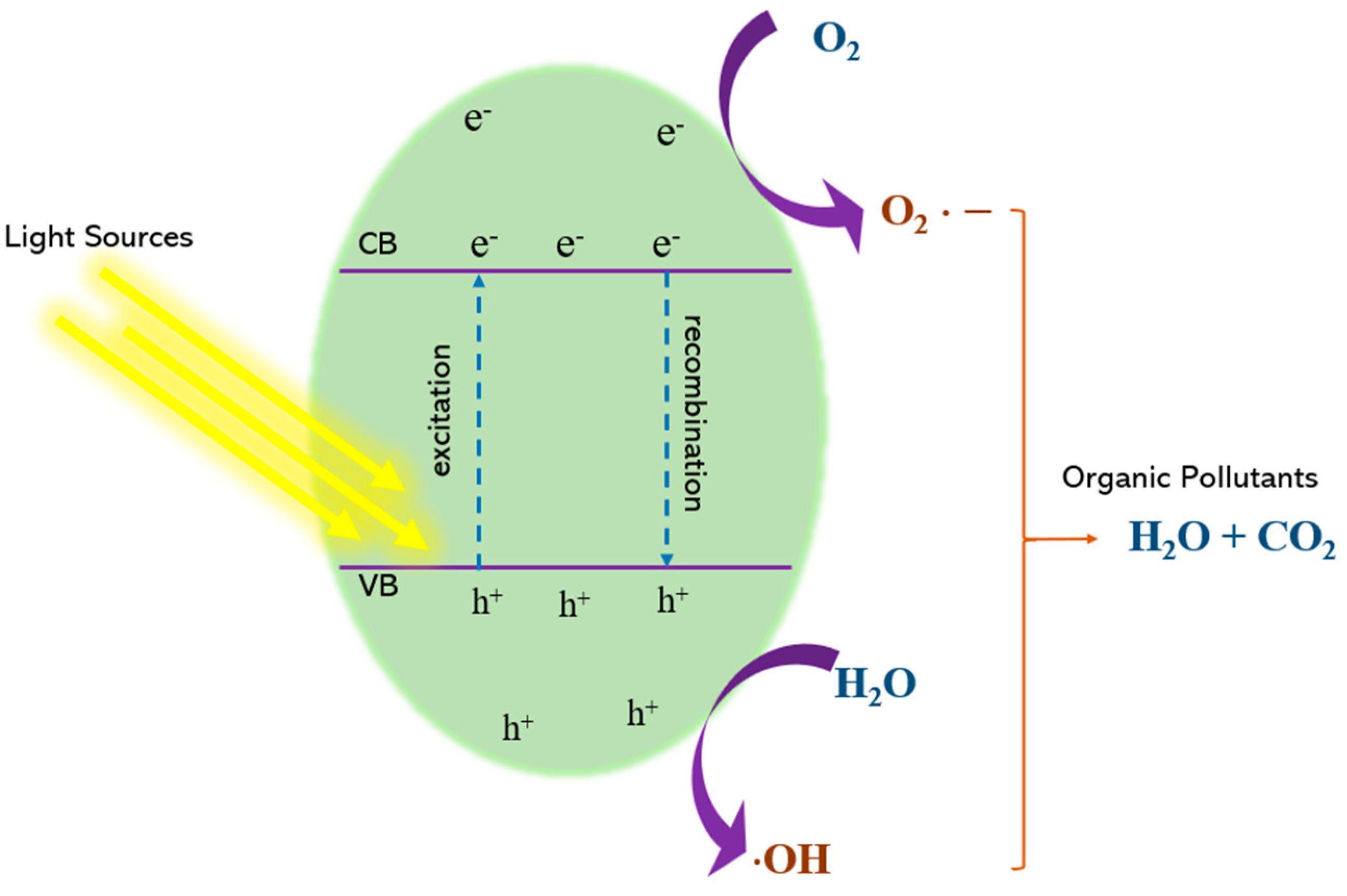
4.1. Principal Mechanism of Photocatalysis

Photocatalysis has been extensively investigated for the breaking down of organic contaminants. Photocatalysis relies on the ability of a photocatalyst to absorb light and initiate or accelerate chemical reactions [26]. Photocatalytic reactions are triggered by exciting a catalyst with photons that have energy equal to or exceeding the semiconductor band gap energy. The photocatalyst absorbs the photon energy, exciting electrons (eˉ) to the conduction band (CB) from the valence band (VB), thereby generating electron–hole pairs. These photogenerated electrons possess high reducing ability, while the resulting holes (h+) are potent oxidizing agents. The excited electrons reduce molecular oxygen (O2) to form reactive superoxide radicals (O2). Simultaneously, h+ migrates to the surface of the photocatalyst and oxidizes H2O to produce O2 or hydroxyl radicals (OH). Although the generated radicals play crucial roles in pollutant degradation, at the same time, the generated h+ can also directly oxidize the organic pollutants [8].
Photocatalysis degrades pollutants through a series of reactions involving wide-band gap semiconductor materials such as ZnO, TiO2, SnO2, CeO2, etc. The band gap energy ( E g ) signifies the difference in energy between the top of the valence band (VB) and the bottom of the conduction band (CB). When light energy exceeds the catalyst’s band gap, it excites eˉ from the VB to CB, creating h+ in the VB [26]. The basic photocatalytic reactions are illustrated below.
s e m i c o n d u c t o r   p h o t o c a t a l y s t + h ν e C B + h V B +
h + + H 2 O     H + + O H
e + O 2 O 2
h + + O H O H
2 e + O 2 + 2 H + H 2 O 2
e + H 2 O 2 O H + O H
o r g a n i c   c o n t a m i n a n t + s e m i c o n d u c t o r   p h o t o c a t a l y s t   ( e C B ) r e d u c t i o n   p r o d u c t
o r g a n i c   c o n t a m i n a n t + s e m i c o n d u c t o r   p h o t o c a t a l y s t   ( h V B + ) o x i d a t i o n   p r o d u c t
o r g a n i c   c o n t a m i n a n t + O H d e g r a d a t e d   p r o d u c t s
To achieve high efficiency in pollutant removal via photocatalysis, several key factors must be considered. The irradiation light’s energy must exceed the photocatalyst’s band gap for the material to absorb the light and generate photocarriers. The valence band must be positioned above the pollutant’s oxidation potential, while the conduction band must thermodynamically favour superoxide radical formation or hydrogen production. O H is essential in degradation processes. If the photocatalyst cannot oxidize water ( O H ) to generate O H , an alternative approach involves the multi-step reduction by photoelectrons, leading to the formation of superoxide radicals, which can subsequently produce O H radicals [9,31].

4.2. Affecting Parameters for Photocatalysis Efficiency

4.2.1. Characteristics of the Catalyst

Photocatalytic activity is substantially affected by the catalyst properties, including its structure, shape, size, and surface area. Owning to small size and higher surface area, nanomaterial-based catalysts outperform bulk materials as they offer substantial large active sites for the absorption of photon and pollutant interaction, enhancing overall photocatalytic activity [17]. Differences in the shape of the catalyst can also lead to variations in photocatalytic activities. Catalysts with tunnel oxide structures, such as titania-based materials, exhibit superior efficiency for hydrogen production from water compared to those with layered structures. Structural variations at the nanoscale level can significantly enhance the photocatalytic breakdown of pollutants.

4.2.2. Catalyst Loading and Pollutant Concentration

Catalyst loading also plays a crucial role in influencing photocatalytic processes. Increasing the amount of the catalyst provides more active sites and enhances electron–hole formation, leading to increased reactive species production like OH• and O2, which facilitate the degradation of pharmaceutical compounds. However, a higher catalyst loading does not always equate to efficiency, since there is an optimal loading concentration beyond which efficiency plateaus. Excessive catalyst concentrations can lead to light scattering and aggregation, reducing the available surface area for the reaction and limiting overall efficiency [32]. For instance, Xie et al. [33] conducted photocatalytic experiments to remove Sulfamethoxazole (SMX) under visible light using different concentrations of 0.625, 1.25, and 2.5 g/L of a photocatalyst, Zn- T i O 2 /pBC. The concentration 1.25 g/L was determined as optimal, which achieved the highest, 81.21%, removal efficiency of 10 mg/L SMX after 3 h of irradiation, while lower and higher concentrations were less effective. Comparatively, catalyst concentrations of 0.625 g/L and 2.5 g/L resulted in 57.73% and 76.28% SMX breakdown underneath identical conditions. This phenomenon proves that increasing the catalyst concentration boosts the active site density on the surface of the catalyst. More active sites absorb more photons, leading to the generation of more reactive species like hydroxyl radicals, which contribute to antibiotic mineralization. Consequently, improved process efficiency and antibiotic eradication can be attained. However, overly high catalyst dosages reduce light absorption due to increased photocatalyst clustering and heightened solution turbidity. Aggregation reduces the catalyst’s active surface area, while turbidity reduces light penetration due to scattering [19]. Thus, catalyst loading directly affects removal efficiency, and optimizing this parameter is crucial for achieving maximum effectiveness [17].
Additionally, the concentration of the target pollutant can also affect the performance of the photocatalyst. While the general rate law indicates that a higher concentration of reactants typically leads to a faster reaction rate, excessively high concentrations can result in the opposite effect. One reason is the imbalance between the number of available active sites and the target molecule, where the active sites become saturated and cannot accommodate all substrate molecules, causing a drop in the reaction rate [34]. Another factor is the light absorbance of the pollutant. When a significant amount of solar light is absorbed by the pollutant molecules instead of the catalyst, it reduces the catalytic efficiency [35].

4.2.3. pH

pH is a critical factor in photocatalytic processes as it influences both the catalyst surface charge and the dissociation of pollutants. When the initial pH is below the catalyst’s point of zero charge, the catalyst surface generally becomes positively charged due to H+ ion adsorption [17]. The pH level dictates the catalyst surface charge properties and the size of the aggregates it creates [36]. Higher pH levels lead to increased adsorption capacity due to the negative surface charge [37]. The morphologies of catalysts, such as BiVO4, are also pH-sensitive, influencing their dispersion and agglomeration [38]. Further, it was demonstrated that as the pH increased from 9 to 10, the surfaces of B i V O 4 nanoparticles became smoother, and further increasing the pH from 10 to 11 caused the catalyst surface to become coarse and clustered, leading to further agglomeration. However, the surface of B i V O 4 at pH 10 was more uniformly dispersed compared to other samples.
Hanafi and Sapawe [36] investigated the effect of pH on the E G Z r O 2 catalyst for degrading remazol brilliant blue (RBB) dye, evaluating pH levels 3, 5, 7, 9, and 11. The best degradation performance was observed at the value of pH 3, achieving 99.8% degradation, followed by 80.1% at pH 5, 54.5% at pH 7, 21.7% at pH 9, and the lowest, 11.3%, at pH 11. The breakdown efficiency decreased with increasing pH. Semiconductor metal oxides generally exhibit amphoteric behaviour, affecting the catalyst surface charge properties during surface reactions. At low pH, the E G Z r O 2 catalyst surface becomes positively charged, attracting more negatively charged RBB anions. This improved electrostatic interaction between the E G Z r O 2 catalyst and RBB dye molecules, improving dye degradation at lower pH values.

4.2.4. Light Source, Light Intensity, and Irradiation Time

As discussed, the working mechanism of photocatalysts requires an excitation light that has enough energy to excite electrons from the valence and to the conduction band. The energy needed is based on the distance from the VB to the CB, i.e., the Eg. If the energy threshold is not met, then the photocatalyst is not activated [8,26]. Commonly used metal oxide photocatalysts, such as TiO2 and ZnO, have wide band gaps of around 3.0 to 3.2; therefore, they can only be excited by UV light. One way to lower the energy requirement is to narrow the band gap through doping or surface heterojunctions, which produces visible light photocatalysts [33,39,40,41,42].
Light intensity and exposure time are also crucial for providing the energy needed to drive the photocatalytic breakdown of organic contaminants. At low light intensities, the process operates linearly because photogenerated electron–hole pairs are used in chemical reactions faster than they can recombine. As light intensity rises, the separation of electron–hole pairs competes with their recombination, leading to a half-order regime [17]. This counterintuitive result was explained mathematically by Meng et al. [43]. They showed that the reciprocal apparent adsorption constant ( K s ) is directly correlated to the square root of the light intensity. As the light intensity rises, K s decreases because the photogenerated hole concentration becomes limited by the availability of H 2 O / O H , causing higher electron–hole recombination rates and lower O H concentrations. Recombination also releases heat, which could further limit photocatalytic activity [44]. Elaziouti et al. [45] demonstrated that light intensity significantly affects the degradation of Congo red using a ZnO photocatalyst. By varying light intensity from 50 J/cm2 to 90 J/cm2, they found that degradation efficiency increased up to 70 J/cm2, after which no further changes occurred. Beyond this intensity, the degradation rate remained constant as all photocatalyst particles were already engaged, indicating that further increases in light intensity did not enhance photocatalytic efficiency.
The irradiation time is also a crucial parameter for photocatalysis processes, as longer exposure leads to higher degradation percentages until an optimal value is reached due to the generation of more hydroxyl radicals. Irradiation time in photodegradation refers to the duration of interaction between the photocatalyst and light rays to generate hydroxyl radicals and the subsequent interaction between these radicals and organic compounds. Hydroxyl radicals are potent oxidizers that degrade pollutants, and an increase in their formation results in higher degradation percentages [46]. Kumar and Pandey [40] examined the impact of irradiation time on methyl green removal using N i L a T i O 2 . They found that methyl green photodegradation improved with extended irradiation, achieving maximum efficiency after 50 min of visible light exposure. This improvement is attributed to the enhanced interaction between dye molecules and the photocatalyst surface, leading to increased degradation efficiency as irradiation time increases.
Photocatalysis offers a powerful, sustainable solution for the degradation of pharmaceutical contaminants in water, addressing many limitations of traditional AOPs. By optimizing key parameters like catalyst characteristics, light intensity, pH, loading, and irradiation time, the efficiency of photocatalytic processes can be significantly enhanced, making it a promising approach for large-scale environmental remediation.

4.3. Photocatalysis Materials

4.3.1. Metal Oxide

Photocatalysis relies on the interaction between light and a material’s electronic band structure. In solids, the electronic band structure comprises two key energy bands: the higher-energy conduction band (CB), and the valence band (VB) of lower energy. The energy difference between these two bands, termed as the band gap, dictates the material’s electronic and optical properties. Solids have different band gaps, resulting in various properties. Based on the band gap width, materials are classified as metals (with overlapping bands), insulators (with a large band gap), and semiconductors (with an intermediate band gap), as shown in Figure 3.
Metals have overlapping band gaps regardless of temperature. Insulators have a very high band gap, making it consequently challenging to excite electrons to the conduction band from the valence band; this band gap is known as the forbidden gap. In semiconductors, the band gap is relatively narrow, enabling electrons to transition to the conduction band from the valence band. A photocatalyst involves light exposure on the catalyst, which acts as thermal energy, exciting electrons to the conduction band from the valence band. Therefore, the width of the band gap is crucial for an efficient photocatalyst, significantly influencing the material’s electrical and optical characteristics [47]. This process is central to the generation of active electron–hole pairs (excitons), whose interactions facilitate the breakdown of pollutants.
Metal oxides are a group of semiconductor materials with various energy gaps, ranging from 1.0 to 4.0 eV. They are highly effective in photocatalytic applications [48]. The functional properties of materials can undergo significant changes when their dimensions are reduced from the micrometre scale to the nanometre scale. Due to this, metal oxide nanomaterials have garnered considerable attention for a variety of applications. It is well established that factors such as electrical conductivity, mechanical properties, surface activity, chemical reactivity, and biological activity can vary based on the size of the material [49]. These nanostructured materials can be synthesized by a variety of industrial methodologies, including precipitation reactions, green synthesis, sol–gel processes, hydrothermal and chemical treatments of precursor reagents, and others. These synthesis methods aim to produce metal oxide nanostructured materials with nanometric particle size and increased surface area of the materials. A larger surface area enhances photocatalysis.
When a photon with energy exceeding the band gap of a semiconductor is absorbed, it generates electron–hole pairs, or excitons, where electrons and holes are bound together by electrostatic forces. The typical distance between a hole and an electron within an exciton is about the Bohr radius [50]. The band gap of a semiconductor influences photocatalytic processes by affecting the electron–hole pairs’ recombination time and electron excitation. The excitons can dissociate into free carriers, with the holes remaining in the VB and the electrons migrating to the CB. The photocatalytic performance of metal oxides is governed by the rate at which these excitons recombine and the energy required to excite the electrons. The efficiency of pollutant degradation depends on maintaining a balance between these processes to maximize charge carrier utilization before recombination occurs [9]. The band gaps of various metal oxides are depicted in Figure 4, which are effective under visible light exposure, possessing appropriate band positions for water reduction reactions. It depicts the band positions of several semiconductors and band gap energies relative to NHE at pH = 0 [26]. Additionally, it should be noted that not all metal oxides can be categorized as semiconductors, as demonstrated by the wide band gap (5.0 eV) of ZrO2.
Metal oxide nanostructures depict unique chemical and physical characteristics because of their high density and the small size of their edges and corners. A large proportion of atoms are located on the particle surface, which increases the reactivity of the nanoparticles by providing a greater effective surface area, allowing them to react more extensively than larger, bulk crystals [26]. The mechanism of metal oxide-based photocatalysts when immersed in an aqueous solution is shown in Figure 5, which depicts the light absorption by metal oxides as catalysts to initiate the photogenerated electrons. As previously mentioned, the photogeneration of electrons can only occur after irradiation by a light source with equal or higher energy. The photoelectrons will participate in reduction reactions, such as hydrogen evolution and the formation of ·OH, and holes will move to the surface of the photocatalysts, while some of the carriers will recombine during the process. These processes accelerate chemical reactions, resulting in the degradation of contaminants [9].
Zinc oxide (ZnO) is extensively employed in photocatalysis because of its excellent chemical stability and high efficacy in purifying water from pollutants. ZnO has a significant band gap of 3.37 eV and can form various nanostructures, including nanosheets, nanowires, nanobelts, and nanorods [49]. Studies, such as the work by Mukherjee et al. [11], have demonstrated ZnO effectiveness in degrading pharmaceutical pollutants like ciprofloxacin; they found that ZnO can achieve a photocatalytic breakdown rate of 0.013 min−1 for ciprofloxacin under optimal conditions (0.5 g/L catalyst dose, natural sunlight, 12 mg/L initial CIP concentration, pH 6.3, and 110 min of irradiation), with an 81% degradation efficiency. Similarly, titanium dioxide ( T i O 2 ) is another metal oxide semiconductor widely used in photocatalysis due to its affordability, exceptional stability, and global availability [49]. T i O 2 generally exhibits 3.0 eV and 3.2 eV optical band gaps for its rutile and anatase phases. Research on T i O 2 photocatalysts indicates that they primarily absorb UV light, limiting their practical applications. However, Abdullah et al. [51] reported that T i O 2 successfully removed 88.30% of ciprofloxacin in 76 min using a 15 W UV-C lamp. To enhance the efficiency of harvesting visible light from the solar irradiation of such semiconductor materials, various techniques and strategies have been explored, such as doping with metals or non-metals, creating heterojunction composites, and engineering the morphology of the photocatalysts.

4.3.2. Quantum Dot-Based Photocatalyst

In contrast to bulk semiconductors with stable valence and conduction band positions, quantum dots (QDs) have unique optoelectronic characteristics controlled by quantum confinement effects. A prominent characteristic of quantum dots is their small size, typically less than 10 nm, which is comparable to their Bohr radius. While bulk semiconductors retain their properties, quantum dots of certain conductive materials like carbon and graphene display a visible range tuneable gap from 1.8 eV to 3.1 eV in their electronic energy states due to quantum confinement effects, based on their size, which directly influences their optical and electronic behaviour. Additionally, the behaviour of excitons within quantum dots can be controlled by adjusting their sizes [52]. This size-dependent quantum confinement results in a widening of the band gap in QDs, allowing for better control over light absorption and exciton behaviour. As the size of QDs decreases, their band gap increases, thus shifting their absorption spectra towards the visible region. Due to the wide, intense visible region absorption spectra, abundant surface-binding properties, and quantum confinement effect, quantum dots present novel and versatile opportunities for harvesting light energy for chemical transformations, positioning them as excellent candidates for advanced photocatalysis applications [53]. The quantum confinement effect enhances not only the optical properties of QDs but also their reactivity due to their rich surface-binding properties. This results in wide, intense visible region absorption spectra, enabling QDs to play a pivotal role in photocatalytic processes. For instance, carbon and graphene quantum dots exhibit a band gap of approximately 1.8 eV to 3.1 eV, making them suitable for visible light photocatalysis.

4.3.3. Two-Dimensional Material Photocatalyst

Two-dimensional (2D) materials include categories such as transition metal carbides, dichalcogenides and nitrides, single-element alkynes, organic materials, nitrides, and transition metal dihalides, which offer remarkable photocatalytic potential because of their unique electronic and structural properties. These materials have extensive applications, including as photocatalysts, and can be synthesized through methods like liquid phase exfoliation, chemical vapour deposition, mechanical exfoliation, and intercalation-assisted expansion and exfoliation [54]. These materials consist of atomic layers stockpiled by weak van der Waals interactions, which enable easy exfoliation and provide an abundance of surface-active sites for catalytic reactions. Two-dimensional layered materials have only a single or a few nanometre-thick layers and a large specific surface area, which exposes more atomic and functional surface structures, providing numerous active sites and enabling more intense reactions [52]. Their atomically thin nature leads to a large surface-to-volume ratio, making these materials highly effective in applications like pollutant degradation, hydrogen evolution, and water splitting.
Photocatalysis using 2D materials can convert light energy into chemical energy, significantly enhancing solar energy utilization efficiency. In particular, MoS2 has gained prominence in photocatalytic research due to its layered structure, high surface area, and tuneable electronic properties. MoS2-based photocatalysts have been utilized across various applications, such as microbial disinfection, photoelectrochemical water splitting, pollutant degradation, hydrogen evolution, and self-cleaning [55]. In a study conducted by Quinn et al. [56], a suspension of MoS2 in Milli-Q filtered water was utilized for the degradation of methylene blue (MB). They successfully achieved a 90% reduction in the original concentration after just 2 h of photocatalysis. However, challenges such as limited long-term dispersion stability in aqueous solutions caused by strong van der Waals forces between layers, low resistance to chemical and photochemical corrosion due to their extremely thin nature, challenges in photoelectrode preparation, and the presence of parasitic chemical reactions limit the broader adoption of 2D materials in photocatalytic applications [52].
Despite these challenges, 2D materials hold immense potential for solar-driven photocatalytic processes due to their high charge carrier mobility and large surface area, which promote efficient light absorption and pollutant interaction. Overcoming the limitations associated with dispersion stability and resistance to degradation could pave the way for more widespread applications of these advanced materials in environmental remediation.

5. Metal Oxide Photocatalysts

Many metal oxides, like TiO2, ZnO, CuO, CeO2, and NiO, have been utilized as photocatalysts due to their unique optical, chemical, and physical properties. These properties include distinct electronic structures, light absorption abilities, and charge transport capacities [57]. Key factors like the composition, size, doping, and shape of metal oxides can impact their photocatalytic efficiency [58]. Among these, T i O 2 is an extensively investigated metal oxide for photocatalytic activity. However, its performance is limited by a high charge carrier recombination, low absorption wavelength, and low surface area [59]. ZnO also shows promise for photocatalysis but faces limitations such as the fast recombination of photogenerated carriers, poor response to visible light, and photo-corrosion in aquatic solutions [60]. To enhance the efficiency of photocatalytic materials, strategies include controlling bulk and surface defects, coupling metal oxides with other materials, doping with metal or non-metal ions, and optimizing the morphology structure of the metal oxide.

5.1. Metal Oxide Morphology

In the field of photocatalysis, especially for the degradation of pharmaceutical pollutants, the morphology of metal oxides plays a pivotal role in dictating their performance. The photocatalysis process for pharmaceutical pollutants involves breaking down large, complex pharmaceutical molecules into smaller ones, such as water, C O 2 , and other environmentally safe byproducts. The efficiency of this process hinges on the semiconductor material’s energy band gap, surface area, and stability. Semiconductor metal oxide photocatalysis is a promising research area because it can address energy and environmental issues sustainably. Effective photocatalytic materials need an appropriate band gap energy, high surface area, long-term stability, suitable morphology, and good recyclability. Nanomaterials, with their high surface-to-volume ratios, offer enhanced redox reaction sites and have garnered considerable interest due to their ability to sustain prolonged catalytic cycles [57].
However, the photocatalytic performance of these materials can often be hindered by morphological limitations. Researchers have noted that poor photocatalytic performance often stems from inadequate morphological and textural properties, such as low surface areas and particle agglomeration [61]. Improving surface area is crucial for enhancing photocatalytic activity. Morphology—whether it is the shape, size, or specific nanostructure—has a profound impact on enhancing light absorption, reducing electron–hole recombination, and increasing the effective surface area for catalysis.
Many studies have shown that unmodified or pristine metal oxide photocatalysts have low efficiency in their photocatalytic abilities. To address this, various nanostructures have been explored to enhance their efficiency. The morphology of nanomaterials significantly affects their energy band gap, which is crucial for photocatalytic performance. Modifying the structure of photocatalyst materials aims to decrease electron–hole recombination rates, increase light absorption, and expand the effective surface area. The high surface-to-volume ratio of nanomaterials provides enhanced surface area for redox reactions [57]. Researchers have identified that poor photocatalytic performance often results from suboptimal morphological and textural properties, such as low surface areas and agglomeration. Increasing surface area is a key factor in improving photocatalytic activity.
Metal oxides can be synthesized using various techniques under controlled conditions, such as sol–gel, microwave-assisted, co-precipitation, solvothermal, and hydrothermal methods. These methods have been used to create suitable and unique metal oxide nanostructures for the applications of photocatalysis [59]. The structural morphology of metal oxides can improve the effective surface area, thereby facilitating photon absorption. The structural, electronic, and light-absorbing characteristics can be fine-tuned through the controlled synthesis of specific morphologies. Investigators have documented various intriguing metal oxide nanostructures, including nanowires, nanorods, nanotubes, nanospheres, and nanoflowers, which have been developed under specific growth conditions for photocatalytic applications. These morphologies exhibit unique capabilities that can improve photocatalytic efficiency, depending on the desired application. Table 4 summarizes works on various metal oxide morphologies.

5.1.1. Nanosphere

Metal oxide nanospheres, despite their seemingly simple spherical morphology, also offer advantages in photocatalysis. Researchers have been actively investigating the formation mechanism of metal oxide nanospheres and their applications as important technological materials. Metal oxide nanospheres, known for their large surface area exposed to radiation, have been extensively studied as photocatalysts for pollutant degradation. Their enhanced performance is due to their high surface area, efficient charge carrier separation, and favourable redox potentials [59]. Several common metal oxide nanospheres, such as TiO2, ZnO, and bismuth oxide, have been studied for pharmaceutical waste degradation [90,91,92]. Additionally, they are coupled with other materials to enhance photocatalytic activity [93,94,95].
It is observed that while nanospheres provide substantial surface area for photocatalytic reactions, their performance may be limited compared to other, more complex morphologies [57]. For example, nanorods and nanowires exhibited higher photocatalytic activity than nanospheres. This is because nanospheres have a relatively lower surface area, which reduces their catalytic efficiency and charge transfer capabilities. Another structure, nanoflowers, consists of assembled rods or sheets resembling a flower. This complex structure provides a large surface area, which is reported to offer better photocatalytic capabilities than nanorods and nanowires [96]. Nanosheets, which are two-dimensional structures, have the highest surface-to-volume ratio, resulting in more electrochemical active sites on the surface [97]. The morphology of metal oxide nanostructures is a key determinant of their photocatalytic efficiency. By tailoring the shape, size, and structural arrangement of these nanomaterials, researchers can significantly improve the light absorption, charge separation, and surface area available for catalytic reactions. As the field advances, the exploration of novel nanostructures and synthesis techniques will continue to drive the advancement of high-performance photocatalysts for the elimination of pharmaceutical contaminants, ultimately contributing to more sustainable and efficient environmental remediation strategies.

5.1.2. Nanorods

Nanorods are one of the most promising morphologies, especially in energy and environmental applications. Researchers have made significant efforts to synthesize metal oxide nanorod structures. Nanorods are rigid tubular structures typically ranging in length from 10 nm to 120 nm, and have lengths significantly higher compared to the diameter, enabling efficient electron transport because of their well-defined and straight structures. Their dimensions can range from nanometres to micrometres, with aspect ratios optimized for applications like photocatalysis. One-dimensional (1D) structures are particularly favoured for their exceptional photocatalytic efficiency in breaking down organic pollutants and generating energy [59]. Nanorods are similar to nanotubes but lack an internal surface. They are less versatile than nanotubes but offer greater thermal stability [98]. Fu et al. [99] demonstrated the hydrothermal synthesis of ZnO nanorods with different aspect ratios for methylene blue (MB) degradation. The MB degradation efficiency reached 80% over five consecutive cycles, outperforming ZnO particles due to their increased exposed surface area. Studies have reported ZnO nanorods with lengths ranging from 780 to 1170 nm and diameters from 91 to 112 nm, while materials, such as V2O5, have been synthesized with lengths up to 1 μm. Other than dyes, ZnO nanorods have also been studied for pharmaceutical waste degradation, such as various antibiotics (i.e., amoxicillin, ciprofloxacin, levofloxacin, ciprofloxacin, cefotaxime) and drugs (i.e., meloxicam, ibuprofen, paracetamol), where all show positive results [100,101,102].

5.1.3. Nanowires

Nanowires closely resemble nanorods but have higher aspect ratios, offering an even more significant boost in photocatalytic performance. The photocatalytic performance improves with higher aspect ratios, as 1D nanostructures provide various geometries that effectively reduce nanoparticle agglomeration, offer numerous active sites, and enable efficient charge carrier transport [59]. Nanowires are extremely thin with typical widths of a few tens of nanometres, and lengths extending into the micrometre range [98]. The photocatalytic performance of nanowire arrays is better than nanotubes due to their porous design, resulting in a larger specific surface area [103]. ZnO nanowires, in particular, have garnered attention for their straightforward synthesis methods and superior photocatalytic activity in pollutant degradation. Liu et al. [73] demonstrated the growth of well-aligned ZnO nanowires, which exhibited methyl red (MR) enhanced degradation underneath UV exposure. Positive results were also achieved against several antibiotics (i.e., sulfamethazine, cephalexin, nitrofurantoin) and drugs (i.e., carbamazepine, diclofenac, lidocaine), further showcasing the benefits of one-dimensional nanostructures [104,105,106]. Nanowires and nanorods share similar structures, but nanowires offer better photocatalytic response and adsorption/desorption capabilities due to their elongated shape.

5.1.4. Nanotube

Nanotubes, with their greater surface-to-volume ratio compared to nanorods and nanowires, reduce the photogenerated electron–hole pair recombination rate and enhance interfacial charge carrier transfer rates, making them highly promising for various applications. The enhanced photocatalytic performance of metal oxides in nanotube arrays is due to their larger surface area, resulting in increased light absorption and the adsorption of organic substances, making them highly efficient for various photocatalytic applications [59]. Nanotubes are similar to nanowires in diameter; nonetheless, they are hollow with a typical aspect ratio of 3 to 5. They can be either multi-walled or single-walled structures [98]. Nanotube photocatalysts remain structurally stable at temperatures up to several hundred degrees Celsius. The interaction points between tubes, the wall thickness, and the nano-interface are crucial for defining their photocatalytic performance [31]. Unlike nanorods, nanotubes are shaped like hollow tubes, which provides them with a larger surface area, significantly reducing the rate of electron–hole recombination. Nanotubes also offer advantages in the one-dimensional ion-exchange direction and light absorption [107]. Wang et al. [80] reported that ZnO nanotubes exhibit superior photocatalytic activity for degrading methyl orange related to other structures, attributed to the uneven distribution of nanoholes on the walls of the porous nanotubes, enhancing photodegradation efficiency under UV radiation. In another study, Bojer et al. [108] synthesized mesostructured ZnO nanotubes capable of degrading ciprofloxacin (antibiotic) more than twice as much as TiO2 P25 nanoparticles at 120 min of irradiation.

5.1.5. Nanoflower

Among various nanostructure systems, nanoflowers are a newly developed class of tiny particles resembling plant flowers at the nanoscale, typically ranging from 100 nm to 500 nm. Flower-like nanostructures have become popular in photocatalytic applications due to their unique properties. Nanoflowers consist of multiple petal layers, providing a larger surface area within a compact structure, rendering them ideal for use in catalysis, biosensors, and drug transport systems [109]. Adjusting reaction conditions and parameters can lead to significant advancements, resulting in flower-like morphologies with enhanced performance. The improved photocatalytic activity is often attributed to oxygen vacancies serving as active sites for charge carrier capture. Their high surface area, combined with their ability to generate multiple light reflections within their structure, leads to improved photocatalytic activity. Flower-like ZnO structures have been shown to outperform traditional nanorods and nanowires in various applications due to their enhanced surface properties [59]. Qu et al. [110] highlighted the high surface area of ZnO nanoflowers, which is beneficial for both enhancing the photocatalytic performance and also for the controlled synthesis of ZnO/carbon dots. Their synthesized material was able to almost completely (up to 95%) degrade pollutants in actual water samples containing antibiotics (cephalexin and ciprofloxacin) and dyes (methylene blue).

5.2. Commonly Used Metal Oxides

Transition metal oxides and their composites demonstrate exceptional photocatalytic capabilities, thereby revolutionizing environmental remediation, particularly in the breaking down of persistent organic contaminants like pharmaceuticals. These metal oxide nanomaterials, designed with precise surface, crystalline, and structural characteristics, act as wide-band gap semiconductors and possess desirable traits such as stability and non-toxicity in water. These properties make them suitable for photocatalytic oxidation or breaking down of harmful organic contaminants [111]. Metal oxides can have various electronic characteristics, ranging from conductive to semiconductive and insulating, which contribute to their unique features, like the wide band gaps that influence their photocatalytic performance. The unique electronic versatility of metal oxides—ranging from conductive to semiconductive and insulating—augments their photocatalytic efficacy by optimizing electron mobility and charge separation, key factors in enhancing pollutant degradation.
A critical feature of metal oxide photocatalysts is their wide band gap, enabling activation under ultraviolet (UV) light, as demonstrated by Nunes et al. [97]. Photocatalysts with such properties can absorb solar radiation, reduce electron–hole recombination rates, and consequently exhibit superior photocatalytic performance in breaking down complex organic pollutants. A diverse array of semiconductor/metal oxide-based nanomaterials including ZnO, SnO2, TiO2, Fe2O3, CdS, WO3, ZrO2, etc., have been extensively studied for their photocatalytic ability to degrade toxic and harmful organic contaminants into the non-toxic byproducts.

5.2.1. Photocatalyst Based on Titanium Oxide

TiO2 has garnered substantial attention as a highly effective photocatalyst, primarily because of its affordability, exceptional chemical stability, non-toxicity, and widespread availability globally. Several methods have been explored to synthesize T i O 2 nanostructures, including hydrothermal, sol–gel, chemical vapour deposition, microwave-assisted, solvothermal, and electrochemical techniques [112]. TiO2 appears in three major crystalline polymorphs, rutile, anatase, and brookite, with anatase typically exhibiting superior photocatalytic activity. TiO2 depicts band gaps of 3.2 eV for anatase and 3.0 eV for the rutile phase, allowing it to absorb photons in the near UV range. However, its photocatalytic activity under sunlight is limited as only 4–5% of solar radiation is UV light, necessitating modifications to enhance visible light absorption [49,111].
TiO2 is typically employed as a photocatalyst in its anatase form, which tends to exhibit greater photocatalytic efficiency compared to the less photoactive rutile phase. The superior photocatalytic activity of anatase is due to three key factors: first, its wider band gap compared to rutile, raising the top of the valence band to larger energy levels relative to the adsorbed molecules’ redox potentials, facilitating more efficient electron transfer and oxidation; second, its indirect band gap, which results in a longer lifespan of photoexcited holes and electrons compared to rutile and brookite’s direct band gaps; and third, the higher oxygen vacancies’ concentration in anatase, leading to superior charge separation efficiency [97].
Recently, Wang et al. [113] prepared TiO2 particles using a solvothermal method, yielding strong UV absorption with a 3.15 eV band gap. When tested for the degradation of tetracycline, TiO2 achieved a breaking down rate of 39% underneath exposure to visible light for 70 min. Furthermore, advanced methodologies like those explored by Parmar and Srivastava [114] utilized artificial neural networks and surface response methodology to optimize the TiO2 photocatalytic degradation of ciprofloxacin, achieving 88.3% degradation efficiency under specific conditions. This study employed a mathematical predictive model utilizing an artificial neural network and surface response methodology. The model evaluated four independent variables, viz initial antibiotic concentration, catalyst dose, reaction time, and solution pH, within a central composite design to study antibiotic breakdown. Under optimal conditions, with a solution pH of 5.04, 75.80 min reaction time, catalyst dose of 44.51 mg/L, and antibiotic concentration of 5.02 mg/L, the photocatalysis process attained an 88.30% degradation of ciprofloxacin. The experimental results were highly consistent with the predicted outputs from the artificial neural network (R2 = 0.975) and surface response methodology (R2 = 0.9969).

5.2.2. Photocatalyst Based on Zinc Oxide

Zinc oxide (ZnO) nanomaterials have demonstrated significant promise as photocatalysts due to their low cost, good stability, high quantum efficiency, and environmentally friendly nature. ZnO has a hexagonal wurtzite structure with a 3.25 eV band gap, making it effective for the photocatalytic breakdown of organic pollutants in wastewater, particularly under UV irradiation [111,112].
Silva et al. [90] demonstrated that pristine ZnO nanoparticles could degrade ciprofloxacin concentrated at 300 µg/L under UV light in less than 6 min, achieving a high degradation efficiency of 63%. Additionally, modified ZnO nanostructures have been developed to enhance visible light absorption. Liu et al. [115] investigated ZnO nanoparticles modified with B i 2 W O 6 for the photocatalytic treatment of amoxicillin concentrated at 20 mg/L. A hydrothermal method was employed to synthesize the catalyst and a reduction in band gap energy was observed, resulting in a high degradation efficiency of amoxicillin at 93.10% under visible light.
Green synthesis approaches have also been explored, as evidenced by Mehmood et al. [116], who employed zinc oxide nanoparticles (GS-ZnONPs) synthesized from leaves of Azadirachta indica (neem) to remove the carbamazepine (CBZ) antiepileptic medicine from wastewater. The research revealed that GS-ZnONPs had 27.55 mg/g adsorption capacity at room temperature. At optimum conditions, 12 mg/L of CBZ at (25 ± 2) °C, 100 mg of the adsorbent, and pH 5.0, the GS-ZnONPs exhibited a high eradication efficiency, effectively removing 92.89% of CBZ from water, highlighting the potential of eco-friendly photocatalyst development.

5.2.3. Photocatalyst Based on Bismuth Oxide

Bismuth oxide (Bi2O3) is another widely studied photocatalyst. Bi2O3, a commonly used oxide semiconductor, has several crystal structures, such as α-Bi2O3 (monoclinic), β-Bi2O3 (tetragonal), γ- B i 2 O 3 (body-centred cubic), and δ-Bi2O3 (cubic fluorite type), each with varying band gaps. These structures’ band gaps are in the decreasing order of δ-Bi2O3 (3.0 eV) > α-Bi2O3 (2.8 eV) > β-Bi2O3 (2.1 eV) > γ-Bi2O3 (1.64 eV) [117]. γ-Bi2O3 has the narrowest band gap, enabling visible light utilization efficiently from the solar spectrum, making it a prominent candidate for photocatalysis. Nonetheless, the photocatalytic performance of pristine Bi2O3 is generally inadequate for antibiotic degradation unless combined with appropriate dopants, particularly for the δ-phase, which has the largest band gap. To enhance Bi2O3 photocatalytic activity, various methods have been employed, such as constructing heterostructures, doping with cations/anions/zerovalent elements, and reducing the band gap. Additionally, preparing Bi2O3 in nanosheet morphology improves its performance as a photocatalyst by providing a higher active surface area [118].
Zhang et al. [119] investigate the photocatalytic activities of Bi2O3 powders by degrading the antibiotics ciprofloxacin (CIP) and tetracycline (TC) underneath visible light exposure (300 W Xenon lamp). The photocatalytic degradation rate for TC was 83.63%, whereas for CIP, it was only 34.90%. The results demonstrated that B i 2 O 3 exhibited better photocatalytic performance in degrading TC compared to CIP, indicating that B i 2 O 3 shows selectivity in the photocatalytic degradation process of different antibiotics. Another approach, using Bi2O3 nanoplates synthesized via ultrasound-assisted methods, improved the breakdown of dyes and antibiotics in aquatic solutions, as shown by Sánchez-Martínez et al. [120]. The photocatalytic performance of the B i 2 O 3 was tested by degrading TC, indigo carmine (IC), and rhodamine B (rhB) in an aquatic solution underneath Xe lamp exposure. The degree of the mineralization of the antibiotic and organic dyes by Bi2O3 was measured using a total organic carbon analysis (TOC), achieving 72% for TC, 13% for IC, and 40% for RhB, after 24 h of Xe lamp exposure. These findings underscore the versatility of Bi2O3 in targeted photocatalytic degradation processes, making it a valuable component in the field of environmental clean-up.
Metal oxide-based photocatalysts, with their diverse electronic properties and adaptable structures, play a pivotal role in addressing pharmaceutical pollutants in water systems. Their effectiveness can be significantly enhanced by incorporating nanocomposites and optimizing light absorption capabilities, further expanding their potential for large-scale environmental applications.

6. Carbon Dots

Carbon-based nanomaterials are extensively investigated due to their long-term chemical stability, non-toxicity, biocompatibility, fluorescence properties, eco-friendliness, inertness, and large effective surface area [121,122]. Among these nanomaterials, carbon dots are particularly interesting for their fascinating optoelectronic characteristics. These zero-dimensional (0D) materials are less than 10 nm in size and consist of sp2 and sp3 carbon atoms with numerous functional groups in their surfaces or polymer chains. Carbon dots exhibit photoluminescence emissions at wavelengths between 320 nm and 450 nm and absorb light at wavelengths ranging from 211 nm to 288 nm [123]. Their intrinsic photoluminescence (PL) and the ability to absorb light within a wide spectral range, from ultraviolet (UV) to visible, render them highly attractive for environmental applications, including photocatalytic processes aimed at pollutant degradation [121].
Carbon dots display quantum confinement effects, creating size-dependent band gaps. When exposed to photons with sufficient energy, these nanomaterials promote electrons to the conduction band from the valence band, forming excitons (electron–hole pairs), which are crucial in light-induced reactions [124]. Their visible light absorption capability and unique light-induced electron transfer properties make carbon dots promising for environmental applications, particularly in photocatalysis. They can act as stand-alone photocatalysts or as multifunctional components within composite photocatalysts, improving light absorption, facilitating efficient electron–hole pair separation, and stabilizing semiconductor photocatalysts [123]. The inclusion of carbon dots within a metal oxide matrix significantly improves electron–hole pair separation, minimizes recombination, and enhances overall photocatalytic efficiency [124].

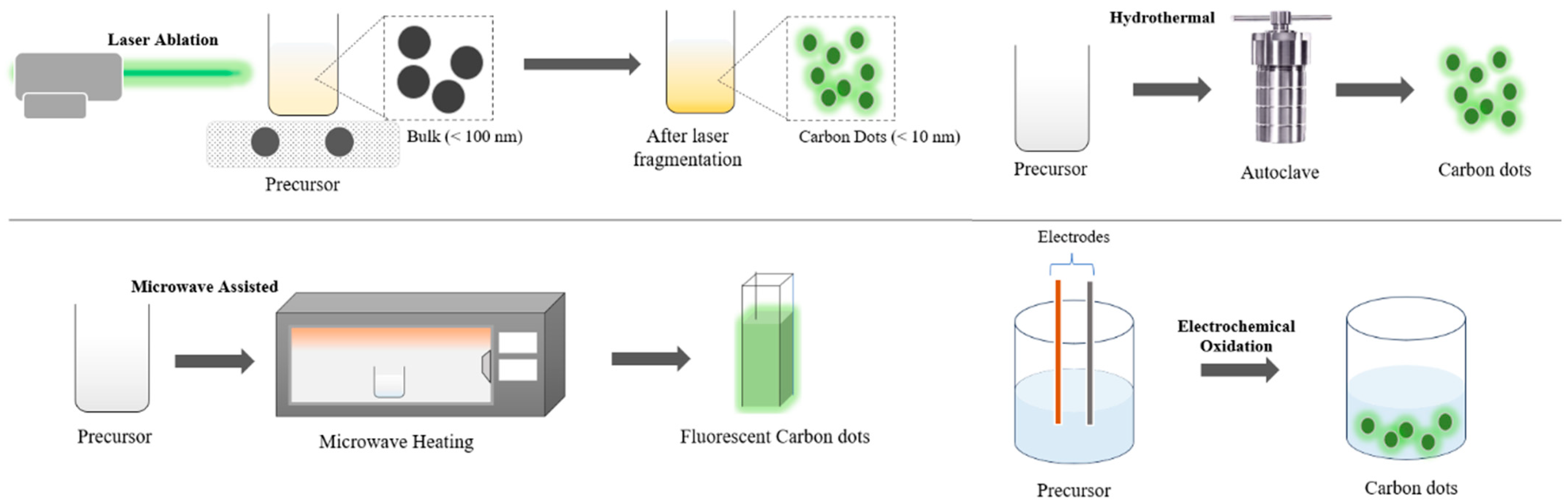
6.1. Synthesis of Carbon Dots

Xu et al. [125] discovered carbon dots through an oxidation process while purifying single-walled carbon nanotubes (SWCNTs). Since then, various approaches have evolved to prepare carbon dots, which can generally be categorized into “bottom-up” and “top-down” approaches. The top-down method involves larger carbon structures breaking down into nanoscale, fluorescent carbon dots using techniques like electrochemical oxidation, laser ablation, and ultrasonic treatment [126]. Conversely, the bottom-up approach involves converting small molecules into carbon dots through carbonization and passivation, hydrothermal, pyrolysis, and microwave-assisted methods [127]. A summary of various bottom-up and top-down approaches is displayed in Table 5.
Numerous food-based carbon sources, such as yoghurt, honey, banana, leaves, egg, rice bran, and garlic, can be used to prepare carbon dots having different quantum yields (QYs). Quantum yield refers to the ratio between the number of photons emitted and the number of photons absorbed, which is critical for optimizing their fluorescence and photocatalytic behaviour [124]. Various methods for single-step growth of fluorescent carbon dots can be achieved for explicit biosensing applications employing natural or synthetic non-toxic precursors [121]. Figure 6 shows the different method approaches for the preparation of carbon dots.
The top-down approach involves breaking down larger carbon materials, like graphite, carbon nanotubes, carbon fibres, and coal, into carbon dots. This is achieved through methods like laser ablation [128], ultrasound [141], and electrochemical methods [142]. Electrochemical oxidation is particularly effective for producing carbon dots due to its high yield, high purity, low cost, good reproducibility, and ease of size modulation. Recent advancements have also emphasized the eco-friendly aspect of synthesis, with water-based electrolytes and alkali-assisted processes yielding highly pure carbon dots. The economical and environmentally friendly high-purity and high-quality carbon dot synthesis using a single-step electrochemical process with water–electrolyte was reported earlier [126]. An alkali-assisted electrochemical method was evolved to produce carbon dots ranging from 1.2 nm to 3.8 nm in size, employing graphite rods as a cathode and as an anode, with electrolyte NaOH/ethanol and operating at current intensities from 10 mA/cm2 to 200 mA/cm2. In contrast, using an acidic electrolyte, H2SO4/ethanol, did not result in carbon dot formation. Liu et al. used a similar approach to produce carbon dots through the electrochemical oxidation of a working graphite electrode with a mixed electrolyte of H2O, NaOH, and ethanol. Applying a 5 V potential for 3 h resulted in carbon dots with a 4 nm average diameter and 11.2% quantum yield. These carbon dots showed photoluminescence intensity dependent on excitation but emitted light at a consistent wavelength regardless of the excitation source. The carbon dots can be employed for the specific detection of Fe3+ ions and cell imaging [143].
Another method for synthesizing carbon dots is the laser ablation of a carbon target in vapour or liquid. This technique uses high-energy lasers to break bonds and generate carbon dots from the target surface. Unlike chemical methods, which are time-intensive and require meticulous control, laser ablation offers a rapid and efficient means of producing carbon dots. Reyes et al. [144] prepared carbon dots by the laser ablation of a solid carbon in acetone. They varied ablation time (150 s, 300 s, 600 s, and 900 s) and laser wavelength (355 nm, 532 nm, and 1064 nm) to study their effects on the optical and morphological properties of the grown particles. The 355 nm wavelength resulted in carbon dots smaller than 5 nm, while longer wavelengths led to larger agglomerations. Increasing ablation time led to decreased photoluminescence intensity and a broader red-shifted peak, with the largest emission quality found employing a 355 nm wavelength for 150 s [143]. The top-down method’s advantages include the potential for scaling up carbon dot production and achieving well-defined structures [145]. However, this technique typically requires conditions like high energy, high potential, and high acidity [121].
The bottom-up approach offers advantages such as simple surface modification in one step and the synthesis of various structures. The mass production of carbon dots is essential for future applications [145]. In this method, carbon dots are created by decomposing or carbonizing small or large precursors using techniques like hydrothermal methods [146], thermal decomposition [147], or microwave irradiation [148]. Hydrothermal carbonization is particularly popular due to its use of abundant and inexpensive precursors, scalability, and non-toxicity. Common carbon sources include organic acids, carbohydrates, fruit juices, and waste peels. Natural carbon sources are preferred for carbon dot production because of their environmental sustainability, and increased availability. Typically, hydrothermal methods involve sealing and reacting organic precursors in a hydrothermal reactor under high temperatures and pressures and extended reaction times [126]. For instance, Qi et al. [149] used pomelo peel to prepare water-soluble N carbon dots having a quantum yield of 76.47% for the sensitive and selective determination of tetracycline via the hydrothermal method. Prasannan and Imae [137] developed a simple one-step hydrothermal carbonization process to synthesize fluorescent carbon dots from orange waste peels and combine them with ZnO to break down naphthol blue-black azo dye.
Microwave irradiation has become a popular alternative due to its simplicity, ease of operation, and efficiency in the preparation of carbon dots. This method produces high-quality pure carbon dots. The uniform heat distribution from microwave treatment ensures consistent particle size across the precursor material [126]. Microwave irradiation accelerated the carbonization of chemical bonds in precursor molecules using electromagnetic waves [121]. Xu et al. [150] reported a heterogeneous synthesis method to achieve N-doped carbon dots by reacting a mixture of urea and calcium citrate underneath microwave power at 800 W for 5 min. The resulting carbon dots released yellowish-green fluorescence in both aqueous and solid phases. Sutanto et al. [151] produced green luminescent carbon dots from the passivation agent urea and carbon source citric acid using 450 W microwave power for 30 min. The bottom-up approach allows for easy surface modification, enabling the integration of carbon dots into photocatalytic composites for environmental applications. The incorporation of heteroatoms like nitrogen (N) further enhances the electronic properties of carbon dots, reducing the band gap and facilitating electron transport [145].

6.2. Properties of Carbon Dots

6.2.1. UV Absorption Property

Carbon dots show wide and intense absorption bands that cover the ultraviolet-to-visible light spectrum. Their absorption spectra are significantly based on their synthetic methods, precursor materials, and dispersion solvents. Typically, carbon dots have one to many absorption peaks in the ultraviolet-to-visible range. Peaks between 220 nm and 270 nm are due to π–π* transitions of C=C and C=N bonds, while peaks in the 280 nm to 350 nm range correspond to n–π* transitions of C–O and C=O bonds. The peaks in the 350 nm to 600 nm range generally come from transition involving functional groups on the surface of carbon dots [127,145]. González et al. [152] effectively produced carbon dots from pyrolytic carbon black. UV–visible spectroscopy demonstrated strong visible range absorption, with the carbon core’s central region peaks at 255 and 272 nm. Variations in sample intensity in the UV-Vis data were attributed to nitrogen doping in the carbon dots. The emission, driven by electron transport, is controlled by the interaction between the lowest unoccupied molecular orbital (LUMO) and the highest occupied molecular orbital (HOMO) [123]. These absorption features are pivotal in determining the photocatalytic potential of carbon dots, as they govern the material’s ability to harness light energy for environmental remediation.

6.2.2. Fluorescence Property

The fluorescence properties of carbon dots are essential for their wide range of applications, especially photocatalysis. These properties include a strong fluorescence stability, narrow emission spectrum, size-dependent (or excitation wavelength-dependent) fluorescence emission, broad excitation spectrum, resistance to photobleaching, and up-conversion luminescence. Mechanisms proposed to explain the luminescent behaviour of carbon dots include quantum confinement effects, emissive traps, carbon excitons, aromatic structures, oxygen-containing groups, passivated surface defects, and free zigzag configurations [127]. This effect can be ascribed to the presence of unique surface features and energy states inside the carbon dots. The influence of size on carbon dots is substantial and affects their photoluminescence (PL) capabilities. Li et al., [153], employed an electrochemical method to prepare carbon dots ranging in size from 1.2 nm to 3.8 nm, demonstrating outstanding light conversion capabilities and size-dependent PL properties. A reverse correlation between the band gap and particle size was noted, with enhancement in particle sizes resulting in the reduction in the band gap [123]. The PL of carbon dots is highly dependent on their size and surface chemistry, with smaller particles exhibiting blue emissions and larger ones showing red-shifted emissions. This size-dependent behaviour, known as quantum confinement, influences the energy gap and governs the emission wavelength. Additionally, surface passivation and functionalization with groups like amines or carboxylates can further modulate the PL intensity and stability, allowing for tuneable optical properties suited for specific applications [127,154].

6.3. Mechanism of Carbon Dots for Photocatalysis

Methods for modifying carbon dots for photocatalysis involve element doping, passivation, surface functionalization, and the creation of a composite. Figure 7 illustrates the distinction between the surface functionalization and passivation of carbon dots. Photocatalyst surface functionalization can aid in the separation of excitons and prevent their recombination, further improving charge migration by trapping photogenerated electrons at various surface sites, thereby controlling the kinetics of redox reactions [155]. This is particularly important in the degradation of pharmaceutical pollutants, where the rapid generation of reactive oxygen species (ROS) is needed to break down complex molecules. Surface passivation can create defect sites on carbon dots, leading to fluorescence emission through the radiative recombination of excitons trapped at these defect locations. Surface passivation also enhances the durability and photostability of carbon dots by preventing photo-corrosion and reducing direct contact with pollutants or reactants.
Doping carbon dots with heteroatoms, particularly nitrogen (N), is a common method to improve their fluorescence capability. In the absence of N-doping, carbon dots have a huge HOMO-LUMO gap, while this gap is reduced through N-doping, requiring less excitation energy [156]. Carbon dots exhibit a broad range of beneficial physicochemical properties because of the diversity of synthetic methods and raw materials, including superior charge transport capabilities, excellent water dispersibility, ultra-small sizes, large surface areas, and excellent electron donors and acceptors [157]. Under light irradiation, carbon dots can generate electron–hole pairs, offering advantages like non-toxicity, low cost, and excellent photoelectric properties, while also stabilizing semiconductor photocatalysts. This makes carbon dots a promising material for photosensitizers, which can function as both electron donors and acceptors.
In the context of photocatalytic processes, carbon dots play a crucial role in three main steps: firstly, they absorb light, promoting the improved generation of electron–hole pairs from the photocatalyst material. This first step is related to its up-conversion luminescence property, in which carbon dots absorb multiple photons and then emit a photon with a shorter wavelength—more high energy [158]. The up-conversion luminescence is expressed by Equation (10), while Equation (11) explains the mechanism by which the carbon dots absorb polyenergetic light,
c a r b o n   d o t s + n h ν 0 c a r b o n   d o t s + h ν
c a r b o n   d o t s + i h ν i c a r b o n   d o t s + h ν
where h ν is greater than E g , h ν 0 , or any h ν i . The emitted photon ( h ν ) is responsible for the electron–hole generation of the photocatalyst, as explained in Equation (1). This implies that the photocatalyst material is not only excited by the light source but also by the photons emitted by carbon dots by up-conversion luminescence, hence improving the electron–hole yield.
In the second step, carbon dots help separate and transfer pairs of electrons and holes, which generate reactive species. Thirdly, these reactive species initiate further photocatalytic reactions. However, the quick recombination rate of electron–hole pairs significantly reduces photocatalytic performance [159]. The excitation process of narrow-band gap semiconductor photocatalysts enhanced with carbon dots can be classified into two categories depending on the type of irradiation source employed for photocatalyst activation. When exposed to UV light, carbon dots function as electron reservoirs, trapping electrons from the conduction band of the photocatalysts. This process decreases the recombination performance of electron–hole pairs [123].
The integration of carbon dots into photocatalytic systems offers a multifaceted approach to tackling environmental challenges, particularly in the degradation of persistent pharmaceutical pollutants. Their unique optical, electronic, and surface properties, coupled with their ability to enhance charge separation and broaden light absorption, make carbon dots a highly promising material in advanced oxidation processes (AOPs).

7. Metal Oxide/Carbon Dot Hybrid Materials

In the present decade, significant focus has been directed towards the enhancement of metal oxides through doping with other nanomaterials or forming multifunctional composites. Among such advanced materials, carbon dots (CDs) have arisen as highly prominent candidates for photocatalytic applications because of their excellent optical and electronic properties. Their remarkable fluorescence behaviour, coupled with their efficient photoelectron transfer capabilities, makes them ideal for use in tandem with metal oxides to form high-performance photocatalysts [160].

7.1. Synthesis Methods of Metal Oxide/Carbon Dots

There are various methods to produce metal oxide/CD materials, with the main goal being to disperse carbon dots on the metal oxide. Two main strategies can be employed to achieve this. One option is first to prepare CDs separately, possibly using the various available methods mentioned in Section 6.1. The obtained CDs are then treated as one of the precursors during the metal oxide synthesis process, as commonly performed when heteroatom doping [161,162,163,164,165,166]. For instance, Huang et al. [166] reported a synthesis of BiOCl/CDs via a one-step hydrothermal method using a Teflon-lined autoclave. In this process, CDs were mixed with Bi(NO3)3·5H2O and NaCl, which served as the precursors for BiOCl. This resulted in BiOCl/CD nanosheets that retained the original morphology of BiOCl, indicating that the addition of CDs did not affect the structural integrity of the metal oxide.
Alternatively, a different approach is to prepare the metal oxide and carbon dots separately before they are mixed [167,168,169,170,171,172]. Liu et al. [167] reported the synthesis of leaf-like BiVO4 decorated with carbon dots, where the BiVO4 and carbon dots were first synthesized using their respective methods before being mixed through hydrothermal treatment using a Teflon-lined autoclave. This approach preserved the distinctive leaf-like morphology of BiVO4. Furthermore, other studies have shown that CDs can also be well dispersed on metal oxides, such as nanosheets, through simple precipitation methods coupled with prior sonication [169,172].
These strategies demonstrate the flexibility of integrating CDs into metal oxides while maintaining their unique morphological features. Ultimately, the choice of method depends on the desired properties and applications of the composite material. By carefully selecting the synthesis approach, researchers can optimize the structural and functional characteristics of metal oxide/CD systems, paving the way for their use in diverse applications, including photocatalysis, sensing, and energy storage.

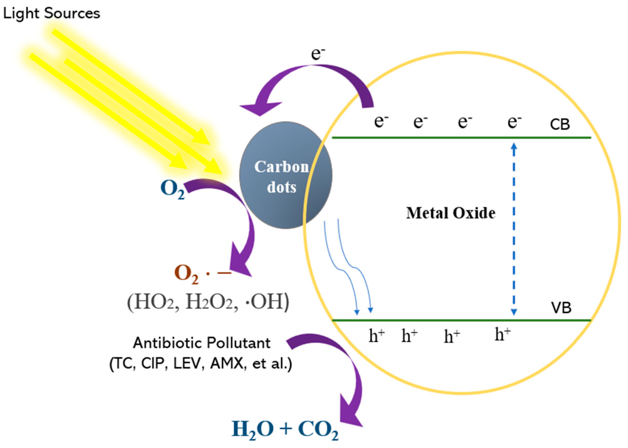
7.2. Mechanism of Metal Oxide/Carbon Dot Photocatalysis

One of the key factors that contribute to the outstanding properties of carbon dots is the quantum confinement effect, which endows them with impressive optoelectronic characteristics. Structurally, carbon dots consist of a graphitic sp2 carbon core surrounded by sp3 carbon atoms, which feature various surface functional groups, like C=O, C–O–C, O=C–OH, and C–O. These functional groups show excellent optical absorption across both UV and visible spectra and have remarkable modification capabilities to convert low-energy photons into high-energy photons. Figure 8 explains the mechanism by which carbon dots modify the surface of metal oxides, effectively boosting their catalytic efficiency. Carbon dots absorb visible light with long wavelengths and subsequently emit short-wavelength UV photons, which photoexcite metal oxides, generating electron–hole pairs and releasing ROS like O H and O 2 , leading to pollutant degradation. These ROS are highly reactive and play a pivotal role in the degradation of pharmaceutical pollutants in aquatic environments. Carbon dots with an appropriate band gap support electron transfer from the metal oxide surface after photoexcitation, thereby improving charge separation and enhancing charge transfer for photocatalytic reactions on the photocatalyst surface [155]. This makes carbon dots valuable as spectral converters for utilizing the full spectrum of sunlight. The synergistic interaction between carbon dots and metal oxides promotes efficient electron transfer to the carbon dots from the surface of the metal oxide after photoexcitation, thereby facilitating improved charge separation and reducing the charge carriers’ recombination. This significantly improves the photocatalytic activity of the composite [14].
Moreover, when carbon dots are incorporated into metal oxide nanocomposites, their unique band gap structure allows for superior light absorption, enabling the utilization of a wider spectrum of sunlight, including both visible and UV light. This property makes carbon dots highly valuable as spectral converters, optimizing the absorption efficiency of the photocatalyst. The combination of carbon dots with metal oxides not only extends the photocatalytic activity into the visible spectrum but also improves the efficiency of photogenerated charge carriers by improving their separation and transport. Consequently, this enhances the overall photonic efficiency of the system [173].
For example, heterostructures formed by carbon dots and wide-band gap metal oxides such as T i O 2 , ZnO, and BiOBr offer significant advantages for photocatalytic applications. These metal oxides are widely recognized for their excellent photocatalytic activity, non-toxicity, high photochemical stability, and cost effectiveness. However, their large band gaps typically restrict their light absorption to the UV range, which is a small portion of the solar spectrum that reaches the Earth’s surface [13]. By integrating carbon dots into these semiconductors, the resulting nanocomposites can efficiently absorb visible light, improving overall photocatalytic performance.
When the nanocomposites are excited, the metal oxide directly absorbs UV radiation while carbon dots absorb visible light. Carbon dots can convert longer-wavelength radiation into shorter-wavelength, higher-energy radiation through up-conversion, making it more suitable for metal oxide absorption. This results in electron–hole separation in both the metal oxide and carbon dots. Carbon dots perform as an electron reservoir by trapping photogenerated electrons from the metal oxide’s conduction band and facilitating their transfer into their valence band, thus increasing the density of surface electron–hole pairs. This process aids in the separation of photoinduced charge carriers and delays charge recombination.
Additionally, under irradiation, electrons generated in the carbon dots’ conduction band react with surface oxygen on the nanocomposite, forming O 2 radicals. These radicals may further react with adsorbed H 2 O , speeding up the production of hydroxyl radicals. Simultaneously, holes in the semiconductor’s valence band may directly react with H 2 O / O H to generate hydroxyl radicals. These radicals, either through combining into hydrogen peroxide or through their intrinsic oxidative properties, can efficiently degrade pharmaceutical contaminants, leading to their non-selective decomposition and eventual mineralization. Essentially, ROS generated in this process plays a vital role in the non-selective decomposition of pollutants.

7.3. Application of Metal Oxide/Carbon Dots for Pharmaceutical Pollutant Remediation

Based on the working mechanism explained above, there are two types of desired outcomes when preparing metal oxide/CDs. The incorporation of CDs can either increase the efficiency of the photocatalyst by acting as an electron reservoir or give additional photocatalytic activity under visible light by utilizing their narrow band gap. The latter is usually the main goal when using superior metal oxide photocatalysts such as ZnO and TiO2, which have good photoactivity but a wide band gap, preventing their utilization under visible light [111,112]. Li et al. [168] demonstrated how the addition of CDs could increase the %degradation of tetracycline using ZnO under visible light irradiation from ~20% in 90 min to over 60%. Furthermore, the addition of halloysite nanotubes (HNTs) was able to increase this number to over 90%. Similar positive results were also shown when incorporating ZnO/CDs for ciprofloxacin degradation under sunlight [11].
On the other hand, the use of CDs as electron reservoirs often plays a critical role when working with visible-light-active metal oxides, as this type of photocatalyst tends to have high electron–hole recombination rates. In this study, we found that many studies have investigated the performance of bismuth-based metal oxide/CD composites [161,163,164,166,167]. Jin et al. [163] demonstrated that a BiVO4/N-doped CD photocatalyst was able to degrade around twice as much tetracycline under 30 min of visible light irradiation, with also twice the rate constant, compared to regular BiVO4. They explained that excited electrons from the conduction band of the metal oxide were able to migrate to the valence band of carbon dots positioned at a lower energy level. This, in turn, inhibits charge recombination.
Furthermore, this property opens the possibility of utilizing other less popular metal oxides that also suffer from low efficiency due to high recombination rates. A study by Huang et al. [162] compared the performance of FeOOH and FeOOH/CD photocatalysts to degrade tetracycline in H2O2/visible light-assisted systems. The results show that FeOOH/CDs were able to fully degrade tetracycline under 40 min, whereas pristine FeOOH was only able to degrade around 20% after 60 min. The addition of CDs was also able to enhance the efficiency of MoO3/g-C3N4 in degrading tetracycline under visible light irradiation to up to 2 times [169]. The same positive results were also seen for CuO2, though Wang et al. demonstrated that the loading percentage of carbon dots plays a key role in enhancing or degrading photocatalytic performance [16].
Table 6 summarizes studies utilizing metal oxide/CD photocatalyst systems for pharmaceutical waste degradation. The integration of metal oxides with carbon dots offers an innovative strategy for enhancing photocatalytic processes aimed at the remediation of pharmaceutical pollutants. The synergistic effects between these materials improve light absorption, boost charge separation efficiency, and aid in the production of ROS, all of which contribute to the breaking down of persistent pollutants in water. Thus, metal oxide/carbon dot nanocomposites hold significant promise as next-generation photocatalysts for environmental applications.

7.4. Health Concerns of Metal Oxide/Carbon Dots

While carbon-based nanoparticles are mostly deemed non-toxic and have good biocompatibility, the toxicity of metal oxides cannot be generalized, as it significantly depends on the material composition, size, surface characteristics, and application environment [121,174]. For example, some metal oxides, such as CuO and Cu2O, have been reported to exhibit genotoxic effects through the induction of oxidative stress. This occurs via the overproduction of ROS, which can damage cellular components, including DNA, proteins, and lipids. At the same time, other metal oxides, such as TiO2 and ZnO, are generally considered safe under specific conditions and have been extensively employed in pharmaceutical formulations and food products. TiO2 nanoparticles are frequently used as food additives and in sunscreens due to their UV-blocking properties, while ZnO nanoparticles are incorporated in cosmetics and antimicrobial coatings [174]. Additionally, the size-dependent effect on toxicity has also been studied in some cases and should not be overlooked [175]. Therefore, the safety of metal oxide/carbon dots should be studied case by case.
Nevertheless, the more prominent concern when applying photocatalyst-based wastewater treatment is preventing the material itself from becoming a secondary pollutant. As recent advances have shifted to nano-sized photocatalysts for enhanced efficiency, the separation and recovery of photocatalyst particles become difficult. Therefore, many studies have touched on the topic of photocatalyst immobilization through the preparation of composites, commonly using polymers [176]. This approach offers a dual benefit: it ensures that the photocatalyst materials remain contained, preventing their release into the water, and mitigates health risks associated with certain metal oxides, such as CuO and CuO2. By trapping the photocatalyst particles within a stable composite, this method reduces environmental contamination while maintaining treatment efficiency. However, the topic of photocatalyst immobilization is a complex subject in its own right and lies beyond the scope of this article.
Table 6. Degradation of Antibiotics by Metal Oxide/Carbon dots.
PhotocatalystCarbon SourcesAntibioticsExperimental ConditionsAntibiotic Removalk
(min−1)
Ref.
NCDs/ B i V O 4 Citric acidTetracycline[catalyst] = 50 mg
[TC] = 5.0 mg/L
Light source = 300 W Xe lamp (visible light)
97% (150 min)-[167]
A g + -CDs- B i 2 W O 6 GlucoseTetracycline[catalyst] = 0.5 g/L (0.01 g)
[TC] = 20 mg/L (20 mL)
Light source = 800 W Xe lamp (UV–visible light)
92% (10 min)
~100% (20 min)
0.09182 [161]
CQD/α-FeOOHCitric acidTetracycline[catalyst] = 0.25 g/L (50 mg)
[TC] = 20 mg/L
pH = 3.09–10.31
Light source = 350 W Xe lamp (visible light)
94.5% (60 min)-[162]
CQDs/ZnO@HNTs Citric acidTetracycline[catalyst] = 80 mg
[TC] = 20 mg/L (100 mL)
Light source = 500 W Xenon lamp (visible light)
92.48% (90 min)-[168]
B i V O 4 /N-CQDs/ A g 3 P O 4 Ammonium citrateTetracycline[catalyst] = 30 mg
[TC] = 10 mg/L (100 mL)
Light source = 300
W Xe lamp (visible light)
88.9% (30 min)0.07097[163]
N-CQDs/ B i 2 W O 6 Ammonium citrateTetracycline[catalyst] = 0.05 g
[TC] = 10 mg/L (100 mL)
Light source = 300 W Xe lamp (visible light)
97% (25 min)0.10684[164]
CQDs/ B i 2 W O 6 Ammonium citrateTetracycline[catalyst] = 0.05 g
[TC] = 10 mg/L (100 mL)
Light source = 300 W Xe lamp (visible light)
83% (25 min)0.0754[164]
Carbon dots/ B i O C l ( C B O C 2 ) ChroellaTetracycline[catalyst] = 15 mg
[TC] = 10 mg/L (100 mL)
pH = 7
Light source = 500 W Xe lamp
99.5% (75 min)0.075[177]
CQDs/ C u 2 O Corn stoverTetracycline[catalyst] = 100 mg
[TC] = 10 mg/L (90 mL)
Light source = Xe lamp (visible light)
92.49% (100 min)-[16]
CDs/g- C 3 N 4 / M o O 3 Citric acid and ureaTetracycline[catalyst] = 30 mg
[TC] = 20 mg/L (50 mL)
Light source = 350 W Xe lamp (visible light)
88.4% (90 min)0.0231[169]
ZIS/CQDsCitric acidTetracycline[catalyst] = 20 mg
[TC] = 10 mg/L (80 mL)
Light source = 250 W Xe lamp (visible light), 350 W mercury lamp (UV)
67.89% (90 min)0.00839[140]
CDs/g- C 3 N 4 / B i P O 4 Citric acidTetracycline[catalyst] = 20 mg
[TC] = 5 mg/L (80 mL)
pH = 4
Light source = 500 W Xe lamp (visible light)
79.3% (210 min)0.01315[170]
A g / C u F e 2 O 4 / C Q D s Citric acidTetracycline[catalyst] = 0.075 g
[TC] = 10 mg/L (250 mL)
pH = 3
Light source = 150 W Xe Lamp (visible light)
100% (75 min)0.1319[171]
CQDs/ B i 3 T i 4 O 12 BambooCiprofloxacin[catalyst] = 50 mg
[CIP] = 15 mg/L (100 mL)
Light source = 300 W Xe lamp (visible light)
65% (90 min)0.01164[172]
T i O 2 /CDs/CdTe QDsPEICiprofloxacin[catalyst] = 50 g
[CIP] = 10 mg/L (100 mL)
Light source = 250 W Xe lamp (visible light)
70% (90 min)0.008[165]
BiOCl/CQDs/rGOSucroseCiprofloxacin[catalyst] = 25 mg
[CIP] = 20 mg/L (50 mL)
Light source = 300 W Xe lamp (visible light)
87% (100 min)0.0146[166]
ZnO/CD NCsTrisodium citrate dihydrateCiprofloxacin[catalyst] = 0.6 g/L
[CIP] = 12 mg/L (50 mL)
pH = 6.3
Light source = natural sunlight
98% (110 min)0.030[11]

8. Conclusions

The presence of pharmaceutical contaminants in water sources poses significant risks to human health. Effective removal of these compounds is essential to ensure the safety of our water supply. Metal oxides, as semiconductors, have been widely utilized in water purification for their ability to efficiently degrade various pharmaceuticals through photocatalysis. However, their application is often limited by issues such as the rapid recombination of photocarriers and high band gap energy, which reduces their industrial efficacy. Moreover, their adsorption capacity is constrained. Various strategies have been explored to address these challenges, including improving the morphology materials until doping with expensive and toxic metals, which can exacerbate charge carrier recombination. To mitigate these limitations, integrating metal oxides with carbon dots shows promise. Metal oxide/carbon dot nanocomposites have demonstrated effective photocatalytic degradation of pharmaceuticals. A review of the literature indicates that carbon dots offer a viable solution to enhance metal oxide performance. Incorporating carbon dots expands the absorption spectrum of the nanocomposites, intensifies light absorption, mitigates photogenerated charge carrier recombination, and enhances pollutant adsorption at active sites. As a result, carbon dots significantly enhance the photocatalytic efficiency of metal oxides under visible light exposure. The further exploration of this composite holds the potential for developing environmentally friendly technologies that utilize sunlight as a clean energy source to remove persistent pharmaceutical pollutants from water sources.
The future of metal oxide/carbon dot nanocomposites for the remediation of pharmaceutical pollutants in water holds significant promise. One of the key areas for further research is optimizing synthesis techniques to improve the uniformity, scalability, and cost effectiveness of these materials. By refining their surface chemistry, researchers can enhance their photocatalytic efficiency, making them viable for real-world water treatment applications. In addition, expanding the range of metal oxides combined with carbon dots—such as exploring emerging materials like tungsten oxide and copper oxide—could lead to the development of nanocomposites with unique electronic properties and enhanced photocatalytic performance. However, the health and safety concerns of using new types of materials should be considered, and strategies to combat any risk should be assessed. Another exciting avenue lies in advancing our understanding of the interaction between carbon dots and metal oxides. In-depth studies using advanced characterization techniques will provide critical insights into the photocatalytic mechanisms, enabling the design of more efficient and selective catalysts. Furthermore, integrating these nanocomposites into hybrid remediation systems that combine multiple treatment methods, such as adsorption and filtration, could significantly improve pollutant degradation efficiency in large-scale water treatment processes. Environmental safety and the long-term impact of these nanomaterials are critical considerations for their widespread use. Future research must focus on assessing their toxicity, degradation behaviour, and potential environmental risks to ensure safe deployment. Finally, maximizing the utilization of the full solar spectrum and addressing real-world scalability challenges—through pilot studies and modular photocatalytic reactors—will be essential for translating the laboratory successes of metal oxide/carbon dot nanocomposites into practical environmental technologies. These advancements could pave the way for sustainable, efficient water purification systems to address the growing issue of pharmaceutical contamination in water resources globally.

Author Contributions

Conceptualization, methodology, software, investigation, writing—original draft preparation, A.S.A.; validation, investigation, data curation, G.A.; writing—review and editing, visualization, F.G.N.; formal analysis, data curation, software, M.M.M.; resources, writing—review and editing, supervision, project administration, funding acquisition, A.T.A.A. All authors have read and agreed to the published version of the manuscript.

Funding

This research received no external funding.

Data Availability Statement

The data presented in this study are available on reasonable request from the corresponding author.

Acknowledgments

The authors acknowledge the CoE Applied Nanotechnology for their support.

Conflicts of Interest

The authors declare no conflicts of interest.

References

  1. Rayaroth, M.P.; Aravindakumar, C.T.; Shah, N.S.; Boczkaj, G. Advanced oxidation processes (AOPs) based wastewater treatment—unexpected nitration side reactions—A serious environmental issue: A review. Chem. Eng. J. 2022, 430, 133002. [Google Scholar] [CrossRef]
  2. Srivastava, S.K. Recent advances in removal of pharmaceutical pollutants in wastewater using metal oxides and carbonaceous materials as photocatalysts: A review. RSC Appl. Interfaces 2024, 1, 340–429. [Google Scholar] [CrossRef]
  3. Shong, B.; Ansari, A.S.; Bent, S.F. Thermally Activated Reactions of Phenol at the Ge(100)-2 × 1 Surface. J. Phys. Chem. C 2020, 124, 23657–23660. [Google Scholar] [CrossRef]
  4. Ansari, A.A.; Sartale, S.D. Narrow size distributed Ag nanoparticles grown by spin coating and thermal reduction: Effect of processing parameters. Mater. Res. Express 2016, 3, 085023. [Google Scholar] [CrossRef]
  5. Raya, S.S.; Ansari, A.S.; Kang, S.G.; Lee, H.-B.; Shong, B. Effect of molecular backbone structure on vapor phase coupling reaction between diiso(thio)cyanates with diamines, diols, and dithiols. Prog. Org. Coatings 2020, 140, 105509. [Google Scholar] [CrossRef]
  6. Khadtare, S.S.; Ansari, A.S.A.; Sartale, S.D.; Jadkar, S.R.; Pathan, H.M. ZnO nanocactus loaded with gold nanoparticles for dye sensitized solar cells. In Proceedings of the 2014 International Renewable and Sustainable Energy Conference (IRSEC), Ouarzazate, Morocco, 17–19 October 2014; pp. 94–96. [Google Scholar]
  7. Ansari, A.A.; Sartale, S.D. The Calculation of Electronic Parameters of Al/TiO2/p-Si MOS Structure Formed Using TiO2 Thin Films Grown by Thermal Oxidation of Sputtered Ti Films. Adv. Sci. Lett. 2016, 22, 1013–1016. [Google Scholar] [CrossRef]
  8. Wang, J.; Zhuan, R. Degradation of antibiotics by advanced oxidation processes: An overview. Sci. Total. Environ. 2020, 701, 135023. [Google Scholar] [CrossRef] [PubMed]
  9. Wei, Z.; Liu, J.; Shangguan, W. A review on photocatalysis in antibiotic wastewater: Pollutant degradation and hydrogen production. Chin. J. Catal. 2020, 41, 1440–1450. [Google Scholar] [CrossRef]
  10. Sharma, S.; Umar, A.; Mehta, S.K.; Ibhadon, A.O.; Kansal, S.K. Solar light driven photocatalytic degradation of levofloxacin using TiO2/carbon-dot nanocomposites. New J. Chem. 2018, 42, 7445–7456. [Google Scholar] [CrossRef]
  11. Mukherjee, I.; Cilamkoti, V.; Dutta, R.K. Sunlight-Driven Photocatalytic Degradation of Ciprofloxacin by Carbon Dots Embedded in ZnO Nanostructures. ACS Appl. Nano Mater. 2021, 4, 7686–7697. [Google Scholar] [CrossRef]
  12. Anucha, C.B.; Altin, I.; Bacaksiz, E.; Stathopoulos, V.N. Titanium dioxide (TiO2)-based photocatalyst materials activity enhancement for contaminants of emerging concern (CECs) degradation: In the light of modification strategies. Chem. Eng. J. Adv. 2022, 10, 100262. [Google Scholar] [CrossRef]
  13. Sharma, S.; Dutta, V.; Singh, P.; Raizada, P.; Rahmani-Sani, A.; Hosseini-Bandegharaei, A.; Thakur, V.K. Carbon quantum dot supported semiconductor photocatalysts for efficient degradation of organic pollutants in water: A review. J. Clean. Prod. 2019, 228, 755–769. [Google Scholar] [CrossRef]
  14. Sendão, R.M.; da Silva, J.C.E.; da Silva, L.P. Photocatalytic removal of pharmaceutical water pollutants by TiO2—Carbon dots nanocomposites: A review. Chemosphere 2022, 301, 134731. [Google Scholar] [CrossRef]
  15. Liu, X.; Yang, Y.; Li, H.; Yang, Z.; Fang, Y. Visible light degradation of tetracycline using oxygen-rich titanium dioxide nanosheets decorated by carbon quantum dots. Chem. Eng. J. 2021, 408, 127259. [Google Scholar] [CrossRef]
  16. Wang, J.; Li, S.; Ma, P.; Guo, Z.; Ma, Q.; Zhao, Q.; Guo, Y.; Zhao, J.; Guan, G. Carbon quantum dots/Cu2O S-scheme heterojunction for enhanced photocatalytic degradation of tetracycline. Colloids Surfaces A Physicochem. Eng. Asp. 2024, 690, 133779. [Google Scholar] [CrossRef]
  17. Nguyen, D.N.; Bui, H.M.; Nguyen, H.Q. Chapter 7—Heterogeneous Photocatalysis for the Removal of Pharmaceutical Compounds. In Current Developments in Biotechnology and Bioengineering; Varjani, S., Pandey, A., Tyagi, R.D., Ngo, H.H., Larroche, C., Eds.; Elsevier: Amsterdam, The Netherlands, 2020; pp. 161–183. [Google Scholar]
  18. Kar, P.; Shukla, K.; Jain, P.; Sathiyan, G.; Gupta, R.K. Semiconductor based photocatalysts for detoxification of emerging pharmaceutical pollutants from aquatic systems: A critical review. Nano Mater. Sci. 2021, 3, 25–46. [Google Scholar] [CrossRef]
  19. Kutuzova, A.; Dontsova, T.; Kwapinski, W. Application of TiO2-Based Photocatalysts to Antibiotics Degradation: Cases of Sulfamethoxazole, trimethoprim and ciprofloxacin. Catalysts 2021, 11, 728. [Google Scholar] [CrossRef]
  20. Papaioannou, C.; Geladakis, G.; Kommata, V.; Batargias, C.; Lagoumintzis, G. Insights in Pharmaceutical Pollution: The Prospective Role of eDNA Metabarcoding. Toxics 2023, 11, 903. [Google Scholar] [CrossRef]
  21. Bayan, E.; Pustovaya, L.; Volkova, M. Recent advances in TiO2-based materials for photocatalytic degradation of antibiotics in aqueous systems. Environ. Technol. Innov. 2021, 24, 101822. [Google Scholar] [CrossRef]
  22. Gao, J.; Rao, S.; Yu, X.; Wang, L.; Xu, J.; Yang, J.; Liu, Q. Dimensional-matched two dimensional/two dimensional TiO2/Bi2O3 step-scheme heterojunction for boosted photocatalytic performance of sterilization and water splitting. J. Colloid Interface Sci. 2022, 628, 166–178. [Google Scholar] [CrossRef] [PubMed]
  23. Polianciuc, S.I.; Gurzău, A.E.; Kiss, B.; Ștefan, M.G.; Loghin, F. Antibiotics in the environment: Causes and consequences. Med. Pharm. Rep. 2020, 93, 231–240. [Google Scholar] [CrossRef]
  24. Bhagat, C.; Kumar, M.; Tyagi, V.K.; Mohapatra, P.K. Proclivities for prevalence and treatment of antibiotics in the ambient water: A review. npj Clean Water 2020, 3, 42. [Google Scholar] [CrossRef]
  25. World Health Organization. Guidance on Wastewater and Solid Waste Management for Manufacturing of Antibiotics; World Health Organization: Geneva, Switzerland, 2024. [Google Scholar]
  26. Kumari, H.; Sonia; Suman; Ranga, R.; Chahal, S.; Devi, S.; Sharma, S.; Kumar, S.; Kumar, P.; Kumar, S.; et al. A Review on Photocatalysis Used For Wastewater Treatment: Dye Degradation. Water Air Soil Pollut. 2023, 234, 349. [Google Scholar] [CrossRef]
  27. Gao, Y.; Wang, Q.; Ji, G.; Li, A. Degradation of antibiotic pollutants by persulfate activated with various carbon materials. Chem. Eng. J. 2022, 429, 132387. [Google Scholar] [CrossRef]
  28. Mishra, N.S.; Reddy, R.; Kuila, A.; Rani, A.; Nawaz, A.; Pichiah, S.; Mukherjee, P. A Review on Advanced Oxidation Processes for Effective Water Treatment. Curr. World Environ. J. 2017, 12, 469–489. [Google Scholar] [CrossRef]
  29. Anwer, M.; Faizi, Y. A review on types and applications of advanced oxidation processes. Int. J. Adv. Eng. Manag. 2022, 4, 1059–1070. [Google Scholar]
  30. Conte, F.; Tommasi, M.; Degerli, S.N.; Forame, E.; Parolini, M.; De Felice, B.; Ramis, G.; Rossetti, I. Comparison of Different Advanced Oxidation Processes (AOPs) and Photocatalysts for the Degradation of Diclofenac. ChemPhotoChem 2024, 8, e202300177. [Google Scholar] [CrossRef]
  31. Mondal, K.; Sharma, A. Recent advances in the synthesis and application of photocatalytic metal–metal oxide core–shell nanoparticles for environmental remediation and their recycling process. RSC Adv. 2016, 6, 83589–83612. [Google Scholar] [CrossRef]
  32. Zhang, D.; Lv, S.; Luo, Z. A study on the photocatalytic degradation performance of a [KNbO3]0.9-[BaNi0.5Nb0.5O3−δ]0.1 perovskite. RSC Adv. 2020, 10, 1275–1280. [Google Scholar] [CrossRef]
  33. Xie, X.; Li, S.; Zhang, H.; Wang, Z.; Huang, H. Promoting charge separation of biochar-based Zn-TiO2/pBC in the presence of ZnO for efficient sulfamethoxazole photodegradation under visible light irradiation. Sci. Total Environ. 2019, 659, 529–539. [Google Scholar] [CrossRef]
  34. Lin, Y.-T.; Weng, C.-H.; Chen, F.-Y. Key operating parameters affecting photocatalytic activity of visible-light-induced C-doped TiO2 catalyst for ethylene oxidation. Chem. Eng. J. 2014, 248, 175–183. [Google Scholar] [CrossRef]
  35. Rajamanickam, D.; Shanthi, M. Photocatalytic degradation of an organic pollutant by zinc oxide—Solar process. Arab. J. Chem. 2016, 9, S1858–S1868. [Google Scholar] [CrossRef]
  36. Hanafi, M.F.; Sapawe, N. Effect of pH on the photocatalytic degradation of remazol brilliant blue dye using zirconia catalyst. Mater. Today Proc. 2020, 31, 260–262. [Google Scholar] [CrossRef]
  37. Tayyebi, A.; Soltani, T.; Lee, B.-K. Effect of pH on photocatalytic and photoelectrochemical (PEC) properties of monoclinic bismuth vanadate. J. Colloid Interface Sci. 2019, 534, 37–46. [Google Scholar] [CrossRef] [PubMed]
  38. Josephine, A.J.; Dhas, C.R.; Venkatesh, R.; Arivukarasan, D.; Christy, A.J.; Monica, S.E.S.; Keerthana, S. Effect of pH on visible-light-driven photocatalytic degradation of facile synthesized bismuth vanadate nanoparticles. Mater. Res. Express 2020, 7, 015036. [Google Scholar] [CrossRef]
  39. Nugroho, F.G.; Agson-Gani, P.A.; Anindita, P.A.; Steky, F.V.; Benu, D.P.; Yuliarto, B.; Dwivany, F.M.; Suendo, V. Prolonging banana shelf life through visible light-induced ethylene scavenging using manganese-decorated TiO2 via KMnO4 reduction. Colloids Surfaces A Physicochem. Eng. Asp. 2024, 691, 133817. [Google Scholar] [CrossRef]
  40. Kumar, A.; Pandey, G. The photocatalytic degradation of methyl green in presence of visible light with photoactive Ni0.10: La0.05: TiO2 nanocomposites. IOSR J. Appl. Chem. 2017, 10, 31–44. [Google Scholar]
  41. Lu, Y.; Lin, Y.; Wang, D.; Wang, L.; Xie, T.; Jiang, T. A high performance cobalt-doped ZnO visible light photocatalyst and its photogenerated charge transfer properties. Nano Res. 2011, 4, 1144–1152. [Google Scholar] [CrossRef]
  42. Wu, Z.; Dong, F.; Zhao, W.; Wang, H.; Liu, Y.; Guan, B. The fabrication and characterization of novel carbon doped TiO2 nanotubes, nanowires and nanorods with high visible light photocatalytic activity. Nanotechnology 2009, 20, 235701. [Google Scholar] [CrossRef]
  43. Meng, Y.; Huang, X.; Wu, Y.; Wang, X.; Qian, Y. Kinetic study and modeling on photocatalytic degradation of para-chlorobenzoate at different light intensities. Environ. Pollut. 2002, 117, 307–313. [Google Scholar] [CrossRef] [PubMed]
  44. Yang, L.; Liu, Z. Study on light intensity in the process of photocatalytic degradation of indoor gaseous formaldehyde for saving energy. Energy Convers. Manag. 2007, 48, 882–889. [Google Scholar] [CrossRef]
  45. Elaziouti, A.; Ahmed, B. ZnO-assisted photocatalytic degradation of congo Red and benzopurpurine 4B in aqueous solution. J. Chem. Eng. Process Technol. 2011, 2, 1–9. [Google Scholar]
  46. Wardhani, S.; Purwonugroho, D.; Fitri, C.W.; Prananto, Y.P. Effect of pH and irradiation time on TiO2-chitosan activity for phenol photo-degradation. In Proceedings of the 8th Annual Basic Science International Conference: Coverage of Basic Sciences Toward the World’s Sustainability Challanges, East Java, Indonesia, 6–7 March 2018. [Google Scholar]
  47. Ahuja, P.; Ujjain, S.K.; Kanojia, R.; Attri, P. Transition Metal Oxides and Their Composites for Photocatalytic Dye Degradation. J. Compos. Sci. 2021, 5, 82. [Google Scholar] [CrossRef]
  48. Araújo, E.S.; Pereira, M.F.G.; da Silva, G.M.G.; Tavares, G.F.; Oliveira, C.Y.B.; Faia, P.M. A Review on the Use of Metal Oxide-Based Nanocomposites for the Remediation of Organics-Contaminated Water via Photocatalysis: Fundamentals, Bibliometric Study and Recent Advances. Toxics 2023, 11, 658. [Google Scholar] [CrossRef] [PubMed]
  49. Pascariu, P.; Gherasim, C.; Airinei, A. Metal Oxide Nanostructures (MONs) as Photocatalysts for Ciprofloxacin Degradation. Int. J. Mol. Sci. 2023, 24, 9564. [Google Scholar] [CrossRef] [PubMed]
  50. Mehta, A.; Mishra, A.; Basu, S.; Shetti, N.P.; Reddy, K.R.; Saleh, T.A.; Aminabhavi, T.M. Band gap tuning and surface modification of carbon dots for sustainable environmental remediation and photocatalytic hydrogen production—A review. J. Environ. Manag. 2019, 250, 109486. [Google Scholar] [CrossRef] [PubMed]
  51. Abdullah, T.A.; Al-Obaidi, Q.; Abdulla, T.A.; Rasheed, R.T.; Al-Azawi, K.; Meharban, F. A Critical Review of the Photocatalytic Degradation of Pharmaceutical Residues by a TiO2-Based Photocatalyst. Hung. J. Ind. Chem. 2023, 51, 65–75. [Google Scholar] [CrossRef]
  52. Yang, X.; Wang, D. Photocatalysis: From Fundamental Principles to Materials and Applications. ACS Appl. Energy Mater. 2018, 1, 6657–6693. [Google Scholar] [CrossRef]
  53. Zhao, L.; Meng, Q.; Fan, X.; Ye, C.; Li, X.; Chen, B.; Ramamurthy, V.; Tung, C.; Wu, L. Photocatalysis with Quantum Dots and Visible Light: Selective and Efficient Oxidation of Alcohols to Carbonyl Compounds through a Radical Relay Process in Water. Angew. Chem. Int. Ed. Engl. 2017, 56, 3020–3024. [Google Scholar] [CrossRef] [PubMed]
  54. Hao, S.; Zhao, X.; Cheng, Q.; Xing, Y.; Ma, W.; Wang, X.; Zhao, G.; Xu, X. A Mini Review of the Preparation and Photocatalytic Properties of Two-Dimensional Materials. Front. Chem. 2020, 8, 582146. [Google Scholar] [CrossRef]
  55. Thomas, N.; Mathew, S.; Nair, K.M.; O’Dowd, K.; Forouzandeh, P.; Goswami, A.; McGranaghan, G.; Pillai, S.C. 2D MoS2: Structure, mechanisms, and photocatalytic applications. Mater. Today Sustain. 2021, 13, 100073. [Google Scholar] [CrossRef]
  56. Quinn, M.D.J.; Ho, N.H.; Notley, S.M. Aqueous Dispersions of Exfoliated Molybdenum Disulfide for Use in Visible-Light Photocatalysis. ACS Appl. Mater. Interfaces 2013, 5, 12751–12756. [Google Scholar] [CrossRef] [PubMed]
  57. Naggar, A.H.; Ahmed, A.S.A.; El-Nasr, T.A.S.; Alotaibi, N.F.; Chong, K.F.; Ali, G.A.M. Morphological Dependence of Metal Oxide Photocatalysts for Dye Degradation. Inorganics 2023, 11, 484. [Google Scholar] [CrossRef]
  58. Kannan, K.; Radhika, D.; Sadasivuni, K.K.; Reddy, K.R.; Raghu, A.V. Nanostructured metal oxides and its hybrids for photocatalytic and biomedical applications. Adv. Colloid Interface Sci. 2020, 281, 102178. [Google Scholar] [CrossRef]
  59. Das, A.; Nikhil, S.K.; Nair, R.G. Influence of surface morphology on photocatalytic performance of zinc oxide: A review. Nano-Struct. Nano-Objects 2019, 19, 100353. [Google Scholar] [CrossRef]
  60. Ferreira, S.H.; Morais, M.; Nunes, D.; Oliveira, M.J.; Rovisco, A.; Pimentel, A.; Águas, H.; Fortunato, E.; Martins, R. High UV and Sunlight Photocatalytic Performance of Porous ZnO Nanostructures Synthesized by a Facile and Fast Microwave Hydrothermal Method. Materials 2021, 14, 2385. [Google Scholar] [CrossRef]
  61. Mohtar, S.S.; Aziz, F.; Ismail, A.F.; Sambudi, N.S.; Abdullah, H.; Rosli, A.N.; Ohtani, B. Impact of Doping and Additive Applications on Photocatalyst Textural Properties in Removing Organic Pollutants: A Review. Catalysts 2021, 11, 1160. [Google Scholar] [CrossRef]
  62. Xie, J.; Wu, Q. One-pot synthesis of ZnO/Ag nanospheres with enhanced photocatalytic activity. Mater. Lett. 2010, 64, 389–392. [Google Scholar] [CrossRef]
  63. Cheng, L.; Zheng, L.; Li, G.; Yin, Q.; Jiang, K. Synthesis of sealed sponge ZnO nanospheres through a novel NH3-evaporation method. Nanotechnology 2008, 19, 075605. [Google Scholar] [CrossRef] [PubMed]
  64. He, Z.; Zhan, L.; Hong, F.; Song, S.; Lin, Z.; Chen, J.; Jin, M. A visible light-responsive iodine-doped titanium dioxide nanosphere. J. Environ. Sci. 2011, 23, 166–170. [Google Scholar] [CrossRef] [PubMed]
  65. Deng, C.; Hu, H.; Ge, X.; Han, C.; Zhao, D.; Shao, G. One-pot sonochemical fabrication of hierarchical hollow CuO submicrospheres. Ultrason. Sonochemistry 2011, 18, 932–937. [Google Scholar] [CrossRef] [PubMed]
  66. Faisal, M.; Ismail, A.A.; Ibrahim, A.A.; Bouzid, H.; Al-Sayari, S.A. Highly efficient photocatalyst based on Ce doped ZnO nanorods: Controllable synthesis and enhanced photocatalytic activity. Chem. Eng. J. 2013, 229, 225–233. [Google Scholar] [CrossRef]
  67. Zhao, J.; Wang, L.; Yan, X.; Yang, Y.; Lei, Y.; Zhou, J.; Huang, Y.; Gu, Y.; Zhang, Y. Structure and photocatalytic activity of Ni-doped ZnO nanorods. Mater. Res. Bull. 2011, 46, 1207–1210. [Google Scholar] [CrossRef]
  68. Alammar, T.; Mudring, A.-V. Facile ultrasound-assisted synthesis of ZnO nanorods in an ionic liquid. Mater. Lett. 2009, 63, 732–735. [Google Scholar] [CrossRef]
  69. Sadollahkhani, A.; Ibupoto, Z.H.; Elhag, S.; Nur, O.; Willander, M. Photocatalytic properties of different morphologies of CuO for the degradation of Congo red organic dye. Ceram. Int. 2014, 40, 11311–11317. [Google Scholar] [CrossRef]
  70. Yin, X.; Liu, L.; Ai, F. Enhanced Photocatalytic Degradation of Methylene Blue by WO3 Nanoparticles Under NIR Light Irradiation. Front. Chem. 2021, 9, 683765. [Google Scholar] [CrossRef]
  71. Ullah, A.; Rahman, L.; Hussain, S.Z.; Abbas, W.; Tawab, A.; Jilani, A.; Bajwa, S.Z.; Khan, W.S.; Riaz, R.; Hussain, I.; et al. Mechanistic insight of dye degradation using TiO2 anchored α-MnO2 nanorods as promising sunlight driven photocatalyst. Mater. Sci. Eng.: B 2021, 271, 115257. [Google Scholar] [CrossRef]
  72. Yuan, S.D.; Chen, S.Q.; Zhu, X.; Xiong, P.; Yang, Y.F.; Hu, Z.H.; Xiong, J. Obtaining Ultra-High Surface Area TiO2 Nanorods via Hydrothermally Transformation of Elongated Titanate Nanotubes. J. Nano Res. 2018, 51, 13–23. [Google Scholar] [CrossRef]
  73. Liu, Y.; Kang, Z.H.; Chen, Z.H.; Shafiq, I.; Zapien, J.A.; Bello, I.; Zhang, W.J.; Lee, S.T. Synthesis, Characterization, and Photocatalytic Application of Different ZnO Nanostructures in Array Configurations. Cryst. Growth Des. 2009, 9, 3222–3227. [Google Scholar] [CrossRef]
  74. Jia, T.; Wang, W.; Long, F.; Zhengyi, F.; Wang, H.; Zhang, Q. Fabrication, characterization and photocatalytic activity of La-Doped ZnO nanowires. J. Alloy. Compd. 2009, 484, 410–415. [Google Scholar] [CrossRef]
  75. Xu, F.; Yuan, Z.-Y.; Du, G.-H.; Ren, T.-Z.; Bouvy, C.; Halasa, M.; Su, B.-L. Simple approach to highly oriented ZnO nanowire arrays: Large-scale growth, photoluminescence and photocatalytic properties. Nanotechnology 2006, 17, 588–594. [Google Scholar] [CrossRef]
  76. Zhao, Y.M.; Hu, W.B.; Xia, Y.D.; Smith, E.F.; Zhu, Y.Q.; Dunnill, C.W.; Gregory, D.H. Preparation and characterization of tungsten oxynitride nanowires. J. Mater. Chem. 2007, 17, 4436–4440. [Google Scholar] [CrossRef]
  77. Sudirman, S.; Atmaja, L.D.P.; Pratama, A.J.; Yuanita, E.; Dharmayani, N.K.T.; Ulfa, M.; Hidayat, R. Highly photocatalytic performance of TiO2 nanowires in the conversion of benzaldehydes to benzoic acid. Acta Chim. Asiana 2023, 6, 315–321. [Google Scholar] [CrossRef]
  78. Hamisu, A.; Gaya, U.; Gaya, A. Effect of alkali strength on the hydrothermal growth of photoactive TiO2 nanowires. J. Nanostructures 2020, 10, 639–651. [Google Scholar]
  79. Wan, X.; Liang, X.; Zhang, C.; Li, X.; Liang, W.; Xu, H.; Lan, S.; Tie, S. Morphology controlled syntheses of Cu-doped ZnO, tubular Zn(Cu)O and Ag decorated tubular Zn(Cu)O microcrystals for photocatalysis. Chem. Eng. J. 2015, 272, 58–68. [Google Scholar] [CrossRef]
  80. Wang, H.; Li, G.; Jia, L.; Wang, G.; Tang, C. Controllable Preferential-Etching Synthesis and Photocatalytic Activity of Porous ZnO Nanotubes. J. Phys. Chem. C 2008, 112, 11738–11743. [Google Scholar] [CrossRef]
  81. Rojviroon, T.; Rojviroon, O.; Sirivithayapakorn, S.; Angthong, S.; Thongpool, V. Application of TiO2 nanotubes as photocatalysts for decolorization of synthetic dye wastewater. Water Resour. Ind. 2021, 26, 100163. [Google Scholar] [CrossRef]
  82. Radhakrishnan, A.; Rejani, P.; Beena, B. CuO nano structures as an ecofriendly nano photo catalyst and antimicrobial agent for environmental remediation. Int. J. Nano Dimens. 2018, 9, 145–157. [Google Scholar]
  83. Choopool, P.; Kooptarnond, K.; Khangkhamano, M.; Rachpech, V. The Effect of Sn(II) Precursor on Morphology and Surface Area of as Synthesis SnO2 Nanotube. Mater. Sci. Forum 2020, 998, 227–232. [Google Scholar] [CrossRef]
  84. Das, A.; Malakar, P.; Nair, R.G. Engineering of ZnO nanostructures for efficient solar photocatalysis. Mater. Lett. 2018, 219, 76–80. [Google Scholar] [CrossRef]
  85. Miao, Y.; Zhang, H.; Yuan, S.; Jiao, Z.; Zhu, X. Preparation of flower-like ZnO architectures assembled with nanosheets for enhanced photocatalytic activity. J. Colloid Interface Sci. 2016, 462, 9–18. [Google Scholar] [CrossRef] [PubMed]
  86. Zhao, X.; Lou, F.; Li, M.; Lou, X.; Li, Z.; Zhou, J. Sol−gel-based hydrothermal method for the synthesis of 3D flower-like ZnO microstructures composed of nanosheets for photocatalytic applications. Ceram. Int. 2014, 40, 5507–5514. [Google Scholar] [CrossRef]
  87. Harris, J.; Silk, R.; Smith, M.; Dong, Y.; Chen, W.-T.; Waterhouse, G.I.N. Hierarchical TiO2 Nanoflower Photocatalysts with Remarkable Activity for Aqueous Methylene Blue Photo-Oxidation. ACS Omega 2020, 5, 18919–18934. [Google Scholar] [CrossRef]
  88. Liu, M.; Lu, W.-M.; Zhao, L.; Zhou, C.-L.; Li, H.-L.; Wang, W.-J. Fabrication and photocatalytical properties of flower-like TiO2 nanostructures. Trans. Nonferrous Met. Soc. China 2010, 20, 2299–2302. [Google Scholar] [CrossRef]
  89. He, Z.; Cai, Q.; Fang, H.; Situ, G.; Qiu, J.; Song, S.; Chen, J. Photocatalytic activity of TiO2 containing anatase nanoparticles and rutile nanoflower structure consisting of nanorods. J. Environ. Sci. 2013, 25, 2460–2468. [Google Scholar] [CrossRef] [PubMed]
  90. Silva, A.R.; Martins, P.M.; Teixeira, S.; Carabineiro, S.A.C.; Kuehn, K.; Cuniberti, G.; Alves, M.M.; Lanceros-Mendez, S.; Pereira, L. Ciprofloxacin wastewater treated by UVA photocatalysis: Contribution of irradiated TiO2 and ZnO nanoparticles on the final toxicity as assessed by Vibrio fischeri. RSC Adv. 2016, 6, 95494–95503. [Google Scholar] [CrossRef]
  91. Jiang, T.; Cheng, L.; Han, Y.; Feng, J.; Zhang, J. One-pot hydrothermal synthesis of Bi2O3-WO3 p-n heterojunction film for photoelectrocatalytic degradation of norfloxacin. Sep. Purif. Technol. 2020, 238, 116428. [Google Scholar] [CrossRef]
  92. Huang, C.; Chen, L.; Li, H.; Mu, Y.; Yang, Z. Synthesis and application of Bi2WO6 for the photocatalytic degradation of two typical fluoroquinolones under visible light irradiation. RSC Adv. 2019, 9, 27768–27779. [Google Scholar] [CrossRef] [PubMed]
  93. Gurung, K.; Ncibi, M.C.; Thangaraj, S.K.; Jänis, J.; Seyedsalehi, M.; Sillanpää, M. Removal of pharmaceutically active compounds (PhACs) from real membrane bioreactor (MBR) effluents by photocatalytic degradation using composite Ag2O/P-25 photocatalyst. Sep. Purif. Technol. 2019, 215, 317–328. [Google Scholar] [CrossRef]
  94. Chen, J.; Wang, M.; Han, J.; Guo, R. TiO2 nanosheet/NiO nanorod hierarchical nanostructures: P–n heterojunctions towards efficient photocatalysis. J. Colloid Interface Sci. 2020, 562, 313–321. [Google Scholar] [CrossRef]
  95. Abdel-Wahab, A.-M.; Al-Shirbini, A.-S.; Mohamed, O.; Nasr, O. Photocatalytic degradation of paracetamol over magnetic flower-like TiO2/Fe2O3 core-shell nanostructures. J. Photochem. Photobiol. A Chem. 2017, 347, 186–198. [Google Scholar] [CrossRef]
  96. Zhao, Q.-E.; Wen, W.; Xia, Y.; Wu, J.-M. Photocatalytic activity of TiO2 nanorods, nanowires and nanoflowers filled with TiO2 nanoparticles. Thin Solid Films 2018, 648, 103–107. [Google Scholar] [CrossRef]
  97. Nunes, D.; Pimentel, A.; Branquinho, R.; Fortunato, E.; Martins, R. Metal Oxide-Based Photocatalytic Paper: A Green Alternative for Environmental Remediation. Catalysts 2021, 11, 504. [Google Scholar] [CrossRef]
  98. Abdulkareem, G.A.; Nurul-Asikin, M.; Hin, T.-Y.Y. Nanomaterials: An Overview of Nanorods Synthesis and Optimization. In Nanorods and Nanocomposites; Sasani, G.M., Soumen, D., Eds.; IntechOpen: Rijeka, Croatia, 2019; Chapter 2. [Google Scholar]
  99. Fu, D.; Han, G.; Meng, C. Size-controlled synthesis and photocatalytic degradation properties of nano-sized ZnO nanorods. Mater. Lett. 2012, 72, 53–56. [Google Scholar] [CrossRef]
  100. Choudhary, S.; Mohapatra, S. Efficient photocatalytic degradation of antibiotic levofloxacin and organic pollutants in water by Pr doped ZnO nanorods. Chem. Phys. Impact 2024, 9, 100687. [Google Scholar] [CrossRef]
  101. Mohammed, R.; Ali, M.E.M.; Gomaa, E.; Mohsen, M. Green ZnO nanorod material for dye degradation and detoxification of pharmaceutical wastes in water. J. Environ. Chem. Eng. 2020, 8, 104295. [Google Scholar] [CrossRef]
  102. Al-Khadhuri, A.; Al-Sabahi, J.; Kyaw, H.H.; Myint, M.T.Z.; Al-Farsi, B.; Al-Abri, M. Photocatalytic degradation toward pharmaceutical pollutants using supported zinc oxide nanorods catalyzed visible light system. Int. J. Environ. Sci. Technol. 2022, 20, 10021–10030. [Google Scholar] [CrossRef]
  103. Jin, Z.; Meng, F.-L.; Jia, Y.; Luo, T.; Sun, B.; Wang, J.; Liu, J.-H.; Huang, X.-J. Porous TiO2 nanowires derived from nanotubes: Synthesis, characterzation and their enhanced photocatalytic properties. Microporous Mesoporous Mater. 2013, 181, 146–153. [Google Scholar] [CrossRef]
  104. Rogé, V.; Guignard, C.; Lamblin, G.; Laporte, F.; Fechete, I.; Garin, F.; Dinia, A.; Lenoble, D. Photocatalytic degradation behavior of multiple xenobiotics using MOCVD synthesized ZnO nanowires. Catal. Today 2018, 306, 215–222. [Google Scholar] [CrossRef]
  105. Nagasundari, S.M.; Muthu, K.; Kaviyarasu, K.; Al Farraj, D.A.; Alkufeidy, R.M. Current trends of Silver doped Zinc oxide nanowires photocatalytic degradation for energy and environmental application. Surfaces Interfaces 2021, 23, 100931. [Google Scholar] [CrossRef]
  106. He, J.; Zhang, Y.; Guo, Y.; Rhodes, G.; Yeom, J.; Li, H.; Zhang, W. Photocatalytic degradation of cephalexin by ZnO nanowires under simulated sunlight: Kinetics, influencing factors, and mechanisms. Environ. Int. 2019, 132, 105105. [Google Scholar] [CrossRef]
  107. Cha, G.; Schmuki, P.; Altomare, M. Anodic TiO2 nanotube membranes: Site-selective Pt-activation and photocatalytic H2 evolution. Electrochimica Acta 2017, 258, 302–310. [Google Scholar] [CrossRef]
  108. Bojer, C.; Schöbel, J.; Martin, T.; Ertl, M.; Schmalz, H.; Breu, J. Clinical wastewater treatment: Photochemical removal of an anionic antibiotic (ciprofloxacin) by mesostructured high aspect ratio ZnO nanotubes. Appl. Catal. B Environ. 2017, 204, 561–565. [Google Scholar] [CrossRef]
  109. Shende, P.; Kasture, P.; Gaud, R. Nanoflowers: The future trend of nanotechnology for multi-applications. Artif. Cells Nanomed. Biotechnol. 2018, 46, 413–422. [Google Scholar] [CrossRef] [PubMed]
  110. Qu, Y.; Xu, X.; Huang, R.; Qi, W.; Su, R.; He, Z. Enhanced photocatalytic degradation of antibiotics in water over functionalized N,S-doped carbon quantum dots embedded ZnO nanoflowers under sunlight irradiation. Chem. Eng. J. 2020, 382, 123016. [Google Scholar] [CrossRef]
  111. Gusain, R.; Gupta, K.; Joshi, P.; Khatri, O.P. Adsorptive removal and photocatalytic degradation of organic pollutants using metal oxides and their composites: A comprehensive review. Adv. Colloid Interface Sci. 2019, 272, 102009. [Google Scholar] [CrossRef]
  112. Velumani, A.; Sengodan, P.; Arumugam, P.; Rajendran, R.; Santhanam, S.; Palanisamy, M. Carbon quantum dots supported ZnO sphere based photocatalyst for dye degradation application. Curr. Appl. Phys. 2020, 20, 1176–1184. [Google Scholar] [CrossRef]
  113. Wang, K.; Liang, L.; Zheng, Y.; Li, H.; Niu, X.; Zhang, D.; Fan, H. Visible light-driven photocatalytic degradation of organic pollutants via carbon quantum dots/TiO2. New J. Chem. 2021, 45, 16168–16178. [Google Scholar] [CrossRef]
  114. Parmar, N.; Srivastava, J.K. Process optimization and kinetics study for photocatalytic ciprofloxacin degradation using TiO2 nanoparticle: A comparative study of Artificial Neural Network and Surface Response Methodology. J. Indian Chem. Soc. 2022, 99, 100584. [Google Scholar] [CrossRef]
  115. Liu, H.; Wang, L.; Wei, S.; Wu, Y.; Zheng, Y.; Yuan, F.; Hou, J. Study on photocatalytic degradation of amoxicillin in wastewater by Bi2WO6/nano-ZnO. Opt. Mater. 2022, 123, 111835. [Google Scholar] [CrossRef]
  116. Mehmood, S.; Ahmed, W.; Rizwan, M.; Bundschuh, J.; Elnahal, A.S.; Li, W. Green synthesized zinc oxide nanoparticles for removal of carbamazepine in water and soil systems. Sep. Purif. Technol. 2024, 334, 125988. [Google Scholar] [CrossRef]
  117. Qin, K.; Zhao, Q.; Yu, H.; Xia, X.; Li, J.; He, S.; Wei, L.; An, T. A review of bismuth-based photocatalysts for antibiotic degradation: Insight into the photocatalytic degradation performance, pathways and relevant mechanisms. Environ. Res. 2021, 199, 111360. [Google Scholar] [CrossRef]
  118. Kirubanithy, K.; Jayaraj, S.K.; Beura, R.; Paramasivam, T. Detailed structural and optical studies of the microwave synthesized β-Bi2O3 nanostructured photocatalysts: Photocatalytic applications on anionic and cationic organic dyes. Environ. Nanotechnol. Monit. Manag. 2022, 17, 100629. [Google Scholar] [CrossRef]
  119. Zhang, G.; Zhang, X.; Wu, Y.; Shi, W.; Guan, W. Rapid microwave-assisted synthesis of Bi2O3 tubes and photocatalytic properties for antibiotics. Micro Nano Lett. 2013, 8, 177–180. [Google Scholar] [CrossRef]
  120. Sánchez-Martínez, D.; Juárez-Ramírez, I.; Torres-Martínez, L.M.; de León-Abarte, I. Photocatalytic properties of Bi2O3 powders obtained by an ultrasound-assisted precipitation method. Ceram. Int. 2016, 42, 2013–2020. [Google Scholar] [CrossRef]
  121. Mansuriya, B.D.; Altintas, Z. Carbon Dots: Classification, Properties, Synthesis, Characterization, and Applications in Health Care—An Updated Review (2018–2021). Nanomaterials 2021, 11, 2525. [Google Scholar] [CrossRef]
  122. Nugroho, F.G.; Ansari, A.S.; Rochman, N.T.; Khadtare, S.S.; Sree, V.G.; Shrestha, N.K.; Hafiyyan, A.F.; Im, H.; Ahmed, A.T.A. Utilizing Indonesian Empty Palm Fruit Bunches: Biochar Synthesis via Temperatures Dependent Pyrolysis. Nanomaterials 2024, 15, 50. [Google Scholar] [CrossRef]
  123. Saputra, A.M.A.; Piliang, A.F.R.; Goei, R.; Ramadhan, H.R.; Gea, S. Synthesis, properties, and utilization of carbon quantum dots as photocatalysts on degradation of organic dyes: A mini review. Catal. Commun. 2024, 187, 106914. [Google Scholar] [CrossRef]
  124. Anpalagan, K.; Yin, H.; Cole, I.; Zhang, T.; Lai, D.T.H. Quantum Yield Enhancement of Carbon Quantum Dots Using Chemical-Free Precursors for Sensing Cr (VI) Ions. Inorganics 2024, 12, 96. [Google Scholar] [CrossRef]
  125. Xu, X.; Ray, R.; Gu, Y.; Ploehn, H.J.; Gearheart, L.; Raker, K.; Scrivens, W.A. Electrophoretic Analysis and Purification of Fluorescent Single-Walled Carbon Nanotube Fragments. J. Am. Chem. Soc. 2004, 126, 12736–12737. [Google Scholar] [CrossRef] [PubMed]
  126. Long, C.; Jiang, Z.; Shangguan, J.; Qing, T.; Zhang, P.; Feng, B. Applications of carbon dots in environmental pollution control: A review. Chem. Eng. J. 2021, 406, 126848. [Google Scholar] [CrossRef]
  127. Kang, C.; Huang, Y.; Yang, H.; Yan, X.F.; Chen, Z.P. A Review of Carbon Dots Produced from Biomass Wastes. Nanomaterials 2020, 10, 2316. [Google Scholar] [CrossRef] [PubMed]
  128. Kaczmarek, A.; Hoffman, J.; Morgiel, J.; Mościcki, T.; Stobiński, L.; Szymański, Z.; Małolepszy, A. Luminescent Carbon Dots Synthesized by the Laser Ablation of Graphite in Polyethylenimine and Ethylenediamine. Materials 2021, 14, 729. [Google Scholar] [CrossRef]
  129. Cui, L.; Ren, X.; Wang, J.; Sun, M. Synthesis of homogeneous carbon quantum dots by ultrafast dual-beam pulsed laser ablation for bioimaging. Mater. Today Nano 2020, 12, 100091. [Google Scholar] [CrossRef]
  130. Zhang, Y.; Park, M.; Kim, H.Y.; Ding, B.; Park, S.-J. A facile ultrasonic-assisted fabrication of nitrogen-doped carbon dots/BiOBr up-conversion nanocomposites for visible light photocatalytic enhancements. Sci. Rep. 2017, 7, 45086. [Google Scholar] [CrossRef]
  131. Zaib, M.; Akhtar, A.; Maqsood, F.; Shahzadi, T. Green Synthesis of Carbon Dots and Their Application as Photocatalyst in Dye Degradation Studies. Arab. J. Sci. Eng. 2021, 46, 437–446. [Google Scholar] [CrossRef]
  132. Guo, F.; Shi, W.; Guan, W.; Huang, H.; Liu, Y. Carbon dots/g-C3N4/ZnO nanocomposite as efficient visible-light driven photocatalyst for tetracycline total degradation. Sep. Purif. Technol. 2017, 173, 295–303. [Google Scholar] [CrossRef]
  133. Jusuf, B.N.; Sambudi, N.S.; Isnaeni; Samsuri, S. Microwave-assisted synthesis of carbon dots from eggshell membrane ashes by using sodium hydroxide and their usage for degradation of methylene blue. J. Environ. Chem. Eng. 2018, 6, 7426–7433. [Google Scholar] [CrossRef]
  134. Jung, H.; Sapner, V.S.; Adhikari, A.; Sathe, B.R.; Patel, R. Recent Progress on Carbon Quantum Dots Based Photocatalysis. Front. Chem. 2022, 10, 881495. [Google Scholar] [CrossRef]
  135. Alkian, I.; Sutanto, H.; Utami, B.A.; Duri, I.R.; A’yuni, D.Q.; Hadiyanto, H. A Review of Carbon dots (CDs) Application in Sensing and Removing Medical Waste. In Proceedings of the 5th International Conference on Energy, Environmental and Information System (ICENIS 2020), Semarang, Indonesia, 12–13 August 2020; p. 06004. [Google Scholar]
  136. Aji, M.P.; Wiguna, P.A.; Susanto; Rosita, N.; Suciningtyas, S.A.; Sulhadi, S. Performance of photocatalyst based carbon nanodots from waste frying oil in water purification. In Proceedings of the 3rd International Conference on Advanced Materials Science and Technology (ICAMST 2015), Semarang, Indonesia, 6–7 October 2015; p. 1725. [Google Scholar]
  137. Prasannan, A.; Imae, T. One-Pot Synthesis of Fluorescent Carbon Dots from Orange Waste Peels. Ind. Eng. Chem. Res. 2013, 52, 15673–15678. [Google Scholar] [CrossRef]
  138. Tyagi, A.; Tripathi, K.M.; Singh, N.; Choudhary, S.; Gupta, R.K. Green synthesis of carbon quantum dots from lemon peel waste: Applications in sensing and photocatalysis. RSC Adv. 2016, 6, 72423–72432. [Google Scholar] [CrossRef]
  139. Chen, P.; Wang, F.; Chen, Z.-F.; Zhang, Q.; Su, Y.; Shen, L.; Yao, K.; Liu, Y.; Cai, Z.; Lv, W.; et al. Study on the photocatalytic mechanism and detoxicity of gemfibrozil by a sunlight-driven TiO2/carbon dots photocatalyst: The significant roles of reactive oxygen species. Appl. Catal. B Environ. 2017, 204, 250–259. [Google Scholar] [CrossRef]
  140. Xu, H.; Jiang, Y.; Yang, X.; Li, F.; Li, A.; Liu, Y.; Zhang, J.; Zhou, Z.; Ni, L. Fabricating carbon quantum dots doped ZnIn2S4 nanoflower composites with broad spectrum and enhanced photocatalytic Tetracycline hydrochloride degradation. Mater. Res. Bull. 2018, 97, 158–168. [Google Scholar] [CrossRef]
  141. Xu, J.; Cui, K.; Gong, T.; Zhang, J.; Zhai, Z.; Hou, L.; Zaman, F.U.; Yuan, C. Ultrasonic-Assisted Synthesis of N-Doped, Multicolor Carbon Dots toward Fluorescent Inks, Fluorescence Sensors, and Logic Gate Operations. Nanomaterials 2022, 12, 312. [Google Scholar] [CrossRef] [PubMed]
  142. An, Q.; Lin, Q.; Huang, X.; Zhou, R.; Guo, X.; Xu, W.; Wang, S.; Xu, D.; Chang, H.-T. Electrochemical synthesis of carbon dots with a Stokes shift of 309 nm for sensing of Fe3+ and ascorbic acid. Dye. Pigment. 2021, 185, 108878. [Google Scholar] [CrossRef]
  143. Chu, K.-W.; Lee, S.L.; Chang, C.-J.; Liu, L. Recent Progress of Carbon Dot Precursors and Photocatalysis Applications. Polymers 2019, 11, 689. [Google Scholar] [CrossRef]
  144. Reyes, D.; Camacho, M.; Camacho, M.; Mayorga, M.; Weathers, D.; Salamo, G.; Wang, Z.; Neogi, A. Laser Ablated Carbon Nanodots for Light Emission. Nanoscale Res. Lett. 2016, 11, 1–11. [Google Scholar] [CrossRef]
  145. Ozyurt, D.; Al Kobaisi, M.; Hocking, R.K.; Fox, B. Properties, synthesis, and applications of carbon dots: A review. Carbon Trends 2023, 12, 100276. [Google Scholar] [CrossRef]
  146. Chang, K.; Zhu, Q.; Qi, L.; Guo, M.; Gao, W.; Gao, Q. Synthesis and Properties of Nitrogen-Doped Carbon Quantum Dots Using Lactic Acid as Carbon Source. Materials 2022, 15, 466. [Google Scholar] [CrossRef]
  147. Wan, J.-Y.; Yang, Z.; Liu, Z.-G.; Wang, H.-X. Ionic liquid-assisted thermal decomposition synthesis of carbon dots and graphene-like carbon sheets for optoelectronic application. RSC Adv. 2016, 6, 61292–61300. [Google Scholar] [CrossRef]
  148. Wang, W.; Li, S.; Yin, P.; Li, J.; Tang, Y.; Yang, M. Response surface methodology optimization for the synthesis of N, S-codoped carbon dots and its application for tetracyclines detection. Chemosphere 2022, 303, 135145. [Google Scholar] [CrossRef]
  149. Qi, H.; Huang, D.; Jing, J.; Ran, M.; Jing, T.; Zhao, M.; Zhang, C.; Sun, X.; Sami, R.; Benajiba, N. Transforming waste into value: Pomelo-peel-based nitrogen-doped carbon dots for the highly selective detection of tetracycline. RSC Adv. 2022, 12, 7574–7583. [Google Scholar] [CrossRef] [PubMed]
  150. Xu, M.; He, G.; Li, Z.; He, F.; Gao, F.; Su, Y.; Zhang, L.; Yang, Z.; Zhang, Y. A green heterogeneous synthesis of N-doped carbon dots and their photoluminescence applications in solid and aqueous states. Nanoscale 2014, 6, 10307–10315. [Google Scholar] [CrossRef] [PubMed]
  151. Sutanto, H.; Alkian, I.; Romanda, N.; Lewa, I.W.L.; Marhaendrajaya, I.; Triadyaksa, P. High green-emission carbon dots and its optical properties: Microwave power effect. AIP Adv. 2020, 10, 055008. [Google Scholar] [CrossRef]
  152. González-González, R.B.; González, L.T.; Madou, M.; Leyva-Porras, C.; Martinez-Chapa, S.O.; Mendoza, A. Synthesis, Purification, and Characterization of Carbon Dots from Non-Activated and Activated Pyrolytic Carbon Black. Nanomaterials 2022, 12, 298. [Google Scholar] [CrossRef]
  153. Li, H.; He, X.; Kang, Z.; Huang, H.; Liu, Y.; Liu, J.; Lian, S.; Tsang, C.H.A.; Yang, X.; Lee, S. Water-Soluble Fluorescent Carbon Quantum Dots and Photocatalyst Design. Angew. Chem. Int. Ed. Engl. 2010, 49, 4430–4434. [Google Scholar] [CrossRef]
  154. Hafiyyan, A.F.; Lathifah, K.; Rayanisaputri, F.R.H.; Syahidah, S.; Khotimah, R.A.N.; Ahmed, A.T.A.; Ansari, A.S.; Rochman, N.T. Carbon dots based fluorescence sensor for P-nitrophesnol. Mater. Today Proc. 2024, in press. [Google Scholar] [CrossRef]
  155. Sakdaronnarong, C.; Sangjan, A.; Boonsith, S.; Kim, D.C.; Shin, H.S. Recent Developments in Synthesis and Photocatalytic Applications of Carbon Dots. Catalysts 2020, 10, 320. [Google Scholar] [CrossRef]
  156. Sahu, Y.; Hashmi, A.; Patel, R.; Singh, A.K.; Susan, A.B.H.; Carabineiro, S.A.C. Potential Development of N-Doped Carbon Dots and Metal-Oxide Carbon Dot Composites for Chemical and Biosensing. Nanomaterials 2022, 12, 3434. [Google Scholar] [CrossRef]
  157. Bhattacharjee, T.; Konwar, A.; Boruah, J.S.; Chowdhury, D.; Majumdar, G. A sustainable approach for heavy metal remediation from water using carbon dot based composites: A review. J. Hazard. Mater. Adv. 2023, 10, 100295. [Google Scholar] [CrossRef]
  158. Wang, B.; Lu, S. The light of carbon dots: From mechanism to applications. Matter 2022, 5, 110–149. [Google Scholar] [CrossRef]
  159. Han, M.; Zhu, S.; Lu, S.; Song, Y.; Feng, T.; Tao, S.; Liu, J.; Yang, B. Recent progress on the photocatalysis of carbon dots: Classification, mechanism and applications. Nano Today 2018, 19, 201–218. [Google Scholar] [CrossRef]
  160. Zhang, Z.; Yi, G.; Li, P.; Zhang, X.; Fan, H.; Zhang, Y.; Wang, X.; Zhang, C. A minireview on doped carbon dots for photocatalytic and electrocatalytic applications. Nanoscale 2020, 12, 13899–13906. [Google Scholar] [CrossRef] [PubMed]
  161. Li, Z.; Zhu, L.; Wu, W.; Wang, S.; Qiang, L. Highly efficient photocatalysis toward tetracycline under simulated solar-light by Ag+-CDs-Bi2WO6: Synergistic effects of silver ions and carbon dots. Appl. Catal. B Environ. 2016, 192, 277–285. [Google Scholar] [CrossRef]
  162. Huang, S.; Zhang, Q.; Liu, P.; Ma, S.; Xie, B.; Yang, K.; Zhao, Y. Novel up-conversion carbon quantum dots/α-FeOOH nanohybrids eliminate tetracycline and its related drug resistance in visible-light responsive Fenton system. Appl. Catal. B Environ. 2020, 263, 118336. [Google Scholar] [CrossRef]
  163. Zhang, J.; Yan, M.; Yuan, X.; Si, M.; Jiang, L.; Wu, Z.; Wang, H.; Zeng, G. Nitrogen doped carbon quantum dots mediated silver phosphate/bismuth vanadate Z-scheme photocatalyst for enhanced antibiotic degradation. J. Colloid Interface Sci. 2018, 529, 11–22. [Google Scholar] [CrossRef]
  164. Zhang, J.; Yuan, X.; Jiang, L.; Wu, Z.; Chen, X.; Wang, H.; Wang, H.; Zeng, G. Highly efficient photocatalysis toward tetracycline of nitrogen doped carbon quantum dots sensitized bismuth tungstate based on interfacial charge transfer. J. Colloid Interface Sci. 2018, 511, 296–306. [Google Scholar] [CrossRef]
  165. Liu, X.; Wang, T.; Lu, Y.; Wang, W.; Zhou, Z.; Yan, Y. Constructing carbon dots and CdTe quantum dots multi-functional composites for ultrasensitive sensing and rapid degrading ciprofloxacin. Sens. Actuators B Chem. 2019, 289, 242–251. [Google Scholar] [CrossRef]
  166. Huang, J.; Chen, W.; Yu, X.; Fu, X.; Zhu, Y.; Zhang, Y. Fabrication of a ternary BiOCl/CQDs/rGO photocatalyst: The roles of CQDs and rGO in adsorption-photocatalytic removal of ciprofloxacin. Colloids Surf. A Physicochem. Eng. Asp. 2020, 597, 124758. [Google Scholar] [CrossRef]
  167. Liu, C.; Zhang, X.; Li, W.; Yu, Y.; Liu, M.; Wang, L.; Li, C.; Zhang, X.; Li, X.; Lin, X. Leaf-like BiVO4 nanostructure decorated by nitrogen-doped carbon quantum dots: Binary heterostructure photocatalyst for enhanced photocatalytic performance. Mater. Res. Bull. 2020, 122, 110640. [Google Scholar] [CrossRef]
  168. Li, J.; Liu, K.; Xue, J.; Xue, G.; Sheng, X.; Wang, H.; Huo, P.; Yan, Y. CQDS preluded carbon-incorporated 3D burger-like hybrid ZnO enhanced visible-light-driven photocatalytic activity and mechanism implication. J. Catal. 2019, 369, 450–461. [Google Scholar] [CrossRef]
  169. Xie, Z.; Feng, Y.; Wang, F.; Chen, D.; Zhang, Q.; Zeng, Y.; Lv, W.; Liu, G. Construction of carbon dots modified MoO3/g-C3N4 Z-scheme photocatalyst with enhanced visible-light photocatalytic activity for the degradation of tetracycline. Appl. Catal. B Environ. 2018, 229, 96–104. [Google Scholar] [CrossRef]
  170. Qian, W.; Hu, W.; Jiang, Z.; Wu, Y.; Li, Z.; Diao, Z.; Li, M. Degradation of Tetracycline Hydrochloride by a Novel CDs/g-C3N4/BiPO4 under Visible-Light Irradiation: Reactivity and Mechanism. Catalysts 2022, 12, 774. [Google Scholar] [CrossRef]
  171. Amiri, R.; Rezaei, A.; Fattahi, N.; Pirsaheb, M.; Rodríguez-Chueca, J.; Moradi, M. Carbon quantum dots decorated Ag/CuFe2O4 for persulfate-assisted visible light photocatalytic degradation of tetracycline: A comparative study. J. Water Process. Eng. 2022, 47, 102742. [Google Scholar] [CrossRef]
  172. Wang, T.; Liu, X.; Ma, C.; Zhu, Z.; Liu, Y.; Liu, Z.; Wei, M.; Zhao, X.; Dong, H.; Huo, P.; et al. Bamboo prepared carbon quantum dots (CQDs) for enhancing Bi3Ti4O12 nanosheets photocatalytic activity. J. Alloys Compd. 2018, 752, 106–114. [Google Scholar] [CrossRef]
  173. Yu, H.; Shi, R.; Zhao, Y.; Waterhouse, G.I.N.; Wu, L.; Tung, C.; Zhang, T. Smart Utilization of Carbon Dots in Semiconductor Photocatalysis. Adv. Mater. 2016, 28, 9454–9477. [Google Scholar] [CrossRef] [PubMed]
  174. Girigoswami, K. Toxicity of Metal Oxide Nanoparticles. Adv. Exp. Med. Biol. 2018, 1048, 99–122. [Google Scholar]
  175. Sahu, D.; Kannan, G.M.; Vijayaraghavan, R. Size-Dependent Effect of Zinc Oxide on Toxicity and Inflammatory Potential of Human Monocytes. J. Toxicol. Environ. Health Part A 2014, 77, 177–191. [Google Scholar] [CrossRef] [PubMed]
  176. Armaković, S.J.; Armaković, S.; Savanović, M.M. Photocatalytic Application of Polymers in Removing Pharmaceuticals from Water: A Comprehensive Review. Catalysts 2024, 14, 447. [Google Scholar] [CrossRef]
  177. Zha, Z.; Lai, J.; Li, Y.; Yang, J.; Cui, S.; Li, Y. The degradation of tetracycline by modified BiOCl nanosheets with carbon dots from the chlorella. J. Alloys Compd. 2021, 855, 157454. [Google Scholar] [CrossRef]
Figure 1. Routes of pharmaceutical pollutants to water resources [20].
Catalysts 15 00134 g001
Figure 2. The general reaction involved and route for the AOP method.
Catalysts 15 00134 g002
Figure 3. Material classification based on band gap width.
Catalysts 15 00134 g003
Figure 4. Numerous metal oxides and their band gap energies [26].
Catalysts 15 00134 g004
Figure 5. Metal oxide mechanism for photocatalysis.
Catalysts 15 00134 g005
Figure 6. Different carbon dot synthesis methods [121].
Catalysts 15 00134 g006
Figure 7. Surface passivation and functionalization of carbon dots.
Catalysts 15 00134 g007
Figure 8. Interaction mechanism of metal oxides and carbon dots.
Catalysts 15 00134 g008
Table 1. Concentration and Uses of Different Antibiotics [24].
Pollutant (Formula)UsesConcentration in WWTPs #PNECres (µg/L)PNECeco (µg/L)
Influent (µg/L)Effluent (µg/L)
Amoxicillin
( C 16 H 19 N 3 O 5 S )
Bacterial infections, and dental abscesses0–6.940–0.06250.250.57
Ciprofloxacin
( C 17 H 18 F N 3 O 3 )
Treatment of mild-to-moderate infections of the urinary and respiratory tracts induced by sensitive microorganisms8.2–45400–31,0000.0230.45
Norfloxacin
( C 16 H 18 F N 3 O 3 )
Curing of urinary tract contagions and prostatitis1.11–18.285–4200.5120
Levofloxacin
( C 18 H 20 F N 3 O 4 )
Therapy for urinary tract infection86.70–3000.251.52
Tetracycline
( C 22 H 24 N 2 O 8 )
Functioning as an antiprotozoal, antibacterial, and antimicrobial agent; protein synthesis inhibitor; and metabolite of Escherichia coli0–48NA0.13.2
# WWTPs = Wastewater Treatment Plants.
Table 2. The Different Strategies for Wastewater Treatment [26].
Water Treatment MethodDescriptionAdvantagesDisadvantages
CoagulationCoagulants function to clump suspended particles together into larger particles.
-
Low capital cost
-
Effortless process
-
High amount of sludge generation
Ion exchangeReplace unwanted ions with more desirable ions, employing a specialized resin.
-
Simple equipment
-
Easy control and maintenance
-
High operational costs
Membrane filtrationPhysical procedure employing synthetic membranes for separating chemicals.
-
Higher eradication efficiency
-
Simple and quick procedure without involving chemicals
-
Costly operation and maintenance
-
Unsuitable for treating substantial quantities of wastewater
Electrochemical treatmentUse of electrical current to facilitate chemical reactions that remove contaminants from water. This method typically utilizes electrodes immersed in water, where oxidation and reduction reactions occur at the electrode surfaces.
-
Efficient technology for recovery of valuable metals
-
High capital and operating cost
ElectrodialysisUses an electric field to selectively remove ions from water. It involves passing water through a series of alternating ion-exchange membranes and applying an electric current.
-
High separation selectivity
-
Regeneration of membrane is easy
-
Pre-treatment of water is not necessary
-
High operational cost due to energy consumption
-
Ions can only be removed
-
Based on water quality, the membrane needs to be selective
Reverse osmosisUses a semi-permeable membrane to remove contaminants from water.
-
Available at domestic to industrial scale
-
The low quantity of energy is sufficient
-
Membranes can be replaced easily
-
Easy to maintain
-
A problem in large-scale treatment due to sludge generation
-
Pre-treatment is essential before purifying the water
-
Membranes are highly sensitive to pH
AdsorptionA mass transfer process of accumulation of chemicals from the liquid phase into the solid phase of adsorbent.
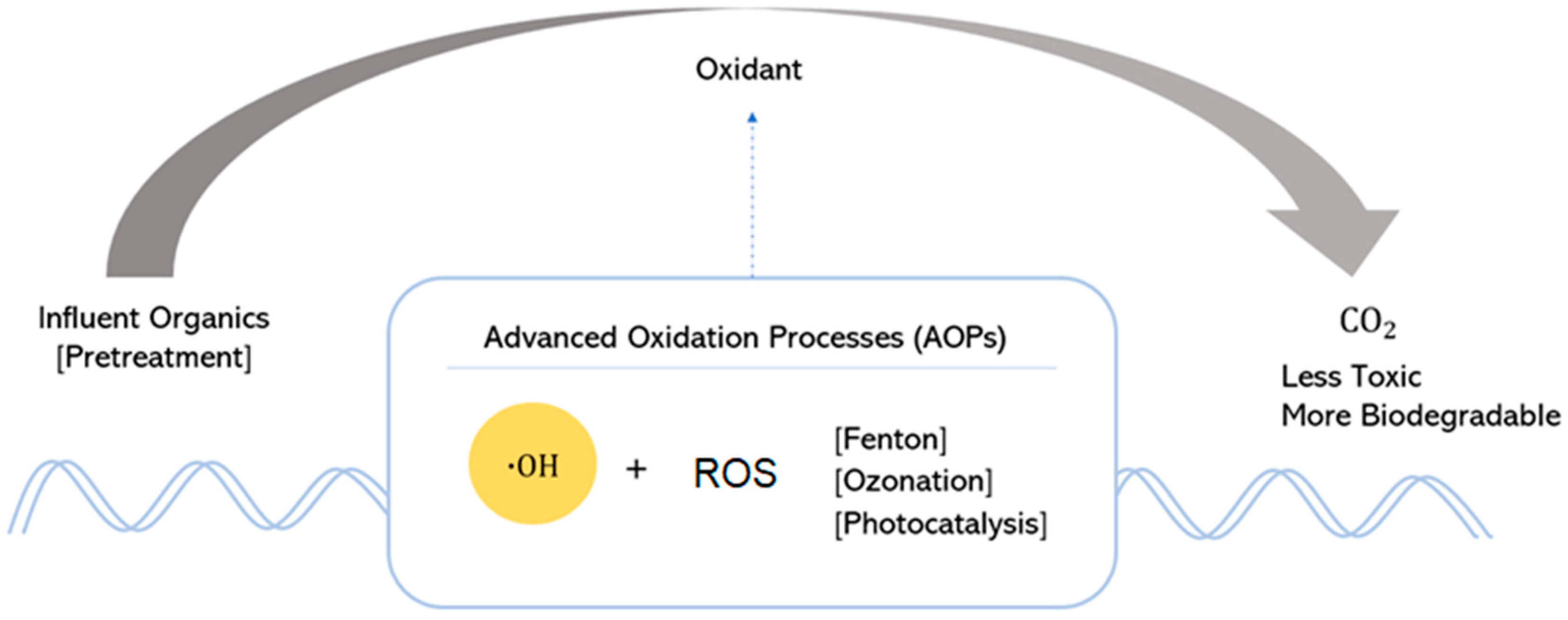
-
Low cost
-
High performance
-
Easy operation
-
Weak selectivity of waste product
Advanced oxidation processesProcesses based on the utilization of highly reactive chemical species that are efficient in oxidizing and degrading organic compounds.
-
High efficiency of pollutant degradation and possibility of complete mineralization in a short period
-
Include environmentally friendly, safe, and sustainable processes
-
Disinfection properties
-
High operation costs
-
In case of incomplete mineralization, post-treatment is required to remove toxicity
Table 3. The Different Methods of AOPs and Their ROS [26].
AOPsROSDescriptionAdvantagesDisadvantages
Fenton-based F e O 2 +
O H
Fe(OOH)+ FeO
OH
OOH
Involves   the   generation   of   hydroxyl   radicals   via   the   reaction   of   hydrogen   peroxide   ( H 2 O 2 ) with iron salts (typically ferrous iron, Fe2+).
-
Enhanced biodegradability
-
Reduced toxicity
-
No energy input
-
Utilization of Fe ions and UV light/electric potential
-
The adaptability of Fe ions to interact with contaminants
-
High chemical consumption
-
Requirement of acidic pH
-
No complete mineralization
-
Slow reaction rate
-
Sludge byproducts containing metal
-
Cost of preparing Fenton reagent
Ozonation O H
H O 2
O2•−
O3•−
Ozone   ( O 3 )   molecules   decompose   in   water ,   producing   hydroxyl   radicals   ( O H )   and   other   ROS   such   as   sin glet   oxygen   ( O 2 1 ), which are highly reactive and capable of oxidizing contaminants.
-
No sludge generation
-
Enhanced biodegradability
-
Reduced toxicity
-
High reaction rate
-
More stable byproducts
-
Effective in pre-treatment for water purification
-
Comparatively inadequate performance in removing COD
-
High cost
-
Self-decomposition
-
Low solubility in water
-
Formation of genotoxic compounds
Electrochemical O H Utilizes electrodes immersed in water to induce oxidation and reduction reactions, facilitating the breakdown of organic contaminants and pathogens.
-
Easy to handle
-
Simplicity in operation
-
In situ generation of H2O2
-
Generation of sludge in some cases
-
Solution pH decreases at the end of the reaction
Photocatalysis O H   O 2
O 2 1
OOH
Absorbs   energy   to   excite   electrons   ( e )   with   strong   reducing   capacity   and   produce   holes   ( h + ) with significant oxidizing potential.
-
Ability to activate the catalyst when exposed to sunlight
-
Total mineralization of pollutants is achievable
-
Accessibility of fairly inexpensive catalysts, like TiO2
-
The shading effect of the catalyst, TiO2, on the light that reaches the pollutant
-
UV light required for activation
-
Improper catalyst selection
-
Radiation wavelength requires high operational costs
Table 4. Metal Oxide Morphology with Different Materials.
MorphologyMetal OxideMethodsPrecursorsExperimental
Conditions
SizeSurface Area (m2/g)Ref.
NanosphereZnOSolvothermalZn(CH3COO)2∙2H2O, AgNO3T = 160 °C
pH = 9.3
Diameter: 400 nm to 500 nm [62]
NH3 evaporationZn(NO3)2, NH4OHEvaporation = 50 °C
Precipitation = 125 °C
pH = 5.4–8.0
Diameter: 80 nm to 130 nm66.12[63]
TiO2HydrothermalTi(SO4)2, glucoseT = 180 °CDiameter: 300 nm to 500 nm76[64]
CuOUltrasound-assistedCu(CH3COO).2H2O, CH4ON2T = 80 °C
pH = 5.94 to 5.08
Diameter: 400 nm to 500 nm59.60[65]
NanorodsZnOHydrothermal(Zn(Ac)2), Ce(NO3)2.6H2OT = 130 °C
pH = 10
Length: 900 nm to 3 μm
Diameter: 70 nm to 85 nm
2.8[66]
HydrothermalZn(Ac)2.2H2O, Ni(Ac)2.4H2OT = 140 °CDiameter: 15 nm to 20 nm
Length: 150 nm to 400 nm
[67]
Hydrothermal(CH3CO2)2Zn·2H2O, (CH3CO2)2Co·4H2OT = 220 °CLength: 0.2 μm to 1.5 μm
Diameter: 90 nm
[41]
Ultrasound-assistedZn(CH3COO)2.2H2O, NaOH, ionic liquid [C4mim][Tf2N]T = 25 °CDiameter: 20 nm
Length: 50 nm to 100 nm
49.93[68]
CuOHydrothermalCu(NO3)2·2.5H2O, C6H12N4Low temperatureLength: 10 nm to 20 nm
Diameter: -
[69]
WO3Wet chemicalsNa2WO4.2H2O T = 180 °C
pH = 1.5
Length: 2 μm
Diameter: 2 μm
[70]
TiO2HydrothermalTi(OBu)4, KMnO4, HCl, isopropanolT = 80 °C
pH = 1.3
[71]
HydrothermalTiO2, NaOHT = 150 °C
Calcination = 600 °C
pH = 6.5
Diameter: 7 nm49.6 to 64.2[42]
HydrothermalNa2O7Ti3 T = 180 °C
pH = 0, 0.5, 1, 2, 7
rpm = 0, 500, 900
Diameter: 5 nm
Length: 100 nm to 1200 nm
26.67 to 109.81[72]
NanowiresZnOHydrothermalZinc foil, C2H8N2 T = 170 °CLength: 3 um
Diameter: 120 nm
[73]
Solvothermal(Zn(Ac)2·2H2O), (La(NO3)3·6H2O)T = 150 °C
Alkaline condition
Diameter: 15 nm to 25 nm [74]
HydrothermalZnSO4, NH4ClT = 60 °C
pH = 11.7
Diameter: 50 nm to 200 nm
Length: 5 μm to 6 μm
[75]
WO3Solvothermal
Thermal reaction
WCl6, cyclohexanol, NH3 pure gasT = 200 °C (solvothermal)
923 K (thermal reaction), 400 sccm min−1
Diameter: 5 nm151[76]
TiO2HydrothermalTiO2, NaOH, KOH, NH4OHT = 180 °C
Neutral pH
Diameter: 25 nm to 30 nm
Length: 54.35 nm
[77]
HydrothermalTiO2, NaOHHydrothermal = 150 °C
Calcination = 500 °C
pH = 6.5
Diameter: 6 nm90.5 to 111.1[42]
HydrothermalTiO2 P25, NaOH/KOH/NH3Hydrothermal = 180 °C
Calcination = 450 °C
pH = 7
Diameter: 5 nm to 20 nm
Length: 74 nm to 367 nm
143.42 to 228.34[78]
NanotubeZnOCo-precipitation(Zn(CH3COO)2·2H2O, Cu(CH3COO)2·H2O, (HOCH2CH2)3N T = 92 °C
pH = 6.5
Length: 2.1 μm
Diameter: 0.21 μm
[79]
HydrothermalZnCl2, NH3T = 95 °CDiameter: 250 nm
Length: 500 nm
[80]
TiO2ElectrochemicalTiVoltage = 20–50 VLength: -
Diameter: 40 nm to 75 nm
[81]
HydrothermalTiO2, NaOHHydrothermal = 150 °C
Calcination = 400 °C
pH = 6.5
Outer diameter: 9 nm to 15 nm
Inner diameter: 4 nm to 7 nm
146.3 to 189.6[42]
CuOMicrowave-assistedCu(CH3COO)2.H2OCalcination = 100 °C
pH = 7
Mean crystallite size: 14 nm55.90[82]
SnO2HydrothermalSnCl2⋅2H2O, HClT = 200 °C
pH = 7
Diameter: 6.6 nm61.99[83]
NanoflowerZnOMicrowave-assistedZn(CH3COO)2.2H2O, KOHT = 180 °CLength: 600 nm to 800 nm Diameter: 150 nm to 200 nm31.75[84]
HydrothermalZn(Ac)2·2H2O, SDST = 100 °CDiameter: 10 μm to 12 μm22.7[85]
Sol–gelZn(NO3)2·6H2O, citric acidT = 100 °C
pH = 14
Diameter: 2 μm to 3 μm11.05[86]
TiO2HydrothermalTi(OBu)4, acetic acid T = 140 °CLength: 10 nm
Diameter: 4.5 μm
79.2[87]
HydrothermalTi, NaOH, H2O2, HNO3T = 150 °C
pH = 7
-134.7[88]
HydrothermalTi(OBu)4, HClT = 180 °CDiameter: 800 nm68 to 185[89]
CuOMicrowave-assistedCu(CH3COO)2.
H2O, NaOH, HMT
T = 90 °CMean crystallite size: 12 nm65.34[82]
Table 5. Synthesis of Carbon dots with Top-down and Bottom-up Approach and Precursors.
MethodCarbon SourcesApplicationRef
Laser ablationGraphite [128]
Polyvinylpyrrolidone (PVP)
Dimethylsulfoxide (DMSO) [129]
UltrasonicationCitric acidRemoval of methylene blue[130]
Elettaria cardamomumRemoval of methylene blue and Congo red[131]
ElectrochemicalGraphiteRemoval of tetracycline[132]
Microwave-assistedEgg shellRemoval of methylene blue[133]
D-glucoseRemoval of methyl orange[134]
Sugar cane juiceRemoval of methylene blue[134]
Citric acidRemoval of ciprofloxacin[135]
HydrothermalWaste frying oilRemoval of methylene blue[136]
Orange peelsRemoval of naphthol black[137]
Lemon peel wasteRemoval of methylene blue[138]
Citric acidRemoval of gemfibrozil[139]
L-ascorbic acidRemoval of levofloxacin[10]
Citric acidRemoval of tetracycline[140]
Disclaimer/Publisher’s Note: The statements, opinions and data contained in all publications are solely those of the individual author(s) and contributor(s) and not of MDPI and/or the editor(s). MDPI and/or the editor(s) disclaim responsibility for any injury to people or property resulting from any ideas, methods, instructions or products referred to in the content.

