
Metal-Assisted Injection Spinning of Ultra Strong Fibers from Megamolecular LC Polysaccharides

 ,
, 
Abstract
1. Introduction
2. Experimental
2.1. Materials
Extraction of Sacran
2.2. Injection Spinning
2.2.1. Preparation of Sacran Solution
2.2.2. Preparation of Metal Ions
2.2.3. Fabrication of Uniaxially Oriented Sacran Complex Fiber
2.3. Measurement
2.3.1. Mechanical Properties
2.3.2. Degree of Swelling
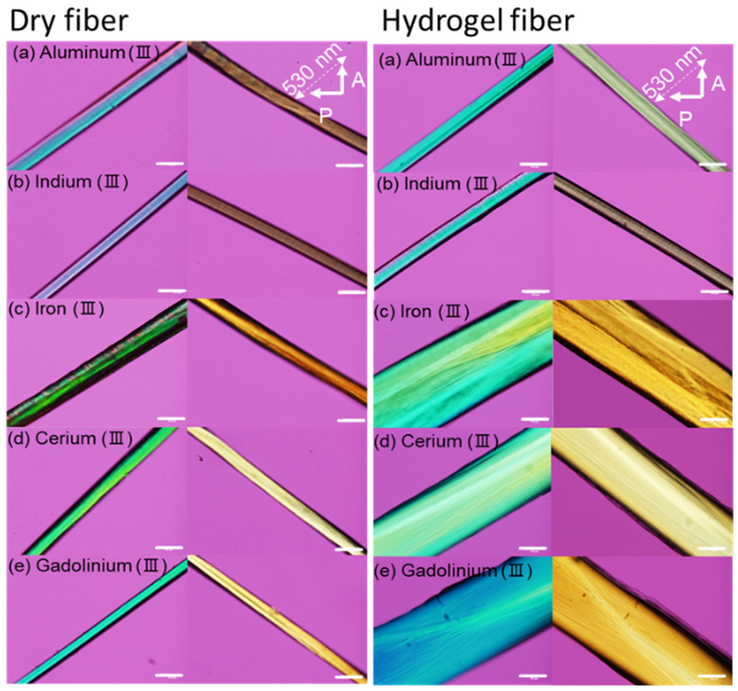
2.3.3. Observation of Orientation of Sacran Complex Fibers Using Crossed-Polarizing Microscope (PLM)
2.3.4. Observation of Sacran Complex Fibers Morphology Using Scanning Electron Microscope (SEM)
3. Results and Discussion
3.1. Fiber Spinning
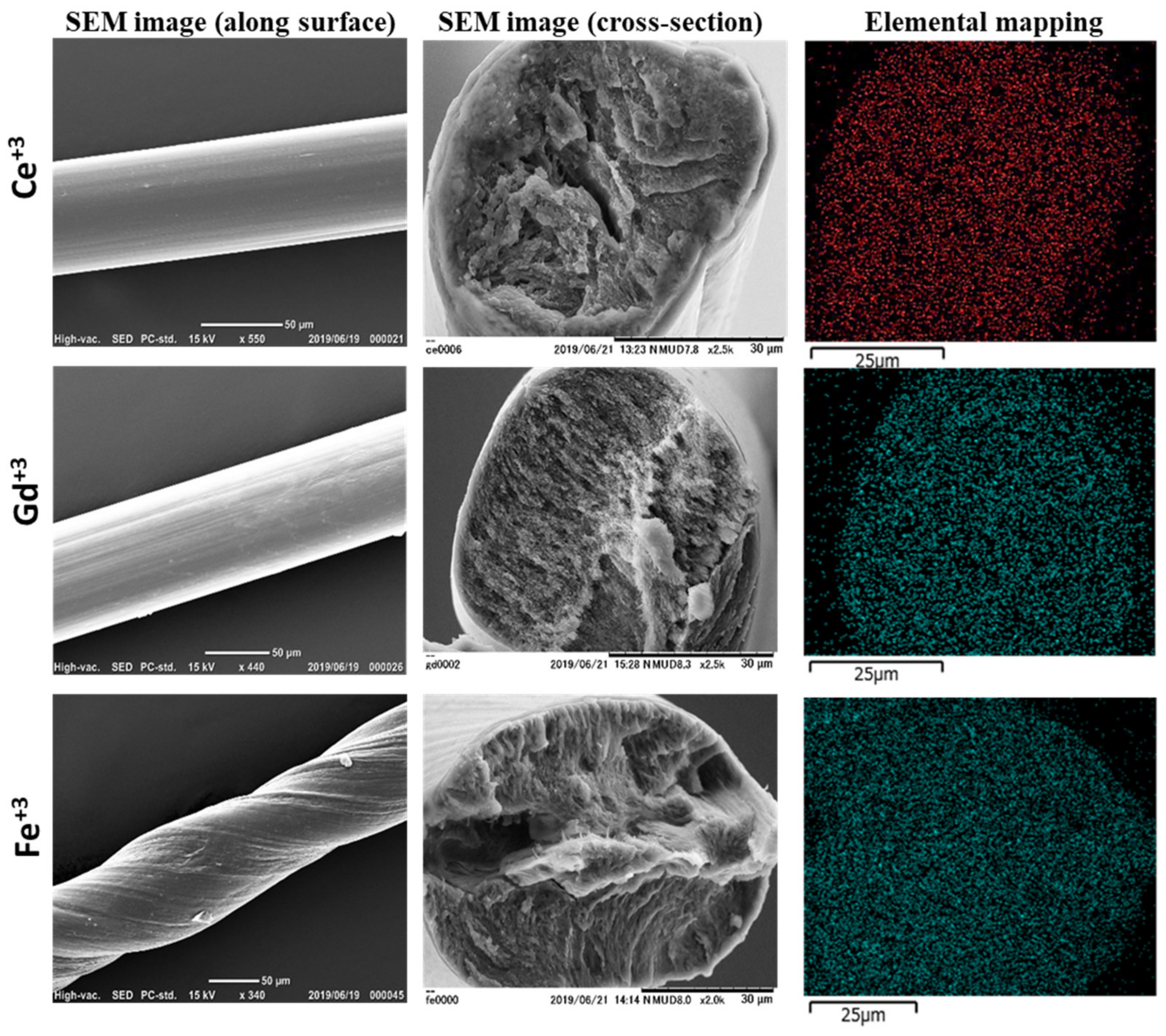
3.2. Structures
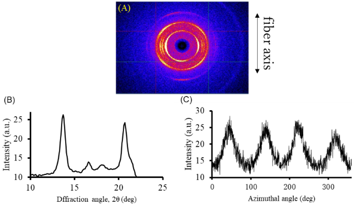
3.3. Structural Orientation
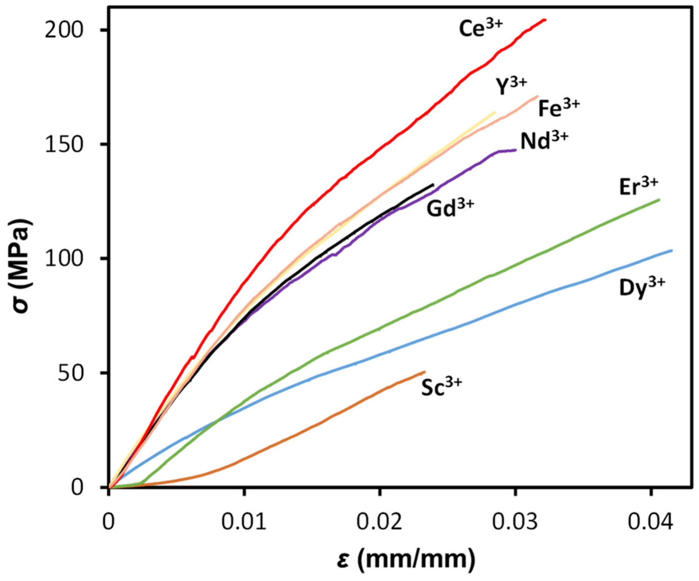
3.4. Mechanical Properties
3.5. Hydrogels
4. Conclusions
Supplementary Materials
Author Contributions
Funding
Institutional Review Board Statement
Data Availability Statement
Conflicts of Interest
References
- Beaumont, M.; Tran, R.; Vera, G.; Niedrist, D.; Rousset, A.; Pierre, R.; Shastri, V.P.; Forget, A. Hydrogel-Forming Algae Polysaccharides: From Seaweed to Biomedical Applications. Biomacromolecules 2021, 22, 1027–1052. [Google Scholar] [CrossRef] [PubMed]
- Delbianco, M.; Seeberger, P.H. Materials science based on synthetic polysaccharides. Mater. Horiz. 2020, 7, 963–969. [Google Scholar] [CrossRef]
- Darabi, S.; Hummel, M.; Rantasalo, S.; Rissanen, M.; Månsson, I.; Hilke, H.; Hwang, B.; Skrifvars, M.; Hamedi, M.M.; Sixta, H.; et al. Green Conducting Cellulose Yarns for Machine-Sewn Electronic Textiles. ACS Appl. Mater. Interfaces 2020, 12, 56403–56412. [Google Scholar] [CrossRef] [PubMed]
- Ma, Y.; You, X.; Rissanen, M.; Schlapp-Hackl, I.; Sixta, H. Sustainable Cross-Linking of Man-Made Cellulosic Fibers with Poly(carboxylic acids) for Fibrillation Control. ACS Sustain. Chem. Eng. 2021, 9, 16749–16756. [Google Scholar] [CrossRef]
- Li, H.; Kruteva, M.; Dulle, M.; Wang, Z.; Mystek, K.; Ji, W.; Pettersson, T.; Wågberg, L. Understanding the Drying Behavior of Regenerated Cellulose Gel Beads: The Effects of Concentration and Nonsolvents. ACS Nano 2022, 16, 2608–2620. [Google Scholar] [CrossRef] [PubMed]
- Chamas, A.; Moon, H.; Zheng, J.; Qiu, Y.; Tabassum, T.; Jang, J.H.; Abu-Omar, M.; Scott, S.L.; Suh, S. Degradation Rates of Plastics in the Environment. ACS Sustain. Chem. Eng. 2020, 8, 3494–3511. [Google Scholar] [CrossRef]
- Deralia, P.K.; du Poset, A.M.; Lund, A.; Larsson, A.; Ström, A.; Westman, G. Oxidation Level and Glycidyl Ether Structure Determine Thermal Processability and Thermomechanical Properties of Arabinoxylan-Derived Thermoplastics. ACS Appl. Bio Mater. 2021, 4, 3133–3144. [Google Scholar] [CrossRef] [PubMed]
- Puanglek, S.; Kimura, S.; Enomoto-Rogers, Y.; Kabe, T.; Yoshida, M.; Wada, M.; Iwata, T. In vitro synthesis of linear α-1,3-glucan and chemical modification to ester derivatives exhibiting outstanding thermal properties. Sci. Rep. 2016, 6, 30479. [Google Scholar] [CrossRef] [PubMed]
- Degli Innocenti, F.; Breton, T. Intrinsic Biodegradability of Plastics and Ecological Risk in the Case of Leakage. ACS Sustain. Chem. Eng. 2020, 8, 9239–9249. [Google Scholar] [CrossRef]
- Plucinski, A.; Lyu, Z.; Schmidt, B.V.K.J. Polysaccharide nanoparticles: From fabrication to applications. J. Mater. Chem. B 2021, 9, 7030–7062. [Google Scholar] [CrossRef]
- Jin, L.; Bai, R. Mechanisms of Lead Adsorption on Chitosan/PVA Hydrogel Beads. Langmuir 2002, 18, 9765–9770. [Google Scholar] [CrossRef]
- Chen, J.; Peng, Q.; Peng, X.; Han, L.; Wang, X.; Wang, J.; Zeng, H. Recent Advances in Mechano-Responsive Hydrogels for Biomedical Applications. ACS Appl. Polym. Mater. 2020, 2, 1092–1107. [Google Scholar] [CrossRef]
- Kim, D.; Kang, S.M. Red Algae-Derived Carrageenan Coatings for Marine Antifouling Applications. Biomacromolecules 2020, 21, 5086–5092. [Google Scholar] [CrossRef] [PubMed]
- Okajima, M.K.; Higashi, T.; Asakawa, R.; Mitsumata, T.; Kaneko, D.; Kaneko, T.; Ogawa, T.; Kurata, H.; Isoda, S. Gelation Behavior by the Lanthanoid Adsorption of the Cyanobacterial Extracellular Polysaccharide. Biomacromolecules 2010, 11, 3172–3177. [Google Scholar] [CrossRef] [PubMed]
- Okajima, M.K.; Miyazato, S.; Kaneko, T. Cyanobacterial Megamolecule Sacran Efficiently Forms LC Gels with Very Heavy Metal Ions. Langmuir 2009, 25, 8526–8531. [Google Scholar] [CrossRef] [PubMed]
- Fujishiro, T.; Ogawa, T.; Matsuoka, M.; Nagahama, K.; Takeshima, Y.; Hagiwara, H. Establishment of a Pure Culture of the Hitherto Uncultured Unicellular Cyanobacterium Aphanothece sacrum, and Phylogenetic Position of the Organism. Appl. Environ. Microbiol. 2004, 70, 3338–3345. [Google Scholar] [CrossRef] [PubMed]
- Okajima, M.K.; Bamba, T.; Kaneso, Y.; Hirata, K.; Fukusaki, E.; Kajiyama, S.; Kaneko, T. Supergiant Ampholytic Sugar Chains with Imbalanced Charge Ratio Form Saline Ultra-absorbent Hydrogels. Macromolecules 2008, 41, 4061–4064. [Google Scholar] [CrossRef]
- Mitsumata, T.; Miura, T.; Takahashi, N.; Kawai, M.; Okajima, M.K.; Kaneko, T. Ionic state and chain conformation for aqueous solutions of supergiant cyanobacterial polysaccharide. Phys. Rev. 2013, 87, 042607. [Google Scholar] [CrossRef] [PubMed]
- Okajima, M.K.; Nakamura, M.; Mitsumata, T.; Kaneko, T. Cyanobacterial Polysaccharide Gels with Efficient Rare-Earth-Metal Sorption. Biomacromolecules 2010, 11, 1773–1778. [Google Scholar] [CrossRef]
- Okeyoshi, K.; Okajima, M.K.; Kaneko, T. The cyanobacterial polysaccharide sacran: Characteristics, structures, and preparation of LC gels. Polym. J. 2020, 53, 81–91. [Google Scholar] [CrossRef]
- Doi, M.; Sagawa, Y.; Tanaka, T.; Mizutani, T.; Okano, Y.; Masaki, H. Defensive Effects of a Unique Polysaccharide, Sacran, to Protect Keratinocytes against Extracellular Stimuli and Its Possible Mechanism of Action. Biol. Pharm. Bull. 2018, 41, 1554–1560. [Google Scholar] [CrossRef] [PubMed]
- Zhang, Y.-Q.; Tanaka, T.; Shibayama, M. Super-absorbency and phase transition of gels in physiological salt solutions. Nature 1992, 360, 142–144. [Google Scholar] [CrossRef]
- Puluhulawa, L.E.; Joni, I.M.; Mohammed, A.F.A.; Arima, H.; Wathoni, N. The Use of Megamolecular Polysaccharide Sacran in Food and Biomedical Applications. Molecules 2021, 26, 3362. [Google Scholar] [CrossRef]
- Phan, T.D.; Mizutani, G.; Li, Y.; Budpud, K.; Okeyoshi, K.; Okajima, M.; Kaneko, T. Sum-Frequency Generation and Scanning Electron Microscope Studies on Second-Harmonic Generation Active Structures of Sacran Aggregates. E-J. Surf. Sci. Nanotechnol. 2022, 20, 98–106. [Google Scholar] [CrossRef]
- Gong, J.P.; Katsuyama, Y.; Kurokawa, T.; Osada, Y. Double-Network Hydrogels with Extremely High Mechanical Strength. Adv. Mater. 2003, 15, 1155–1158. [Google Scholar] [CrossRef]
- Haraguchi, K.; Takehisa, T.; Fan, S. Effects of Clay Content on the Properties of Nanocomposite Hydrogels Composed of Poly(N-isopropylacrylamide) and Clay. Macromolecules 2002, 35, 10162–10171. [Google Scholar] [CrossRef]
- Sakai, T.; Akagi, Y.; Matsunaga, T.; Kurakazu, M.; Chung, U.; Shibayama, M. Highly Elastic and Deformable Hydrogel Formed from Tetra-arm Polymers. Macromol. Rapid Commun. 2010, 31, 1954–1959. [Google Scholar] [CrossRef]


| Metal Ions | E (GPa) | σ (GPa) | ε (mm/mm) | U (kJ/m3) |
|---|---|---|---|---|
| Sc+3 | 2.0 ± 0.4 | 0.05 ± 0.01 | 0.03 ± 0.01 | 0.73 ± 0.24 |
| Dy+3 | 2.8 ± 0.4 | 0.11 ± 0.03 | 0.04 ± 0.01 | 2.06 ± 0.58 |
| Er+3 | 2.9 ± 0.4 | 0.12 ± 0.05 | 0.04 ± 0.02 | 2.17 ± 0.86 |
| Nd+3 | 4.7 ± 1.8 | 0.15 ± 0.06 | 0.03 ± 0.02 | 2.76 ± 0.91 |
| Gd+3 | 5.2 ± 0.9 | 0.14 ± 0.03 | 0.02 ± 0.01 | 2.15 ± 0.89 |
| Y+3 | 5.4 ± 0.8 | 0.15 ± 0.05 | 0.02 ± 0.01 | 3.69 ± 0.81 |
| Fe+3 | 5.4 ± 0.4 | 0.17 ± 0.04 | 0.03 ± 0.01 | 3.91 ± 0.41 |
| Ce+3 | 5.4 ± 0.6 | 0.19 ± 0.05 | 0.03 ± 0.01 | 5.55 ± 2.06 |
Disclaimer/Publisher’s Note: The statements, opinions and data contained in all publications are solely those of the individual author(s) and contributor(s) and not of MDPI and/or the editor(s). MDPI and/or the editor(s) disclaim responsibility for any injury to people or property resulting from any ideas, methods, instructions or products referred to in the content. |
© 2024 by the authors. Licensee MDPI, Basel, Switzerland. This article is an open access article distributed under the terms and conditions of the Creative Commons Attribution (CC BY) license (https://creativecommons.org/licenses/by/4.0/).
Share and Cite
Ali, M.A.; Singh, M.; Zhang, S.; Kaneko, D.; Okajima, M.K.; Kaneko, T. Metal-Assisted Injection Spinning of Ultra Strong Fibers from Megamolecular LC Polysaccharides. Polymers 2024, 16, 1099. https://doi.org/10.3390/polym16081099
Ali MA, Singh M, Zhang S, Kaneko D, Okajima MK, Kaneko T. Metal-Assisted Injection Spinning of Ultra Strong Fibers from Megamolecular LC Polysaccharides. Polymers. 2024; 16(8):1099. https://doi.org/10.3390/polym16081099
Chicago/Turabian StyleAli, Mohammad Asif, Maninder Singh, Shuo Zhang, Daisaku Kaneko, Maiko Kaneko Okajima, and Tatsuo Kaneko. 2024. "Metal-Assisted Injection Spinning of Ultra Strong Fibers from Megamolecular LC Polysaccharides" Polymers 16, no. 8: 1099. https://doi.org/10.3390/polym16081099
APA StyleAli, M. A., Singh, M., Zhang, S., Kaneko, D., Okajima, M. K., & Kaneko, T. (2024). Metal-Assisted Injection Spinning of Ultra Strong Fibers from Megamolecular LC Polysaccharides. Polymers, 16(8), 1099. https://doi.org/10.3390/polym16081099