Article

Differentially Expressed Transcription Factors during Male and Female Cone Development in Pinus halepensis

1
Institute of Plant Sciences, Agricultural Research Organization, Volcani Center, Rishon LeZion 7505101, Israel
2
The Robert H. Smith Institute of Plant Sciences and Genetics in Agriculture, The Hebrew University of Jerusalem, Rehovot 76100, Israel
3
Department of Plant and Environmental Sciences, Weizmann Institute of Science, Rehovot 76100, Israel
*
Author to whom correspondence should be addressed.
Agronomy 2022, 12(7), 1588; https://doi.org/10.3390/agronomy12071588
Submission received: 1 February 2022 / Revised: 18 May 2022 / Accepted: 28 June 2022 / Published: 30 June 2022
(This article belongs to the Special Issue Flowering and Flower Development in Plants)

Abstract

:
The molecular regulation of induction and development of reproductive organs is well established in angiosperms, while it is slowly accumulating in gymnosperms. Here, we conducted comparative transcriptomic profiles at different stages of male and female cone development in Pinus halepensis. Our aim was to reveal transcription factor encoding genes involved in reproductive induction and development. For accurate developmental stage identification, histological analysis preceded the molecular analysis. The flowering induction genes FT/TFL were expressed mainly at the late developmental stages of the male cone, suggesting involvement in vegetative bud dormancy instead of flowering induction. The male cone development was associated with the expression of the C-class PhMADS2, and the B-class genes PhDAL11 and PhDAL13, while the female cone development was associated with the expression of the C-class MADS1 and DAL14 genes. This study adds valuable knowledge to the profile of transcription factors and MADS-box genes regulating cone development in gymnosperms.

1. Introduction

Reproduction in plants includes induction and organ differentiation stages. The molecular regulation of these processes is well characterized in angiosperms compared to gymnosperms [1,2,3,4,5]. Yet, the current genomic tools permit a slow bridging of this gap and provide valuable knowledge to understand cone development in conifers.
Flowering induction in angiosperms is regulated by transcription factors, including LEAFY (LFY), APETALA1 (AP1), CAULIFLOWER (CAL), and FRUITFUL (FUL) [1]. Other key regulators are genes belonging to the FLOWERING LOCUS T (FT)/TERMINAL FLOWER 1 (TFL1) family [6,7]. The FT protein (also known as florigen) is produced in the leaves, then transported to the meristem, where it forms a complex with the FLOWERING LOCUS D (FD) transcription factor, to induce flowering. In contrast, TFL1 is produced in the meristem and forms a complex with FD that inhibits the expression of the flowering-promoting genes LFY and the MADS-box gene, APETALA1 (AP1), preventing flowering induction [8,9]. SUPPRESSOR OF OVEREXPRESSION OF CO 1 (SOC1), encoding a MADS-box transcription factor, is another key gene in flowering induction [2,10,11]. The photoperiodic regulation of SOC1 is mediated by CONSTANS (CO), mainly through FT, which acts as an activator. SOC1 directly activates LFY and AGL24, a MADS-box transcription factor [2]. Genes encoding basic helix-loop-helix-type (bHLH) transcription factors are also related to flowering induction. These include FLOWERING BHLH (FBH1-4), which together with TEOSINTE BRANCHED 1/CYCLOIDEA/PROLIFERATING CELL NUCLEAR ANTIGEN FACTOR (TCP) positively regulate CO [3].
Studies on induction of reproductive organs in gymnosperms, specifically in the Pinaceae, indicate similar and dissimilar mechanisms to angiosperms [12]. Contrary to the one copy of LFY in angiosperms, gymnosperms possess two copies; LFY and Needly (NLY) [13,14,15]. Both are expressed in vegetative buds, male, and female developing cones [14,16,17,18]. However, the Picea abies LFY (PaLFY) mRNA levels are slightly increased prior to the female cone setting in the early-cone-setting mutant acrocona [18]. Similarly, the Pinus caribaea LFY homolog, PcLFY, is found to be expressed dominantly in the female cone in early developmental stages [19]. Furthermore, both LFY and NLY from Pinus radiata as well as LFY from P. caribaea largely rescued the Arabidopsis lfy mutant phenotype, suggesting a role in transition to the reproductive stage [15,17,19].
The gymnosperm homologous of FT/TFL1 was identified in Pinaceae [20,21]. The expression levels of the P. abies homologs PaFTL1 and PaFTL2 reached their maximum level before growth cessation, suggesting a role in dormancy regulation. Moreover, expression of FTL1 and FTL2 from P. abies and Pinus tabuliformis in Arabidopsis inhibited floral transition, demonstrating the functional homology of these genes to the FT/TFL1 in angiosperms [21,22]. A SOC1 homolog of P. abies, DEFICIENCE-AGAMOUS-LIKE 19 (DAL19) was highly expressed in the acrocona mutant prior to cone set, suggesting a role in flower induction similarly to SOC1 [18].
Following flower induction, the development of flower organs is regulated mainly by transcription factors from the MADS-box family [23]. The ABC model, later expanded to the ABCDE model, describes the interactions between gene groups that regulate the different floral organs development [5,24]. In Arabidopsis, overlapping expression of E-class genes, which include SEPALLATA1,2,3,4 (SEP1,2,3,4) and AGL6-like, and A-class genes, which include AP1 and APETALA2 (AP2), induce sepal formation; E-class together with B-class, which include APETALA3 (AP3) and PISTILLATA (PI), and A-class induce petal formation; E-class together with B- and C-class, which include AGAMOUS (AG) genes, form stamens; E- and C-class induce carpels, while E- together with C- and D-classes are responsible for ovule formation [5,25].
Many genes homologous to angiosperm floral organ determining genes were found in gymnosperms [5,21]. It appears that B-class homologous genes, including the P. radiata PrDGL and DAL11-13 from P. abies and P. tabuliformis, are expressed specifically in developing male cones [21,26,27]. Gymnosperm genes homologous to angiosperm C-class genes include PrMADS1-3 in P. radiata, as well as DAL2 in P. abies and P. tabuliformis [21,28,29]. The DAL2 homologs in other conifers such as Podocarpus reichei and Taxus globose exhibit female cone expression [30]. Interestingly, the DAL10 MADS-box gene from P. abies, which do not show homology to any known angiosperm gene, exhibits a male specific function similar to that of B-class genes [31]. The overexpression of the P. tabuliformis DAL10 homolog in Arabidopsis resulted in very early flowering induction, suggesting also a role in the transition to the reproductive phase in gymnosperms [21].
According to the gymnosperms model, overlapping C- and B-class gene expression results in male cone development, while the expression of only C-class genes causes female cone development. In both cases, the tetramers contain A class proteins [5]. A recent study revealed several physical interactions among MADS-box proteins in P. tabuliformis, supporting the hypothesis of the requirement for tetramerization of MADS-box proteins [21]. Furthermore, the large number of MADS-box homologous genes in conifers suggest complex regulatory interaction.
P. halepensis is a monoecious species (bearing male and female cones on the same tree), which begins its reproductive phase with the production of female cones approximately at the age of five years. Once the tree grows sufficiently in size, it begins to form male cones as well, usually one to two years later [32,33]. An early female cone development and a spatial distribution of the cones on a mature tree (female cones at the upper part and male cones at the middle and lower part, Supplementary Figure S1) facilitate outcrossing, ensuring high genetic diversity [32]. Female cone buds occur in a lateral position on a vegetative shoot bud. The female cone contains ovule-bearing scales (megasporophylls) attached to bracts and arranged spirally on a central axis, while the male cones are organized in clusters around a central axis (Figure 1). Vegetative to reproductive transition occurs in October–November depending on the provenance and environmental conditions, and cone development continues until March when pollination takes place [34].
To gain knowledge on the molecular regulation of the reproductive phase of P. halepensis, we analyzed differentially expressed genes encoding for transcription factors at different stages of male and female cones development. The transcriptomic analysis was accompanied by histological analysis to achieve an accurate developmental stage identification. We identified several TF families that were dominantly expressed in either female or male cones, and also showed the cone specific expression of several MADS-box genes.

2. Materials and Methods

2.1. Plant Material

Plant material was collected from Pinus halepensis trees that grow at the Volcani Center (31.9872, 34.8212, Rishon LeZion, Israel). For transcriptome analysis, samples (13–18 shoot apices from 3–5 trees) were collected every week from December 2019 to March 2020 between 10:00 and 11:00 a.m. to minimize the effect of the circadian rhythm. Each shoot apex was immediately cut longitudinally into two parts. One part was frozen in liquid nitrogen and stored at −80 °C for mRNA extraction and sequencing, while the other half was fixated in formaldehyde-acetic acid–alcohol (FAA, 10:5:50 in double-distilled water) solution for histological analysis. The aim of the histological analysis was to determine the developmental stage and meristem identity (male/female/vegetative) of each collected sample.
For candidate gene analysis, samples were collected in a similar way from October 2020 to February 2021.

2.2. Histological Analysis

Samples in FAA were put in a vacuum chamber for 15 min and were stored overnight at 4 °C. Following dehydration in an ethanol series (70, 80, 90, and 100%, 30 min each), the samples went through a gradual Histoclear solution change up to 100% Histoclear. Overnight incubation at room temperature with Paraplast chips (Leica, Wetzlar, Germany, Paraplast Plus) was followed by several hours of incubation at 42 °C. Dissolved pure paraffin was changed twice a day for four days at 62 °C oven. The samples were embedded in blocks, sectioned into 10 μm width longitudinal sections (using Leica, RM2245 Microtome), and mounted on slides, which were incubated overnight on a 40 °C plate. The sections were then stained with Fast Green and Safranin [35], observed in a light microscope (Nikon Eclipse Ni-E), and images were taken using DS-Ri2 camera (Nikon, Tokyo, Japan).

2.3. Sample Selection for RNA Sequencing

Following histological analysis, terminal buds were chosen for mRNA sequencing according to their developmental stage [36] (Table 1). All developmental stages were identified, except for stage 1 for vegetative, male, and female, which was absent (probably due to late sampling). For each stage, we included three biological replicates.

2.4. RNA Isolation, Library Preparation and Sequencing

The selected buds were ground with liquid nitrogen, using a mortar and pestle. Total RNA was extracted with SpectrumTM Plant Total RNA Kit (Sigma, St Louis, MO, USA) following the manufacturer’s instructions, and the RNA quantity was determined using a NanoDrop 1000 spectrophotometer (Thermo Fisher Scientific, Inc.). The RNA samples were sent to Macrogen Inc. (Amsterdam, the Netherlands) for the following sequencing procedure: RNA quality analysis using the 2200 TapeStation System (Agilent Technologies, Santa Clara, CA, USA), mRNA enrichment, library preparation using Illumina TruSeq RNA Library v2 kit, and 100 bp paired end sequencing by Illumina NovaSeq (Illumina, San Diego, CA, USA).

2.5. Transcriptome Analysis: Assembly and Differential Expression Analysis

A transcriptome database was de novo assembled using Trinity software (version: v2.1.1) [37] based on the sequences derived from the RNA sequencing. The transcript quantification from the RNA-Seq data was performed using the Bowtie2 aligner [38] and the expectation maximization method (RSEM) by estimating maximum likelihood expression levels [39] via the perl script align_and_estimate_abundance.pl with –est_method RSEM from Trinity protocol [40]. According to the sample correlation matrix (Supplementary Figure S2), three samples showed expression patterns which were not sufficiently correlated with their equivalent biological replicates. These samples (Male stage 3 replicate 1, Female stage 2 replicate 2, and Vegetative stage 1 replicate 1) were excluded from further analyses. Differential expression analyses were done using the edgeR R package [41].

2.6. Sequence Similarity and Functional Annotation

The merged transcriptome was used as a query term for a search of the NCBI non-redundant (nr) protein database that was carried out with the DIAMOND program [42]. Homologous sequences were also identified by searching the Swiss-Prot database with the BLASTx tool [43] and an E-value threshold of 10–5. The search results were imported into Blast2GO version 4.0 [44] for gene ontology (GO) assignments. Enzyme codes and KEGG pathway annotations were based on the mapping of GO terms to their enzyme codes. The KAAS tool (Kegg Automatic Annotation Server; http://www.genome.jp/tools/kaas/, accessed on 1 July 2021) was used for KEGG orthology and KEGG pathway assignments. Gene ontology enrichment analysis was carried out using the Blast2GO program based on Fisher’s exact test [45] with multiple testing correction of false discovery rate (FDR) [46]. The threshold was set as FDR with a corrected p-value of less than 0.05. Gene ontology analysis was done by comparing the GO terms in the test sample to the GO terms in a background reference. Every comparison had hundreds to thousands of DE genes. In order to focus on transcription factors, the differentially expressed (DE) genes were blasted against the transcription factors database PlantFTDB (http://planttfdb.gao-lab.org/, accessed on 1 September 2021). In addition, the homologs of the MADS-box genes were isolated from the identified DE transcription factors.

2.7. Phylogenetic Analysis

A phylogenetic tree of the MADS-box transcription factors was constructed with MEGA X [47] using the maximum likelihood method. In silico translated amino acid sequences were used for this analysis. One isoform was selected from each gene, according to the highest protein annotation score. The P. halepensis proteins were aligned using the MUCSLE algorithm [48] with MADS-box protein sequences from Arabidopsis (retrieved from The Arabidopsis Information Resource, TAIR) and other conifer MADS-box proteins (retrieved from UniProt, https://www.uniprot.org/, accessed on 1 September 2021). The conserved MADS and K domains together with the linker between them were used for the alignment. A thousand replicates were used for bootstrapping. The evolutionary history was inferred by using the maximum likelihood method and JTT matrix-based model [49]. Initial tree(s) for the heuristic search were obtained automatically by applying Neighbor-Join and BioNJ algorithms to a matrix of pairwise distances estimated using the JTT model, and then selecting the topology with a superior log likelihood value.

2.8. Quantitative Polymerase Chain Reaction (qPCR) Analysis

Candidate genes were analyzed by qPCR in samples that were collected in the second year (see above). Primers were designed to amplify 70–200 bp segment of each candidate gene (Supplementary Table S1). Each type of bud at each developmental stage was represented by three to four biological replicates, with two technical replicates for each. Since sample collection for the qPCR analysis started earlier in the second year than in the first year, it included earlier developmental stages; 1, 3 and 4 for female, and 2 and 3 for male. RNA was extracted as described above from all the samples and diluted equally to reach a concentration of 1000 ng/μL. DNA was degraded using RQ1 RNase-Free DNase (Promega, Madison, WI, USA). cDNA was synthesized using the qScript cDNA Synthesis Kit (QIAGEN, Hilden, Germany). Finally, qPCR was done using ABsolute Blue QPCR Mix, SYBR Green, ROX (Thermo Scientific™, Waltham, MA, USA). The expression level of the chosen genes was normalized according to the expression levels of the P. halepensis homologs of UPL6 (E3 ubiquitin-protein ligase) TRINITY_DN211436_c2_g2_i2. This gene showed little variation in expression levels between the different bud types, suggesting reliability for qPCR relative expression levels normalization. The relative gene expression was calculated according to the 2−ΔΔCT method [50], using the housekeeping gene CT values, and the average CT values of vegetative stage 1 tissues, defined as the control group. The derived values were converted to its logarithmic base 2 for stabilization of variances.

3. Results

3.1. Phenotypic Characterization of the Developmental Stages

To gain insights into the genes involved in reproduction in P. halepensis, we collected the three different bud types, vegetative, female and male at different developmental stages (Figure 1), for RNA-seq, and a year later for qPCR analysis as described in Material and Methods. The buds presented in Figure 1 and the histology analysis in Figure 2 represent all stages identified in this study.
At their early developmental stage, the vegetative, female and male closed buds look similar (Figure 1A,D,H). Soon after the buds elongate and swell, their different morphology becomes clearly visible (Figure 1B,E,I). The vegetative shoot develops needles, which further elongate (Figure 1C), and the female cone emerges through the top of the scales (Figure 1F,G), while the many small male cones emerge from the bud scales to be ready for pollen release (Figure 1J,K).

3.2. Histological Analysis

Histological analysis that was performed on half of each sample (see Materials and Method) revealed the developmental stages of the three different bud types (Figure 2). In early developmental stage, the lateral meristems of the vegetative bud are still not fully developed and enclosed within the bud scales, this stage was called “vegetative stage 1” (VS1). At stage 2 (VS2), the lateral meristems are apparent, and the developing brachyblasts (dwarf shoots) are growing outside the bud scales. At stage 3 (VS3), the bud is fully developed and the elongated needles are visible.
At stage 1 of the female cone developmental (FS1), a huge swollen structure with a few lateral primordial bracts appear, enclosed within the bud scales. At stage 2 (FS2), the bud is enlarged in size, the bract primordia are elongated, and the ovuliferous scale anlagen becomes visible. During stage 3 (FS3), developed bracts cover the developing ovuliferous scales. At stage 4 (FS4), the cone is fully developed with ovules, ready to be pollinated. The developmental stages presented in Figure 2 for the female cone are consistent with the previously defined stages [51].
The first stage of the male cone development is characterized by lateral microsporophyll primordia. In the second stage (MS2), a swollen structure, hidden within the bud scales, contains developing microsporophyll. At MS3, the cones are emerged from the bud scales, and the microsporophyll contains a microsporangium (pollen sac), with pollen mother cells surrounded by a tapetal layer. In the final stage of male cone development (MS4), the cone contains fully developed pollen grains ready to be carried by wind for pollination.

3.3. Transcriptome Analysis of the Three Bud Types at Early and Late Differentiation Stages

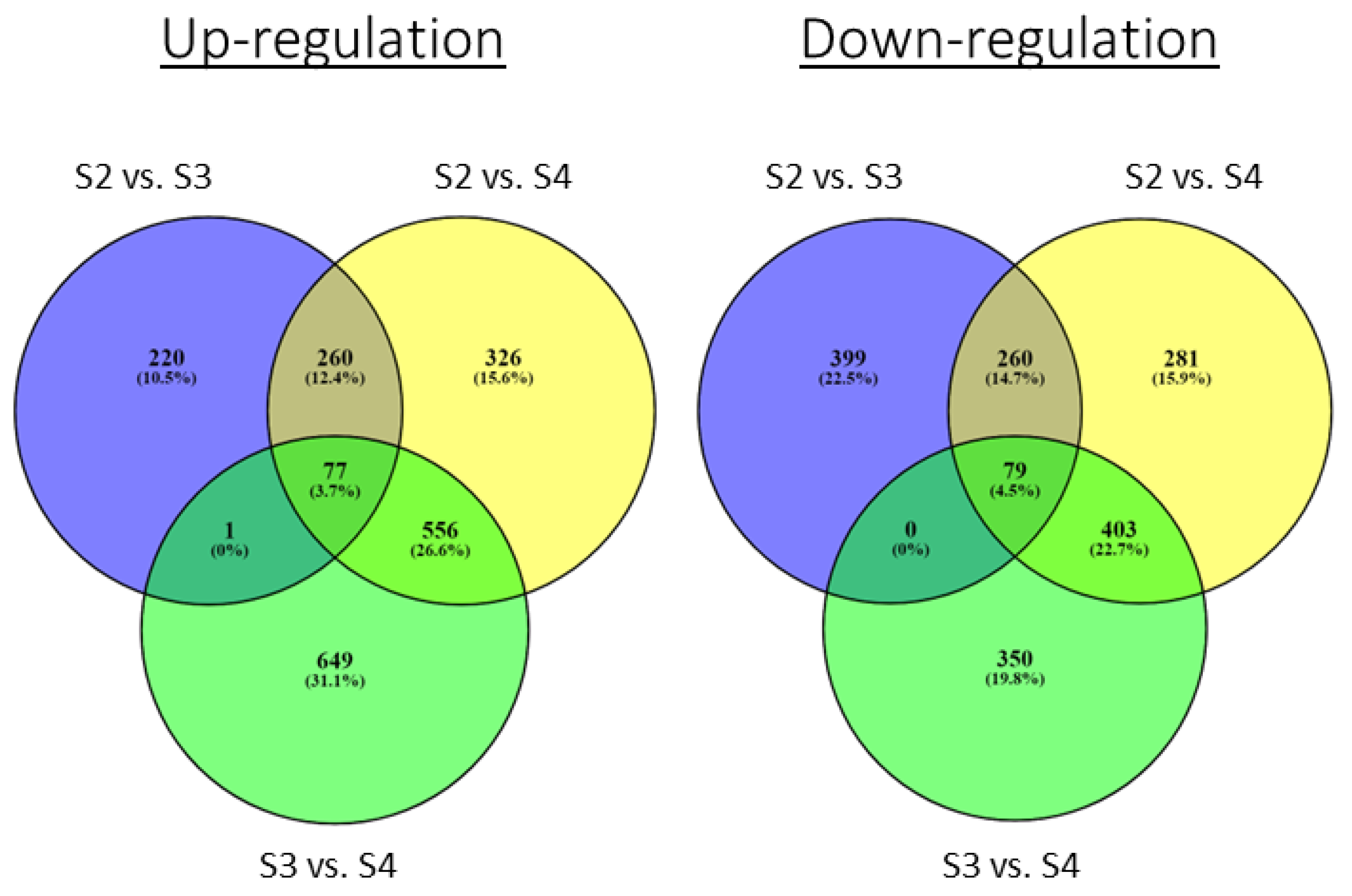
Based on the developmental stages determined by the histology analysis in the first year, we realized that we started our sampling a little late. Thus, the selected buds for RNA-seq represented stages 2 and 3 for vegetative bud (VS2 and VS3) and male cone (MS2 and MS3), and stages two to four for the female cone (FS2-FS4). Twenty-one cDNA libraries yielded an average of 44 million 100 bp paired-end reads, which were assembled using Trinity as described in Material and Methods. The transcriptome catalog contained 546,815 transcripts corresponding to 414,456 genes. The average contig length was 735.42 bp, with an N50 size of 1487 bp corresponding to a total length of 402 Mbp. Annotating the transcriptome catalog resulted in 81,521 contigs. Differential gene expression analyses between the three buds, both at early and late developmental stages, revealed that the number of DE genes between male and vegetative types is the highest, whereas between the female and the vegetative types is the lowest (Table 2, full list of genes in Supplementary Table S2). Moreover, more genes were DE at late developmental stages than in early developmental stages (Table 2), reflecting the advanced differentiation of each organ. Comparison between stage 2 and stage 3 of each bud type revealed that the vegetative bud exhibits the smallest number of DE between the two stages, whereas the male cones exhibited the largest number of DE genes. A similar number of genes were up- and down-regulated between stage 2 and stage 3 in all three bud types (Table 2). Shared expressed genes between the three developmental stages of the female cone are presented in Figure 3. It appears that 649 genes were uniquely up-regulated in developmental FS3 compared to FS4, suggesting that at the latter stage, some processes were completed.

3.4. Transcription Factors Families Involved in Early and Late Stages of Cone Development

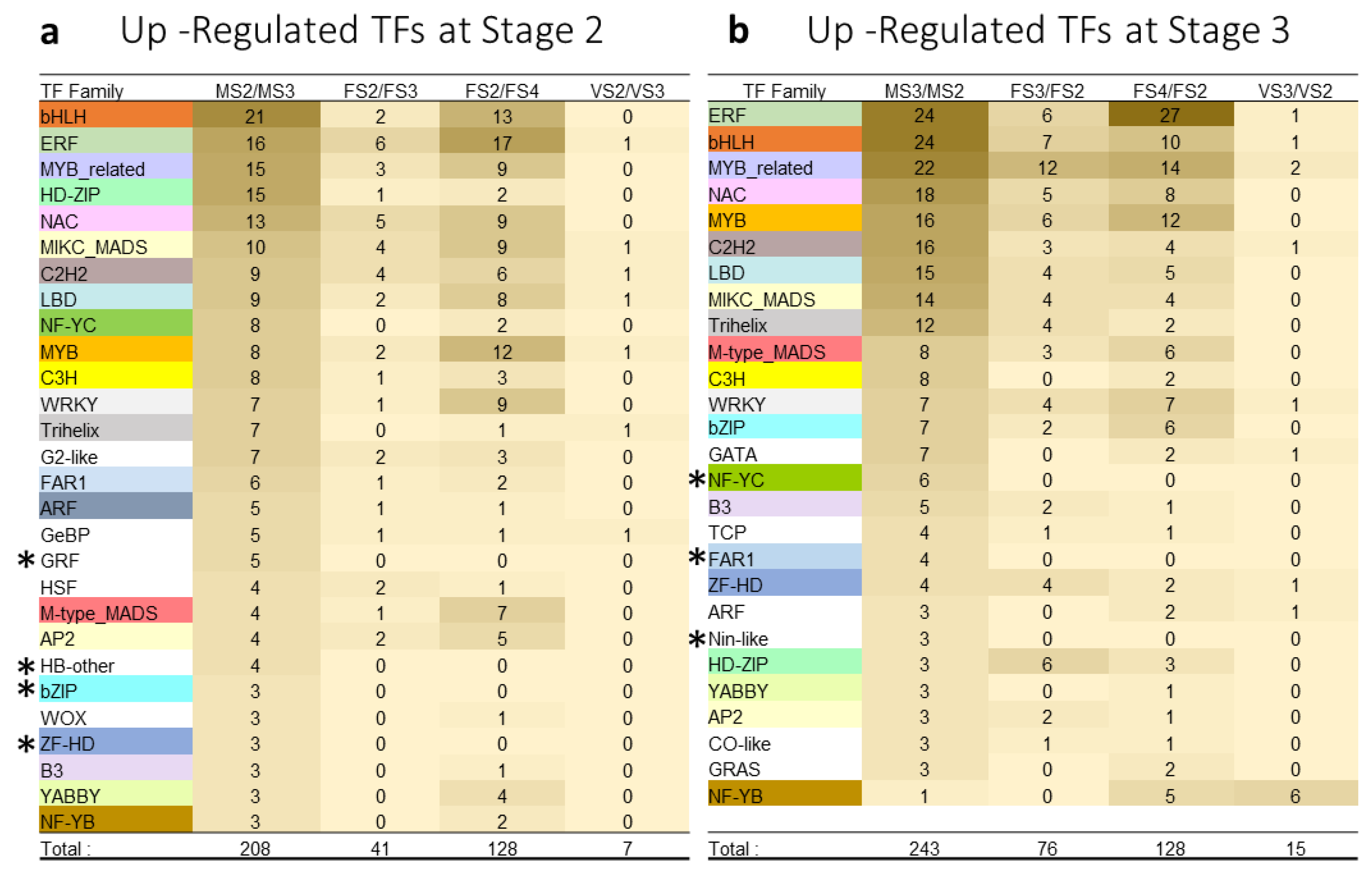
To identify transcription factors related to cone development, we blasted the DE genes against the transcription factors database PlantFTDB (http://planttfdb.gao-lab.org/, accessed on 1 September 2021) and grouped the genes into TF families (Figure 4). Included in Figure 4 are TF families with at least three members differentially expressed in at least one comparison. The number of TFs that were DE during the male cone development was dramatically higher than DE TFs during the female cone development (Figure 4). For female cone development, more TFs were up-regulated in the late than in the early developmental stages. The most represented TF families (>10 members in both stages) in the male cone were bHLH, ERF, MYB-related, NAC, and MIKC-MADS (Figure 4). The MYB-related family was highly represented in the late female stages, while the ERF, bHLH, and MYB families were highly represented in the most advanced developmental stage, FS4. Interestingly, the HD-ZIP family was dominantly expressed at MS2 with 15 family members. In addition, male cone development exhibited TFs (GRF, HB-other, bZIP, ZF-HD, NF-YC, FAR1, and Nin-like) that were not DE during the female or vegetative bud development. MICK_MADS and M-type_MADS were enriched in the early and late stages of male and female cone development (Figure 4).

3.5. Differentially Expressed TFs Involved in Flowering Induction

The LFY homolog gene (TRINITY_DN212345_c0_g2) exhibiting 97.5% similarity to the P. radiata NLY (Accession U76757) had higher expression level in MS2 compared to MS3 (log2FC = 2.48, Supplementary Table S2). Except for that, LFY homologs did not show differential expression between meristem types or developmental stages.
Two FT/TFL1 homologous contigs (TRINITY_DN197941_c0_g2 and TRINITY_DN197941_c0_g3) were up-regulated in the male cones at S3, compared to vegetative buds at S3 (log2FC = 10.33 and 5.75, respectively), and also up-regulated in MS3 compared to MS2 (log2FC = 4.8 and 10.5, respectively). Both contigs exhibit high similarity (93% and 97%, respectably) to the Pinus armandii FT/TFL protein. TRINITY_DN197941_c0_g3 (FT/TFL1) was found to decline in female cones as development proceeded. In addition, a MOTHER OF FT and TFL1-like homolog (MFT) (TRINITY_DN203996_c0_g3) was highly expressed at S3 of the female cone (Supplementary Table S2, Figure S3).
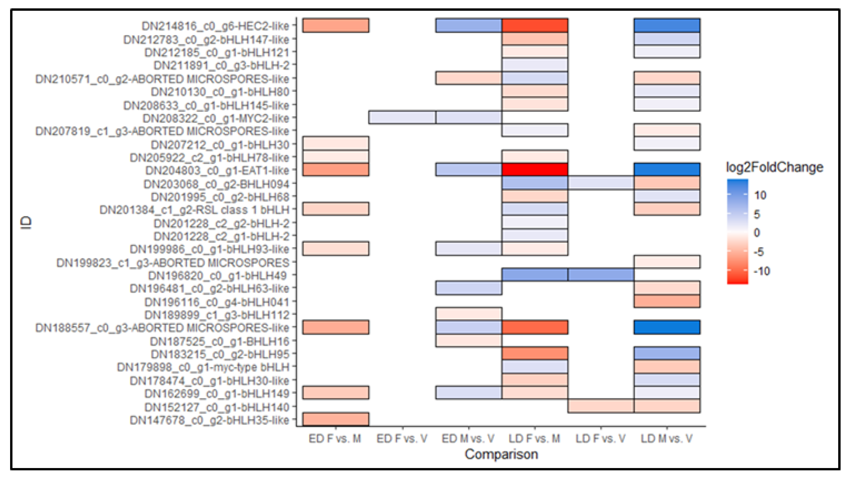
From the bHLH family, 31 TFs were identified as DE among the six comparisons between different bud types at early and late developmental stages (Figure 5). DE bHLH TFs were more pronounced in the male than the female cones. Early differentiated female cones had one up-regulated gene, a MYC2–like homolog. Early differentiated male cones had seven up-regulated genes, including homologous to HEC2, EAT1 and bHLH63. Three down-regulated genes in early differentiated male cones included homologs of ABORTED MICROSPORES, bHLH112 and bHLH16. Late differentiated female cones had two up-regulated genes (bHLH094, bHLH49 homologs) and one down-regulated gene (BHLH140). Like MADS-box genes, the most DE bHLH TFs were up-regulated in late differentiated male cones. Substantial down-regulation of the bHLH041 homolog was observed in late differentiated male cones (Figure 5).

3.6. Differentially Expressed TF Involved in Female Cone Differentiation

Altogether, 25 MADS-box TFs were identified as DE in six comparisons (Figure 6). Three of them, homologs of DAL3 were up-regulated in early differentiating female cones. Late differentiating female tissues had six up-regulated genes, including DAL14, MADS1, MADS2 and MADS5 homologous genes. Those tissues had five down-regulated genes as well, among them are DAL13, AGL1 and SOC1 homologous genes, together with two putative MADS-box genes.

3.7. Differentially Expressed TF Involved in Male Cone Differentiation

Seven genes, including homologs of MADS1, MADS2, DAL3, DAL11 and DAL13, were up-regulated in early differentiating male cones. Late differentiated male cones were the most prominent bud type in terms of the magnitude of DE MADS-box genes, with nine up-regulated genes (DAL3, DAL5, DAL11, DAL13, DAL14, MADS2, AGL66, and a putative MADS-box homologous gene), and seven down-regulated genes (MADS-box 2, MADS8, SOC1, MADS5, AGL1, DAL19, and a putative MADS-box homologous gene).

3.8. Phylogenetic Analysis

The importance of MADS-box genes in flowering regulation as well as other developmental processes, led us to focus on those genes for further analysis. We found that most of MADS-box homologs had several isoforms. Thus, we generated a phylogenetic tree of the 25 identified MADS-box TFs together with homologs from Arabidopsis and representative gymnosperms, including P. radiata, Ginkgo biloba, and P. abies. The 25 TFs were divided into 14 different clades, while the clade containing the higher number of P. halepensis MADS-box proteins was the sister clade of the Arabidopsis SOC1 TF, which included the P. abies proteins DAL3 and DAL19 as well (Supplementary Figure S4).

3.9. Differentially Expressed Gene Validation

Eight MADS-box genes were chosen for further characterization by qPCR. We chose them due to their potential role in cone development manifested by their up-regulation in at least one developmental stage (Figure 6; Supplementary Table S3). Each gene was represented by its most expressed isoform. The selected genes were named according to their closest coniferous homologous inferred from their closest NCBI BLASTP results, as well as the derived phylogenetic tree.
Homogeny of variances between the different bud types was determined by the Bartlett test. We found the variances to be unequal in PhDAL11 and PhMADS1. Therefore, significant differences in PhDAL11 and PhMADS1 were determined by six different t-tests, under the assumption of unequal variances. Bonferroni correction was applied in PhMADS1 and PhDAL11 resulting in α values of 0.0083. The Tukey–Kramer HSD test supported significant differences found by the independent t-tests. Therefore, the differences in relative expression levels of all genes were determined using the Tukey–Kramer HSD test.
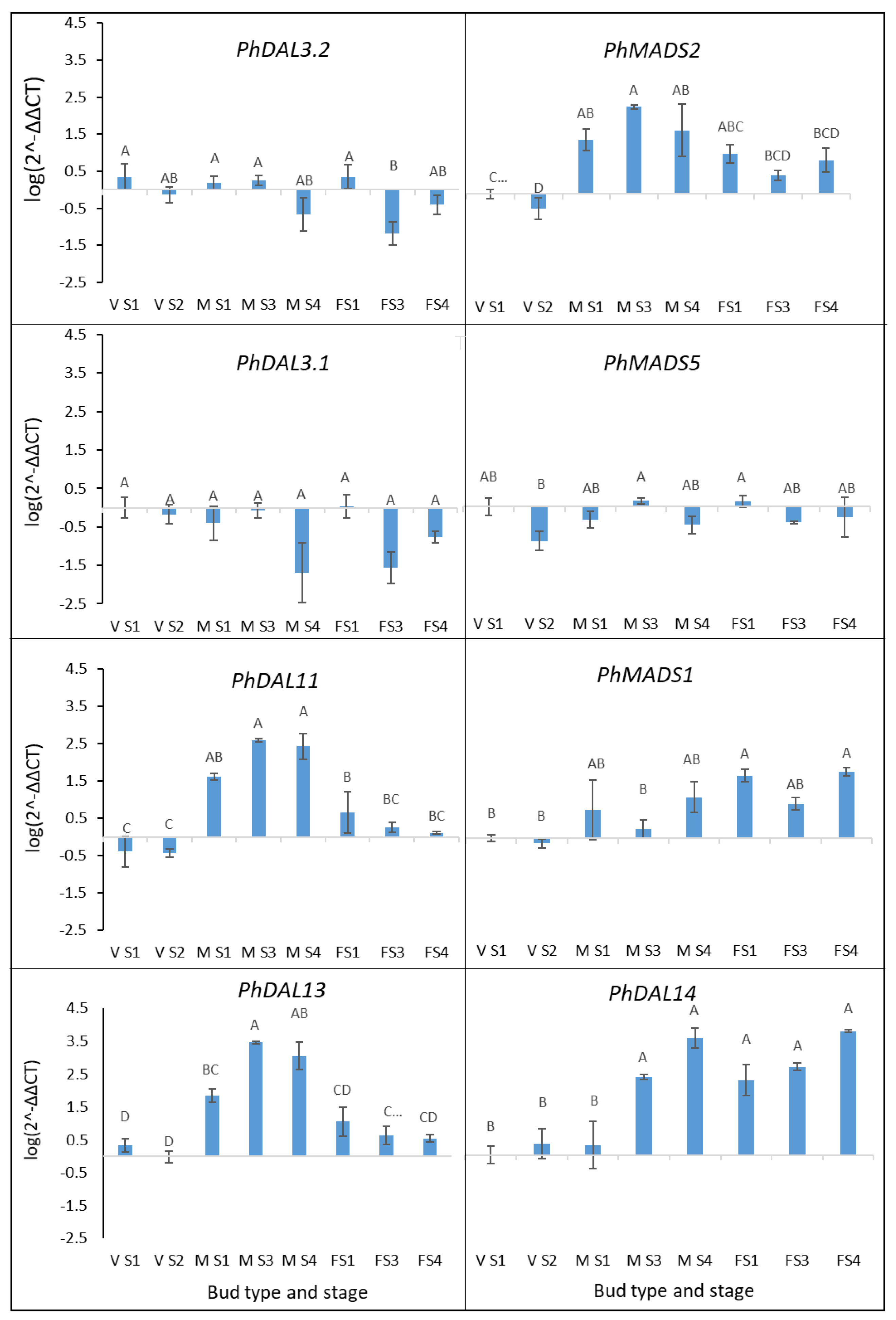
The qPCR showed that PhDAL11 and PhDAL13 were highly expressed across the male cone developmental stages (Figure 7). PhDAL14, PhMADS1, and PhMADS2 were highly expressed across the female cone developmental stages; however, they were also highly expressed at late developmental stages of the male cone. The expression of PhMADS1 was increasing gradually throughout the female cones development, while PhMADS2 showed a weak correlation with cone development. The expression patterns of all genes described above, corresponded to the RNA-seq results (Figure 6, Supplementary Table S3). However, the expression patterns of PhDAL3.1, PhDAL3.2, and PhMADS5 were not consistent with the RNA-seq results (Figure 7).

4. Discussion

This study describes the involvement of transcription factors in the induction and development of the male and female cones in P. halepensis. Identification of the different developmental stages through histology prior to the molecular analysis allowed us to identify differentially expressed TFs at early and late developmental stages. This study enriches our knowledge on the regulation of reproductive development in gymnosperms, and particularly in pines.
By analyzing the transcriptome of vegetative, male, and female tissues at different developmental stages, we found that the higher number of DE genes correlated with the male tissues (Table 2). That may be attributed to a more complexed regulation of male compared to female cone development [5]. Male cone development follows the BC model, according to which expression of both B- and C-class TFs are required, whereas female cone development requires solely C-class TFs [5]. DE genes were also prominent in all tissues in late compared to early developmental stages, suggesting that cone structure and tissue specification at late developmental stages are complex.

4.1. Reproductive Development Is Accompanied by Enriched TF Expression

TFs that were DE in early and late cone developmental stages were enriched in the five families bHLH, ERF, MYB-related, NAC, and MIKC-MADS (Figure 4). These TF families are essential in many processes throughout the plant’s life cycle. In cone development, the bHLH family was prominent with more than 20 genes in early and late male cone development stages. This suggests that bHLH genes may have a significant regulatory role in reproductive organ development in conifers, similar to what was previously described in Arabidopsis [3]. A study on Arabidopsis and the liverwort Marchantia polymorpha showed that bHLH genes are crucial for the formation of generative cells that give rise to the male gametes, suggesting that bHLH may be key regulators of reproductive development across plant species [52]. Recently, it was shown that the bHLH expression in P. tabuliformis is age dependent, suggesting a role in the vegetative growth transition [53]. In light of these reports, the current study results, which suggest massive involvement of bHLH genes in the reproductive stage, encourage additional studies in conifers. Another noticeable TF family is the ERF, a subfamily within the AP2/ERF super-family [54]. We found that most ERF genes were up-regulated in late female cone development (Figure 4). AP2 homologs, AP2L1 and AP2L2, were identified in P. abies and Pinus thunbergii. AP2L1 was dominantly expressed in the female cone, whereas AP2L2 was expressed in both cone types in both species [55,56]. In addition, PaAP2L2 was able to rescue the ap2-1 Arabidopsis mutant phenotype [57]. These functional studies imply that ERF TFs in P. halepensis likely possess AP2/ERF homologs regulating late stages of female cone development.

4.2. Floral Induction Homologous Genes

Unexpectedly, we found only one LFY homolog, which was DE in the male cone tissue only. Its expression level was higher at stage 2 compared to stage 3. This result is inconsistent with previous studies that showed high expression of LFY and NLY in early stages of male and female cone development [58]. It has been shown that NLY is expressed in reproductive as well as in vegetative tissue in pines. However, its capacity to rescue the Arabidopsis lfy mutant implies that it also plays a role in flowering induction [17]. In the current study, the absence of LFY homologs in the DE genes may be due to late tissue collection for the RNA-seq analysis, after the expression peak of LFY and NLY.
The expression patterns of the two FT/TFL1 genes imply a role in late male cone development (Figure S3A). One of those genes (TRINITY_DN197941_c0_g3) had the highest expression level at FS2 and then declined as development progressed (Figure S3B). The FT/TFL homologs exhibited a similar expression level in vegetative and female buds. Yet, an additional FT/TFL homolog (a MOTHER OF FT and TFL1-like, Table S2) was DE between the three developmental stages of the female cone, with highest expression level seen at stage 3, suggesting that FT/TFL may have a role in female cone development. The opposite expression pattern of the FT/TFL1 homologs in female and male cones indicates a different function, which may be antagonistic. It has been shown that FT/TFL1 homologs from conifers act similarly to TFL1 in repressing instead of promoting flowering, and do not seem to have a major role in the reproductive development of pines [20,22]. A recent publication reporting on age-related gene expression in Pinus tabuliformis suggests that interactions between the MADS-box genes MADS11 and DAL1 mediate flowering induction in pines [53]. The same study showed that the over-expression of PtMADS11 in Arabidopsis rescued the late flowering ft phenotype, implying a role in flowering induction [53]. Since in our study we did not include pre-cone differentiation stages in the RNA-seq analysis, we could not confirm that MADS11 and DAL1 are involved in the flowering induction in P. halepensis.
The PhDAL3.1 and PhDAL3.2 genes exhibited high expression level during early differentiation stages of both cone types. The closest homologous gene of PhDAL3.1 and PhDAL3.2 is DAL3, which belongs to an orthologous sister clade and thus to the entire angiosperm SOC1 clade [58]. Our phylogenetic analysis also classified the P. halepensis DAL3 homologs in a sister clade of the SOC1 clade, together with the P. abies DAL3 (Figure S4). The central role of SOC1 in the transition to flowering in Arabidopsis through multiple pathways [2] also implies a central role for PhDAL3.1, PhDAL3.2 in pine, assuming functional similarity between angiosperms and gymnosperms. The qPCR results showed no difference in PhDAL3.1, PhDAL3.2 expression between both cone types and the vegetative buds, in all developmental stages. The only significant difference observed was a lower expression of PhDAL3.2 in FS3 cones. Therefore, our data suggest a role for PhDAL3.1 and PhDAL3.2 in the development of all bud types, including vegetative growth.
Although the RNA-seq data showed that PhMADS5 is up-regulated in late developing female cones, the qPCR results found no difference in the expression levels of this gene in any specific tissue (Figure 7). The closest Arabidopsis homolog of PhMADS5 is SHORT VEGETATIVE PHASE (SVP), involved in flowering time regulation and transition to flowering [59,60]. Like SOC1 orthologs, SVP is a multiple copies family in gymnosperms [61], which indicates that like SOC1, different SVP paralogs may have a different role in developmental processes in conifers.

4.3. Genes Involved in Male and Female Cone Development

We found that the MADS-box genes PhDAL11 and PhDAL13 expressed dominantly in the Male cones. These genes were previously found to be phylogenetically related to B- class MADS-box genes, expressed specifically in male cones of P. abies and P. tabuliformis [58,62], and had similar phenotypic effects to endogenous Arabidopsis B-class proteins when expressed ectopically in Arabidopsis [27]. A new study published recently further demonstrated the male cone specific expression of DAL11 and DAL13 in P. tabuliformis, and showed physical interactions between these two proteins [21]. Together with these studies, our study supports the model proposing that B- class genes are essential for male identity determination [5]. An additional B-class gene examined in the current study, PhMADS2, was up-regulated in both cone types, yet most prominently in the male cone. Niu et al. [21] showed that PtMADS2 interacts with PtDAL11 and PtDAL13, supporting the involvement of PhMADS2 in the male cone development.
PhMADS1 was up-regulated in female cones (Figure 7). The most compatible alignment of PhMADS1 in Arabidopsis was the AP1 protein, which is an A-class TF, important for both floral meristem identity transition and the development of petal and sepals [1,5]. MADS1 in P. radiata was shown to be highly expressed in early cone development and was localized to the ovuliferousscale primordia [13]. MADS1 homologs from P. tabuliformis, PtMADS1 and PtMADS10 were also specifically expressed in the developing female cone [21]. They were shown to physically interact with other female-related MADS-box proteins, such as PtDAL14 and PtDAL1, to regulate the female cone development. It is likely that PhMADS1 plays a similar role in P. halepensis.
In our study, PhDAL14 was highly expressed throughout the female cone development; however, it was also up-regulated in the male cone during the middle to late developmental stages (Figure 7). This expression pattern is in agreement with PaDAL14 expression in P. abies and P. tabuliformis female and male cone development [21,58]. AGL6 is the most closely related Arabidopsis protein to PhDAL14. AGL6 has an E function of floral organ development, with additional functions including floral meristem regulation, ovule and seed development [63]. The function of AGL6 and other E class proteins in angiosperms seem similar to that of PhDAL14 in conifers. This suggests that PhDAL14 is an essential component for both female and male cone development [28,64]. Physical interactions between DAL14 and the male-specific proteins DAL11 and DAL13 in P. tabuliformis support its involvement in regulating both cone types [21].
Function of the genes presented in the current study is confirmed by heterologous complementation done in Arabidopsis mutants in similar studies, as mentioned above. However, gene silencing via the CRISPR/Cas9 approach offers a better understanding of gene function, assuming that transformation and regeneration platforms are available for coniferous species. Silencing key genes in the development of the female pine cone can result in sterile trees, an outcome desirable for both commercial and ecological purposes. Sterile trees might direct the energy to vegetative growth, offering a great economic potential for timber production. In particular, sterility in P. halepensis might prevent the negative environmental impacts caused by uncontrolled regeneration in forests, thereby increasing the efficiency of forest management.

5. Conclusions

Our study contributes to the accumulating evidence of the molecular regulation of the reproduction phase in conifers. The expression pattern of the P. halepensis MADS-box genes was very similar to that found in other conifers, as well as to the newly studied homologs from P. tabuliformis [21], suggesting highly conserved mechanisms in Pinaceae. Yet, the large number of transcription factors revealed in our study suggests that the reproductive phase in Pinaceae is much more complex. We believe that this study will urge further investigation into the regulation of reproductive development in conifers.

Supplementary Materials

The following supporting information can be downloaded at: https://www.mdpi.com/article/10.3390/agronomy12071588/s1, Figure S1: A P. halepensis tree with male and female cones; Figure S2: Heatmap of Pearson correlation analysis evaluating the similarity between samples; Figure S3: Differential expression of FT/TFL1 homologs between different developmental stages of the male (A) and female (B) cones. Figure S4: Maximum likelihood phylogenetic analysis of 25 MADS-box proteins from P. halepensis related to MADS-box proteins from other species. Table S1: Primers used in qPCR; Table S2: Transcriptome analysis of Pinus halepensis cones development; Table S3: Selected P. halepensis MADS-box genes and their expression patterns.

Author Contributions

Conceptualization, R.D.-S. and G.R.; methodology, G.R.; validation, G.R., H.Z. and H.F.; formal analysis, A.F.; investigation, G.R.; data curation, G.R.; writing—original draft preparation, G.R.; writing—review and editing, R.D.-S., G.R. and L.E.W.; supervision, R.D.-S. and L.E.W.; project administration, R.D.-S.; funding acquisition, R.D.-S. All authors have read and agreed to the published version of the manuscript.

Funding

This research received no external funding.

Informed Consent Statement

Not applicable.

Data Availability Statement

Data archiving statement. Data of this project have been deposited with links to BioProject accession number SUB11014167 in The National Center for Biotechnology Information (NCBI). The raw data from the Illumina sequencing have been deposited in NCBI Sequence Read Archive (SRA)—Accession numbers will be provided later, and before publication.

Acknowledgments

Gilad Reisfeld acknowledges financial support from the Appleby Foundation.

Conflicts of Interest

The authors declare no conflict of interest.

References

  1. Ferrandiz, C.; Gu, Q.; Martienssen, R.; Yanofsky, M.F. Redundant regulation of meristem identity and plant architecture by FRUITFULL, APETALA1 and CAULIFLOWER. Development 2000, 127, 725–734. [Google Scholar] [CrossRef]
  2. Lee, J.; Lee, I. Regulation and function of SOC1, a flowering pathway integrator. J. Exp. Bot. 2010, 61, 2247–2254. [Google Scholar] [CrossRef] [Green Version]
  3. Ito, S.; Song, Y.H.; Josephson-Day, A.R.; Miller, R.J.; Breton, G.; Olmstead, R.G.; Imaizumi, T. FLOWERING BHLH transcriptional activators control expression of the photoperiodic flowering regulator CONSTANS in Arabidopsis. Proc. Natl. Acad. Sci. USA 2012, 109, 3582–3587. [Google Scholar] [CrossRef] [Green Version]
  4. Silva, C.S.; Puranik, S.; Round, A.; Brennich, M.; Jourdain, A.; Parcy, F.; Hugouvieux, V.; Zubieta, C. Evolution of the Plant Reproduction Master Regulators LFY and the MADS Transcription Factors: The Role of Protein Structure in the Evolutionary Development of the Flower. Front. Plant Sci. 2016, 6, 1193. [Google Scholar] [CrossRef] [Green Version]
  5. Theißen, G.; Melzer, R.; Rümpler, F. MADS-domain transcription factors and the floral quartet model of flower development: Linking plant development and evolution. Development 2016, 143, 3259–3271. [Google Scholar] [CrossRef] [Green Version]
  6. Kobayashi, Y.; Kaya, H.; Goto, K.; Iwabuchi, M.; Araki, T. A Pair of Related Genes with Antagonistic Roles in Mediating Flowering Signals. Science 1999, 286, 1960–1962. [Google Scholar] [CrossRef]
  7. Wickland, D.P.; Hanzawa, Y. The FLOWERING LOCUS T/TERMINAL FLOWER 1 Gene Family: Functional Evolution and Molecular Mechanisms. Mol. Plant 2015, 8, 983–997. [Google Scholar] [CrossRef] [Green Version]
  8. Moraes, T.S.; Dornelas, M.C.; Martinelli, A.P. FT/TFL1: Calibrating Plant Architecture. Front. Plant Sci. 2019, 10, 97. [Google Scholar] [CrossRef] [Green Version]
  9. Liljegren, S.J.; Gustafson-Brown, C.; Pinyopich, A.; Ditta, G.S.; Yanofsky, M.F. Interactions among APETALA1, LEAFY, and TERMINAL FLOWER1 specify meristem fate. Plant Cell 1999, 11, 1007–1018. [Google Scholar] [CrossRef] [Green Version]
  10. Samach, A.; Onouchi, H.; Gold, S.E.; Ditta, G.S.; Schwarz-Sommer, Z.; Yanofsky, M.F.; Coupland, G. Distinct Roles of CONSTANS Target Genes in Reproductive Development of Arabidopsis. Science 2000, 288, 1613–1616. [Google Scholar] [CrossRef] [Green Version]
  11. Borner, R.; Kampmann, G.; Chandler, J.; Gleißner, R.; Wisman, E.; Apel, K.; Melzer, S. A MADS domain gene involved in the transition to flowering in Arabidopsis. Plant J. 2000, 24, 591–599. [Google Scholar] [CrossRef]
  12. De La Torre, A.R.; Piot, A.; Liu, B.; Wilhite, B.; Weiss, M.; Porth, I. Functional and morphological evolution in gymnosperms: A portrait of implicated gene families. Evol. Appl. 2020, 13, 210–227. [Google Scholar] [CrossRef] [Green Version]
  13. Mouradov, A.; Glassick, T.; Hamdorf, B.; Teasdale, R.D. Molecular control of early cone development inPinus radiata. Protoplasma 1999, 208, 3–12. [Google Scholar] [CrossRef]
  14. Mellerowicz, E.J.; Horgan, K.; Walden, A.; Coker, A.; Walter, C. PRFLL—A Pinus radiata homologue of FLORICAULA and LEAFY is expressed in buds containing vegetative shoot and undifferentiated male cone primordia. Planta 1998, 206, 619–629. [Google Scholar] [CrossRef]
  15. Maizel, A.; Busch, M.A.; Tanahashi, T.; Perkovic, J.; Kato, M.; Hasebe, M.; Weigel, D. The Floral Regulator LEAFY Evolves by Substitutions in the DNA Binding Domain. Science 2005, 308, 260–263. [Google Scholar] [CrossRef]
  16. Carlsbecker, A.; Tandre, K.; Johanson, U.; Englund, M.; Engström, P. The MADS-box gene DAL1 is a potential mediator of the juvenile-to-adult transition in Norway spruce (Picea abies). Plant J. 2004, 40, 546–557. [Google Scholar] [CrossRef]
  17. Mouradov, A.; Glassick, T.; Hamdorf, B.; Murphy, L.; Fowler, B.; Marla, S.; Teasdale, R.D. NEEDLY, a Pinus radiata ortholog of FLORICAULA/LEAFY genes, expressed in both reproductive and vegetative meristems. Proc. Natl. Acad. Sci. USA 1998, 95, 6537–6542. [Google Scholar] [CrossRef] [Green Version]
  18. Uddenberg, D.; Reimegård, J.; Clapham, D.; Almqvist, C.; von Arnold, S.; Emanuelsson, O.; Sundström, J.F. Early Cone Setting in Picea abies acrocona Is Associated with Increased Transcriptional Activity of a MADS Box Transcription Factor. Plant Physiol. 2012, 161, 813–823. [Google Scholar] [CrossRef] [Green Version]
  19. Dornelas, M.C.; Rodriguez, A.P.M. A Floricaula/Leafy gene homolog is preferentially expressed in developing female cones of the tropical pine Pinus caribaea var. caribaea. Genet. Mol. Biol. 2005, 28, 299–307. [Google Scholar] [CrossRef] [Green Version]
  20. Klintenäs, M.; Pin, P.A.; Benlloch, R.; Ingvarsson, P.K.; Nilsson, O. Analysis of conifer FLOWERING LOCUS T/TERMINAL FLOWER1-like genes provides evidence for dramatic biochemical evolution in the angiosperm FT lineage. New Phytol. 2012, 196, 1260–1273. [Google Scholar] [CrossRef]
  21. Niu, S.; Li, J.; Bo, W.; Yang, W.; Zuccolo, A.; Giacomello, S.; Chen, X.; Han, F.; Yang, J.; Song, Y.; et al. The Chinese pine genome and methylome unveil key features of conifer evolution. Cell 2022, 185, 204–217.e14. [Google Scholar] [CrossRef]
  22. Karlgren, A.; Gyllenstrand, N.; Källman, T.; Sundström, J.F.; Moore, D.; Lascoux, M.; Lagercrantz, U. Evolution of the PEBP Gene Family in Plants: Functional Diversification in Seed Plant Evolution. Plant Physiol. 2011, 156, 1967–1977. [Google Scholar] [CrossRef] [Green Version]
  23. Theißen, G. Development of floral organ identity: Stories from the MADS house. Curr. Opin. Plant Biol. 2001, 4, 75–85. [Google Scholar] [CrossRef]
  24. Weigel, D.; Meyerowitz, E.M. Activation of Floral Homeotic Genes in Arabidopsis. Science 1993, 261, 1723–1726. [Google Scholar] [CrossRef]
  25. Bowman, J.L.; Smyth, D.R.; Meyerowitz, E.M. Genetic interactions among floral homeotic genes of Arabidopsis. Development 1991, 112, 1–20. [Google Scholar] [CrossRef]
  26. Mouradov, A.; Hamdorf, B.; Teasdale, R.D.; Kim, J.T.; Winter, K.-U.; Theißen, G. A DEF/GLO-like MADS-box gene from a gymnosperm: Pinus radiata contains an ortholog of angiosperm B class floral homeotic genes. Dev. Genet. 1999, 25, 245–252. [Google Scholar] [CrossRef]
  27. Sundström, J.; Engström, P. Conifer reproductive development involves B-type MADS-box genes with distinct and different activities in male organ primordia. Plant J. 2002, 31, 161–169. [Google Scholar] [CrossRef]
  28. Mouradov, A.; Glassick, T.V.; Hamdorf, B.A.; Murphy, L.C.; Marla, S.S.; Yang, Y.; Teasdale, R.D. Family of MADS-Box Genes Expressed Early in Male and Female Reproductive Structures of Monterey Pine. Plant Physiol. 1998, 117, 55–62. [Google Scholar] [CrossRef] [Green Version]
  29. Tandre, K.; Albert, V.A.; Sundas, A.; Engstrom, P. Conifer homologues to genes that control floral development in angiosperms. Plant Mol. Biol. 1995, 27, 69–78. [Google Scholar] [CrossRef]
  30. Englund, M.; Carlsbecker, A.; Engström, P.; Vergara-Silva, F. Morphological “primary homology” and expression of AG-subfamily MADS-box genes in pines, podocarps, and yews. Evol. Dev. 2011, 13, 171–181. [Google Scholar] [CrossRef]
  31. Carlsbecker, A.; Sundström, J.; Tandre, K.; Englund, M.; Kvarnheden, A.; Johanson, U.; Engström, P. The DAL10 gene from Norway spruce (Picea abies) belongs to a potentially gymnosperm-specific subclass of MADS-box genes and is specifically active in seed cones and pollen cones. Evol. Dev. 2003, 5, 551–561. [Google Scholar] [CrossRef]
  32. Ne’eman, G.; Goubitz, S.; Werger, M.J.A.; Shmida, A. Relationships between tree size, crown shape, gender segregation and sex allocation in Pinus halepensis, a Mediterranean pine tree. Ann. Bot. 2011, 108, 197–206. [Google Scholar] [CrossRef] [Green Version]
  33. Climent, J.; Prada, M.A.; Calama, R.; Chambel, M.R.; de Ron, D.S.; Alía, R. To grow or to seed: Ecotypic variation in reproductive allocation and cone production by young female Aleppo pine (Pinus halepensis, Pinaceae). Am. J. Bot. 2008, 95, 833–842. [Google Scholar] [CrossRef]
  34. Weinstein, A. Geographic variation and phenology of Pinus halepensis, P. brutia and P. eldarica in Israel. For. Ecol. Manag. 1989, 27, 99–108. [Google Scholar] [CrossRef]
  35. Brandizzi, F. Ruzin SE. 1999. Plant microtechnique and microscopy. 322 pp. Oxford, New York: Oxford University Press. £32.50 (softback). Ann. Bot. 2000, 86, 708. [Google Scholar] [CrossRef] [Green Version]
  36. Asgari, F.; Irian, S.; Jonoubi, P.; Majd, A. Meristem Structure, Development of Cones and Microsporogenesis of Tehran Pine (Pinus Eldarica Medw.). J. Plant Dev. 2014, 21, 83–93. [Google Scholar]
  37. Grabherr, M.G.; Haas, B.J.; Yassour, M.; Levin, J.Z.; Thompson, D.A.; Amit, I. Full-length transcriptome assembly from RNA-Seq data without a reference genome. Nat. Biotechnol. 2011, 29, 644–652. [Google Scholar] [CrossRef] [Green Version]
  38. Langmead, B.; Trapnell, C.; Pop, M.; Salzberg, S.L. Ultrafast and memory-efficient alignment of short DNA sequences to the human genome. Genome Biol. 2009, 10, R25. [Google Scholar] [CrossRef] [Green Version]
  39. Li, B.; Dewey, C.N. RSEM: Accurate transcript quantification from RNA-Seq data with or without a reference genome. BMC Bioinform. 2011, 12, 323. [Google Scholar] [CrossRef] [Green Version]
  40. Haas, B.J.; Papanicolaou, A.; Yassour, M.; Grabherr, M.; Blood, P.D.; Bowden, J.; Couger, M.B.; Eccles, D.; Li, B.; Lieber, M.; et al. De novo transcript sequence reconstruction from RNA-seq using the Trinity platform for reference generation and analysis. Nat. Protoc. 2013, 8, 1494–1512. [Google Scholar] [CrossRef]
  41. Robinson, M.D.; McCarthy, D.J.; Smyth, G.K. edgeR: A Bioconductor package for differential expression analysis of digital gene expression data. Bioinformatics 2010, 26, 139–140. [Google Scholar] [CrossRef] [Green Version]
  42. Buchfink, B.; Xie, C.; Huson, D.H. Fast and sensitive protein alignment using DIAMOND. Nat. Methods 2015, 12, 59–60. [Google Scholar] [CrossRef]
  43. Altschul, S.F.; Gish, W.; Miller, W.; Myers, E.W.; Lipman, D.J. Basic Local Alignment Search Tool. J. Mol. Biol. 1990, 215, 403–410. [Google Scholar] [CrossRef]
  44. Conesa, A.; Götz, S.; García-Gómez, J.M.; Terol, J.; Talón, M.; Robles, M. Blast2GO: A universal tool for annotation, visualization and analysis in functional genomics research. Bioinformatics 2005, 21, 3674–3676. [Google Scholar] [CrossRef] [Green Version]
  45. Upton, G.J.G. Fisher’s exact test. J. R. Stat. Soc. Ser. A (Stat. Soc.) 1992, 155, 395–402. [Google Scholar] [CrossRef]
  46. Benjamini, Y.; Hocberg, Y. Controlling the false discovery rate: A practical and powerful approach to multiple testing. J. R. Stat. Soc. 1995, 57, 289–300. [Google Scholar] [CrossRef]
  47. Kumar, S.; Stecher, G.; Li, M.; Knyaz, C.; Tamura, K. MEGA X: Molecular Evolutionary Genetics Analysis across Computing Platforms. Mol. Biol. Evol. 2018, 35, 1547–1549. [Google Scholar] [CrossRef]
  48. Edgar, R.C. MUSCLE: Multiple sequence alignment with high accuracy and high throughput. Nucleic Acids Res. 2004, 32, 1792–1797. [Google Scholar] [CrossRef] [Green Version]
  49. Jones, D.T.; Taylor, W.R.; Thornton, J.M. The rapid generation of mutation data matrices from protein sequences. Bioinformatics 1992, 8, 275–282. [Google Scholar] [CrossRef]
  50. Livak, K.J.; Schmittgen, T.D. Analysis of relative gene expression data using real-time quantitative PCR and the 2(-Delta Delta C(T)) Method. Methods 2001, 25, 402–408. [Google Scholar] [CrossRef]
  51. Bramlett, D.L. Recognizing Developmental Stages in Southern Pine Flowers: The Key to Controlled Pollination; Southeastern Forest Experiment Station: Asheville, NC, USA, 1980. [Google Scholar]
  52. Yamaoka, S.; Nishihama, R.; Yoshitake, Y.; Ishida, S.; Inoue, K.; Saito, M.; Okahashi, K.; Bao, H.; Nishida, H.; Yamaguchi, K.; et al. Generative Cell Specification Requires Transcription Factors Evolutionarily Conserved in Land Plants. Curr. Biol. 2018, 28, 479–486.e5. [Google Scholar] [CrossRef] [PubMed] [Green Version]
  53. Ma, J.-J.; Chen, X.; Song, Y.-T.; Zhang, G.-F.; Zhou, X.-Q.; Que, S.-P.; Mao, F.; Pervaiz, T.; Lin, J.-X.; Li, Y.; et al. MADS-box transcription factors MADS11 and DAL1 interact to mediate the vegetative-to-reproductive transition in pine. Plant Physiol. 2021, 187, 247–262. [Google Scholar] [CrossRef] [PubMed]
  54. Feng, K.; Hou, X.-L.; Xing, G.-M.; Liu, J.-X.; Duan, A.-Q.; Xu, Z.-S.; Li, M.-Y.; Zhuang, J.; Xiong, A.-S. Advances in AP2/ERF super-family transcription factors in plant. Crit. Rev. Biotechnol. 2020, 40, 750–776. [Google Scholar] [CrossRef]
  55. Shigyo, M.; Ito, M. Analysis of gymnosperm two-AP2-domain-containing genes. Dev. Genes Evol. 2004, 214, 105–114. [Google Scholar] [CrossRef]
  56. Vahala, T.; Oxelman, B.; Arnold, S.v. Two APETALA2-like genes of Picea abies are differentially expressed during development. J. Exp. Bot. 2001, 52, 1111–1115. [Google Scholar] [CrossRef] [Green Version]
  57. Nilsson, L.; Carlsbecker, A.; Sundås-Larsson, A.; Vahala, T. APETALA2 like genes from Picea abies show functional similarities to their Arabidopsis homologues. Planta 2007, 225, 589–602. [Google Scholar] [CrossRef]
  58. Carlsbecker, A.; Sundström, J.F.; Englund, M.; Uddenberg, D.; Izquierdo, L.; Kvarnheden, A.; Vergara-Silva, F.; Engström, P. Molecular control of normal and acrocona mutant seed cone development in Norway spruce (Picea abies) and the evolution of conifer ovule-bearing organs. New Phytol. 2013, 200, 261–275. [Google Scholar] [CrossRef]
  59. Klocko, A.L.; Lu, H.; Magnuson, A.; Brunner, A.M.; Ma, C.; Strauss, S.H. Phenotypic Expression and Stability in a Large-Scale Field Study of Genetically Engineered Poplars Containing Sexual Containment Transgenes. Front. Bioeng. Biotechnol. 2018, 6, 100. [Google Scholar] [CrossRef]
  60. Li, C.; Chen, C.; Gao, L.; Yang, S.; Nguyen, V.; Shi, X.; Siminovitch, K.; Kohalmi, S.E.; Huang, S.; Wu, K.; et al. The Arabidopsis SWI2/SNF2 Chromatin Remodeler BRAHMA Regulates Polycomb Function during Vegetative Development and Directly Activates the Flowering Repressor Gene SVP. PLoS Genet. 2015, 11, e1004944. [Google Scholar] [CrossRef] [Green Version]
  61. Chen, F.; Zhang, X.; Liu, X.; Zhang, L. Evolutionary Analysis of MIKCc-Type MADS-Box Genes in Gymnosperms and Angiosperms. Front. Plant Sci. 2017, 8, 895. [Google Scholar] [CrossRef] [Green Version]
  62. Niu, S.; Yuan, H.; Sun, X.; Porth, I.; Li, Y.; El-Kassaby, Y.A.; Li, W. A transcriptomics investigation into pine reproductive organ development. New Phytol. 2016, 209, 1278–1289. [Google Scholar] [CrossRef]
  63. Dreni, L.; Zhang, D. Flower development: The evolutionary history and functions of the AGL6 subfamily MADS-box genes. J. Exp. Bot. 2016, 67, 1625–1638. [Google Scholar] [CrossRef] [Green Version]
  64. Gramzow, L.; Weilandt, L.; Theißen, G. MADS goes genomic in conifers: Towards determining the ancestral set of MADS-box genes in seed plants. Ann. Bot. 2014, 114, 1407–1429. [Google Scholar] [CrossRef] [Green Version]
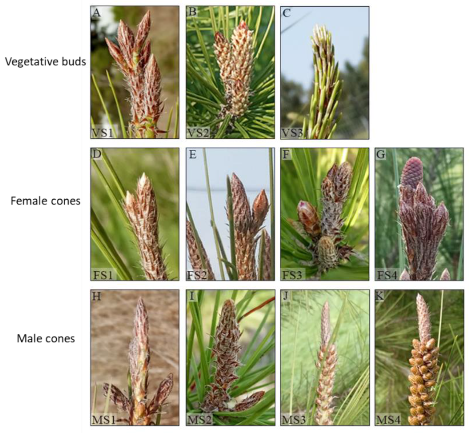
Figure 1. Developmental stages of vegetative buds, and female and male cones in P. halepensis. (AC) Vegetative buds at stages 1–3. (A) VS1, Early vegetative buds. (B) VS2, Dwarf shoots are visible. (C) VS3, Needles are visible. (DG) Female cones. (D) FS1, Tightly enclosed bud. (E) FS2, Elongated and swelled bud. (F) FS3, Cone emergence through the top of the scales. (G) FS4, A visible cone. (HK) Male cones. (H) MS1, Tightly enclosed bud. (I) MS2, Apparent buds. (J) MS3, Cone emergence from the scales. (K) MS4, Fully developed cones.
Agronomy 12 01588 g001
Figure 2. Longitudinal sections of vegetative, female and male P. halepensis buds at different developmental stages. (AD) Vegetative buds developmental stages. (A) S1, Dwarf shoot (Brachyblasts) primordia (DSP) with apical meristem (AM). (B) (S2) and (C) (S3), Dwarf shoots (DS) emerge through the scales. (D) S4, Visible needles (VN). (EH) Female cones developmental stages. (E) S1, Bulge-like structure enclosed within the scales with few lateral bract anlagen. (F) S2, bract primordia (BP) and ovuliferous primordia (OP). (G) S3, Bracts (B) are fully developed covering the developing ovuliferous scales. (H) S4, bract and ovuliferous scale (OS) complex with developing ovule (O). (IL) Male cones developmental stages. (I) S1, Lateral microsporophyll primordia (MIP). (J) S2, Young cones hidden within the scales, contains developing microsporophyll. (K) S3, Cone emergence from bud scales and the microsporophyll (MI) contains microsporangium (MS). (L) S4, fully developed cones with pollen sacs (PS) and pollen mother cells (P). Scale Bar, (AE,I) 500 μM, (FH,JL) 250 μM.
Agronomy 12 01588 g002
Figure 3. Venn diagram of up-regulated and down-regulated genes showing shared genes between the female cone three developmental stages. S2–S4, developmental stages.
Agronomy 12 01588 g003
Figure 4. Heat-map of up-regulated transcription factors (TF) grouped into families involved in cone development. Included are families which showed at least three up-regulated members in at least one meristem type in the early (a) and late (b) stages of cone development. Color intensity of the heat-map reflects the number of genes. Common families between the early and late stages share the same color, while uncommon families are not colored. Asterisks indicate TF families found only in the male cone. MS2, Male Stage 2; MS3, Male Stage 3; FS2, Female Stage 2; FS3, Female Stage 3; FS4, Female Stage 4; VS2, Vegetative Stage 2; VS3, Vegetative Stage 3.
Agronomy 12 01588 g004
Figure 5. bHLH TFs differentially expressed in six comparisons. Gene ID is composed of the TRINITY accession ID, followed by NCBI BLASTP derived homolog. ED, Early development (Stage 2); LD, Late development (Stage 3); F, female; V, vegetative; M, male.
Agronomy 12 01588 g005
Figure 6. MADS-box TFs differentially expressed in six comparisons. Gene ID is composed of the TRINITY accession ID, followed by NCBI BLASTP derived homolog gene. ED, Early development (Stage 2); LD, Late development (Stage 3); F, female; V, vegetative; M, male.
Agronomy 12 01588 g006
Figure 7. Relative expression levels of eight MADS-box genes in vegetative buds, male, and female cones during several developmental stages. Uppercase letters indicate significant differences (p < 0.05). Bars represent standard errors. VS1, Vegetative Stage 1; VS2, Vegetative Stage 2; MS1, Male Stage 1; MS3, Male Stage 3; FS1, Female Stage 1; FS3, Female Stage 3; FS4, Female Stage 4.
Agronomy 12 01588 g007
Table 1. Developmental stages of vegetative shoot, and male and female buds.
MeristemStageDescription
Vegetative 1 *Undifferentiated lateral meristems.
2Differentiated non-visible needles.
3Visible elongated needles.
Male 1 *Male flowering bud clearly identified.
2Differentiated microsporophyll.
3Developed pollen sacs with a tapetal layer on the edges, containing pollen mother cells.
Female 1 *Female flowering bud (ovulate cone) clearly identified.
2Bract differentiation, ovuliferous scales are not differentiated.
3Ovuliferous scales are at early differentiation.
4Ovuliferous scales are differentiated, ovules are visible.
* Stages that were missing from the collected samples for RNA-seq.
Table 2. Number of differentially expressed genes between the different bud types, and between the developmental stages in each bud type.
ComparisonUp-RegulatedDown-RegulatedTotal
Early developmental StageFS2 vs. MS2417405822
FS2 vs. VS29288180
MS2 vs. VS25196891208
Late developmental StageFS3 vs. MS3238221154497
FS3 vs. VS3102013002320
MS3 vs. VS3204225215563
Vegetative bud developmentVS2 vs. VS3139224363
Male cone developmentMS2 vs. MS3220021554355
Female cone developmentFS2 vs. FS35587381296
FS2 vs. FS4121910232242
FS3 vs. FS412838322115
S, developmental stage; F, female; M, male; V, vegetative; DE, differentially expressed.
Publisher’s Note: MDPI stays neutral with regard to jurisdictional claims in published maps and institutional affiliations.