Article

Nanopriming-Induced Enhancement of Cucumber Seedling Development: Exploring Biochemical and Physiological Effects of Silver Nanoparticles

Research Group FiVe-A, Plant Physiology Unit, Genetic, Physiology and Microbiology Department, Faculty of Biological Sciences, Universidad Complutense de Madrid, Ciudad Universitaria, E-28040 Madrid, Spain
*
Author to whom correspondence should be addressed.
Agronomy 2024, 14(8), 1866; https://doi.org/10.3390/agronomy14081866
Submission received: 29 July 2024 / Revised: 20 August 2024 / Accepted: 20 August 2024 / Published: 22 August 2024

Abstract

:
Nanopriming, a technique that involves treating seeds with nanoparticles, is gaining attention for enhancing seed germination and seedling growth. This study explored the effects of silver nanoparticles (AgNPs), synthesized using Ascorbic acid, Caffeic acid, and Gallic acid, on cucumber seedling development. The nanoparticles, characterized by spherical morphology and distinct optical properties, showed varying effects based on the type and concentration of the reducing agents used. AgNP treatments generally led to higher germination rates and improved shoot and root growth compared to controls. Biochemical analyses revealed that these treatments influenced plant physiology, affecting reactive oxygen species (ROS) production, oxidative stress markers, and the content of amino acids, phenolic compounds, flavonoids, and soluble sugars. Specifically, certain AgNP treatments reduced oxidative stress, while others increased oxidative damage. Additionally, variations in free amino acids and phenolic and flavonoid contents were noted, suggesting complex interactions between nanoparticles and plant biochemical pathways. These findings highlight the potential of nanopriming in agriculture and underscore the need for further research to optimize nanoparticle formulations for different plant species.

1. Introduction

The continuous global population growth, with projections estimating around 9 billion people by 2050, has significantly increased food demand. Anticipated agricultural demand is expected to rise by 50–70% compared to current levels, presenting substantial challenges [1]. Fertilizers and pesticides play a crucial role in agricultural production by providing essential nutrients and protecting crops from pests and diseases. However, their excessive use has led to detrimental environmental and health impacts, including soil chemical alterations, water pollution, and the introduction of harmful substances into the food chain [2]. Balancing production with sustainability is imperative for ensuring food security, necessitating the exploration of new technologies like nanotechnology to enhance agricultural production sustainably.
Nanotechnology, dedicated to synthesizing and developing nanomaterials, offers innovative solutions for sustainable agricultural practices [3]. Specifically, nanopriming, which involves using nanoparticles in a priming solution that contacts seeds, has shown promise. This technique facilitates seed germination and growth by allowing nanoparticles to penetrate the seed coat, altering metabolism and signaling pathways [4]. Nanopriming not only enhances germination and growth but also offers protection against seed-borne pathogens due to the antifungal and antibacterial properties of nanoparticles.
Understanding nanoparticle application methods is crucial, with nanopriming gaining prominence for its effectiveness in promoting seed germination and seedling growth [5]. Traditional seed priming techniques, such as hydropriming, osmopriming, and hormonal priming, have limitations, including significant waste and environmental contamination [6]. In contrast, nanopriming addresses these issues by utilizing smaller quantities of nanoparticles.
Green synthesis emerges as a sustainable alternative for producing nanoparticles. This method minimizes waste, energy consumption, and material usage, reducing environmental impacts. Biological agents like algae, fungi, bacteria, and plants can transform metallic ions into nanoparticles, with plant extracts offering a rapid, cost-effective, and environmentally friendly approach [7,8].
The dualistic impact of silver nanoparticles (AgNPs) on plant systems is noteworthy, with both positive and negative effects reported. Positive impacts include enhanced agromorphological, antioxidant, and enzymatic aspects in plants like Helianthus annuus [1] and improved germination and growth in Satureja hortensis [9]. However, negative effects such as growth inhibition and cellular viability reduction have been observed in plants like Lemna gibba and rice (Oryza sativa L.), highlighting the need for comprehensive understanding [10,11].
Focusing on cucumber (Cucumis sativus L.), one of the oldest cultivated vegetables, this study aims to explore the effects of AgNPs synthesized through green methods on cucumber seeds and seedlings. Cucumbers are not only nutritionally valuable but also hold medicinal and cosmetic potential, making them an important subject for scientific investigation [12,13]. By examining the biochemical and physiological aspects of AgNP nanopriming, this research seeks to contribute to the evolving field of nanotechnology in agriculture, addressing global food security and sustainability.

2. Materials and Methods

2.1. Green Synthesis of Silver Nanoparticles (AgNPs) and Reaction Mixture Preparation

The green synthesis of AgNPs was performed using silver nitrate (AgNO3) and antioxidant agents: Gallic acid (GA), Caffeic acid (CA), and Ascorbic acid (AA) (all supplied by Sigma-Aldrich Química SL, Madrid, Spain). Two concentrations of AgNO3 solutions (10 mM and 100 μM) were initially prepared. In a test tube, 10 mL of each antioxidant agent solution was combined with 50 μL of the AgNO3 solution to initiate nanoparticle synthesis. The mixture was kept in the dark at 60 °C for 5 days, leading to the deposition of nanoparticles at the bottom of the tube. The procedure was repeated to ensure a sufficient quantity of nanoparticles for further analysis.

2.2. Characterization of Silver Nanoparticles

A range of analytical techniques was employed to characterize the synthesized silver nanoparticles:
  • UV-Visible Spectrophotometry: The UV-visible absorption spectra of the Ag nanoparticle solutions were recorded between 300 and 500 nm using a UV-visible spectrophotometer.
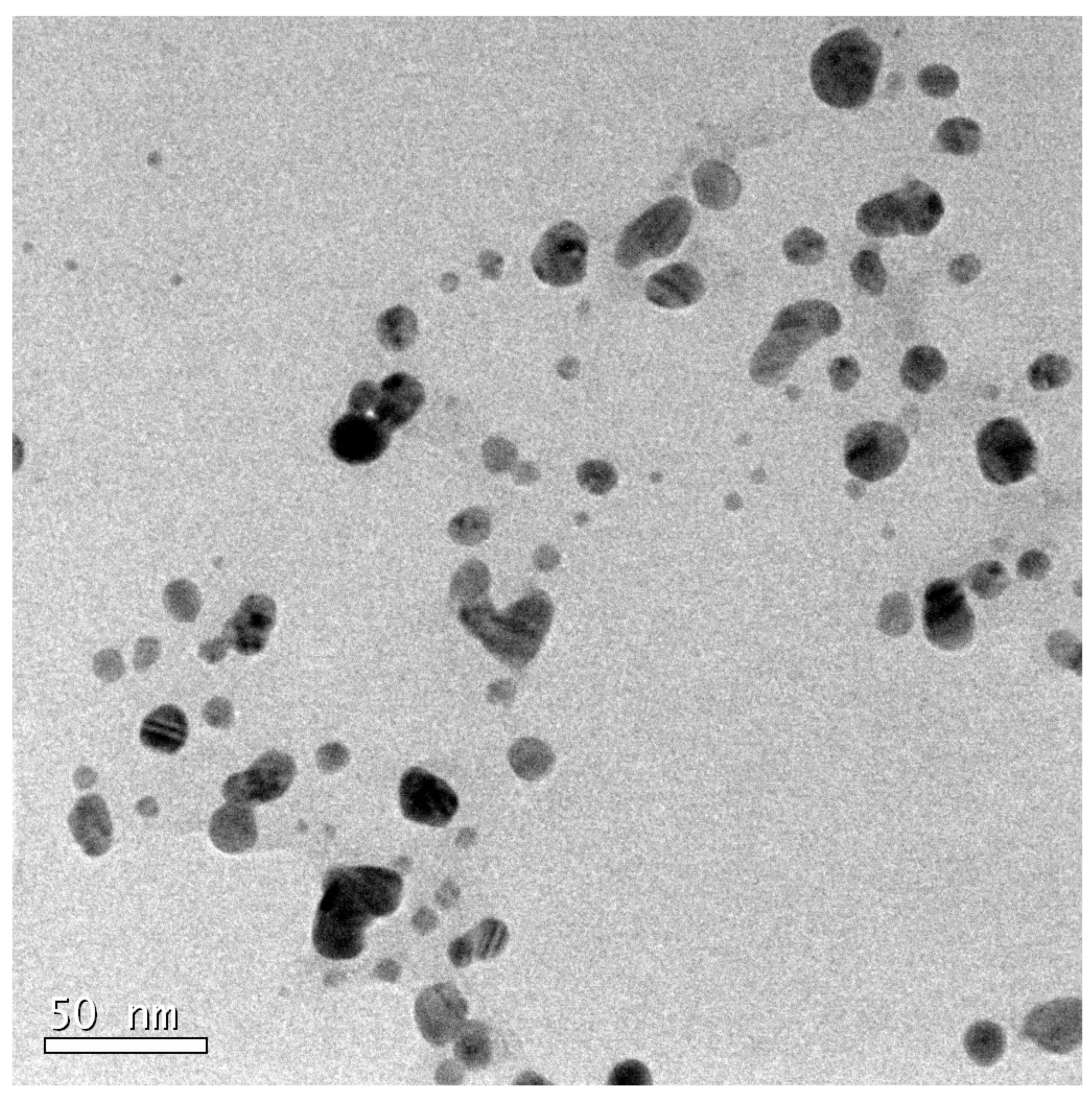
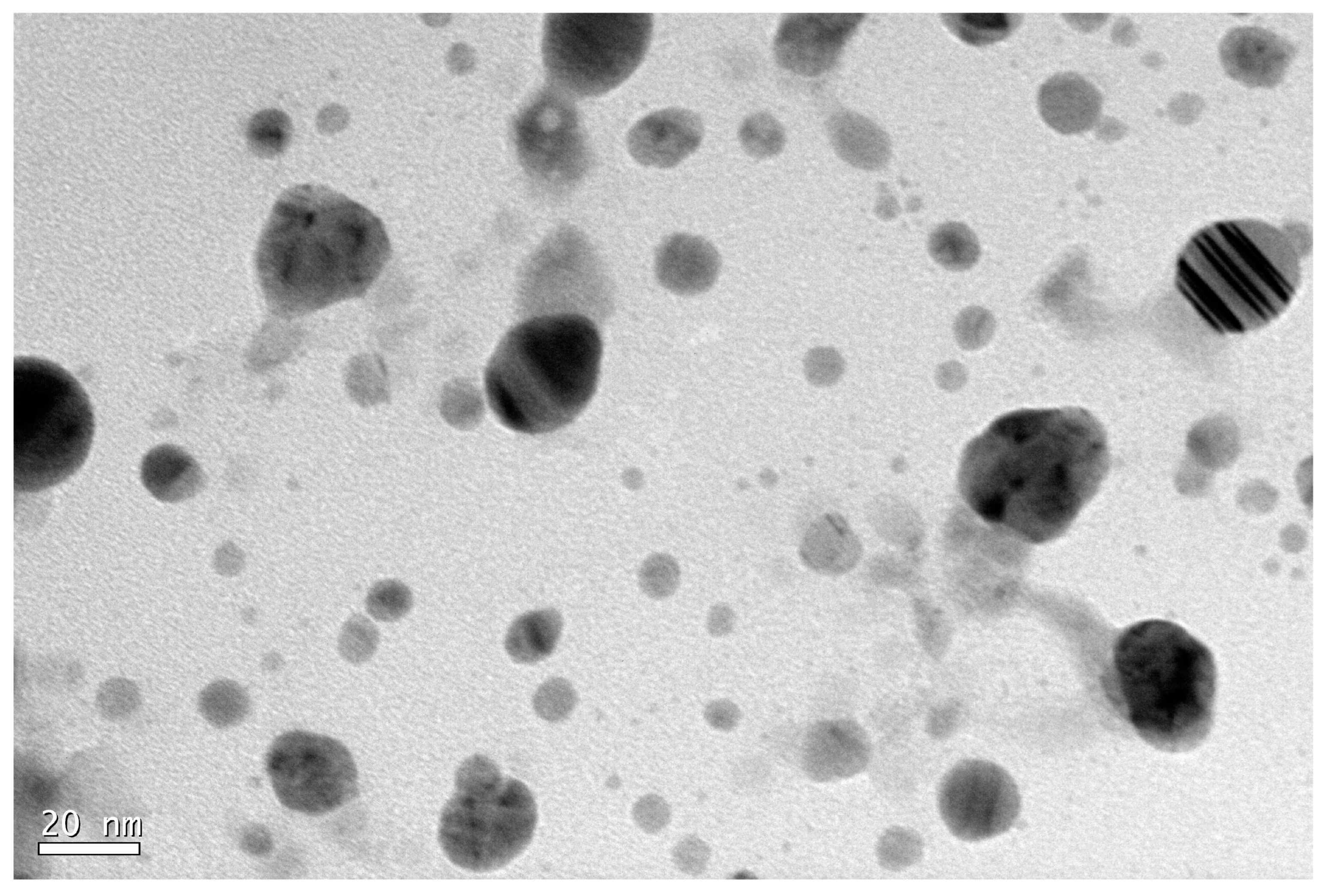
  • Transmission Electron Microscopy (TEM): Nanoparticles were suspended in 1 mL of water. A drop of this solution was placed on a carbon-coated copper grid and air-dried. TEM analysis was performed using a JEOL2100 microscope at the Centro Nacional de Microscopía Electrónica, Madrid.
  • X-ray Powder Diffraction (XRD): XRD profiles were obtained using an Empyrean Cu LFF X-ray diffractometer within a 2θ range from 2° to 80°. The analyses were conducted at the Research Support Centre (CAI) for X-Ray Diffraction, Universidad Complutense de Madrid. The resulting diffraction patterns were compared with reference patterns from the International Centre for Diffraction Data (ICDD) database.
  • Fourier-Transform Infrared Spectroscopy (FTIR): FTIR measurements were carried out using a JASCO (FT/IR-6200) spectrophotometer at the CAI of Physical and Chemical Techniques, Complutense University of Madrid. The interpretation of results followed guidelines provided in infrared spectroscopy tables [14].

2.3. Nanopriming of Cucumber Seeds with AgNPs and In Vitro Cultivation

Cucumber seeds (Cucumis sativus L.), ‘Marketer’ variety, were sourced from Vilmorin-Mikado Ibérica, Spain. All equipment was sterilized meticulously. The experiment included various nanopriming treatments for seed germination, such as water-control, bulk AgNO3 controls, and AgNPs from GA, CA, and AA. Each treatment involved a 20 h imbibition period in darkness. The experiment was replicated twice to enhance statistical robustness.

2.4. In Vitro Cultivation of Cucumber Seedlings

After the 20 h nanopriming period, 12 seeds from each solution were placed in Petri dishes with moist filter paper and kept in the dark for 3 days at 22 ± 2 °C. Nine germinated seeds from each dish were transplanted into seed trays and grown in controlled conditions (22 ± 2 °C, 16 h light/8 h dark photoperiod) for one month.

2.5. Measurement of Morphological Parameters during Germination

On the third day post-sowing, various morphological parameters were measured, including shoot and root lengths, secondary root presence, and germination percentage. The shoot–root ratio was computed by dividing shoot length by root length.

2.6. Measurement of Photosynthetic Parameters

Photosynthetic parameters, including SPAD values and chlorophyll fluorescence (Fv/Fm, PIabs), were measured after one month using the SPAD-502 meter and HANDY PEA+ device, respectively. Photosynthetic pigment content was assessed using lyophilized plant material following the Rainbow protocol [15].

2.7. Measurement of Biochemical Parameters

Biochemical analyses were conducted after one month of growth, using methodologies from López-Hidalgo et al. [15]. Oxidative stress was measured by detecting superoxide anion through NBT reduction, analyzed using ImageJ® software [16].

2.8. Statistical Analysis

Statistical analysis was performed using STATISTICA® v7 software. A simple ANOVA was conducted to compare means between different treatments. Statistically significant differences (p < 0.05) were classified using the Duncan multiple range test. Means are presented with their standard deviations.

3. Results

3.1. Synthesis and Characterization of Silver Nanoparticles Using Reducing Agents

The introduction of Gallic acid to silver nitrate led to discernible changes in coloration, resulting in grey nanoparticles (GA-AgNPs). This reaction, albeit not immediate, exhibited high optical density values, and UV-visible spectrum analysis revealed a prominent peak at 440 nm, corresponding to 70 nm-sized particles. Electron microscopy unveiled aggregates and confirmed the coating of nanoparticles with Gallic acid. FTIR spectra identified specific functional groups, and XRD analysis confirmed the presence of silver in various oxidation states (Figure 1).
Caffeic acid induced an immediate reaction, resulting in dark grey nanoparticles (CA-AgNPs) with plasmon resonance bands at 421 nm and 467 nm, representing 50 nm and 80 nm-sized particles. FTIR spectra elucidated the presence of Caffeic acid on nanoparticle surfaces. XRD analysis identified specific oxidation states of silver, offering insights into the synthesized nanoparticles’ structural properties (Figure 2).
Ascorbic acid promptly reacted with silver nitrate, yielding gold-colored nanoparticles (AA-AgNPs) with distinct plasmon resonance bands at 402 nm, 421 nm, and 467 nm. Electron microscopy highlighted 20 nm-sized particles, and FTIR spectra demonstrated similarities between Ascorbic acid and the synthesized nanoparticles. XRD analysis verified different oxidation states of silver, providing a comprehensive characterization (Figure 3).

3.2. Morphological Parameters

3.2.1. No Adverse Effects on Germination Observed with Nanopriming Treatments

The results reveal favorable germination percentages for all treatments, except for the AgNO3 treatment, which served as a negative control with no seed germination (Table 1). Minor variations are observed between the 100 μM and 10 mM treatments, with the 100 μM treatments showing slightly lower percentages, around 80–90%. The control and 10 mM treatments exhibit identical germination percentages.

3.2.2. Silver Nanoparticle Treatments Enhanced Both Shoot and Root Lengths in Cucumber Seedlings

The morphological parameters of shoot and root length provide valuable insights into the initial effects of nanoparticle treatments on cucumber plants (Figure 4). The results underscore significant disparities among treatments. Notably, the water-control treatment exhibits the lowest mean shoot length, markedly inferior to the highest mean recorded in the CA-AgNPs (100 μM) treatment, which reaches approximately 3.6 cm. When comparing the control with alternative treatments, a noteworthy discrepancy of at least 1 cm becomes evident. Interestingly, nanoparticle treatments display close clustering, with notable distinctions observed primarily between AA-AgNPs (10 mM) and CA-AgNPs (100 μM). Upon analyzing individual reducing agents, slight disparities emerge between the 10 mM and 100 μM treatments, although more pronounced in Ascorbic acid treatments.
Next, root length also exhibits significant differences between treatments with and without nanoparticles (Figure 5). The lowest values recorded are in the water-control and AA-AgNPs (10 mM) treatments, both around 14 cm, about 5 cm lower than the GA-AgNPs (100 μM) and CA-AgNPs (100 μM) treatments, which displayed the highest values. This disparity highlights the pronounced impact of nanoparticle treatments on root development, with certain treatments promoting notably longer roots compared to others.
Lastly, regarding the shoot–root ratio, no significant differences (Duncan’s Test, p < 0.05) have been observed for the relationship between shoot and root lengths, with the exception of the control group and the AA-AgNPs (100 μM) treatments. These findings suggest a consistent proportionality between shoot and root lengths across most treatments, indicating balanced growth dynamics. However, the exception of the control group and the AA-AgNPs (100 μM) treatments underscores specific treatment effects on shoot–root ratio.

3.3. Photosynthetic Parameters

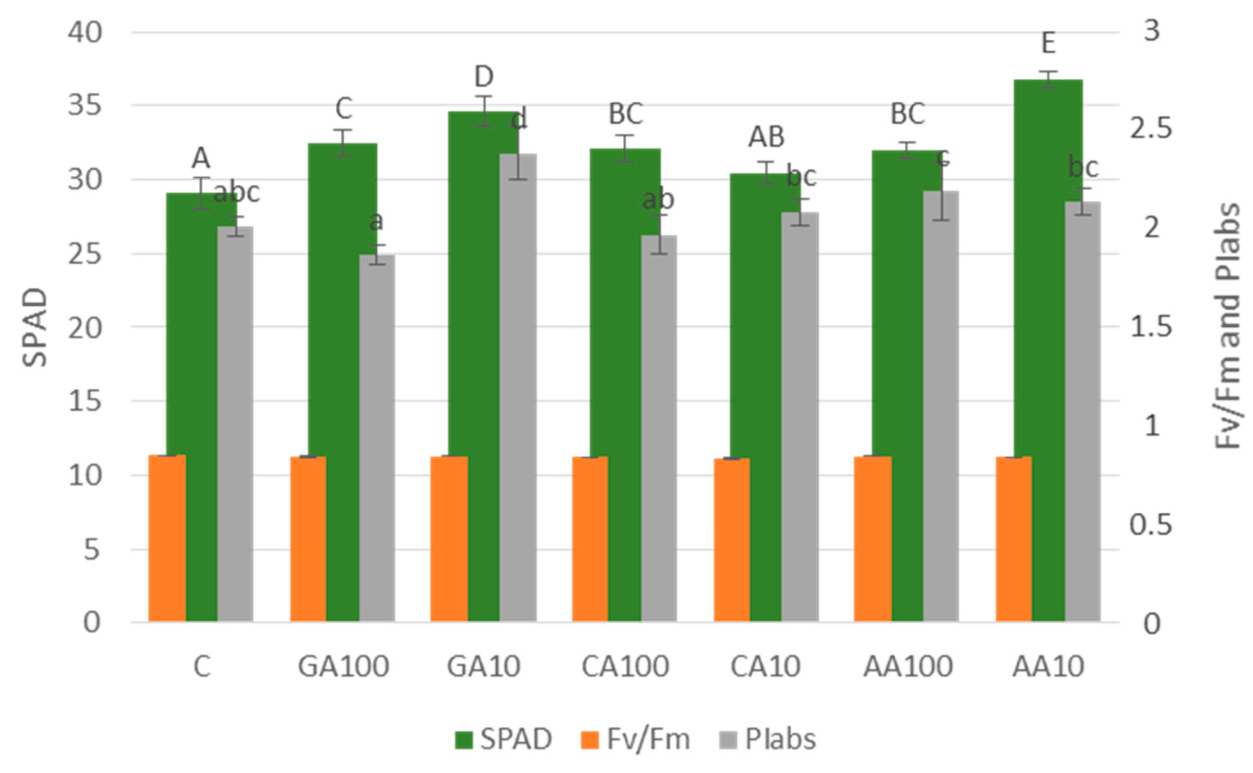
3.3.1. Nano-Priming Treatments Significantly Improved the Chlorophyll Index of Cucumber Seedlings

To estimate the chlorophyll content in leaves, the chlorophyll index (SPAD) was employed. As depicted in Figure 6, significant differences were observed between the control and AgNPs treatments. The lowest SPAD values belong to the control treatment, significantly (p < 0.05) surpassed by all nanoparticle treatments, except CA-AgNPs (10 mM). Noteworthy is the minimal difference between the 100 μM and 10 mM treatments for most reducing agents, except for AA-AgNPs, where differences are more pronounced.

3.3.2. Chlorophyll Fluorescence

Two parameters, maximum photosystem II photochemical efficiency (Fv/Fm) and potential photosynthetic index of photosystem II (PIabs), were considered to determine chlorophyll fluorescence. Fv/Fm values are relatively consistent across treatments, with no significant differences observed between control, AA-AgNPs (100 μM), and GA-AgNPs treatments (Figure 6). PIabs shows significant differences between treatments with and without nanoparticles. The highest and lowest values are within Gallic acid-derived AgNPs treatments. The control value lies in between other treatments, showing significant differences only with CA-AgNPs and AA-AgNPs (10 mM) treatments.

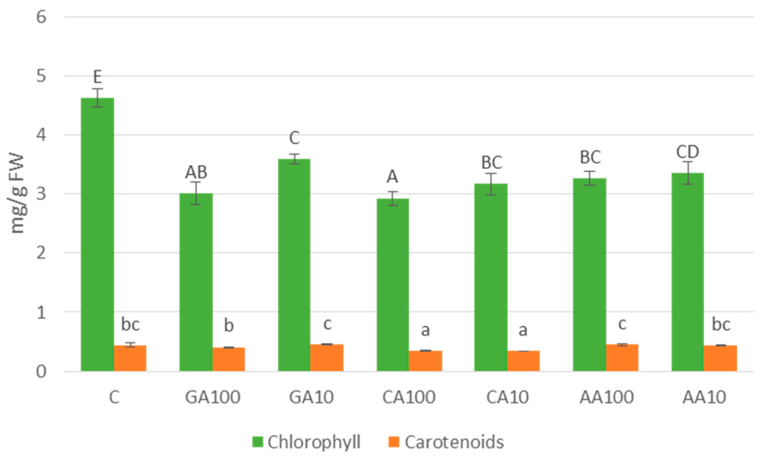
3.3.3. Photosynthetic Pigment Content: Chlorophyll and Carotenoids

For a comprehensive understanding of the photosynthetic process, the total content of chlorophylls and carotenoids in cucumber seedling aerial parts was measured.
Regarding chlorophyll content (Figure 7), the water-control treatment stands out with significantly higher values than all AgNPs treatments. Among nanoparticle treatments, 100 μM treatments consistently showed lower values than 10 mM treatments. Notably, GA-AgNPs (10 mM) exhibited the highest value among all nanoparticle treatments.
As for carotenoid content (Figure 7), significant differences were found between the water-control and CA-AgNPs treatments. The highest recorded value belongs to GA-AgNPs (10 mM), slightly surpassing the control and AA-AgNPs treatments. CA-AgNPs treatments have the lowest carotenoid content with no significant differences between both tested concentrations.

3.4. Biochemical Parameters

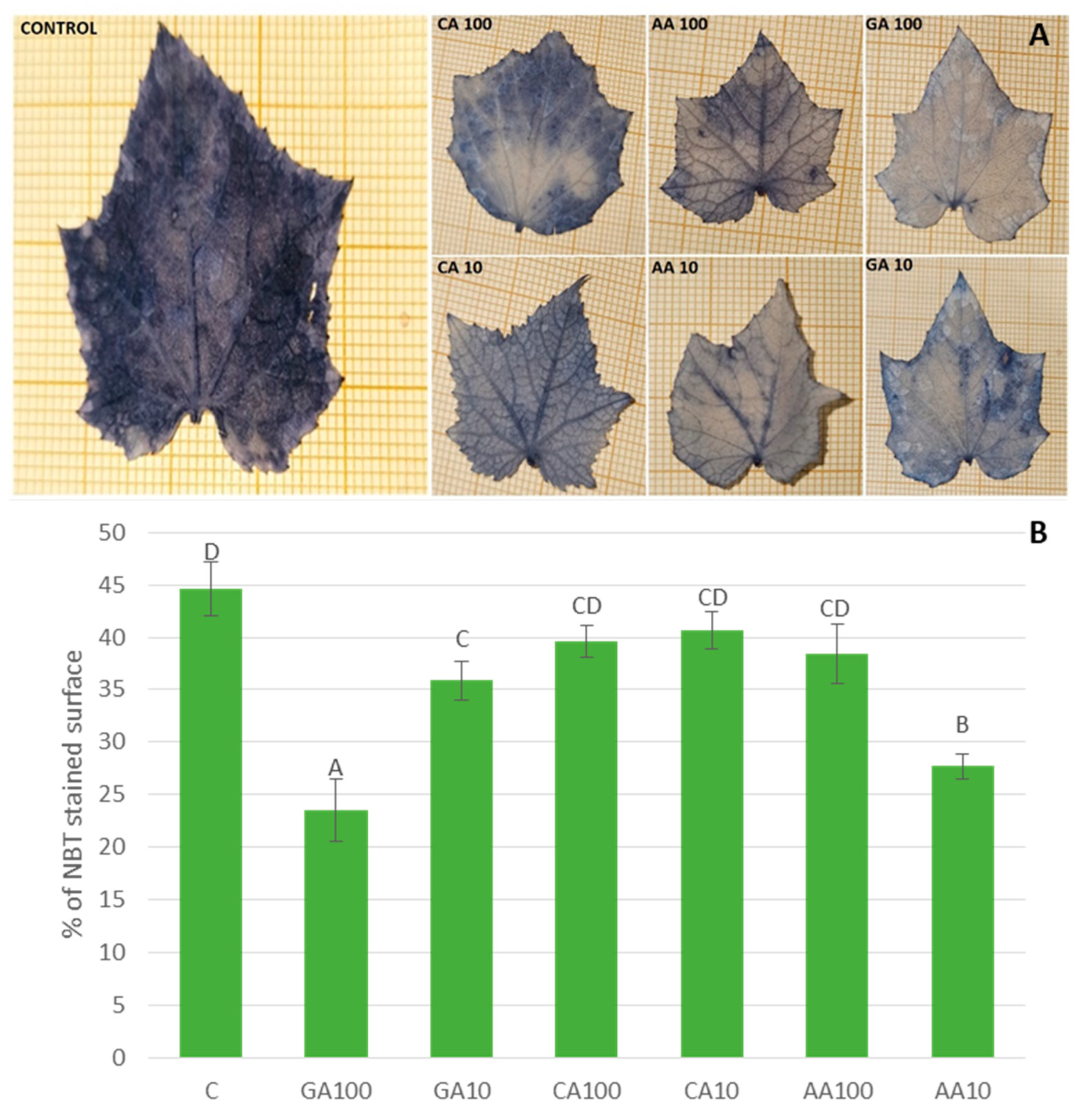
3.4.1. Malondialdehyde Content Varied with Different Nano-Priming Treatments, Indicating Changes in Oxidative Stress Levels

Results indicate a larger stained (NBT) area in control plant leaves compared to leaves from nanoparticle treatments (Figure 8A). A greater stained surface signifies a higher concentration of superoxide anion, indicating increased oxidative stress. Figure 8B illustrates the oxidative stress percentages for each nanopriming treatment. Control seedlings and those treated with CA-AgNPs exhibit significantly higher oxidative stress compared to other treatments. GA-AgNPs (10 mM) and AA-AgNPs (100 μM) treatments show high percentages, slightly lower than those mentioned earlier.
Malondialdehyde (MDA) content, a cellular oxidative damage indicator, varies significantly among treatments (Figure 9). GA-AgNPs treatments, especially GA-AgNPs (100 μM), show the lowest values, denoted by treatment GA100 and GA10. Conversely, CA-AgNPs treatments, particularly CA-AgNPs (10 mM), exhibit the highest malondialdehyde content. Treatments AA100 and AA10 display intermediate MDA levels. These results reflect significantly higher cellular oxidative damage in CA-AgNPs nanopriming treatments and AA-AgNPs (10 mM) compared to the water-control. GA-AgNPs (100 μM) seedlings demonstrate significantly lower oxidative damage compared to other treatments.

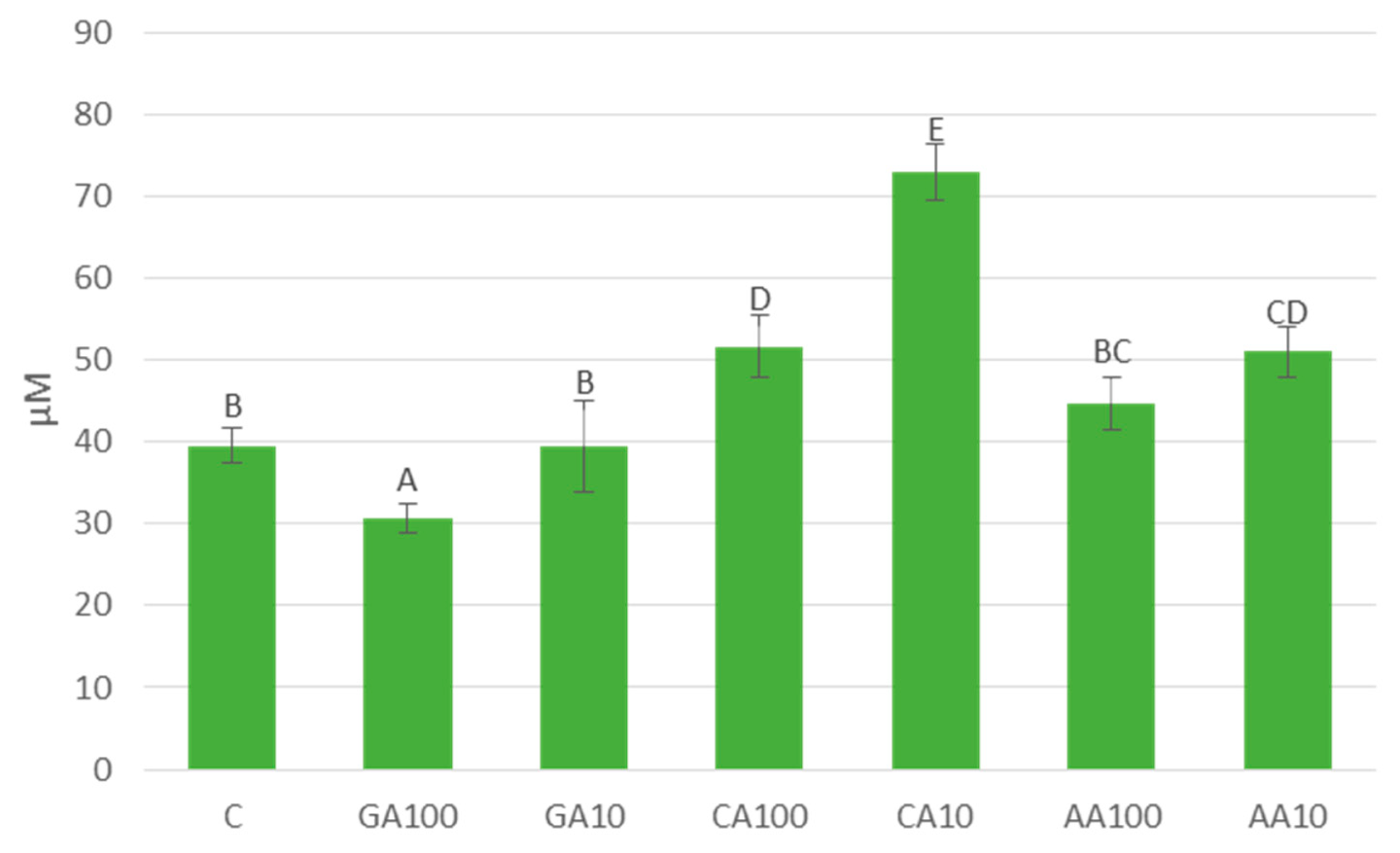
3.4.2. Silver Nanoparticles Induced Significant Changes in Free Amino Acids Content in Cucumber Seedlings

The content of free amino acids, crucial indicators of cellular metabolism, also varies significantly among treatments (Figure 10). The water-control treatment exhibits the highest content of free amino acids (56.71 mg/g FW), followed by treatments GA10 and CA10. Conversely, the lowest levels are observed in treatment CA100. Treatments GA100, AA100, and AA10 display intermediate levels of free amino acids.

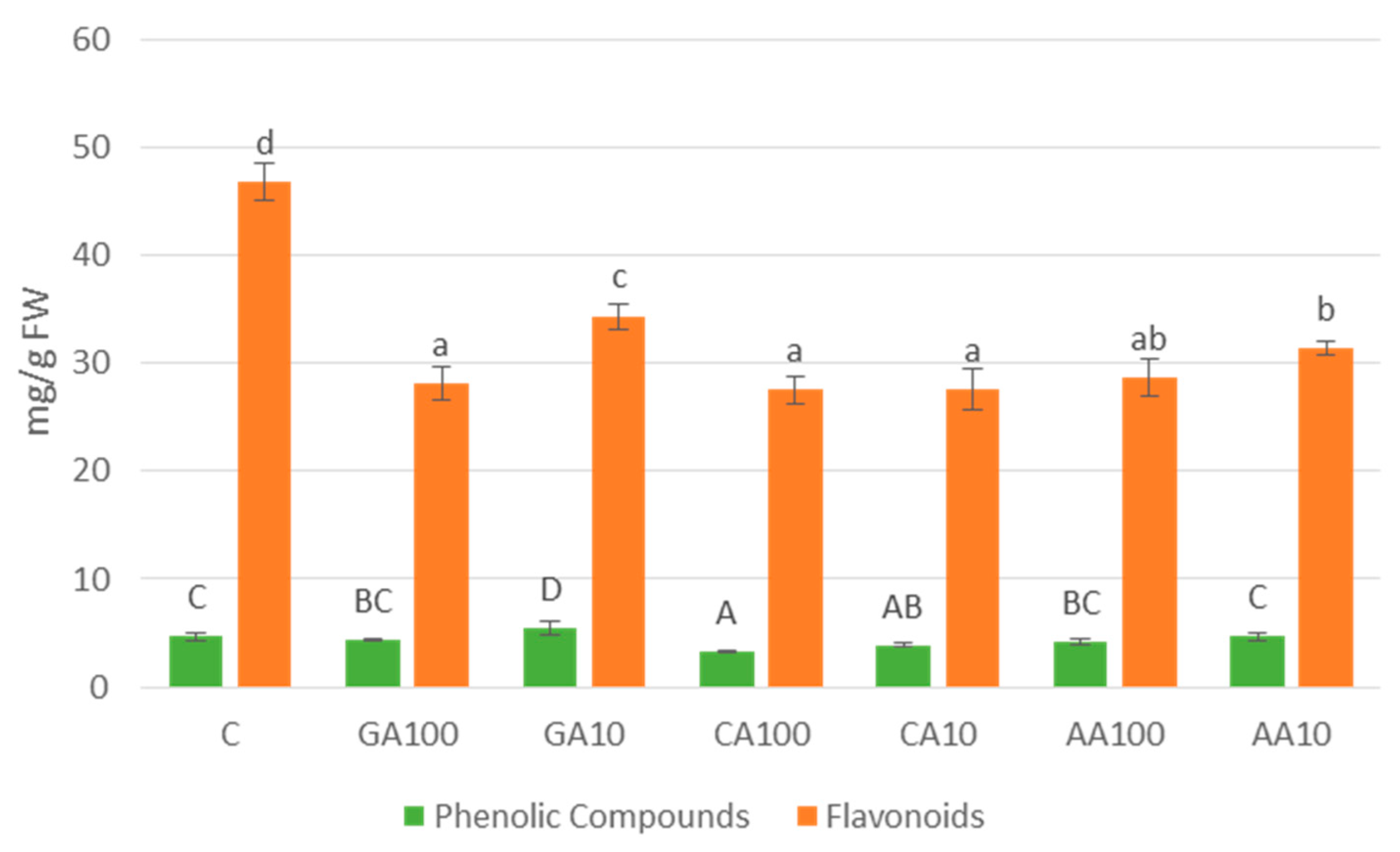
3.4.3. Variation in Total Phenolic Compounds and Flavonoid Contents Observed across Different Nano-Priming Treatments

The content of phenolic compounds and flavonoids, key secondary metabolites with various physiological roles, exhibits significant variability across treatments (Figure 11). Phenolic compound content ranges from 3.27 mg/g FW in treatment CA100 to 5.44 mg/g FW in treatment GA10, while flavonoid content ranges from 2.75 mg/g FW in treatment CA10 to 3.67 mg/g FW in treatment C. Notably, treatments GA10, GA100, and CA10 display relatively higher levels of phenolic compounds compared to other treatments. Conversely, treatment CA100 exhibits the lowest phenolic compound content. Similarly, treatments GA10, GA100, and CA10 also exhibit relatively higher levels of flavonoids compared to other treatments.

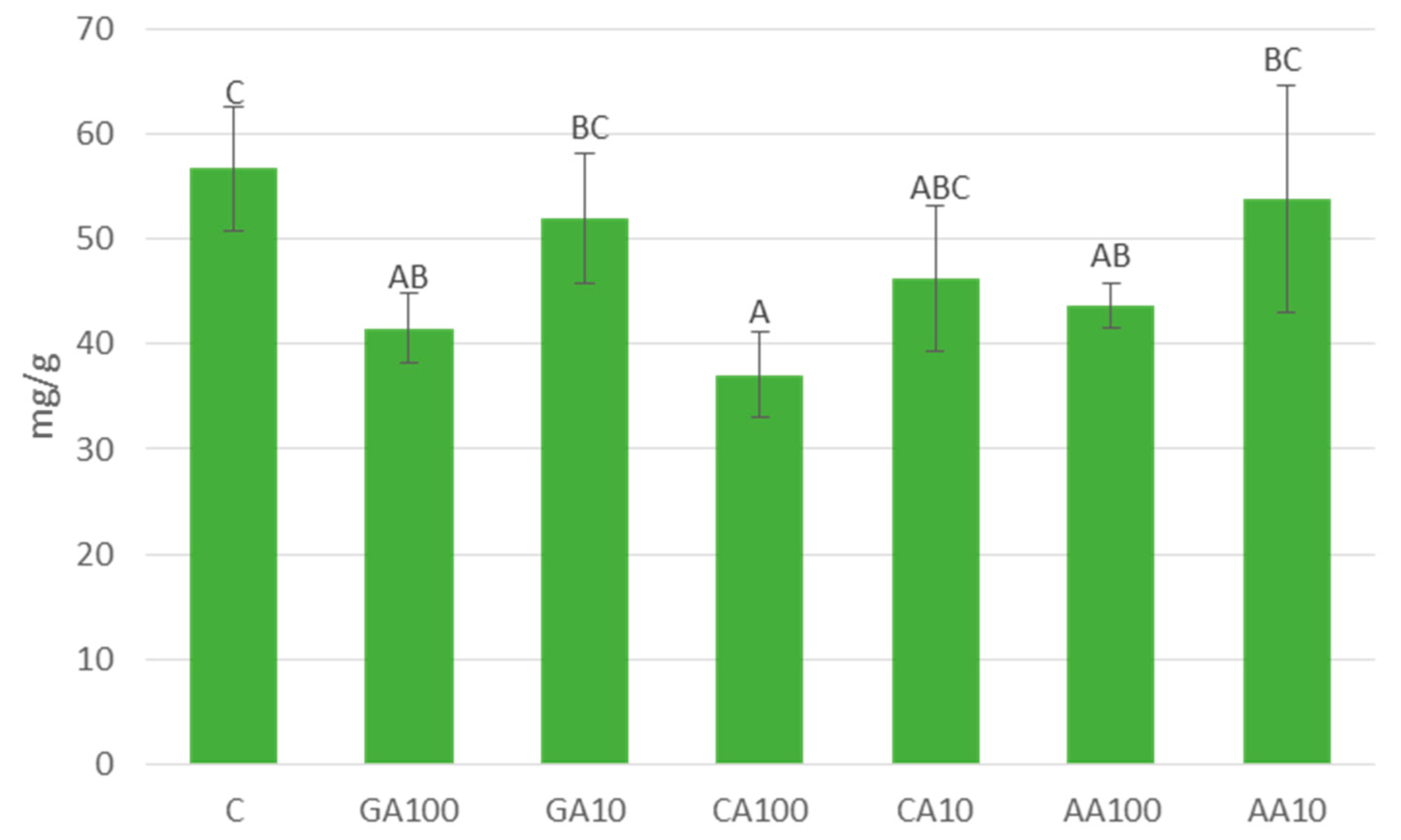
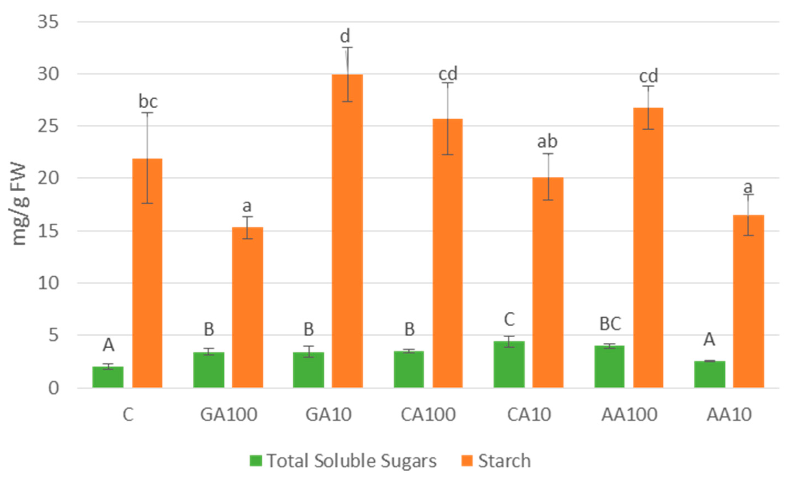
3.4.4. Nano-Priming Treatments Altered Total Soluble Sugars and Starch Contents in Cucumber Seedlings

The content of total soluble sugars and starch, vital components of carbohydrate metabolism, displays significant variation across treatments (Figure 12). Total soluble sugars content ranges from 2.00 mg/g FW in treatment C to 4.42 mg/g FW in treatment CA10, while starch content ranges from 15.34 mg/g FW in treatment GA100 to 29.97 mg/g FW in treatment CA10. Notably, treatments CA10 and CA100 exhibit relatively higher levels of total soluble sugars compared to other treatments. Conversely, treatment C displays the lowest total soluble sugars content. Regarding starch content, treatments CA10 and CA100 also display relatively higher levels compared to other treatments, with treatment CA10 (29.97 mg/g FW) exhibiting the highest starch content. Conversely, treatment GA100 exhibits the lowest starch content (15.34 mg/g FW).

4. Discussion

The nanoparticles synthesized with Ascorbic acid, Caffeic acid, and Gallic acid all exhibit a spherical morphology, as observed in the characterization studies. The spherical shape is a common trait across the various reducing agents used in the synthesis process [17]. This characteristic is noteworthy, as the spherical morphology often contributes to the stability and biocompatibility of nanoparticles [18]. The uniformity in shape may have implications for the interactions of these nanoparticles with biological systems, influencing their potential applications in fields such as medicine and agriculture.
In the case of Gallic acid, the synthesis resulted in grey nanoparticles with a prominent peak at 440 nm, suggesting the presence of 70 nm-sized particles. The observed optical properties hint at potential applications in fields that are sensitive to specific wavelengths, showcasing the versatility of Gallic acid-induced nanoparticles. On the other hand, ascorbic acid-mediated synthesis yielded gold-colored nanoparticles with distinct plasmon resonance bands at 402 nm, 421 nm, and 467 nm. Caffeic acid-induced nanoparticles exhibited an immediate reaction and varied particle sizes, highlighting the influence of its aromatic structure on nanoparticle characteristics. These unique optical characteristics contribute to the distinctive coloration of the nanoparticles and may have implications for applications in sensing or imaging [19,20], where specific wavelengths are crucial for optimal performance. The variation in optical features between Gallic acid, Ascorbic acid, and Caffeic acid-synthesized nanoparticles underscores the significance of choosing the appropriate reducing agent based on the desired nanoparticle properties for specific applications.
The FTIR signatures of the reducing agents, Gallic acid, Ascorbic acid, and Caffeic acid, along with the corresponding silver nanoparticles, revealed intriguing similarities and distinctions. In the case of Ascorbic acid, remarkable parallels were observed, including stretching vibrations of the C=C bond, enol-hydroxyl peak, and O–H (hydroxyl) stretching bond, along with characteristic bands between 500 and 600 cm−1, indicative of silver oxide lattice vibrations. Likewise, Gallic acid-coated nanoparticles exhibited distinct peaks corresponding to functional groups in Gallic acid, emphasizing the surface interaction and stabilization of nanoparticles. Caffeic acid-induced nanoparticles showcased a unique FTIR spectrum, highlighting the influence of its aromatic structure on nanoparticle characteristics. The adsorption of oxidized Caffeic acid onto the silver nanoparticles was confirmed by distinctive peaks and a broad band indicating silver oxide. The XRD analysis further identified various oxidation states of silver, suggesting a potential role for Ascorbic, Gallic, and Caffeic acids as capping agents in nanoparticle synthesis [17].
While some studies suggest that the size and surface characteristics of nanoparticles may influence their interaction with living organisms, it is important to acknowledge that specific effects can vary depending on factors such as the type of nanoparticles, concentration, exposure duration, and the biological system in question. In our study, we aim to investigate whether the differences in size and capping agents of nanoparticles synthesized with Gallic acid, Ascorbic acid, and Caffeic acid could have implications for their interaction with biological systems, such as plants.
The specific mechanisms underlying the interactions of nanoparticles within seeds are still being explored. The germination results of cucumber seeds showed a consistently high germination percentage across all nanopriming treatments, similar to what has been observed in other studies [21]. Notably, the AgNO3 control exhibited no seed germination, making it an effective negative control [22]. The minor differences in germination percentages between the 100 μM and 10 mM treatments suggest a concentration-dependent response, with slightly lower percentages observed in the 100 μM treatments.
The treatment of plants with silver nanoparticles (AgNPs) could enhance overall growth and development by interacting with proteins, enzymes, and carbohydrates that influence the production of plant growth regulators [23]. These regulators play crucial roles in cell division, elongation, and cellular processes such as electron transport and the regulation/production of substances like ethylene or reactive oxygen species. The complex interaction of these processes is reflected in variations in total biomass development, affecting both roots and shoots.
Indeed, when examining morphological parameters such as shoot and root length, a notable positive impact of nanopriming on cucumber seedling development was observed. The significant differences between treatments with and without nanoparticles underscore the potential impact of nanopriming on cucumber seedling morphology, similar to what has been demonstrated in other species [13].
The effects of silver nanoparticles (AgNPs) are not uniform across all parts of the plant. Roots appear to exhibit a more significant response to nanoparticle treatments compared to shoots, which could be explained by the fact that roots are the first tissues to come into direct contact with silver nanoparticles [24] and are responsible for the continuous absorption of nutrients.
It is particularly noteworthy that the shorter shoot length in the control treatment, compared to the nanoparticle treatments, suggests a positive influence on shoot development. The enhanced growth observed in seedlings exposed to silver nanoparticles (AgNPs), especially in treatments with Caffeic acid (CA) and Gallic acid (GA), suggests a positive influence on the development of both shoots and roots. Our results, which demonstrate the positive effects of nanoparticles in stimulating growth, align with previous studies that highlight the benefits of nanopriming for seed germination and seedling development [13,25].
Although the mechanisms underlying the observed effects are not yet fully understood, interactions between nanoparticles and proteins, enzymes, and carbohydrates could influence the production of plant growth regulators, affecting cell division, elongation, and other cellular processes [23,26].
The variations between reducing agents highlight the importance of understanding nanoparticle characteristics and their influence on morphological outcomes [23]. While the literature reports diverse results in nanoparticle–seed interactions, our findings align with studies showing positive effects on seed germination and seedling development. The results consistently demonstrated that silver nanoparticles (AgNPs) synthesized with Gallic acid (GA-AgNPs) significantly improved shoot and root length in cucumber seedlings compared to other treatments and controls. This suggests that GA-AgNPs could enhance cell elongation and division, possibly due to their impact on the production of plant growth regulators [27].
However, it is important to recognize that these positive effects are not universal; other studies have reported negative or inhibitory responses in different plant species and with various nanoparticles [28,29,30]. This underscores the need for a nuanced understanding of nanoparticle characteristics, such as size and concentration, as well as their specific interactions with different plant species [31,32], to optimize their use in agriculture.
Although the aerial part has shown notable growth in nanoparticle treatments, it is crucial to evaluate how efficient this development is in terms of its photosynthetic capacity. It is important to consider that, although the Fv/Fm values in several treatments suggest potential damage to the photosynthetic apparatus (Fv/Fm value of less than 0.8, reflecting damage to the reaction center of photosystem II [26], the SPAD values do not show a drastic decrease. This seemingly contradictory behavior may be due to factors affecting SPAD readings. SPAD measurements, which offer an indirect estimate of chlorophyll content, can be influenced by leaf structure, thickness, and water content, which might alter light absorbance and, therefore, the SPAD measurement. Additionally, uneven chlorophyll distribution within the leaf could also affect SPAD values independently of the actual chlorophyll concentration [32].
These factors suggest that discrepancies between SPAD values and chlorophyll content could reflect complex interactions influenced by treatment conditions, underscoring the need for further investigation into leaf physiology and measurement techniques. Therefore, although the decrease in Fv/Fm indicates that there may be some level of damage to the photosynthetic apparatus, this suggests that the plants might be experiencing moderate stress or are somehow compensating for the negative effects on their photosynthetic apparatus.
A deeper understanding of the physiological aspects, particularly ROS generation, is necessary to unravel the potential oxidative stress and cellular damage induced by AgNPs, in which the dosage and size of AgNPs also play a crucial role, as an excess can lead to phytotoxicity [13].
The primary mechanism underlying AgNP-induced phytotoxicity is the overproduction of ROS, leading to oxidative stress in plant cells. Four types of ROS, including singlet oxygen, superoxide, hydrogen peroxide, and hydroxyl radical, play a key role in this process [33]. The excessive production of ROS under stress conditions can result in lipid peroxidation, damaging cell membrane permeability, altering cellular structure, and directly affecting proteins and DNA, ultimately leading to cell death and growth inhibition in plants [34].
The percentage of oxidative stress observed in our study differs significantly from recent literature, with notable variations between the control treatment and the AG100 and AA10 treatments. Interestingly, the remaining nanoparticle treatments exhibit percentages similar to the control, suggesting that those gNPs do not induce oxidative stress, and, in the case of GA100 and AA10, actually reduce oxidative stress significantly. This aligns with findings in tobacco plants, where low levels of ROS were observed after AgNP treatments [35]. Similarly, in a rice seedling study, exposure to AgNPs resulted in lower ROS levels compared to untreated seedlings, potentially contributing to growth stimulation [36].
The variation in malondialdehyde (MDA) content among the treatments indicates significant oxidative damage at the cellular level, with AC and AA10 nanopriming treatments inducing higher oxidative damage compared to the control. In contrast, the GA100 treatment shows significantly lower oxidative damage. This aligns with previous studies reporting increased MDA formation in soybeans (Glycine max L.) and Arabidopsis thaliana L. after exposure to AgNPs [37,38]. However, the results challenge the expected trend, as gallic acid-derived nanoparticles in our study show lower MDA values. Our study reveals complex dynamics in ROS production and oxidative stress. While some treatments induce oxidative damage, others mitigate it, challenging conventional expectations.
The ability of GA-AgNPs to enhance antioxidant systems and mitigate oxidative stress suggests that crops treated with these nanoparticles could exhibit increased resistance to abiotic stresses such as drought, salinity, and extreme temperatures. Furthermore, the improved uptake and utilization of nutrients observed in this study could lead to more efficient nutrient management, reducing the need for excessive fertilizer application and thereby minimizing environmental pollution. The concentration-dependent nature of these effects underscores the importance of optimal nanoparticle dosing to avoid potential phytotoxic effects [13].
Surprisingly, the free amino acid content was significantly higher in the control treatment compared to the AgNPs 100 μM treatments. Notably, AC treatments consistently reported the lowest levels of free amino acids, suggesting a potential negative impact of AgNPs. These results diverge from other literature’s findings, where interactions with nanoparticles typically lead to an overall increase in amino acid content. Studies in tomato (Lycopersicon esculentum Mill.) plants [39] and Arabidopsis thaliana [40] have reported linear increases in amino acids, particularly in response to stress. However, contrasting results exist, such as in wheat (Triticum aestivum L.) and rice, where exposure to AgNPs did not induce significant changes in amino acid content [17].
In line with our results, specific cases have been documented where certain amino acids experienced substantial decreases following AgNP exposure. In a cucumber study, exposure to AgNPs resulted in a significant dose-dependent reduction of glycine and asparagine content [41], paralleling our findings. These amino acids are integral to plant metabolism, and their alteration can have profound implications for the physiological state of the plant.
The recorded phenolic compound contents exhibited considerable heterogeneity among treatments. Notably, the GA10 treatment demonstrated the highest phenolic compound content, slightly surpassing the control treatment. In contrast, treatments with other AgNP concentrations, except for AC treatments, showed similar phenolic compound levels. These findings align with studies on watermelon (Citrullus lanatus (Thunb.) Matsum. y Nakai) [42] and kale (Brassica oleracea var. sabellica L.) [43], suggesting that most AgNP treatments may not significantly affect phenolic compound content. A positive impact on phenolic content was observed in the GA10 treatment, consistent with studies on fava beans (Phaseolus vulgaris L.) [44], tomatoes [45], and potatoes (Solanum tuberosum L.) [46].
Flavonoid contents displayed marked differences between the control treatment and AgNP treatments. The control treatment exhibited significantly higher flavonoid levels compared to AgNP treatments, with GA10 reporting the highest flavonoid content among nanoparticle treatments. This reduction in flavonoid content aligns with findings in other species, where AgNP treatments resulted in significantly lower flavonoid content compared to controls [45,46]. However, contrasting results have been documented in tomatoes and potatoes, where AgNPs induced a slight increase in flavonoid content [45,47].
Significant differences in total soluble sugar content were observed among treat-ments, with all nanoparticle treatments showing higher levels compared to the control. CA10 exhibited the most substantial increase, suggesting a clear positive effect of AgNPs on soluble sugar content. Similar findings in studies by Kumar et al. [4] and Salih et al. [45] support this positive impact, with the former reporting a 20% increase in total soluble sugar content in winged bean seedlings treated with AgNPs.
Contrasting results have been reported in studies on lemons, where increasing AgNP concentrations led to a progressive reduction in total sugar content [48]. Additionally, a significant decrease in total and reducing sugar content was observed in rice seedlings exposed to AgNPs [49]. These discrepancies emphasize the need for a nuanced understanding of the specific effects of AgNPs on different plant species.
Starch content varied significantly among treatments, with no clear correlation between nanoparticle concentration or type and starch levels. Each antioxidant agent demonstrated a treatment that significantly surpassed the control in starch content. This suggests that the effects on starch content depend on the nanoparticle type and concentration. A study on common beans and corn by Salama [50] provided a similar pattern, where different concentrations of AgNPs yielded diverse results in carbohydrate content.
The results of this study underscore the significant impact that the choice of reducing agent and concentration has on the outcomes of silver nanoparticle (AgNP) treatments in cucumber seedlings. Silver nanoparticles synthesized with Gallic acid (GA-AgNPs) at a concentration of 10 mM consistently demonstrated the most favorable effects across multiple parameters, including shoot and root lengths, chlorophyll content, and oxidative stress reduction. These findings suggest that GA-AgNPs could be highly effective in enhancing plant growth and stress resilience, potentially making them a valuable tool in agricultural practices, particularly in environments subjected to abiotic stresses like drought or high salinity.
Conversely, Caffeic acid-synthesized nanoparticles (CA-AgNPs) at the same concentration resulted in the highest starch content but also induced increased oxidative stress. This dual effect indicates that while CA-AgNPs might promote carbohydrate storage, they could also lead to higher oxidative damage under certain conditions. The accumulation of phenolic compounds and flavonoids, observed predominantly in CA-AgNP-treated plants, further supports their potential role in enhancing plant defense mechanisms. However, the associated oxidative stress could limit their applicability, highlighting the need for careful consideration when selecting CA-AgNPs for specific agricultural purposes.
Ascorbic acid-synthesized nanoparticles (AA-AgNPs) demonstrated intermediate effects across most of the evaluated parameters, such as shoot and root growth, oxidative stress, and chlorophyll content. While AA-AgNPs did not outperform the other treatments in any specific category, their balanced profile suggests a more generalized benefit, potentially offering a moderate improvement in both growth and stress resistance without the extremes observed with GA- and CA-AgNPs.
These differential effects imply that the selection of AgNP type and concentration can be strategically tailored to meet specific agricultural objectives. For example, GA-AgNPs could be particularly advantageous in enhancing growth and reducing oxidative stress in crops exposed to harsh environmental conditions. In contrast, CA-AgNPs might be more suitable for boosting starch content and defense mechanisms, albeit with the trade-off of increased oxidative stress. AA-AgNPs could serve as a more balanced option, providing moderate benefits across a range of physiological processes.
In conclusion, this study not only reveals the nuanced roles of different AgNPs in plant physiology but also emphasizes the importance of optimizing nanopriming treatments based on the specific needs of the crop and the environmental context. Future research could further refine these findings by exploring the long-term effects of these treatments and their applicability to other crops and stress conditions.
Further research is essential to decipher the intricacies of nanoparticle-plant interactions and their physiological consequences, contributing to the broader understanding of nanomaterial applications in agriculture.

5. Conclusions

In conclusion, our study shows the diverse impacts of nanopriming with silver nanoparticles (AgNPs) synthesized using Gallic acid, Ascorbic acid, and Caffeic acid on cucumber seed germination and seedling development. Nanopriming treatments significantly enhanced cucumber seedling development, as evidenced by increased shoot and root lengths compared to the control. The observed variations among reducing agents underscore the importance of understanding nanoparticle characteristics in influencing morphological outcomes. Moreover, our findings shed light on the biochemical responses of cucumber seedlings to nanopriming treatments. While nanopriming induced alterations in oxidative stress and malondialdehyde (MDA) content, specific treatments exhibited mitigation or exacerbation of oxidative damage, challenging conventional expectations. The analysis of free amino acids and secondary metabolites revealed intricate responses to nanopriming treatments, highlighting the potential implications for plant physiology and metabolism. Contrasting results among treatments underscore the complexity of nanoparticle–plant interactions. Our study offers valuable insights into both the potential benefits and challenges associated with nanoparticle applications in agriculture, with implications for the optimization of nanopriming protocols, highlighting the necessity for further research to elucidate specific molecular mechanisms and optimize application strategies.

Author Contributions

Conceptualization, A.G.-G. and B.P.; methodology, B.P.; validation, A.G.-G. and B.P.; formal analysis, A.G.-G.; investigation, H.d.D.; resources, A.G.-G.; data curation, B.P.; writing—original draft preparation, H.d.D.; writing—review and editing, A.G.-G.; visualization, B.P.; supervision, B.P. All authors have read and agreed to the published version of the manuscript.

Funding

This research received no external funding.

Data Availability Statement

Data are contained within the article.

Acknowledgments

We thank Raquel Alonso Valenzuela for her technical assistance and to the Centro Nacional de Microscopía Electrónica, the Research Support Centre (CAI) for Spectroscopy and Correlation (CESCO) and the Research Support Centre (CAI) for X-ray Diffraction of the Universidad Complutense de Madrid. Graphical abstract: Created with BioRender.com (accessed on 28 July 2024).

Conflicts of Interest

The authors declare no conflicts of interest.

References

  1. Batool, S.U.; Javed, B.; Sohail; Zehra, S.S.; Mashwani, Z.-u.-R.; Raja, N.I.; Khan, T.; ALHaithloul, H.A.S.; Alghanem, S.M.; Al-Mushhin, A.A.M.; et al. Exogenous Applications of Bio-fabricated Silver Nanoparticles to Improve Biochemical, Antioxidant, Fatty Acid and Secondary Metabolite Contents of Sunflower. Nanomaterials 2021, 11, 1750. [Google Scholar] [CrossRef]
  2. León-Silva, S.; Fernández-Luqueño, F.; López-Valdez, F. Silver Nanoparticles (AgNP) in the Environment: A Review of Potential Risks on Human and Environmental Health. Water Air Soil Pollut. 2016, 227, 306. [Google Scholar] [CrossRef]
  3. Elgorban, A.M.; El-Samawaty, A.E.-R.M.; Yassin, M.A.; Sayed, S.R.; Adil, S.F.; Elhindi, K.M.; Bakri, M.; Khan, M. Antifungal silver nanoparticles: Synthesis, characterization and biological evaluation. Biotechnol. Biotechnol. Equip. 2016, 30, 56–62. [Google Scholar] [CrossRef]
  4. Kumar, V.K.; Muthukrishnan, S.; Rajalakshmi, R. Phytostimulatory effect of phytochemical fabricated nanosilver (AgNPs) on Psophocarpus tetragonolobus (L.) DC. seed germination: An insight from antioxidative enzyme activities and genetic similarity studies. Curr. Plant Biol. 2020, 23, 100158. [Google Scholar] [CrossRef]
  5. Mishra, S.; Singh, H.B. Biosynthesized silver nanoparticles as a nanoweapon against phytopathogens: Exploring their scope and potential in agriculture. Appl. Microbiol. Biotechnol. 2015, 99, 1097–1107. [Google Scholar] [CrossRef]
  6. Shelar, A.; Singh, A.V.; Maharjan, R.S.; Laux, P.; Luch, A.; Gemmati, D.; Tisato, V.; Singh, S.P.; Santilli, M.F.; Shelar, A.; et al. Sustainable agriculture through multi-disciplinary seed nanopriming: Prospects of opportunities and challenges. Cells 2021, 10, 2428. [Google Scholar] [CrossRef]
  7. Abdelghany, T.M.; Al-Rajhi, A.M.H.; Al Abboud, M.A.; Alawlaqi, M.M.; Magdah, A.G.; Helmy, E.A.M.; Mabrouk, A.S. Recent Advances in Green Synthesis of Silver Nanoparticles and Their Applications: About Future Directions. A Review. BioNanoScience 2017, 8, 5–16. [Google Scholar] [CrossRef]
  8. Ovais, M.; Khalil, A.T.; Raza, A.; Khan, M.A.; Ahmad, I.; Islam, N.U.; Saravanan, M.; Ubaid, M.F.; Ali, M.; Shinwari, Z.K. Green synthesis of silver nanopar-ticles via plant extracts: Beginning a new era in cancer theranostics. Nanomedicine 2016, 12, 3157–3177. [Google Scholar] [CrossRef]
  9. Nejatzadeh, F. Effect of silver nanoparticles on salt tolerance of Satureja hortensis l. during in vitro and in vivo germination tests. Heliyon 2021, 7, e05981. [Google Scholar] [CrossRef]
  10. Oukarroum, A.; Barhoumi, L.; Pirastru, L.; Dewez, D. Silver nanoparticle toxicity effect on growth and cellular viability of the aquatic plant Lemna gibba. Environ. Toxicol. Chem. 2013, 32, 902–907. [Google Scholar] [CrossRef]
  11. Thuesombat, P.; Hannongbua, S.; Akasit, S.; Chadchawan, S. Effect of silver nanoparticles on rice (Oryza sativa L. cv. KDML 105) seed germination and seedling growth. Ecotoxicol. Environ. Saf. 2014, 104, 302–309. [Google Scholar] [CrossRef]
  12. Mallik, J.; Das, P.; Das, S. Pharmacological activity of Cucumis sativus L.—A complete overview. Asian J. Pharm. Res. Dev. 2013, 1, 1–6. [Google Scholar]
  13. Sharma, P.; Bhatt, D.; Zaidi, M.G.H.; Saradhi, P.P.; Khanna, P.K.; Arora, S. Silver Nanoparticle-Mediated Enhancement in Growth and Antioxidant Status of Brassica juncea. Appl. Biochem. Biotechnol. 2012, 167, 2225–2233. [Google Scholar] [CrossRef] [PubMed]
  14. Callejas, F.R. Tablas de Espectroscopía Infrarroja; Departamento de Física y Química, UNAM (Universidad Nacional Autónoma de México): Mexico City, Mexico, 2000. [Google Scholar]
  15. López-Hidalgo, C.; Meijón, M.; Lamelas, L.; Valledor, L. The rainbow protocol: A sequential method for quantifying pigments, sugars, free amino acids, phenolics, flavonoids and MDA from a small amount of sample. Plant Cell Environ. 2021, 44, 1977–1986. [Google Scholar] [CrossRef]
  16. Liu, G.; Zhang, M.; Jin, Y.; Fan, X.; Xu, J.; Zhu, Y.; Fu, Z.; Pan, X.; Qian, H. The Effects of Low Concentrations of Silver Nanoparticles on Wheat Growth, Seed Quality, and Soil Microbial Communities. Water Air Soil Pollut. 2017, 228, 348. [Google Scholar] [CrossRef]
  17. Al-Zahrani, S.; Astudillo-Calderón, S.; Pintos, B.; Pérez-Urria, E.; Manzanera, J.A.; Martín, L.; Gomez-Garay, A. Role of Synthetic Plant Extracts on the Production of Silver-Derived Nanoparticles. Plants 2021, 10, 1671. [Google Scholar] [CrossRef]
  18. Xie, W.; Guo, Z.; Gao, F.; Gao, Q.; Wang, D.; Liaw, B.-S.; Cai, Q.; Sun, X.; Wang, X.; Zhao, L. Shape-, size- and structure-controlled synthesis and biocompatibility of iron oxide nanoparticles for magnetic theranostics. Theranostics 2018, 8, 3284–3307. [Google Scholar] [CrossRef]
  19. Ghodake, G.; Shinde, S.; Kadam, A.; Saratale, R.G.; Saratale, G.D.; Syed, A.; Shair, O.; Alsaedi, M.; Kim, D.-Y. Gallic acid-functionalized silver nanoparticles as colorimetric and spectrophotometric probe for detection of Al3+ in aqueous medium. J. Ind. Eng. Chem. 2019, 82, 243–253. [Google Scholar] [CrossRef]
  20. Liu, Y.; Lu, Y.; Yang, X.; Zheng, X.; Wen, S.; Wang, F.; Vidal, X.; Zhao, J.; Liu, D.; Zhou, Z.; et al. Amplified stimulated emission in upconversion nanoparticles for super-resolution nanoscopy. Nature 2017, 543, 229–233. [Google Scholar] [CrossRef]
  21. Savithramma, N.; Ankanna, S.; Bhumi, G. Effect of nanoparticles on seed germination and seedling growth of Boswellia ovalifoliolata an endemic and endangered medicinal tree taxon. Nano Vis. 2012, 2, 2. [Google Scholar]
  22. Hojjat, S.S.; Hojjat, H. Effect of Nano Silver on Seed Germination and Seedling Growth in Fenugreek Seed. ETP Int. J. Food Eng. 2015, 1, 106–110. [Google Scholar] [CrossRef]
  23. Rezvani, N.; Sorooshzadeh, A.; Farhadi, N. Effect of nano-silver on growth of saffron in flooding stress. Int. J. Agric. Biosyst. Eng. 2012, 6, 11–16. [Google Scholar]
  24. Štefanić, P.P.; Jarnević, M.; Cvjetko, P.; Biba, R.; Šikić, S.; Tkalec, M.; Cindrić, M.; Letofsky-Papst, I.; Balen, B. Comparative proteomic study of phytotoxic effects of silver nanoparticles and silver ions on tobacco plants. Environ. Sci. Pollut. Res. 2019, 26, 22529–22550. [Google Scholar] [CrossRef]
  25. Yasur, J.; Rani, P.U. Environmental effects of nanosilver: Impact on castor seed germination, seedling growth, and plant physiology. Environ. Sci. Pollut. Res. 2013, 20, 8636–8648. [Google Scholar] [CrossRef]
  26. Gomez-Garay, A.; Pintos, B.; Manzanera, J.A.; Lobo, C.; Villalobos, N.; Martín, L. Uptake of CeO2 Nanoparticles and Its Effect on Growth of Medicago arborea In Vitro Plantlets. Biol. Trace Element Res. 2014, 161, 143–150. [Google Scholar] [CrossRef]
  27. Tripathi, D.; Singh, M.; Pandey-Rai, S. Crosstalk of nanoparticles and phytohormones regulate plant growth and metabolism under abiotic and biotic stress. Plant Stress 2022, 6, 100107. [Google Scholar] [CrossRef]
  28. Gomez-Garay, A.; Pintos, B.; Manzanera, J.A.; Prada, C.; Martin, L.; Galan, J.M.G.Y. Nanoceria and bulk cerium oxide effects on the germination of asplenium adiantum-nigrum spores. For. Syst. 2016, 25, e067. [Google Scholar] [CrossRef]
  29. El-Temsah, Y.S.; Joner, E.J. Impact of Fe and Ag nanoparticles on seed germination and differences in bioavailability during exposure in aqueous suspension and soil. Environ. Toxicol. 2011, 27, 42–49. [Google Scholar] [CrossRef]
  30. Dimkpa, C.O.; McLean, J.E.; Martineau, N.; Britt, D.W.; Haverkamp, R.; Anderson, A.J. Silver nanoparticles disrupt wheat (Triticum aestivum L.) growth in a sand matrix. Environ. Sci. Technol. 2013, 47, 1082–1090. [Google Scholar] [CrossRef]
  31. Mehmood, A. Brief overview of the application of silver nanoparticles to improve growth of crop plants. IET Nanobiotechnol. 2018, 12, 701–705. [Google Scholar] [CrossRef]
  32. Uddling, J.; Gelang-Alfredsson, J.; Piikki, K.; Pleijel, H. Evaluating the relationship between leaf chlorophyll concentration and SPAD-502 chlorophyll meter readings. Photosynth. Res. 2007, 91, 37–46. [Google Scholar] [CrossRef]
  33. Mourato, M.; Reis, R.; Martins, L.L. Characterization of plant antioxidative system in response to abiotic stresses: A focus on heavy metal toxicity. Adv. Sel. Plant Physiol. Asp. 2012, 12, 1–17. [Google Scholar]
  34. Yan, A.; Chen, Z. Impacts of Silver Nanoparticles on Plants: A Focus on the Phytotoxicity and Underlying Mechanism. Int. J. Mol. Sci. 2019, 20, 1003. [Google Scholar] [CrossRef]
  35. Cvjetko, P.; Zovko, M.; Štefanić, P.P.; Biba, R.; Tkalec, M.; Domijan, A.M.; Vrček, I.V.; Letofsky-Papst, I.; Šikić, S.; Balen, B. Phytotoxic effects of silver nanoparticles in tobacco plants. Environ. Sci. Pollut. Res. 2018, 25, 5590–5602. [Google Scholar] [CrossRef]
  36. Gupta, S.D.; Agarwal, A.; Pradhan, S. Phytostimulatory effect of silver nanoparticles (AgNPs) on rice seedling growth: An insight from antioxidative enzyme activities and gene expression patterns. Ecotoxicol. Environ. Saf. 2018, 161, 624–633. [Google Scholar] [CrossRef] [PubMed]
  37. De la Torre-Roche, R.; Hawthorne, J.; Musante, C.; Xing, B.; Newman, L.A.; Ma, X.; White, J.C. Impact of Ag nanoparticle exposure on p,p′-DDE bioaccumulation by Cucurbita pepo (Zucchini) and Glycine max (Soybean). Environ. Sci. Technol. 2013, 47, 718–725. [Google Scholar] [CrossRef] [PubMed]
  38. Nair, P.M.G.; Chung, I.M. Assessment of silver nanoparticle-induced physiological and molecular changes in Arabidopsis thaliana. Environ. Sci. Pollut. Res. 2014, 21, 8858–8869. [Google Scholar] [CrossRef]
  39. Karami Mehrian, S.; Heidari, R.; Rahmani, F. Effect of silver nanoparticles on free amino acids content and antioxidant defense system of tomato plants. Indian J. Plant Physiol. 2015, 20, 257–263. [Google Scholar] [CrossRef]
  40. Liu, Y.-S.; Chang, Y.-C.; Chen, H.-H. Silver nanoparticle biosynthesis by using phenolic acids in rice husk extract as reducing agents and dispersants. J. Food Drug Anal. 2018, 26, 649–656. [Google Scholar] [CrossRef]
  41. Zhang, H.; Du, W.; Peralta-Videa, J.R.; Gardea-Torresdey, J.L.; White, J.C.; Keller, A.A.; Guo, H.; Ji, R.; Zhao, L. Metabolomics Reveals How Cucumber (Cucumis sativus) Reprograms Metabolites To Cope with Silver Ions and Silver Nanoparticle-Induced Oxidative Stress. Environ. Sci. Technol. 2018, 52, 8016–8026. [Google Scholar] [CrossRef]
  42. Acharya, P.; Jayaprakasha, G.K.; Crosby, K.M.; Jifon, J.L.; Patil, B.S. Nanoparticle-Mediated Seed Priming Improves Germination, Growth, Yield, and Quality of Watermelons (Citrullus lanatus) at multi-locations in Texas. Sci. Rep. 2020, 10, 5037. [Google Scholar] [CrossRef]
  43. Tymoszuk, A. Silver Nanoparticles Effects on In Vitro Germination, Growth, and Biochemical Activity of Tomato, Radish, and Kale Seedlings. Materials 2021, 14, 5340. [Google Scholar] [CrossRef] [PubMed]
  44. Younis, M.E.; Abdel-Aziz, H.M.M.; Heikal, Y.M. Nanopriming technology enhances vigor and mitotic index of aged Vicia faba seeds using chemically synthesized silver nanoparticles. S. Afr. J. Bot. 2019, 125, 393–401. [Google Scholar] [CrossRef]
  45. Salih, A.M.; Qahtan, A.A.; Al-Qurainy, F.; Al-Munqedhi, B.M. Impact of Biogenic Ag-Containing Nanoparticles on Ger-mination Rate, Growth, Physiological, Biochemical Parameters, and Antioxidants System of Tomato (Solanum tuberosum L.) In Vitro. Processes 2022, 10, 825. [Google Scholar] [CrossRef]
  46. Bagherzadeh Homaee, M.; Ehsanpour, A.A. Physiological and biochemical responses of potato (Solanum tuberosum) to silver nanoparticles and silver nitrate treatments under in vitro conditions. Indian J. Plant Physiol. 2015, 20, 353–359. [Google Scholar] [CrossRef]
  47. Azeez, L.; Lateef, A.; Adebisi, S.A. Silver nanoparticles (AgNPs) biosynthesized using pod extract of Cola nitida enhances antioxidant activity and phytochemical composition of Amaranthus caudatus Linn. Appl. Nanosci. 2017, 7, 59–66. [Google Scholar] [CrossRef]
  48. Mosa, W.F.; Mackled, M.I.; Abdelsalam, N.R.; Behiry, S.I.; Al-Askar, A.A.-A.; Basile, A.; Abdelkhalek, A.; Elsharkawy, M.M.; Salem, M.Z.M. Impact of Silver Na-noparticles on Lemon Growth Performance: Insecticidal and Antifungal Activities of Essential Oils From Peels and Leaves. Front. Plant Sci. 2022, 13, 898846. [Google Scholar] [CrossRef]
  49. Nair, P.M.G.; Chung, I.M. Physiological and molecular level effects of silver nanoparticles exposure in rice (Oryza sativa L.) seedlings. Chemosphere 2014, 112, 105–113. [Google Scholar] [CrossRef]
  50. Salama, H.M. Effects of silver nanoparticles in some crop plants, common bean (Phaseolus vulgaris L.) and corn (Zea mays L.). Int. Res. J. Biotechnol. 2012, 3, 190–197. [Google Scholar]
Figure 1. TEM image of AgNPs synthesized via the reaction of Gallic acid with AgNO3.
Agronomy 14 01866 g001
Figure 2. TEM image of AgNPs synthesized via the reaction of Caffeic acid with AgNO3.
Agronomy 14 01866 g002
Figure 3. TEM image of AgNPs synthesized via the reaction of Ascorbic acid with AgNO3.
Agronomy 14 01866 g003
Figure 4. Images of cucumber plants subjected to different nanopiming treatments with silver nanoparticles: Control; AA100 (Ascorbic acid + AgNO3 100 μM); AA10 (Ascorbic acid + AgNO3 10 mM); AC100 (Caffeic acid + AgNO3 100 μM); AC10 (Caffeic acid + AgNO3 10 mM); AG100 (Gallic acid + AgNO3 100 μM); AG10 (Gallic acid + AgNO3 10 mM).
Agronomy 14 01866 g004
Figure 5. Mean values of shoot length, root length, shoot–root ratio of cucumber seedlings treated with different silver nanopriming solutions (silver nanoparticles were synthetized by Gallic acid (GA), Caffeic acid (CA), and Ascorbic acid (AA) and 10 mM and 100 μM AgNO3). Different letters denote significant differences between treatments based on Duncan’s multiple range test (p < 0.05).
Agronomy 14 01866 g005
Figure 6. Mean values of SPAD values, Fv/Fm ratio, and PIabs of cucumber seedlings treated with different silver nanopriming solutions (silver nanoparticles were synthetized by Gallic acid (GA), Caffeic acid (CA), and Ascorbic acid (AA) and 10 mM and 100 μM AgNO3). Different letters denote significant differences between treatments based on Duncan’s multiple range test (p < 0.05).
Agronomy 14 01866 g006
Figure 7. Mean values of chlorophyll and carotenoid contents of cucumber seedlings treated with different silver nanopriming solutions (silver nanoparticles were synthetized by Gallic acid (GA), Caffeic acid (CA), and Ascorbic acid (AA) and 10 mM and 100 μM AgNO3). Different letters denote significant differences between treatments based on Duncan’s multiple range test (p < 0.05).
Agronomy 14 01866 g007
Figure 8. Representative NBT-stained leaves of cucumber plants of each nanopriming treatment (A): Control; AA 100 (Ascorbic acid + AgNO3 100 μM); AA 10 (Ascorbic acid + AgNO3 100 mM); AC 100 (Caffeic acid + AgNO3 100 μM); AC 10 (Caffeic acid + AgNO3 100 mM); AG 100 (Gallic acid + AgNO3 100 μM); AG 10 (Gallic acid + AgNO3 100 mM). (B) The histogram represents the quantitative analysis of formazan deposits area in the leaves for the plants from each treatment. Different letters accompanying the data in the figure denote statistically distinct groups (p ≤ 0.05) via Duncan’s test.
Agronomy 14 01866 g008
Figure 9. Mean values of malondialdehyde (MDA) content of cucumber seedlings treated with different silver nanopriming solutions (silver nanoparticles were synthetized by Gallic acid (GA), Caffeic acid (CA), and Ascorbic acid (AA) and 10 mM and 100 μM AgNO3). Different letters denote significant differences between treatments based on Duncan’s multiple range test (p < 0.05).
Agronomy 14 01866 g009
Figure 10. Mean values of free amino acids content of cucumber seedlings treated with different silver nanopriming solutions (silver nanoparticles were synthetized by Gallic acid (GA), Caffeic acid (CA), and Ascorbic acid (AA) and 10 mM and 100 μM AgNO3). Different letters denote significant differences between treatments based on Duncan’s multiple range test (p < 0.05).
Agronomy 14 01866 g010
Figure 11. Mean values of phenolic compounds and flavonoid content of cucumber seedlings treated with different silver nanopriming solutions (silver nanoparticles were synthetized by Gallic acid (GA), Caffeic acid (CA), and Ascorbic acid (AA) and 10 mM and 100 μM AgNO3). Different letters denote significant differences between treatments based on Duncan’s multiple range test (p < 0.05).
Agronomy 14 01866 g011
Figure 12. Mean values of total soluble sugars and starch contents of cucumber seedlings treated with different silver nanopriming solutions (silver nanoparticles were synthetized by Gallic acid (GA), Caffeic acid (CA), and Ascorbic acid (AA) and 10 mM and 100 μM AgNO3). Different letters denote significant differences between treatments based on Duncan’s multiple range test (p < 0.05).
Agronomy 14 01866 g012
Table 1. Influence of silver nanoparticles synthesized with diverse organic acids (Gallic Acid—GA, Caffeic Acid—CA, and Ascorbic Acid—AA) on cucumber seed germination. Evaluation of two nanoparticle concentrations in nanopriming: 100 μM and 10 mM, in comparison with bulk AgNO3.
Priming TreatmentConcentrationGermination (%)
Water-Control-100
Bulk AgNO3100 μM0
10 mM0
GA-AgNPs100 μM100
10 mM100
CA-AgNPs100 μM88.8 ± 9.6
10 mM100
AA-AgNPs100 μM83.3
10 mM100
Disclaimer/Publisher’s Note: The statements, opinions and data contained in all publications are solely those of the individual author(s) and contributor(s) and not of MDPI and/or the editor(s). MDPI and/or the editor(s) disclaim responsibility for any injury to people or property resulting from any ideas, methods, instructions or products referred to in the content.

