Article

Improvement in the Sequential Extraction of Phycobiliproteins from Arthrospira platensis Using Green Technologies

by
Wanida Pan-utai
1,*,
Siriluck Iamtham
2,3,4,
Sumitra Boonbumrung
5 and
Juta Mookdasanit
6
1
Department of Applied Microbiology, Institute of Food Research and Product Development, Kasetsart University, Chatuchak, Bangkok 10900, Thailand
2
Department of Science, Faculty of Liberal Arts and Science, Kasetsart University, Kamphaeng Saen, Nakhon Pathom 73140, Thailand
3
Center for Agricultural Biotechnology, Kasetsart University, Kamphaeng Saen Campus, Nakhon Pathom 73140, Thailand
4
Center of Excellence on Agricultural Biotechnology—(AG-BIO/PERDO-CHE), Bangkok 10900, Thailand
5
Department of Food Chemistry and Physics, Institute of Food Research and Product Development, Kasetsart University, Chatuchak, Bangkok 10900, Thailand
6
Department of Fishery Products, Faculty of Fisheries, Kasetsart University, Chatuchak, Bangkok 10900, Thailand
*
Author to whom correspondence should be addressed.
Life 2022, 12(11), 1896; https://doi.org/10.3390/life12111896
Submission received: 20 October 2022 / Revised: 10 November 2022 / Accepted: 12 November 2022 / Published: 15 November 2022
(This article belongs to the Special Issue Microalgae Metabolites)

Abstract

:
Arthrospira platensis (commercially known as Spirulina) is an excellent source of phycobiliproteins, especially C-phycocyanin. Phycobiliproteins are significant bioactive compounds with useful biological applications. The extraction process plays a significant role in downstream microalga production and utilisation. The important pigments found in A. platensis include chlorophyll and carotenoids as nonpolar pigments and phycobiliproteins as polar pigments. Supercritical fluid extraction (SFE) as a green extraction technology for the high-value metabolites of microalgae has potential for trends in food and human health. The nonpolar bioactive compounds, chlorophyll and carotenoids of A. platensis, were primarily separated using supercritical carbon dioxide (SC-CO2) solvent-free fluid extraction pressure; the temperature and ethanol as cosolvent conditions were compared. The residue from the A. platensis cells was subjected to phycobiliprotein extraction. The phosphate and water extraction of A. platensis SFE residue were compared to evaluate phycobiliprotein extraction. The SFE results exhibited higher pressure (350 bar) and temperature extraction (50 °C) with ethanol-free extraction and increased nonpolar pigment. Phycobiliprotein yield was obtained from A. platensis SFE residue by ethanol-free buffer extraction as a suitable process with antioxidant properties. The C-phycocyanin was isolated and enhanced to 0.7 purity as food grade. This developed method can be used as a guideline and applied as a sustainable process for important pigment extraction from Arthrospira microalgae.

1. Introduction

Arthrospira platensis (commercially known as Spirulina) is a filamentous cyanobacterium and blue-green microalga commonly supplemented in functional foods [1,2], nutraceuticals [3,4] and animal feed [5] and is also used for biofuel production [6]. A. platensis can utilise carbon dioxide as a nutrient source for biomass production [7]. Arthrospira biomass is a rich source of both macro and micronutrients and is used as a food and dietary supplement due to its therapeutic properties such as antioxidant and anti-inflammatory activities [1,8,9]. Arthrospira is also a good source of natural proteins, carbohydrates, lipids, vitamins, enzymes and pigments including chlorophyll, carotenoids and phycocyanin [10,11]. Arthrospira can be cultivated in open systems on a large scale for high biomass production [12], making it a significant and interesting natural source for valuable biosubstances and functional ingredients. The WHO has designated Arthrospira as a superfood due to its potentially rich source of chemicals with biological activities that can also be employed as functional components. Recently, increased knowledge of the properties of the health-boosting nutrients and nutraceuticals has improved lifestyles. As a result, Arthrospira microalgae are now attracting increased interest in the nutritional and food science fields [13].
Arthrospira is also a valuable source of natural pigments including chlorophyll, carotenoids and phycobiliprotein, especially C-phycocyanin (C-PC) [11,14]. The expanded use of Arthrospira has coincided with increased consumer knowledge of the value of natural colourants and their advantages in terms of nutrition, pharmacology and health. As a result, more natural colours are being used, with spirulina serving as a popular source of these pigments, particularly in the food and cosmetic sectors [15]. Chlorophyll is one of the main photosynthetic pigments in natural systems with a role in the photosynthetic process of absorbing light, transferring energy and transporting electrons. Chlorophyll is also utilised in several industries for its photophysical and photochemical capabilities, such as food colouring and optically active centres for luminescent solar concentrators [16,17]. Microalgae are the main source of valuable compounds obtained through photosynthesis [18]. Carotenoids are insoluble in water and found in Arthrospira [19]. The bioavailability of carotenoids from Arthrospira has promising potential as a source of provitamin A which has high antioxidant activity and ameliorates cardiovascular disorders, cancer and anti-aging activity [20]. Natural carotenoids are used in industrial applications as food colourants, feed additives, cosmetics and pharmaceuticals [21].
Phycobiliproteins (PBPs) are the light-harvesting pigment proteins of phycobilisomes (antenna complexes), which act as photosynthetic accessory pigments in cyanobacteria [22]. PBPs can be divided into three groups on the basis of their absorption characteristics as C-phycocyanin (C-PC, blue pigment), allophycocyanin (APC, light-blue pigment) and phycoerythrin (PE, red pigment) [23,24]. Phycobiliproteins are used for various diagnostic scientific research and therapeutic purposes [25] and mainly consist of C-phycocyanin (C-PC) which is a well-known pigment with antioxidant, anti-inflammatory and anticarcinogenic activities [26]. Phycocyanin serves as the main photosynthetic pigment autotroph of Arthrospira [27]. Phycobiliproteins can be used as safe alternatives to synthetic colours, which are frequently poisonous or otherwise dangerous in food, cosmetics and pharmaceutical products [28]. Phycobiliproteins can be extracted using physical and chemically assisted methods; however, various aspects such as organism composition, stability and cell-wall resistance affect the choice of approach [29]. The biosynthetic recovery output of PBPs from biomass should also be considered and various levels of purity are necessary depending on how the PBPs will be used [30]. The primary phycobiliprotein in most blue-green algae is C-phycocyanin (C-PC) [31]. Phycobiliproteins have a market value of USD 5000–33,000 g−1 depending on quality as a natural pigment in the food, cosmetic, medical and biotechnology sectors [32]. The purity grade of phycobiliprotein has a significant impact on its commercial market value [33], with an extract purity of phycobiliprotein at 0.56–0.70 considered as food grade [34], 1.5 as cosmetic grade, 3.9 as reactive grade and greater than 4.0 as analytical grade [35]. Currently, several technologies exist for the commercial extraction of pigments from algae. The colours released from the ruptured cell wall of microalgae can be extracted using organic solvent extraction [36], pressurised solvent extraction [37], ionic liquid extraction, high-pressure homogenisation, ultrasonication [38] and supercritical carbon dioxide fluid extraction [39]. A. platensis is a photosynthetic multicellular blue-green microalga that is cultivated on a large scale for the commercial processing of biomass and bio-products [40]. Microalgae are becoming increasingly important, particularly for their composition because they contain high-value substances such as carotenoids, chlorophylls and phycobiliproteins [41]. Therefore, efficient methods for obtaining pigments and bio-products from microalgae are needed. These bioactive chemicals can be successfully extracted using SC-CO2 extraction as an environmentally friendly method [42].
Supercritical carbon dioxide (SC-CO2) fluid extraction is a green technology aimed at replacing organic solvent extraction [43]. The supercritical fluid extraction (SFE) approach is used by a variety of sectors to extract valuable bioactive chemicals [44]. The SFE technique has been widely employed as a separation technology in the food processing and pharmaceutical sectors for efficient and selective component extraction. SC-CO2 fluid extraction has evolved as a more environmentally friendly method than traditional petroleum-based solvent extraction procedures. SC-CO2 is a solvent that is widely used in SFE [45]. The special properties of SC-CO2 make it appealing for isolating essential oils, neutral lipids, flavours, perfumes, antioxidants and pigments from both terrestrial and non-terrestrial biomasses [46]. Most nonpolar solutes can be separated by SC-CO2 since it is lipophilic. In comparison to solvent extraction, CO2 separation is straightforward and leaves no residues in the extract [47]. Utilisation of the SC-CO2 approach focuses on the extraction of hydrophobic antioxidant chemicals [48]. A small amount of cosolvents can induce microalgal cells to expand, allowing fast mass transfer of analytes from the matrix [49]. The SC-CO2 extraction technique uses pressures and temperatures greater than the CO2 critical point [50]. Several studies have employed the SC-CO2 extraction process to purify active components (carotenoids and linolenic acid), oil and caffeic acid from microalgae [51,52,53]. SFE is a novel environmentally friendly technical method employed in the food and pharmaceutical industries to avoid the harmful organic solvents that use CO2 above its critical temperature and pressure points [54]. The addition of cosolvents induces the expansion of microalgal cells, allowing rapid mass transfer of analytes from the matrix. Carbon dioxide is the most commonly used SFE solvent in the pharmaceutical sector and is classed as safe by the USFDA [55]. The methods and processes of bioactive substance extraction have been improved to maximise the utilisation of biosubstances from microalgae. Nowadays, SFE is a popular green technology to extract nonpolar pigments from microalgae.
Arthrospira platensis is one of the main sources of natural commercial phycobiliproteins, especially C-phycocyanin. Therefore, the objectives of this study were to evaluate the efficiency of (i) extraction processes for nonpolar pigments: chlorophyll and carotenoids, using SFE as a green technology, before water-soluble phycobiliprotein extraction from Arthrospira biomass and (ii) evaluation of the improvement in the sequential phycobiliprotein extraction of cell biomass residues after SFE by comparing phosphate and water extraction with ultrasound-assisted extraction.

2. Materials and Methods

2.1. Microalgal Production

Arthrospira platensis IFRPD 1182 microalgae were prepared by the Institute of Food Research and Product Development, Kasetsart University, Thailand. The starter culture was maintained and prepared in Zarrouk medium [56] composed of (per litre) 16.8 g NaHCO3, 2.5 g NaNO3, 0.5 g K2HPO4, 1.0 g K2SO4, 1.0 g NaCl, 0.2 g Mg2SO4·7H2O 0.04 g CaCl2·7H2O, 0.01 g FeSO4·7H2O and 0.08 g EDTA. One millilitre each of vitamin A5 and B5 micronutrients was added into the medium. The micronutrient solution of A5 was composed of (per litre) 2.86 g H3BO3, 1.81 g MnCl2·4H2O, 0.22 g ZnSO4·7H2O, 0.08 g CuSO4·5H2O and 0.01 g MoO3. The B5 micronutrient solution was composed of (per litre) 22.9 mg NH4VO3, 96.0 mg K3Cr2(SO4)4·24H2O, 47.8 mg NiSO4·7H2O, 17.9 mg Na2WO3, 44.0 mg Co(NO3)2·6H2O and 40 mg Ti2(SO4)3. A. platensis was cultured and incubated in chamber equipment with temperature controlled at 30 °C [57]. Light intensity was controlled at a photon flux density of 162 µmol·m−2·s−1 using fluorescent 18 W daylight lamps with a 16 h/8 h light/dark cycle. Air mixed with 2% (v/v) CO2 at 0.67 vvm was added via continuous bubbling through a PTFE membrane filter. The A. platensis starter was grown for 7–14 days until reaching the log phase and then used at 10% (v/v) for biomass production. A. platensis was produced in Zarrouk medium in 200 L working volume in 500 L raceway ponds with a paddle wheel operated at 15 rpm. Average light photon flux density was 471 µmol·m−2·s−1 during open pond production with batch cultivation. The biomass of A. platensis was grown to the exponential phase for 15–20 days, with biomass concentration reaching 1 g·L−1. Cells were harvested using a 60 µm nylon membrane filter and washed with clean tap water until no residue remained in the culture medium. Then, the harvested cells were oven-dried at 65 °C for 4–6 h in a hot air oven. The oven-dried biomass of A. platensis was milled to 0.5 mm sample size using a mill grinder (ZM-1, Retsch, Haan, Germany) for use in the experiments.

2.2. Chlorophyll and Carotenoid Pigments Using Supercritical Fluid Extraction

Oven-dried A. platensis microalgal biomass was investigated for pigments extracted using SFE as a green technology. The chlorophyll and carotenoid pigments (nonpolar bioactive compounds) in A. platensis were extracted using an SC-CO2 pilot unit with a helix SFE System (Applied Separations Inc., Allentown, PA, USA). An overview of the SC-CO2 system is shown in Figure 1. The system included a solvent and carbon dioxide pump, a back-pressure regulator (BPR), a 1 L extractor vessel enclosed in a heating jacket, a pressure transmitter and a sample collector. The 5 g dry weight of A. platensis oven-dried biomass was added to a high-pressure stainless-steel extractor vessel. The experiments were performed with and without ethanol at 10% (w/w) of samples as the cosolvent. Polypropylene wool was used to mediate the inlet and outlet of the vessel. Static extraction was performed for 60 min, followed by dynamic extraction for 240 min, under various conditions of pressure at 250 and 350 bar with temperatures of 40 and 50 °C. Two main extraction experiments were compared: with and without ethanol as a cosolvent during SFE extraction (Table 1). Optimal conditions were cited from previous studies [58,59]. The flow rate was controlled at 3 litres per minute (LPM). The pigments were extracted and collected into the sample collector. All experiments were performed in duplicate. The extracted samples were kept in the dark at −20 °C for analysis. The residues of A. platensis biomass after nonpolar pigment extraction in the extractor were collected for sequential phycobiliprotein extraction.

2.3. Sequential Phycobiliprotein Extraction

A. platensis biomass residues after each SFE experiment (in extractor) were sequentially extracted for phycobiliproteins (PBPs), whereas oven-dried A. platensis biomass without SFE was used as the control. PBP extraction was performed using a biomass concentration of 0.02 g·mL−1 in 0.01 M phosphate extraction (0.01 M PB, pH 7.0) and water extraction (distilled water). The experiments were performed using ultrasonic-assisted extraction at frequency 35 kHz and power 320 W (DT 100H, Bandelin, Germany), for 30 min [60]. Temperature was maintained at around 25 °C by ice addition in an ultrasonic bath. The samples were incubated in the dark at 25 °C for 24 h and crude phycobiliproteins were collected from the mixtures by centrifugation at 3461× g for 10 min (EBA 200, Hettich, Tuttlingen, Germany). All experiments were performed in triplicate.

2.4. C-Phycocyanin Isolation

Crude phycobiliproteins were purified and concentrated using ultrafiltration with molecular weight cut-off (MWCO) of 100 kDa (Amicon Ultra-15 Centrifugal Filter Unit, Millipore, Merck, Darmstadt, Germany). The C-phycocyanin isolate was collected after centrifuging at 5000× g for 10 min at 20 °C (Model 6000, Kubota, Tokyo, Japan) for further analysis. All experiments were performed in triplicate.

2.5. Carotenoid and Chlorophyll Determination

Optical densities of the SC-CO2 fluid extracted samples were measured at 470, 645 and 662 nm using a UV–Vis spectrophotometer (SP-8001, UV–Vis Spectrophotometer, Metertech, Taiwan), with 100 (% v/v) acetone set as the blank. Total carotenoid and chlorophyll concentrations were calculated using the following equations [61]:
Chlorophyll   a   ( mg · mL 1 ) = 11.75 OD 662 2.350 OD 645 .
Chlorophyll   b   ( mg · mL 1 ) = 18.61 OD 645 3.960 OD 662 .
Total   carotenoids   ( μ g · mL 1 ) = [ 1000 OD 470 2.27 Chlorophyll   a 81.4   Chlorophyll   b ] 227
All samples were determined in duplicate, with chlorophyll and carotenoid contents expressed as milligram per gram dried biomass (mg·g−1) and microgram per gram dried biomass (µg·g−1), respectively.

2.6. Phycobiliprotein Determination

Optical densities of the extracted samples from sequential phycobiliprotein extraction were measured at 562, 615 and 652 nm using a UV–Vis spectrophotometer (SP-8001, UV–Vis Spectrophotometer, Metertech, Taipei, Taiwan). Concentrations of C-PC, APC and PE were combined as total phycobiliprotein concentration according to the following equations [60]:
C PC   ( mg · mL 1 ) = OD 615 0.474 OD 652 5.34 .
APC   ( mg · mL 1 ) = OD 652 0.208 OD 615 5.09 .
PE   ( mg · mL 1 ) = OD 562 2.41 C PC 0.849 APC 9.62 .
where C-PC, APC and PE are C-phycocyanin, allophycocyanin and phycoerythrin concentration. All samples were determined in duplicate, with phycobiliprotein concentration expressed as milligrams per millilitre (mg·mL−1) and phycobiliprotein extraction yields expressed as milligrams per gram of dried biomass (mg·g−1).
C PC   Yield   ( mg · g 1 ) = C PC V Dried   Biomass .
APC   Yield   ( mg · g 1 ) = APC V Dried   Biomass
PE   Yield   ( mg · g 1 ) = PE V Dried   Biomass .

2.7. Extract Purity

Extract purity of the phycobiliproteins was determined according to the absorbance at 562, 615, 652 and 280 nm using a UV–Vis spectrophotometry (SP-8001, UV–Vis Spectrophotometer, Metertech, Taipei, Taiwan) according to the following equations [60]:
C PC   = OD 615 OD 280 .
APC   = OD 652 OD 280 .
PE   = OD 562 OD 280 .
The purities of C-phycocyanin (C-PC), allophycocyanin (APC) and phycoerythrin (PE) fractions were calculated using the ratios of absorbance at 615, 652 and 562 divided by 280 nm, while absorbance at 280 nm revealed total protein concentration in the extracted samples [62].

2.8. Total Phenolic Content

Total phenolic content of the extracted samples from sequential phycobiliprotein extraction was determined using the Folin–Ciocâlteu colourimetric method with slight modifications [14]. Briefly, 20 μL of the sample was mixed with 100 µL of 0.2 N Folin–Ciocâlteu solution (SRL, Mumbai, India) and 80 µL of 0.7 M sodium carbonate solution, followed by incubation at room temperature for 8 min. Then, 50 μL of distilled water was added to the mixture, before incubating at 40 °C for 30 min. The absorbance was measured at 750 nm using a microplate reader (M965+, Microplate Reader, Metertech, Taipei, Taiwan). Gallic acid was used as the standard. All samples were determined in duplicate, with results expressed as mg gallic equivalent (mg GA·g−1).

2.9. ABTS Assay

The ABTS radical-scavenging antioxidant activity of the extracted samples from sequential phycobiliprotein extraction was determined following a previously described method [63] with slight modifications. Briefly, the ABTS radical solution was prepared from the reaction between 505.05 µL of 7 mM ABTS (2,2-azino-bis (3-ethaylbenzothiazoline-6-sulphonic acid) diammonium salt) (SRL, Mumbai, India) and 5.05 µL of 245 mM ammonium persulphate. The mixture was kept in the dark at room temperature for 16 h and then diluted with distilled water to an optical density of 0.7 at 750 nm. Then, 10 μL of sample was mixed with 190 µL of ABTS solution. The mixture was kept in the dark for 5 min. The absorbance was measured at 750 nm using a microplate reader. Ascorbic acid (Sigma-Aldrich, Singapore) was used as the antioxidant standard. All samples were determined in duplicate, with antioxidant capacity expressed as mg ascorbic acid equivalent (mg vitamin C·g−1).

2.10. FRAP Assay

The ferric ion reducing antioxidant power assay of the extracted samples from sequential phycobiliprotein extraction was determined according to the method of Renugadevi et al. [64] with slight modifications. Briefly, the reagent was prepared from 300 mM sodium acetate (pH 3.6) and 10 mM TPTZ (2,4,6-tris (2-pyridyl)-s-triazine) (SRL, India) in 40 mM HCl and 20 mM ferric chloride (Sigma-Aldrich, Singapore) at volumes of 25, 2.5 and 2.5 mL respectively. Then, 10 μL of sample was mixed with 190 µL of FRAP reagent before incubating in the dark for 30 min. The absorbance was measured at 593 nm using a microplate reader. Ascorbic acid (Sigma-Aldrich, Singapore) was used as the standard. All samples were determined in duplicate, with results expressed as mg ascorbic acid equivalent (mg vitamin C·g−1).

2.11. Statistical Analysis

All parameters from the experiments were statistically analysed by one-way analysis of variance (ANOVA) using SPSS 12.0 (SPPS, Inc., Armonk, NY, USA). Multiple comparisons in all experiments were conducted using Duncan’s multiple range test (DMRT) with a significance level of 0.05.

3. Results and Discussion

3.1. Supercritical Fluid Extraction (SFE)

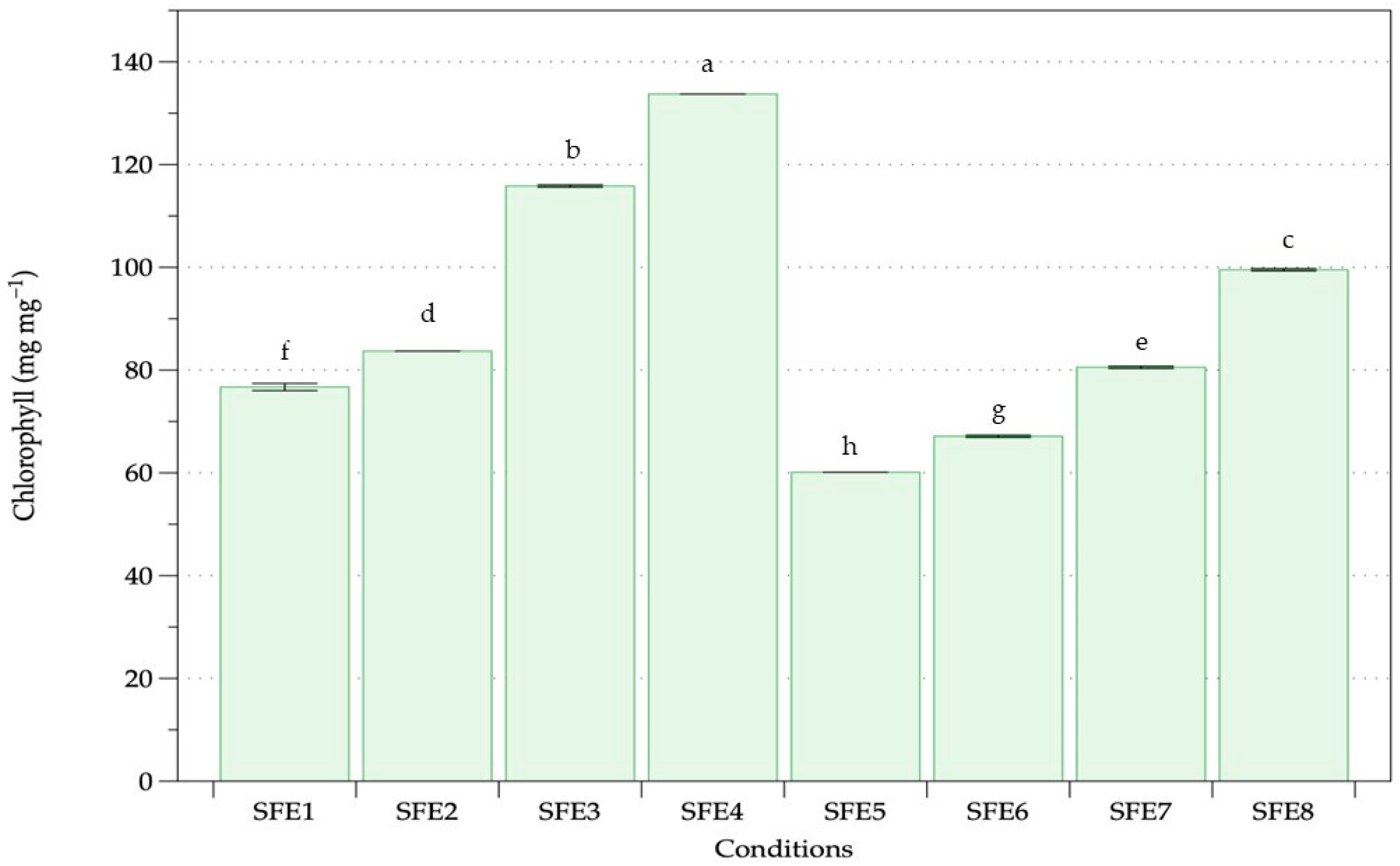
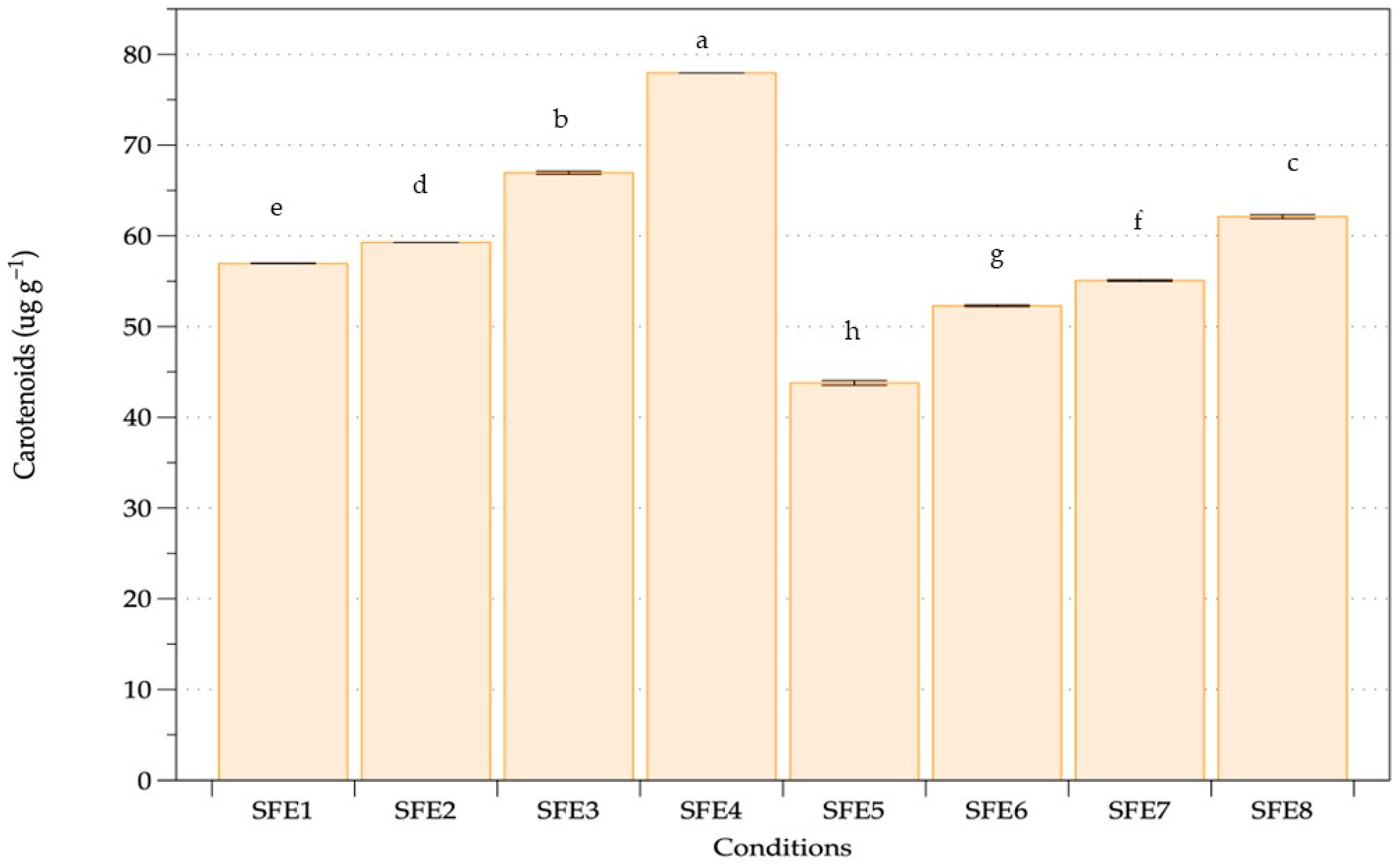
Nonpolar pigment extraction including chlorophyll and carotenoids of A. platensis oven-dried biomass was performed with various pressure, temperature and cosolvent assistance (Table 1). Two main groups without (SFE1–SFE4) and with (SFE5–SFE8) ethanol as a cosolvent were compared. The chlorophyll content of A. platensis using SFE under different conditions is shown in Figure 2. The extraction results revealed a chlorophyll content range of 60.12–133.72 mg·mg−1 of dry weight biomass. Higher pressure and temperature achieved higher chlorophyll content both with and without ethanol. The highest amount of chlorophyll extracted was 133.73 mg·mg−1 obtained from 350 bar, 50 °C and without ethanol. The carotenoid contents of A. platensis using SFE under various conditions are shown in Figure 3, with a range of 43.79–77.95 μg·g−1 of dry weight biomass. The highest amount of carotenoid extracted was obtained at the highest pressure and temperature of SC-CO2 without ethanol. Higher carotenoid content was observed when increasing the pressure and temperature of extraction both with and without ethanol as a co-solvent. Equal pressure and temperature extraction without ethanol showed higher chlorophyll and carotenoid extraction than with ethanol.
SFE at high pressure and temperature without cosolvent was evaluated for extraction of chlorophylls and carotenoids from Arthrospira. Increasing temperature at a constant pressure gave higher chlorophyll and carotenoid extraction, whereas increasing pressure at a constant temperature gave higher chlorophyll and carotenoid. Both pressure and temperature were influencing parameters using SC-CO2. The results concurred with previous studies of microalgae and seaweed extraction performed using SC-CO2, where higher pressure gave higher extraction and faster kinetic extraction [65]. At higher pressure, the enhancement of carbon dioxide density improved the extraction process with enhanced solubility, while constant temperature and increasing pressure increased yield with faster kinetic extraction due to the relationship between pressure and density [66]. Temperature plays an important role in the SFE process [67]. Pigment extraction is dependent on a delicate equilibrium between the reduction in supercritical carbon dioxide density and the increase in pigment vapour pressure as the temperature rises, essentially representing pigment solubility in the solvent [68]. Higher temperatures assisted higher solute solubility, hence boosting solute mass transfer in the matrix. Our findings were similar to previous results. High pressure and temperature of SFE at 450 bar and 60 °C gave highest carotenoids from A. platensis as a suitable green extraction technology [59].
Ethanol at 10% co-solvent did not affect chlorophyll and carotenoid extraction from A. platensis. Increasing ethanol percentage increased the extraction and yield of more polar compounds [69]. Nonpolar extracts are generally used to remove or extract nonpolar substances from biomass as unwanted glycosides and lipids [70]. Previous studies revealed that the optimal yield of chlorophylls and carotenoids from Nannocholopsis gaditana, Synechococcus sp. and Dunaliella salina was obtained using SFE with ethanol as a cosolvent compared with the conventional method (methanol extraction) [71]. SFE has various advantages over conventional extraction methods using hexane, petroleum ether, chloroform, ethanol and methanol to recover nonpolar biosubstances from algae [72]. Higher carotenoid and chlorophyll extraction from Dunaliella salina was obtained using SFE than by ultrasound-assisted extraction. [73]. Carotenoid extraction from Chlorella vulgaris was obtained using SC-CO2 fluid extraction under 350 bar and 40 °C and was more difficult than hydrocarbon extraction [74]. The highest yield of pigment depended on the microalgal type, cultivation procedure and other factors.

3.2. Sequential Phycobiliprotein Extraction

Previous studies on PBP extraction from Arthrospira revealed the optimised conditions to be sonication-assisted extraction with incubation at 25 °C for 24 h [60]. PBPs were extracted under various conditions, with concentration, extraction yield and extract purity shown in Table 2, Table 3 and Table 4. Phosphate buffer resulted in a higher yield of PBPs than water. C-PC, APC, PE and PBP concentration ranges were 0.57–1.17, 0.08–0.31, 0.03–0.09 and 0.67–1.55 mg·mL−1, respectively (Table 2). Highest concentration of C-PC was achieved for SFE4 using phosphate buffer but was not significantly different from SFE1–SFE3. Highest concentrations of APC and PE were achieved for SFE1 using phosphate buffer. The highest PBP concentration was achieved for SFE4. In general, greater PBP yields were obtained following SFE without ethanol cosolvent assistance (SFE1-4). The control experiments showed the lowest parameters in all cases. Table 3 shows the yield of PBP extraction under various conditions. The yield ranges of C-PC, APC, PE and PBP were 29.18–56.09, 4.05–14.55, 1.31–4.46 and 34.54–73.40 mg·g−1, respectively. The SFE4 condition with phosphate buffer extraction provided the highest C-PC yield, whereas the SFE1 condition with phosphate buffer extraction gave the highest APC and PE yields. The highest PBP yield was achieved for the SFE4 condition with phosphate buffer extraction. The control experiments showed the lowest extract yield. Table 3 shows the extract purity of the phycobiliprotein extraction from A. platensis SFE residues. The maximum extract purities of C-PC, APC and PE were 0.61, 0.24 and 0.30, respectively, following phosphate buffer extraction. C-PC extracted under the SFE5 condition with phosphate buffer showed maximum extract purity, which was non-significantly different from SFE1 with phosphate buffer extraction. The control experiments resulted in the lowest extract purity. The sequential extraction of phycobiliproteins from Arthrospira cell residues following SFE led to improvement in concentration, extraction yield and extract purity compared to the control.
For A. platensis residues prepared from SFE without ethanol as cosolvent, PBP concentration and extraction yield were higher than ethanol-assisted extraction. All experiments achieved higher C-PC, APC, PE and PBP concentration and extraction yield than the control experiment (A. platensis without SFE). PBPs are a complex group containing C-PC, APC and PE as the major classes of water-soluble pigments [75]. The SFE process with CO2 separated the nonpolar pigment. Hence, A. platensis residues from SC-CO2 remained mainly as PBP water-soluble pigments with fewer nonpolar contaminants. The variable pressure and temperature conditions of the SFE process of A. platensis did not affect PBP extraction from A. platensis biomass residues. Our results showed that phycobiliproteins extracted after SFE without ethanol showed a higher yield of phycobiliproteins. Previous results showed the yields of C-PC, APC, PE and PBPs without the SFE process to be 43.57, 2.59, 3.60 and 40.72 mg g−1, respectively. Thus, our results showed higher PBPs from A. platensis residues in the SFE process. Water can be used as a green extraction solvent [76]. Both chlorophyll and carotenoid extraction of A. platensis with SC-CO2 and PBP water extraction of A. platensis SFE residues were enhanced using green technologies and a sustainable extraction process. Previous studies of water extraction of PBPs from marine Spirulina maxima using ultrasonication extraction at 20–100 kHz achieved a high concentration and yield of PBP extraction [77]. High levels of proteins were also found in PBPs from Arthrospira [34]. One of the key factors influencing aggregation and dissociation to produce monomers, trimers, hexamers and other oligomers in solution is the pH value. Trimers were produced from C-PC with highest solubility at pH 7.0 [78]. Our results showed that concentration and yield of PBPs under phosphate buffer extraction were higher than using water extraction. PBPs are extracted more effectively due to their enhanced solubility and diffusion rate at pH 7.0 maintained under phosphate buffer. When utilising diluted phosphate buffer for extraction, the osmotic shock may result in cell wall rupture [38]. Therefore, our results confirmed the optimal condition of PBP extraction using cell residues from SFE, as well as the suitability of phosphate buffer for extraction.
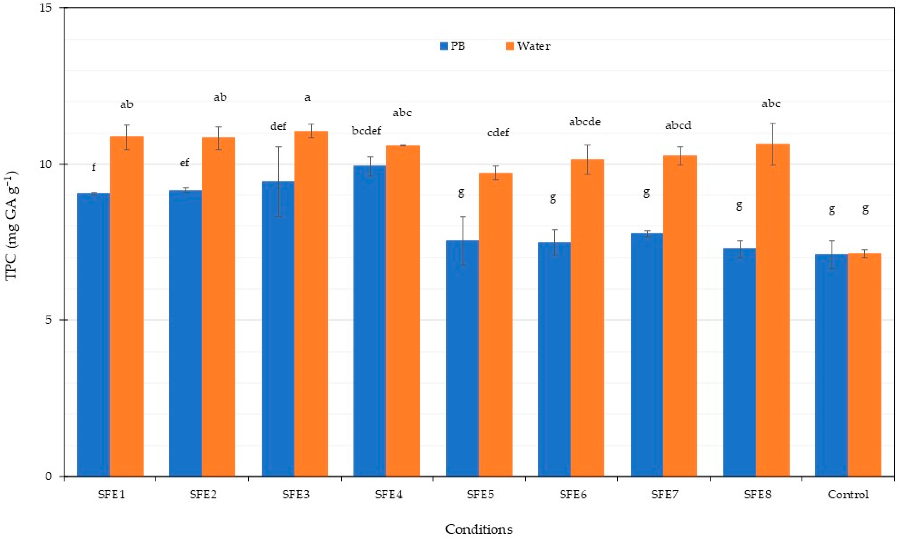
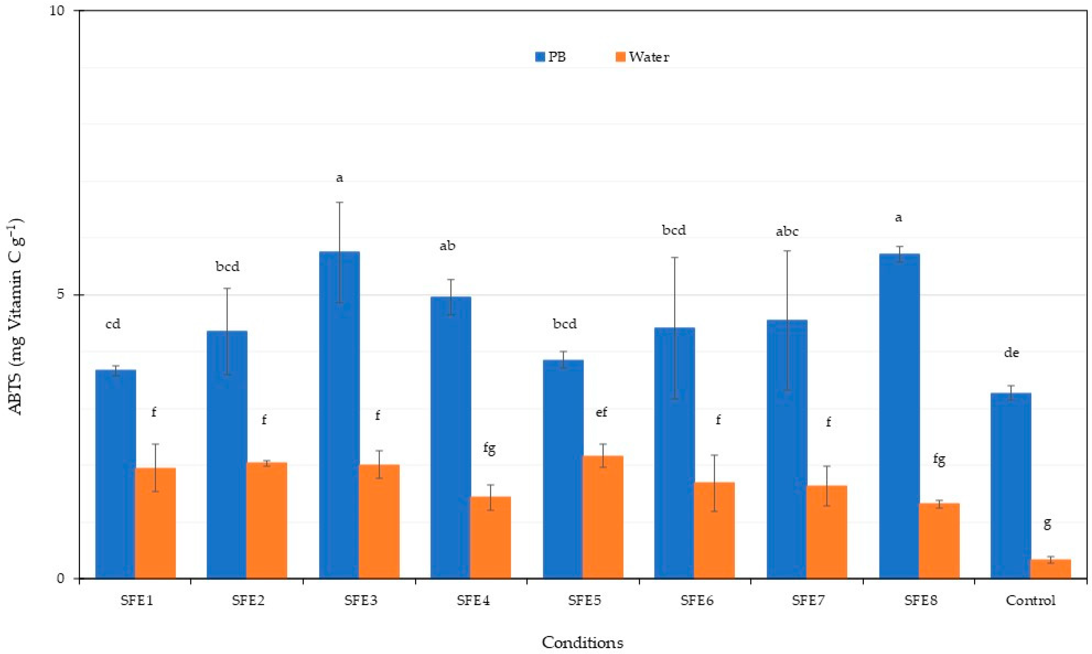
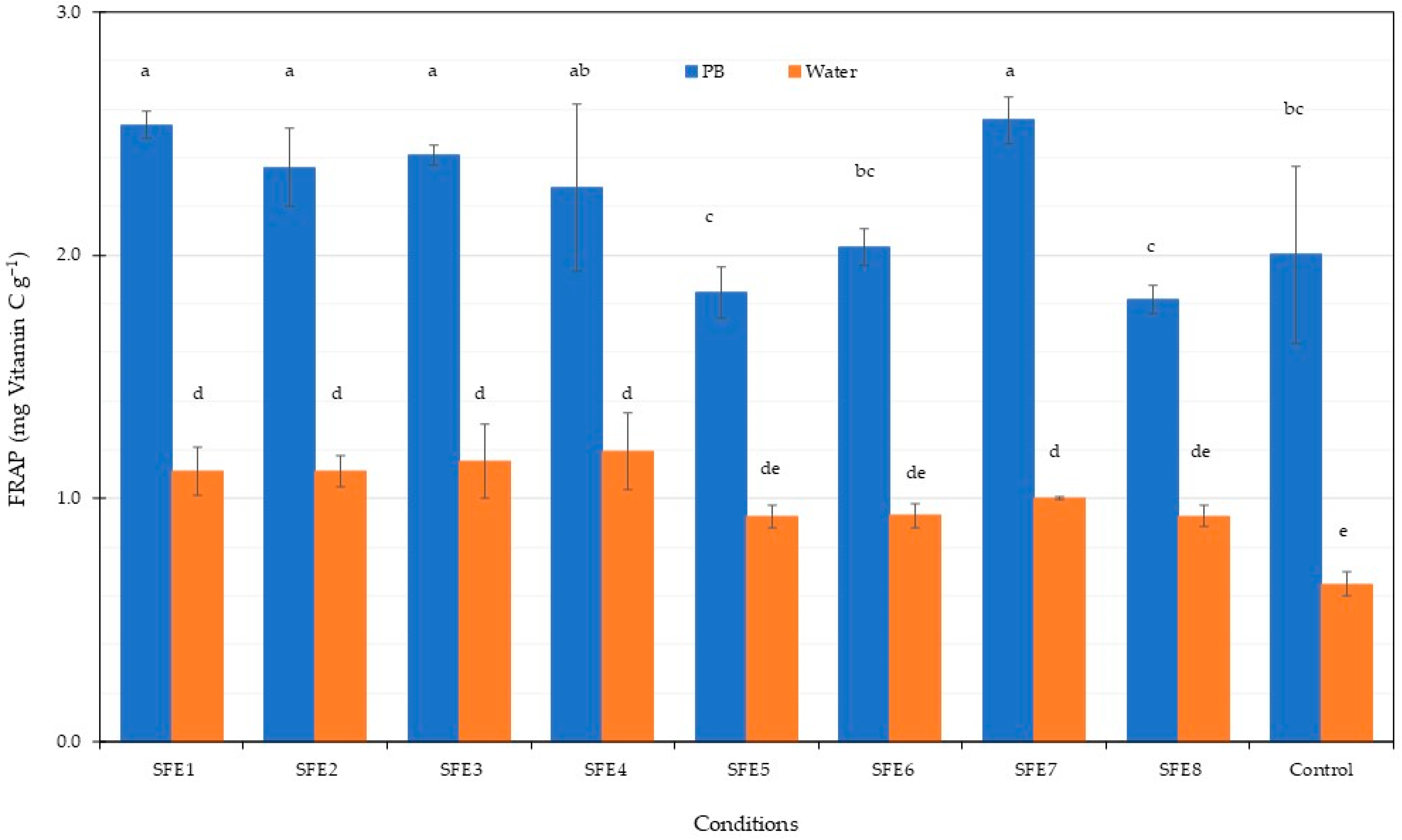
The total phenolic content in the PBP extract from Arthrospira after SFE is shown in Figure 4. All experiments with water gave higher TPC compared to the phosphate buffer (PB). A TPC of approximately 10 mg·g−1 was obtained from water extraction. The control experiments gave the lowest TPC in all cases. Previous results found that water extraction from Stypocaulon scoparium algae gave the highest TPC among several solvents including water-methanol, methanol and ethanol [79]. Therefore, water was a suitable solvent for total phenolic content extraction. The antioxidant potential of the PBP extract from Arthrospira residues after SFE was assessed using ABTS and FRAP methodologies, with results shown in Figure 5 and Figure 6. ABTS and FRAP antioxidant assay involves a single electron transfer process; however, the ABTS is distinguished by antioxidant reducing power, which is measured by the ability to reduce a coloured stable free radical (ABTS•+), while FRAP is distinguished by the antioxidant chemical ability to reduce Fe3+ ions to blue Fe2+ ions [80]. The ABTS radical-scavenging activity of the PBP extract was higher following phosphate buffer extraction compared to water extraction. Maximum ABTS at 5.7 mg·g−1 was achieved for the SFE3 and SFE8 conditions. The FRAP antioxidant activities of the PBP extract from Arthrospira residues after SFE were higher following phosphate buffer extraction compared to water extraction, ranging from 1.82 to 2.56 mg·g−1. Our results indicated that PBP extracts contained phenolic compounds and exhibited antioxidant capacity. Antioxidant activity is not only caused by phenolic substances [79]. C-PC blue colourant was observed from A. platensis SFE residues as the main pigment. Therefore, antioxidant properties in our results were caused by PBP extraction from A. platensis. Higher PBP extraction was exhibited from phosphate buffer extraction and the assay also gave higher antioxidant activity. Previous results showed the antioxidant activities of various C-PC concentrations from A. platensis using the ABTS assay [81]. The scavenging capacity of ABTS increased concentration dependently [82]. The antioxidant properties of C-PC can be used as a food supplement. The previous study of C-PC purified by ultrafiltration presented the antioxidant activity against the ABTS 206.36 µmol Trolox g−1 of ice cream with C-PC incorporated increasing the antioxidant activity after digestion [63]. Data from both ABTS and FRAP assays showed that PBPs had significant antioxidant properties and could be considered as food for human health improvement. C-PC showed high antioxidant activity which could be applied in several sectors. Reactive oxygen species (ROS) and oxidative processes are recognised as playing an ameliorating role in a number of illnesses including atherosclerosis, diabetes and Alzheimer’s disease [82].

3.3. C-Phycocyanin Isolation

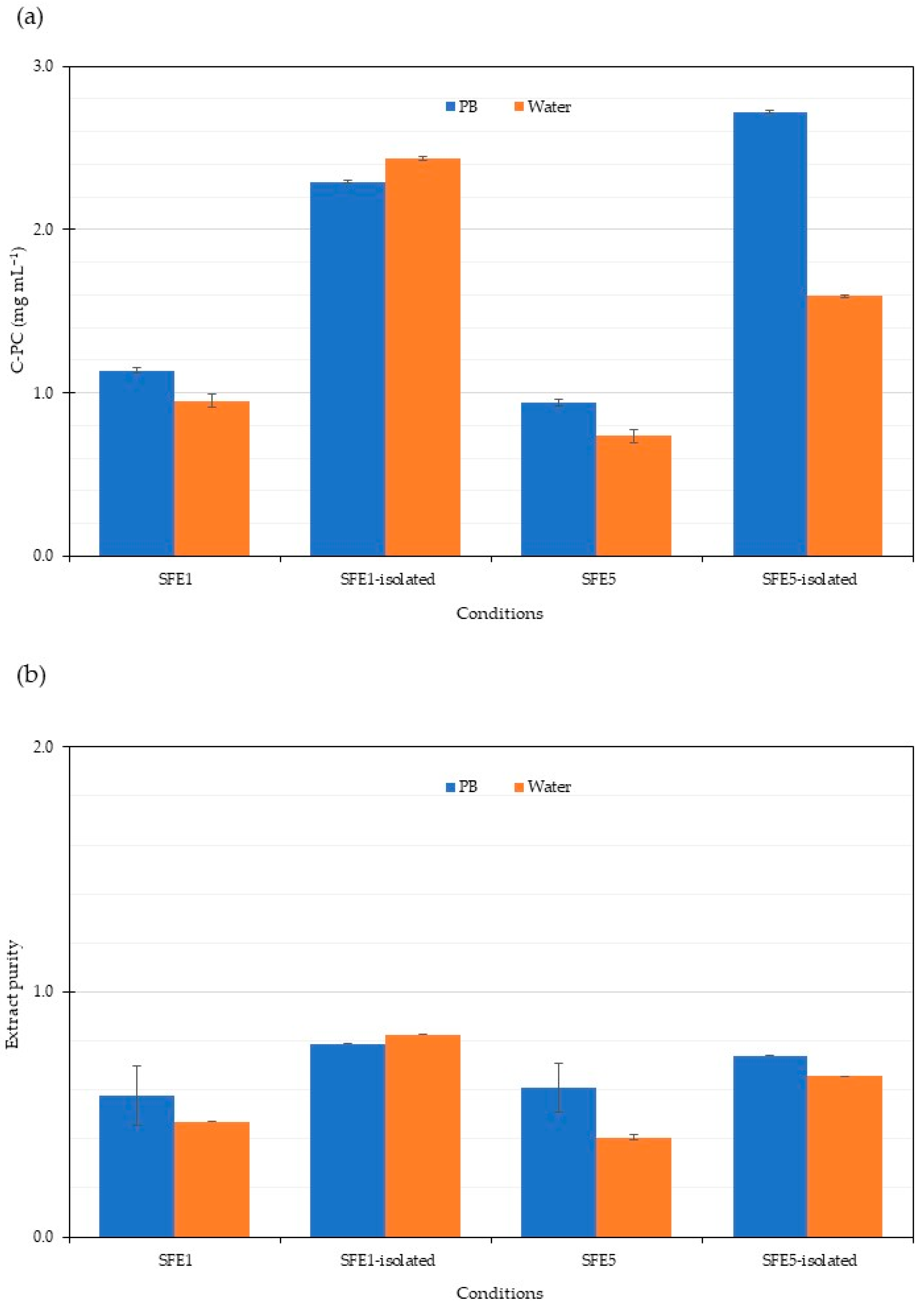
C-PC is the main PBP extracted from Arthrospira. Price and C-PC quality are directly correlated, with higher cost corresponding to a purer product [83]. Crude C-phycocyanin supernatants from SFE1 and SFE5 cell residues following phosphate and water extraction were selected to improve C-PC purity by ultrafiltration. C-PC concentration and purity (Figure 7) revealed an increase to approximately 0.7, similar to other purification methods. Previous results from several steps of purification of C-PC were studied. The purification of C-PC using activated charcoal for 24 h gave the highest purity of 1.2 [60]. Our results in this study used a short time for purification of 15 min. Crude C-PC isolated using ammonium sulphate precipitation followed by ion exchange chromatography gave higher purity than our results [84]. Crude C-PC showed improved purity for ammonium sulphate precipitation, ultrafiltration, gel filtration, and ion exchange chromatography [85]. The purification procedures follow several steps to attain high purity of C-PC as a valuable high cost bioproduct.

4. Conclusions

A. platensis is a commercially available blue-green microalga that is used as a food source for human health. Supercritical fluid extraction (SFE) was successfully applied to obtain the chlorophylls, carotenoids and phycobiliproteins remaining in cell residues. The optimal yield of nonpolar biocompounds (chlorophylls and carotenoids) was achieved using SFE at high pressure and temperature. Ethanol as a cosolvent did not improve extraction during the SC-CO2 process. The optimal yield of phycobiliproteins from SFE residues was achieved using a phosphate buffer extraction without cosolvent, while the purity of C-phycocyanin (C-PC) was improved.

Author Contributions

Conceptualisation, W.P.-u.; methodology, W.P.-u., S.B. and J.M.; formal analysis, W.P.-u., S.B. and J.M.; writing—original draft preparation, W.P.-u.; writing—review and editing, W.P.-u.; visualisation, W.P.-u., S.I., S.B. and J.M.; project administration, W.P.-u.; funding acquisition, W.P.-u. All authors have read and agreed to the published version of the manuscript.

Funding

This research was funded by the National Research Council of Thailand, grant number NRCT5-TRG63002-01, 2020–2021, the Center of Excellence on Agricultural Biotechnology, Science and Technology Postgraduate Education and Research Development Office, Office of the Higher Education Commission, Ministry of Education, Science, Research, and Innovation (AG-BIO/PERDO-CHE) Thailand, and the Kasetsart University Research and Development (KURDI) and Institute of Food Research and Product Development (IFRPD), Kasetsart University, Thailand.

Institutional Review Board Statement

Not applicable.

Informed Consent Statement

Not applicable.

Data Availability Statement

Not applicate.

Conflicts of Interest

The authors declare no conflict of interest. The funders had no role in the design of the study; the collection, analyses or interpretation of data; the writing of the manuscript, or in the decision to publish the results.

References

  1. Masten Rutar, J.; Hudobivnik, M.J.; Nečemer, M.; Vogel Mikuš, K.; Arčon, I.; Ogrinc, N. Nutritional Quality and Safety of the Spirulina Dietary Supplements Sold on the Slovenian Market. Foods 2022, 11, 849. [Google Scholar] [CrossRef] [PubMed]
  2. AlFadhly, N.K.Z.; Alhelfi, N.; Altemimi, A.B.; Verma, D.K.; Cacciola, F.; Narayanankutty, A. Trends and Technological Advancements in the Possible Food Applications of Spirulina and Their Health Benefits: A Review. Molecules 2022, 27, 5584. [Google Scholar] [CrossRef] [PubMed]
  3. Wollina, U.; Voicu, C.; Gianfaldoni, S.; Lotti, T.; França, K.; Tchernev, G. Arthrospira platensis—Potential in Dermatology and Beyond. Open Access Maced. J. Med. Sci. 2018, 6, 176–180. [Google Scholar] [CrossRef] [PubMed] [Green Version]
  4. Jung, C.H.G.; Braune, S.; Waldeck, P.; Küpper, J.-H.; Petrick, I.; Jung, F. Morphology and Growth of Arthrospira platensis during Cultivation in a Flat-Type Bioreactor. Life 2021, 11, 536. [Google Scholar] [CrossRef] [PubMed]
  5. Altmann, B.A.; Rosenau, S. Spirulina as Animal Feed: Opportunities and Challenges. Foods 2022, 11, 965. [Google Scholar] [CrossRef]
  6. Ebhodaghe, S.O.; Imanah, O.E.; Ndibe, H. Biofuels from microalgae biomass: A review of conversion processes and procedures. Arab. J. Chem. 2021, 15, 103591. [Google Scholar] [CrossRef]
  7. Iglina, T.; Iglin, P.; Pashchenko, D. Industrial CO2 Capture by Algae: A Review and Recent Advances. Sustainability 2022, 14, 3801. [Google Scholar] [CrossRef]
  8. Jaeschke, D.P.; Mercali, G.D.; Marczak, L.D.F.; Müller, G.; Frey, W.; Gusbeth, C. Extraction of valuable compounds from Arthrospira platensis using pulsed electric field treatment. Bioresour. Technol. 2019, 283, 207–212. [Google Scholar] [CrossRef]
  9. Finamore, A.; Palmery, M.; Bensehaila, S.; Peluso, I. Antioxidant, Immunomodulating, and Microbial-Modulating Activities of the Sustainable and Ecofriendly Spirulina. Oxidat. Med. Cell. Longev. 2017, 2017, 3247528. [Google Scholar] [CrossRef] [Green Version]
  10. Salla, A.C.V.; Margarites, A.C.; Seibel, F.I.; Holz, L.C.; Brião, V.B.; Bertolin, T.E.; Colla, L.M.; Costa, J.A.V. Increase in the carbohydrate content of the microalgae Spirulina in culture by nutrient starvation and the addition of residues of whey protein concentrate. Bioresour. Technol. 2016, 209, 133–141. [Google Scholar] [CrossRef]
  11. Soni, R.A.; Sudhakar, K.; Rana, R.S. Spirulina—From growth to nutritional product: A review. Trends Food Sci. Technol. 2017, 69, 157–171. [Google Scholar] [CrossRef] [Green Version]
  12. Soni, R.A.; Sudhakar, K.; Rana, R.S. Comparative study on the growth performance of Spirulina platensis on modifying culture media. Energy Rep. 2019, 5, 327–336. [Google Scholar] [CrossRef]
  13. Koli, D.K.; Rudra, S.G.; Bhowmik, A.; Pabbi, S. Nutritional, Functional, Textural and Sensory Evaluation of Spirulina Enriched Green Pasta: A Potential Dietary and Health Supplement. Foods 2022, 11, 979. [Google Scholar] [CrossRef] [PubMed]
  14. Park, W.S.; Kim, H.-J.; Li, M.; Lim, D.H.; Kim, J.; Kwak, S.-S.; Kang, C.-M.; Ferruzzi, M.G.; Ahn, M.-J. Two Classes of Pigments, Carotenoids and C-Phycocyanin, in Spirulina Powder and Their Antioxidant Activities. Molecules 2018, 23, 2065. [Google Scholar] [CrossRef] [PubMed] [Green Version]
  15. Chethana, S.; Nayak, C.A.; Madhusudhan, M.C.; Raghavarao, K.S.M.S. Single step aqueous two-phase extraction for downstream processing of C-phycocyanin from Spirulina platensis. J. Food Sci. Technol. 2014, 52, 2415–2421. [Google Scholar] [CrossRef] [Green Version]
  16. Martins, M.; Albuquerque, C.M.; Pereira, C.F.; Coutinho, J.A.P.; Neves, M.G.P.M.S.; Pinto, D.C.G.A.; Faustino, M.A.F.; Ventura, S.P.M. Recovery of Chlorophyll a Derivative from Spirulina maxima: Its Purification and Photosensitizing Potential. ACS Sustain. Chem. Eng. 2021, 9, 1772–1780. [Google Scholar] [CrossRef]
  17. Fischer, W.W.; Hemp, J.; Johnson, J.E. Evolution of oxygenic photosynthesis. Annu. Rev. Earth Planet. Sci. 2016, 44, 647–683. [Google Scholar] [CrossRef]
  18. Falkowski, P.G.; Raven, J.A. Aquatic Photosynthesis; Princeton University Press: Princeton, NJ, USA, 2013. [Google Scholar]
  19. Assunção, L.S.; Bezerra, P.Q.M.; Poletto, V.S.H.; Rios, A.D.O.; Ramos, I.G.; Ribeiro, C.D.F.; Machado, B.A.S.; Druzian, J.I.; Costa, J.A.V.; Nunes, I.L. Combination of carotenoids from Spirulina and PLA/PLGA or PHB: New options to obtain bioactive nanoparticles. Food Chem. 2020, 346, 128742. [Google Scholar] [CrossRef]
  20. Liang, R.; Shoemaker, C.F.; Yang, X.; Zhong, F.; Huang, Q. Stability and Bioaccessibility of β-Carotene in Nanoemulsions Stabilized by Modified Starches. J. Agric. Food Chem. 2013, 61, 1249–1257. [Google Scholar] [CrossRef]
  21. Christaki, E.; Bonos, E.; Giannenas, I.; Florou-Paneri, P. Functional properties of carotenoids originating from algae. J. Sci. Food Agric. 2013, 93, 5–11. [Google Scholar] [CrossRef]
  22. Kannaujiya, V.K.; Sundaram, S.; Sinha, R.P. Phycobiliproteins: Recent Developments and Future Applications; Springer: Berlin/Heidelberg, Germany, 2017. [Google Scholar]
  23. Bermejo, R.; Ruiz, E.; Acien, F. Recovery of B-phycoerythrin using expanded bed adsorption chromatography: Scale-up of the process. Enzym. Microb. Technol. 2007, 40, 927–933. [Google Scholar] [CrossRef]
  24. Christaki, E.; Bonos, E.; Florou-Paneri, P. Chapter 14—Innovative Microalgae Pigments as Functional Ingredients in Nutrition. In Handbook of Marine Microalgae; Kim, S.-K., Ed.; Academic Press: Boston, MA, USA, 2015; pp. 233–243. [Google Scholar]
  25. Tavanandi, H.A.; Mittal, R.; Chandrasekhar, J.; Raghavarao, K. Simple and efficient method for extraction of C-Phycocyanin from dry biomass of Arthospira platensis. Algal Res. 2018, 31, 239–251. [Google Scholar] [CrossRef]
  26. De la Jara, A.; Ruano-Rodriguez, C.; Polifrone, M.; Assunçao, P.; Brito-Casillas, Y.; Wägner, A.M.; Serra-Majem, L. Impact of dietary Arthrospira (Spirulina) biomass consumption on human health: Main health targets and systematic review. J. Appl. Phycol. 2018, 30, 2403–2423. [Google Scholar] [CrossRef]
  27. Ali, S.K.; Saleh, A.M. Spirulina—An overview. Int. J. Pharm. Pharm. Sci. 2012, 4, 9–15. [Google Scholar]
  28. Kaur, S.; Khattar, J.I.S.; Singh, Y.; Singh, D.P.; Ahluwalia, A.S. Extraction, purification and characterisation of Phycocyanin from Anabaena fertilissima PUPCCC 410.5: As a natural and food grade stable pigment. J. Appl. Phycol. 2019, 31, 1685–1696. [Google Scholar] [CrossRef]
  29. Vinothkanna, A.; Sekar, S. Diagnostic Applications of Phycobiliproteins. In Pigments from Microalgae Handbook; Springer: Berlin/Heidelberg, Germany, 2020; pp. 585–610. [Google Scholar]
  30. Li, W.; Su, H.-N.; Pu, Y.; Chen, J.; Liu, L.-N.; Liu, Q.; Qin, S. Phycobiliproteins: Molecular structure, production, applications, and prospects. Biotechnol. Adv. 2019, 37, 340–353. [Google Scholar] [CrossRef]
  31. Chiong, T.; Acquah, C.; Lau, S.Y.; Khor, E.H.; Danquah, M.K. Chapter 12—Microalgal-Based Protein By-Products: Extraction, Purification, and Applications. In Protein Byproducts; Singh Dhillon, G., Ed.; Academic Press: Cambridge, MA, USA, 2016; pp. 213–234. [Google Scholar]
  32. Sekar, S.; Chandramohan, M. Phycobiliproteins as a commodity: Trends in applied research, patents and commercialization. J. Appl. Phycol. 2007, 20, 113–136. [Google Scholar] [CrossRef]
  33. Lauceri, R.; Chini Zittelli, G.; Torzillo, G. A simple method for rapid purification of phycobiliproteins from Arthrospira platensis and Porphyridium cruentum biomass. Algal Res. 2019, 44, 101685. [Google Scholar] [CrossRef]
  34. Dagnino-Leone, J.; Figueroa, C.P.; Castañeda, M.L.; Youlton, A.D.; Vallejos-Almirall, A.; Agurto-Muñoz, A.; Pavón Pérez, J.; Agurto-Muñoz, C. Phycobiliproteins: Structural aspects, functional characteristics, and biotechnological perspectives. Comput. Struct. Biotechnol. J. 2022, 20, 1506–1527. [Google Scholar] [CrossRef]
  35. Sala, L.; Moraes, C.C.; Kalil, S.J. Cell pretreatment with ethylenediaminetetraacetic acid for selective extraction of C-phycocyanin with food grade purity. Biotechnol. Prog. 2018, 34, 1261–1268. [Google Scholar] [CrossRef]
  36. Hosikian, A.; Lim, S.; Halim, R.; Danquah, M.K. Chlorophyll Extraction from Microalgae: A Review on the Process Engineering Aspects. Int. J. Chem. Eng. 2010, 2010, 391632. [Google Scholar] [CrossRef]
  37. Montero, L.; Sedghi, M.; García, Y.; Almeida, C.; Safi, C.; Engelen-Smit, N.; Cifuentes, A.; Mendiola, J.A.; Ibáñez, E. Pressurized Liquid Extraction of Pigments from Chlamydomonas sp. and Chemical Characterization by HPLC–MS/MS. J. Anal. Test. 2018, 2, 149–157. [Google Scholar] [CrossRef]
  38. Nemer, G.; Louka, N.; Vorobiev, E.; Salameh, D.; Nicaud, J.-M.; Maroun, R.G.; Koubaa, M. Mechanical Cell Disruption Technologies for the Extraction of Dyes and Pigments from Microorganisms: A Review. Fermentation 2021, 7, 36. [Google Scholar] [CrossRef]
  39. Pagels, F.; Pereira, R.N.; Vicente, A.A.; Guedes, A.C. Extraction of Pigments from Microalgae and Cyanobacteria—A Review on Current Methodologies. Appl. Sci. 2021, 11, 5187. [Google Scholar] [CrossRef]
  40. Banayan, S.; Jahadi, M.; Khosravi-Darani, K. Pigment Productions by Spirulina platensis as a Renewable Resource. J. Appl. Biotechnol. Rep. 2022, 9, 614–621. [Google Scholar] [CrossRef]
  41. Martí-Quijal, F.J.; Ramon-Mascarell, F.; Pallarés, N.; Ferrer, E.; Berrada, H.; Phimolsiripol, Y.; Barba, F.J. Extraction of Antioxidant Compounds and Pigments from Spirulina (Arthrospira platensis) Assisted by Pulsed Electric Fields and the Binary Mixture of Organic Solvents and Water. Appl. Sci. 2021, 11, 7629. [Google Scholar] [CrossRef]
  42. Basa’Ar, O.; Fatema, S.; Alrabie, A.; Mohsin, M.; Farooqui, M. Supercritical carbon dioxide extraction of Triognella foenum graecum Linn seeds: Determination of bioactive compounds and pharmacological analysis. Asian Pac. J. Trop. Biomed. 2017, 7, 1085–1091. [Google Scholar] [CrossRef]
  43. Sahena, F.; Zaidul, I.; Jinap, S.; Karim, A.; Abbas, K.; Norulaini, N.; Omar, A. Application of supercritical CO2 in lipid extraction—A review. J. Food Eng. 2009, 95, 240–253. [Google Scholar] [CrossRef]
  44. Ruiz-Domínguez, M.C.; Medina, E.; Salinas, F.; Bugueño, W.; Fuentes, J.-L.; Vílchez, C.; Garbayo, I.; Cerezal-Mezquita, P. Methodological Optimization of Supercritical Fluid Extraction of Valuable Bioactive Compounds from the Acidophilic Microalga Coccomyxa onubensis. Antioxidants 2022, 11, 1248. [Google Scholar] [CrossRef]
  45. Díaz-Reinoso, B.; Moure, A.; Domínguez, H.; Parajó, J.C. Supercritical CO2 Extraction and Purification of Compounds with Antioxidant Activity. J. Agric. Food Chem. 2006, 54, 2441–2469. [Google Scholar] [CrossRef]
  46. Wei, M.-C.; Wang, C.-S.; Wei, D.-H.; Yang, Y.-C. Insights into the Supercritical CO2 Extraction of Perilla Oil and Its Theoretical Solubility. Processes 2021, 9, 239. [Google Scholar] [CrossRef]
  47. Bahadar, A.; Khan, M.B.; Asim, M.A.; Jalwana, K. Chapter 21—Supercritical Fluid Extraction of Microalgae (Chlorella vulagaris) Biomass. In Handbook of Marine Microalgae; Kim, S.-K., Ed.; Academic Press: Boston, MA, USA, 2015; pp. 317–330. [Google Scholar]
  48. Vafaei, N.; Rempel, C.B.; Scanlon, M.G.; Jones, P.J.H.; Eskin, M.N.A. Application of Supercritical Fluid Extraction (SFE) of Tocopherols and Carotenoids (Hydrophobic Antioxidants) Compared to Non-SFE Methods. AppliedChem 2022, 2, 68–92. [Google Scholar] [CrossRef]
  49. Molino, A.; Mehariya, S.; Di Sanzo, G.; LaRocca, V.; Martino, M.; Leone, G.P.; Marino, T.; Chianese, S.; Balducchi, R.; Musmarra, D. Recent developments in supercritical fluid extraction of bioactive compounds from microalgae: Role of key parameters, technological achievements and challenges. J. CO2 Util. 2020, 36, 196–209. [Google Scholar] [CrossRef]
  50. Uwineza, P.A.; Waśkiewicz, A. Recent Advances in Supercritical Fluid Extraction of Natural Bioactive Compounds from Natural Plant Materials. Molecules 2020, 25, 3847. [Google Scholar] [CrossRef]
  51. Patias, L.D.; Fernandes, A.S.; Petry, F.C.; Mercadante, A.Z.; Jacob-Lopes, E.; Zepka, L.Q. Carotenoid profile of three microalgae/cyanobacteria species with peroxyl radical scavenger capacity. Food Res. Int. 2017, 100, 260–266. [Google Scholar] [CrossRef] [PubMed]
  52. Poojary, M.M.; Barba, F.J.; Aliakbarian, B.; Donsì, F.; Pataro, G.; Dias, D.A.; Juliano, P. Innovative Alternative Technologies to Extract Carotenoids from Microalgae and Seaweeds. Mar. Drugs 2016, 14, 214. [Google Scholar] [CrossRef] [Green Version]
  53. Pyne, S.; Paria, K. Optimization of extraction process parameters of caffeic acid from microalgae by supercritical carbon dioxide green technology. BMC Chem. 2022, 16, 1–11. [Google Scholar] [CrossRef]
  54. Kankala, R.K.; Zhang, Y.S.; Wang, S.-B.; Lee, C.-H.; Chen, A.-Z. Supercritical Fluid Technology: An Emphasis on Drug Delivery and Related Biomedical Applications. Adv. Healthc. Mater. 2017, 6, 1700433. [Google Scholar] [CrossRef] [Green Version]
  55. Chakravarty, P.; Famili, A.; Nagapudi, K.; Al-Sayah, M.A. Using Supercritical Fluid Technology as a Green Alternative During the Preparation of Drug Delivery Systems. Pharmaceutics 2019, 11, 629. [Google Scholar] [CrossRef] [Green Version]
  56. Zarrouk, C. Contribution à l’étude d’une Cyanophycée. Influence de Divers Facteurs Physiques et Chimiques sur la Croissanceet la Photosynthèse de Spirulina maxima. Ph.D. Thesis, University of Paris, Paris, France, 1996. [Google Scholar]
  57. Pan-Utai, W.; Poopat, N.; Parakulsuksatid, P. Photoautotrophic Cultivation of Arthrospira maxima for Protein Accumulation under Minimum Nutrient Availability. Appl. Food Biotechnol. 2020, 7, 225–234. [Google Scholar] [CrossRef]
  58. Deniz, I.; Ozen, M.O.; Yesil-Celiktas, O. Supercritical fluid extraction of phycocyanin and investigation of cytotoxicity on human lung cancer cells. J. Supercrit. Fluids 2016, 108, 13–18. [Google Scholar] [CrossRef]
  59. Esquivel-Hernández, D.A.; López, V.H.; Rodríguez-Rodríguez, J.; Alemán-Nava, G.S.; Cuéllar-Bermúdez, S.P.; Rostro-Alanis, M.; Parra-Saldívar, R. Supercritical Carbon Dioxide and Microwave-Assisted Extraction of Functional Lipophilic Compounds from Arthrospira platensis. Int. J. Mol. Sci. 2016, 17, 658. [Google Scholar] [CrossRef] [PubMed] [Green Version]
  60. Pan-Utai, W.; Iamtham, S. Extraction, purification and antioxidant activity of phycobiliprotein from Arthrospira platensis. Process Biochem. 2019, 82, 189–198. [Google Scholar] [CrossRef]
  61. Lichtenthaler, H.K.; Wellburn, A.R. Determinations of total carotenoids and chlorophylls a and b of leaf extracts in different solvents. Analysis 1983, 11, 591–592. [Google Scholar] [CrossRef] [Green Version]
  62. Liu, L.; Chen, X.-L.; Zhang, X.-Y.; Zhang, Y.-Z.; Zhou, B.-C. One-step chromatography method for efficient separation and purification of R-phycoerythrin from Polysiphonia urceolata. J. Biotechnol. 2005, 116, 91–100. [Google Scholar] [CrossRef]
  63. De Amarante, M.C.A.; Braga, A.R.C.; Sala, L.; Kalil, S.J. Colour stability and antioxidant activity of C-phycocyanin-added ice creams after in vitro digestion. Food Res. Int. 2020, 137, 109602. [Google Scholar] [CrossRef]
  64. Renugadevi, K.; Nachiyar, C.V.; Sowmiya, P.; Sunkar, S. Antioxidant activity of phycocyanin pigment extracted from marine filamentous cyanobacteria Geitlerinema sp. TRV57. Biocatal. Agric. Biotechnol. 2018, 16, 237–242. [Google Scholar] [CrossRef]
  65. Crampon, C.; Boutin, O.; Badens, E. Supercritical Carbon Dioxide Extraction of Molecules of Interest from Microalgae and Seaweeds. Ind. Eng. Chem. Res. 2011, 50, 8941–8953. [Google Scholar] [CrossRef]
  66. Michalak, I.; Dmytryk, A.; Wieczorek, P.P.; Rój, E.; Łęska, B.; Górka, B.; Messyasz, B.; Lipok, J.; Mikulewicz, M.; Wilk, R.; et al. Supercritical Algal Extracts: A Source of Biologically Active Compounds from Nature. J. Chem. 2015, 2015, 597140. [Google Scholar] [CrossRef] [Green Version]
  67. Priyadarsani, S.; Patel, A.S.; Kar, A.; Dash, S. Process optimization for the supercritical carbondioxide extraction of lycopene from ripe grapefruit (Citrus paradisi) endocarp. Sci. Rep. 2021, 11, 10273. [Google Scholar] [CrossRef]
  68. Macías-Sánchez, M.; Mantell, C.; Rodríguez, M.; de la Ossa, E.M.; Lubián, L.; Montero, O. Supercritical fluid extraction of carotenoids and chlorophyll a from Synechococcus sp. J. Supercrit. Fluids 2007, 39, 323–329. [Google Scholar] [CrossRef]
  69. Ariff, M.A.M.; Yusri, A.M.; Razak, N.A.A.; Jaapar, J. Effect of CO2 flow rate, co-solvent and pressure behavior to yield by supercritical CO2 extraction of Mariposa Christia Vespertilionis leaves. AIP Conf. Proc. 2018, 2045, 020072. [Google Scholar] [CrossRef]
  70. Radzali, S.A.; Markom, M.; Saleh, N.M. Co-Solvent Selection for Supercritical Fluid Extraction (SFE) of Phenolic Compounds from Labisia pumila. Molecules 2020, 25, 5859. [Google Scholar] [CrossRef] [PubMed]
  71. Macías-Sánchez, M.D.; Serrano, C.M.; Rodríguez, M.R.; de la Ossa, E.M.; Lubián, L.M.; Montero, O. Extraction of carotenoids and chlorophyll from microalgae with supercritical carbon dioxide and ethanol as cosolvent. J. Sep. Sci. 2008, 31, 1352–1362. [Google Scholar] [CrossRef]
  72. Santana, A.; Jesus, S.; Larrayoz, M.; Filho, R. Supercritical Carbon Dioxide Extraction of Algal Lipids for the Biodiesel Production. Procedia Eng. 2012, 42, 1755–1761. [Google Scholar] [CrossRef] [Green Version]
  73. Macías-Sánchez, M.D.; Mantell, C.; Rodriguez, M.; de la Ossa, E.M.; Lubian, L.; Montero, O. Comparison of supercritical fluid and ultrasound-assisted extraction of carotenoids and chlorophyll a from Dunaliella salina. Talanta 2009, 77, 948–952. [Google Scholar] [CrossRef]
  74. Palavra, A.; Coelho, J.; Barroso, J.; Rauter, A.; Fareleira, J.; Mainar, A.; Urieta, J.; Nobre, B.; Gouveia, L.; Mendes, R.; et al. Supercritical carbon dioxide extraction of bioactive compounds from microalgae and volatile oils from aromatic plants. J. Supercrit. Fluids 2011, 60, 21–27. [Google Scholar] [CrossRef]
  75. Zhang, Y.; Rehmmann, L. Chapter 19—Extraction of high-value compounds from marine biomass via ionic liquid-based techniques. In Innovative and Emerging Technologies in the Bio-Marine Food Sector; Garcia-Vaquero, M., Rajauria, G., Eds.; Academic Press: Cambridge, MA, USA, 2022; pp. 417–439. [Google Scholar]
  76. Castro-Puyana, M.; Marina, M.L.; Plaza, M. Water as green extraction solvent: Principles and reasons for its use. Curr. Opin. Green Sustain. Chem. 2017, 5, 31–36. [Google Scholar] [CrossRef]
  77. Choi, W.Y.; Lee, H.Y. Effect of Ultrasonic Extraction on Production and Structural Changes of C-Phycocyanin from Marine Spirulina maxima. Int. J. Mol. Sci. 2018, 19, 220. [Google Scholar] [CrossRef] [Green Version]
  78. Aftari, R.V.; Rezaei, K.; Mortazavi, A.; Bandani, A.R. The Optimized Concentration and Purity of Spirulina platensis C-Phycocyanin: A Comparative Study on Microwave-Assisted and Ultrasound-Assisted Extraction Methods. J. Food Process. Preserv. 2015, 39, 3080–3091. [Google Scholar] [CrossRef]
  79. López, A.; Rico, M.; Rivero, A.; de Tangil, M.S. The effects of solvents on the phenolic contents and antioxidant activity of Stypocaulon scoparium algae extracts. Food Chem. 2011, 125, 1104–1109. [Google Scholar] [CrossRef]
  80. Vieira, M.V.; Turkiewicz, I.P.; Tkacz, K.; Fuentes-Grünewald, C.; Pastrana, L.M.; Fuciños, P.; Wojdyło, A.; Nowicka, P. Microalgae as a Potential Functional Ingredient: Evaluation of the Phytochemical Profile, Antioxidant Activity and In-Vitro Enzymatic Inhibitory Effect of Different Species. Molecules 2021, 26, 7593. [Google Scholar] [CrossRef] [PubMed]
  81. Aoki, J.; Sasaki, D.; Asayama, M. Development of a method for phycocyanin recovery from filamentous cyanobacteria and evaluation of its stability and antioxidant capacity. BMC Biotechnol. 2021, 21, 1–10. [Google Scholar] [CrossRef] [PubMed]
  82. Wu, H.-L.; Wang, G.-H.; Xiang, W.-Z.; Li, T.; He, H. Stability and Antioxidant Activity of Food-Grade Phycocyanin Isolated from Spirulina platensis. Int. J. Food Prop. 2016, 19, 2349–2362. [Google Scholar] [CrossRef] [Green Version]
  83. Sukhinov, D.V.; Gorin, K.V.; Romanov, A.O.; Gotovtsev, P.M.; Sergeeva, Y.E. Increased C-phycocyanin extract purity by flocculation of Arthrospira platensis with chitosan. Algal Res. 2021, 58, 102393. [Google Scholar] [CrossRef]
  84. Kissoudi, M.; Sarakatsianos, I.; Samanidou, V. Isolation and purification of food-grade C-phycocyanin from Arthrospira platensis and its determination in confectionery by HPLC with diode array detection. J. Sep. Sci. 2017, 41, 975–981. [Google Scholar] [CrossRef]
  85. De Amarante, M.C.A.; Braga, A.R.C.; Sala, L.; Moraes, C.C.; Kalil, S.J. Design strategies for C-phycocyanin purification: Process influence on purity grade. Sep. Purif. Technol. 2020, 252, 117453. [Google Scholar] [CrossRef]
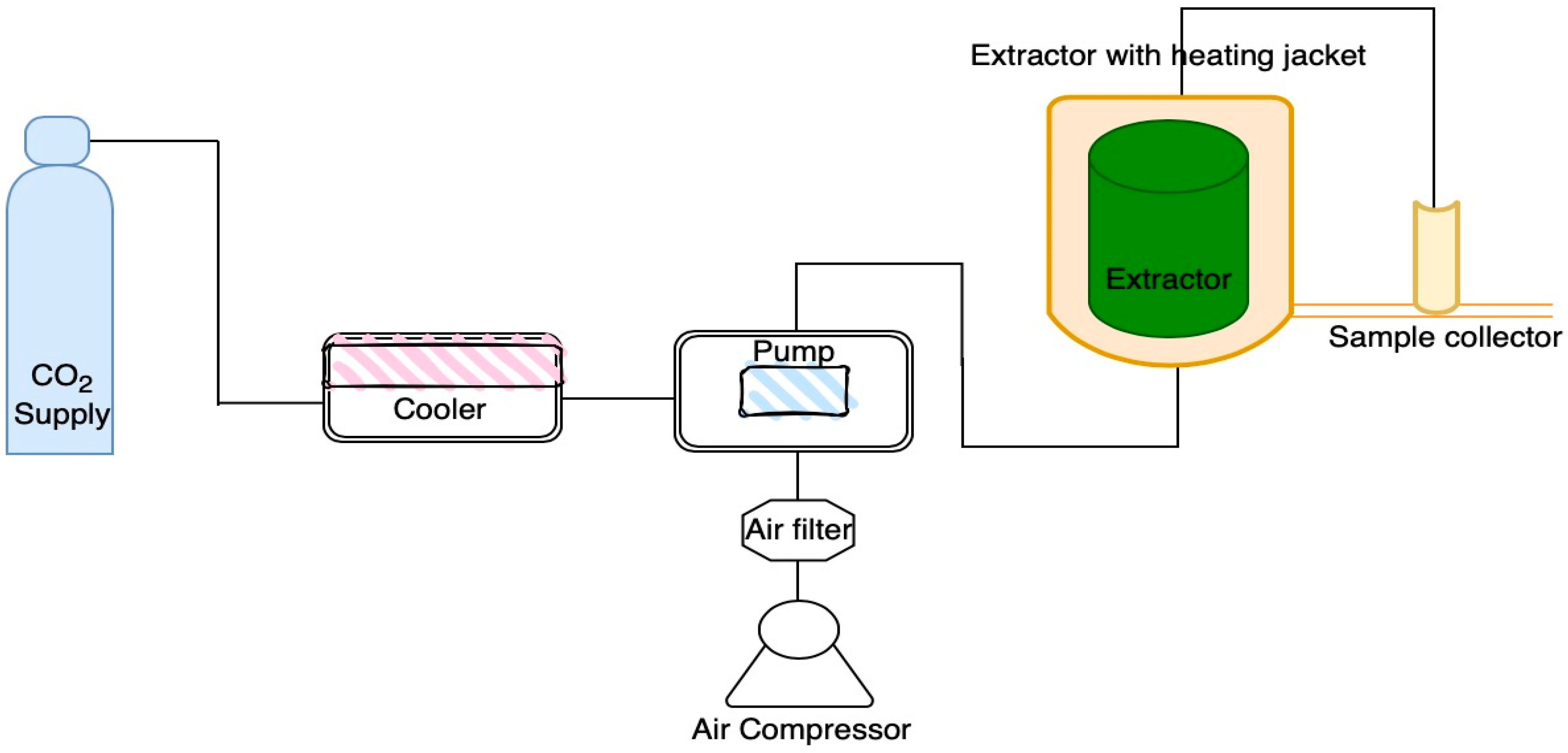
Figure 1. Overview of the SC-CO2 system.
Life 12 01896 g001
Figure 2. Chlorophyll content of A. platensis following SFE under various conditions. Different letters indicate significant differences (p < 0.05). Data were calculated from duplicate experimental values ± standard deviation (SD). Abbreviations of conditions of SFE (SFE1–SFE8) are shown in Table 1.
Life 12 01896 g002
Figure 3. Carotenoid content of A. platensis following SFE under various conditions. Different letters indicate significant differences (p < 0.05). Data were calculated from duplicate experimental values ± standard deviation (SD). Abbreviations of conditions of SFE (SFE1–SFE8) are shown in Table 1.
Life 12 01896 g003
Figure 4. Total phenolic content of A. platensis SFE residues under various conditions. Different letters indicate significant differences (p < 0.05). Data were calculated from triplicate experimental values ± standard deviation (SD). PB and water are phosphate and water extraction. The abbreviations of conditions of SFE (SFE1-SFE8) are shown in Table 1.
Life 12 01896 g004
Figure 5. ABTS radical scavenging activity of A. platensis SFE residues under various conditions. Different letters indicate significant differences (p < 0.05). Data were calculated from triplicate experimental values ± standard deviation (SD). PB and water are phosphate and water extraction. The abbreviations of conditions of SFE (SFE1–SFE8) are shown in Table 1.
Life 12 01896 g005
Figure 6. Ferric reducing antioxidant power assay of A. platensis SFE residues under various conditions. Different letters indicate significant differences (p < 0.05). Data were calculated from triplicate experimental values ± standard deviation (SD). PB and water are phosphate and water extraction. The abbreviations of conditions of SFE (SFE1–SFE8) are shown in Table 1.
Life 12 01896 g006
Figure 7. Purity of C-PC isolated from A. platensis SFE residues. Data were calculated from triplicate experimental values ± standard deviation (SD). PB and water are phosphate and water extraction. (a) C-PC concentration and (b) Extract purity of C-PC isolated from A. platensis SFE residues
Life 12 01896 g007
Table 1. Experimental conditions for supercritical fluid extraction (SFE) of A. platensis.
ExperimentPressureTemperatureCosolvent
(bar)(°C)(% w/w)
SFE125040None
SFE225050None
SFE335040None
SFE435050None
SFE52504010% ethanol
SFE62505010% ethanol
SFE73504010% ethanol
SFE83505010% ethanol
Table 2. Phycobiliprotein concentration of A. platensis SFE residues under various conditions. Data in the same column with different superscripts are significantly different (p < 0.05). Data were calculated from triplicate experimental values ± standard deviation (SD). C-PC, APC, PE and PBP are C-phycocyanin, allophycocyanin, phycoerythrin and total phycobiliprotein concentration. The abbreviations of conditions of SFE (SFE1–SFE8) are shown in Table 1.
ExperimentPhycobiliprotein Concentration (mg·mL−1)
C-PCAPCPEPBP
Phosphate buffer extraction
SFE11.139 a ± 0.020.307 a ± 0.010.094 a ± 0.001.540 a ± 0.03
SFE21.140 a ± 0.030.298 ab ± 0.010.093 ab ± 0.011.530 a ± 0.01
SFE31.156 a ± 0.020.275 b ± 0.010.082 c ± 0.001.513 a ± 0.02
SFE41.174 a ± 0.030.291 ab ± 0.010.085 bc ± 0.011.550 a ± 0.04
SFE50.945 b ± 0.020.200 c ± 0.010.062 d ± 0.001.207 b ± 0.03
SFE60.886 bcd ± 0.010.184 cd ± 0.010.059 de ± 0.001.129 bcd ± 0.03
SFE70.959 b ± 0.070.207 c ± 0.020.065 d ± 0.011.231 b ± 0.10
SFE80.860 bcde ± 0.090.189 cd ± 0.020.060 d ± 0.011.109 bcd ± 0.12
Control0.766 ef ± 0.090.142 f ± 0.010.045 f ± 0.000.953 ef ± 0.11
Water extraction
SFE10.953 b ± 0.040.193 c ± 0.010.059 de ± 0.001.205 b ± 0.06
SFE20.960 b ± 0.020.200 c ± 0.010.063 d ± 0.001.223 b ± 0.02
SFE30.911 bc ± 0.070.196 c ± 0.010.064 d ± 0.001.171 bc ± 0.07
SFE40.957 b ± 0.030.196 c ± 0.010.060 d ± 0.001.214 b ± 0.03
SFE50.737 f ± 0.040.135 f ± 0.010.043 f ± 0.000.915 f ± 0.05
SFE60.788 def ± 0.000.148 ef ± 0.000.046 f ± 0.000.982 ef ± 0.01
SFE70.839 cde ± 0.000.167 de ± 0.000.051 ef ± 0.001.056 cde ± 0.01
SFE80.806 def ± 0.010.155 ef ± 0.000.049 f ± 0.001.011 def ± 0.01
Control0.568 g ± 0.020.079 g ± 0.010.025 g ± 0.000.672 g ± 0.03
Table 3. Phycobiliprotein extraction yield from A. platensis SFE residues under various conditions. Data in the same column with different superscripts are significantly different (p < 0.05). Data were calculated from triplicate experimental values ± standard deviation (SD). C-PC, APC, PE and PBP are C-phycocyanin, allophycocyanin, phycoerythrin and total phycobiliprotein. The abbreviations of conditions of SFE (SFE1–SFE8) are shown in Table 1.
ExperimentsPhycobiliprotein Extraction Yield (mg·g−1)
C-PCAPCPEPBP
Phosphate buffer extraction
SFE154.072 a ± 0.9314.550 a ± 0.014.460 a ± 0.0573.082 a ± 0.98
SFE254.486 a ± 2.2714.216 ab ± 0.154.417 a ± 0.4073.119 a ± 1.72
SFE356.091 a ± 0.7313.325 b ± 0.663.988 b ± 0.2773.404 a ± 1.66
SFE455.021 a ± 0.2713.636 ab ± 0.053.985 b ± 0.0472.643 a ± 0.17
SFE546.864 bc ± 1.209.926 c ± 0.453.053 c ± 0.1559.843 bc ± 1.80
SFE643.038 cde ± 0.848.930 cd ± 0.132.873 cd ± 0.0454.841 cde ± 0.76
SFE746.428 bc ± 2.8410.015 c ± 1.023.160 c ± 0.4559.603 bc ± 4.31
SFE841.206 de ± 2.909.034 cd ± 0.922.874 cd ± 0.1853.114 def ± 3.99
Control38.965 ef ± 4.657.227 f ± 0.672.300 e ± 0.0248.492 fg ± 5.35
Water extraction
SFE146.945 bc ± 2.259.525 c ± 0.572.897 cd ± 0.0959.367 bc ± 2.91
SFE247.703 b ± 0.949.924 c ± 0.353.139 c ± 0.0760.765 b ± 1.36
SFE345.344 bcd ± 3.019.770 c ± 0.363.203 c ± 0.0158.316 bcd ± 3.37
SFE447.462 bc ± 1.469.706 c ± 0.282.998 c ± 0.0360.167 bc ± 1.71
SFE536.610 f ± 2.006.710 f ± 0.442.145 e ± 0.0845.465 g ± 2.52
SFE639.238 ef ± 0.197.349 ef ± 0.032.302 e ± 0.1048.889 fg ± 0.33
SFE741.770 de ± 0.128.303 de ± 0.112.521 de ± 0.0752.594 ef ± 0.30
SFE840.135 ef ± 0.167.734 ef ± 0.022.463 e ± 0.1250.333 efg ± 0.30
Control29.180 g ± 1.144.052 g ± 0.331.306 f ± 0.1434.539 h ± 1.61
Table 4. Extract purity of A. platensis SFE residues under various conditions. Data in the same column with different superscripts are significantly different (p < 0.05). Data were calculated from triplicate experimental values ± standard deviation (SD). C-PC, APC and PE are C-phycocyanin allophycocyanin and phycoerythrin. The abbreviations of conditions of SFE (SFE1-SFE8) are shown in Table 1.
ExperimentsExtract Purity
C-PCAPCPE
Phosphate buffer extraction
SFE10.578 ab ± 0.120.240 a ± 0.050.299 ab ± 0.06
SFE20.472 cd ± 0.020.193 bcde ± 0.010.243 cde ± 0.02
SFE30.498 bcd ± 0.010.195 bcd ± 0.010.250 bcd ± 0.01
SFE40.536 abc ± 0.060.214 abc ± 0.030.270 abc ± 0.03
SFE50.611 a ± 0.100.228 ab ± 0.040.303 a ± 0.05
SFE60.412 d ± 0.000.153 defg ± 0.000.205 de ± 0.00
SFE70.485 bcd ± 0.010.183 cdef ± 0.000.242 cde ± 0.00
SFE80.398 d ± 0.060.151 efg ± 0.030.200 de ± 0.03
Control0.418 d ± 0.000.148 fg ± 0.000.204 de ± 0.01
Water extraction
SFE10.470 cd ± 0.000.173 cdef ± 0.000.231 cde ± 0.00
SFE20.473 cd ± 0.010.176 cdef ± 0.010.235 cde ± 0.01
SFE30.448 cd ± 0.030.169 defg ± 0.010.226 cde ± 0.01
SFE40.497 bcd ± 0.010.183 cdef ± 0.000.245 cd ± 0.01
SFE50.407 d ± 0.010.144 fg ± 0.010.199 de ± 0.01
SFE60.414 d ± 0.010.148 fg ± 0.000.202 de ± 0.00
SFE70.419 d ± 0.020.153 defg ± 0.010.205 de ± 0.01
SFE80.434 cd ± 0.010.156 defg ± 0.000.213 de ± 0.00
Control0.405 d ± 0.010.130 g ± 0.000.190 e ± 0.00
Publisher’s Note: MDPI stays neutral with regard to jurisdictional claims in published maps and institutional affiliations.