Article

Detection of Three Sarcocystis Species (Apicomplexa) in Blood Samples of the Bank Vole and Yellow-Necked Mouse from Lithuania

by
Petras Prakas
*,
Naglis Gudiškis
,
Neringa Kitrytė
,
Dovilė Laisvūnė Bagdonaitė
and
Laima Baltrūnaitė
Nature Research Centre, Akademijos Str. 2, 08412 Vilnius, Lithuania
*
Author to whom correspondence should be addressed.
Life 2024, 14(3), 365; https://doi.org/10.3390/life14030365
Submission received: 27 February 2024 / Revised: 7 March 2024 / Accepted: 9 March 2024 / Published: 10 March 2024
(This article belongs to the Special Issue Trends in Microbiology 2024)

Abstract

:
The genus Sarcocystis is an abundant group of Apicomplexa parasites found in mammals, birds, and reptiles. These parasites are characterised by the formation of sarcocysts in the muscles of intermediate hosts and the development of sporocysts in the intestines of definitive hosts. The identification of Sarcocystis spp. is usually carried out in carcasses of animals, while there is a lack of studies on the detection of Sarcocystis species in blood samples. In the current study, blood samples of 214 yellow-necked mice (Apodemus flavicollis) and 143 bank voles (Clethrionomys glareolus) from Lithuania were examined for Sarcocystis. The molecular identification of Sarcocystis was carried out using nested PCR of cox1 and 28S rRNA and subsequent sequencing. Sarcocystis spp. were statistically (p < 0.01) more frequently detected in the bank vole (6.3%) than in yellow-necked mice (0.9%). The analysed parasites were observed in four different habitats, such as mature deciduous forest, bog, natural meadow, and arable land. Three species, Sarcocystis funereus, Sarcocystis myodes, and Sarcocystis cf. glareoli were confirmed in the bank vole, whereas only Sarcocystis myodes were found in yellow-necked mice. The obtained results are important in the development of molecular identification of Sarcocystis parasites in live animals.

1. Introduction

Sarcocystis (Apicomplexa: Sarcocystidae) is a genus of intracellular parasites that were first discovered in 1843 by F. Miescher in the muscles of the house mouse (Mus musculus) [1]. All Sarcocystis species use two hosts to complete their life cycle. Hosts of Sarcocystis species are usually determined by a prey–predator ecological relationship [2]. Intermediate hosts are infected by consuming food or water contaminated with mature sporocysts of Sarcocystis spp. After intake, sporozoites are released from the intestine of the host and enter the bloodstream where the schizogony takes place. Schizogony consists of several stages while the number of generations and the type of host cell varies based on Sarcocystis species [1,3]. Asexual reproduction results in the formation of sarcocysts in muscle tissues or CNS [1,4,5]. Definitive hosts become infected through the ingestion of tissues containing mature sarcocysts. Upon ingestion, sexual reproduction occurs within the small intestines of the definitive host. After the sporulation of oocysts, sporocysts are released into the environment together with the faecal matter [1,6].
Some Sarcocystis species are pathogenic to intermediate hosts, whereas in most cases they are not hazardous to definitive hosts [1]. These parasites are often found in livestock and annually cause losses in the animal husbandry industry [7]. Rodents are important for the transmission of various diseases [8]. However, limited data exists regarding the pathogenic Sarcocystis species that utilize rodents as intermediate hosts. Sarcocystis glareoli and Sarcocystis microti, which were previously assigned to the genus Frenkelia, form cysts in the brain of various rodent species [9]. The latter two Sarcocystis species are transmitted through the common buzzard (Buteo buteo) and potentially other members of the genus Buteo [10]. Additionally, Sarcocystis singaporensis has been used as a biological control agent against rats in a variety of agroforestry and agricultural environments [11].
To date, over 40 different Sarcocystis species have been identified in rodents. However, due to the low level of research on different rodent species, it is suggested that the true number of Sarcocystis spp. in these hosts is higher [12,13]. In recent years, several new Sarcocystis species have been described in rodents [14,15,16]. It should be noted that Sarcocystis spp. are most thoroughly examined in the house mouse (Mus musculus) and brown rat (Rattus norvegicus). However, data on the prevalence and richness of Sarcocystis species infecting wild mice and voles is limited.
Only some species of Sarcocystis produce sarcocysts that are visible to the naked eye, while the cysts of the other species of this genus are microscopic. The use of light or an electron microscope allows the differentiation of Sarcocystis species based on the size, and shape of the sarcocysts, their wall structure, and the morphometric features of bradyzoites that are located inside the cysts [16,17,18]. Nonetheless, the microscopical characterization and the isolation of sarcocysts from host tissues require specific competencies [19]. Additionally, the detection of morphologically similar Sarcocystis species in tissues of closely related intermediate hosts complicates microscopical analysis [1,20]. Therefore, the list of known Sarcocystis species is revised and new species are described by using combined morphological and molecular methods [21]. Currently, ribosomal RNA genes (28S rRNA or 18S rRNA), internal spacer region 1 (ITS1), and mitochondrial cytochrome oxidase 1 (cox1) are mostly used for Sarcocystis species identification [12]. The selection of the genetic regions for the identification of the parasites depends on its host taxonomic group as it has been shown that Sarcocystis species co-evolved with their hosts [22].
To date, Sarcocystis parasites were mainly examined in animal carcasses by the aforementioned methods [23]. Sarcocystis species identification in blood would allow the detection of the parasite in living organisms. Conventional immunological methods are poorly suitable for the separation of Sarcocystis species due to the difficulties in the generation of species–specific antibodies [1,24]. Therefore, DNA analysis methods are appropriate for the diagnosis of Sarcocystis spp. in blood samples [25]. The first attempt to use molecular methods in blood samples of llamas instead of muscle tissues for the identification of Sarcocystis species was conducted in 2016 in Argentina [26]. A year later, the first known attempt at using the blood of rodents to identify Sarcocystis spp. was carried out in Japan. Even though the number of screened samples was small, Sarcocystis species were successfully detected in some of them [27]. In the following years, two unrelated studies were carried out in Nigeria and Turkey in which researchers successfully identified the DNA of Sarcocystis species in the blood samples of rodents [28,29].
An increasing amount of data indicates that 18S rRNA is unsuitable for distinguishing sequences of related Sarcocystis species [17,21,30,31]. A recent study indicates that amplification of ITS1 sequences from Sarcocystis spp. in rodents proves to be challenging [22]. Consequently, 28S rRNA and cox1 genetic regions were used for our study. Low concentrations of Sarcocystis species DNA in the bloodstream limit the use of conventional PCR methods. Thus, the nested PCR approach was applied in the current study to generate enough of the amplified products.
In 2016–2017, research was carried out to determine if rodents in Lithuania are infected with blood parasites such as Babesia spp., Trypanosoma spp., and Hepatozoon spp. [32]. Blood samples collected by Baltrūnaitė et al. [32] and additional samples gathered between 2018 and 2019 were employed in this investigation. For this study, we selected two of the most common and abundant rodent species in Lithuania, the yellow-necked mouse (Apodemus flavicollis) and the bank vole (Clethrionomys glareolus). These rodents dominate in forests but are also frequent in other habitats [33]. Thus, the objective of our work was to determine the prevalence of Sarcocystis spp. and to molecularly identify parasite species in the blood samples of A. flavicollis and C. glareolus from Lithuania.

2. Materials and Methods

2.1. Blood Sample Collection

The study was carried out in Lithuania, Vilnius, and Molėtai districts, in 2016–2019 (May–November) (Figure 1).
A highly fragmented landscape composed of various open and forest habitats was typical of the study area. The habitats ranged from the ones not disturbed by human activity (e.g., natural meadows, bogs) to the intensively used (e.g., arable land). During the four-year period, a total of 357 blood samples were collected, with 214 of the samples belonging to A. flavicollis and 143 to C. glareolus. Small mammals were trapped in various habitats, namely mature deciduous forests, and mixed forests, planted young forests, bogs, natural and shrubby meadows, and arable land. Sherman live traps were baited with pieces of bread soaked in sunflower oil and bedding material. Traps were set in the evening and checked early in the morning. Live animals were humanely killed by cervical dislocation. Blood from the heart was collected in SET buffer immediately after death and stored in the freezer at −20 °C until further molecular analysis [34]. Trapped rodents were identified to species and weighed. The sex and age (juveniles, sub-adults, adults) were determined during dissection. The age was based on the atrophy of the thymus gland and the status of reproductive organs [35,36,37].

2.2. Molecular Analysis of A. flavicollis and C. glareolus Blood Samples

Total DNA extraction was performed using standard ammonium acetate protocol [38]. Nested PCR of 28S rRNA and cox1 was employed to detect the presence of Sarcocystis spp. in the blood samples of A. flavicollis and C. glareolus. External primer pair of SF1/SR5 and internal primer pair of SgraucoF1/SgraucoR1 were used for the amplification of cox1 sequences, while the Sgrau281/Sgrau282 and Sgrau283/Sgrau284 primer pairs were employed for the 28S rRNA [22].
The first PCR reaction was carried out in 25 µL reaction volume, containing 12.5 µL of Dream Taq PCR Master Mix (Thermo Fisher Scientific Baltics, Vilnius, Lithuania), 7.5 µL of nuclease-free water, 0.5 µM of each primer and 4 µL of the template DNA. For the second step of the PCR, 2 µL of the products obtained from the initial PCR were utilized. The rest of the reaction volume contained 12.5 µL of Dream Taq PCR Master Mix (Thermo Fisher Scientific Baltics, Vilnius, Lithuania), 9.5 µL nuclease-free water, and 0.5 µM of each primer. Water was used as the negative control instead of the DNA template for both steps of the nested PCR. DNA extracted from Sarcocystis myodes cysts [16] served as positive control in this study.
The amplification of the first PCR was conducted using the following program: initial denaturation at 95 °C for 5 min, followed by 35 cycles of 45 s at 95 °C, 55 s at 59–61 °C (depending on the annealing temperature of primers), followed by 65 s at 72 °C and the final extension for 7 min at 72 °C. The second round of the PCR was performed as follows: initial denaturation at 95 °C for 5 min, followed by 35 cycles of 35 s at 95 °C, 45 s at 59 °C, 55 s at 72 °C and ended with the final extension at 72 °C for 7 min. After completion of each PCR step products were visualized on 1% agarose gels using electrophoresis.
All the positive samples were purified with alkaline phosphatase FastAP and exonuclease ExoI (Thermo Fisher Scientific Baltics, Vilnius, Lithuania) according to the instructions of the manufacturer. The purified samples were sequenced with forward and reverse primers used for a second PCR step. Big-Dye® Terminator v3.1 Cycle Sequencing Kit (Thermo Fisher Scientific, Vilnius, Lithuania) and the 3500 Genetic Analyzer (Applied Biosystems, Foster City, CA, USA) were employed for Sanger sequencing. All the acquired chromatograms were pure, without double peaks. Nucleotide BLAST (http://blast.ncbi.nlm.nih.gov/, accessed on 27 January 2024) tool was utilized to compare sequences detected in this study with Sarcocystis spp. sequences in the NCBI GenBank database.
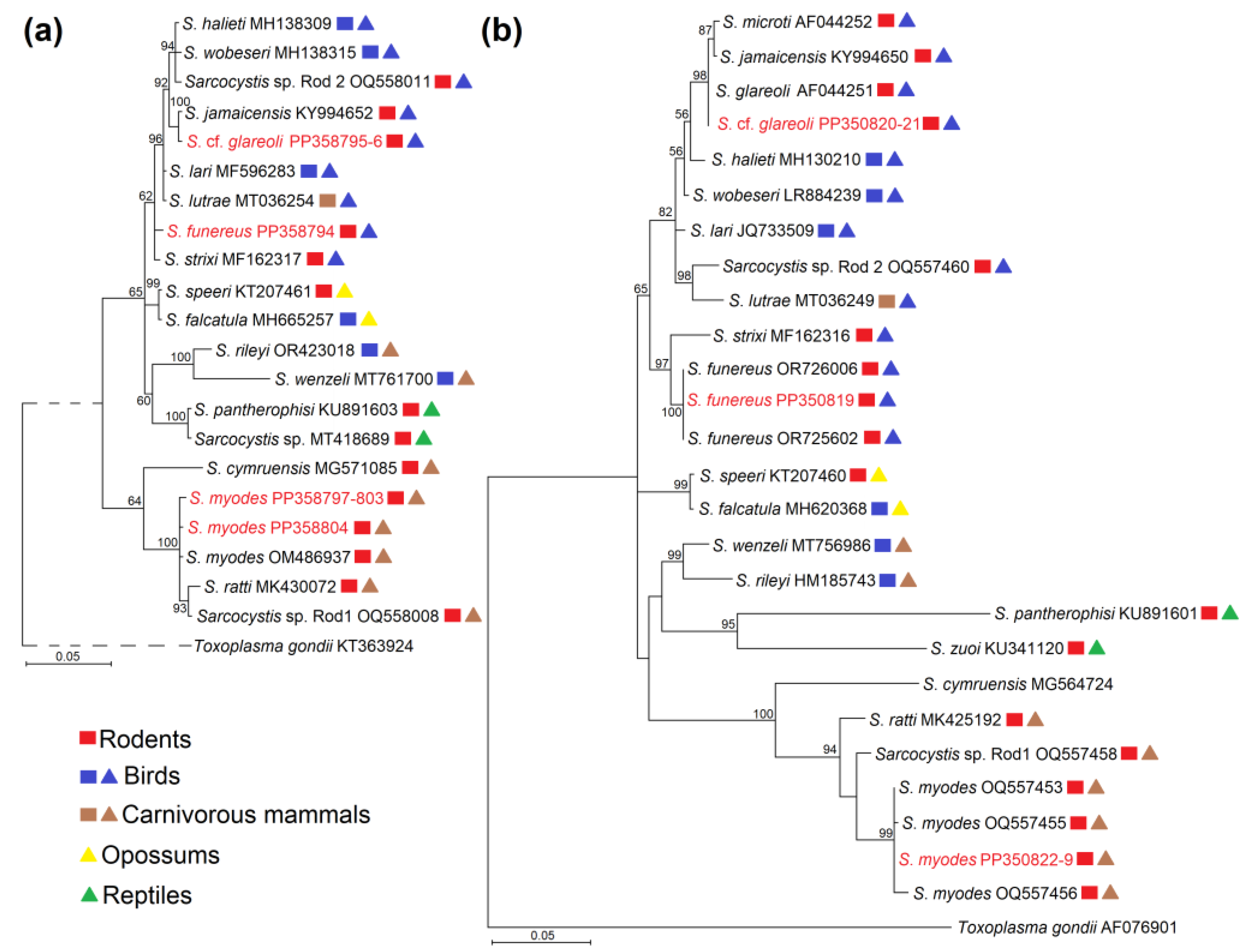
Multiple alignments of cox1 and 28S rRNA sequences were created with the help of the MUSCLE algorithm implemented in MEGA7 [39]. The alignment of cox1 contained 22 taxa and 619 nucleotide positions without gaps, whilst 28S rRNA alignment consisted of 27 taxa and 803 nucleotide positions including gaps. The selection of evolutionary models and generation of phylogenetic trees was performed while using TOPALi v2.5 [40]. The Bayesian method was applied to uncover phylogenetic relationships. The F81 + G and HKY + G nucleotide substitution models were chosen for cox1 and 28S rRNA analysis. Toxoplasma gondii was chosen as the outgroup in the generation of phylogenetic trees. The analyses were carried out in two runs, using one million generations with a sample frequency of 10 and 25% burn-in value. The 28S rRNA and cox1 sequences of the Sarcocystis spp. from the present study are available in GenBank under the accession numbers PP350819–PP350829 and PP358794–PP358804.

2.3. Statistical Analysis

Quantitative parasitology 3.0 software was utilized for statistical tests [41]. Sterne’s exact method was applied to determine a 95% confidence interval (CI) for the detection rates of Sarcocystis spp. Fisher’s exact test was utilized to assess the statistical significance of the differences comparing the prevalence of Sarcocystis spp. observed in the samples of A. flavicollis and C. glareolus.

3. Results

3.1. Detection Rates of Sarcocystis spp. in the Blood Samples of A. flavicollis and C. glareolus

For this study, 143 samples of C. glareolus were collected from traps in seven different habitats, such as mature deciduous forests and mixed forests, planted young forests, bogs, natural and shrubby meadows, and arable land (Table 1). The number of specimens caught in each habitat ranged from 8 to 34. Molecular examination of blood samples revealed the presence of Sarcocystis spp. in individuals collected from four different habitats: arable land, bog, mature deciduous forest, and natural meadow. In the latter habitats, the rate of positive samples ranged from 11.11% (95% CI = 2.02–33.02) to 12.50% (95% CI = 0.64–50.00). Differences in detection rates of Sarcocystis spp. were statistically insignificant when comparing the habitats tested (p > 0.05). The overall frequency of Sarcocystis spp. in the investigated blood samples of C. glareolus was 6.29% (95% CI = 3.19–11.45).
Additionally, 214 individuals of A. flavicollis were caught in seven different habitats, including mature deciduous forests and mixed forests, planted young forests, bogs, natural and shrubby meadows, and arable land (Table 2). In all but two habitats, between 29 and 63 individuals were trapped. In the remaining two habitats, planted young forest and bog, only five and eight individuals were collected, respectively. Molecular analysis of blood samples revealed the presence of Sarcocystis spp. in individuals collected only in arable land 3.17% (95% CI = 0.57–10.87). Statistically significant differences in detection rates of Sarcocystis spp. across the analysed habitats were not observed (p > 0.05). The detection frequency was significantly lower in A. flavicollis (0.93%; 95% CI = 0.17–3.40) compared to C. glareolus (p = 0.009).

3.2. Molecular Characterisation of Sarcocystis spp. in A. flavicollis and C. glareolus

In this study, 11 isolates were successfully characterized within partial cox1 and 28S rRNA. The length of the analysed cox1 sequences was 619 bp, while 28S rRNA sequences ranged between 721 bp and 735 bp. At the cox1 loci, sequences obtained in this study shared a similarity of 94.67–100% amongst themselves, while at the 28S rRNA loci the similarity was 87.25–100%. Thus, 28S rRNA was more variable compared with the cox1 for the Sarcocystis spp. detected. Based on the molecular analysis, three Sarcocystis species (Sarcocystis funereus, S. myodes and Sarcocystis cf. glareoli) were identified (Table 3).
According to the analysis of cox1 gene fragments, the sequences of S. myodes demonstrated 99.84–100% similarity compared to the sequences of this species deposited in the NCBI GenBank. Additionally, fragments acquired from the amplification of the 28S rRNA loci demonstrated intraspecific similarity of 99.46–100% in comparison with other S. myodes sequences. Based on 28S rRNA, the sequences of S. myodes showed the highest similarity to those of Sarcocystis ratti and Sarcocystis sp. Rod1 (96.46–97.82%).
The second detected Sarcocystis species (isolates LTKrCgla7A and LTKrCgla25C) exhibited the highest genetic similarity with two Sarcocystis species (S. glareoli and S. microti) previously classified in the genus Frenkelia and Sarcocystis jamaicensis (Table 3). However, cox1 sequences of S. glareoli and S. microti are yet to be established. The analysed Sarcocystis isolate displayed 100% similarity with S. jamaicensis within the cox1. Based on 28S rRNA, this organism differed from S. jamaicensis by two single nucleotide polymorphisms (SNPs) and by a single indel (insertion/deletion) from S. glareoli. Therefore, as nucleotide substitutions have more value for species divergence than indels [42], this Sarcocystis organism was denoted as S. cf. glareoli.
The last found species in this investigation was recently described as S. funereus [43]. At 28S rRNA, obtained sequences shared 99.72–100% similarity with those of S. funereus, 95.60% with those of Sarcocystis lari, and 95.25% with those of Sarcocystis strixi. Cox1 sequences of S. funereus have not been identified to date. Thus, based on this gene, sequences of S. funereus obtained in our work demonstrated the highest similarity with those of S. lari, Sarcocystis lutrae, and S. strixi (99.35–99.52%).

3.3. Phylogenetic Analysis of Identified Sarcocystis Species

The phylogenetic analyses based on two analysed loci (cox1 and 28S rRNA) resulted in longer branches of phylograms when using 28S rRNA sequences (Figure 2). In the 28S rRNA phylogenetic tree, sequences of three Sarcocystis species detected in this work were grouped with other isolates of the corresponding species, thus confirming the identification of the species studied. In both phylograms, two of the examined species, S. funereus and S. cf. glareoli were placed together with Sarcocystis species employing rodents, birds, and carnivorous mammals as their intermediate hosts and birds of their identified or presumed definitive hosts according to phylogenetic studies. Meanwhile, S. myodes was grouped into several Sarcocystis species using rodents and carnivorous mammals as their defined or proposed definitive hosts. Based on sequences of more variable 28S rRNA, S. cf. glareoli was most closely related to S. jamaicensis and S. microti, whereas S. funereus was sister species to S. strixi and finally S. myodes was grouped with S. ratti and Sarcocystis sp. Rod1.

4. Discussion

4.1. Prevalence of Sarcocystis spp. in Rodents

This study is the first documented attempt at investigating blood samples of C. glareolus and A. flavicollis for the detection of Sarcocystis species. In prior Lithuanian studies, sarcocysts were found in the muscles of several rodent species, including the brown rat (Rattus norvegicus), black rat (Rattus rattus), the bank vole, common vole (Microtus arvalis), tundra vole (Alexandromys oeconomus), field vole (Microtus agrestis), yellow-necked mouse, and striped field mouse (Apodemus agrarius) [16,44,45,46,47].
Specifically, the reported prevalence of Sarcocystis parasites in C. glareolus in Lithuania varies from 1.34% to 14.38% [16,22,45,48,49]. In this study, DNA of Sarcocystis spp. was detected in 6.29% (95% CI = 3.19–11.45) of C. glareolus blood samples, which is consistent with results from prior studies using muscle samples in Lithuania. Similar detection rates were observed in the Czech Republic and Finland, with Sarcocystis spp. detection in C. glareolus being relatively low, at 1.46% and 6%, respectively [50,51]. Nevertheless, studies on other vole species highlight significantly higher Sarcocystis species prevalence. In China, a study found that 25% of large oriental voles (Eothenomys miletus) were infected with Sarcocystis species [14]. Similarly, in Argentina, wild Cricetidae species exhibited a prevalence of 16.08% [12], while in Japan, 16.85% of Bedford’s red-backed voles (Clethrionomys rufocanus bedfordiae) were infected [52]. Moreover, research conducted in the Netherlands on M. arvalis reported seasonal variations in Sarcocystis spp. prevalence among voles, ranging from 6% to 33% [53].
Formerly, species producing cysts in the muscle tissues were classified under the genus Sarcocystis [54], while parasite species forming cysts in the brains of small mammals were placed under the genus Frenkelia [55] due to differences in sarcocyst morphology and its location. However, the merging of these genera has been proposed by several authors based on phylogenetic studies [9,56]. In Lithuania, it was reported that the prevalence of Frenkelia spp. in C. glareolus was 21.11% [57]. By comparison, in the Czech Republic, the prevalence of Frenkelia spp. was 38.5% [51], while in Germany it ranged from 10.34% to 55.93% [58,59], and in France, it varied from 1.02% to 48.14% [60,61].
Investigations on the prevalence of Sarcocystis spp. and Frenkelia spp. in A. flavicollis are notably scarce. In Lithuania, previously reported detection rates of Sarcocystis parasite in muscles ranged from 0% to 0.84%, consistent with the findings of this investigation, which reported a prevalence of 0.93% (95% CI = 0.17–3.40) [22,45,48,49]. In studies conducted in the Czech Republic and Spain, no Sarcocystis species were identified in the muscle samples of mice [51,62]. Taking into consideration other species of the genus Apodemus, higher infection rates of Sarcocystis spp. were recorded, namely in the large Japanese field mouse (Apodemus speciosus) and the small Japanese field mouse (Apodemus argenteus), the prevalence of Sarcocystis spp. was 18.99% and 21.43%, respectively [52]. In a previous Lithuanian study, Frenkelia spp. was absent in all A. flavicollis specimens [57]. Meanwhile, the prevalence of Frenkelia spp. in A. flavicollis from the Czech Republic was 2.40% [51], and in Germany, it ranged from 0.45% [59] to 8.54% (95% CI = 4.2–16.6) [58]. In summary, studies carried out in Lithuania, Germany, and the Czech Republic indicate that Sarcocystis spp. is more commonly found in C. glareolus than in A. flavicollis (present study, [51,57,58,59]).

4.2. Detection of Sarcocystis spp. in Blood Samples of Intermediate Hosts

During this investigation, blood samples were used to determine the prevalence and Sarcocystis spp. richness in Lithuania for the first time. Additionally, prior documented research on blood samples to identify Sarcocystis spp. has been conducted in Argentina, Japan, Turkey, Nigeria, and Australia, indicating precedence for such studies [25,26,27,28,29,63]. Three separate studies utilized rodent blood samples for investigation, successfully detecting Sarcocystis spp. in the blood of grey-sided vole (Myodes rufocanus), wood mouse (Apodemus sylvaticus), black rat and brown rat [27,28,29]. Remarkably, the prevalence of Sarcocystis spp. in M. rufocanus from Japan was relatively high, reaching 16.67%; nonetheless, this observation could be attributed to the small sample size (n = 6) that was examined [27]. Blood samples collected in Nigeria and Turkey from A. sylvaticus, R. rattus, and R. norvegicus exhibited an overall prevalence of Sarcocystis parasites ranging between 0.19% and 2.1% [28,29], aligning with the low prevalence rates reported in our study.
Regrettably, in this study, it was not possible to compare data from samples collected from blood and muscle tissues of the same individuals. Despite this, a study in Lithuania was carried out recently to identify Sarcocystis spp. in the muscle samples of rodents from commercial orchards [22]. Due to the sheer volume of the samples, the authors pooled muscle samples of the rodents that belonged to the same species, digested them, and identified parasite species using nested PCR and sequencing. In the study, the prevalence of Sarcocystis spp. in A. flavicollis was recorded as 0.84% (95% CI = 0.15–2.75), a finding that correlates closely with the results of our investigation, which showed a detection rate of 0.93% (95% CI = 0.17–3.40). Meanwhile, the rate of Sarcocystis infection in the muscles of C. glareolus was 1.34% (95% CI = 0.08–6.43) [22], showing a lower detection rate compared to our study, in which the rate was 6.29% (95% CI = 3.19–11.45). The variance in the results may be due to the different stages of Sarcocystis spp. infection, distinct sample collection locations, and potential seasonal effects [3,53]. Findings from a study conducted on camelids suggest that the DNA of Sarcocystis spp. can be detectable in the blood during the initial phases of the infection but become undetectable once encystment occurs [25].
The identification of three Sarcocystis species in this study highlights the significance of utilizing blood samples from intermediate hosts as a valuable method for examining both Sarcocystis prevalence and species diversity. This is the first report of S. funereus in C. glareolus from Lithuania, although molecular studies of muscle tissues were conducted before [16,22]. Although the molecular method used in this investigation is not suitable for the detection of Sarcocystis species coinfections. To identify more than one Sarcocystis species in a single sample, other methods, e.g., cloning, might be used [64].

4.3. Characteristics of Sarcocystis spp. in A. flavicollis and C. glareolus

Our molecular analysis showed the presence of three Sarcocystis species in A. flavicollis and C. glareolus from Lithuania. Among these species, S. myodes was only recently identified and characterised in the muscles of a single C. glareolus specimen [16]. Subsequently, sarcocysts of S. myodes have been detected in other intermediate hosts, such as A. flavicollis, A. agrarius, and M. arvalis [22]. Morphological analysis revealed sarcocysts of S. myodes to be microscopic (600–3000 × 70–220 µm) with a thin (~1 µm), smooth cyst wall lacking visible protrusions [16]. The characteristics of S. myodes sarcocysts closely resemble those of S. microti and S. glareoli. However, the latter species are exclusively found in the brains of small mammals [10,65]. Molecular analysis data on 18S rRNA and 28S rRNA loci demonstrates reliable differences between the three species [16,47,66]. While S. glareoli and S. microti utilise buzzards (Buteo spp.) as their definitive host [16,65], the primary host of S. myodes remains speculative due to insufficient data. Nevertheless, phylogenetic analysis suggests a potential association of S. myodes with predatory mammals [22].
Additionally, S. myodes shares structural similarities in its sarcocyst wall with S. cernae found in the common vole, as well as with Sarcocystis montanaensis discovered in the prairie vole (Microtus achrogaster), the long-tailed vole (Microtus longicaudus), and the eastern meadow vole (Microtus pennsylvanicus) [67,68,69]. However, the bradyzoites of Sarcocystis cernae (8–9 × 2–2.5 µm) [67] and S. montanaensis (9.8–12.2 × 2.2–4.3 µm) [68] differ from those of S. myodes (9.6–12.0 × 3.1–4.6 μm) [16]. A variety of snake species serve as definitive hosts in the life cycle of S. montanaensis, while S. cernae exclusively relies on the common kestrel (Falco tinnunculus) according to transmission experiments [68,69]. Thus, S. myodes differs from other Sarcocystis species found in different vole species, as determined through morphological, genetic, and phylogenetic analyses [16]. In addition, S. myodes displays sequence resemblance to S. ratti, found in the muscles of black rats across four loci (18S rRNA, 28S rRNA, cox1, ITS1) [16,47]. Morphologically, both parasite species share similar sarcocyst size, shape, and wall structure, although S. myodes exhibits longer bradyzoites compared to those of S. ratti (7.5–9.3 × 3.9–4.8 µm) [47].
During our study, one of the Sarcocystis organisms was identified tentatively as S. cf. glareoli. The lack of molecular data for the S. glareoli species, which was once placed under the genus Frenkelia, causes a significant challenge in the identification of this species. Currently, only sequences of the 18S rRNA and 28S rRNA genes have been obtained [9,10]. Apart from cox1, which was used in numerous studies [12,16,21,47,70,71], additional genetic markers, such as ITS1, mitochondrial cytochrome b (cytb), complete ITS15.8S rRNA–ITS2 region, two apicoplast genes—RNA polymerase beta subunit (rpoB) and caseinolytic protease C (clpC) demonstrate promising prospects for improved discrimination of Sarcocystis spp. in small mammals [47,66,72,73].
The last Sarcocystis species identified in our investigation is S. funereus, recently detected in the Tengmalm’s owl (Aegolius funereus) population in Finland [43]. Formerly identified as Sarcocystis sp. isolate Af1, this species lacked comprehensive data concerning its intermediate host and sarcocyst structure, despite characterisation using four genetic loci (ITS1, cox1, 18S rRNA, and 28S rRNA) [74]. However, S. funereus has been differentiated from other Sarcocystis species primarily through the utilization of the 28S rRNA and ITS1 region [43]. The latter region demonstrates superior sensitivity in discerning genetic variances among Sarcocystis species with birds and carnivores as intermediate hosts [74,75,76]. While the natural intermediate host remains elusive, experimental findings proposed C. glareolus as a potential candidate [77]. Notably, A. funereus exhibits a dietary preference for voles, with bank voles comprising more than 40% of its diet [78]. Our investigation provides additional evidence supporting the hypothesis that C. glareolus serves as the natural intermediate host for S. funereus.

5. Conclusions

Our comprehensive study marks a significant advance in the research of Sarcocystis spp. by using blood samples for the investigation of these parasites in two rodent species, A. flavicollis and C. glareolus. The findings revealed varying frequencies of Sarcocystis spp. across habitats, with no statistically significant differences observed in detection rates. Moreover, prior investigations carried out in Lithuania, Germany, and the Czech Republic indicate that C. glareolus tends to have higher rates of Sarcocystis spp. detection compared to A. flavicollis, which is consistent with the prevalence rates observed in the current study. Additionally, molecular analysis revealed three distinct Sarcocystis species—S. myodes, S. cf. glareoli, and S. funereus, with the latter being identified in Lithuania for the first time. Thus, blood samples can be successfully used for the studies of Sarcocystis spp. richness in small mammals.

Author Contributions

Conceptualisation, L.B. and P.P.; methodology, L.B. and P.P.; software, N.G. and P.P.; validation, L.B., N.K. and P.P.; formal analysis, N.G. and D.L.B.; investigation, L.B., N.K., N.G. and D.L.B.; resources, L.B., N.K. and P.P.; writing—original draft preparation, L.B., N.G., D.L.B. and P.P.; writing—review and editing, L.B., N.G., D.L.B., N.K. and P.P.; visualization, P.P.; supervision, P.P.; funding acquisition, L.B. and P.P. All authors have read and agreed to the published version of the manuscript.

Funding

This research received no external funding.

Institutional Review Board Statement

The research was conducted according to Lithuanian laws. The use of wild mammals for research is regulated by the Republic of Lithuania Law of Wildlife (22 June 2010, No. XI-920) and the Order of the Director of the State Food and Veterinary Service (30 June 2014, No. D1-369/B1-380, as last amended on 15 February 2014, No. D1-119/B1-197). According to the current procedure, no permit was required to perform the investigation in the current study. Investigated species are not classified as protected species in accordance with the Republic of Lithuania Law on protected Animals, Plants and Mushrooms (Order of the Director of the State Food and Veterinary Service, Chapter III, Article 50.2). The study was approved by the Animal Welfare Committee of the Nature Research Centre (protocol No GGT-2, 15 November 2016).

Informed Consent Statement

Not applicable.

Data Availability Statement

The 28S rRNA and cox1 sequences of S. funereus, S. myodes and S. cf. glareoli are available via the NCBI GenBank database under accession numbers PP350819–PP350829 and PP358794–PP358804.

Conflicts of Interest

The authors declare no conflicts of interest.

References

  1. Dubey, J.P.; Calero-Bernal, R.; Rosenthal, B.M.; Speer, C.A.; Fayer, R. Sarcocystosis of Animals and Humans, 2nd ed.; CRC Press: Boca Raton, FL, USA, 2016. [Google Scholar]
  2. Fayer, R. Sarcocystis spp. in Human Infections. Clin. Microbiol. Rev. 2004, 17, 894–902. [Google Scholar] [CrossRef]
  3. Decker Franco, C.; Schnittger, L.; Florin-Christensen, M. Sarcocystis. In Parasitic Protozoa of Farm Animals and Pets; Springer International Publishing: Cham, Switzerland, 2018; pp. 103–124. [Google Scholar] [CrossRef]
  4. Dubey, J.P.; Lindsay, D.S.; Saville, W.J.A.; Reed, S.M.; Granstrom, D.E.; Speer, C.A. A Review of Sarcocystis neurona and Equine Protozoal Myeloencephalitis (EPM). Vet. Parasitol. 2001, 95, 89–131. [Google Scholar] [CrossRef] [PubMed]
  5. Miller, M.A.; Barr, B.C.; Nordhausen, R.; James, E.R.; Magargal, S.L.; Murray, M.; Conrad, P.A.; Toy-Choutka, S.; Jessup, D.A.; Grigg, M.E. Ultrastructural and Molecular Confirmation of the Development of Sarcocystis neurona Tissue Cysts in the Central Nervous System of Southern Sea Otters (Enhydra lutris nereis). Int. J. Parasitol. 2009, 39, 1363–1372. [Google Scholar] [CrossRef] [PubMed]
  6. Levine, N.D. The Taxonomy of Sarcocystis (Protozoa, Apicomplexa) Species. J. Parasitol. 1986, 72, 372. [Google Scholar] [CrossRef] [PubMed]
  7. Martínez-Navalón, B.; Anastasio-Giner, B.; Cano-Fructuoso, M.; Sanchez-Martínez, P.; Llopis-Morant, A.; Perez-Castarlenas, B.; Goyena, E.; Berriatua Fernández De Larrea, E. Short Communication. Sarcocystis Infection: A Major Cause of Carcass Condemnation in Adult Sheep in Spain. Span. J. Agric. Res. 2012, 10, 388. [Google Scholar] [CrossRef]
  8. Dahmana, H.; Granjon, L.; Diagne, C.; Davoust, B.; Fenollar, F.; Mediannikov, O. Rodents as Hosts of Pathogens and Related Zoonotic Disease Risk. Pathogens 2020, 9, 202. [Google Scholar] [CrossRef]
  9. Votýpka, J.; Hypša, V.; Jirku, M.; Flegr, J.; Vavra, J.; Lukes, J. Molecular Phylogenetic Relatedness of Frenkelia spp. (Protozoa, Apicomplexa) to Sarcocystis falcatula Stiles 1893: Is the Genus Sarcocystis Paraphyletic? J. Eukaryot. Microbiol. 1998, 45, 137–141. [Google Scholar] [CrossRef]
  10. Mugridge, N.B.; Morrison, D.A.; Johnson, A.M.; Luton, K.; Dubey, J.P. Phylogenetic Relationships of the Genus Frenkelia: A Review of Its History and New Knowledge Gained from Comparison of Large Subunit Ribosomal Ribonucleic Acid Gene Sequences. Int. J. Parasitol. 1999, 29, 957–972. [Google Scholar] [CrossRef]
  11. Jäkel, T.; Khoprasert, Y.; Endepols, S.; Archer-Baumann, C.; Suasa-ard, K.; Promkerd, P.; Kliemt, D.; Boonsong, P.; Hongnark, S. Biological Control of Rodents Using Sarcocystis singaporensis. Int. J. Parasitol. 1999, 29, 1321–1330. [Google Scholar] [CrossRef]
  12. Bentancourt Rossoli, J.V.; Moré, G.; Soto-Cabrera, A.; Moore, D.P.; Morrell, E.L.; Pedrana, J.; Scioli, M.V.; Campero, L.M.; Basso, W.; Hecker, Y.P.; et al. Identification of Sarcocystis spp. in Synanthropic (Muridae) and Wild (Cricetidae) Rodents from Argentina. Parasitol. Res. 2024, 123, 31. [Google Scholar] [CrossRef]
  13. Canova, V.; Helman, E.; Robles, M.D.R.; Abba, A.M.; Moré, G. First Report of Sarcocystis spp. (Apicomplexa, Sarcocystidae) in Lagostomus maximus (Desmarest, 1917) (Rodentia, Chinchillidae) in Argentina. Int. J. Parasitol. Parasites Wildl. 2023, 20, 180–186. [Google Scholar] [CrossRef] [PubMed]
  14. Hu, J.-J.; Liu, Q.; Yang, Y.-F.; Esch, G.W.; Guo, Y.-M.; Zou, F.-C. Sarcocystis eothenomysi n. sp. (Apicomplexa: Sarcocystidae) from the Large Oriental Vole Eothenomys miletus (Thomas) (Cricetidae: Microtinae) from Anning, China. Syst. Parasitol. 2014, 89, 73–81. [Google Scholar] [CrossRef] [PubMed]
  15. Hu, J.-J.; Liu, T.-T.; Liu, Q.; Esch, G.W.; Chen, J.-Q. Sarcocystis clethrionomyelaphis Matuschka, 1986 (Apicomplexa: Sarcocystidae) Infecting the Large Oriental Vole Eothenomys miletus (Thomas) (Cricetidae: Microtinae) and Its Phylogenetic Relationships with Other Species of Sarcocystis Lankester, 1882. Syst. Parasitol. 2015, 91, 273–279. [Google Scholar] [CrossRef]
  16. Rudaitytė-Lukošienė, E.; Jasiulionis, M.; Balčiauskas, L.; Prakas, P.; Stirkė, V.; Butkauskas, D. Morphological and Molecular Description of Sarcocystis myodes n. sp. from the Bank Vole (Clethrionomys glareolus) in Lithuania. Biology 2022, 11, 512. [Google Scholar] [CrossRef]
  17. El-Morsey, A.; Abdo, W.; Sultan, K.; Elhawary, N.M.; AbouZaid, A.A. Ultrastructural and Molecular Identification of the Sarcocysts of Sarcocystis tenella and Sarcocystis arieticanis Infecting Domestic Sheep (Ovis aries) from Egypt. Acta Parasit. 2019, 64, 501–513. [Google Scholar] [CrossRef] [PubMed]
  18. Gjerde, B.; De La Fuente, C.; Alunda, J.M.; Luzón, M. Molecular Characterisation of Five Sarcocystis Species in Domestic Sheep (Ovis aries) from Spain. Parasitol. Res. 2020, 119, 215–231. [Google Scholar] [CrossRef]
  19. Feng, Y.; Guo, R.; Sang, X.; Zhang, X.; Li, M.; Li, X.; Yang, N.; Jiang, T. A Systematic Meta-Analysis of Global Sarcocystis Infection in Sheep and Goats. Pathogens 2023, 12, 902. [Google Scholar] [CrossRef]
  20. Hu, J.-J.; Huang, S.; Wen, T.; Esch, G.W.; Liang, Y.; Li, H.-L. Sarcocystis spp. in Domestic Sheep in Kunming City, China: Prevalence, Morphology, and Molecular Characteristics. Parasite 2017, 24, 8. [Google Scholar] [CrossRef]
  21. Jäkel, T.; Raisch, L.; Richter, S.; Wirth, M.; Birenbaum, D.; Ginting, S.; Khoprasert, Y.; Mackenstedt, U.; Wassermann, M. Morphological and Molecular Phylogenetic Characterization of Sarcocystis kani sp. nov. and Other Novel, Closely Related Sarcocystis spp. Infecting Small Mammals and Colubrid Snakes in Asia. Int. J. Parasitol. Parasites Wildl. 2023, 22, 184–198. [Google Scholar] [CrossRef]
  22. Prakas, P.; Stirkė, V.; Šneideris, D.; Rakauskaitė, P.; Butkauskas, D.; Balčiauskas, L. Protozoan Parasites of Sarcocystis spp. in Rodents from Commercial Orchards. Animals 2023, 13, 2087. [Google Scholar] [CrossRef]
  23. Baranauskaitė, A.; Strazdaitė-Žielienė, Ž.; Servienė, E.; Butkauskas, D.; Prakas, P. Molecular Identification of Protozoan Sarcocystis in Different Types of Water Bodies in Lithuania. Life 2022, 13, 51. [Google Scholar] [CrossRef]
  24. Moré, G.; Bacigalupe, D.; Basso, W.; Rambeaud, M.; Venturini, M.C.; Venturini, L. Serologic Profiles for Sarcocystis sp. and Neospora caninum and Productive Performance in Naturally Infected Beef Calves. Parasitol. Res. 2010, 106, 689–693. [Google Scholar] [CrossRef]
  25. Decker Franco, C.; Romero, S.; Ferrari, A.; Schnittger, L.; Florin-Christensen, M. Detection of Sarcocystis aucheniae in Blood of Llama Using a Duplex Semi-Nested PCR Assay and Its Association with Cyst Infestation. Heliyon 2018, 4, e00928. [Google Scholar] [CrossRef]
  26. Martin, M.; Decker Franco, C.; Romero, S.; Carletti, T.; Schnittger, L.; Florin-Christensen, M. Molecular Detection of Sarcocystis aucheniae in the Blood of Llamas from Argentina. Rev. Argent. Microbiol. 2016, 48, 200–205. [Google Scholar] [CrossRef]
  27. Moustafa, M.A.M.; Shimozuru, M.; Mohamed, W.; Taylor, K.R.; Nakao, R.; Sashika, M.; Tsubota, T. First Molecular Detection and Characterization of Hepatozoon and Sarcocystis spp. in Field Mice and Voles from Japan. Parasitol. Res. 2017, 116, 2321–2325. [Google Scholar] [CrossRef] [PubMed]
  28. Kamani, J.; Harrus, S.; Nachum-Biala, Y.; Gutiérrez, R.; Mumcuoglu, K.Y.; Baneth, G. Prevalence of Hepatozoon and Sarcocystis spp. in Rodents and Their Ectoparasites in Nigeria. Acta Trop. 2018, 187, 124–128. [Google Scholar] [CrossRef]
  29. Usluca, S.; Celebi, B.; Karasartova, D.; Gureser, A.S.; Matur, F.; Oktem, M.A.; Sozen, M.; Karatas, A.; Babur, C.; Mumcuoglu, K.Y.; et al. Molecular Survey of Babesia microti (Aconoidasida: Piroplasmida) in Wild Rodents in Turkey. J. Med. Entomol. 2019, 56, 1605–1609. [Google Scholar] [CrossRef]
  30. Gjerde, B.; Hilali, M.; Mawgood, S.A. Molecular Characterisation of Three Regions of the Nuclear Ribosomal DNA Unit and the Mitochondrial cox1 Gene of Sarcocystis fusiformis from Water Buffaloes (Bubalus bubalis) in Egypt. Parasitol. Res. 2015, 114, 3401–3413. [Google Scholar] [CrossRef] [PubMed]
  31. Sudan, V.; Shanker, D.; Paliwal, S.; Kumar, R.; Singh, A. Phylogenetics of Sarcocystis fusiformis Isolates Based on 18S rRNA and cox1 Genes. Microb. Pathog. 2021, 159, 105144. [Google Scholar] [CrossRef]
  32. Baltrūnaitė, L.; Kitrytė, N.; Križanauskienė, A. Blood Parasites (Babesia, Hepatozoon and Trypanosoma) of Rodents, Lithuania: Part I. Molecular and Traditional Microscopy Approach. Parasitol. Res. 2020, 119, 687–694. [Google Scholar] [CrossRef]
  33. Prūsaitė, J. Fauna of Lithuania: Mammals; Mokslas: Vilnius, Lithuania, 1988; p. 295. [Google Scholar]
  34. Hellgren, O.; Waldenström, J.; Bensch, S. A New PCR Assay for Simultaneous Studies of Leucocytozoon, Plasmodium, and Haemoproteus from Avian Blood. J. Parasitol. 2004, 90, 797–802. [Google Scholar] [CrossRef] [PubMed]
  35. Terman, R.C. Weights of Selected Organs of Deer Mice (Peromyscus maniculatus bairdii) from Asymp-Totic Laboratory Populations. J. Mammal. 1969, 50, 311–320. [Google Scholar] [CrossRef]
  36. Myllymäki, A. Demographic Mechanisms in the Fluctuating Populations of the Field Vole Microtus agrestis. Oikos 1977, 29, 468–493. [Google Scholar] [CrossRef]
  37. Balčiauskas, L.; Balčiauskienė, L.; Janonytė, A. Reproduction of the Root Vole (Microtus oeconomus) at the Edge of Its Distribution Range. Turk. J. Zool. 2012, 36, 668–675. [Google Scholar] [CrossRef]
  38. Sambrook, J.; Fritsch, E.F.; Maniatis, T. Molecular Cloning: A Laboratory Manual; Cold Spring Harbor Laboratory Press: Cold Spring Harbor, NY, USA, 1989. [Google Scholar]
  39. Kumar, S.; Stecher, G.; Tamura, K. MEGA7: Molecular Evolutionary Genetics Analysis Version 7.0 for Bigger Datasets. Mol. Biol. Evol. 2016, 33, 1870–1874. [Google Scholar] [CrossRef]
  40. Milne, I.; Wright, F.; Rowe, G.; Marshall, D.F.; Husmeier, D.; McGuire, G. TOPALi: Software for Automatic Identification of Recombinant Sequences within DNA Multiple Alignments. Bioinformatics 2004, 20, 1806–1807. [Google Scholar] [CrossRef] [PubMed]
  41. Reiczigel, J.; Marozzi, M.; Fábián, I.; Rózsa, L. Biostatistics for Parasitologists—A Primer to Quantitative Parasitology. Trends Parasitol. 2019, 35, 277–281. [Google Scholar] [CrossRef] [PubMed]
  42. Chen, J.-Q.; Wu, Y.; Yang, H.; Bergelson, J.; Kreitman, M.; Tian, D. Variation in the Ratio of Nucleotide Substitution and Indel Rates across Genomes in Mammals and Bacteria. Mol. Biol. Evol. 2009, 26, 1523–1531. [Google Scholar] [CrossRef]
  43. Máca, O.; Kouba, M.; Langrová, I.; Panská, L.; Korpimäki, E.; González-Solís, D. The Tengmalm’s Owl Aegolius funereus (Aves, Strigidae) as the Definitive Host of Sarcocystis funereus sp. nov. (Apicomplexa). Front. Vet. Sci. 2024, 11, 1356549. [Google Scholar] [CrossRef]
  44. Arnastauskiené, T.; Grikieniené, J. Infection of small mammals with sarcosporidians in the South-Eastern Baltic region. Ecology 1993, 2, 47–56. [Google Scholar]
  45. Grikienienė, J.; Malakauskas, M.; Mažeikytė, R.; Balčiauskas, L.; Senutaitė, J. Muscle Parasites (Sarcocystis, Trichinella, Alaria) of Wild Mammals in Lithuania. Theriol. Litu. 2001, 1, 29–46. [Google Scholar]
  46. Prakas, P.; Butkauskas, D. Protozoan Parasites from Genus Sarcocystis and Their Investigations in Lithuania. Ekologija 2012, 58, 45–58. [Google Scholar] [CrossRef]
  47. Prakas, P.; Kirillova, V.; Gavarāne, I.; Grāvele, E.; Butkauskas, D.; Rudaitytė-Lukošienė, E.; Kirjušina, M. Morphological and Molecular Description of Sarcocystis ratti n. sp. from the Black Rat (Rattus rattus) in Latvia. Parasitol. Res. 2019, 118, 2689–2694. [Google Scholar] [CrossRef] [PubMed]
  48. Grikienienė, J. Investigations into Endoparasites of Small Mammals in the Environs of Lake Drūkšiai. Acta Zool. Litu. 2005, 15, 109–114. [Google Scholar] [CrossRef]
  49. Grikienienė, J.; Mažeikytė, R. Investigation of Sarcosporidians (Sarcocystis) of Small Mammals in Kamasta Landscape Reserve and Its Surroundings. Acta Zool. Litu. 2000, 10, 55–68. [Google Scholar] [CrossRef]
  50. Soveri, T.; Henttonen, H.; Tanskanen, R.; Husu-Kallio, J.; Haukisalmi, V.; Sukura, A.; Laakkonen, J. Disease Patterns in Field and Bank Vole Populations during a Cyclic Decline in Central Finland. Comp. Immunol. Microbiol. Infect. Dis. 2000, 23, 73–89. [Google Scholar] [CrossRef]
  51. Svobodová, M.; Vorisek, P.; Votýpka, J.; Weidinger, K. Heteroxenous Coccidia (Apicomplexa: Sarcocystidae) in the Populations of Their Final and Intermediate Hosts: European Buzzard and Small Mammals. Acta Protozool. 2004, 43, 251–260. [Google Scholar]
  52. Inoue, I.; Yamada, M.; Yoshimi, Y.; Imai, Y.; Utsugi, I.; Suzuki, R.; Nogami, S.; Fujita, E.; Takahashi, K.; Tsuchiya, K.; et al. Prevalence of Sarcocystis (Protozoa, Apicomplexa) in Voles in Japan. Jpn. J. Parasitol. 1990, 39, 415–417. [Google Scholar]
  53. Hoogenboom, I.; Dijkstra, C. Sarcocystis cernae: A Parasite Increasing the Risk of Predation of Its Intermediate Host, Microtus arvalis. Oecologia 1987, 74, 86–92. [Google Scholar] [CrossRef]
  54. Tillmann, T.; Kamino, K.; Mohr, U. Sarcocystis muris—A Rare Case in Laboratory Mice. Lab. Anim. 1999, 33, 390–392. [Google Scholar] [CrossRef] [PubMed]
  55. Meingassner, J.G.; Burtscher, H. Doppelinfektion Des Gehirns Mit Frenkelia Species Und Toxoplasma gondii Bei Chinchilla laniger [Double Infection of the Brain with Frenkelia Species and Toxoplasma gondii in Chinchilla laniger—Author’s Trans.]. Vet. Pathol. 1977, 14, 146–153. [Google Scholar] [CrossRef] [PubMed]
  56. Modrý, D.; Votýpka, J.; Svobodová, M. Note on the Taxonomy of Frenkelia microti (Findlay & Middleton, 1934) (Apicomplexa: Sarcocystidae). Syst. Parasitol. 2004, 58, 185–187. [Google Scholar] [CrossRef] [PubMed]
  57. Grikienienė, J.; Mažeikytė, R.; Balčiauskas, L. The First Data on Brain Parasites of the Genus Frenkelia (Protista: Coccidia) in Some Small Rodent Species in Lithuania. Acta Zool. Litu. 2003, 13, 21–27. [Google Scholar] [CrossRef]
  58. Krücken, J.; Blümke, J.; Maaz, D.; Demeler, J.; Ramünke, S.; Antolová, D.; Schaper, R.; Von Samson-Himmelstjerna, G. Small Rodents as Paratenic or Intermediate Hosts of Carnivore Parasites in Berlin, Germany. PLoS ONE 2017, 12, e0172829. [Google Scholar] [CrossRef] [PubMed]
  59. Waindok, P.; Özbakış-Beceriklisoy, G.; Janecek-Erfurth, E.; Springer, A.; Pfeffer, M.; Leschnik, M.; Strube, C. Parasites in Brains of Wild Rodents (Arvicolinae and Murinae) in the City of Leipzig, Germany. Int. J. Parasitol. Parasites Wildl. 2019, 10, 211–217. [Google Scholar] [CrossRef]
  60. Fichet-Calvet, E.; Kia, E.B.; Giraudoux, P.; Quéré, J.P.; Delattre, P.; Ashford, R.W. Frenkelia Parasites in a Small Mammal Community. Dynamics of Infection and Effect on the Host. Parasite 2004, 11, 301–310. [Google Scholar] [CrossRef]
  61. Deter, J.; Bryja, J.; Chaval, Y.; Galan, M.; Henttonen, H.; Laakkonen, J.; Voutilainen, L.; Vapalahti, O.; Vaheri, A.; Salvador, A.R.; et al. Association between the DQA MHC Class II Gene and Puumala Virus Infection in Myodes glareolus, the Bank Vole. Infect. Genet. Evol. 2008, 8, 450–458. [Google Scholar] [CrossRef]
  62. Fernández-Escobar, M.; Millán, J.; Chirife, A.D.; Ortega-Mora, L.M.; Calero-Bernal, R. Molecular Survey for Cyst-Forming Coccidia (Toxoplasma gondii, Neospora caninum, Sarcocystis spp.) in Mediterranean Periurban Micromammals. Parasitol. Res. 2020, 119, 2679–2686. [Google Scholar] [CrossRef]
  63. Huaman, J.L.; Pacioni, C.; Forsyth, D.M.; Pople, A.; Hampton, J.O.; Helbig, K.J.; Carvalho, T.G. Evaluation of Haemoparasite and Sarcocystis Infections in Australian Wild Deer. Int. J. Parasitol. Parasites Wildl. 2021, 15, 262–269. [Google Scholar] [CrossRef]
  64. Basso, W.; Alvarez Rojas, C.A.; Buob, D.; Ruetten, M.; Deplazes, P. Sarcocystis Infection in Red Deer (Cervus elaphus) with Eosinophilic Myositis/Fasciitis in Switzerland and Involvement of Red Foxes (Vulpes vulpes) and Hunting Dogs in the Transmission. Int. J. Parasitol. Parasites Wildl. 2020, 13, 130–141. [Google Scholar] [CrossRef]
  65. Upton, S.J.; McKown, R.D. The Red-Tailed Hawk, Buteo jamaicensis, a Native Definitive Host of Frankelia microti (Apicomplexa) in North America. J. Wildl. Dis. 1992, 28, 85–90. [Google Scholar] [CrossRef] [PubMed]
  66. Zeng, H.; Guo, Y.; Ma, C.; Deng, S.; Hu, J.; Zhang, Y. Redescription and Molecular Characterization of Sarcocysts of Sarcocystis cymruensis from Norway Rats (Rattus norvegicus) and Sarcocystis ratti from Black Rats (R. rattus) in China. Parasitol. Res. 2020, 119, 3785–3791. [Google Scholar] [CrossRef] [PubMed]
  67. Černá, Z.; Loučková, M. Microtus arvalis as the Intermediate Host of a Coccidian from the Kestrel (Falco tinnunculus). Folia Parasitol. 1976, 23, 110. [Google Scholar]
  68. Dubey, J. Sarcocystis montanaenis and S. microti sp. n. from the Meadow Vole (Microtus pennsylvanicus). Proc. Helminthol. Soc. Wash. 1983, 50, 318–324. [Google Scholar]
  69. Lindsay, D.S.; Upton, S.J.; Blagburn, B.L.; Toivio-Kinnucan, M.; McAllister, C.T.; Trauth, S.E. Sporocysts Isolated from the Southern Copperhead (Agkistrodon contortrix contortrix) Produce Sarcocystis montanaensis-like Sarcocysts in Prairie Voles (Microtus ochrogastei). J. Wildl. Dis. 1991, 27, 148–152. [Google Scholar] [CrossRef]
  70. Antunes Murata, F.H.; Cerqueira-Cézar, C.K.; Thompson, P.C.; Tiwari, K.; Mowery, J.D.; Verma, S.K.; Rosenthal, B.M.; Sharma, R.N.; Dubey, J.P. Sarcocystis cymruensis: Discovery in Western Hemisphere in the Brown Rat (Rattus norvegicus) from Grenada, West Indies: Redescription, Molecular Characterization, and Transmission to IFN-γ Gene Knockout Mice via Sporocysts from Experimentally Infected Domestic Cat (Felis catus). Parasitol. Res. 2018, 117, 1195–1204. [Google Scholar] [CrossRef]
  71. Pérez, P.O.; Wibbelt, G.; Brinkmann, A.; Puentes, J.A.G.; Tuh, F.Y.Y.; Lakim, M.B.; Nitsche, A.; Wells, K.; Jäkel, T. Description of Sarcocystis scandentiborneensis sp. nov. from Treeshrews (Tupaia minor, T. tana) in Northern Borneo with Annotations on the Utility of COI and 18S rDNA Sequences for Species Delineation. Int. J. Parasitol. Parasites Wildl. 2020, 12, 220–231. [Google Scholar] [CrossRef]
  72. Watthanakaiwan, V.; Sukmak, M.; Hamarit, K.; Kaolim, N.; Wajjwalku, W.; Muangkram, Y. Molecular Characterization of the Ribosomal DNA Unit of Sarcocystis singaporensis, Sarcocystis zamani and Sarcocystis zuoi from Rodents in Thailand. J. Vet. Med. Sci. 2017, 79, 1412–1418. [Google Scholar] [CrossRef]
  73. Hu, J.; Sun, J.; Guo, Y.; Zeng, H.; Zhang, Y.; Tao, J. Infection of the Asian Gray Shrew Crocidura attenuata (Insectivora: Soricidae) with Sarcocystis attenuati n. sp. (Apicomplexa: Sarcocystidae) in China. Parasit. Vectors 2022, 15, 13. [Google Scholar] [CrossRef]
  74. Máca, O.; Kouba, M.; Korpimäki, E.; González-Solís, D. Molecular Identification of Sarcocystis sp. (Apicomplexa, Sarcocystidae) in Offspring of Tengmalm’s Owls, Aegolius funereus (Aves, Strigidae). Front. Vet. Sci. 2021, 8, 804096. [Google Scholar] [CrossRef] [PubMed]
  75. Máca, O. Molecular Identification of Sarcocystis lutrae in the European Otter (Lutra lutra) and the European Badger (Meles meles) from the Czech Republic. Parasitol. Res. 2018, 117, 943–945. [Google Scholar] [CrossRef] [PubMed]
  76. Juozaitytė-Ngugu, E.; Švažas, S.; Šneideris, D.; Rudaitytė-Lukošienė, E.; Butkauskas, D.; Prakas, P. The Role of Birds of the Family Corvidae in Transmitting Sarcocystis Protozoan Parasites. Animals 2021, 11, 3258. [Google Scholar] [CrossRef]
  77. Wiesner, J. A New Sarcosporidian Species [Unnamed] of Clethrionomys glareolus Inhabiting the Owl Aegolius funereus as Definitive Host. J. Protozool. 1981, 27, 72A. [Google Scholar]
  78. Khalil, H.; Ecke, F.; Evander, M.; Hörnfeldt, B. Selective Predation on Hantavirus-Infected Voles by Owls and Confounding Effects from Landscape Properties. Oecologia 2016, 181, 597–606. [Google Scholar] [CrossRef] [PubMed]
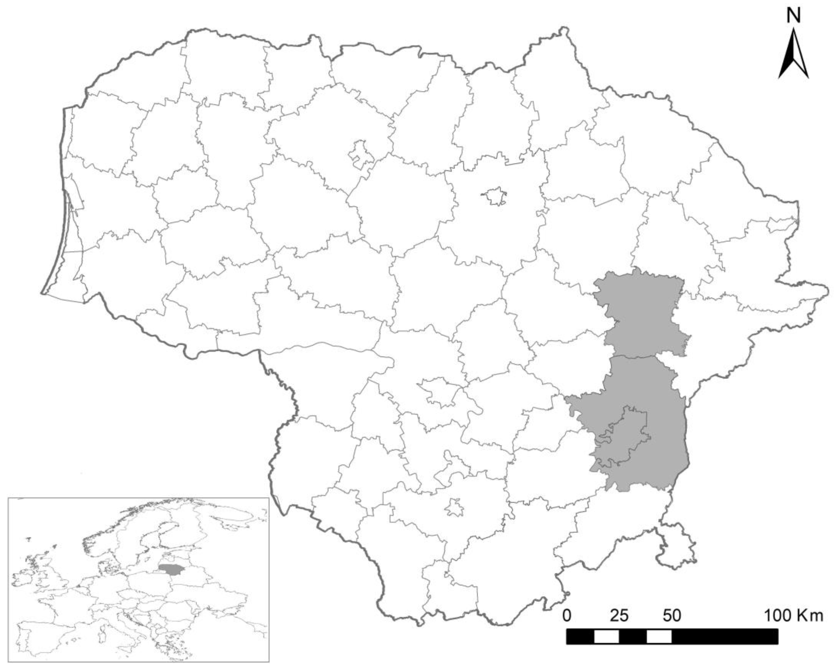
Figure 1. Small mammal trapping areas in Lithuania during the 2016–2019 period. Sampled districts are marked in grey.
Life 14 00365 g001
Figure 2. The phylogenetic relationships of the Sarcocystis spp. identified in blood samples of C. glareolus and A. flavicollis based on cox1 (a) and 28S rRNA (b) sequences. Phylograms were generated using Bayesian methods, scaled according to the branch length, and rooted on Toxoplasma gondii. The posterior probability support values are indicated next to branches, and GenBank accession numbers are given behind the species name. The sequences of three Sarcocystis species (S. funereus, S. myodes, and S. cf. glareoli) obtained in this work are shown in red. The coloured rectangles and triangles indicate the identified or presumed intermediate hosts and definitive hosts of Sarcocystis species, respectively.
Life 14 00365 g002
Table 1. Detection rates of Sarcocystis spp. in blood samples of C. glareolus.
HabitatTrappedInfected (%)Sarcocystis Species
Mature deciduous forest182 (11.11%)S. myodes
Mature mixed forest330-
Planted young forest100-
Bog81 (12.50%)S. myodes
Natural meadow344 (11.76%)S. myodes and S. funereus *
Shrubby meadow220-
Arable land182 (11.11%)S. cf. glareoli
Total1439 (6.29%)
* Sarcocystis myodes was detected in three samples and S. funereus was detected in a single sample.
Table 2. Detection rates of Sarcocystis spp. in blood samples of A. flavicollis.
HabitatTrappedInfected
Mature deciduous forest330
Mature mixed forest450
Planted young forest50
Bog80
Natural meadow310
Shrubby meadow290
Arable land632 (3.17%) *
Total2142 (0.93%)
* Only one species S. myodes was confirmed by molecular methods.
Table 3. Sarcocystis species identified in this study, and the percentage of similarity compared with the most closely related species.
Genetic Similarity with the Most Closely Related Species by Different Genes
Sarcocystis speciescox1Sarcocystis species28S rRNA
S. myodes
(619 bp)
S. myodes (99.84–100%), Sarcocystis sp. Rod1 (99.52–99.68%), S. ratti (99.03–99.19%)S. myodes
(735 bp)
S. myodes (99.46–100%), Sarcocystis sp. Rod1 (97.82%), S. ratti (96.46%)
S. cf. glareoli
(619 bp)
S. jamaicensis (100%), Sarcocystis sp. SCMW1 (99.68%), S. lutrae, S. corvusi, S. columbae, S. halieti, S. lari (99.52%), S. wobeseri, S. cornixi, Sarcocystis sp. ex Accipiter cooperi, Sarcocystis sp. Rod2 (99.35%), S. turdusi (99.19%), S. caninum, S. arctica, S. strixi, S. cf. strixi (99.03%)S. cf. glareoli
(726 bp)
S. glareolid (99.86%), S. jamaicensis (99.72%), S. microti (98.62%)
S. funereus
(619 bp)
S. strixi (99.52%), S. lutrae, S. lari (99.35%), Sarcocystis sp. Ex Accipiter cooperi, Sarcocystis sp. SCMW1 (99.19%), S. corvusi, S. columbae, S. halieti (99.03%)S. funereus
(721 bp)
S. funereus (99.72–100%), S. lari (95.60%), S. strixi (95.25%)
The lengths of the analysed fragment are indicated in parentheses under the name of Sarcocystis species.
Disclaimer/Publisher’s Note: The statements, opinions and data contained in all publications are solely those of the individual author(s) and contributor(s) and not of MDPI and/or the editor(s). MDPI and/or the editor(s) disclaim responsibility for any injury to people or property resulting from any ideas, methods, instructions or products referred to in the content.