Article

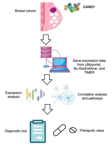
Bioinformatics Analysis of the Prognostic Significance of CAND1 in ERα-Positive Breast Cancer

Department of Pharmacology, Faculty of Medicine, Kuwait University, Kuwait City 13110, Kuwait
Diagnostics 2022, 12(10), 2327; https://doi.org/10.3390/diagnostics12102327
Submission received: 3 September 2022 / Revised: 25 September 2022 / Accepted: 26 September 2022 / Published: 27 September 2022
(This article belongs to the Special Issue Breast Cancer Biomarkers)

Abstract

:
The identification of novel prognostic biomarkers for breast cancer is an unmet clinical need. Cullin-associated and neddylation-dissociated 1 (CAND1) has been implicated in mediating carcinogenesis in prostate and lung cancers. In addition, CAND1 is an established prognostic biomarker for worse prognosis in liver cancer. However, the prognostic significance of CAND1 in breast cancer has not yet been explored. In this study, Breast Cancer Gene-Expression Miner (Bc-GenExMiner) and TIMER2.0 were utilized to explore the mRNA expression of CAND1 in ERα-positive breast cancer patients. The Kaplan–Meier plotter was used to explore the relationship between CAND1 expression and several prognostic indicators. The Gene Set Cancer Analysis (GSCA) web server was then used to explore the pathways of the genes that correlate with CAND1 in ERα-positive breast cancer. Immune infiltration was investigated using Bc-GenExMiner. Our bioinformatics analysis illustrates that breast cancer patients have higher CAND1 compared to normal breast tissue and that ERα-positive breast cancer patients with a high expression of CAND1 have poor overall survival (OS), distant metastasis-free survival (DMFS), and relapse-free survival (RFS) outcomes. Higher CAND1 expression was observed in histologic grade 3 compared to grades 2 and 1. Our results revealed that CAND1 positively correlates with lymph nodes and negatively correlates with the infiltration of immune cells, which is in agreement with published reports. Our findings suggest that CAND1 might mediate invasion and metastasis in ERα-positive breast cancer, possibly through the activation of estrogen and androgen signaling pathways; however, experiments should be carried out to further explore the role of CAND1 in activating the androgen and estrogen signaling pathways. In conclusion, the results suggest that CAND1 could be used as a potential novel biomarker for worse prognosis in ERα-positive breast cancer.

Graphical Abstract

1. Introduction

CAND1 gene encodes the CAND1 protein, which is predominantly localized in the cytoplasm [1]. CAND1 binds to unneddylated cullins [1,2,3] and regulates cullin–RING ubiquitin ligases [4,5,6]. These ligases play a role in the ubiquitinoylation of proteins that are degraded by the proteasome system and control the stability of substrates involved in transcription and cell cycle [1,7,8,9]. In cancer, these ligases are frequently dysregulated [7,10]. It has been shown that several members of the ubiquitin-ligase machinery are promising molecular targets for breast cancer therapeutic, including OTUB1 [11,12].
CAND1 has been implicated in mediating carcinogenesis, in which the downregulation of CAND1 induces apoptosis in prostate cancer cell lines [13]. Other published reports show that CAND1 is overexpressed in prostate cancer tissue compared to normal tissue [14,15]. It has been proposed that high CAND1 expression increases the degradation of tumor suppressor proteins, thereby inhibiting apoptosis [15]. In addition, CAND1 overexpression has been shown to promote the proliferation of prostate cancer by stabilizing PLK4 [14]. CAND1 has also been implicated in mediating proliferation and metastasis in lung cancer, in which targeting CAND1 by miR-33a decreases the proliferation and migration [16]. Moreover, it has been shown that CAND1 correlates with poor OS in liver cancer, in which silencing CAND1 suppresses the proliferation of liver cancer by inducing caspase-8/RIP1-dependent apoptosis [17]. CAND1 overexpression has also been observed in oligodendroglial cancer compared to normal tissue [18]. Additionally, targeting CAND1 with miR-148b-3p regulates Schwann cells migration [19]. miR-148b-3p has also been implicated in mediating the proliferation and migration of breast cancer cell lines [20,21].
Taking into account the fact that CAND1 overexpression has been shown to promote cancer proliferation and metastasis in several cancers, including lung, prostate, and liver cancers [11,12,13,14,15], and that CAND1 interacts with miR-148b-3p, which is a mediator of ERα-positive breast cancer cell metastasis [17,18,19], it is crucial to investigate the prognostic value of CAND1 in ERα-positive breast cancer. Moreover, given that there is a great demand for new biomarkers to predict outcomes and enhance individualized treatment due to breast cancer heterogeneity and that there is a lack of information about CAND1′s potential as a breast cancer prognostic biomarker, the aim of this study was to explore the significance of CAND1 in ERα-positive breast cancer by utilizing several bioinformatics tools.
Prompted by the lack of information about CAND1’s potential as an ERα-positive breast cancer biomarker, comprehensive bioinformatics analysis was carried out utilizing several databases to evaluate the significance of CAND1 as a diagnostic tool. Therefore, we evaluated the significance of CAND1 expression in ERα-positive breast cancer via bioinformatics analysis of the clinical indicators and survival data in several large online databases, including Bc-GenExMiner and TIMER2.0. In this research, the Kaplan–Meier plotter was utilized to assess the relationship between CAND1 expression and several prognostic indicators, whereas the GSCA web server was used to explore the pathways of the genes that correlate with CAND1. In addition, the cBio Cancer Genomics Portal was utilized to assess the relationship between CAND1 expression and different clinical indicators, including lymph nodes and histologic grades. The Bc-GenExMiner web server was also utilized to explore the infiltration of immune cells.

2. Materials and Methods

2.1. Investigating CAND1 mRNA Expression in Breast Cancer Tissue and Normal Breast Tissue

The mRNA expression of CAND1 in breast cancer and normal breast tissue was investigated using two different databases. Bc-GenExMiner version 4.1, which is a mining tool of published annotated genomic data, was utilized to investigate the expression of CAND1 in normal breast tissue (n = 92), tumor-adjacent tissue (n = 89), ERα-positive breast cancer tissue (n = 530), and ERα-negative breast cancer tissue (http://bcgenex.ico.unicancer.fr/BC-GEM/GEM-Accueil.php?js=1) (accessed on 31 August 2022) [22]. The TIMER2.0 database was also utilized to explore the expression of CAND1 in normal breast tissue (n = 1093) and in breast cancer (n = 112) (http://timer.cistrome.org/) (accessed on 20 August 2022) [23].

2.2. Kaplan–Meier Survival Curve Analysis

The Kaplan–Meier plotter website (http://kmplot.com/analysis/) (accessed on 15 August 2022) was utilized to explore the prognostic value of CAND1 in ERα-positive breast cancer patients [24]. The website was used to generate Kaplan–Meier plots for CAND1 in ERα-positive breast cancer patients by selecting the following parameters: OS, DMFS, and RFS; probe set option: all probe sets per gene; split patients by: upper quartile; follow-up threshold: 240 months [25]. Logrank p-values of <0.05 were considered statistically significant.

2.3. The Infiltration of Immune Cells

Pearson’s correlation coefficients between gene markers of immune cells and CAND1 were investigated using bc-GenExMiner [26]. Several gene markers of immune cells were included in the analysis, such as M1 and M2 macrophages, T cells, natural killer cells, and B cells. A p-value of <0.0001 was considered significant.

2.4. The Correlation between CAND1 mRNA and Neoplasm Histologic Grades, Hormone Therapy, and Lymph Nodes in ERα-Positive Breast Cancer Patients

The cBio Cancer Genomics Portal (MA, USA) (http://cbioportal.org) (accessed on 18 May 2022) was utilized, in which the METABRIC dataset was downloaded, which contains 1900 breast cancer patients (Table S1) [27]. The distribution of the expression of CAND1 mRNA, patients receiving hormone therapy, neoplasm histologic grades, and lymph nodes examined positive were measured across samples. CAND1 expression levels were considered high if their z-scores were higher or equal to the 75th percentile of the distribution [28].

2.5. Identification of Genes That Correlate with CAND1 in ERα-Positive Breast Cancer Patients

Genes that positively correlate with CAND1 in ERα-positive breast cancer patients with a Pearson’s correlation coefficient of >0.5 and a p-value of <0.0001 were identified using bc-GenExMiner (Table 1). Moreover, miRNAs that target CAND1 were identified (Table S1).

2.6. Association of Genes That Correlate with CAND1 in ERα-Positive Breast Cancer with Signaling Pathways

The GSCA web server (Houston, TX, USA) (http://bioinfo.life.hust.edu.cn/GSCA/#/) (accessed on 10 August 2022) was utilized to explore the involvement of the genes that correlate with CAND1 in activating and inhibiting pathways related to cancer, such as RTK, RAS/MAPK, estrogen receptor signaling, androgen receptor signaling, and TSC/mTOR [29]. Breast-invasive carcinoma patients with available expression data and who had paired samples (paired tumor–normal tissue) were included in the analysis.

2.7. Statistical Analysis

The grpahs were ploted using PRISM 8 GraphPad Inc. software (San Diego, CA, USA). The Kruskal–Wallis test was used to assess the significance of the differences between more than two groups, while the Mann–Whitney test was used to assess the significance of the differences between two groups.

3. Results

3.1. CAND1 mRNA Is Upregulated in ERα-Positive Breast Cancer Patients Compared to Tumor-Adjacent and Normal Breast Tissue

CAND1 mRNA expression in ERα-positive breast cancer patients was explored using Bc-GenExMiner and TIMER2.0. Significant upregulation of CAND1 mRNA expression was observed in breast cancer tissue compared to normal breast tissue (Figure 1a,b). Significant upregulation of CAND1 mRNA expression was also observed in ERα-positive breast cancer patients compared to ERα-negative breast cancer, tumor adjacent, and normal breast tissue (Figure 1a).

3.2. CAND1 mRNA Expression Correlates with Poor Prognosis in ERα-Positive Breast Cancer Patients

The prognostic value of CAND1 in ERα-positive breast cancer was explored using the Kaplan–Meier plotter website. It was observed that CAND1 significantly correlates with worse OS (Figure 2a), worse DMFS (Figure 2b), and worse RFS (Figure 2c) in ERα-positive breast cancer patients.

3.3. CAND1 Correlates Negatively with Various Gene Markers of Immune Cells in ERα-Positive Breast Cancer Patients

Taking into account that CAND1 correlates with poor prognosis in ERα-positive breast cancer patients and that the infiltration of immune cells correlates with better prognosis in breast cancer [30,31,32], an investigation of the type of the immune cells infiltrated in ERα-positive breast cancer patients was conducted. The results indicated that CAND1 correlates negatively with various immune cells, including T cells, B cells, monocytes, M1 and M2 macrophages, and neutrophils (Figure 3).

3.4. CAND1 mRNA Positively Correlates with Neoplasm Histologic Grades, Hormone Therapy, and Lymph Nodes in ERα-Positive Breast Cancer Patients

The METABRIC dataset in the cBio Cancer Genomics Portal was explored to validate our observation that CAND1 correlates with worse prognosis in ERα-positive breast cancer patients. CAND1 mRNA significantly correlates with lymph nodes examined positive in ERα-positive breast cancer patients (Figure 4a). A significant positive correlation was observed between CAND1 mRNA expression and neoplasm histologic grades in ERα-positive breast cancer patients, in which significant upregulation of CAND1 was observed in grade 3 compared to grades 2 and 1 (Figure 4b). In addition, significantly higher CAND1 mRNA was observed in ERα-positive breast cancer patients who received hormone therapy compared to ERα-positive breast cancer patients who did not receive hormone therapy (Figure 4c).

3.5. Identifying Genes That Positively Correlate with CAND1 in ERα-Positive Breast Cancer Patients

It was vital to investigate the genes that show a significant positive correlation with CAND1 in ERα-positive breast cancer patients to explore the pathways in which CAND1 exerts its effect on prognosis, as described in the summary of the experimental design (Figure 5).
The genes that showed a significant positive Pearson’s correlation (correlation coefficient > 0.5) with CAND1 in ERα-positive breast cancer patients were selected (Table 1).

3.6. Genes That Correlate with CAND1 in ERα-Positive Breast Cancer Patients Are Involved in the Activation of the Androgen and Estrogen Signaling Pathways

To explore the function of CAND1 in ERα-positive breast cancer, genes that showed a significant positive correlation with CAND1 were selected to identify the potential pathways that they are involved in. It was observed that the genes that significantly correlate with CAND1 expression in ERα-positive breast cancer patients were involved in the activation of the androgen and estrogen signaling pathways (Figure 6). A summary of the main results is shown in Figure S1.

4. Discussion

The results showed that CAND1 mRNA expression was upregulated in breast cancer patients compared to tumor-adjacent and normal breast tissue and that CAND1 expression was upregulated in ERα-positive breast cancer compared to ERα-negative breast cancer, suggesting that CAND1 might predict prognosis in ERα-positive breast cancer. A published report agrees with our findings, showing that CAND1 was upregulated in ERα-positive breast cancer compared to ERα-negative breast cancer [33]. In addition, CAND1 upregulation compared to normal tissue has also been observed in other cancers, including prostate and liver cancers [15,17].
The prognostic value of CAND1 in ERα-positive breast cancer was further investigated using the Kaplan–Meier plotter, in which CAND1 showed to correlate positively with worse prognosis in ERα-positive breast cancer, suggesting that CAND1 might act as a predictive biomarker for worse prognosis in ERα-positive breast cancer. Although the prognostic value of CAND1 in ERα-positive breast cancer has not been extensively studied, it has been suggested that CAND1 correlates with worse OS in breast cancer [34]. Additionally, CAND1 has been shown to serve as a potential biomarker for worse prognosis in liver and prostate cancers [15,17]. Moreover, the significant upregulation of CAND1 in neoplasm histologic grade 3 compared to grades 2 and 1 supports our previous observation that CAND1 correlates with worse prognosis in ERα-positive breast cancer. Interestingly, CAND1 interacts with CUL4, which is a cullin family member that has been shown to correlate with poor OS in breast cancer [35,36,37]. TUBB, which interacts with CAND1 [4], has also been shown to correlate with poor OS in ERα-positive breast cancer [38].
To further validate our observation that CAND1 correlates with poor prognosis in ERα-positive breast cancer, the infiltration of immune cells was investigated, as infiltration correlates with better prognosis in breast cancer [30,31,32]. The significant negative correlations between CAND1 and several immune cells agree with our previous observation that CAND1 correlates with poor prognosis in ERα-positive breast cancer. Although the correlation between CAND1 and the infiltration of immune cells in breast cancer has not been studied before, a published report showed that the infiltration of CD4 memory T cells and neutrophils correlates positively with CAND1 in lung adenocarcinoma [39]. This positive correlation might be due to the fact that the authors used a different approach to assess the correlation. The authors investigated the correlation between the infiltration of immune cells and a group of genes, including CAND1, RRM2, and SLC2A1, and they did not assess the correlation between the infiltration of immune cells and CAND1 alone.
Given that CAND1 correlates positively with DMSF and lymph nodes in ERα-positive breast cancer patients, CAND1 might be involved in mediating invasion and metastasis in ERα-positive breast cancer patients. A published report showed similar results, in which higher CAND1 was observed in breast cancer with lymph node metastasis compared to breast cancer without lymph node metastasis [40]. Although the role of CAND1 in mediating invasion in breast cancer has not been investigated yet, CAND1 has been shown to stabilize and synergize with PLK4 to induce centriole overduplication in prostate cancer [14]. PLK4 has been implicated in mediating metastasis and invasion by mediating the events of actin cytoskeleton [41]. These observations suggest that CAND1 might mediate invasion and metastasis in ERα-positive breast cancer, possibly through the stabilization of PLK4. In addition, CAND1 might mediate metastasis through the interaction with miR-148b-3p, which has been implicated in mediating the migration of breast cancer cell lines [20,21]; however, the exact mechanism remains to be unidentified.
Our results showed that CAND1 might be involved in the activation of the androgen and estrogen signaling pathways in ERα-positive breast cancer, as the genes that strongly correlate with CAND1 in ERα-positive breast cancer were involved in activating these pathways. The androgen and estrogen signaling pathways have been shown to mediate invasion and metastasis in different cancers, including breast cancer [42,43,44,45,46]. This suggests that the correlation observed between CAND1 and metastasis in ERα-positive breast cancer might be due to the activation of the estrogen and androgen signaling pathways. Although the role of CAND1 in regulating the estrogen signaling pathway has not been extensively studied, it has been observed that the estrogen receptor positively regulates CAND1 in ERα-positive breast cancer cell lines [47]. This suggests that CAND1 might be involved in this signaling pathway. The role of CAND1 in regulating the androgen receptor has been studied in prostate cancer, in which CAND1 has been shown to mediate cancer aggressiveness, possibly not through the androgen receptor signaling pathway, as it was shown to mediate cancer aggressiveness in androgen receptor-positive and -negative prostate cancer cell lines [15]. Given that miR-148a regulates the expression of CAND1 and that miR-148a is an androgen-responsive miRNA [13,48,49], the androgen signaling pathway might indirectly regulate CAND1 or CAND1 might play a role in this pathway. However, further experiments should be carried out to explore the involvement of CAND1 in the androgen and estrogen signaling pathways.
Given that the results indicate that CAND1 correlates with worse prognosis in ERα-positive breast cancer and that significantly higher CAND1 occurs in ERα-positive breast cancer patients who received hormone therapy compared to ERα-positive breast cancer patients who did not receive hormone therapy, clinicians should be aware when using hormone therapy to treat ERα-positive breast cancer patients, as it might increase CAND1 expression in patients.
Despite the above results and advantages of such bioinformatics analysis, some limitations need to be addressed. Specifically, the possible mechanism by which CAND1 activates the androgen and estrogen signaling pathways remains unknown. Further mechanistic analysis of CAND1 using in vitro and animal models is needed in order to robustly elucidate these mechanisms. Such analysis will aid in the development of novel therapeutics specifically targeted at Erα-positive breast cancer patients with upregulated CAND1. Moreover, combining bioinformatics with computer-aided diagnosis techniques, including cloud-based extreme learning machine, intuitionistic fuzzy active contour method, and neutosophic set-based medical image analysis, is crucial to validate our observations and to further explore the prognostic value of CAND1 in Erα-positive breast cancer, as these techniques outperform other techniques in diagnosing cancers and predicting outcomes [50,51,52].

5. Conclusions

In conclusion, it was shown that CAND1 is significantly upregulated in breast cancer patients compared to normal breast tissue, and higher CAND1 expression was observed in histologic grade 3 compared to grades 2 and 1 in ERα-positive breast cancer patients. In addition, it was found that CAND1 correlates positively with DMSF and lymph nodes in ERα-positive breast cancer patients. These results suggest that CAND1 could be used as a potential novel biomarker for worse prognosis in ERα-positive breast cancer, as it correlates positively with worse OS, RFS, and DFMS. Our results suggest that CAND1 might mediate invasion and metastasis in ERα-positive breast cancer, possibly through the activation of the estrogen and androgen signaling pathways. Despite the promising results of such bioinformatics analyses, using in vitro and animal models is necessary in order to robustly elucidate the significance of CAND1 in mediating invasion and metastasis in ERα-positive breast cancer and to explore the role of CAND1 in activating the androgen and estrogen signaling pathways.

Supplementary Materials

The following supporting information can be downloaded at: https://www.mdpi.com/article/10.3390/diagnostics12102327/s1, Table S1: METABRIC dataset; Table S2: List of miRNAs that target CAND1; Figure S1: Summary of the main results of the study. Refs. [53,54,55] were mentioned in Table S2 in the supplementary material.

Funding

This research received no external funding.

Informed Consent Statement

Not applicable.

Data Availability Statement

Not applicable.

Conflicts of Interest

The author declares no conflict of interest.

References

  1. Chua, Y.; Boh, B.; Ponyeam, W.; Hagen, T. Regulation of cullin RING E3 ubiquitin ligases by CAND1 in vivo. PLoS ONE 2011, 6, 16071. [Google Scholar] [CrossRef] [PubMed]
  2. Bosu, D.; Kipreos, T. Cullin-RING ubiquitin ligases: Global regulation and activation cycles. Cell Div. 2008, 3, 7. [Google Scholar] [CrossRef] [PubMed]
  3. Helmstaedt, K.; Schwier, E.; Christmann, M.; Nahlik, K.; Westermann, M.; Harting, R.; Grond, S.; Busch, S.; Braus, G. Recruitment of the inhibitor Cand1 to the cullin substrate adaptor site mediates interaction to the neddylation site. Mol. Biol. Cell 2011, 22, 153–164. [Google Scholar] [CrossRef] [PubMed]
  4. Bennett, E.; Rush, J.; Gygi, S.; Harper, J. Dynamics of Cullin-RING Ubiquitin Ligase Network Revealed by Systematic Quantitative Proteomics. Cell 2010, 143, 951–965. [Google Scholar] [CrossRef]
  5. Lydeard, J.; Schulman, B.; Harper, J. Building and remodelling Cullin–RING E3 ubiquitin ligases. EMBO Rep. 2013, 14, 1050–1061. [Google Scholar] [CrossRef]
  6. Soucy, T.; Smith, P.; Rolfe, M. Targeting NEDD8-Activated Cullin-RING Ligases for the Treatment of Cancer. Clin. Cancer Res. 2009, 15, 3912–3916. [Google Scholar] [CrossRef]
  7. Fouad, S.; Wells, O.; Hill, M.; D’Angiolella, V. Cullin Ring Ubiquitin Ligases (CRLs) in Cancer: Responses to Ionizing Radiation (IR) Treatment. Front. Physiol. 2019, 10, 1144. Available online: https://www.frontiersin.org/articles/10.3389/fphys.2019.01144 (accessed on 1 September 2022). [CrossRef]
  8. Genschik, P.; Sumara, I.; Lechner, E. The emerging family of CULLIN3-RING ubiquitin ligases (CRL3s): Cellular functions and disease implications. EMBO J. 2013, 17, 2307–2320. [Google Scholar] [CrossRef]
  9. Diaz, S.; Wang, K.; Sjögren, B.; Liu, X. Roles of Cullin-RING Ubiquitin Ligases in Cardiovascular Diseases. Biomolecules 2022, 12, 416. [Google Scholar] [CrossRef]
  10. Qi, J.; Ronai, Z. Dysregulation of ubiquitin ligases in cancer. Drug Resist. Updat. Rev. Comment. Antimicrob. Anticancer Chemother. 2015, 23, 1–11. [Google Scholar] [CrossRef] [Green Version]
  11. Wang, Y.; Dai, J.; Zeng, Y.; Guo, J.; Lan, J. E3 Ubiquitin Ligases in Breast Cancer Metastasis: A Systematic Review of Pathogenic Functions and Clinical Implications. Front. Oncol. 2021, 11, 752604. [Google Scholar] [CrossRef] [PubMed]
  12. Iskandar, A.; Zulkifli, N.; Ahmad, M.; Das, K.; Zulkifle, N. OTUB1 expression and interaction network analyses in MCF-7 breast cancer cells. Gene Rep. 2021, 24, 101273. [Google Scholar] [CrossRef]
  13. Murata, T.; Takayama, K.; Urano, T.; Horie-Inoue, K.; Ikeda, K.; Takahashi, S.; Kawazu, C.; Hasegawa, A.; Ouchi, Y.; Homma, Y.; et al. miR-148a is an androgen-responsive microRNA that promotes LNCaP prostate cell growth by repressing its target CAND1 expression. Prostate Cancer Prostatic Dis. 2010, 13, 356–361. [Google Scholar] [CrossRef] [PubMed]
  14. Korzeniewski, N.; Hohenfellner, M.; Duensing, S. CAND1 Promotes PLK4-Mediated Centriole Overduplication and Is Frequently Disrupted in Prostate Cancer. Neoplasia 2012, 14, 799–806. [Google Scholar] [CrossRef] [PubMed]
  15. Eigentler, A.; Tymoszuk, P.; Zwick, J.; Schmitz, A.; Pircher, A.; Kocher, F.; Schlicker, A.; Lesche, R.; Schafer, G.; Theurl, I.; et al. The Impact of Cand1 in Prostate Cancer. Cancers 2020, 12, 428. [Google Scholar] [CrossRef] [PubMed]
  16. Kang, M.; Li, Y.; Zhao, Y.; He, S.; Shi, J. miR-33a inhibits cell proliferation and invasion by targeting CAND1 in lung cancer. Clin. Transl. Oncol. 2018, 20, 457–466. [Google Scholar] [CrossRef] [PubMed]
  17. Che, Z.; Liu, F.; Zhang, W.; Mcgrath, M.; Hou, D.; Chen, P.; Song, C.; Yang, D. Targeting CAND1 promotes caspase-8/RIP1-dependent apoptosis in liver cancer cells. Am. J. Transl. Res. 2018, 10, 1357–1372. [Google Scholar]
  18. Lv, Z.; Bao, B.; Liang, P. Screening and Authentication of Key Genes and Prognostic Value Analysis in Oligodendroglial Tumor. Res. Square 2020. [Google Scholar] [CrossRef]
  19. Qian, T.; Zhao, L.; Wang, J.; Li, P.; Qin, J.; Liu, Y.; Yu, B.; Ding, F.; Gu, X.; Zhou, S. miR-148b-3p promotes migration of Schwann cells by targeting cullin-associated and neddylation-dissociated 1. Neural Regen. Res. 2016, 11, 1001–1005. [Google Scholar] [CrossRef]
  20. Dai, W.; He, J.; Zheng, L.; Bi, M.; Hu, F.; Chen, M.; Niu, H.; Yang, J.; Luo, Y.; Tang, W.; et al. miR-148b-3p, miR-190b, and miR-429 Regulate Cell Progression and Act as Potential Biomarkers for Breast Cancer. J. Breast Cancer 2019, 22, 219–236. [Google Scholar] [CrossRef]
  21. Dorraki, N.; Ghale-Noie, Z.; Ahmadi, N.; Keyvani, V.; Bahadori, R.; Nejad, A.; Aschner, M.; Pourghadamya, H.; Mollazadeh, S.; Mirzaei, H. miRNA-148b and its role in various cancers. Epigenomics 2021, 13, 1939–1960. [Google Scholar] [CrossRef] [PubMed]
  22. Jézéquel, P.; Gouraud, W.; Azzouz, F.; Guerin-Charbonnel, C.; Juin, P.; Lasla, H.; Campone, M. Bc-GenExMiner 4.5: New mining module computes breast cancer differential gene expression analyses. Database 2021, 1–10. [Google Scholar] [CrossRef] [PubMed]
  23. Li, T.; Fu, J.; Zeng, Z.; Cohen, D.; Li, J.; Chen, Q.; Liu, X. TIMER2.0 for analysis of tumor-infiltrating immune cells. Nucleic Acids Res. 2020, 48, W509–W514. [Google Scholar] [CrossRef] [PubMed]
  24. Győrffy, B. Survival analysis across the entire transcriptome identifies biomarkers with the highest prognostic power in breast cancer. Comput. Struct. Biotechnol. J. 2021, 19, 4101–4109. [Google Scholar] [CrossRef]
  25. Li, Q.; Birkbak, N.; Gyorffy, B.; Szallasi, Z.; Eklund, A. Jetset: Selecting the optimal microarray probe set to represent a gene. BMC Bioinform. 2011, 12, 474. [Google Scholar] [CrossRef]
  26. Tang, Z.; Kang, B.; Li, C.; Chen, T.; Zhang, Z. GEPIA2: An enhanced web server for large-scale expression profiling and interactive analysis. Nucleic Acids Res. 2019, 47, W556–W560. [Google Scholar] [CrossRef]
  27. Cerami, E.; Gao, J.; Dogrusoz, U.; Gross, B.; Sumer, S.; Aksoy, B.; Jacobsen, A.; Byrne, C.; Heuer, M.; Larsson, E.; et al. The cBio Cancer Genomics Portal: An Open Platform for Exploring Multidimensional Cancer Genomics Data. Cancer Discov. 2017, 7, 736–740. [Google Scholar] [CrossRef]
  28. Cereda, M.; Gambardella, G.; Benedetti, L.; Iannelli, F.; Patel, D.; Basso, G.; Guerra, R.; Mourikis, T.; Puccio, I.; Sinha, S.; et al. Patients with genetically heterogeneous synchronous colorectal cancer carry rare damaging germline mutations in immune-related genes. Nat. Commun. 2016, 7, 12072. [Google Scholar] [CrossRef]
  29. Liu, C.; Hu, F.; Xia, M.; Han, L.; Zhang, Q.; Guo, A. GSCALite: A web server for gene set cancer analysis. Bioinformatics 2018, 34, 3771–3772. [Google Scholar] [CrossRef]
  30. Manuel, M.; Tredan, O.; Bachelot, T.; Clapisson, G.; Courtier, A.; Parmentier, G.; Rabeony, T.; Grives, A.; Perez, S.; Mouret, J.; et al. Lymphopenia combined with low TCR diversity (divpenia) predicts poor overall survival in metastatic breast cancer patients. Oncoimmunology 2012, 1, 432–440. [Google Scholar] [CrossRef]
  31. Adams, S.; Gray, R.; Demaria, S.; Goldstein, L.; Perez, E.; Shulman, L.; Martino, S.; Wang, M.; Jones, V.; Saphner, T.; et al. Prognostic value of tumor-infiltrating lymphocytes in triple-negative breast cancers from two phase III randomized adjuvant breast cancer trials: ECOG 2197 and ECOG 1199. J. Clin. Oncol. Off. J. Am. Soc. 2014, 32, 2959–2966. [Google Scholar] [CrossRef] [PubMed]
  32. Ali, H.; Chlon, L.; Pharoah, P.; Markowetz, F.; Caldas, C. Patterns of Immune Infiltration in Breast Cancer and Their Clinical Implications: A Gene-Expression-Based Retrospective Study. PLoS Med. 2016, 13, e1002194. [Google Scholar] [CrossRef]
  33. Rezaul, K.; Thumar, J.; Lundgren, D.; Eng, J.; Claffey, K.; Wilson, L.; Han, D. Differential Protein Expression Profiles in Estrogen Receptor–Positive and –Negative Breast Cancer Tissues Using Label-Free Quantitative Proteomics. Genes Cancer 2010, 1, 251–271. [Google Scholar] [CrossRef] [PubMed]
  34. Mamoor, S. A single nucleotide variant on chromosome 12 residing within CAND1 distinguishes patients with luminal B breast cancer. OSF Preprints 2022. [Google Scholar] [CrossRef]
  35. Reichermeier, K.; Straube, R.; Reitsma, J.; Sweredoski, M.; Rose, C.; Moradian, A.; Besten, W.; Hinkle, T.; Verschueren, E.; Petzold, G.; et al. PIKES Analysis Reveals Response to Degraders and Key Regulatory Mechanisms of the CRL4 Network. Mol. Cell. 2020, 77, 1092–1106.e9. [Google Scholar] [CrossRef]
  36. Liu, A.; Zhang, S.; Shen, Y.; Lei, R.; Wang, Y. Association of mRNA expression levels of Cullin family members with prognosis in breast cancer: An online database analysis. Medicine 2019, 98, e16625. [Google Scholar] [CrossRef] [PubMed]
  37. Feng, M.; Wang, Y.; Bi, L.; Wang, H.; Mao, Z.; Wei, G. CRL4ADTL degrades DNA-PKcs to modulate NHEJ repair and induce genomic instability and subsequent malignant transformation. Oncogene 2021, 40, 2096–2111. [Google Scholar] [CrossRef]
  38. Alhammad, R. Bioinformatics Identification of TUBB as Potential Prognostic Biomarker for Worse Prognosis in ERα-Positive and Better Prognosis in ERα-Negative Breast Cancer. Diagnostics 2022, 12, 2067. [Google Scholar] [CrossRef]
  39. Wang, G.; Zhao, M.; Li, G.; Zheng, F.; Xu, G.; Hong, X. m7G-Associated subtypes, tumor microenvironment, and validation of prognostic signature in lung adenocarcinoma. Front. Genet. 2022, 13, 954840. [Google Scholar] [CrossRef]
  40. Wang, Y.; Chen, X.; Tian, Y.; Liu, L.; Su, P. Decreased Expression of circ_0000160 in Breast Cancer With Axillary Lymph Node Metastasis. Front. Mol. Biosci. 2022, 8, 690826. Available online: https://www.frontiersin.org/articles/10.3389/fmolb.2021.690826 (accessed on 19 September 2022). [CrossRef]
  41. Kazazian, K.; Go, C.; Wu, H.; Brashavitskaya, O.; Xu, R.; Dennis, J.; Gringras, A.; Swallow, C. Plk4 Promotes Cancer Invasion and Metastasis through Arp2/3 Complex Regulation of the Actin Cytoskeleton. Cancer Res. 2017, 77, 434–447. [Google Scholar] [CrossRef] [PubMed] [Green Version]
  42. Iacopetta, D.; Rechoum, Y.; Fuqua, S. The Role of Androgen Receptor in Breast Cancer. Drug Discov. Today Dis. Mech. 2012, 9, e19–e27. [Google Scholar] [CrossRef] [PubMed]
  43. Michmerhuizen, A.; Spratt, D.; Pierce, L.; Speers, C. Are we there yet? Understanding androgen receptor signaling in breast cancer. NPJ Breast Cancer 2020, 6, 47. [Google Scholar] [CrossRef] [PubMed]
  44. Bleach, R.; McIlroy, M. The Divergent Function of Androgen Receptor in Breast Cancer; Analysis of Steroid Mediators and Tumor Intracrinology. Front. Endocrinol. 2018, 9, 594. [Google Scholar] [CrossRef] [PubMed]
  45. Khatun, A.; Shimozawa, M.; Kito, H.; Kawaguchi, M.; Fujimoto, M.; Ri, M.; Kajikuri, J.; Niwa, S.; Fujii, M.; Ohya, S. Transcriptional Repression and Protein Degradation of the Ca2+-Activated K+ Channel KCa1.1 by Androgen Receptor Inhibition in Human Breast Cancer Cells. Front. Physiol. 2018, 9, 312. Available online: https://www.frontiersin.org/articles/10.3389/fphys.2018.00312 (accessed on 30 August 2022). [CrossRef] [PubMed]
  46. Roy, S.; Vadlamudi, R. Role of Estrogen Receptor Signaling in Breast Cancer Metastasis. Int. J. Breast Cancer. 2012, 2012, 654698. [Google Scholar] [CrossRef]
  47. Cheng, R.; Qi, L.; Kong, X.; Wang, Z.; Fang, Y.; Wang, J. Identification of the Significant Genes Regulated by Estrogen Receptor in Estrogen Receptor-Positive Breast Cancer and Their Expression Pattern Changes When Tamoxifen or Fulvestrant Resistance Occurs. Front. Genet. 2020, 11, 538734. [Google Scholar] [CrossRef]
  48. Venz, S.; Junker, H.; Ultsch, E.; Hetke, F.; Kruger, E.; Burchardt, M.; Caetano-Pinto, P.; Roennau, C. Identification of the Regulatory Targets of miR-3687 and miR-4417 in Prostate Cancer Cells Using a Proteomics Approach. Int. J. Mol. Sci. 2022, 23, 10565. [Google Scholar] [CrossRef]
  49. Jalava, S.; Urbanucci, A.; Latonen, L.; Waltering, K.; Sahu, B.; Janne, O.; Seppala, J.; Lahdesmaki, H.; Tammela, T.; Visakorpi, T. Androgen-regulated miR-32 targets BTG2 and is overexpressed in castration-resistant prostate cancer. Oncogene 2012, 31, 4460–4471. [Google Scholar] [CrossRef]
  50. Koundal, D.; Sharma, B. Challenges and future directions in neutrosophic set-based medical image analysis. In Neutrosophic Set in Medical Image Analysis; Academic Press: Cambridge, MA, USA, 2019; pp. 313–343. [Google Scholar] [CrossRef]
  51. Koundal, D.; Sharma, B.; Guo, Y. Intuitionistic based segmentation of thyroid nodules in ultrasound images. Comput. Biol. Med. 2020, 121, 103776. [Google Scholar] [CrossRef]
  52. Lahoura, V.; Singh, H.; Aggarwal, A.; Sharma, B.; Mohammed, M.; Damasevicius, R.; Kadry, S.; Cengiz, K. Cloud Computing-Based Framework for Breast Cancer Diagnosis Using Extreme Learning Machine. Diagnostics 2021, 11, 241. [Google Scholar] [CrossRef] [PubMed]
  53. Varga, A.; Marton, E.; Markovics, A.; Penyige, A.; Balogh, I.; Nagy, B.; Szilagyi, M. Suppressing the PI3K/AKT Pathway by miR-30d-5p Mimic Sensitizes Ovarian Cancer Cells to Cell Death Induced by High-Dose Estrogen. Biomedicines 2022, 10, 2060. [Google Scholar] [CrossRef]
  54. Gao, C.; Wei, J.; Tang, T.; Huang, Z. Role of microRNA-33a in malignant cells (Review). Oncol. Lett. 2020, 20, 2537–2556. [Google Scholar] [CrossRef] [PubMed]
  55. Xu, J.; Chen, Z.; Wang, Y.; Wang, X.; Chen, L.; Yuan, T.; Tang, X.; Lu, Y.; Chen, H.; Chen, M.; et al. Several circulating miRNAs related to hyperlipidemia and atherosclerotic cardiovascular diseases. Lipids Health Dis. 2019, 18, 104. [Google Scholar] [CrossRef] [PubMed] [Green Version]
Figure 1. (a) CAND1 mRNA expression in normal, tumor-adjacent, ERα-positive and -negative breast cancer tissues using bc-GenExMiner; (b) CAND1 mRNA expression in normal and breast cancer tissue using TIMER2.0. ** p < 0.01, *** p < 0.001, and **** p < 0.0001.
Diagnostics 12 02327 g001
Figure 2. Survival curves evaluating the prognostic value of CAND1 in ERα-positive breast cancer patients: (a) Analysis for OS; (b) analysis for DMFS; (c) analysis for RFS.
Diagnostics 12 02327 g002
Figure 3. Pearson’s correlation coefficient between CAND1 mRNA and several gene markers of immune cells in ERα-positive breast cancer patients, including T cells, B cells, monocytes, macrophages M1 and M2, and neutrophils. **** p < 0.0001.
Diagnostics 12 02327 g003
Figure 4. Plots evaluating CAND1 expression in ERα-positive breast cancer patients according to different clinical parameters: (a) Analysis for lymph nodes examined positive; (b) analysis for neoplasm histologic grades; (c) analysis for ERα-positive breast cancer patients who received hormone therapy and patients who did not receive hormone therapy. * p < 0.05 and ** p < 0.01.
Diagnostics 12 02327 g004
Figure 5. Schematic presentation of the experimental design followed in this study.
Diagnostics 12 02327 g005
Figure 6. Heat map showing the activated and inhibited pathways based on genes that significantly correlate with CAND1 in ERα-positive breast cancer patients.
Diagnostics 12 02327 g006
Table 1. Genes that correlate positively with CAND1 in ERα-positive breast cancer patients.
Gene SymbolCorrelation Coefficient 1p-ValueNumber of Patients
CCT20.6541<0.00013685
LEMD30.6339<0.00013685
NUP1070.6324<0.00013685
XPOT0.6155<0.00013685
TBK10.6058<0.00013685
PCBP2P20.5962<0.0001530
CNOT20.5901<0.00013685
ANKRD18EP0.5863<0.0001530
XPOTP10.5848<0.0001530
YEATS40.5838<0.00013685
CPSF60.5686<0.00013685
RAB210.5648<0.00013685
FRS20.5577<0.00013685
LARP40.5474<0.00013685
YWHAZP40.5464<0.0001530
MDM20.5444<0.00013685
MDM10.5387<0.00013685
SUDS3P10.5379<0.0001530
LRRC37A16P0.5342<0.0001530
PTGES3P30.5318<0.0001530
SCYL20.5234<0.00013685
PTGES30.5193<0.00013685
NUS1P10.5097<0.0001530
FAM3C2P0.5081<0.0001530
XPO10.5045<0.00013685
TCAF1P10.5041<0.0001530
RBM120.5014<0.00013685
1 Pearson’s correlation coefficient.
Publisher’s Note: MDPI stays neutral with regard to jurisdictional claims in published maps and institutional affiliations.

