Review

Ultrasound Imaging of Facial Vascular Neural Structures and Relevance to Aesthetic Injections: A Pictorial Essay

1
Department of Physical Medicine and Rehabilitation, National Taiwan University Hospital, Bei-Hu Branch, Taipei 10845, Taiwan
2
Department of Physical Medicine and Rehabilitation, College of Medicine, National Taiwan University, Taipei 10048, Taiwan
3
Center for Regional Anesthesia and Pain Medicine, Wang-Fang Hospital, Taipei Medical University, Taipei 11600, Taiwan
4
Department of Physical Medicine and Rehabilitation, Taichung Veterans General Hospital, Taichung 407219, Taiwan
5
Department of Surgery, Division of Plastic Surgery, National Taiwan University Hospital, Taipei 10048, Taiwan
6
Department of Rehabilitation Medicine, Charles University, First Faculty of Medicine and General University Hospital in Prague, 12800 Prague, Czech Republic
7
Physical and Rehabilitation Medicine Unit, Department of Biomedical and Neuromotor Science, Istituto di Ricovero e Cura a Carattere Scientifico Rizzoli Orthopedic Institute, 40136 Bologna, Italy
8
Department of Physical and Rehabilitation Medicine, Hacettepe University Medical School, Ankara 06100, Turkey
*
Author to whom correspondence should be addressed.
Diagnostics 2022, 12(7), 1766; https://doi.org/10.3390/diagnostics12071766
Submission received: 31 May 2022 / Revised: 12 July 2022 / Accepted: 18 July 2022 / Published: 21 July 2022

Abstract

:
The facial and submental regions are supplied by complicated neurovascular networks; therefore, facial aesthetic injections may be associated with serious adverse events such as skin necrosis and blindness. Pre-injection localization of neurovascular structures using high-resolution ultrasound can theoretically prevent unexpected complications. Therefore, a systematic protocol that focuses on these facial neurovascular structures is warranted. In this pictorial essay, we discuss the sonoanatomy of facial and submental neurovascular structures and its relevance to aesthetic injections. Moreover, we have highlighted the mechanisms underlying potential neurovascular injuries during aesthetic injections.

1. Introduction

Facial aesthetic injections are widely used in clinical practice; the most common indications include reduction of wrinkles and facial contouring. Injectables include botulinum toxin [1], fillers (hyaluronic acid, silicone, and autologous fat) [2] and deoxynucleic acid [3]. Botulinum toxin blocks the neuromuscular junction, which results in relaxation of the underlying muscles and is therefore used to eliminate wrinkles. Fillers used for volume augmentation are useful for contouring atrophic/concave regions. Deoxynucleic acid is useful to reduce local fullness in areas of excessive fat accumulation. However, the facial and submental regions are supplied by a complicated neurovascular network; therefore, post-injection complications represent a significant challenge to cosmetologists [4,5].
Ultrasonography is known to provide high-resolution imaging of superficial soft tissues [6,7]. Power Doppler imaging enables investigators to scrutinize even tiny vasculature without the administration of contrast agents [8]. Although sound beams cannot penetrate the bony cortex, ultrasonography is a sensitive tool to delineate the neurovascular foramina on facial bones [7]. A recent review discussed details of the sonoanatomy of facial muscles but not of facial neurovascular structures [9]. In this pictorial essay, we discuss the sonoanatomy of the neurovascular structures in the facial and submental regions and its relevance to aesthetic injections, aiming to facilitate pre-procedure ultrasound scanning in a systematic manner. We hope the accidental injury to the vessels and nerves on the face can thus be decreased during aesthetic injections. Figure 1, Figure 2 and Figure 3 illustrate the main neurovascular structures over the frontal, lateral, and submental regions of the face, respectively. These figures have been adapted to the face of our co-author (H.-C. C) with permission obtained for publication. All sonographic images were obtained using a 10–25 MHz high-frequency linear transducer (X-Cube 90, Alpinion Medical Systems Co. Ltd., Anyang, Korea).

2. Aesthetic Injections and Relevant Neurovascular Structures

2.1. Upper Face

Facial wrinkles are the most common indication for botulinum toxin injections [10]. Horizontal forehead lines can be corrected by injections into the frontalis muscle. Likewise, injections into the corrugator supercilii and procerus are useful to eliminate glabellar frown lines. Lateral canthal lines, commonly referred to as ‘crow’s feet’, can be reduced by injections into the lateral orbital portions of the orbicularis oculi muscle.
The forehead and glabella represent the most common sites for filler injections [11]. The injectables should be introduced within the loose connective tissue above the periosteum for wrinkle reduction and volume expansion. Furthermore, injections into the periorbital area can be used for correction of ‘sunken eyes.’
Vessels that course across the middle of the forehead include the supratrochlear artery/vein and the supraorbital artery/vein [12,13]. These are accompanied by the supraorbital and supratrochlear nerves [14]. The lateral forehead is supplied by the frontal branch of the superficial temporal artery and vein [15] (Figure 1).

2.2. Middle Face

Temporalis muscle hypertrophy may accompany masseter muscle atrophy, and botulinum injections are useful in such cases. Injections into the risorius and zygomaticus major muscles are occasionally used to improve facial asymmetry. Injections into the levator labii superioris alaeque nasi and levator labii superioris are shown to be useful to correct a gummy smile [9].
Filler injections into the temporal area are useful to correct atrophy secondary to age-induced fat loss [11]. Medial infraorbital region injections are used to trim the tear trough, which is a deep crease between the lower eyelid and upper cheek. Flattening of the malar eminence can be corrected by injections into the cheek, which is referred to as malar augmentation. Injections into the buccal fat pad layer are useful to contour anterior sunken cheeks. Subzygomatic depression can be corrected through injections into the superficial fat of the subcutaneous layer above the superficial musculoaponeurotic system distal to the zygomatic arch. Nasolabial fold injections can improve age-induced deepening of nasolabial lines. Injections directed into the layer between the nasalis muscle and the nasal cartilage increase the projection of the nasal bridge.
The dorsal nasal artery/vein and the angular artery/vein run in close proximity to or on the nose [16,17]. The cheek and upper perioral region are supplied by the superior labial artery/vein and the infraorbital artery/vein [18,19], and the superficial temporal and deep temporal arteries/veins supply the temporal area (Figure 2) [20]. These vessels are accompanied by the auriculotemporal, infraorbital, and facial nerves (Figure 2) [14].

2.3. Lower Face

Injections into the masseter muscle are used to contour a square face and for treatment of bruxism [21]. Mentalis muscle injections improve a cobblestone chin. Injections into the depressor labii inferioris or depressor anguli oris muscle are used to treat a droopy face [9].
Filler injections administered medial to the depressor anguli oris muscle can reduce prominent marionette lines. Injections over the mental region improve the appearance of an under-projected chin [11].
The inferior labial artery/vein and the facial artery/vein are important vessels of the lower face (Figure 1) [22]. Innervation of the lower face is primarily via the mental nerve [14].

2.4. Submental Region

Submental fullness is the most common indication for aesthetic injections into the submental region. Deoxycholic acid is approved by the Food and Drug Administration of the United States for reduction of submental fat [3]. Although these injections are considered safe and effective, adverse reactions such as local tenderness, edema, numbness, bruising, and hematoma have been reported [3]. The submental artery/vein and the hypoglossal nerve are important neurovascular structures that should be protected during submental injections (Figure 3) [23,24].

3. Mechanism of Neurovascular Injury

Facial aesthetic injections are associated with risks such as bruising, edema, skin discoloration, and infection. Nerve injuries may occur secondary to direct trauma such as needle piercing and nerve laceration. Based on severity (from mild to severe), peripheral nerve injury is categorized as neurapraxia, axonotmesis, and neurotmesis [25,26]. Mild nerve injury leads to temporary sensory/motor impairment, which usually recovers within 2 to 3 weeks. However, severe injury (for example, nerve transection) can cause long-term anesthesia over the innervated region.
Vascular compromise observed after filler injection [27] is attributable to filler-induced vessel compression, particularly after enlargement of the volume owing to absorption of water nearby. Direct puncture of vessels can lead to antegrade or retrograde flow of fillers and trigger embolic events, which may result in serious complications such as skin necrosis, blindness, and stroke.

4. Sonoanatomy of the Neurovascular Structures of the Upper Face

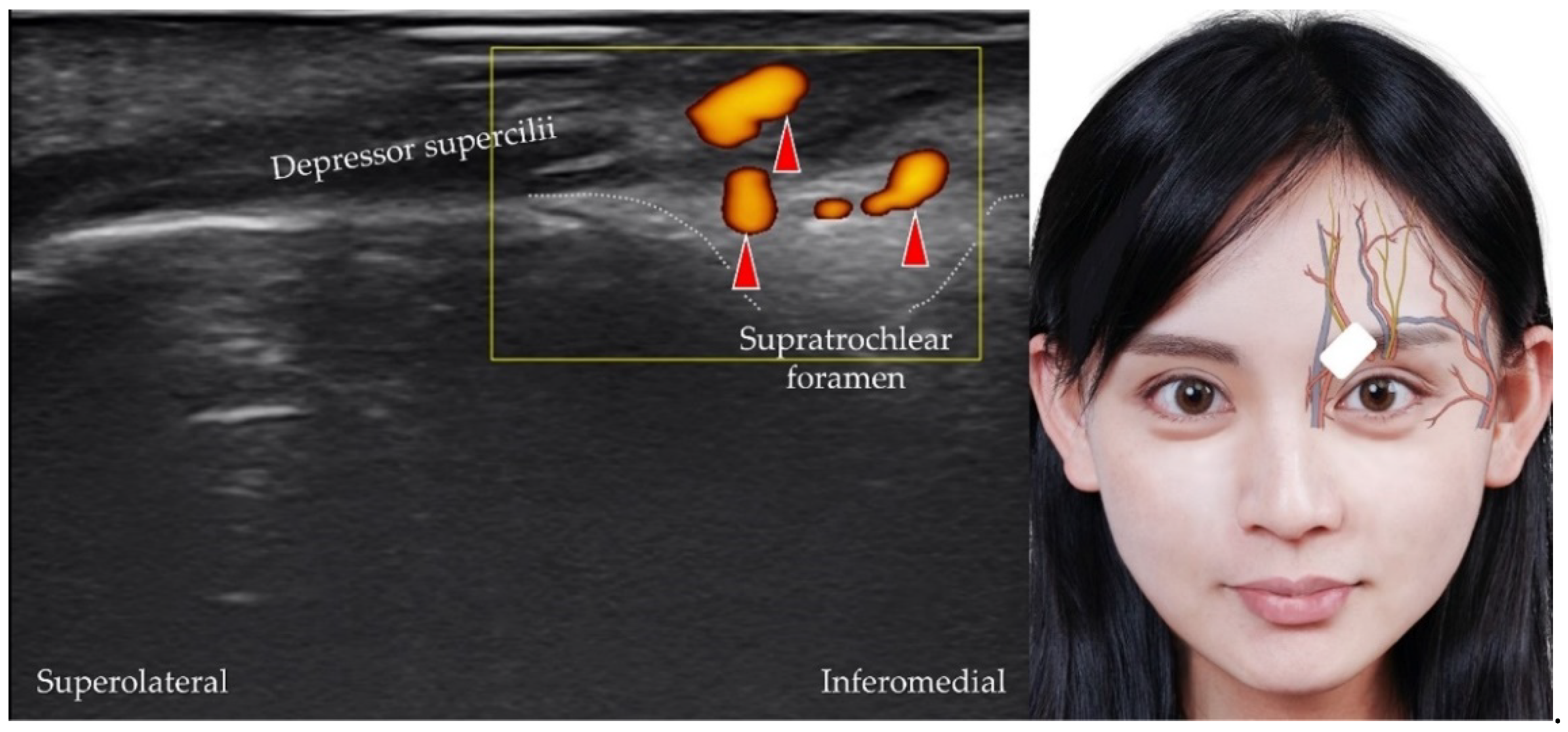
4.1. Supratrochlear Artery, Vein, and Nerve

The supratrochlear artery is the terminal branch of the ophthalmic artery [12]. It originates from the supratrochlear foramen, which is a small aperture at the medial edge of the cranial orbital margin of the frontal bone. The transducer is placed over the medial orbital rim in the horizontal plane, and the medial edge of the transducer is pivoted to the medial canthus. The supratrochlear artery is visualized as it emerges from the supratrochlear foramen [28] (Figure 4). The supratrochlear nerve (Figure 5) [29] (which originates from the frontal nerve, which itself is a branch of the ophthalmic nerve) and vein may be visualized in addition to the supratrochlear artery.

4.2. Supraorbital Artery, Vein, and Nerve

The supraorbital artery also branches from the ophthalmic artery and emerges from the supraorbital foramen, a small opening near the midpoint of the superior orbital margin in the frontal bone [12]. The transducer is placed over the center of the superior orbital rim. The supraorbital artery originates from the supraorbital foramen (Figure 6), and the supraorbital nerve [30,31] (also a terminal branch of the frontal nerve) and vein are identified along with the supraorbital artery (Figure 7).

4.3. Frontal Branch of the Superficial Temporal Artery/Vein

The superficial temporal artery branches into the terminal frontal branch near the level of the tragus [20,32]. The frontal branch travels anteriorly to supply the lateral frontal region. The transducer is placed over the lateral part of the forehead, slightly cranial to the eyebrows. The frontal branches of the superficial temporal artery (and vein) can be visualized coursing over the frontalis muscle (Figure 8). Table 1 summarizes the techniques used to scan the neurovascular structures of the upper face.

5. Sonoanatomy of the Neurovascular Structures of the Middle Face

5.1. Dorsal Nasal Artery and Vein

The dorsal nasal artery is a terminal branch of the ophthalmic artery [33] and emerges from the medial orbital rim and descends along the nasal septum. A communicating branch, known as the intercanthal artery, may be identified at the nasal bridge. The transducer is placed in the horizontal plane near the medial orbital rim to visualize the short axis of the dorsal nasal artery (and vein) on the procerus, levator labii superioris alaeque nasi, and nasalis muscles (Figure 9) [9,34].

5.2. Angular Artery and Vein

The angular artery is the terminal portion of the facial artery and ascends along the nasolabial sulcus to the medial orbital rim [17]. The transducer is placed in the horizontal plane lateral to the ala of the nose, and the angular artery can be identified coursing over the levator labii superioris alaeque nasi and levator labii superioris muscles [35]. The angular vein is seen more lateral than the angular artery (Figure 10).

5.3. Superior Labial Artery and Vein

The superior labial artery originates from the facial artery and courses along the cranial edge of the lip [22]. The transducer is placed in the sagittal plane medial to the angle of the mouth. The superior labial artery/vein can be visualized coursing over or under the orbicularis oris muscle (Figure 11) [22,36].

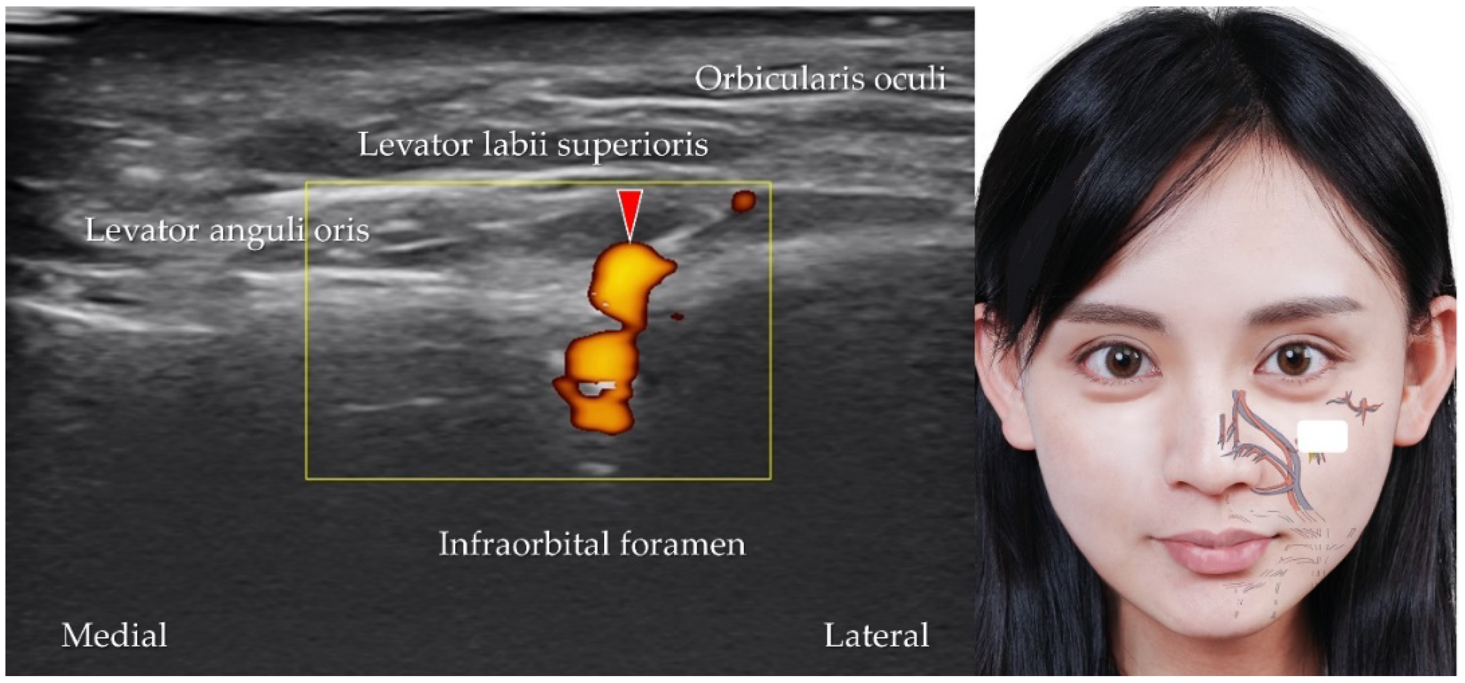
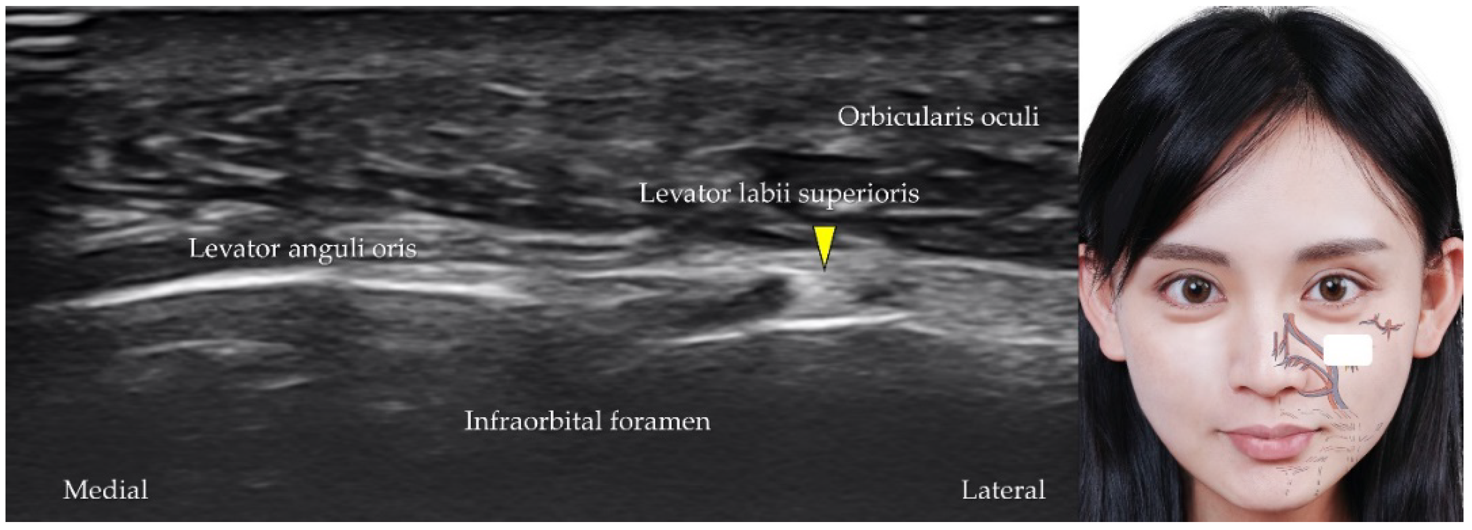
5.4. Infraorbital Artery, Vein, and Nerve

The infraorbital artery is the terminal branch of the maxillary artery [19]. The transducer is placed in the horizontal plane slightly distal to the inferior eyelid. The infraorbital artery (Figure 12), vein, and nerve (a branch from the maxillary nerve, Figure 13) can be visualized as they emerge from the infraorbital foramen [37].

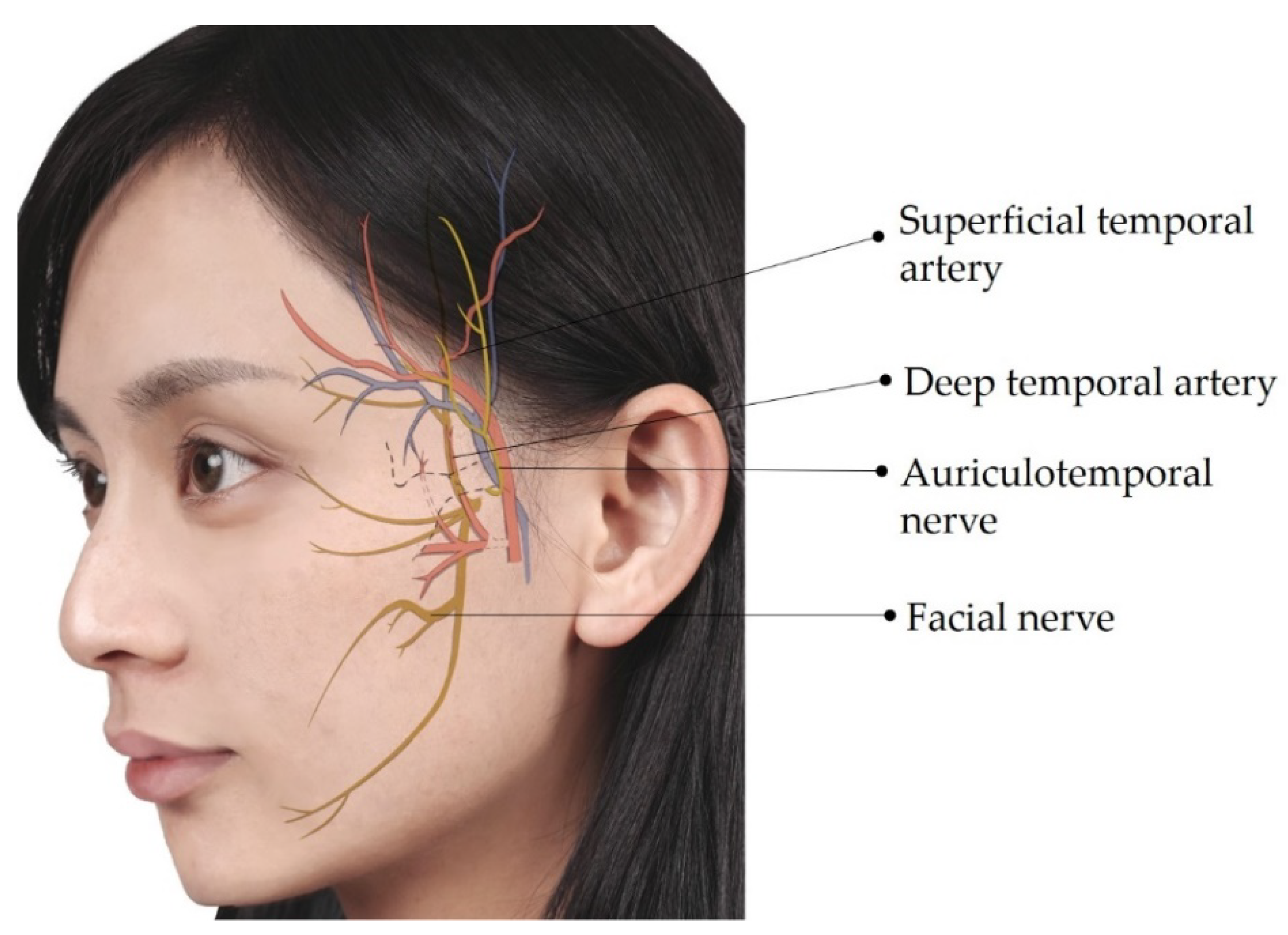
5.5. Superficial Temporal Artery and Vein

The superficial temporal artery originates from the external carotid artery and further divides into the frontal and parietal branches [15]. The transducer is placed in the horizontal plane cranial to the zygomatic arch. The parietal branch can be identified superficial to the temporal fascia (Figure 14) [38].

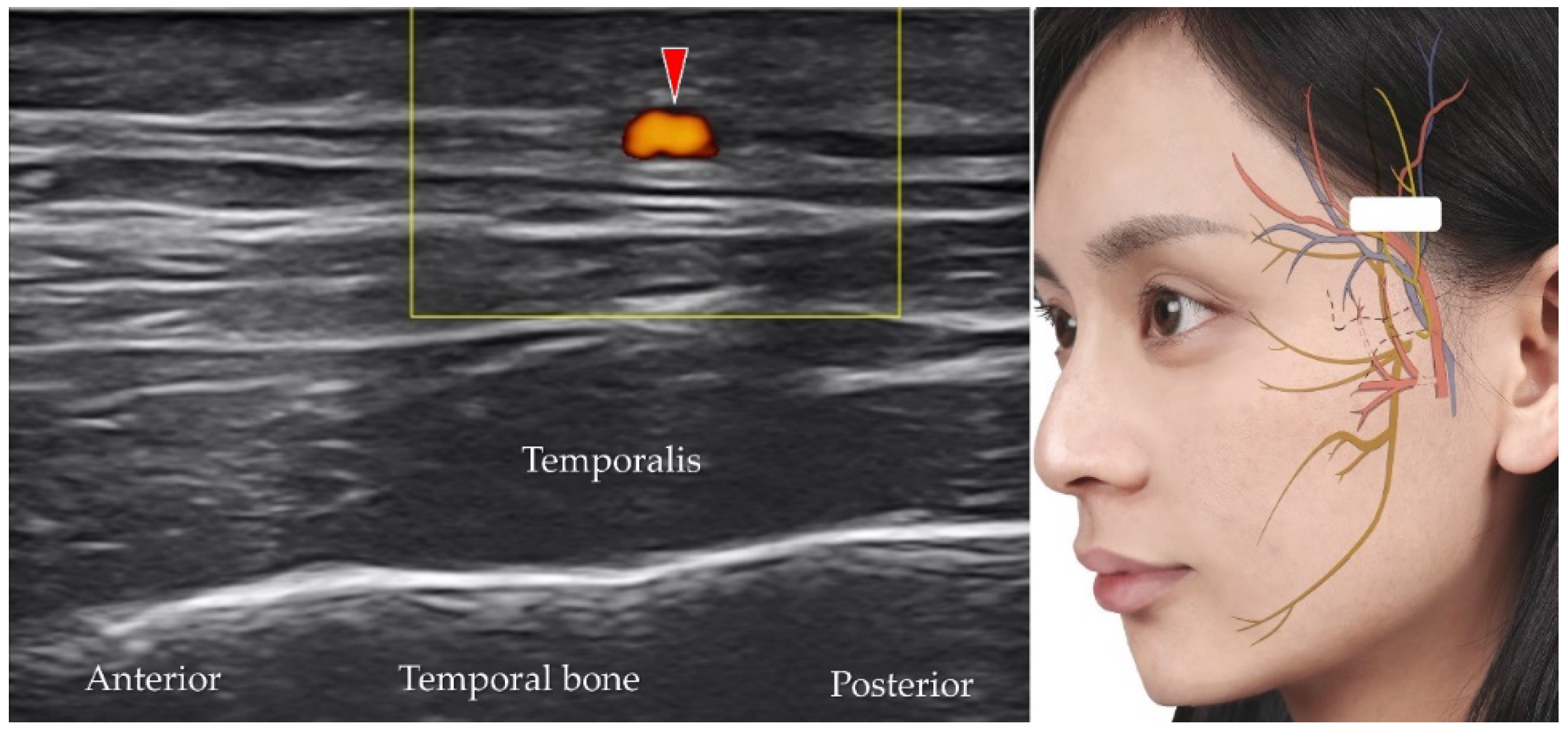
5.6. Deep Temporal Artery and Vein

The deep temporal artery, which shows an anterior and a posterior branch [39] originates from the maxillary artery, which itself is a branch of the external carotid artery. The transducer is placed in the horizontal plane, cranial to the zygomatic arch, to identify the temporalis muscle. The deep temporal artery can be visualized between the temporalis muscle and the periosteum of the temporal bone (Figure 15).

5.7. Auriculotemporal Nerve
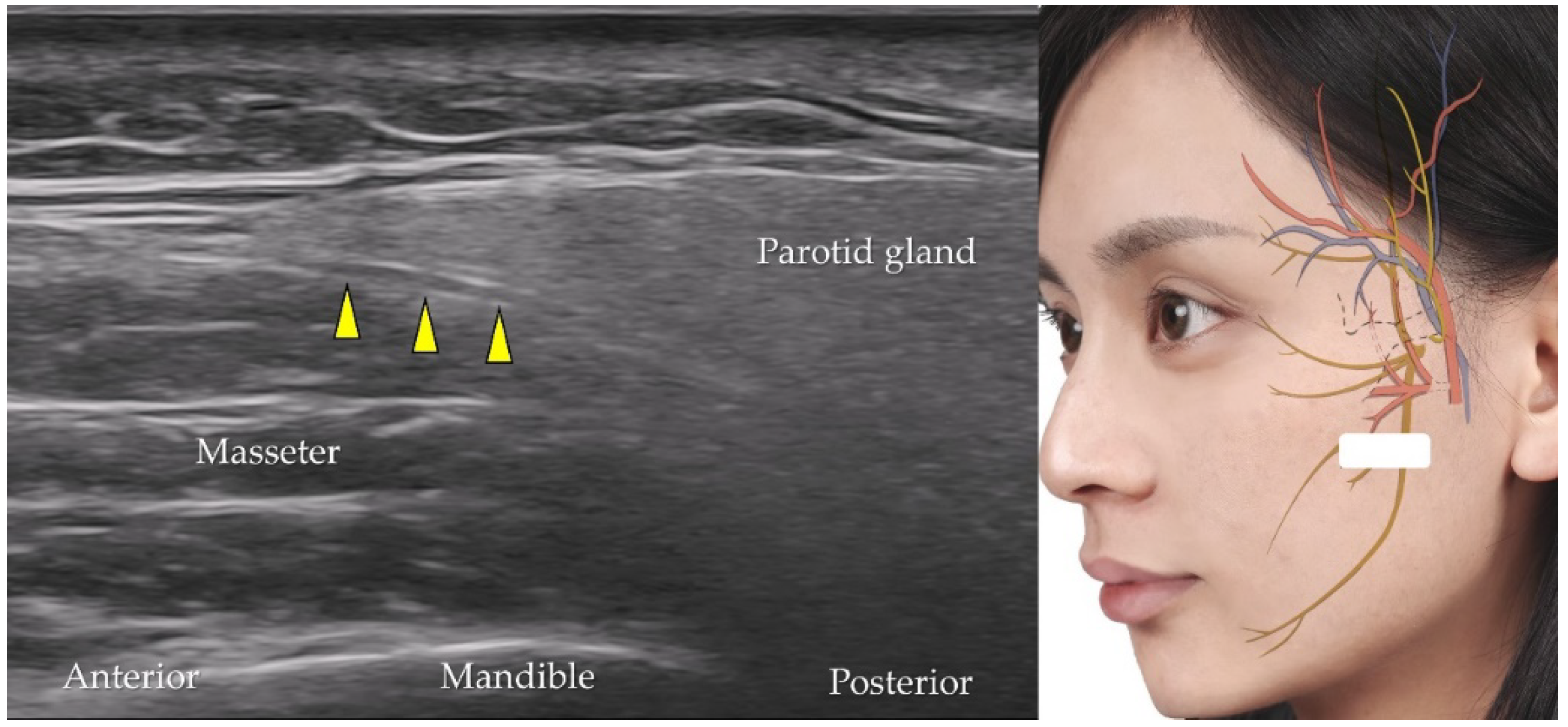
The auriculotemporal nerve branches from the mandibular nerve and provides sensory innervation to the jaw, ears, and scalp [14]. The transducer is placed in the horizontal plane slightly anterior to the tragus. The short axis of the auriculotemporal nerve can be observed adjacent to the superior temporal artery (Figure 16) [40].

5.8. Facial Nerve

After emerging from the brain stem, the facial nerve travels through the facial canal in the temporal bone [41] and exits the skull bone through the stylomastoid foramen to enter the parotid gland. The transducer is placed in the horizontal plane at the cranial level of the mandibular ramus. The long axis of the facial nerve is observed as a tubular hypoechoic structure within the parotid gland (Figure 17) [42]. Table 2 summarizes the techniques used to scan neurovascular structures of the middle face.

6. Sonoanatomy of the Neurovascular Structures of the Lower Face

6.1. Facial Artery and Vein

The facial artery, a branch of the external carotid artery [43], courses superficial to the posterior belly of the digastric muscle, the stylohyoid muscle and the submandibular gland before it ascends toward the nasolabial fold after it crosses the caudal border of the mandible. The transducer is placed in the horizontal plane at the midpoint of the mandibular body to visualize the facial artery (and vein) (Figure 18).

6.2. Inferior Labial Artery and Vein

The inferior labial artery, which branches from the facial artery at the inferolateral aspect of the angle of the mouth, Ref. [22] courses along the caudal edge of the lower lip between the orbicularis oris and the mucous membrane. The transducer is placed in the sagittal plane over the lower lip to the inferior labial artery (and vein) along the short axis (Figure 19).

6.3. Mental Nerve

The mental nerve is a branch of the mandibular nerve that provides sensation to the chin and the lower lip [14]. The transducer is placed in the horizontal plane, caudal to the lower lip, and lateral to the mental protuberance. The mental nerve can be identified as it emerges from the mental foramen (Figure 20).

7. Sonoanatomy of the Neurovascular Structures of the Submental Region

7.1. Submental Artery

The submental artery branches from the facial artery near the border of the mandibular body and supplies the submandibular and sublingual glands [23]. The transducer is placed in the coronal plane over the submental region, and the submental artery can be visualized coursing superficial to the mylohyoid muscle (Figure 21).

7.2. Hypoglossal Nerve

The hypoglossal nerve is the 12th cranial nerve that innervates the extrinsic and intrinsic muscles of the tongue [24]. The transducer is positioned in the coronal plane over the submental area and is moved from the posterior to the anterior aspect. The hypoglossal nerve can be visualized as a hypoechoic circular structure between the hypoglossal and mylohyoid muscles (Figure 22). The lingual artery and vein may accompany the hypoglossal nerve in the submental region. Table 3 summarizes the techniques used to scan the neurovascular structures of the lower face and submental region.

8. Factors Associated with Ultrasound-Guided Aesthetic Injections into the Facial and Submental Regions

Although several pilot studies have reported the feasibility of ultrasound-guided facial aesthetic injections, palpation-based interventions remain widely popular among injectors. Factors that limit ultrasound guidance for facial injections include the following: (A) Facial muscles are thin and superficial [9]; therefore, they need to be delineated using transducers with higher frequencies (for example, 20 MHz). (B) The facial region is not a large plain surface, which interferes with the use of an in-plane approach. Therefore, unlike ultrasound-guided injections into peripheral joints, ultrasound guidance for facial cosmetic injections warrants the following strategy, which is different from conventional ultrasound-guided injections:
(i)
Injectors unfamiliar with real-time guided injections should perform ultrasound imaging using a marking pen to confirm important neurovascular structures. This method facilitates palpation-based injections after removing the transducer and provides a guideline to avoid the marked regions.
(ii)
Injectors familiar with ultrasound-guided techniques should prepare an ultrasound machine equipped with a high-frequency (>20 MHz) transducer, which also has a small footprint (<2.5 cm in width). The out-of-plane approach, which allows multiple injections in one scanning plane, is recommended to shorten the needle pathway under the skin.

9. Conclusions

High-resolution ultrasonography enables visualization of facial vessels and nerves. Inadvertent injections into facial neurovascular structures can result in serious adverse effects, which may be prevented by cautious pre-intervention ultrasound scanning. Despite the complicated neurovascular network of the facial region, the systematic protocol presented in this pictorial essay will serve as a guideline for investigators to familiarize themselves with the imaging technique and develop expertise in the aforementioned ultrasound-guided approach to facial aesthetic injections.

Author Contributions

Conceptualization, W.-T.W. and K.-V.C.; methodology, K.-V.C., H.-C.C. and L.-R.C.; software, L.-R.C. and H.-C.C.; validation, C.-H.K., K.M., V.R. and L.Ö.; writing—original draft preparation, W.-T.W.; writing—review and editing, K.-V.C.; funding acquisition, K.-V.C. All authors have read and agreed to the published version of the manuscript.

Funding

This study was made possible by (1) research funding from the Community and Geriatric Medicine Research Center, National Taiwan University Hospital, Bei-Hu Branch, Taipei, Taiwan; (2) Ministry of Science and Technology (MOST 106-2314-B-002-180-MY3, 109-2314-B-002-114-MY3, and 109-2314-B-002-127), and (3) Taiwan Society of Ultrasound in Medicine.

Institutional Review Board Statement

Not applicable.

Informed Consent Statement

Not applicable.

Data Availability Statement

Data are contained within the main text of the manuscript.

Conflicts of Interest

The authors declare no conflict of interest.

References

  1. Nestor, M.; Cohen, J.L.; Landau, M.; Hilton, S.; Nikolis, A.; Haq, S.; Viel, M.; Andriopoulos, B.; Prygova, I.; Foster, K.; et al. Onset and Duration of AbobotulinumtoxinA for Aesthetic Use in the Upper face: A Systematic Literature Review. J. Clin. Aesthet. Dermatol. 2020, 13, E56–E83. [Google Scholar] [PubMed]
  2. Akinbiyi, T.; Othman, S.; Familusi, O.; Calvert, C.; Card, E.B.; Percec, I. Better Results in Facial Rejuvenation with Fillers. Plast. Reconstr. Surg. Glob. Open 2020, 8, e2763. [Google Scholar] [CrossRef] [PubMed]
  3. Humphrey, S.; Femmer, P.; Beleznay, K.; Carruthers, J.D.A. Deoxycholic Acid for Submental Fullness and More: Real-World Experience With 202 Patients. Dermatol. Surg. 2019, 45, 624–627. [Google Scholar] [CrossRef]
  4. Li, D.; Zhang, H. Facial Injections and Blindness: A Review on Anatomy. Ann. Plast. Surg. 2022, 88, 233–236. [Google Scholar] [CrossRef]
  5. Singh, K.; Nooreyezdan, S. Nonvascular Complications of Injectable Fillers-Prevention and Management. Indian J. Plast. Surg. 2020, 53, 335–343. [Google Scholar] [CrossRef]
  6. Hsu, P.C.; Chang, K.V.; Wu, W.T.; Wang, J.C.; Ozcakar, L. Effects of Ultrasound-Guided Peritendinous and Intrabursal Corticosteroid Injections on Shoulder Tendon Elasticity: A Post Hoc Analysis of a Randomized Controlled Trial. Arch. Phys. Med. Rehabil. 2021, 102, 905–913. [Google Scholar] [CrossRef]
  7. Ozcakar, L.; Kara, M.; Chang, K.V.; Carl, A.B.; Akkaya, N.; Tok, F.; Chen, W.S.; Wang, T.G.; Tekin, L.; Ulasl, A.M.; et al. Nineteen reasons why physiatrists should do musculoskeletal ultrasound: EURO-MUSCULUS/USPRM recommendations. Am. J. Phys. Med. Rehabil. 2015, 94, e45–e49. [Google Scholar] [CrossRef]
  8. Chang, K.V.; Wu, S.H.; Lin, S.H.; Shieh, J.Y.; Wang, T.G.; Chen, W.S. Power Doppler presentation of shoulders with biceps disorder. Arch. Phys. Med. Rehabil. 2010, 91, 624–631. [Google Scholar] [CrossRef]
  9. Wu, W.T.; Chang, K.V.; Chang, H.C.; Chen, L.R.; Kuan, C.H.; Kao, J.T.; Wei, L.Y.; Chen, Y.J.; Han, D.S.; Ozcakar, L. Ultrasound Imaging of the Facial Muscles and Relevance with Botulinum Toxin Injections: A Pictorial Essay and Narrative Review. Toxins 2022, 14, 101. [Google Scholar] [CrossRef]
  10. Small, R. Botulinum toxin injection for facial wrinkles. Am. Fam. Phys. 2014, 90, 168–175. [Google Scholar]
  11. Crowley, J.S.; Kream, E.; Fabi, S.; Cohen, S.R. Facial Rejuvenation with Fat Grafting and Fillers. Aesthet. Surg. J. 2021, 41, S31–S38. [Google Scholar] [CrossRef] [PubMed]
  12. Agorgianitis, L.; Panagouli, E.; Tsakotos, G.; Tsoucalas, G.; Filippou, D. The Supratrochlear Artery Revisited: An Anatomic Review in Favor of Modern Cosmetic Applications in the Area. Cureus 2020, 12, e7141. [Google Scholar] [CrossRef] [PubMed] [Green Version]
  13. Kato, N.; Outi, H. Relation of the supraorbital nerve and vessels to the notch and foramen of the supraorbital margin. Okajimas Folia Anat. Jpn. 1962, 38, 411–424. [Google Scholar] [CrossRef] [PubMed] [Green Version]
  14. Allam, A.E.; Khalil, A.A.F.; Eltawab, B.A.; Wu, W.T.; Chang, K.V. Ultrasound-Guided Intervention for Treatment of Trigeminal Neuralgia: An Updated Review of Anatomy and Techniques. Pain Res. Manag. 2018, 2018, 5480728. [Google Scholar] [CrossRef]
  15. Imanishi, N.; Nakajima, H.; Minabe, T.; Chang, H.; Aiso, S. Venous drainage architecture of the temporal and parietal regions: Anatomy of the superficial temporal artery and vein. Plast. Reconstr. Surg. 2002, 109, 2197–2203. [Google Scholar] [CrossRef]
  16. Tansatit, T.; Apinuntrum, P.; Phetudom, T. Facing the Worst Risk: Confronting the Dorsal Nasal Artery, Implication for Non-surgical Procedures of Nasal Augmentation. Aesthet. Plast. Surg. 2017, 41, 191–198. [Google Scholar] [CrossRef]
  17. Gombolevskiy, V.; Gelezhe, P.; Morozov, S.; Melnikov, D.V.; Vorontsov, A.; Kulberg, N.; Frank, K.; Gotkin, R.H.; Lachman, N.; Cotofana, S. The Course of the Angular Artery in the Midface: Implications for Surgical and Minimally Invasive Procedures. Aesthet. Surg. J. 2021, 41, 805–813. [Google Scholar] [CrossRef]
  18. Lee, S.H.; Gil, Y.C.; Choi, Y.J.; Tansatit, T.; Kim, H.J.; Hu, K.S. Topographic anatomy of the superior labial artery for dermal filler injection. Plast. Reconstr. Surg. 2015, 135, 445–450. [Google Scholar] [CrossRef]
  19. Kim, H.-S.; Lee, K.-L.; Gil, Y.-C.; Hu, K.-S.; Tansatit, T.; Kim, H.-J. Topographic Anatomy of the Infraorbital Artery and Its Clinical Implications for Nasolabial Fold Augmentation. Plast. Reconstr. Surg. 2018, 142, 273e–280e. [Google Scholar] [CrossRef]
  20. Pinar, Y.A.; Govsa, F. Anatomy of the superficial temporal artery and its branches: Its importance for surgery. Surg. Radiol. Anat. 2006, 28, 248–253. [Google Scholar] [CrossRef]
  21. Fernández-Núñez, T.; Amghar-Maach, S.; Gay-Escoda, C. Efficacy of botulinum toxin in the treatment of bruxism: Systematic review. Med. Oral Patol. Oral Cir. Bucal. 2019, 24, e416–e424. [Google Scholar] [CrossRef] [PubMed]
  22. Cotofana, S.; Alfertshofer, M.; Schenck, T.L.; Bertucci, V.; Beleznay, K.; Ascher, B.; Lachmann, N.; Green, J.B.; Swift, A.; Frank, K. Anatomy of the Superior and Inferior Labial Arteries Revised: An Ultrasound Investigation and Implication for Lip Volumization. Aesthet. Surg. J. 2020, 40, 1327–1335. [Google Scholar] [CrossRef] [PubMed]
  23. Atamaz Pinar, Y.; Govsa, F.; Bilge, O. The anatomical features and surgical usage of the submental artery. Surg. Radiol. Anat. 2005, 27, 201–205. [Google Scholar] [CrossRef] [PubMed]
  24. Meng, S.; Reissig, L.F.; Tzou, C.H.; Meng, K.; Grisold, W.; Weninger, W. Ultrasound of the Hypoglossal Nerve in the Neck: Visualization and Initial Clinical Experience with Patients. AJNR Am. J. Neuroradiol. 2016, 37, 354–359. [Google Scholar] [CrossRef] [Green Version]
  25. Robinson, L.R. Traumatic injury to peripheral nerves. Muscle Nerve 2000, 23, 863–873. [Google Scholar] [CrossRef]
  26. Hsu, P.C.; Chang, K.V.; Mezian, K.; Nanka, O.; Wu, W.T.; Yang, Y.C.; Meng, S.; Ricci, V.; Ozcakar, L. Sonographic Pearls for Imaging the Brachial Plexus and Its Pathologies. Diagnostics 2020, 10, 324. [Google Scholar] [CrossRef]
  27. DeLorenzi, C. Complications of Injectable Fillers, Part 2: Vascular Complications. Aesthet. Surg. J. 2014, 34, 584–600. [Google Scholar] [CrossRef] [Green Version]
  28. Cotofana, S.; Velthuis, P.J.; Alfertshofer, M.; Frank, K.; Bertucci, V.; Beleznay, K.; Swift, A.; Gavril, D.L.; Lachman, N.; Schelke, L. The Change of Plane of the Supratrochlear and Supraorbital Arteries in the Forehead-An Ultrasound-Based Investigation. Aesthet. Surg. J. 2021, 41, NP1589–NP1598. [Google Scholar] [CrossRef]
  29. Janis, J.E.; Hatef, D.A.; Hagan, R.; Schaub, T.; Liu, J.H.; Thakar, H.; Bolden, K.M.; Heller, J.B.; Kurkjian, T.J. Anatomy of the Supratrochlear Nerve: Implications for the Surgical Treatment of Migraine Headaches. Plast. Reconstr. Surg. 2013, 131. [Google Scholar] [CrossRef]
  30. Napier, A.; De Jesus, O.; Taylor, A. Supraorbital Nerve Block; StatPearls Publishing LLC.: Treasure Island, FL, USA, 2022. [Google Scholar]
  31. Ren, H.; Shen, Y.; Luo, F. Treatment of Supraorbital Neuralgia Using Ultrasound-Guided Radiofrequency Thermocoagulation of the Supraorbital Nerve: A Retrospective Study. J. Pain Res. 2020, 13, 251–259. [Google Scholar] [CrossRef] [Green Version]
  32. Koziej, M.; Wnuk, J.; Polak, J.; Trybus, M.; Pękala, P.; Pękala, J.; Hołda, M.; Antoszewski, B.; Tomaszewski, K. The superficial temporal artery: A meta-analysis of its prevalence and morphology. Clin. Anat. 2020, 33, 1130–1137. [Google Scholar] [CrossRef] [PubMed]
  33. Tansatit, T.; Jitaree, B.; Uruwan, S.; Rungsawang, C. Anatomical Study of the Dorsal Nasal Artery to Prevent Visual Complications during Dorsal Nasal Augmentation. Plast. Reconstr. Surg. Glob. Open 2021, 9. [Google Scholar] [CrossRef] [PubMed]
  34. Alfertshofer, M.G.; Frank, K.; Moellhoff, N.; Helm, S.; Freytag, L.; Mercado-Perez, A.; Hargiss, J.B.; Dumbrava, M.; Green, J.B.; Cotofana, S. Ultrasound Anatomy of the Dorsal Nasal Artery as it Relates to Liquid Rhinoplasty Procedures. Facial Plast. Surg. Clin. N. Am. 2022, 30, 135–141. [Google Scholar] [CrossRef] [PubMed]
  35. Yoshimatsu, H.; Harima, M.; Iida, T.; Narushima, M.; Karakawa, R.; Nakatsukasa, S.; Yamamoto, T.; Hayashi, A. Use of the Distal Facial Artery (Angular Artery) for Supermicrosurgical Midface Reconstruction. Plast Reconstr. Surg. Glob. Open 2019, 7, e1978. [Google Scholar] [CrossRef]
  36. Lee, K.L.; Lee, H.J.; Youn, K.H.; Kim, H.J. Positional relationship of superior and inferior labial artery by ultrasonography image analysis for safe lip augmentation procedures. Clin. Anat. 2020, 33, 158–164. [Google Scholar] [CrossRef] [PubMed]
  37. Michalek, P.; Donaldson, W.; McAleavey, F.; Johnston, P.; Kiska, R. Ultrasound imaging of the infraorbital foramen and simulation of the ultrasound-guided infraorbital nerve block using a skull model. Surg. Radiol. Anat. 2013, 35, 319–322. [Google Scholar] [CrossRef]
  38. Karahaliou, M.; Vaiopoulos, G.; Papaspyrou, S.; Kanakis, M.A.; Revenas, K.; Sfikakis, P.P. Colour duplex sonography of temporal arteries before decision for biopsy: A prospective study in 55 patients with suspected giant cell arteritis. Arthritis Res. Ther. 2006, 8, R116. [Google Scholar] [CrossRef] [Green Version]
  39. Nikolis, A.; Enright, K.M.; Troupis, T.; Koutsilieris, M.; Stratigos, A.J.; Rigopoulos, D.; Cotofana, S. Topography of the deep temporal arteries and implications for performing safe aesthetic injections. J. Cosmet. Dermatol. 2022, 21, 608–614. [Google Scholar] [CrossRef]
  40. Zhou, H.; Xue, Y.; Liu, P. Application of auriculotemporal nerve block and dextrose prolotherapy in exercise therapy of TMJ closed lock in adolescents and young adults. Head Face Med. 2021, 17, 11. [Google Scholar] [CrossRef]
  41. Tawfik, E.A. Sonographic characteristics of the facial nerve in healthy volunteers. Muscle Nerve 2015, 52, 767–771. [Google Scholar] [CrossRef]
  42. Tawfik, E.A.; Walker, F.O.; Cartwright, M.S. Neuromuscular ultrasound of cranial nerves. J. Clin. Neurol. 2015, 11, 109–121. [Google Scholar] [CrossRef] [PubMed] [Green Version]
  43. Lee, H.J.; Won, S.Y.; O, J.; Hu, K.S.; Mun, S.Y.; Yang, H.M.; Kim, H.J. The facial artery: A Comprehensive Anatomical Review. Clin. Anat. 2018, 31, 99–108. [Google Scholar] [CrossRef] [PubMed]
Figure 1. Illustration showing the frontal facial neurovascular structures. Blue structures indicate veins, which are mostly accompanied by arteries (in red) with the same nomenclature.
Diagnostics 12 01766 g001
Figure 2. Illustration showing the lateral facial neurovascular structures. Blue structures indicate veins, which are mostly accompanied by arteries (in red) with the same nomenclature.
Diagnostics 12 01766 g002
Figure 3. Illustration showing the submental facial neurovascular structures. Blue structures indicate veins, which are mostly accompanied by arteries (in red) with the same nomenclature.
Diagnostics 12 01766 g003
Figure 4. Power Doppler ultrasound imaging of the supratrochlear artery (red arrowheads) and its branches. White square, footprint of the ultrasound transducer. White dashed line, border of the supratrochlear foramen.
Diagnostics 12 01766 g004
Figure 5. Ultrasound imaging of the supratrochlear nerve (yellow arrowhead). White square, footprint of the ultrasound transducer.
Diagnostics 12 01766 g005
Figure 6. Power Doppler ultrasound imaging of the supraorbital artery (red arrowheads) and its branches. White square, footprint of the ultrasound transducer.
Diagnostics 12 01766 g006
Figure 7. Ultrasound imaging of the supraorbital nerve (yellow arrowhead). White square, footprint of the ultrasound transducer.
Diagnostics 12 01766 g007
Figure 8. Power Doppler ultrasound imaging of the frontal branch of the superficial temporal artery (red arrowheads) and its branches. White square, footprint of the ultrasound transducer.
Diagnostics 12 01766 g008
Figure 9. Power Doppler ultrasound imaging of the dorsal nasal artery (red arrowheads) and its branches. White square, footprint of the ultrasound transducer.
Diagnostics 12 01766 g009
Figure 10. Power Doppler ultrasound imaging of the angular artery (red arrowheads). White square, footprint of the ultrasound transducer.
Diagnostics 12 01766 g010
Figure 11. Power Doppler ultrasound imaging of the superior labial artery (red arrowheads). White square, footprint of the ultrasound transducer.
Diagnostics 12 01766 g011
Figure 12. Power Doppler ultrasound imaging of the infraorbital artery (red arrowhead). White square, footprint of the ultrasound transducer.
Diagnostics 12 01766 g012
Figure 13. Ultrasound imaging of the infraorbital nerve (yellow arrowhead). White square, footprint of the ultrasound transducer.
Diagnostics 12 01766 g013
Figure 14. Power Doppler ultrasound imaging of the parietal branch of the superficial temporal artery (red arrowhead). White square, footprint of the ultrasound transducer.
Diagnostics 12 01766 g014
Figure 15. Power Doppler ultrasound imaging of the parietal branch of the deep temporal artery (red arrowhead). White square, footprint of the ultrasound transducer.
Diagnostics 12 01766 g015
Figure 16. Power Doppler ultrasound imaging of the main trunk of the superficial temporal artery (red arrowhead) and the auriculotemporal nerve (yellow arrowhead). White square, footprint of the ultrasound transducer.
Diagnostics 12 01766 g016
Figure 17. Ultrasound imaging of the facial nerve (yellow arrowheads). White square, the footprint of the ultrasound transducer.
Diagnostics 12 01766 g017
Figure 18. Power Doppler ultrasound imaging of the facial artery (red arrowhead). White square, footprint of the ultrasound transducer.
Diagnostics 12 01766 g018
Figure 19. Power Doppler ultrasound imaging of the inferior labial artery (red arrowhead). White square, footprint of the ultrasound transducer.
Diagnostics 12 01766 g019
Figure 20. Ultrasound imaging of the mental nerve (yellow arrowhead). White square, footprint of the ultrasound transducer.
Diagnostics 12 01766 g020
Figure 21. Power Doppler ultrasound imaging of the submental artery (red arrowhead). White square, footprint of the ultrasound transducer.
Diagnostics 12 01766 g021
Figure 22. Ultrasound imaging of the hypoglossal nerve (yellow arrowhead). White square, footprint of the ultrasound transducer.
Diagnostics 12 01766 g022
Table 1. Summary of the anatomy and scanning techniques for the neurovascular structures of the upper face.
Vessel/NerveOrigin/Drain toSupplyTransducer Position
Supratrochlear arteryOphthalmic arteryMedial aspect of the upper eye lid forehead, glabella and frontalis muscleThe transducer is placed over the medial orbital rim in the horizontal plane
Supratrochlear veinSupratrochlear foramenAnterior forehead and scalpThe transducer is placed over the medial orbital rim in the horizontal plane
Supratrochlear nerveFrontal nerve, branch of the ophthalmic nerve (V1)Cutaneous innervation of the lateral lower forehead and upper eyelidThe transducer is placed over the medial orbital rim in the horizontal plane
Supraorbital arteryOphthalmic arteryUpper eyelid and skin of the forehead and the scalpThe transducer is placed in proximity over the center of the superior orbital rim
Supraorbital veinAngular veinForehead, eyebrow, and upper eyelidThe transducer is placed in proximity over the center of the superior orbital rim
Supraorbital nerveFrontal nerve, branch of the ophthalmic nerve (V1)Cutaneous sensation of the lateral forehead and upper eyelidThe transducer is placed in proximity over the center of the superior orbital rim
Frontal branch of the superficial temporal arterySuperficial temporal arteryLateral aspect of the forehead with anastomosis of the supraorbital arteryThe transducer is placed over the lateral part of the forehead slightly cranial to the eyebrow
Frontal branch of the superficial temporal veinSuperficial temporal veinLateral aspect of the foreheadThe transducer is placed over the lateral part of the forehead slightly cranial to the eyebrow
Table 2. Summary of the anatomy and scanning techniques for the neurovascular structures over the middle face.
Vessel/NerveOrigin/Drain toSupplyTransducer Position
Dorsal nasal arteryOphthalmic arteryLacrimal sac and tip of the noseThe transducer is placed in the horizontal plane near the medial orbital rim
Dorsal nasal veinAngular veinLateral side of the noseThe transducer is placed in the horizontal plane near the medial orbital rim
Angular arteryFacial arteryLacrimal sac, nose, lower eyelid, and orbicularis oculi musclesThe transducer can be placed in the horizontal plane lateral to the ala of the nose
Angular veinFacial veinMedial canthus, and nose and upper lipThe transducer can be placed in the horizontal plane lateral to the ala of the nose lateral to the angular artery
Superior labial arteryFacial arteryUpper lipThe transducer is placed in the sagittal plane medial to the angle of the mouth
Superior labial veinFacial veinUpper lipThe transducer is placed in the sagittal plane medial to the angle of the mouth
Infraorbital arteryMaxillary arteryLacrimal sac, upper incisor and canine teethThe transducer is placed in the horizontal plane slightly distal to the inferior eyelid
Infraorbital veinPterygoid venous plexusLacrimal sac, upper incisor and canine teethThe transducer is placed in the horizontal plane slightly distal to the inferior eyelid
Infraorbital nerveMaxillary nerve (CN V2)Lower eyelid, anterior cheek, and upper lipThe transducer is placed in the horizontal plane slightly distal to the inferior eyelid
Superficial temporal arteryExternal carotid arteryParietal branch for the posterior temporal area and frontal branch for the lateral foreheadThe transducer can be placed in the horizontal plane cranial to the zygomatic arch
Superficial temporal veinRetromandibular veinPosterior temporal area and lateral foreheadThe transducer can be placed in the horizontal plane cranial to the zygomatic arch
Deep temporal arteryMaxillary arteryTemporalis muscle and temporal fossaThe transducer is placed in the horizontal plane cranial to the zygomatic arch to identify the vessels under the temporalis muscle
Deep temporal veinRetromandibular veinTemporalis muscle and temporal fossaThe transducer is placed in the horizontal plane cranial to the zygomatic arch to identify the vessels under the temporalis muscle
Auriculotemporal nerveMandibular nerve (CN V3)Auricle, external acoustic meatus, and temporal regionThe transducer is placed in the horizontal plane slightly anterior to the tragus
Facial nervePons of the brain stemMotor to facial expression muscles, the posterior belly of digastric, stylohyoid; sensory to taste (anterior two-thirds of tongue)The transducer is placed in the horizontal plane at the cranial level of the mandibular ramus
Table 3. Summary of the anatomy and scanning techniques for the neurovascular structures over the lower face and submental region.
Vessel/NerveOrigin/Drain toSupplyTransducer Position
Lower face
Facial arteryExternal carotid arteryAnterior upper neck and anterior mandible regionThe transducer is placed in the horizontal plane at the midpoint of the mandibular body
Facial veinRetromandibular or internal jugular veinAnterior upper neck and anterior mandible regionThe transducer is placed in the horizontal plane at the midpoint of the mandibular body
Inferior labial arteryFacial arteryLower lipThe transducer is placed in the sagittal plane over the lower lip
Inferior labial VeinFacial veinLower lipThe transducer is placed in the sagittal plane over the lower lip
Mental nerveMandibular nerve (CN V3)Cutaneous sensation of the anterior chin and lower lipThe transducer is placed in the horizontal plane caudal to the lower lip and lateral to the mental protuberance
Submental region
Submental arteryFacial arterySubmental areaThe transducer is placed in the coronal plane over the submental region
Hypoglossal nerveBrain stemTongue muscleThe transducer is positioned in the coronal plane over the submental area
Publisher’s Note: MDPI stays neutral with regard to jurisdictional claims in published maps and institutional affiliations.

