Next Issue
Volume 13, September-2
Previous Issue
Volume 13, August-2


Diagnostics, Volume 13, Issue 17 (September-1 2023) – 141 articles

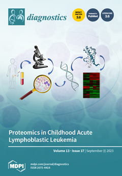
Cover Story (view full-size image): Acute Lymphoblastic Leukemia (ALL) is the most common cancer among children and one of the successful cancer therapeutics that is accomplished via the implementation of risk-directed chemotherapy. Nevertheless, leukemia remains a leading cause of cancer-related deaths. The application of next-generation sequencing tools has illustrated the genomic landscape of ALL. However, underlying dynamic changes at the protein level remain a challenge. Proteomics is a cutting-edge technology aimed at deciphering the mechanisms, pathways, and the degree to which the proteome impacts leukemia subtypes. The analysis of high-throughput global proteomic profiles represents an opportunity to unveil new biological markers and druggable targets for innovative and customized therapies providing new prospects for precision-medicine strategies. View this paper
  • Issues are regarded as officially published after their release is announced to the table of contents alert mailing list.
  • You may sign up for e-mail alerts to receive table of contents of newly released issues.
  • PDF is the official format for papers published in both, html and pdf forms. To view the papers in pdf format, click on the "PDF Full-text" link, and use the free Adobe Reader to open them.
Order results
Result details
Section
Select all
Export citation of selected articles as:
13 pages, 485 KiB  
Review
Selective IgM Deficiency: Evidence, Controversies, and Gaps
by Ivan Taietti, Martina Votto, Maria De Filippo, Matteo Naso, Lorenza Montagna, Daniela Montagna, Amelia Licari, Gian Luigi Marseglia and Riccardo Castagnoli
Diagnostics 2023, 13(17), 2861; https://doi.org/10.3390/diagnostics13172861 - 4 Sep 2023
Cited by 3 | Viewed by 3066
Abstract
Selective Immunoglobulin M deficiency (SIgMD) has been recently included in the inborn errors of immunity (IEI) classification by the International Union of Immunological Societies Expert Committee. The understanding of SIgMD is still extremely limited, especially so in cases of SIgMD in the pediatric [...] Read more.
Selective Immunoglobulin M deficiency (SIgMD) has been recently included in the inborn errors of immunity (IEI) classification by the International Union of Immunological Societies Expert Committee. The understanding of SIgMD is still extremely limited, especially so in cases of SIgMD in the pediatric population. The epidemiology of SIgMD in the pediatric population is still unknown. The pathogenesis of SIgMD remains elusive, and thus far no genetic nor molecular basis has been clearly established as a definitive cause of this primary immunodeficiency. Recurrent respiratory infections represent the main clinical manifestations in children, followed by allergic and autoimmune diseases. No conclusive data on the correct therapeutic management of SIgMD are available. Although, for most SIgMD patients, Ig replacement therapy is not required, it may be recommended for patients with significantly associated antibody deficiency and recurrent or severe infections. Prophylactic antibiotics and the prompt treatment of febrile illness are crucial. There is insufficient evidence on the prognosis of this condition. Therefore, further studies are required to define the disease trajectories and to increase our understanding of the molecular mechanisms underlying SIgMD in order to facilitate a better clinical, immunological, and prognostic characterization of the condition and develop tailored therapeutic management strategies. Full article
Show Figures

Figure 1

11 pages, 2092 KiB  
Article
Evaluating the Efficiency of the Cobas 6800 System for BK Virus Detection in Plasma and Urine Samples
by Junhyup Song, Sinyoung Kim, Eunmin Kwak and Younhee Park
Diagnostics 2023, 13(17), 2860; https://doi.org/10.3390/diagnostics13172860 - 4 Sep 2023
Cited by 3 | Viewed by 1443
Abstract
We evaluated the overall performance of the Cobas 6800 BKV test in detecting BK virus (BKV). We examined the imprecision of the Cobas 6800 BKV test and compared the qualitative and quantitative results obtained from the Cobas 6800 BKV test and the Real-Q [...] Read more.
We evaluated the overall performance of the Cobas 6800 BKV test in detecting BK virus (BKV). We examined the imprecision of the Cobas 6800 BKV test and compared the qualitative and quantitative results obtained from the Cobas 6800 BKV test and the Real-Q BKV quantification assay. We assessed 88 plasma and 26 urine samples collected between September and November 2022 from patients with BKV infection using the Real-Q BKV quantitative assay. The lognormal coefficient of variation indicated that the inter-assay precision of the Cobas 6800 BKV test ranged from 13.86 to 33.83%. A strong correlation was observed between the quantitative results obtained using the Cobas 6800 BKV test and the Real-Q BKV quantification assay for plasma samples. The Spearman’s rank correlation coefficients (ρ) for plasma, polymerase chain reaction (PCR) media-stabilized urine, and raw urine samples were 0.939, 0.874, and 0.888, respectively. Our analyses suggest that the Cobas 6800 BKV test is suitable for clinical applications owing to the strong correlation between the results obtained using this test and the Real-Q BKV quantification assay in plasma and urine samples. Furthermore, utilizing fresh raw urine samples can be a viable approach for the Cobas 6800 BKV test as it is less labor- and time-intensive. Full article
(This article belongs to the Collection Diagnostic Virology)
Show Figures

Figure 1

12 pages, 4294 KiB  
Article
Identification of Autoantibodies to a Hybrid Insulin Peptide in Type 1 Diabetes
by Janet M. Wenzlau, Yong Gu, Aaron Michels, Marian Rewers, Kathryn Haskins and Liping Yu
Diagnostics 2023, 13(17), 2859; https://doi.org/10.3390/diagnostics13172859 - 4 Sep 2023
Cited by 4 | Viewed by 1770
Abstract
Type 1 diabetes (T1D) is a chronic autoimmune disease that attacks the insulin-producing b cells of the pancreatic islets. Autoantibodies to b cell proteins typically appear in the circulation years before disease onset, and serve as the most accurate biomarkers of T1D risk. [...] Read more.
Type 1 diabetes (T1D) is a chronic autoimmune disease that attacks the insulin-producing b cells of the pancreatic islets. Autoantibodies to b cell proteins typically appear in the circulation years before disease onset, and serve as the most accurate biomarkers of T1D risk. Our laboratory has recently discovered novel b cell proteins comprising hybrid proinsulin:islet amyloid polypeptide peptides (IAPP). T cells from a diabetic mouse model and T1D patients are activated by these hybrid peptides. In this study, we asked whether these hybrid molecules could serve as antigens for autoantibodies in T1D and prediabetic patients. We analyzed sera from T1D patients, prediabetics and healthy age-matched donors. Using a highly sensitive electrochemiluminescence assay, sera were screened for binding to recombinant proinsulin:IAPP probes or truncated derivatives. Our results show that sera from T1D patients contain antibodies that bind larger hybrid proinsulin:IAPP probes, but not proinsulin or insulin, at significantly increased frequencies compared to normal donors. Examination of sera from prediabetic patients confirms titers of antibodies to these hybrid probes in more than 80% of individuals, often before seroconversion. These results suggest that hybrid insulin peptides are common autoantigens in T1D and prediabetic patients, and that antibodies to these peptides may serve as valuable early biomarkers of the disease. Full article
Show Figures

Figure 1

20 pages, 8165 KiB  
Article
A Deep Learning Model Based on Capsule Networks for COVID Diagnostics through X-ray Images
by Gabriela Rangel, Juan C. Cuevas-Tello, Mariano Rivera and Octavio Renteria
Diagnostics 2023, 13(17), 2858; https://doi.org/10.3390/diagnostics13172858 - 4 Sep 2023
Cited by 5 | Viewed by 1504
Abstract
X-ray diagnostics are widely used to detect various diseases, such as bone fracture, pneumonia, or intracranial hemorrhage. This method is simple and accessible in most hospitals, but requires an expert who is sometimes unavailable. Today, some diagnoses are made with the help of [...] Read more.
X-ray diagnostics are widely used to detect various diseases, such as bone fracture, pneumonia, or intracranial hemorrhage. This method is simple and accessible in most hospitals, but requires an expert who is sometimes unavailable. Today, some diagnoses are made with the help of deep learning algorithms based on Convolutional Neural Networks (CNN), but these algorithms show limitations. Recently, Capsule Networks (CapsNet) have been proposed to overcome these problems. In our work, CapsNet is used to detect whether a chest X-ray image has disease (COVID or pneumonia) or is healthy. An improved model called DRCaps is proposed, which combines the advantage of CapsNet and the dilation rate (dr) parameter to manage images with 226 × 226 resolution. We performed experiments with 16,669 chest images, in which our model achieved an accuracy of 90%. Furthermore, the model size is 11M with a reconstruction stage, which helps to avoid overfitting. Experiments show how the reconstruction stage works and how we can avoid the max-pooling operation for networks with a stride and dilation rate to downsampling the convolution layers. In this paper, DRCaps is superior to other comparable models in terms of accuracy, parameters, and image size handling. The main idea is to keep the model as simple as possible without using data augmentation or a complex preprocessing stage. Full article
(This article belongs to the Special Issue Deep Learning-Based Models for Medical Imaging Diagnosis)
Show Figures

Figure 1

13 pages, 600 KiB  
Review
Hepatic Inflammatory Pseudotumor—Focusing on Its Heterogeneity
by Soo Ryang Kim, Soo Ki Kim, Yu-ichiro Koma, Motoko Sasaki, Akira Asai and Hiroki Nishikawa
Diagnostics 2023, 13(17), 2857; https://doi.org/10.3390/diagnostics13172857 - 4 Sep 2023
Cited by 3 | Viewed by 1308
Abstract
Hepatic inflammatory pseudotumors (IPTs) are defined as benign, non-malignant, non-metastasizing tumors characterized by the presence of myofibroblastic spindle cells, hetorogenous populations of inflammatory cells, particularly plasma cells, lymphocytes and macrophages, as well as locations of fibrosis and necrosis without cellular anaplasia or atypical [...] Read more.
Hepatic inflammatory pseudotumors (IPTs) are defined as benign, non-malignant, non-metastasizing tumors characterized by the presence of myofibroblastic spindle cells, hetorogenous populations of inflammatory cells, particularly plasma cells, lymphocytes and macrophages, as well as locations of fibrosis and necrosis without cellular anaplasia or atypical mitoses. Despite subsequent reports in the references, hepatic IPT remains difficult to diagnose; while posing major issues specifically for its differential diagnosis compared with that of other various benign diseases and malignant hepatic tumors. Histopathological findings are always a requisite for confirming the diagnosis, particularly given that the pathogenesis of IPT remains ambiguous to date. Hepatic IPT is a heterogeneous entity in terms of its clinical features, pathological findings, and pathogenesis. Once the diagnosis is confirmed, however, needless surgery such as wedge resection and lobectomy should be avoided. Here, we discuss the heterogeneity of hepatic IPT, its clinical features, pathological findings, and pathogenesis, and describe its differential diagnosis. Full article
(This article belongs to the Section Pathology and Molecular Diagnostics)
Show Figures

Figure 1

10 pages, 7257 KiB  
Article
Impact of Phacoemulsification Parameters on Central Retinal Thickness Change Following Cataract Surgery
by Lars H. B. Mackenbrock, Isabella D. Baur, Grzegorz Łabuz, Gerd U. Auffarth and Ramin Khoramnia
Diagnostics 2023, 13(17), 2856; https://doi.org/10.3390/diagnostics13172856 - 4 Sep 2023
Cited by 3 | Viewed by 1195
Abstract
Cataract surgery can lead to inflammatory processes in the retina due to its invasive nature, resulting in prolonged recovery times and reduced functional outcomes. The aim of the current study is to explore the impact that phacoemulsification parameters have on macular thickness following [...] Read more.
Cataract surgery can lead to inflammatory processes in the retina due to its invasive nature, resulting in prolonged recovery times and reduced functional outcomes. The aim of the current study is to explore the impact that phacoemulsification parameters have on macular thickness following surgery. This prospective single-center study enrolled 46 healthy patients (46 eyes) who underwent uneventful cataract surgery. Retinal thickness was assessed using optical coherence tomography (OCT) preoperatively, as well as 1, 4, and 12 weeks after surgery. The macula was divided into a central (CMT), inner (IMT), and outer ring (OMT). Cataract density was automatically determined using an anterior segment OCT and a custom MATLAB script. Corrected distance visual acuity (CDVA), intraocular pressure (IOP) as well as cumulative dissipated energy (CDE), ultrasound time (UT), and fluids used during phacoemulsification were recorded. Retinal thickness and volume increased significantly following cataract surgery, reaching its maximum 4 weeks post-operatively. Statistically significant correlations were found between the CDE and IMT, OMT and retinal volume change (rIMT = 0.356, rOMT = 0.298, rvolume = 0.357 with p < 0.05) as well as between the ultrasound time and IMT, OMT, and retinal volume change (rIMT = 0.369, rOMT = 0.293 and rvolume = 0.409 with p < 0.05). Changes in CMT did not correlate with any surgical metrics. Additionally, no correlation was found to the amount of fluid used, whether CDVA or IOP. However, a link between nuclear cataract density and changes in OMT (r = 0.310, p < 0.05) was established. How ultrasound energy impacts the choroidea, and to what extent retinal metabolism changes after surgery, needs to be explored in future studies. Full article
(This article belongs to the Section Pathology and Molecular Diagnostics)
Show Figures

Figure 1

14 pages, 2878 KiB  
Article
A Combination of FPU-Net and Feature Clustering Methods for Accurate Segmentation of Femoral Neck in Radiographic Diagnosis
by Y. Y. Chen, C. H. Chuang, S. L. Hsieh, T. L. Lin and C. J. Hsu
Diagnostics 2023, 13(17), 2855; https://doi.org/10.3390/diagnostics13172855 - 4 Sep 2023
Viewed by 1192
Abstract
In this study, we develop an innovative method that assists computer-aided diagnosis in the determination process of the exact location of the femoral neck junction in plain radiographs. Our algorithm consists of two phases, i.e., coarse prediction and fine matching, which are implemented [...] Read more.
In this study, we develop an innovative method that assists computer-aided diagnosis in the determination process of the exact location of the femoral neck junction in plain radiographs. Our algorithm consists of two phases, i.e., coarse prediction and fine matching, which are implemented by supervised deep learning method and unsupervised clustering, respectively. In coarse prediction, standard masks are first produced by a specialist and trained in our proposed feature propagation network (FPU-Net) with supervised learning on the femoral neck dataset. In fine matching, the standard masks are first classified into different categories using our proposed three parameters with unsupervised learning. The predicted mask from FPU-Net is matched with each category of standard masks by calculating the values of intersection of union (IOU), and finally the predicted mask is substituted by the standard mask with the largest IOU value. A total of 4320 femoral neck parts in anterior–posterior (AP) pelvis radiographs collected from China Medical University Hospital database were used to test our method. Simulation results show that, on the one hand, compared with other segmentation methods, the method proposed in this paper has a larger IOU value and better suppression of noise outside the region of interest; on the other hand, the introduction of unsupervised learning for fine matching can help in the accurate localization segmentation of femoral neck images. Accurate femoral neck segmentation can assist surgeons to diagnose and reduce the misdiagnosis rate and burden. Full article
(This article belongs to the Section Machine Learning and Artificial Intelligence in Diagnostics)
Show Figures

Figure 1

17 pages, 2237 KiB  
Review
Orofacial Pain and Dentistry Management: Guidelines for a More Comprehensive Evidence-Based Approach
by Mauro Labanca, Marzia Gianò, Caterina Franco and Rita Rezzani
Diagnostics 2023, 13(17), 2854; https://doi.org/10.3390/diagnostics13172854 - 4 Sep 2023
Cited by 6 | Viewed by 4402
Abstract
Orofacial pain represents one of the most common health problems that negatively affects the activities of daily living. However, the mechanisms underlying these conditions are still unclear, and their comprehensive management is often lacking. Moreover, even if pain is a common symptom in [...] Read more.
Orofacial pain represents one of the most common health problems that negatively affects the activities of daily living. However, the mechanisms underlying these conditions are still unclear, and their comprehensive management is often lacking. Moreover, even if pain is a common symptom in dentistry, differential diagnostic procedures are needed to exclude other pain origins. Misinterpretation of the pain origin, in fact, can lead to misdiagnosis and to subsequent mismanagement. Pain in the orofacial area is the most common reason for patients to visit the dentist, but this area is complex, and the pain could be associated with the hard and soft tissues of the head, face, oral cavity, or to a dysfunction of the nervous system. Considering that the origins of orofacial pain can be many and varied, a thorough assessment of the situation is necessary to enable the most appropriate diagnostic pathway to be followed to achieve optimal clinical and therapeutic management. Full article
(This article belongs to the Special Issue Advances in the Diagnosis of Oral Diseases)
Show Figures

Figure 1

21 pages, 4143 KiB  
Article
AI Evaluation of Imaging Factors in the Evolution of Stage-Treated Metastases Using Gamma Knife
by Calin G. Buzea, Razvan Buga, Maria-Alexandra Paun, Madalina Albu, Dragos T. Iancu, Bogdan Dobrovat, Maricel Agop, Viorel-Puiu Paun and Lucian Eva
Diagnostics 2023, 13(17), 2853; https://doi.org/10.3390/diagnostics13172853 - 4 Sep 2023
Cited by 1 | Viewed by 1594
Abstract
Background: The study investigated whether three deep-learning models, namely, the CNN_model (trained from scratch), the TL_model (transfer learning), and the FT_model (fine-tuning), could predict the early response of brain metastases (BM) to radiosurgery using a minimal pre-processing of the MRI images. The dataset [...] Read more.
Background: The study investigated whether three deep-learning models, namely, the CNN_model (trained from scratch), the TL_model (transfer learning), and the FT_model (fine-tuning), could predict the early response of brain metastases (BM) to radiosurgery using a minimal pre-processing of the MRI images. The dataset consisted of 19 BM patients who underwent stereotactic-radiosurgery (SRS) within 3 months. The images used included axial fluid-attenuated inversion recovery (FLAIR) sequences and high-resolution contrast-enhanced T1-weighted (CE T1w) sequences from the tumor center. The patients were classified as responders (complete or partial response) or non-responders (stable or progressive disease). Methods: A total of 2320 images from the regression class and 874 from the progression class were randomly assigned to training, testing, and validation groups. The DL models were trained using the training-group images and labels, and the validation dataset was used to select the best model for classifying the evaluation images as showing regression or progression. Results: Among the 19 patients, 15 were classified as “responders” and 4 as “non-responders”. The CNN_model achieved good performance for both classes, showing high precision, recall, and F1-scores. The overall accuracy was 0.98, with an AUC of 0.989. The TL_model performed well in identifying the “progression” class, but could benefit from improved precision, while the “regression” class exhibited high precision, but lower recall. The overall accuracy of the TL_model was 0.92, and the AUC was 0.936. The FT_model showed high recall for “progression”, but low precision, and for the “regression” class, it exhibited a high precision, but lower recall. The overall accuracy for the FT_model was 0.83, with an AUC of 0.885. Conclusions: Among the three models analyzed, the CNN_model, trained from scratch, provided the most accurate predictions of SRS responses for unlearned BM images. This suggests that CNN models could potentially predict SRS prognoses from small datasets. However, further analysis is needed, especially in cases where class imbalances exist. Full article
Show Figures

Figure 1

19 pages, 18409 KiB  
Article
Brain–Computer Interface: The HOL–SSA Decomposition and Two-Phase Classification on the HGD EEG Data
by Mary Judith Antony, Baghavathi Priya Sankaralingam, Shakir Khan, Abrar Almjally, Nouf Abdullah Almujally and Rakesh Kumar Mahendran
Diagnostics 2023, 13(17), 2852; https://doi.org/10.3390/diagnostics13172852 - 3 Sep 2023
Cited by 2 | Viewed by 1560
Abstract
An efficient processing approach is essential for increasing identification accuracy since the electroencephalogram (EEG) signals produced by the Brain–Computer Interface (BCI) apparatus are nonlinear, nonstationary, and time-varying. The interpretation of scalp EEG recordings can be hampered by nonbrain contributions to electroencephalographic (EEG) signals, [...] Read more.
An efficient processing approach is essential for increasing identification accuracy since the electroencephalogram (EEG) signals produced by the Brain–Computer Interface (BCI) apparatus are nonlinear, nonstationary, and time-varying. The interpretation of scalp EEG recordings can be hampered by nonbrain contributions to electroencephalographic (EEG) signals, referred to as artifacts. Common disturbances in the capture of EEG signals include electrooculogram (EOG), electrocardiogram (ECG), electromyogram (EMG) and other artifacts, which have a significant impact on the extraction of meaningful information. This study suggests integrating the Singular Spectrum Analysis (SSA) and Independent Component Analysis (ICA) methods to preprocess the EEG data. The key objective of our research was to employ Higher-Order Linear-Moment-based SSA (HOL–SSA) to decompose EEG signals into multivariate components, followed by extracting source signals using Online Recursive ICA (ORICA). This approach effectively improves artifact rejection. Experimental results using the motor imagery High-Gamma Dataset validate our method’s ability to identify and remove artifacts such as EOG, ECG, and EMG from EEG data, while preserving essential brain activity. Full article
(This article belongs to the Special Issue Biomedical Signal Processing and Analysis)
Show Figures

Figure 1

20 pages, 2090 KiB  
Article
Preoperative Assessment of Perianal Fistulas with Combined Magnetic Resonance and Tridimensional Endoanal Ultrasound: A Prospective Study
by Nikolaos Varsamis, Christoforos Kosmidis, Grigorios Chatzimavroudis, Fani Apostolidou Kiouti, Christoforos Efthymiadis, Vasilis Lalas, Chrysi Maria Mystakidou, Christina Sevva, Konstantinos Papadopoulos, George Anthimidis, Charilaos Koulouris, Alexandros Vasileios Karakousis, Konstantinos Sapalidis and Isaak Kesisoglou
Diagnostics 2023, 13(17), 2851; https://doi.org/10.3390/diagnostics13172851 - 3 Sep 2023
Cited by 5 | Viewed by 2354
Abstract
Background: we designed a prospective study of diagnostic accuracy that compared pelvic MRI and 3D-EAUS with pelvic MRI alone in the preoperative evaluation and postoperative outcomes of patients with perianal fistulas. Methods: the sample size was 72 patients and this was divided into [...] Read more.
Background: we designed a prospective study of diagnostic accuracy that compared pelvic MRI and 3D-EAUS with pelvic MRI alone in the preoperative evaluation and postoperative outcomes of patients with perianal fistulas. Methods: the sample size was 72 patients and this was divided into two imaging groups. MRI alone was performed on the first group. Both MRI and 3D-EAUS were performed in parallel on the second group. Surgical exploration took place after two weeks and was the standard reference. Park’s classification, the presence of a concomitant abscess or a secondary tract, and the location of the internal opening were recorded. All patients were re-evaluated for complete fistula healing and fecal incontinence six months postoperatively. All of the collected data were subjected to statistical analysis. Results: the MRI group included 36 patients with 42 fistulas. The MRI + 3D-EAUS group included 36 patients with 46 fistulas. The adjusted sensitivity and negative predictive value were 1.00 for most fistula types in the group that underwent combined imaging. The adjusted specificity improved for intersphincteric fistulas in the same group. The adjusted balanced accuracy improved for all fistula types except rectovaginal. The combination of imaging methods showed improved diagnostic accuracy only in the detection of a secondary tract. The healing rate at six months was 100%. Fecal incontinence at six months did not present a statistically significant difference between the two groups (Fisher’s exact test p-value > 0.9). Patients with complex perianal fistulas had a statistically significant higher probability of undergoing a second surgery (x2 test p-value = 0.019). Conclusions: the combination of pelvic MRI and 3D-EAUS showed improved metrics of diagnostic accuracy and should be used in the preoperative evaluation of all patients with perianal fistulas, especially those with complex types. Full article
(This article belongs to the Special Issue Diagnostic Imaging in Gastrointestinal Diseases)
Show Figures

Figure 1

14 pages, 2858 KiB  
Systematic Review
The Role of Ultrasound in Epicutaneo-Caval Catheter Insertion in Neonates: Systematic Review, Meta-Analysis and Future Perspectives
by Vito D’Andrea, Valentina Cascini, Rosellina Russo, Alessandro Perri, Giorgia Prontera, Gina Ancora, Giovanni Vento, Gabriele Lisi and Giovanni Barone
Diagnostics 2023, 13(17), 2850; https://doi.org/10.3390/diagnostics13172850 - 3 Sep 2023
Cited by 8 | Viewed by 1520
Abstract
Chest and abdominal X-rays after the insertion of an epicutaneo-caval catheter in infants are the standard method of checking the tip location in many neonatal intensive care units. The role of ultrasound in the tip location of the epicutaneo-caval catheter in neonates has [...] Read more.
Chest and abdominal X-rays after the insertion of an epicutaneo-caval catheter in infants are the standard method of checking the tip location in many neonatal intensive care units. The role of ultrasound in the tip location of the epicutaneo-caval catheter in neonates has been the subject of many recent studies. This systematic review investigates the accuracy of epicutaneo-caval catheter tip location by comparing ultrasound and conventional radiology. We performed a systematic literature search in multiple databases. The selection of studies yielded nineteen articles. The systematic review and meta-analysis were performed according to PRISMA (Preferred Reporting Items for Systematic reviews and Meta-analysis). The analyses showed that ultrasound is a better imaging technique for epicutaneo-caval catheter tip location in the neonatal intensive care unit than conventional radiology. By improving operator training and selecting a standardized echography protocol, ultrasound could become the gold standard for visualizing the epicutaneo-caval catheter tip in the neonatal intensive care unit. This would have some important benefits: (1) increased accuracy in tip location (2); a more rapid use of the central venous access (3); and a significant reduction in radiation exposure. Full article
(This article belongs to the Special Issue Maternal–Fetal and Neonatal Diagnostics)
Show Figures

Figure 1

15 pages, 3249 KiB  
Article
Predicting Future Incidences of Cardiac Arrhythmias Using Discrete Heartbeats from Normal Sinus Rhythm ECG Signals via Deep Learning Methods
by Yehyun Kim, Myeonggyu Lee, Jaeung Yoon, Yeji Kim, Hyunseok Min, Hyungjoo Cho, Junbeom Park and Taeyoung Shin
Diagnostics 2023, 13(17), 2849; https://doi.org/10.3390/diagnostics13172849 - 3 Sep 2023
Cited by 6 | Viewed by 3458
Abstract
This study aims to compare the effectiveness of using discrete heartbeats versus an entire 12-lead electrocardiogram (ECG) as the input for predicting future occurrences of arrhythmia and atrial fibrillation using deep learning models. Experiments were conducted using two types of inputs: a combination [...] Read more.
This study aims to compare the effectiveness of using discrete heartbeats versus an entire 12-lead electrocardiogram (ECG) as the input for predicting future occurrences of arrhythmia and atrial fibrillation using deep learning models. Experiments were conducted using two types of inputs: a combination of discrete heartbeats extracted from 12-lead ECG and an entire 12-lead ECG signal of 10 s. This study utilized 326,904 ECG signals from 134,447 patients and categorized them into three groups: true–normal sinus rhythm (T-NSR), atrial fibrillation–normal sinus rhythm (AF-NSR), and clinically important arrhythmia–normal sinus rhythm (CIA-NSR). The T-NSR group comprised patients with at least three normal rhythms in a year and no atrial fibrillation or arrhythmias history. Clinically important arrhythmia included atrial fibrillation, atrial flutter, atrial premature contraction, atrial tachycardia, ventricular premature contraction, ventricular tachycardia, right and left bundle branch block, and atrioventricular block over the second degree. The AF-NSR group included normal sinus rhythm paired with atrial fibrillation or atrial flutter within 14 days, and the CIA-NSR group comprised normal sinus rhythm paired with CIA occurring within 14 days. Three deep learning models, ResNet-18, LSTM, and Transformer-based models, were utilized to distinguish T-NSR from AF-NSR and T-NSR from CIA-NSR. The experiments demonstrated the potential of using discrete heartbeats in predicting future arrhythmia and atrial fibrillation incidences extracted from 12-lead electrocardiogram (ECG) signals alone, without any additional patient information. The analysis reveals that these discrete heartbeats contain subtle patterns that deep learning models can identify. Focusing on discrete heartbeats may lead to more timely and accurate diagnoses of these conditions, improving patient outcomes and enabling automated diagnosis using ECG signals as a biomarker. Full article
(This article belongs to the Section Machine Learning and Artificial Intelligence in Diagnostics)
Show Figures

Figure 1

18 pages, 5957 KiB  
Article
Precision in Dermatology: Developing an Optimal Feature Selection Framework for Skin Lesion Classification
by Tallha Akram, Riaz Junejo, Anas Alsuhaibani, Muhammad Rafiullah, Adeel Akram and Nouf Abdullah Almujally
Diagnostics 2023, 13(17), 2848; https://doi.org/10.3390/diagnostics13172848 - 2 Sep 2023
Cited by 2 | Viewed by 1651
Abstract
Melanoma is widely recognized as one of the most lethal forms of skin cancer, with its incidence showing an upward trend in recent years. Nonetheless, the timely detection of this malignancy substantially enhances the likelihood of patients’ long-term survival. Several computer-based methods have [...] Read more.
Melanoma is widely recognized as one of the most lethal forms of skin cancer, with its incidence showing an upward trend in recent years. Nonetheless, the timely detection of this malignancy substantially enhances the likelihood of patients’ long-term survival. Several computer-based methods have recently been proposed, in the pursuit of diagnosing skin lesions at their early stages. Despite achieving some level of success, there still remains a margin of error that the machine learning community considers to be an unresolved research challenge. The primary objective of this study was to maximize the input feature information by combining multiple deep models in the first phase, and then to avoid noisy and redundant information by downsampling the feature set, using a novel evolutionary feature selection technique, in the second phase. By maintaining the integrity of the original feature space, the proposed idea generated highly discriminant feature information. Recent deep models, including Darknet53, DenseNet201, InceptionV3, and InceptionResNetV2, were employed in our study, for the purpose of feature extraction. Additionally, transfer learning was leveraged, to enhance the performance of our approach. In the subsequent phase, the extracted feature information from the chosen pre-existing models was combined, with the aim of preserving maximum information, prior to undergoing the process of feature selection, using a novel entropy-controlled gray wolf optimization (ECGWO) algorithm. The integration of fusion and selection techniques was employed, initially to incorporate the feature vector with a high level of information and, subsequently, to eliminate redundant and irrelevant feature information. The effectiveness of our concept is supported by an assessment conducted on three benchmark dermoscopic datasets: PH2, ISIC-MSK, and ISIC-UDA. In order to validate the proposed methodology, a comprehensive evaluation was conducted, including a rigorous comparison to established techniques in the field. Full article
Show Figures

Figure 1

14 pages, 4203 KiB  
Article
Age Matters: A Comparative Study of RF Heating of Epicardial and Endocardial Electronic Devices in Pediatric and Adult Phantoms during Cardiothoracic MRI
by Fuchang Jiang, Kaylee R. Henry, Bhumi Bhusal, Pia Sanpitak, Gregory Webster, Andrada Popescu, Christina Laternser, Daniel Kim and Laleh Golestanirad
Diagnostics 2023, 13(17), 2847; https://doi.org/10.3390/diagnostics13172847 - 2 Sep 2023
Cited by 2 | Viewed by 1504
Abstract
This study focused on the potential risks of radiofrequency-induced heating of cardiac implantable electronic devices (CIEDs) in children and adults with epicardial and endocardial leads of varying lengths during cardiothoracic MRI scans. Infants and young children are the primary recipients of epicardial CIEDs, [...] Read more.
This study focused on the potential risks of radiofrequency-induced heating of cardiac implantable electronic devices (CIEDs) in children and adults with epicardial and endocardial leads of varying lengths during cardiothoracic MRI scans. Infants and young children are the primary recipients of epicardial CIEDs, though the devices have not been approved as MR conditional by the FDA due to limited data, leading to pediatric hospitals either refusing the MRI service to most pediatric CIED patients or adopting a scan-all strategy based on results from adult studies. The study argues that risk–benefit decisions should be made on an individual basis. We used 120 clinically relevant epicardial and endocardial device configurations in adult and pediatric anthropomorphic phantoms to determine the temperature rise during RF exposure at 1.5 T. The results showed that there was significantly higher RF heating of epicardial leads than endocardial leads in the pediatric phantom, but not in the adult phantom. Additionally, body size and lead length significantly affected RF heating, with RF heating up to 12 °C observed in models based on younger children with short epicardial leads. The study provides evidence-based knowledge on RF-induced heating of CIEDs and highlights the importance of making individual risk–benefit decisions when assessing the potential risks of MRI scans in pediatric CIED patients. Full article
(This article belongs to the Special Issue Updates in Cardiothoracic Imaging)
Show Figures

Figure 1

13 pages, 3009 KiB  
Article
Retro-Mode in NIDEK Mirante: A Comparative Analysis with Other Imaging Modalities for AMD and CSR
by Adam Wylęgała, Przemysław Wozniak, Bogumiła Sędziak-Marcinek, Bartłomiej Bolek, Dominika Szkodny and Edward Wylęgała
Diagnostics 2023, 13(17), 2846; https://doi.org/10.3390/diagnostics13172846 - 2 Sep 2023
Viewed by 1477
Abstract
Background: Retro-mode is a novel technique capable of creating pseudo-3D images of the retina. However, its clinical utility remains unknown. This study aimed to evaluate the Nidek Mirante multimodal imaging platform for ocular assessment in patients with various retinal conditions. Methods: A total [...] Read more.
Background: Retro-mode is a novel technique capable of creating pseudo-3D images of the retina. However, its clinical utility remains unknown. This study aimed to evaluate the Nidek Mirante multimodal imaging platform for ocular assessment in patients with various retinal conditions. Methods: A total of 115 participants with central serous chorioretinopathy (CSR) and age-related macular degeneration (AMD) were included. Two experienced graders independently evaluated the images, and statistical analysis was performed to assess interclass correlation coefficients (ICC) between graders and modalities; Results: For CSR detection, retro-mode demonstrated exceptionally high ICC rates (ICC = 1; 100%), while color and autofluorescence (FAF) showed moderate coefficients (0.69 and 0.78, respectively). The detection of pigment epithelial detachment was high across all methods, with only retro-mode deviated right (DR) allowing detection in 69% of cases, while retro-mode DR and deviated left (DL) achieved 100% detection. FAF-green achieved a 95% detection rate. In detecting retinal atrophy, most modalities demonstrated high detection rates, with the lowest detection rates offered by retro-mode DL (ICC = 0.85) and DR (ICC = 0.89), while retro-mode ring aperture offered 0.97. Infra-red and fluorescein angiography imaging offered the highest detection rates among the tested modalities, with 97% and 100%, respectively. Conclusion: Retro-mode showed promise for comprehensive ocular evaluation and diagnosis, with certain imaging modalities demonstrating higher accuracy in detecting specific retinal features. Full article
(This article belongs to the Special Issue State of the Art in Retinal Optical Coherence Tomography Images)
Show Figures

Figure 1

10 pages, 686 KiB  
Article
Genotypic Frequencies of Mutations Associated with Alpha-1 Antitrypsin Deficiency in Unrelated Bone Marrow Donors from the Murcia Region Donor Registry in the Southeast of Spain
by Irene Cuenca, Carmen Botella, María Rosa Moya-Quiles, Víctor Jimenez-Coll, José Antonio Galian, Helios Martinez-Banaclocha, Manuel Muro-Pérez, Alfredo Minguela, Isabel Legaz and Manuel Muro
Diagnostics 2023, 13(17), 2845; https://doi.org/10.3390/diagnostics13172845 - 2 Sep 2023
Viewed by 1281
Abstract
Alpha-1 antitrypsin (AAT1) deficiency (AAT1D) is an inherited disease with an increased risk of chronic obstructive pulmonary disease (COPD), liver disease, and skin and blood vessel problems. AAT1D is caused by mutations in the SERPINE1 gene (Serine Protease Inhibitor, group A, member 1). [...] Read more.
Alpha-1 antitrypsin (AAT1) deficiency (AAT1D) is an inherited disease with an increased risk of chronic obstructive pulmonary disease (COPD), liver disease, and skin and blood vessel problems. AAT1D is caused by mutations in the SERPINE1 gene (Serine Protease Inhibitor, group A, member 1). Numerous variants of this gene, the Pi system, have been identified. The most frequent allelic variants are Pi*M, Pi*S, and Pi*Z. The development of COPD requires both a genetic predisposition and the contribution of an environmental factor, smoking being the most important. Studies on this deficiency worldwide are very scarce, and it is currently considered a rare disease because it is underdiagnosed. The aim of this study was to analyze the genotypic frequencies of mutations associated with AAT1 deficiency in unrelated bone marrow donors from the donor registry of the Region of Murcia in southeastern Spain due to the high risk of presenting with different pathologies and underdiagnosis in the population. A total of 112 DNA-healthy voluntary unrelated bone marrow donors from different parts of the Region of Murcia were analyzed retrospectively. AAT1 deficiency patient testing involved an automated biochemical screening routine. The three main variants, Pi*M, Pi*Z, and Pi*S, were analyzed in the SERPINE1 gene. Our results showed a frequency of 3.12% of the Pi*Z (K342) mutation in over 224 alleles tested in the healthy population. The frequency of Pi*S (V264) was 11.1%. The frequency of the haplotype with the most dangerous mutation, EK342 EE264, was 4.46%, and the frequency of EK342 EV264 was 1.78% in the healthy population. Frequencies of other EE342 EV264-mutated haplotypes accounted for 18.7%. As for the EE342 VV264 haplotype, 0.89% of the total healthy population presented heterozygous for the EV264 mutation and one individual presented homozygous for the VV264 mutation. In conclusion, the frequencies of Pi mutations in the healthy population of the Region of Murcia were not remarkably different from the few studies reported in Spain. The genotype and haplotype frequencies followed the usual pattern. Health authorities should be aware of this high prevalence of the Pi*S allelic variant and pathological genotypes such as Pi*MZ and Pi*SZ in the healthy population if they consider screening the smoking population. Full article
(This article belongs to the Special Issue Genetics and Cytopathology Testing in Disease Diagnosis)
Show Figures

Figure 1

13 pages, 905 KiB  
Review
Migraine Aura—Catch Me If You Can with EEG and MRI—A Narrative Review
by Franz Riederer, Johannes Beiersdorf, Adrian Scutelnic and Christoph J. Schankin
Diagnostics 2023, 13(17), 2844; https://doi.org/10.3390/diagnostics13172844 - 2 Sep 2023
Cited by 1 | Viewed by 3902
Abstract
Roughly one-third of migraine patients suffer from migraine with aura, characterized by transient focal neurological symptoms or signs such as visual disturbance, sensory abnormalities, speech problems, or paresis in association with the headache attack. Migraine with aura is associated with an increased risk [...] Read more.
Roughly one-third of migraine patients suffer from migraine with aura, characterized by transient focal neurological symptoms or signs such as visual disturbance, sensory abnormalities, speech problems, or paresis in association with the headache attack. Migraine with aura is associated with an increased risk for stroke, epilepsy, and with anxiety disorder. Diagnosis of migraine with aura sometimes requires exclusion of secondary causes if neurological deficits present for the first time or are atypical. It was the aim of this review to summarize EEG an MRI findings during migraine aura in the context of pathophysiological concepts. This is a narrative review based on a systematic literature search. During visual auras, EEG showed no consistent abnormalities related to aura, although transient focal slowing in occipital regions has been observed in quantitative studies. In contrast, in familial hemiplegic migraine (FHM) and migraine with brain stem aura, significant EEG abnormalities have been described consistently, including slowing over the affected hemisphere or bilaterally or suppression of EEG activity. Epileptiform potentials in FHM are most likely attributable to associated epilepsy. The initial perfusion change during migraine aura is probably a short lasting hyperperfusion. Subsequently, perfusion MRI has consistently demonstrated cerebral hypoperfusion usually not restricted to one vascular territory, sometimes associated with vasoconstriction of peripheral arteries, particularly in pediatric patients, and rebound hyperperfusion in later phases. An emerging potential MRI signature of migraine aura is the appearance of dilated veins in susceptibility-weighted imaging, which may point towards the cortical regions related to aura symptoms (“index vein”). Conclusions: Cortical spreading depression (CSD) cannot be directly visualized but there are probable consequences thereof that can be captured Non-invasive detection of CSD is probably very challenging in migraine. Future perspectives will be elaborated based on the studies summarized. Full article
Show Figures

Figure 1

11 pages, 1826 KiB  
Article
Reliability of Cone Beam Computed Tomography in Predicting Implant Treatment Outcomes in Edentulous Patients
by Abdulaziz Alhossan, Yu-Cheng Chang, Tun-Jan Wang, Yu-Bo Wang and Joseph P. Fiorellini
Diagnostics 2023, 13(17), 2843; https://doi.org/10.3390/diagnostics13172843 - 2 Sep 2023
Viewed by 1243
Abstract
Since the development of CBCT has been utilized in dentistry, the images of the CBCT can assist the surgeon to evaluate the anatomy carefully. Despite the value of radiology evaluation, implant procedures may require additional consideration rather than only evaluating the anatomical factors. [...] Read more.
Since the development of CBCT has been utilized in dentistry, the images of the CBCT can assist the surgeon to evaluate the anatomy carefully. Despite the value of radiology evaluation, implant procedures may require additional consideration rather than only evaluating the anatomical factors. The purpose of this study is to evaluate the predictability of using CBCT alone to plan for implant placement in edentulous patients digitally. CBCT images were analyzed by clinicians, measuring the maxillary and mandibular ridge heights and widths digitally of four predetermined implant sites in the maxillary and two selected implant sites in the mandibular arches of 91 patients planning for implant-supported overdenture. A total of 47 patients out of the 91 had completed implant placement on the edentulous ridge, contributing to 55 upper and/or lower arches (136 dental implants). Both predictabilities are low, implying that CBCT planning for implant placement on the edentulous ridge is not a good index and is insufficient to predict the surgical procedures as a solo method. The findings of this study indicate that digital planning by CBCT is insufficient to serve as an individual tool to predict implant procedures. Further information and evaluation must be considered for implant placement in the edentulous ridge. Full article
(This article belongs to the Special Issue Diagnosis and Treatment of Craniofacial and Oral Disease)
Show Figures

Figure 1

15 pages, 3471 KiB  
Article
Quantitative Chest X-ray Radiomics for Therapy Response Monitoring in Patients with Pulmonary Tuberculosis
by Tamarisk Du Plessis, William Ian Duncombe Rae, Gopika Ramkilawon, Neil Alexander Martinson and Mike Michael Sathekge
Diagnostics 2023, 13(17), 2842; https://doi.org/10.3390/diagnostics13172842 - 1 Sep 2023
Viewed by 1169
Abstract
Tuberculosis (TB) remains the second leading cause of death globally from a single infectious agent, and there is a critical need to develop improved imaging biomarkers and aid rapid assessments of responses to therapy. We aimed to utilize radiomics, a rapidly developing image [...] Read more.
Tuberculosis (TB) remains the second leading cause of death globally from a single infectious agent, and there is a critical need to develop improved imaging biomarkers and aid rapid assessments of responses to therapy. We aimed to utilize radiomics, a rapidly developing image analysis tool, to develop a scoring system for this purpose. A chest X-ray radiomics score (RadScore) was developed by implementing a unique segmentation method, followed by feature extraction and parameter map construction. Signature parameter maps that showed a high correlation to lung pathology were consolidated into four frequency bins to obtain the RadScore. A clinical score (TBscore) and a radiological score (RLscore) were also developed based on existing scoring algorithms. The correlation between the change in the three scores, calculated from serial X-rays taken while patients received TB therapy, was evaluated using Spearman’s correlation. Poor correlations were observed between the changes in the TBscore and the RLscore (0.09 (p-value = 0.36)) and the TBscore and the RadScore (0.02 (p-value = 0.86)). The changes in the RLscore and the RadScore had a much stronger correlation of 0.22, which is statistically significant (p-value = 0.02). This shows that the developed RadScore has the potential to be a quantitative monitoring tool for responses to therapy. Full article
(This article belongs to the Section Pathology and Molecular Diagnostics)
Show Figures

Figure 1

16 pages, 1175 KiB  
Article
Risk Factors and Interpretation of Inconclusive Endoscopic Ultrasound-Guided Fine Needle Aspiration Cytology in the Diagnosis of Solid Pancreatic Lesions
by Renáta Bor, Béla Vasas, Anna Fábián, Mónika Szűcs, Zsófia Bősze, Anita Bálint, Mariann Rutka, Klaudia Farkas, Tibor Tóth, Tamás Resál, Péter Bacsur, Tamás Molnár and Zoltán Szepes
Diagnostics 2023, 13(17), 2841; https://doi.org/10.3390/diagnostics13172841 - 1 Sep 2023
Cited by 2 | Viewed by 1340
Abstract
Background: The inconclusive cytological findings of endoscopic ultrasound-guided fine needle aspiration (EUS-FNA) remain a major clinical challenge and often lead to treatment delays. Methods: Patients who had undergone EUS-FNA sampling for solid pancreas lesions between 2014 and 2021 were retrospectively enrolled. The “atypical” [...] Read more.
Background: The inconclusive cytological findings of endoscopic ultrasound-guided fine needle aspiration (EUS-FNA) remain a major clinical challenge and often lead to treatment delays. Methods: Patients who had undergone EUS-FNA sampling for solid pancreas lesions between 2014 and 2021 were retrospectively enrolled. The “atypical” and “non-diagnostic” categories of the Papanicolaou Society of Cytopathology System were considered inconclusive and the “negative for malignancy” category of malignancy was suspected clinically. We determined the frequency and predictors of inconclusive cytological finding. Results: A total of 473 first EUS-FNA samples were included, of which 108 cases (22.83%) were inconclusive. Significant increases in the odds of inconclusive cytological findings were observed for lesions with a benign final diagnosis (OR 11.20; 95% CI 6.56–19.54, p < 0.001) as well as with the use of 25 G FNA needles (OR 2.12; 95% CI 1.09–4.01, p = 0.023) compared to 22 G needles. Furthermore, the use of a single EUS-FNA technique compared to the combined use of slow-pull and standard suction techniques (OR 1.70; 95% CI 1.06–2.70, p = 0.027) and less than three punctures per procedure led to an elevation in the risk of inconclusive cytology (OR 2.49; 95% CI 1.49–4.14, p < 0.001). Risk reduction in inconclusive cytology findings was observed in lesions between 2–4 cm (OR 0.40; 95% CI 0.23–0.68, p = 0.001) and >4 cm (OR 0.16; 95% CI 0.08–0.31, p < 0.001) compared to lesions ≤2 cm. Conclusions: The more than two punctures per EUS-FNA sampling with larger-diameter needle (19 G or 22 G) using the slow-pull and standard suction techniques in combination may decrease the probability of inconclusive cytological findings. Full article
(This article belongs to the Special Issue Editorial Board Members’ Collection Series—Advances in Ultrasound)
Show Figures

Figure 1

12 pages, 3674 KiB  
Article
The Diagnostic Accuracy of Artificial Intelligence in Radiological Markers of Normal-Pressure Hydrocephalus (NPH) on Non-Contrast CT Scans of the Brain
by Dittapong Songsaeng, Poonsuta Nava-apisak, Jittsupa Wongsripuemtet, Siripra Kingchan, Phuriwat Angkoondittaphong, Phattaranan Phawaphutanon and Akara Supratak
Diagnostics 2023, 13(17), 2840; https://doi.org/10.3390/diagnostics13172840 - 1 Sep 2023
Cited by 2 | Viewed by 4046
Abstract
Diagnosing normal-pressure hydrocephalus (NPH) via non-contrast computed tomography (CT) brain scans is presently a formidable task due to the lack of universally agreed-upon standards for radiographic parameter measurement. A variety of radiological parameters, such as Evans’ index, narrow sulci at high parietal convexity, [...] Read more.
Diagnosing normal-pressure hydrocephalus (NPH) via non-contrast computed tomography (CT) brain scans is presently a formidable task due to the lack of universally agreed-upon standards for radiographic parameter measurement. A variety of radiological parameters, such as Evans’ index, narrow sulci at high parietal convexity, Sylvian fissures’ dilation, focally enlarged sulci, and more, are currently measured by radiologists. This study aimed to enhance NPH diagnosis by comparing the accuracy, sensitivity, specificity, and predictive values of radiological parameters, as evaluated by radiologists and AI methods, utilizing cerebrospinal fluid volumetry. Results revealed a sensitivity of 77.14% for radiologists and 99.05% for AI, with specificities of 98.21% and 57.14%, respectively, in diagnosing NPH. Radiologists demonstrated NPV, PPV, and an accuracy of 82.09%, 97.59%, and 88.02%, while AI reported 98.46%, 68.42%, and 77.42%, respectively. ROC curves exhibited an area under the curve of 0.954 for radiologists and 0.784 for AI, signifying the diagnostic index for NPH. In conclusion, although radiologists exhibited superior sensitivity, specificity, and accuracy in diagnosing NPH, AI served as an effective initial screening mechanism for potential NPH cases, potentially easing the radiologists’ burden. Given the ongoing AI advancements, it is plausible that AI could eventually match or exceed radiologists’ diagnostic prowess in identifying hydrocephalus. Full article
Show Figures

Figure 1

12 pages, 2253 KiB  
Article
Endoscopic Ultrasound-Guided Fine Needle Biopsy in the Diagnostic Work-Up of Deep-Seated Lymphadenopathies and Spleen Lesions: A Monocentric Experience
by Flaminia Bellisario, Fabia Attili, Fabrizia Campana, Federica Borrelli de Andreis, Silvia Bellesi, Elena Maiolo, Eleonora Alma, Rosalia Malafronte, Giuseppe Macis, Luigi Maria Larocca, Salvatore Annunziata, Francesco D’Alò and Stefan Hohaus
Diagnostics 2023, 13(17), 2839; https://doi.org/10.3390/diagnostics13172839 - 1 Sep 2023
Cited by 2 | Viewed by 1530
Abstract
EUS-FNB has been introduced in clinical practice as a less invasive diagnostic approach with respect to surgery. We performed a single-center retrospective study on the diagnostic efficacy of EUS-guided FNB, including 171 patients with lymph nodes, splenic, and extranodal lesions that underwent EUS [...] Read more.
EUS-FNB has been introduced in clinical practice as a less invasive diagnostic approach with respect to surgery. We performed a single-center retrospective study on the diagnostic efficacy of EUS-guided FNB, including 171 patients with lymph nodes, splenic, and extranodal lesions that underwent EUS for FNB at our institution. Excluding 12 patients who did not undergo FNB and 25 patients with a previous diagnosis of a solid tumor, we included 134 patients with clinical/radiological suspect of a lymphoproliferative disease, including 20 patients with a previous history of lymphoma. Out of the 134 biopsies, material of diagnostic quality was obtained in 111 procedures (84.3%). Histological examination of the EUS-FNB samples produced an actionable diagnosis in 100 cases (74.6%). Among the patients without an actionable diagnosis, a second, different diagnostic procedure produced a further eight diagnoses of lymphoma. Therefore, the sensitivity of EUS-FNB for diagnosing lymphomas was calculated to be 86.4% (51/59). Assignment of lymphomas to WHO classification subtypes was possible in 47/51 (92%) of the cases. In conclusion, EUS-FNB is an effective procedure for the histological characterization of lesions that are suspected to be lymphoproliferative disease, allowing for an actionable diagnosis in 75% of cases. Full article
(This article belongs to the Section Medical Imaging and Theranostics)
Show Figures

Figure 1

11 pages, 872 KiB  
Article
Heart Rate Variability as a Surrogate Marker of Severe Chronic Coronary Syndrome in Patients with Obstructive Sleep Apnea
by Christopher Seifen, Maria Zisiopoulou, Katharina Ludwig, Johannes Pordzik, Muthuraman Muthuraman and Haralampos Gouveris
Diagnostics 2023, 13(17), 2838; https://doi.org/10.3390/diagnostics13172838 - 1 Sep 2023
Cited by 1 | Viewed by 1396
Abstract
Background and Objectives: Obstructive sleep apnea (OSA) is a known risk factor for chronic coronary syndrome (CCS). CCS and OSA are separately associated with significant changes in heart rate variability (HRV). In this proof-of-concept study, we tested whether HRV values are significantly different [...] Read more.
Background and Objectives: Obstructive sleep apnea (OSA) is a known risk factor for chronic coronary syndrome (CCS). CCS and OSA are separately associated with significant changes in heart rate variability (HRV). In this proof-of-concept study, we tested whether HRV values are significantly different between OSA patients with concomitant severe CCS, and OSA patients without known CCS. Material and Methods: The study comprised a retrospective assessment of the historical and raw polysomnography (PSG) data of 32 patients who presented to a tertiary university hospital with clinical complaints of OSA. A total of 16 patients (four females, mean age 62.94 ± 2.74 years, mean body mass index (BMI) 31.93 ± 1.65 kg/m2) with OSA (median apnea-hypopnea index (AHI) 39.1 (30.5–70.6)/h) and severe CCS were compared to 16 patients (four females, mean age 62.35 ± 2.06 years, mean BMI 32.19 ± 1.07 kg/m2) with OSA (median AHI 40 (30.6–44.5)/h) but without severe CCS. The short–long-term HRV (in msec) was calculated based on the data of a single-lead electrocardiogram (ECG) provided by one full-night PSG, using the standard deviation of the NN, normal-to-normal intervals (SDNN) and the heart rate variability triangular index (HRVI) methods, and compared between the two groups. Results: A significant reduction (p < 0.05) in both SDNN and HRVI was found in the OSA group with CCS compared to the OSA group without CCS. Conclusions: Severe CCS has a significant impact on short–long-term HRV in OSA patients. Further studies in OSA patients with less-severe CCS may shed more light onto the involved mechanistic processes. If confirmed in future larger studies, this physiologic metric has the potential to provide a robust surrogate marker of severe CCS in OSA patients. Full article
(This article belongs to the Special Issue Moving beyond Current Diagnosis of Sleep-Disordered Breathing)
Show Figures

Figure 1

12 pages, 30959 KiB  
Article
The Role of Temporomandibular Joint Arthroscopy for Diagnosis and Surgical Management of Synovial Chondromatosis
by Salvatore Sembronio, Luca Raccampo, Alessandro Tel, Michele Di Cosola, Stefania Troise, Giovanni Dell’Aversana Orabona and Massimo Robiony
Diagnostics 2023, 13(17), 2837; https://doi.org/10.3390/diagnostics13172837 - 1 Sep 2023
Cited by 1 | Viewed by 1509
Abstract
Objective: We report the experience of our maxillo-facial surgery unit into the diagnostic and the therapeutic role of arthroscopy of temporomandibular joint (TMJ) synovial chondromatosis (SC). Materials and Methods: A series of sixteen patients with an imaging, arthroscopical, and histological diagnosis of SC [...] Read more.
Objective: We report the experience of our maxillo-facial surgery unit into the diagnostic and the therapeutic role of arthroscopy of temporomandibular joint (TMJ) synovial chondromatosis (SC). Materials and Methods: A series of sixteen patients with an imaging, arthroscopical, and histological diagnosis of SC treated with arthroscopy was selected. The surgeries were conducted in the Department of Maxillo-facial surgery, Academic hospital of Udine, from January 2016 to December 2022. Medical history, clinical examination, imaging, arthroscopical, and histological characteristics were recorded and then reviewed and discussed. Results: Clinical improvement, both in pain and in maximum incisal opening (MIO), were noticed in whole patients. Histologically, according to Milgram’s classification, the sample was fairly homogeneous. Arthroscopic treatment was successful in 87.5% of the patients. Only two cases of SC relapse were registered and were then submitted to open surgery to perform a total sinovectomy. The data collected were used to develop an SC classification proposal based on clinical, radiological (magnetic resonance imaging), arthroscopical, and histopathological characteristics. Conclusions: TMJ arthroscopy must be considered the first line of treatment for SC, leaving open surgery to relapses cases and those cases with extraarticular extension. A univocal classification is essential to best stage and prognostically characterise this pathology. Full article
Show Figures

Figure 1

5 pages, 1310 KiB  
Interesting Images
Synchronous High-Grade Squamous Intraepithelial Lesion of the Fimbria of the Fallopian Tube in a 51-Year-Old Woman with Invasive Squamous Cell Carcinoma of the Uterine Cervix
by Anne-Sophie Wegscheider, Nikolas Tauber, Kirsten Graubner, Gudrun Ziegeler, Michael Behr, Christoph Lindner and Axel Niendorf
Diagnostics 2023, 13(17), 2836; https://doi.org/10.3390/diagnostics13172836 - 1 Sep 2023
Viewed by 1092
Abstract
Primary squamous cell carcinoma or squamous intraepithelial lesion of the fallopian tube is a very rare finding with only a small number of cases worldwide. We describe the case of a 51-year-old woman, undergoing an abdominal hysterectomy after the diagnosis of an HPV-associated [...] Read more.
Primary squamous cell carcinoma or squamous intraepithelial lesion of the fallopian tube is a very rare finding with only a small number of cases worldwide. We describe the case of a 51-year-old woman, undergoing an abdominal hysterectomy after the diagnosis of an HPV-associated invasive squamous cell carcinoma of the uterine cervix with the unexpected detection of an HPV16-positive high-grade squamous intraepithelial lesion of the fimbria of the right fallopian tube in the resection specimen. The finding of an isolated, HPV-associated squamous intraepithelial lesion in the fallopian tube raises the question of a de novo development in this body compartment (after exclusion of a continuous metastatic spread from the uterine cervix) by taking a virus-associated field effect into account and should encourage the inclusion of this possibility when examining the fallopian tube in a routine setting. Full article
(This article belongs to the Special Issue Pathology and Diagnosis of Gynecologic Diseases, 2nd Edition)
Show Figures

Figure 1

18 pages, 5406 KiB  
Article
Crohn’s Disease Prediction Using Sequence Based Machine Learning Analysis of Human Microbiome
by Metehan Unal, Erkan Bostanci, Ceren Ozkul, Koray Acici, Tunc Asuroglu and Mehmet Serdar Guzel
Diagnostics 2023, 13(17), 2835; https://doi.org/10.3390/diagnostics13172835 - 1 Sep 2023
Cited by 2 | Viewed by 1996
Abstract
Human microbiota refers to the trillions of microorganisms that inhabit our bodies and have been discovered to have a substantial impact on human health and disease. By sampling the microbiota, it is possible to generate massive quantities of data for analysis using Machine [...] Read more.
Human microbiota refers to the trillions of microorganisms that inhabit our bodies and have been discovered to have a substantial impact on human health and disease. By sampling the microbiota, it is possible to generate massive quantities of data for analysis using Machine Learning algorithms. In this study, we employed several modern Machine Learning techniques to predict Inflammatory Bowel Disease using raw sequence data. The dataset was obtained from NCBI preprocessed graph representations and converted into a structured form. Seven well-known Machine Learning frameworks, including Random Forest, Support Vector Machines, Extreme Gradient Boosting, Light Gradient Boosting Machine, Gaussian Naïve Bayes, Logistic Regression, and k-Nearest Neighbor, were used. Grid Search was employed for hyperparameter optimization. The performance of the Machine Learning models was evaluated using various metrics such as accuracy, precision, fscore, kappa, and area under the receiver operating characteristic curve. Additionally, Mc Nemar’s test was conducted to assess the statistical significance of the experiment. The data was constructed using k-mer lengths of 3, 4 and 5. The Light Gradient Boosting Machine model overperformed over other models with 67.24%, 74.63% and 76.47% accuracy for k-mer lengths of 3, 4 and 5, respectively. The LightGBM model also demonstrated the best performance in each metric. The study showed promising results predicting disease from raw sequence data. Finally, Mc Nemar’s test results found statistically significant differences between different Machine Learning approaches. Full article
(This article belongs to the Section Machine Learning and Artificial Intelligence in Diagnostics)
Show Figures

Figure 1

10 pages, 1452 KiB  
Article
Prognostic Value of FDG PET/CT in Patients with Nodal Peripheral T-Cell Lymphoma
by Woo Hee Choi, Eun Ji Han, Joo Hyun O, Eun Kyoung Choi, Joon-Il Choi, Gyeongsin Park, Byung-Ock Choi, Young-Woo Jeon, Gi-June Min and Seok-Goo Cho
Diagnostics 2023, 13(17), 2834; https://doi.org/10.3390/diagnostics13172834 - 1 Sep 2023
Cited by 1 | Viewed by 1270
Abstract
This study evaluated the prognostic significance of FDG PET/CT in patients with nodal peripheral T-cell lymphoma (PTCL). We retrospectively reviewed patients with histologically confirmed nodal PTCL who underwent FDG PET/CT at baseline, after three cycles of first-line chemotherapy (interim), and at the end [...] Read more.
This study evaluated the prognostic significance of FDG PET/CT in patients with nodal peripheral T-cell lymphoma (PTCL). We retrospectively reviewed patients with histologically confirmed nodal PTCL who underwent FDG PET/CT at baseline, after three cycles of first-line chemotherapy (interim), and at the end of therapy. Response was assessed visually using the Deauville 5-point scale (D5PS); scores of 1, 2, and 3 were considered PET-negative, and scores of 4 and 5 were considered PET-positive. The associations between FDG PET/CT findings and survival were assessed using Cox regression analysis. A total of 79 patients (44 males and 35 females; median age 56 years) were included in this study. In response assessment, 17 (22%) had an interim PET-positive result and 10 (13%) had an end-of-therapy PET-positive result. During a median follow-up of 50 months, 37 patients (47%) presented with disease progression and 30 patients (38%) died. The estimated 5-year progression-free survival (PFS) and overall survival (OS) were 57% and 64%, respectively. An interim PET-positive result was the only significant indicator of PFS. Higher International Prognostic Index and end-of-therapy PET-positive result were significant independent prognostic factors of OS. Interim and end-of-therapy FDG PET/CT responses based on D5PS are meaningful in predicting the outcomes of patients with nodal PTCL. Full article
(This article belongs to the Special Issue 18F-FDG PET/CT: Current and Future Clinical Applications)
Show Figures

Figure 1

14 pages, 1808 KiB  
Systematic Review
The Reliability of Ultrasonographic Assessment of Depth of Invasion: A Systematic Review with Meta-Analysis
by Marco Nisi, Stefano Gennai, Filippo Graziani and Rossana Izzetti
Diagnostics 2023, 13(17), 2833; https://doi.org/10.3390/diagnostics13172833 - 1 Sep 2023
Cited by 1 | Viewed by 942
Abstract
Depth of invasion (DOI) has been recognized to be a strong prognosticator for oral squamous cell carcinoma (OSCC). Several diagnostic techniques can be employed for DOI assessment, however intraoral ultrasonography has been increasingly applied for the intraoral evaluation of OSCCs. The aim of [...] Read more.
Depth of invasion (DOI) has been recognized to be a strong prognosticator for oral squamous cell carcinoma (OSCC). Several diagnostic techniques can be employed for DOI assessment, however intraoral ultrasonography has been increasingly applied for the intraoral evaluation of OSCCs. The aim of the present study is to review the evidence on the application of intraoral ultrasonography to the assessment of DOI in patients affected by OSCC. A systematic electronic and manual literature search was performed, and data from eligible studies were reviewed, selected, and extracted. The studies had to report the correlation between DOI estimated with ultrasonography versus histopathology. A meta-analysis was conducted on the quantitative data available. Sixteen articles were included in the review following the screening of the initial 228 studies retrieved from the literature. The meta-analysis showed a significant correlation between ultrasonographic and histopathologic measurements (p < 0.01). The studies were all at low/moderate risk of bias. Ultrasonography appears a valuable tool for DOI assessment. Full article
Show Figures

Figure 1

29 pages, 2019 KiB  
Review
Touching Base with Some Mediterranean Diseases of Interest from Paradigmatic Cases at the “Magna Graecia” University Unit of Infectious Diseases: A Didascalic Review
by Ferdinando Carmelo Pio Lionello, Salvatore Rotundo, Gabriele Bruno, Gabriella Marino, Helen Linda Morrone, Paolo Fusco, Chiara Costa, Alessandro Russo, Enrico Maria Trecarichi, Anna Beltrame and Carlo Torti
Diagnostics 2023, 13(17), 2832; https://doi.org/10.3390/diagnostics13172832 - 1 Sep 2023
Cited by 2 | Viewed by 1989
Abstract
Among infectious diseases, zoonoses are increasing in importance worldwide, especially in the Mediterranean region. We report herein some clinical cases from a third-level hospital in Calabria region (Southern Italy) and provide a narrative review of the most relevant features of these diseases from [...] Read more.
Among infectious diseases, zoonoses are increasing in importance worldwide, especially in the Mediterranean region. We report herein some clinical cases from a third-level hospital in Calabria region (Southern Italy) and provide a narrative review of the most relevant features of these diseases from epidemiological and clinical perspectives. Further, the pathogenic mechanisms involved in zoonotic diseases are reviewed, focusing on the mechanisms used by pathogens to elude the immune system of the host. These topics are of particular concern for individuals with primary or acquired immunodeficiency (e.g., people living with HIV, transplant recipients, patients taking immunosuppressive drugs). From the present review, it appears that diagnostic innovations and the availability of more accurate methods, together with better monitoring of the incidence and prevalence of these infections, are urgently needed to improve interventions for better preparedness and response. Full article
Show Figures

Figure 1

Previous Issue
Back to TopTop