Article

Diagnostic Accuracy of ki-67 Labeling Index in Endoscopic Ultrasonography-Fine-Needle Aspiration Cytology and Biopsy of Pancreatic Neuroendocrine Neoplasms

1
Department of Pathology, Uijeongbu Eulji Medical Center, Eulji University School of Medicine, Uijeongbu-si 11759, Gyeonggi-do, Republic of Korea
2
Department of Internal Medicine, Uijeongbu Eulji Medical Center, Eulji University School of Medicine, Uijeongbu-si 11759, Gyeonggi-do, Republic of Korea
*
Author to whom correspondence should be addressed.
These authors contributed equally to this work.
Diagnostics 2023, 13(17), 2756; https://doi.org/10.3390/diagnostics13172756
Submission received: 29 June 2023 / Revised: 6 August 2023 / Accepted: 23 August 2023 / Published: 25 August 2023
(This article belongs to the Section Medical Imaging and Theranostics)

Abstract

:
Background: This study aimed to compare the diagnostic accuracy of the Ki-67 labeling index (LI) between endoscopic ultrasonography-fine-needle aspiration cytology/biopsy (EUS-FNAC/FNB) and surgical specimens of pancreatic neuroendocrine neoplasms (PanNENs). Methods: Conventional meta-analysis and diagnostic test accuracy (DTA) reviews were performed on 17 eligible studies. The DTA review involved calculating the sensitivity, specificity, diagnostic odds ratio (OR), and area under the curve (AUC) of the summary receiver operating characteristic (SROC) curve. In addition, subgroup analysis was conducted based on EUS-FNAC and FNB, tumor grade, and tumor size. Results: The overall concordance rate of WHO grade based on Ki-67 LI between the EUS-FNAC/FNB and the surgical specimen was 0.767 (95% confidence interval (CI), 0.713–0.814). Concordance rates of the EUS-FNAC and EUS-FNB subgroups were 0.741 (95% CI, 0.681–0.794) and 0.839 (95% CI, 0.738–0.906), respectively. In the DTA review for grade 3, the sensitivity and specificity were calculated to be 0.786 (95% CI, 0.590–0.917) and 0.998 (95% CI, 0.987–1.000), respectively. The diagnostic OR and AUC of the SROC curve were 150.220 (95% CI, 46.145–489.000) and 0.983, respectively. The sensitivity and specificity were observed to be highest in the grade 1 and 3 subgroups, respectively. Conclusions: Higher concordance of tumor grade based on Ki-67 LI was observed between EUS-FNAC/FNB and surgical specimens, indicating the potential usefulness of Ki-67 LI in predicting PanNEN tumor grade in EUS-FNAC/FNB.

1. Introduction

Pancreatic neuroendocrine neoplasms (PanNENs) account for 2–5% of all pancreatic neoplasms [1,2]. They are classified into well-differentiated and poorly differentiated NENs [1]. The grade of PanNENs is determined based on the mitotic rate and Ki-67 labeling index (LI). While most PanNENs exhibit indolent behavior, there are instances of recurrence [3,4]. The recurrence of PanNENs is associated with factors, such as tumor size, Ki-67 LI, and tumor grade [4,5]. Endoscopic ultrasonography (EUS) is a commonly used preoperative diagnostic tool for evaluating pancreatic lesions. PanNEN specimens were usually collected using an EUS-fine-needle aspiration cytology/fine-needle biopsy (EUS-FNAC/FNB) [6]. The tumor grade of PanNENs can be assessed using the EUS-FNAC/FNB specimens. Despite a relatively strong correlation between the preoperative tumor grade and the tumor grade of the surgical specimens [7,8,9,10,11,12,13,14,15,16,17,18,19,20,21,22,23], the assessment of tumor grade based on EUS-FNAC/FNB samples is challenging in daily practice due to limitations in sample volume or cellularity. The diagnosis and grading of PanNENs can be particularly challenging when dealing with smaller tumor sizes [24,25]. Accurate diagnosis and tumor grading through EUS-FNAC/FNB can significantly influence the choice of treatment modality. The assessment of Ki-67 LI assessment using EUS-FNAC/FNB plays a vital role in the accuracy and usefulness of preoperative examinations. The accuracy of Ki-67 LI diagnosis has been investigated in several studies, but the sample size of PanNENs in each study was limited [7,8,9,10,11,12,13,14,15,16,17,18,19,20,21,22,23]. Furthermore, the concordance between Ki-67 LI in EUS-FNAC/FNB and surgical specimens has shown variability across different reports [7,8,9,10,11,12,13,14,15,16,17,18,19,20,21,22,23]. Given the limitations of individual studies, a meta-analysis can offer a more comprehensive assessment of the diagnostic accuracy of Ki-67 LI in PanNENs, providing a robust evaluation of its diagnostic value.
The objective of this study was to assess the diagnostic test accuracy of the Ki-67 LI in EUS-FNAC/FNB for PanNENs using a meta-analysis and diagnostic test accuracy (DTA) review approach. The study examined the concordance rates of Ki-67 LI between EUS-FNAC/FNB and surgical specimens and performed a subgroup analysis based on tumor grade, sampling method, and tumor size.

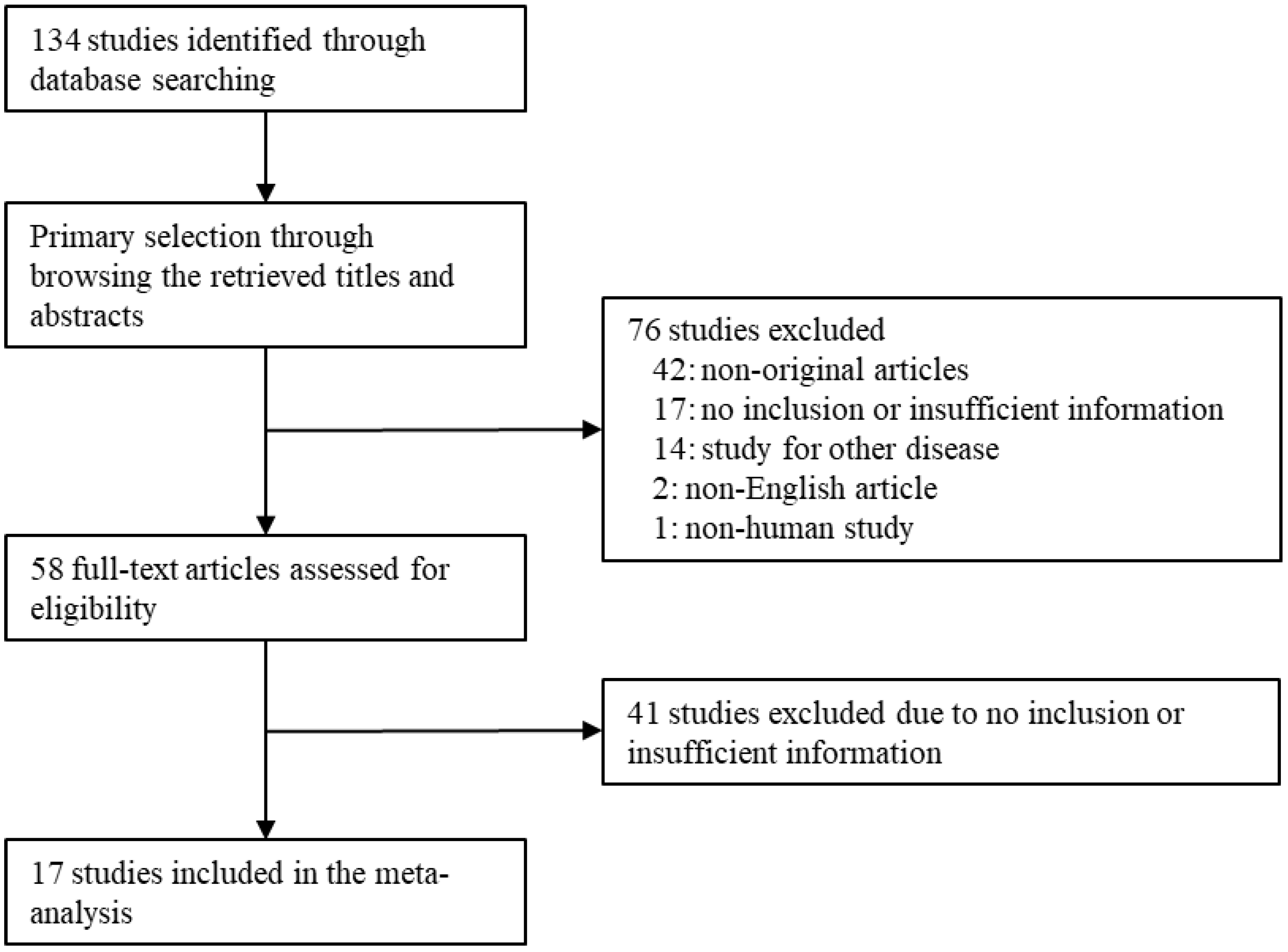
2. Materials and Methods

2.1. Published Study Search and Selection Criteria

Relevant articles were obtained by searching the PubMed databases through 20 January 2023. Searching was performed using the following keywords: “(pancreas or pancreatic) AND (EUS or endoscopic ultrasound or aspiration cytology) AND (Ki-67 or Ki67 or proliferation index)”. The titles and abstracts of all searched articles were screened for exclusion. Review articles and previous meta-analyses were also screened to obtain additional eligible studies. Searched results were then reviewed, and articles were included if the study investigated the PanNENs and there was information for Ki-67 immunohistochemistry. The articles that were case reports or non-original articles, or non-English language publications were excluded.

2.2. Data Extraction

Data from all eligible studies were extracted by two individual authors. Extracted data from each of the eligible studies included the following [7,8,9,10,11,12,13,14,15,16,17,18,19,20,21,22,23]: first author’s name, year of publication, study location, number of patients analyzed, types of obtained EUS samples, tumor grade of aspiration and surgical specimen, and so on. For the meta-analysis and diagnostic test accuracy review, all data associated with the Ki-67 LI of PanNEN were extracted. Any disagreements were resolved by consensus.

2.3. Statistical Analysis

For performing the meta-analysis, all data were analyzed using the Comprehensive Meta-Analysis software package (Biostat, Englewood, NJ, USA). The concordance rates of tumor grade by Ki-67 labeling index were investigated between EUS-FNAC/FNB and surgical specimens. In addition, the subgroup analysis based on types of obtained EUS samples, tumor grade, and tumor size was performed. Because the eligible studies used various evaluation methods for Ki-67 IHC of PanNEN and had different populations of patients, a random-effects model was more appropriate than a fixed-effects model. Heterogeneity between the eligible studies was checked using P statistics (p-value). In addition, the significance of difference between subgroups was evaluated using a meta-regression test. To evaluate publication bias, Begg’s funnel plot and Egger’s test were conducted. The results with p < 0.05 were considered statistically significant. If significant publication bias was found, the fail-safe N and trim-fill tests were additionally conducted to confirm the degree of publication bias. The results were considered statistically significant with p < 0.05.
In the present study, the diagnostic test accuracy (DTA) review of the Ki-67 labeling index of PanNEN based on tumor grade was performed using the Meta-Disc program (version 1.4; Biostatics, the Ramon y Cajal Hospital, Madrid, Spain) [26]. The pooled sensitivity and specificity, diagnostic odds ratio (OR) were calculated from individual data of each eligible study. By plotting ‘sensitivity’ and ‘1-specificity’ of each study, the SROC curve (summary receiver operating characteristic curve) was firstly constructed and the curve fitting was performed through linear regression using the Littenberg and Moses linear model [27]. Because each of the data were heterogeneous, the accuracy data were pooled by fitting a SROC curve and measuring the value of the area under the curve (AUC) [26]. An area under the curve (AUC) close to 1 means the test is strong, and close to 0.5 means the test is considered poor. Subgroup analysis based on the PanNEN grade was conducted.

3. Results

3.1. Subsection

Selection and Characteristics

Through database searching, 134 articles were identified. We excluded 76 articles of the searched studies through the screening. These 76 articles were excluded due to non-original articles (n = 42), no inclusion or insufficient information (n = 17), studies for other diseases (n = 14), non-English articles (n = 2), and non-human study (n = 1). Next, we reviewed the full text of 58 articles. Forty-one articles were excluded due to no inclusion or insufficient information. Finally, 17 articles were included in this meta-analysis and DTA review (Figure 1 and Table 1).

3.2. Concordance Rate of Tumor Grade of Pancreatic Neuroendocrine Neoplasm between EUS-FNAC/FNB and Surgical Specimen

First, the overall concordance rate was investigated between EUS-FNAC/FNB and surgical specimens. The estimated concordance rate was 0.767 (95% CI 0.713–0.814) (Table 2). In a subgroup analysis based on EUS sampling, the concordance rates of FNAC and FNB were 0.741 (95% CI 0.681–0.794) and 0.839 (95% CI 0.738–0.906), respectively. Although the concordance rate of EUS-FNB was higher than that of EUS-FNAC, there was no statistical significance (p = 0.071 in the meta-regression test). Next, the subgroup analysis based on the tumor grade of PanNEN was performed. The concordance rates of grade 1/2 and grade 3 subgroups were 0.772 (95% CI 0.722–0.816) and 0.743 (95% CI 0.628–0.945), respectively. In the grade 1/2 subgroup, the concordance rates of EUS-FNAC and EUS-FNB were 0.745 (95% CI 0.695–0.789) and 0.846 (95% CI 0.722–0.921), respectively. The concordance rates of grade 1 and 2 PanNENs were 0.772 (95% CI 0.712–0.820) and 0.741 (95% CI 0.655–0.812), respectively. In addition, in the grade 3 subgroup, the concordance rates of EUS-FNAC and EUS-FNB were 0.879 (95% CI 0.660–0.965) and 0.667 (95% CI 0.154–0.957), respectively. The concordance rate of grade 3 was lower in EUS-FNB than in EUS-FNAC. However, the statistical significance between EUS-FNAC and EUS-FNB was not found in the subgroup analysis (p = 0.356 in the meta-regression test). In PanNEN with less than 2 cm, the concordance rate was 0.797 (95% CI 0.726–0.853). In the subgroup analysis, the concordance rates of grade 1 and 2 PanNENs were 0.877 (95% CI 0.791–0.930) and 0.685 (95% CI 0.414–0.870), respectively. There was a significant difference of concordance rates between grade 1 and 2 subgroups in PanNENs with less than 2 cm (p = 0.021 in the meta-regression test).

3.3. Diagnostic Test Accuracy Review of Ki-67 in the Grading of Pancreatic Neuroendocrine Neoplasm

A DTA review was performed for the sensitivity, specificity, diagnostic OR, and AUC on SROC of EUS-FNAC/FNB compared to the surgical specimen. We evaluated the diagnostic accuracy of EUS-FNAC/FNB in predicting the tumor grade using Ki-67. The comparison between grade 1/2 and 3 subgroups was performed. In grade 3 subgroup, the pooled sensitivity and specificity were 0.786 (95% CI 0.590–0.917) and 0.998 (95% CI 0.987–1.000), respectively (Table 3). The diagnostic OR and AUC on SROC in grade 3 subgroup were 150.220 and 0.983, respectively. In the subgroup analysis based on tumor grade, the pooled sensitivities of PanNEN grades 1 and 2 were 0.908 (95% CI 0.876–0.937) and 0.599 (95% CI 0.534–0.661), respectively. The pooled specificities of PanNEN grades 1 and 2 were 0.616 (95% CI 0.557–0.674) and 0.904 (95% CI 0.872–0.930), respectively. The diagnostic ORs were 14.467 (95% CI 8.892–23.536) and 13.971 (95% CI 8.364–23.335) in PanNEN grades 1 and 2 subgroups, respectively. In addition, the AUC on SROC were 0.871 and 0.859 in PanNEN grades 1 and 2 subgroups, respectively. In PanNEN with less than 2 cm, the pooled sensitivities of PanNEN grades 1 and 2 were 0.852 (95% CI 0.771–0.913) and 0.667 (95% CI 0.498–0.809), respectively. The pooled specificities of PanNEN grades 1 and 2 were 0.675 (95% CI 0.509–0.814) and 0.844 (95% CI 0.762–0.906), respectively. In PanNEN grade 1 subgroup, the diagnostic OR and AUC on SROC were 15.319 (95% CI 5.915–39.677) and 0.841, respectively. In PanNEN grade 2 subgroup, the diagnostic OR and AUC on SROC were 13.093 (95% CI 5.143–33.332) and 0.834, respectively.

4. Discussion

To the best of our knowledge, this study represents the first meta-analysis and DTA review focusing on the assessment of tumor grading using Ki-67 LI between EUS-FNAC/FNB and surgical specimens of PanNENs. The increasing number of incidentally detected PanNENs can be attributed to advancements in imaging technology and improved opportunities for evaluation [23]. Treatment decisions for non-functioning PanNENs can pose significant challenges. The treatment of PanNENs is determined on the basis of their size and grade. The histological grade of PanNENs is determined by mitosis and Ki-67 LI. According to the guidelines, PanNENs are classified into three categories, grades 1 to 3, based on Ki-67 LI. When PanNEN is suspected during EUS-FNAC/FNB, the measurement of Ki-67 LI can provide valuable information for guiding treatment decisions. Given the importance of Ki-67 LI on EUS-FNAC/FNB in treatment decision-making, it is imperative to determine the concordance between Ki-67 LI results from EUS-FNAC/FNB and surgical specimens. However, the limited information available from individual studies examining this concordance emphasizes the significance of our results.
The advancements in EUS for diagnosing pancreatic lesions have significantly improved its diagnostic accuracy. However, the evaluation of specimens obtained through EUS-FNAC/FNB remains a challenging aspect. In the case of PanNENs, EUS-FNAC/FNB is commonly regarded as the most accurate and safe diagnostic method. Ki-67 LI is essential for tumor grading in PanNENs, specifically in EUS-FNAC/FNB specimens. The WHO classifies PanNENs into grades 1 to 3, with each grade determined based on mitotic rate and Ki-67 LI. The concordance between EUS-FNAC/FNB and surgical specimens can be utilized to evaluate the diagnostic accuracy of PanNEN grading. Notably, the reported concordance rates exhibit variations across different studies [7,8,9,10,11,12,13,14,15,16,17,18,19,20,21,22,23]. The concordance rates of individual studies ranged from 50.0 to 100.0% in our meta-analysis [7,8,9,10,11,12,13,14,15,16,17,18,19,20,21,22,23]. The tumor grade of PanNENs, as determined from surgical specimens, is considered definitive. However, in situations where surgical removal is not feasible, preoperative EUS-FNAC/FNB specimens can be used to assess Ki-67 LI, providing valuable information for tumor grading. The advancements in EUS techniques have increased the importance of assessing Ki-67 LI in preoperative specimens. However, it should be noted that the assessment of concordance in Ki-67 LI between preoperative and operative specimens may be limited to operable patients only. Therefore, obtaining comprehensive information from individual studies may be challenging. Performing a meta-analysis along with a DTA review can provide a comprehensive assessment of diagnostic accuracy. We evaluated the accuracy of Ki-67 LI in assessing the tumor grade of PanNEN using EUS-FNAC/FNB through a DTA review. We specifically explored the potential benefits of using a core biopsy needle (FNB) for sampling, which may help overcome the limitations associated with the FNA technique. A previous meta-analysis indicated the superiority of FNB over FNA in evaluating pancreatic masses [28]. However, there is a lack of comprehensive data on the concordance of Ki-67 LI between FNAC and FNB in previous studies. Although not statistically significant, FNB exhibited a slightly higher concordance rate compared to FNAC (0.839 vs. 0.741; p = 0.071 in the meta-regression test). Interestingly, in the grade 3 subgroup, FNAC showed a higher concordance rate compared to FNB (0.879 vs. 0.667). Notably, the concordance rate of EUS-FNAC in the grade 3 subgroup was significantly higher compared to that in grade 1 and 2, or EUS-FNB subgroups. However, in routine clinical practice, the difference in concordance rates may have limited significance as FNAC and FNB are typically performed simultaneously.
If the tumor size is less than 2 cm, it is classified as low-stage, which has implications for treatment decision-making. Guidelines recommend a wait-and-see approach for asymptomatic patients with tumors less than 2 cm [5,29]. However, there may be a mismatch between guideline recommendations and their application in clinical practice [15]. Previous studies have examined the concordance of Ki-67 LI in PanNENs less than 2 cm. In this meta-analysis, we further analyzed PanNENs less than 2 cm in size. The concordance rate for tumors less than 2 cm was 0.797 (95% CI, 0.726–0.853). Subgroup analysis based on tumor grade yielded concordance rates of grades 1 and 2 of 0.877 (95% CI 0.791–0.930) and 0.685 (95% CI 0.414–0.870), respectively. There was a significant difference between the grade 1 and 2 subgroups (p = 0.021 in the meta-regression test). In clinical practice, this finding can be useful for evaluating tumor grading in PanNENs smaller than 2 cm. The DTA review showed higher sensitivity and lower specificity in the grade 1 subgroup compared to the grade 2 subgroup. However, the diagnostic OR and AUC of the SROC were not significantly different between grades 1 and 2.
Discrepancies between EUS-FNAC/FNB and surgical specimens can be attributed to various factors. One important factor is tumor heterogeneity. According to a previous report, grade 2 PanNENs exhibit a greater heterogeneity compared to grade 1 and 3 tumors [9]. Hasegawa et al. demonstrated that the dispersion of Ki- 67 LI was significantly higher in grade 2 tumors compared to grade 1 tumors [30]. Considering the limitations of EUS-FNAC/FNB, it may not be possible to examine the entire lesion, and the evaluation of hotspots can be limited due to the limited number of specimens. Ki-67 LI should be evaluated in at least 500 tumor cells in the hotspot area for accurate tumor grading. Thus, the number of specimens obtained with EUS-FNAC/FNB can impact the concordance rates as it may limit the evaluation of a sufficient number of tumor cells. The following guidelines provided by the World Health Organization (WHO) [1] and the European Neuroendocrine Tumor Society [5] outline the criteria for tumor grading based on Ki-67 LI: (1) grade 1, Ki-67 LI < 2%; (2) grade 2, Ki-67 LI between 3 and 20%; and (3) grade 3, Ki-67 > 20%. If the Ki-67 LI value is close to the threshold (2 or 20%), caution should be exercised in interpreting the results. This variability in tumor grading can significantly impact concordance rates.
The current study has several limitations that need to be acknowledged. Firstly, we were unable to evaluate the concordance rate based on EUS-FNAC/FNB cellularity due to insufficient information available in the eligible studies. It is recommended to count in more than 500 cells for accurate assessment. Secondly, a detailed analysis based on the specific clones of Ki-67 antibodies used could not be conducted due to insufficient information. Lastly, the evaluation method of Ki-67 LI, whether it was eyeballing or automated calculations, could not be analyzed in subgroup analysis due to insufficient information.

5. Conclusions

In conclusion, our study demonstrates a high level of concordance between Ki-67 LI measured using EUS-FNAC/FNB and surgical specimens in patients with PanNENs. Notably, when considering PanNENs smaller than 2 cm, we observed a significant concordance rate of Ki-67 LI in both the grade 1 and 2 subgroups. The findings of our DTA review, underscore the value of Ki-67 LI assessment through EUS-FNAC/FNB as a reliable method for predicting the tumor grade of PanNENs. However, further study is needed to evaluate the difference in accuracy for low cellularity of EUS-FNAC/FNB.

Author Contributions

Conceptualization, J.-S.P. and N.Y.K.; methodology, J.-S.P.; software, J.-S.P.; data curation, K.-W.M., I.H.O. and D.H.L.; writing—original draft preparation, J.-S.P. and N.Y.K.; writing—review and editing, K.-W.M. and B.K.S. All authors have read and agreed to the published version of the manuscript.

Funding

This research received no external funding.

Institutional Review Board Statement

Not applicable.

Informed Consent Statement

Not applicable.

Data Availability Statement

Not applicable.

Conflicts of Interest

The authors declare no conflict of interest.

References

  1. WHO Classification of Tumours Editorial Board. Digestive System Tumours. Lyon (France): International Agency for Research on Cancer, 5th ed.; WHO classification of tumours series; International Agency for Research on Cancer: Lyon, France, 2019; Volume 1. [Google Scholar]
  2. Yao, J.C.; Hassan, M.; Phan, A.; Dagohoy, C.; Leary, C.; Mares, J.E.; Abdalla, E.K.; Fleming, J.B.; Vauthey, J.N.; Rashid, A.; et al. One hundred years after “carcinoid”: Epidemiology of and prognostic factors for neuroendocrine tumors in 35,825 cases in the United States. J. Clin. Oncol. 2008, 26, 3063–3072. [Google Scholar] [CrossRef] [PubMed]
  3. Dong, D.-H.; Zhang, X.-F.; Lopez-Aguiar, A.G.; Poultsides, G.; Makris, E.; Rocha, F.; Kanji, Z.; Weber, S.; Fisher, A.; Fields, R.; et al. Resection of pancreatic neuroendocrine tumors: Defining patterns and time course of recurrence. HPB 2019, 22, 215–223. [Google Scholar] [CrossRef] [PubMed]
  4. Zhang, X.F.; Xue, F.; Dong, D.H.; Alexandra, L.A.; George, P.; Eleftherios, M.; Flavio, R.; Zaheer, K.; Sharon, W.; Alexander, F.; et al. New nodal staging for primary pancreatic neuroendocrine tumors: A multi-institutional and national data analysis. Ann. Surg 2021, 274, e28–e35. [Google Scholar] [CrossRef] [PubMed]
  5. Falconi, M.; Eriksson, B.; Kaltsas, G.; Bartsch, D.K.; Capdevila, J.; Caplin, M.; Kos-Kudla, B.; Kwekkeboom, D.; Rindi, G.; Klöppel, G.; et al. Vienna Consensus Conference participants. ENETS consensus guidelines update for the management of patients with functional pancreatic neuroendocrine tumors and non-functional pancreatic neuroendocrine tumors. Neuroendocrinology 2016, 103, 153–171. [Google Scholar] [CrossRef] [PubMed]
  6. Hosoda, W.; Takagi, T.; Mizuno, N.; Shimizu, Y.; Sano, T.; Yamao, K.; Yatabe, Y. Diagnostic approach to pancreatic tumors with the specimens of endoscopic ultrasound-guided fine needle aspiration. Pathol. Int. 2010, 60, 358–364. [Google Scholar] [CrossRef]
  7. Abi-Raad, R.; Lavik, J.P.; Barbieri, A.L.; Zhang, X.; Adeniran, A.J.; Cai, G. Grading Pancreatic Neuroendocrine Tumors by Ki-67 Index Evaluated on Fine-Needle Aspiration Cell Block Material. Am. J. Clin. Pathol. 2020, 153, 74–81. [Google Scholar] [CrossRef]
  8. Boutsen, L.; Jouret-Mourin, A.; Borbath, I.; van Maanen, A.; Weynand, B. Accuracy of Pancreatic Neuroendocrine Tumour Grading by Endoscopic Ultrasound-Guided Fine Needle Aspiration: Analysis of a Large Cohort and Perspectives for Improvement. Neuroendocrinology 2018, 106, 158–166. [Google Scholar] [CrossRef]
  9. Crinò, S.F.; Ammendola, S.; Meneghetti, A.; Bernardoni, L.; Conti Bellocchi, M.C.; Gabbrielli, A.; Landoni, L.; Paiella, S.; Pin, F.; Parisi, A.; et al. Comparison between EUS-guided fine-needle aspiration cytology and EUS-guided fine-needle biopsy histology for the evaluation of pancreatic neuroendocrine tumors. Pancreatology 2021, 21, 443–450. [Google Scholar] [CrossRef]
  10. Cui, Y.; Khanna, L.G.; Saqi, A.; Crapanzano, J.P.; Mitchell, J.M.; Sethi, A.; Gonda, T.A.; Kluger, M.D.; Schrope, B.A.; Allendorf, J.; et al. The Role of Endoscopic Ultrasound-Guided Ki67 in the Management of Non-Functioning Pancreatic Neuroendocrine Tumors. Clin. Endosc. 2020, 53, 213–220. [Google Scholar] [CrossRef]
  11. Arco, C.D.D.; Pérez, J.Á.D.; Medina, L.O.; Valera, J.S.; Aceñero, M.J.F. Reliability of Ki-67 Determination in FNA Samples for Grading Pancreatic Neuroendocrine Tumors. Endocr. Pathol. 2016, 27, 276–283. [Google Scholar] [CrossRef]
  12. Leo, M.D.; Poliani, L.; Rahal, D.; Auriemma, F.; Anderloni, A.; Ridolfi, C.; Spaggiari, P.; Capretti, G.; Tommaso, L.D.; Preatoni, P.; et al. Pancreatic Neuroendocrine Tumours: The Role of Endoscopic Ultrasound Biopsy in Diagnosis and Grading Based on the WHO 2017 Classification. Dig. Dis. 2019, 37, 325–333. [Google Scholar] [PubMed]
  13. Farrell, J.M.; Pang, J.C.; Kim, G.E.; Tabatabai, Z.L. Pancreatic neuroendocrine tumors: Accurate grading with Ki-67 index on fine-needle aspiration specimens using the WHO 2010/ENETS criteria. Cancer Cytopathol. 2014, 122, 770–778. [Google Scholar] [CrossRef] [PubMed]
  14. Grosse, C.; Noack, P.; Silye, R. Accuracy of grading pancreatic neuroendocrine neoplasms with Ki-67 index in fine-needle aspiration cellblock material. Cytopathology 2019, 30, 187–193. [Google Scholar] [CrossRef] [PubMed]
  15. Heidsma, C.M.; Tsilimigras, D.I.; Rocha, F.; Abbott, D.E.; Fields, R.; Smith, P.M.; Poultsides, G.A.; Cho, C.; van Eijck, C.; van Dijkum, E.N.; et al. US Neuroendocrine Tumor Study Group. Clinical relevance of performing endoscopic ultrasound-guided fine-needle biopsy for pancreatic neuroendocrine tumors less than 2 cm. J. Surg Oncol. 2020, 122, 1393–1400. [Google Scholar] [CrossRef] [PubMed]
  16. Hwang, H.S.; Kim, Y.; An, S.; Kim, S.J.; Kim, J.Y.; Kim, S.Y.; Hwang, D.W.; Park, D.H.; Lee, S.S.; Kim, S.C.; et al. Grading by the Ki-67 Labeling Index of Endoscopic Ultrasound-Guided Fine Needle Aspiration Biopsy Specimens of Pancreatic Neuroendocrine Tumors Can Be Underestimated. Pancreas 2018, 47, 1296–1303. [Google Scholar] [CrossRef] [PubMed]
  17. Kalantri, S.; Bakshi, P.; Verma, K. Grading of pancreatic neuroendocrine tumors on endoscopic ultrasound-guided fine-needle aspiration using Ki-67 index and 2017 World Health Organization criteria: An analysis of 32 cases. Cytojournal 2020, 17, 21. [Google Scholar] [CrossRef]
  18. Laskiewicz, L.; Jamshed, S.; Gong, Y.; Ainechi, S.; LaFemina, J.; Wang, X. The diagnostic value of FNA biopsy in grading pancreatic neuroendocrine tumors. Cancer Cytopathol. 2018, 126, 170–178. [Google Scholar] [CrossRef]
  19. Leeds, J.S.; Nayar, M.K.; Bekkali, N.L.H.; Wilson, C.H.; Johnson, S.J.; Haugk, B.; Darne, A.; Oppong, K.W. Endoscopic ultrasound-guided fine-needle biopsy is superior to fine-needle aspiration in assessing pancreatic neuroendocrine tumors. Endosc. Int. Open 2019, 7, E1281–E1287. [Google Scholar] [CrossRef]
  20. Paiella, S.; Landoni, L.; Rota, R.; Valenti, M.; Elio, G.; Crinò, S.F.; Manfrin, E.; Parisi, A.; Cingarlini, S.; D’Onofrio, M.; et al. Endoscopic ultrasound-guided fine-needle aspiration for the diagnosis and grading of pancreatic neuroendocrine tumors: A retrospective analysis of 110 cases. Endoscopy 2020, 52, 988–994. [Google Scholar] [CrossRef]
  21. Piani, C.; Franchi, G.M.; Cappelletti, C.; Scavini, M.; Albarello, L.; Zerbi, A.; Arcidiacono, P.G.; Bosi, E.; Manzoni, M.F. Cytological Ki-67 in pancreatic endocrine tumours: An opportunity for pre-operative grading. Endocr. Relat. Cancer 2008, 15, 175–181. [Google Scholar] [CrossRef]
  22. Sugimoto, M.; Takagi, T.; Hikichi, T.; Suzuki, R.; Watanabe, K.; Nakamura, J.; Kikuchi, H.; Konno, N.; Waragai, Y.; Asama, H.; et al. Efficacy of endoscopic ultrasonography-guided fine needle aspiration for pancreatic neuroendocrine tumor grading. World J. Gastroenterol. 2015, 21, 8118–8124. [Google Scholar] [CrossRef] [PubMed]
  23. Tacelli, M.; Petrone, M.C.; Capurso, G.; Muffatti, F.; Andreasi, V.; Partelli, S.; Doglioni, C.; Falconi, M.; Arcidiacono, P.G. Diagnostic accuracy of EUS-FNA in the evaluation of pancreatic neuroendocrine neoplasms grading: Possible clinical impact of misclassification. Endosc. Ultrasound 2021, 10, 372–380. [Google Scholar] [CrossRef]
  24. Polkowski, M.; Jenssen, C.; Kaye, P.; Carrara, S.; Deprez, P.; Fernández-Esparrach, G.; Eisendrath, P.; Aithal, G.P.; Arcidiacono, P.; Barthet, M.; et al. Technical aspects of endoscopic ultrasound (EUS)-guided sampling in gastroenterology: European Society of Gastrointestinal Endoscopy (ESGE) Technical Guideline—March 2017. Endoscopy 2017, 49, 989–1006. [Google Scholar] [CrossRef] [PubMed]
  25. Siddiqui, A.A.; Brown, L.J.; Hong, S.K.; Draganova-Tacheva, R.A.; Korenblit, J.; Loren, D.E.; Kowalski, T.E.; Solomides, C. Relationship of pancreatic mass size and diagnostic yield of endoscopic ultrasound-guided fine needle aspiration. Dig. Dis. Sci. 2011, 56, 3370–3375. [Google Scholar] [CrossRef] [PubMed]
  26. Zamora, J.; Abraira, V.; Muriel, A.; Khan, K.; Coomarasamy, A. Meta-DiSc: A software for meta-analysis of test accuracy data. BMC Med. Res. Methodol. 2006, 6, 31. [Google Scholar] [CrossRef]
  27. Moses, L.E.; Shapiro, D.; Littenberg, B. Combining independent studies of a diagnostic test into a summary ROC curve: Data-analytic approaches and some additional considerations. Stat Med. 1993, 12, 1293–1316. [Google Scholar] [CrossRef]
  28. Li, H.; Li, W.; Zhou, Q.Y.; Fan, B. Fine needle biopsy is superior to fine needle aspiration in endoscopic ultrasound guided sampling of pancreatic masses: A meta-analysis of randomized controlled trials. Medicine 2018, 97, e0207. [Google Scholar] [CrossRef]
  29. NANETS treatment guidelines. Well-differentiated neuroendocrine tumors of the stomach and pancreas. Pancreas 2010, 39, 735–752. [Google Scholar] [CrossRef]
  30. Hasegawa, T.; Yamao, K.; Hijioka, S.; Bhatia, V.; Mizuno, N.; Hara, K.; Imaoka, H.; Niwa, Y.; Tajika, M.; Kondo, S.; et al. Evaluation of Ki- 67 index in EUS-FNA specimens for the assessment of malignancy risk in pancreatic neuroendocrine tumors. Endoscopy 2014, 46, 32–38. [Google Scholar] [CrossRef]
Figure 1. Flow chart of the searching strategy.
Diagnostics 13 02756 g001
Table 1. Main characteristics of the eligible studies.
Author and
Publication Year
LocationSpecimenAntibody CloneNumber
of
Patients
Surgical
Specimen
Tumor Size
(cm, Mean ± SD)
G1G2G3
Abi-Raad 2020 [7]USAFNAC with CBMIB-149272203.00 ± 1.90
Boutsen 2018 [8]BelgiumFNACMIB-157302342.85 ± 2.16
Crinò 2021 [9]ItalyFNAC with CBMIB-16913401.98 ± 1.50
FNB 73462522.15 ± 1.33
Cui 2020 [10]USAFNAC30–9372485ND
Díaz Del Arco 2016 [11]SpainFNACND107303.20 ± 3.28
Di Leo 2019 [12]ItalyFNBND2520412.10 ± 1.49
Farrell 2014 [13]USAFNACMIB-12215523.03 ± 1.73
Grosse 2019 [14]AustriaFNAC with CBND152943.84 ± 1.8
Heidsma 2020 [15]USAFNACND6346161ND
Hwang 2018 [16]KoreaFNB 33201033.30 ± 2.20
Kalantri 2020 [17]IndiaFNAC with CBBGX-29711443ND
Laskiewicz 2018 [18]USAFNACMIB-12615110ND
Leeds 2019 [19]UKFNAC with CBND2316702.57 ± 0.31
FNB 26121403.25 ± 0.36
Paiella 2020 [20]ItalyFNACND77482812.45 ± 1.34
Piani 2008 [21]ItalyFNACMIB-11811613.05 ± 2.77
Sugimoto 2015 [22]JapanFNACMIB-185302.57 ± 1.32
Tacelli 2021 [23]ItalyFNAC with CBMIB-1112595032.39 ± 0.31
SD, standard deviation; FNAC, fine-needle aspiration cytology; CB, cell block; FNB, fine-needle biopsy; ND, no description.
Table 2. The estimated concordance rates of WHO grade using ki-67 labeling index between aspirated and surgical specimens in pancreatic neuroendocrine neoplasms.
Number
of
Subsets
Fixed Effect
[95% CI]
Heterogeneity Test
[p-Value]
Random Effect
[95% CI]
Egger’s
Test
[p-Value]
Overall190.754 [0.719, 0.786]0.0060.767 [0.713, 0.814]0.080
 FNAC150.734 [0.694, 0.771]0.0390.741 [0.681, 0.794]0.136
 FNB a40.840 [0.770, 0.892]0.1400.839 [0.738, 0.906]0.826
 Grade 1/2190.757 [0.722, 0.790]0.0280.772 [0.722, 0.816]0.024
  FNAC150.739 [0.699, 0.776]0.1940.745 [0.695, 0.789]0.052
  FNB b40.840 [0.766, 0.894]0.0620.846 [0.722, 0.921]0.352
  Grade 1190.756 [0.713, 0.794]0.0640.772 [0.712, 0.820]0.026
  Grade 2170.732 [0.657, 0.796]0.3100.741 [0.655, 0.812]0.062
 Grade 360.743 [0.628, 0.945]0.9660.743 [0.628, 0.945]0.019
  FNAC50.879 [0.660, 0.965]0.9990.879 [0.660, 0.965]<0.001
  FNB c10.667 [0.154, 0.957]1.0000.667 [0.154, 0.957]-
Tumor size, less than 2 cm60.797 [0.726, 0.853]0.7770.797 [0.726, 0.853]0.204
 Grade 1 d50.877 [0.791, 0.930]0.9390.877 [0.791, 0.930]0.385
 Grade 230.665 [0.453, 0.827]0.2760.685 [0.414, 0.870]0.757
CI, Confidence interval; FNAC, fine-needle aspiration cytology; FNB, fine-needle biopsy. a, p = 0.071 in the meta-regression test; b, p = 0.063 in the meta-regression test; c, p = 0.356 in the meta-regression test; d, p = 0.021 in the meta-regression test.
Table 3. Sensitivity, specificity, diagnostic odds ratio and area under curve of summary receiver operation characteristics curve of evaluating WHO grade using ki-67 labeling index in endoscopic sonography-guided fine-needle aspiration.
Number
of
Subsets
Sensitivity (%)
[95% CI]
Specificity (%)
[95% CI]
Diagnostic OR
[95% CI]
AUC
on SROC
Overall
 Grade 1 *180.908 [0.876, 0.937]0.616 [0.557, 0.674]14.467 [8.892, 23.536]0.871
 Grade 2 *170.599 [0.534, 0.661]0.904 [0.872, 0.930]13.971 [8.364, 23.335]0.859
 Grade 3 #100.786 [0.590, 0.917]0.998 [0.987, 1.000]150.220 [46.145, 489.000]0.983
Tumor size, less than 2 cm
 Grade 140.852 [0.771, 0.913]0.675 [0.509, 0.814]15.319 [5.915, 39.677]0.841
 Grade 240.667 [0.498, 0.809]0.844 [0.762, 0.906]13.093 [5.143, 33.332]0.834
CI, Confidence interval; OR, Odds ratio; AUC, Area under curve; SROC, summary receiver operating characteristic. *, reference: grade 3; #, reference: grade 1 and 2.
Disclaimer/Publisher’s Note: The statements, opinions and data contained in all publications are solely those of the individual author(s) and contributor(s) and not of MDPI and/or the editor(s). MDPI and/or the editor(s) disclaim responsibility for any injury to people or property resulting from any ideas, methods, instructions or products referred to in the content.

