
Tumid Lupus Erythematosus (TLE): A Review of a Rare Variant of Chronic Cutaneous Lupus Erythematosus (cCLE) with Emphasis on Differential Diagnosis

 ,
,  ,
,  and
and 
Abstract
1. Introduction
- (1)
- Acute cutaneous lupus erythematosus (ACLE), typically presents with a malar rash, which is a butterfly shaped rash across the cheeks and bridge of the nose but that may also involve the scalp, neck, and upper chest [2]. From a histopathological point of view, ACLE presents interface dermatitis, with vacuolar degeneration of basal keratinocytes, often accompanied by lymphocytic infiltration and a perivascular and periadnexal inflammation, with some degrees of dermal edema and, in some cases, signs of leukocytoclastic vasculitis. Furthermore, epidermal changes such as hyperkeratosis, focal parakeratosis, and dyskeratosis can be appreciated. Finally, mucin deposition in the dermis is another potential feature of this form of CLE [3].
- (2)
- Subacute cutaneous lupus erythematosus (SCLE) is characterized by nonscarring, psoriasiform, or annular lesions predominantly found on sun-exposed areas such as the upper back, shoulders, extensor surfaces of the arms, and neck and that can also present as widespread erythematous plaques [4]. Histologically, SCLE presents interface dermatitis with basal cell vacuolization and hyperkeratosis with some degree of follicular plugging, which is a common finding in SCLE and contributes to the characteristic scaling and follicular papules seen in SCLE lesions. At the level of the dermis, it is possible to appreciate a dense inflammatory infiltrate around blood vessels (perivascular) and hair follicles (perifollicular) with lymphocytes, histiocytes, and occasionally eosinophils infiltrate these areas, contributing to the inflammatory response. Finally, in some cases of SCLE, there are mucin deposition and dermal changes such as edema [5].
- (3)
- Chronic cutaneous lupus erythematosus (CCLE) encompasses several subtypes: discoid lupus erythematosus (DLE) presents with well-defined, scaly, erythematous plaques often with follicular plugging and atrophy and in which the lesions typically occur on the face, scalp, and ears, but can also affect other areas of the body.
2. Epidemiology
3. Etiopathogenesis
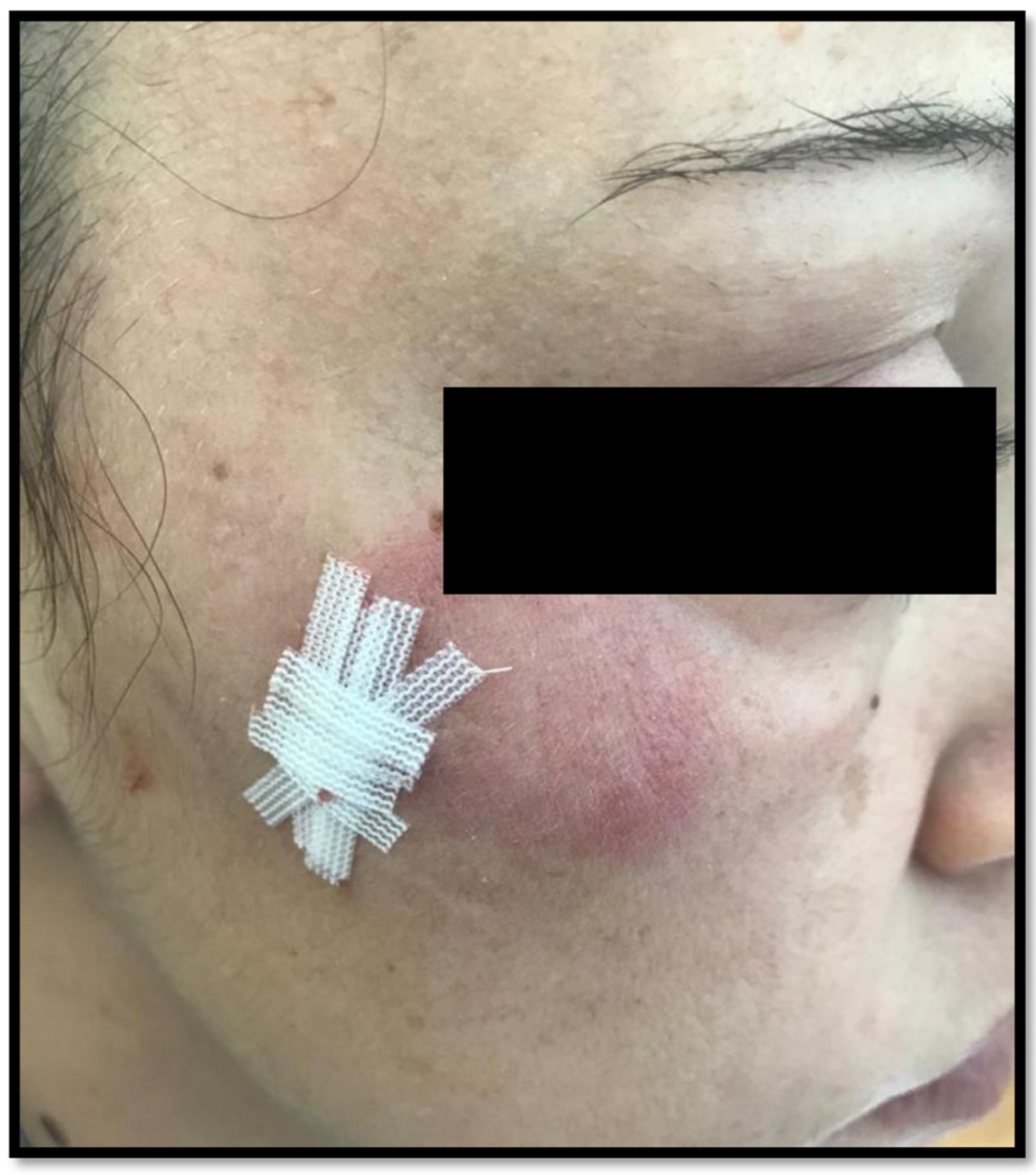
4. Clinical Picture
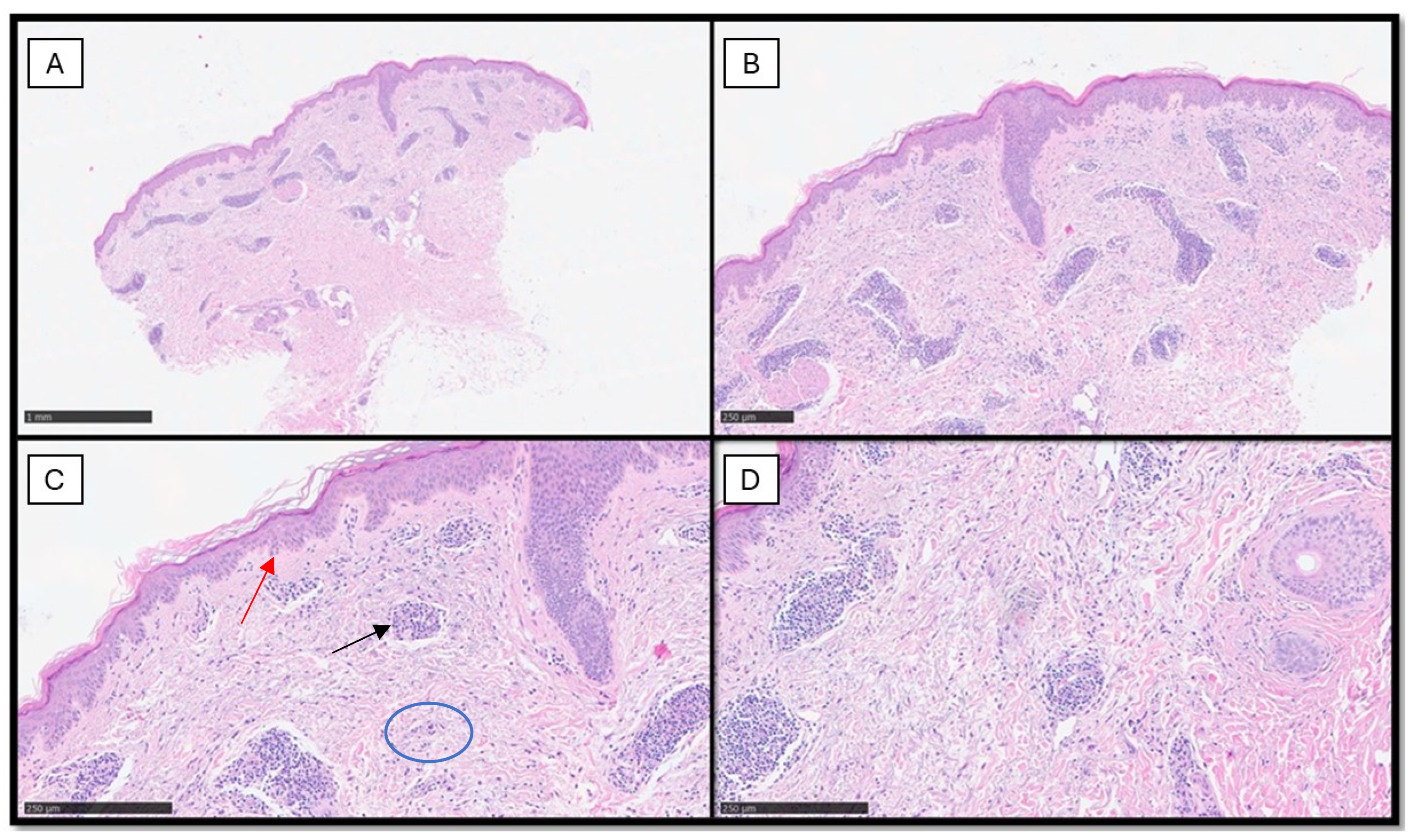
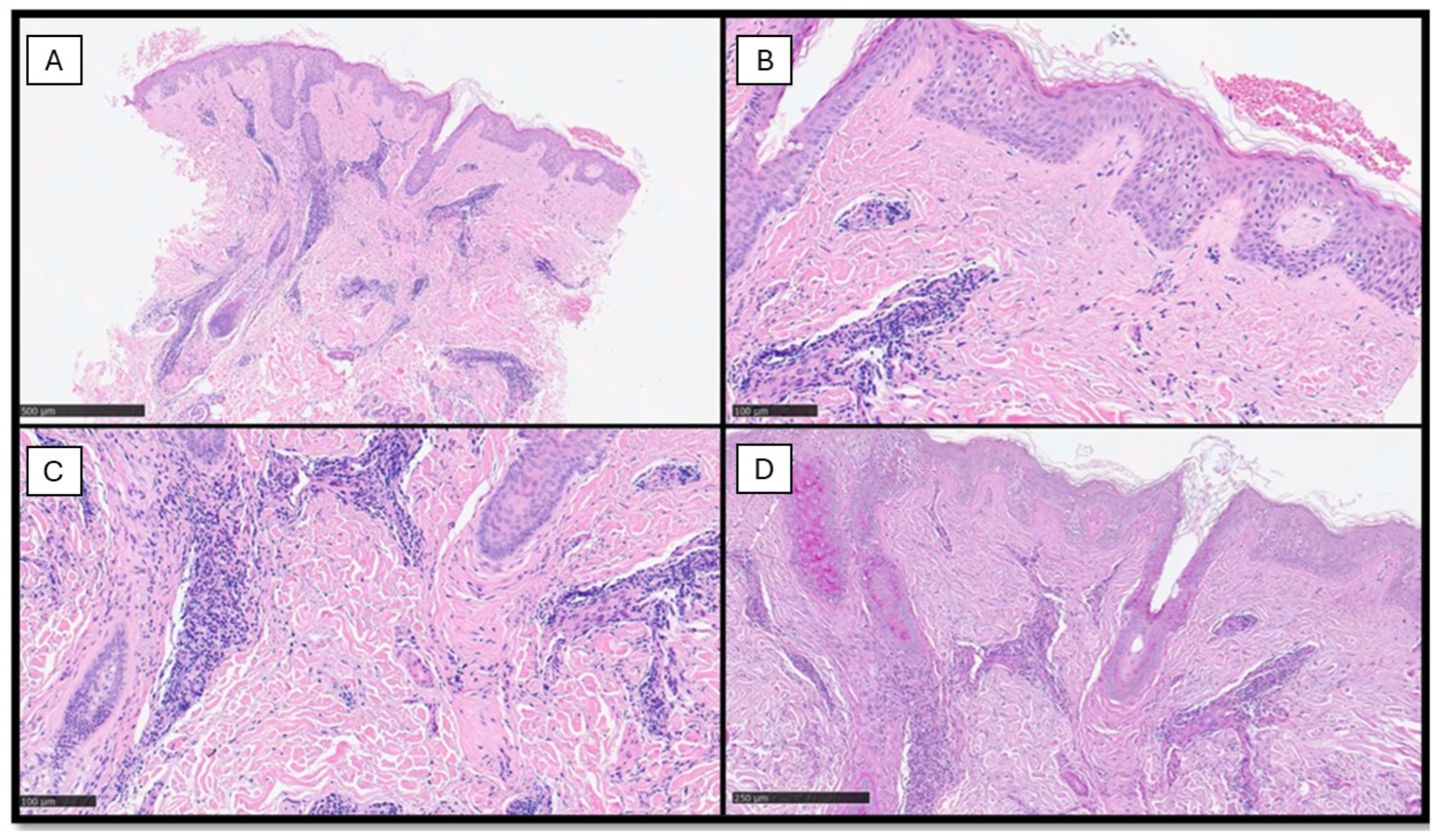
5. Histopathology
6. Classification of TLE
7. Prognosis
8. Therapy
9. Differential Diagnosis
9.1. Jessner–Kanof Infiltrate (Pseudolymphoma)
9.2. Polymorphous Light Eruption (PLE)
9.3. Reticular Erythematous Mucinosis (REM)
9.4. Granuloma Faciale (GF)
9.5. Cutaneous Manifestations of B-Chronic Lymphocytic Leukemia (B-CLL)
10. Associations Rarely Reported
11. The Larger Series of TLE Reported in Literature
12. Conclusions
Author Contributions
Funding
Institutional Review Board Statement
Informed Consent Statement
Data Availability Statement
Acknowledgments
Conflicts of Interest
References
- Petty, A.J.; Floyd, L.; Henderson, C.; Nicholas, M.W. Cutaneous Lupus Erythematosus: Progress and Challenges. Curr. Allergy Asthma Rep. 2020, 20, 12. [Google Scholar] [CrossRef]
- Walling, H.W.; Sontheimer, R.D. Cutaneous lupus erythematosus: Issues in diagnosis and treatment. Am. J. Clin. Dermatol. 2009, 10, 365–381. [Google Scholar] [CrossRef] [PubMed]
- Filho, J.P.M.; Peixoto, R.L.; Martins, L.G.; de Melo, S.D.; de Carvalho, L.L.; Pereira, A.K.F.d.T.C.; Freire, E.A.M. Lupus erythematosus: Considerations about clinical, cutaneous and therapeutic aspects. An. Bras. Dermatol. 2014, 89, 118–125. [Google Scholar] [CrossRef]
- Niebel, D.; de Vos, L.; Fetter, T.; Brägelmann, C.; Wenzel, J. Cutaneous Lupus Erythematosus: An Update on Pathogenesis and Future Therapeutic Directions. Am. J. Clin. Dermatol. 2023, 24, 521–540. [Google Scholar] [CrossRef]
- Poh, Y.J.; Alrashid, A.; Sangle, S.R.; Higgins, E.; Benton, E.; McGibbon, D.; D’cruz, D.P. Proton pump inhibitor induced subacute cutaneous lupus erythematosus: Clinical characteristics and outcomes. Lupus 2022, 31, 1078–1083. [Google Scholar] [CrossRef]
- Zhou, W.; Wu, H.; Zhao, M.; Lu, Q. New insights into the progression from cutaneous lupus to systemic lupus erythematosus. Expert Rev. Clin. Immunol. 2020, 16, 829–837. [Google Scholar] [CrossRef]
- Joseph, A.K.; Windsor, B.; Hynan, L.S.; Chong, B.F. Discoid lupus erythematosus skin lesion distribution and characteristics in Black patients: A retrospective cohort study. Lupus Sci. Med. 2021, 8, e000514. [Google Scholar] [CrossRef] [PubMed]
- Green, P.J.; Pasternak, S. Hypertrophic and Ulcerated Discoid Lupus Erythematosus. J. Cutan. Med. Surg. 2012, 16, 453–457. [Google Scholar] [CrossRef] [PubMed]
- Kuhn, A.; Richter-Hintz, D.; Oslislo, C.; Ruzicka, T.; Megahed, M.; Lehmann, P. Lupus erythematosus tumidus—A neglected subset of cutaneous Lupus erythematosus: Report of 40 cases. Arch. Dermatol. 2000, 136, 1033–1041. [Google Scholar] [CrossRef]
- Alexiades-Armenakas, M.R.; Baldassano, M.; Bince, B.; Werth, V.; Bystryn, J.-C.; Kamino, H.; Soter, N.A.; Franks, A.G., Jr. Tumid lupus erythematosus: Criteria for classification with immunohistochemical analysis. Arthritis Care Res. 2003, 49, 494–500. [Google Scholar] [CrossRef]
- Sontheimer, R. The lexicon of cutaneous lupus erythematosus-A review and personal perspective on the nomenclature and classification of the cutaneous manifestations of lupus erythematosus. Lupus 1997, 6, 84–95. [Google Scholar] [CrossRef] [PubMed]
- Hoffmann, E. Demonstrationen: Lupus erythematodes tumidus. Derm Zeitschr 1909, 16, 159–160. [Google Scholar]
- Vieira, V.; Del Pozo, J.; Yebra-Pimentel, M.T.; Martínez, W.; Fonseca, E. Lupus erythematosus tumidus: A series of 26 cases. Int. J. Dermatol. 2006, 45, 512–517. [Google Scholar] [CrossRef] [PubMed]
- Schmitt, V.; Meuth, A.; Amler, S.; Kuehn, E.; Haust, M.; Messer, G.; Bekou, V.; Sauerland, C.; Metze, D.; Köpcke, W.; et al. Lupus erythematosus tumidus is a separate subtype of cutaneous lupus erythematosus. Br. J. Dermatol. 2010, 162, 64–73. [Google Scholar] [CrossRef]
- Kuhn, A.; Sonntag, M.; Ruzicka, T.; Lehmann, P.; Megahed, M. Histopathologic findings in lupus erythematosus tumidus: Review of 80 patients. J. Am. Acad. Dermatol. 2003, 48, 901–908. [Google Scholar] [CrossRef] [PubMed]
- Gilliam, J.N.; Sontheimer, R.D. Distinctive cutaneous subsets in the spectrum of lupus erythematosus. J. Am. Acad. Dermatol. 1981, 4, 471–475. [Google Scholar] [CrossRef] [PubMed]
- Saleh, D.; Grubbs, H.; Koritala, T.; Crane, J.S. Tumid Lupus Erythematosus; StatPearls Publishing: St. Petersburg, FL, USA, 2023. [Google Scholar]
- WHO ICD–10/2019 Revision. Available online: https://icd.who.int/browse10/2019/en (accessed on 17 January 2024).
- Sonntag, M.; Lehmann, P.; Megahed, M.; Ruzicka, T.; Kuhn, A. Lupus erythematosus tumidus in Childhood. Dermatology 2003, 207, 188–192. [Google Scholar] [CrossRef]
- Biazar, C.; Sigges, J.; Patsinakidis, N.; Ruland, V.; Amler, S.; Bonsmann, G.; Kuhn, A. Cutaneous lupus erythematosus: First multicenter database analysis of 1002 patients from the European Society of Cutaneous Lupus Erythematosus (EUSCLE). Autoimmun. Rev. 2013, 12, 444–454. [Google Scholar] [CrossRef] [PubMed]
- Liu, E.; Daze, R.P.; Moon, S. Tumid Lupus Erythematosus: A Rare and Distinctive Variant of Cutaneous Lupus Erythematosus Masquerading as Urticarial Vasculitis. Cureus 2020, 12, e8305. [Google Scholar] [CrossRef]
- Vale, E.C.S.d.; Garcia, L.C. Cutaneous lupus erythematosus: A review of etiopathogenic, clinical, diagnostic and therapeutic aspects. An. Bras. Dermatol. 2023, 98, 355–372. [Google Scholar] [CrossRef]
- Kuhn, A.; Sonntag, M.; Richter-Hintz, D.; Oslislo, C.; Megahed, M.; Ruzicka, T.; Lehmann, P. Phototesting in lupus erythe-matosus: A 15-year experience. J. Am. Acad. Dermatol. 2001, 45, 86–95. [Google Scholar] [CrossRef] [PubMed]
- Yu, C.; Chang, C.; Zhang, J. Immunologic and genetic considerations of cutaneous lupus erythematosus: A comprehensive review. J. Autoimmun. 2013, 41, 34–45. [Google Scholar] [CrossRef] [PubMed]
- Böckle, B.C.; Sepp, N.T. Smoking is highly associated with discoid lupus erythematosus and lupus erythematosus tumidus: Analysis of 405 patients. Lupus 2015, 24, 669–674. [Google Scholar] [CrossRef] [PubMed]
- Schneider, S.W.; Staender, S.; Schlüter, B.; Luger, T.A.; Bonsmann, G. Infliximab-induced lupus erythematosus tumidus in a patient with rheumatoid arthritis. Arch. Dermatol. 2006, 142, 109–116. [Google Scholar] [CrossRef] [PubMed]
- Sohl, S.; Renner, R.; Winter, U.; Bodendorf, M.; Paasch, U.; Simon, J.C.; Treudler, R. Drug-induced lupus erythematosus tu-midus during treatment with adalimumab. Hautarzt 2009, 60, 826–829. [Google Scholar] [CrossRef] [PubMed]
- Schepis, C.; Lentini, M.; Siragusa, M.; Batolo, D. ACE-inhibitor-induced drug eruption resembling lymphocytic infiltration (of jessner-kanof) and Lupus erythematosus tumidus. Dermatology 2004, 208, 354–355. [Google Scholar] [CrossRef] [PubMed]
- Böckle, B.C.; Baltaci, M.; Weyrer, W.; Sepp, N.T. Bortezomib-induced lupus erythematosus Tumidus. Oncologist 2009, 14, 637–639. [Google Scholar] [CrossRef]
- Abadías-Granado, I.; Sánchez-Bernal, J.; Felipo-Berlanga, F.; Ara-Martín, M. Coexistence of Tumid Lupus Erythematosus and Discoid Lupus Erythematosus. Actas Dermosifiliogr. 2019, 110, 253–255. [Google Scholar] [CrossRef]
- Jatwani, K.; Chugh, K.; Osholowu, O.S.; Jatwani, S. Tumid Lupus Erythematosus and Systemic Lupus Erythematosus: A Report on Their Rare Coexistence. Cureus 2020, 12, e7545. [Google Scholar] [CrossRef]
- Gambichler, T.; Pätzholz, J.; Schmitz, L.; Lahner, N.; Kreuter, A. FOXP3+ and CD39+ regulatory T cells in subtypes of cutaneous lupus erythematosus. J. Eur. Acad. Dermatol. Venereol. 2015, 29, 1972–1977. [Google Scholar] [CrossRef]
- Rodríguez-Caruncho, C.; Bielsa, I. Lupus erythematosus tumidus: A clinical entity still being defined. Actas Dermosifiliogr. 2011, 102, 668–674. [Google Scholar] [CrossRef] [PubMed]
- Patsinakidis, N.; Kautz, O.; Gibbs, B.F.; Raap, U. Lupus erythematosus tumidus: Clinical perspectives. Clin. Cosmet. Investig. Dermatol. 2019, 2019, 707–719. [Google Scholar] [CrossRef] [PubMed]
- Shen, A.; Fisher, J.M.; Kuklinski, L.F.; Hogeling, M. A teen with blaschkolinear tumid lupus erythematosus. JAAD Case Rep. 2022, 21, 1–5. [Google Scholar] [CrossRef] [PubMed]
- Chronic Cutaneous Lupus 2023. Available online: https://www.pathologyoutlines.com/topic/skinnontumorlupussle.html (accessed on 17 February 2024).
- Hood, A.F. The Lives of Lesions: Chronology in Dermatopathology. Arch. Dermatol. 1985, 121, 809. [Google Scholar] [CrossRef]
- Cerroni, L. Skin Lymphoma, The Illustrated Guide, 5th ed.; Wiley Blackwell: Hoboken, NJ, USA, 2020. [Google Scholar]
- Weldemann, A.; Ziepert, M.; Kreuz, M.; Dumann, K.; Simon, J.C.; Kunz, M.; Ziemer, M. Lupus erythematosus: Correlation of clinical and histological findings and proposal for a modified disease classification. JDDG: J. Dtsch. Dermatol. Ges. 2021, 19, 1591–1599. [Google Scholar] [CrossRef] [PubMed]
- Tan, B.C.H.; Tang, I.; Bonin, J.; Koelmeyer, R.; Hoi, A. The performance of different classification criteria for systemic lupus erythematosus in a real-world rheumatology department. Rheumatology 2022, 61, 4509–4513. [Google Scholar] [CrossRef] [PubMed]
- Gougerot, H.; Burnier, R. Lupus érythémateux tumidus. Bull. Soc. Fr. Dermatol. Syphil. 1930, 37, 1219–1292. [Google Scholar]
- Rockl, H. Erythematodes tumidus; a case history with reference to the problem of erythematodic lymphocytoma. Hautarzt 1954, 5, 422–423. [Google Scholar] [PubMed]
- Vilanova, X. The profundus and tumidus forms of lupus erythe-matosus; report of a case and comments. Rev. Clin. Sep. 1950, 36, 388–393. [Google Scholar]
- Sanchez-Melendez, S.; Malik, R.; Kanwar, R.; Yang, K.; Nambudiri, V.E. Clinical epidemiology of tumid lupus erythematosus: A retrospective review of 179 patients. J. Am. Acad. Dermatol. 2024, 90, 202–204. [Google Scholar] [CrossRef]
- Kuhn, A.; Ruzicka, T. Classification of cutaneous lupus erythematosus. In Cutaneous Lupus Erythematosus; Kuhn, A., Lehmann, P., Ruzicka, T., Eds.; Springer: New York, NY, USA, 2005; pp. 53–57. [Google Scholar]
- Rodriguez-Caruncho, C.; Bielsa, I.; Fernández-Figueras, M.T.; Roca, J.; Carrascosa, J.M.; Ferrándiz, C. Lupus erythematosus tumidus: A clinical and histological study of 25 cases. Lupus 2015, 24, 751–755. [Google Scholar] [CrossRef]
- Kuhn, A.; Bein, D.; Bonsmann, G. The 100th anniversary of lupus erythematosus tumidus. Autoimmun. Rev. 2009, 8, 441–448. [Google Scholar] [CrossRef]
- Stead, J.; Headley, C.; Ioffreda, M.; Kovarik, C.; Werth, V. Coexistence of tumid lupus erythematosus with systemic lupus er-ythematosus and discoid lupus erythematosus: A report of two cases of tumid lupus. J. Clin. Rheumatol. 2008, 14, 338–341. [Google Scholar] [CrossRef]
- Sigges, J.; Biazar, C.; Landmann, A.; Ruland, V.; Patsinakidis, N.; Amler, S.; Bonsmann, G.; Kuhn, A. Therapeutic strategies evaluated by the European Society of Cutaneous Lupus Erythematosus (EUSCLE) Core Set Questionnaire in more than 1000 patients with cutaneous lupus erythematosus. Autoimmun. Rev. 2013, 12, 694–702. [Google Scholar] [CrossRef]
- Pona, A.; Cardenas-de la Garza, J.A.; Broderick, A.; Sangueza, O.P.; Niehaus, A.G.; Bowers, N.; Pichardo, R.O. Lupus Erythematosus Tumidus Clinical Characteristics and Treatment: A Retrospective Review of 25 Patients. Cutis 2022, 109, 330. [Google Scholar] [CrossRef]
- Marmor, M.F.; Kellner, U.; Lai, T.Y.; Melles, R.B.; Mieler, W.F.; American Academy of Ophthalmology. Recommendations on Screening for Chloroquine and Hydroxychloroquine Retinopathy (2016 Revision). Ophthalmology 2016, 123, 1386–1394. [Google Scholar] [CrossRef]
- Kuhn, A.; Aberer, E.; Bata-Csörgő, Z.; Caproni, M.; Dreher, A.; Frances, C.; Gläser, R.; Klötgen, H.; Landmann, A.; Marinovic, B.; et al. S2k guideline for treatment of cutaneous lupus erythematosus—Guided by the European Dermatology Forum (EDF) in cooperation with the European Academy of Dermatology and Venereology (EADV). J. Eur. Acad. Dermatol. Venereol. 2017, 31, 389–404. [Google Scholar] [CrossRef]
- Kreuter, A.; Tigges, C.; Hunzelmann, N.; Oellig, F.; Lehmann, P.; Hofmann, S.C. Rituximab in the treatment of recalcitrant generalized lupus erythematosus tumidus. J. Dtsch. Dermatol. Ges. 2017, 15, 729–731. [Google Scholar] [CrossRef]
- Verdelli, A.; Coi, A.; Marzano, A.; Antiga, E.; Cozzani, E.; Quaglino, P.; La Placa, M.; Benucci, M.; De Simone, C.; Papini, M.; et al. Autoantibody profile and clinical patterns in 619 Italian patients with cutaneous lupus erythematosus. J. Eur. Acad. Dermatol. Venereol. 2019, 33, 742–752. [Google Scholar] [CrossRef]
- Kanof, M.; Jessner, N.B. Lymphocytic infiltration of the skin. Arch. Dermatol. 1953, 68, 447–449. [Google Scholar]
- Weber, F.; Schmuth, M.; Fritsch, P.; Sepp, N. Lymphocytic infiltration of the skin is a photosensitive variant of lupus erythematosus: Evidence by phototesting. Br. J. Dermatol. 2001, 144, 292–296. [Google Scholar] [CrossRef]
- Rémy-Leroux, V.; Léonard, F.; Lambert, D.; Wechsler, J.; Cribier, B.; Thomas, P.; Adamski, H.; Marguery, M.-C.; Aubin, F.; Leroy, D.; et al. Comparison of histopathologic–clinical characteristics of Jessner’s lymphocytic infiltration of the skin and lupus erythematosus tumidus: Multicenter study of 46 cases. J. Am. Acad. Dermatol. 2008, 58, 217–223. [Google Scholar] [CrossRef]
- McKee, P.H. Superficial and deep perivascular inflammatory dermatoses. In Pathology of the Skin with Clinical Correlations, 3rd ed.; McKee, P.H., Calonje, E., Granter, S.R., Eds.; Elsevier: Philadephia, PA, USA, 2005; pp. 269–271. [Google Scholar]
- Steigleder, G.K.; Gartmann, H.; Linker, U. REM syndrome: Reticular erythematous mucinosis (round-cell erythematosis), a new entity? Br. J. Dermatol. 1974, 91, 191–199. [Google Scholar] [CrossRef]
- Perry, H.O.; Kierland, R.R.; Montgomery, H. Plaque-Like Form of Cutaneous Mucinosis. Arch. Dermatol. 1960, 82, 980–985. [Google Scholar] [CrossRef]
- Bleehen, S.; Slater, D.; Mahood, J.; Church, R. Reticular erythematous mucinosis: Light and electron microscopy, irnmunofluorescence and histochemical findings. Br. J. Dermatol. 1982, 106, 9–18. [Google Scholar] [CrossRef]
- Vanuytrecht-Henderickx, D.; Dewolf-Peeters, C.; Degreef, H. Morphological study of the reticular erythematous mucinosis syndrome. Dermatology 1984, 168, 163–169. [Google Scholar] [CrossRef]
- Izumi, T.; Tajima, S.; Harada, R.; Nishikawa, T. Reticular erythematous mucinosis syndrome: Glycosaminoglycan synthesis by fibroblasts and abnormal response to interleukin-1β. Dermatology 1996, 192, 41–45. [Google Scholar] [CrossRef]
- Del Pozo, J.; Martinez, W.; Almagro, M.; Yebra, M.; Garcia-Silva, J.; Fonseca, E. Reticular erythematous mucinosis syndrome. Report of a case with positive immunofluorescence. Clin. Exp. Dermatol. 1997, 22, 234–236. [Google Scholar] [CrossRef]
- Al Dhafiri, M.; Kaliyadan, F. Granuloma Faciale; StatPearls Publishing: Treasure Island, FL, USA, 2024. [Google Scholar]
- Georgiadou, N.M.; Singh, S.M.; Singh, M.M.H. Lupus Erythematosus Tumidus Associated With Hemophagocytosis. Am. J. Dermatopathol. 2022, 44, 519–522. [Google Scholar] [CrossRef]
- Choonhakarn, C.; Poonsriaram, A.; Chaivoramukul, J. Lupus erythematosus tumidus. Int. J. Dermatol. 2004, 43, 815–818. [Google Scholar] [CrossRef]
- Boggio, F.; Lora, V.; Cota, C.; Pereira, A.; Müllegger, R.; Prieto-Torres, L.; Cerroni, L. Cutaneous hemophagocytosis: Clinicopathologic features of 21 cases. J. Am. Acad. Dermatol. 2018, 78, 377–382. [Google Scholar] [CrossRef]
- Kerl, K.; Wolf, I.H.; Cerroni, L.; Wolf, P.; French, L.E.; Kerl, H. Hemophagocytosis in Cutaneous Autoimmune Disease. Am. J. Dermatopathol. 2015, 37, 539–543. [Google Scholar] [CrossRef]
- Magaña, M.; Castellanos, G.; Meurehg, C.C.; Magaña-Mainero, M.-R. Lupus Erythematosus Tumidus: Clinical and Pathological Features in a Series of 20 Patients. Am. J. Dermatopathol. 2022, 44, 469–477. [Google Scholar] [CrossRef]


| Type of LE | Clinical Features | Topography | Histopathological Findings |
|---|---|---|---|
| TLE | Annular, erythematosus, edematous, urticarial plaques | Sun-exposed areas (face, chest, upper back, upper extremities) | Abundant dermal mucin deposition, superficial and deep perivascular and periadnexal lymphocytic infiltrate, with occasional edema in the papillary dermis |
| DLE | Erythematosus, hyperkeratotic lesions with frequent atrophic scarring | Head and neck, extensor aspects of the arms | Hyperkeratosis, follicular plugging, vacuolar degeneration, thickening of the dermo-epidermal junction |
| SCLE | Annular, polycyclic lesions with scaly surface, nonscarring, erythematosus | Sun-exposed areas | Vacuolar degeneration with a superficial perivascular and interstitial lymphocytic infiltrate |
Disclaimer/Publisher’s Note: The statements, opinions and data contained in all publications are solely those of the individual author(s) and contributor(s) and not of MDPI and/or the editor(s). MDPI and/or the editor(s) disclaim responsibility for any injury to people or property resulting from any ideas, methods, instructions or products referred to in the content. |
© 2024 by the authors. Licensee MDPI, Basel, Switzerland. This article is an open access article distributed under the terms and conditions of the Creative Commons Attribution (CC BY) license (https://creativecommons.org/licenses/by/4.0/).
Share and Cite
Daruish, M.; Ambrogio, F.; Foti, C.; Filosa, A.; Cazzato, G. Tumid Lupus Erythematosus (TLE): A Review of a Rare Variant of Chronic Cutaneous Lupus Erythematosus (cCLE) with Emphasis on Differential Diagnosis. Diagnostics 2024, 14, 780. https://doi.org/10.3390/diagnostics14070780
Daruish M, Ambrogio F, Foti C, Filosa A, Cazzato G. Tumid Lupus Erythematosus (TLE): A Review of a Rare Variant of Chronic Cutaneous Lupus Erythematosus (cCLE) with Emphasis on Differential Diagnosis. Diagnostics. 2024; 14(7):780. https://doi.org/10.3390/diagnostics14070780
Chicago/Turabian StyleDaruish, Maged, Francesca Ambrogio, Caterina Foti, Alessandra Filosa, and Gerardo Cazzato. 2024. "Tumid Lupus Erythematosus (TLE): A Review of a Rare Variant of Chronic Cutaneous Lupus Erythematosus (cCLE) with Emphasis on Differential Diagnosis" Diagnostics 14, no. 7: 780. https://doi.org/10.3390/diagnostics14070780
APA StyleDaruish, M., Ambrogio, F., Foti, C., Filosa, A., & Cazzato, G. (2024). Tumid Lupus Erythematosus (TLE): A Review of a Rare Variant of Chronic Cutaneous Lupus Erythematosus (cCLE) with Emphasis on Differential Diagnosis. Diagnostics, 14(7), 780. https://doi.org/10.3390/diagnostics14070780