Abstract
This study investigates the microstructural evolution and mechanical properties of high-nickel shipbuilding steel during thermal processing using high-temperature confocal laser-scanning microscopy (HTCLSM). An in situ observation of the heating and holding processes reveals critical insights into phase transformations, grain-growth behavior, and the formation of precipitates. The experimental results demonstrate that austenitization begins at approximately 700 °C, with significant grain-boundary nucleation. At 900 °C, the formation of black precipitates was observed, and their persistence up to temperatures exceeding 1000 °C was confirmed. Scanning electron microscopy (SEM) and energy-dispersive spectroscopy (EDS) analyses identified these precipitates as chromium carbides (Cr7C3), which significantly contribute to the material’s strength. A comprehensive analysis using transmission electron microscopy (TEM) confirmed the presence and distribution of Cr7C3 within the grains and along grain boundaries. These findings provide a deeper understanding of the microstructural dynamics in high-nickel steels, guiding the optimization of heat-treatment processes to enhance mechanical properties for maritime applications.
1. Introduction
The performance of advanced steels is significantly affected by factors such as grain size, subgrain boundaries, phase composition, and precipitates [1,2,3]. These microstructural features are influenced by thermal treatments and controlled rolling processes, which can optimize material properties through grain refinement and phase transformations [4,5,6,7]. However, the primary challenge in this field is the difficulty of directly observing and understanding microstructural changes during high-temperature processes due to limitations in traditional experimental techniques [8,9]. This lack of direct observation impedes the development of a comprehensive understanding of the relationships between thermal processing, microstructural evolution, and mechanical properties. In this study, we aimed to address this gap by utilizing advanced in situ observation techniques to analyze the high-temperature microstructural evolution of high-nickel steel. Specifically, we explored the formation and transformation of precipitates, grain-growth behavior, and phase changes during heating and holding processes, focusing on their impact on the mechanical properties of the material.
Confocal laser-scanning microscopy (CLSM), developed in the 1980s, offers a solution for the in situ observation of microstructural changes at high temperatures [10,11,12]. CLSM provides a high resolution and can generate clear, three-dimensional images through simultaneous multi-fluorescence observations, advantages not available with traditional optical microscopy. Lasertec Corporation in Japan has advanced CLSM technology by integrating confocal laser scanning, infrared heating, and tensile testing into high-temperature confocal laser-scanning microscopy (HTCLSM) [13,14]. This innovation has become an essential tool for visually studying processes such as melting, solidification, and the high-temperature deformation of materials. The Lasertec confocal laser-scanning microscope uses a He–Ne laser source and precise confocal spatial filtering to form a conjugate image. The laser creates a point light source at the focal plane of the objective lens, scanning the sample. Spatial filtering at the detection pinhole in the focal plane of the measurement lens effectively suppresses stray fluorescence and interference from out-of-focus planes. This results in high-signal-to-noise-ratio optical tomographic images. The laser’s monochromatic nature and short depth of focus yield a high longitudinal resolution, enabling non-destructive layer scanning and fluorescence intensity measurements at different sample depths. The optical sections from various focal planes are reconstructed into a three-dimensional structure of the sample. The development of HTCLSM has significantly advanced the in situ observation of material microstructure transformations, such as high-temperature phase changes in steel, high-temperature precipitation processes, ferrite–austenite transformations, and austenite decomposition [15,16,17].
HTCLSM’s capability to observe in real time microstructural changes under high temperatures makes it indispensable for material research and development. It is particularly useful in studying phase transformations in steels, for which rapid heating and cooling rates can be simulated to mimic industrial processes. This allows for the investigation of phenomena such as austenitization, martensitic transformation, and carbide precipitation, providing valuable insights into the kinetics and thermodynamics of these transformations [18,19,20]. These advancements have deepened the understanding of microstructural transformations in materials. Microstructural evolution during thermal processing is a complex interplay of nucleation, growth, and the coarsening of different phases [21,22,23]. The precise control of these processes is vital for achieving the desired mechanical properties in advanced materials. For instance, in steels, the formation and dissolution of carbides, nitrides, and other intermetallic compounds during heating and cooling significantly affect hardness, strength, and toughness [24,25,26]. Understanding these transformations at the microscopic level allows for the development of heat-treatment schedules that optimize performance characteristics for specific applications.
By employing high-temperature confocal laser-scanning microscopy (HTCLSM) combined with SEM and EDS analyses, this work seeks to provide novel insights into the microstructural transformations occurring in high-nickel steels, particularly in relation to carbide precipitation performance. Our findings offer valuable data for optimizing the thermal processing of these steels, enhancing their industrial applications. Additionally, the methodologies employed in this study can be applied to other alloy systems, facilitating advancements in various high-performance materials
2. Experimental Materials and Methods
2.1. Experimental Materials
The chemical composition of the steel used in this study was experimentally measured through optical emission spectroscopy (OES) (Waltham, MA, USA), and the results are detailed in Table 1. Steel ingots were produced using a 50-kg vacuum-induction furnace and subsequently forged into blocks with dimensions of 120 mm × 120 mm × 260 mm. These blocks were then soaked at 1200 °C for a minimum of one hour to ensure uniform temperature distribution and homogenization. Following the soaking process, the blocks were hot-rolled into plates with a cross-sectional dimension of 20 mm × 150 mm.

Table 1.
Chemical composition of the steel (wt.%).
2.2. High-Temperature In Situ Equipment
The in situ observation of the material heating process was conducted using high-temperature confocal laser-scanning microscopy (HTCLSM, Yonekura MFG. CO., LTD., Miyagi, Japan). This method’s sophisticated equipment (Yonekura MFG. CO., LTD., Miyagi, Japan) integrates multiple advanced systems, including a high-temperature furnace, an imaging system, a temperature control system, and a computer system. Additionally, it features a tensile furnace, a protective gas system, a cooling system, a workbench control system, a tensile control system, and an image detection system, as illustrated in Figure 1.
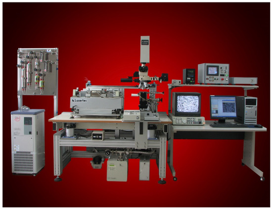
Figure 1.
HTCLSM schematic.
HTCLSM is particularly suited to observing the microstructural evolution of materials at elevated temperatures. The high-temperature furnace provides precise thermal conditions required for studying phase transformations, grain growth, and other high-temperature phenomena. The imaging system, equipped with a He–Ne laser source, ensures high-resolution and high-contrast images by utilizing confocal spatial filtering. This allows for the suppression of out-of-focus light and stray fluorescence, resulting in clear, three-dimensional reconstructions of the sample’s microstructure. The temperature control system ensures accurate and stable thermal conditions during experiments, which are critical for the reproducibility and reliability of the observations. Integration with a computer system allows for real-time data acquisition and analysis, facilitating the monitoring and recording of microstructural changes as they occur. The protective gas system, usually involving an inert gas like argon, is employed to prevent the oxidation and contamination of the sample during high-temperature exposure. The cooling system is used to control the sample’s cooling rate, which is essential for studying the effects of different cooling regimes on the microstructure and properties of the material.
The HTCLSM setup provides a comprehensive platform for in situ high-temperature studies, combining precise thermal and mechanical control with advanced imaging capabilities to enable a detailed analysis of microstructural evolution and its impact on material properties.
2.3. In Situ Observation Samples
The samples used for in situ microstructural observations were cylindrical with dimensions of φ7.5 mm × 3.5 mm. Prior to observation, the sample surfaces were meticulously ground, polished, and cleaned. Due to the self-imaging capabilities of HTCLSM, the etching of the polished samples was not necessary.
Once prepared, the samples were placed into an Al2O3 crucible, which was then inserted into a heating furnace. The furnace was evacuated to a vacuum pressure of 1 × 10⁻⁶ MPa, followed by the introduction of a protective gas to prevent oxidation and contamination during heating. Various parameters of the HTCLSM were set to ensure optimal conditions for in situ observation.
The heating and holding procedures for the in situ observation samples are depicted in Figure 2. The samples were initially heated at a rate of 4 °C/s to 900 °C and held at this temperature for 5 min. Subsequently, one group of samples was further heated to 1150 °C at the same rate and held for an additional 5 min before being rapidly cooled via forced inert gas cooling to room temperature at a rate of 10 °C/s. Another group was directly cooled to room temperature after the initial hold at 900 °C at the same manner.
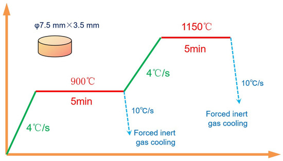
Figure 2.
Heating procedure for in situ observation.
After the completion of the in situ observation experiments, scanning electron microscopy (SEM) was conducted using a JEOL JSM-7800F microscope (Akishima, Japan), while transmission electron microscopy (TEM) was carried out on a JEOL JEM-2100 microscope (Akishima, Japan) at 200 kV. TEM samples were prepared through mechanical thinning, followed by ion milling to achieve electron transparency. This meticulous process ensures that the observed changes in microstructure are accurately captured and analyzed, providing valuable insights into the behavior of the material under controlled thermal conditions. The combination of HTCLSM’s advanced imaging capabilities with precise thermal control allows for a comprehensive study of microstructural evolution, which is critical for optimizing material properties and processing techniques.
3. Results and Discussion
3.1. In Situ Observation of Microstructural Transformation
The in situ observation of the microstructural transformation during the heating process was conducted using high-temperature confocal laser-scanning microscopy (HTCLSM). The changes in the material’s microstructure during heating are shown in Figure 3.
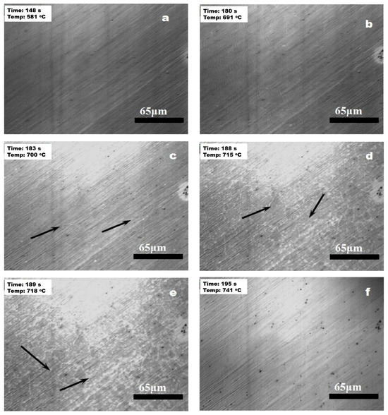
Figure 3.
In situ observation of microstructural changes during heating: (a) 148 s; (b) 180 s; (c) 183 s; (d) 188 s; (e) 189 s; and (f) 195.
In Figure 3, the numbers in the top left corner indicate the time (seconds) and the real-time temperature (°C) of the sample. Initially, at temperatures below 580 °C, no significant microstructural changes were observed, indicating that the material remained stable within this temperature range. However, as the temperature reached 700 °C, the material began to enter the austenitization phase. Typically, grain boundaries, subgrain boundaries, and defects are common sites for nucleation. As shown by the arrows in Figure 3c,d, austenite nuclei initially formed along the grain boundaries, where the white dot-like formations can be observed along the boundary. These nuclei appeared as fine, discrete particles along the grain boundaries, highlighting the initial stages of austenite formation. As the temperature continues to rise, these austenite nuclei expanded and merged, forming a uniform austenitic structure, as depicted in Figure 3e. By approximately 740 °C, the microstructure was predominantly composed of austenite. The transformation to austenite within the temperature range of 700 °C to 740 °C was relatively rapid, occurring over approximately 12 s. This rapid transformation can be attributed to the high mobility of grain boundaries at elevated temperatures, facilitating the quick formation and growth of austenite grains.
When the temperature reached 900 °C, precipitates started to form within the microstructure. The changes in the microstructure and the formation of precipitates during the holding period at 900 °C are depicted in Figure 4.
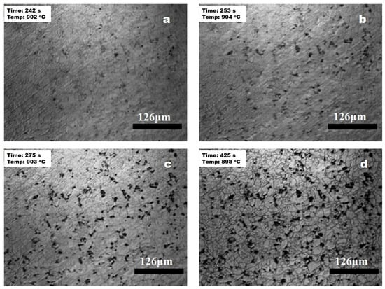
Figure 4.
Microstructural changes during holding at 900 °C: (a) 242 s; (b) 253 s; (c) 275 s; and (d) 425 s.
Figure 4 illustrates that the sample reached the set temperature of 900 °C after 240 s, at which point black precipitates began to appear (Figure 4a). As the holding time increased, the quantity of dark precipitates began to rise, as shown in Figure 4b, which depicts the in situ morphology of the dark phase after approximately 10 s of holding. Over time, the extent of the dark phase continued to increase, reaching its peak at around 30 s, as illustrated in Figure 4c. During the subsequent holding period, the number and distribution of the dark precipitates remained largely unchanged, as shown in Figure 4d. Furthermore, a comparison of Figure 4c,d reveals that the color in Figure 4d appears darker, suggesting a slight increase in the quantity of precipitates during the holding process. The dark appearance of these precipitates can be attributed to their interaction with trace amounts of gases in the observation chamber, which may have resulted in the formation of oxide layers on the precipitate surfaces. Additionally, the high resolution and in situ capabilities of HTCLSM allow for the real-time tracking of these microstructural changes, providing valuable insights into the kinetics of precipitation and growth mechanisms.
The evolution of the microstructure as the temperature was increased from 900 °C to 1130 °C, followed by a 5-min hold, is shown in Figure 5.
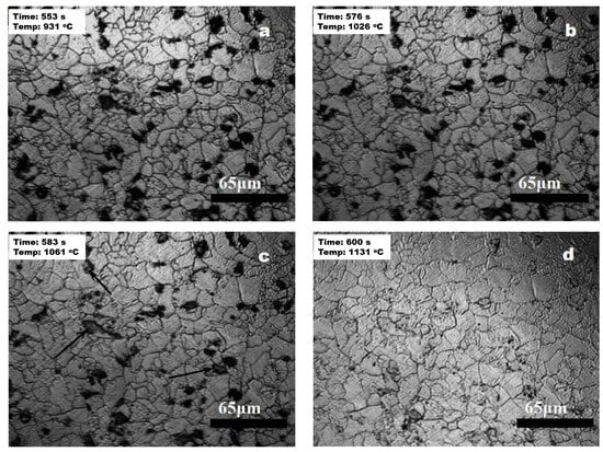
Figure 5.
Microstructural evolution from 900 °C to 1131 °C: (a) 553 s; (b) 576 s; (c) 583 s; and (d) 600 s.
As the temperature increased from 900 °C to 930 °C, in situ observations indicated that the extent and distribution of the dark phase remained largely unchanged, as shown in Figure 5a. The dark phase remained stable even as the temperature reached 1026 °C. Upon further heating, after 7 s, the temperature rose to 1061 °C, at which point the dark precipitates began to dissolve, with their quantity and size gradually decreasing, as shown in Figure 5c. As the temperature continued to rise, the dark phase continued to dissolve, and after approximately 17 s, no dark precipitates were visible through the in situ observations at a temperature of 1131 °C, as shown in Figure 5d. Beyond this temperature, the microstructure remained essentially unchanged despite further heating. The dissolution of precipitates at high temperatures can be explained by the increased solubility of the precipitate-forming elements in the matrix at elevated temperatures. This process is critical for homogenizing the microstructure and eliminating potential sites for crack initiation, thereby enhancing the overall mechanical properties of the material.
These in situ observations provide comprehensive insights into the phase transformations and precipitation behavior of the material at elevated temperatures. Understanding these transformations is essential for optimizing thermal processing parameters, such as heating rates, holding times, and cooling rates, to achieve desired microstructural characteristics and mechanical properties. The real-time data obtained from HTCLSM enables a detailed analysis of the dynamic processes occurring within the material, facilitating the development of advanced materials with tailored properties for specific applications.
3.2. SEM Analysis of Precipitates
Through in situ observations using HTCLSM, it was evident that black precipitates formed during the holding process at 900 °C. These precipitates persisted even when the temperature was increased to above 1000 °C, indicating their thermal stability. The appearance of these precipitates during the heating process likely influenced the mechanical properties of the material. To further investigate the nature and characteristics of these precipitates, a sample subjected to the same heating rate and soaked at 900 °C for 5 min was rapidly cooled to room temperature to preserve the dark precipitated phase particles. Scanning electron microscopy (SEM) and energy-dispersive spectroscopy (EDS) analyses were then conducted on the precipitates, as shown in Figure 6 and Figure 7.
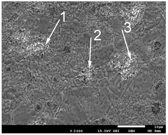
Figure 6.
SEM morphology of in situ observation sample.
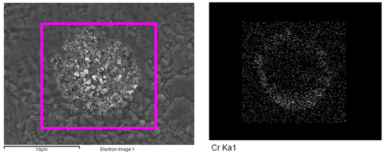
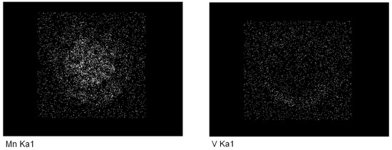
Figure 7.
Mapping analysis of alloying elements in dark precipitated phases.
Figure 6 presents the SEM morphology of the sample, highlighting areas with dark precipitates. The elemental compositions of three specific positions marked by arrows in Figure 6 are detailed in Table 2.
Table 2.
Energy spectrum analysis results (wt%).
In Figure 7, the mapping analysis of the alloying elements in the dark precipitated phases shows areas similar to those marked by arrows in Figure 6, which contain dark particles. Initially, the precipitates appeared circular, but as they grew, their shapes became irregular. EDS was employed to analyze the composition of these precipitated phases. The chemical compositions of the three positions indicated in Figure 6 reveal that the dark precipitated phases contained significantly higher concentrations of alloying elements such as Cr, V, and Mn compared to the matrix. This suggests that the precipitation of the dark phases may be due to the segregation of these alloying elements during the thermal process.
The presence of precipitates, especially those enriched with alloying elements like Cr, V, and Mn, can have profound effects on the mechanical properties of the material. These elements can influence the hardness, strength, and toughness of steel. For instance, vanadium and chromium are known to form carbides that can enhance the hardness and wear resistance of the material. Manganese can improve hardenability and tensile strength. The thermal stability of these precipitates up to temperatures above 1000 °C suggests that they play a significant role in the high-temperature performance of the material. The segregation and subsequent precipitation of alloying elements at elevated temperatures could lead to the localized strengthening or weakening of the microstructure, depending on the nature and distribution of the precipitates.
3.3. Determination of Precipitate Composition
After in situ observation at 900 °C, a 3-mm layer was removed from the surface of the sample, followed by grinding and polishing. The sample was then etched with a 5% nitric acid alcohol solution, and the carbides precipitated during the 900 °C in-situ observation process were examined using Scanning Electron Microscopy (SEM). The results are shown in Figure 8. From Figure 8, it can be seen that carbides precipitated during the in-situ observation are present along the grain boundaries and subgrain boundaries, as well as within the grains.
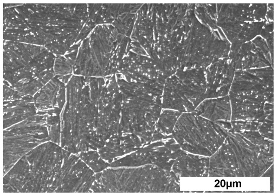
Figure 8.
Microstructure of precipitates from the in situ observation sample.
To identify the types of precipitates formed during the heating process, transmission electron microscopy (TEM) was employed to analyze the samples. The results are shown in Figure 9.
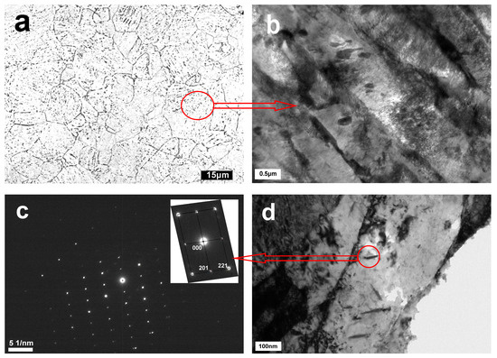
Figure 9.
Microstructure at the inner surface of the specimen: (a) optical micrograph; (b,d) TEM micrograph; and (c) diffraction pattern.
From Figure 9, it is evident that precipitates are formed and distributed within the grains, along the grain boundaries, and at the martensite lath boundaries, as illustrated in Figure 9a. Figure 9b,d show TEM micrographs where the precipitation particles are clearly visible. These images indicate that the particles tend to form at the boundaries and dislocations, which act as nucleation sites for the precipitates. The diffraction pattern in Figure 9c confirms that the particles are chromium carbides, specifically Cr7C3. The presence of Cr7C3, which is significantly harder than ordinary cementite, can substantially increase the steel’s strength. This is due to the fact that Cr7C3 carbides have a higher hardness and wear resistance, providing enhanced reinforcement to the steel matrix. Additionally, Cr7C3 precipitates impede dislocation movement, thereby increasing the material’s yield strength and hardness.
The precipitation and dissolution processes of Cr7C3 have a significant impact on the mechanical properties of steel [27]. Figure 10 illustrates the effect of temperature on the diffusion coefficient of Cr7C3 in steel.
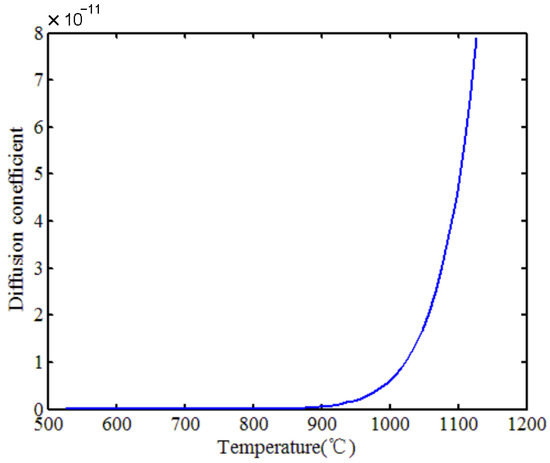
Figure 10.
Diffusion coefficient curve of Cr7C3 carbide.
The variation in the diffusion coefficient of Cr7C3 aligns with the in situ observation results presented in this paper. As seen from the diffusion coefficient curve, when the temperature exceeded 1000 °C, the diffusion coefficient increased by at least two orders of magnitude. This explains why, at 1026 °C, the particles began to dissolve into the matrix and completely dissolved at around 1131 °C, corroborating the previous findings. The diffusion behavior of Cr7C3, with a marked increase in the diffusion coefficient above 1000 °C, highlights the dynamic nature of these precipitates during thermal processing. Understanding this behavior is crucial for optimizing heat-treatment protocols to control the distribution and size of Cr7C3 precipitates, thereby tailoring the material’s properties to specific applications.
The identification of chromium carbides (Cr7C3) as the primary precipitates provides significant insights into the thermal stability and mechanical enhancement of high-nickel steel [28]. The formation of Cr7C3 at grain boundaries and dislocations suggests a strong interaction between these precipitates and the microstructural features of steel, contributing to the observed increase in strength.
In conclusion, the TEM analysis provided a comprehensive understanding of the precipitate composition and its implications for the mechanical properties of high-nickel steel. The formation of Cr7C3 precipitates and their interaction with the microstructure play a pivotal role in enhancing the mechanical properties of the material. These findings contribute to the broader knowledge required for developing advanced materials with superior performance, facilitating the design of more robust and reliable engineering solutions.
4. Conclusions
This research significantly advances the understanding of microstructural transformations in high-nickel steels and demonstrates the critical role of advanced microscopy techniques in material science. The comprehensive insights gained from HTCLSM, SEM, EDS, and TEM analyses provide a robust framework for optimizing the heat-treatment processes of high-nickel shipbuilding steel. The key findings are summarized as follows:
(1) In situ observations indicated that austenitization began at approximately 700 °C, with nucleation primarily occurring at grain boundaries. The austenitization process concluded at around 740 °C, leading to substantial microstructural changes.
(2) At 900 °C, the formation of black precipitates was observed, which persisted even when the temperature exceeded 1000 °C. These precipitates were identified as chromium carbides (Cr7C3) through TEM analyses. The Cr7C3 precipitates contribute to the material’s overall mechanical strength due to their hard, dispersed nature.
(3) During the in situ observation, Cr7C3 precipitates began to dissolve into the matrix at temperatures above 1026 °C, with complete dissolution occurring at around 1131 °C.
Author Contributions
Conceptualization, G.S.; data curation, G.S. and Q.W.; formal analysis, G.S., Z.L., S.Z., and Q.W.; investigation, G.S., S.Z., and Q.W.; methodology, G.S., S.Z., and Q.W.; supervision, G.S. and Z.L.; writing—original draft, G.S.; writing—review and editing, G.S. and S.Z. All authors have read and agreed to the published version of the manuscript.
Funding
The authors acknowledge financial support from the Kunlun Talent Project of Qinghai Province (2023-QLGKLYCZX-022).
Data Availability Statement
The data presented in this study are available only upon request from the corresponding author due to privacy concerns.
Conflicts of Interest
The authors declare that they have no known competing financial interests or personal relationships that could have appeared to influence the work reported in this paper.
References
- Okuda, K.; Yamamitsu, K.; Xu, X.; Miyamoto, G. Change in NbC Precipitates during Cold Rolling of Nb-added IF Steel Sheet. Tetsu Hagane 2023, 109, 323–332. [Google Scholar] [CrossRef]
- Zhao, B.; Ai, F.; Yuan, Q.; Wu, H.; Cheng, J.; Wang, S.; Xie, D. Effect of Normalizing on Impact and Corrosion Resistance of Low-temperature Service Seamless Steel Pipe. J. Phys. Conf. Ser. 2023, 2463, 012011. [Google Scholar] [CrossRef]
- Huang, S.; Li, C.; Li, Z.; Zhuang, C.; Zeng, Z.; Wang, J. Effect of Thermal Simulation Process on Microstructure of Seismic Steel Bars. Materials 2022, 15, 3438. [Google Scholar] [CrossRef]
- Isobe, K.; Kumagai, Y.; Satou, T. Effects of Cooling Conditions and γ Grain Size on Each Behavior of Transformations, 3-Dimensional Thermal Deformation and Stress Generation during Immersion Cooling of Steel Bloom. ISIJ Int. 2023, 63, 919–929. [Google Scholar] [CrossRef]
- Napal, U.G.; Segarra, M.A.; Lazcano, B.E.; Sivo, A. Thermal Mass Effect on the Solution Cooling Rate and on HIPped Astroloy Component Properties. Materials 2022, 15, 1434. [Google Scholar] [CrossRef]
- Yan, M.; Sun, J.; Huang, H.; Chen, L.; Dong, K.; Chen, Z. Effect of hot rolling and cooling process on microstructure and properties of 2205/Q235 clad plate. J. Iron Steel Res. Int. 2018, 25, 1113–1122. [Google Scholar] [CrossRef]
- Kuziak, R.; Kania, Z.; Pidvysots, V.; Roelofs, H.; Pernach, M.; Pietrzyk, M. Through Process Modelling of Rolling and Controlled Cooling of TRIP Assisted Bainitic Steel Rods and Prediction of the Retained Austenite in Products. Mater. Sci. Forum 2017, 892, 23–33. [Google Scholar] [CrossRef]
- Liu, G.; Liu, J.; Zhang, J.; Zhang, M.; Feng, Y. Microstructure Evolution and Mechanical Properties of Medium Carbon Martensitic Steel during Warm Rolling and Annealing Process. Materials 2021, 14, 6900. [Google Scholar] [CrossRef]
- Li, X.; Zhou, X.; Jiang, Q.; Liu, Z. The Prediction of the Mechanical Properties for Hot-Rolled Nb Micro-Alloyed Dual-Phase Steel Based on Microstructure Characteristics. JOM 2023, 75, 2225–2234. [Google Scholar] [CrossRef]
- Dige, I.; Nilsson, H.; Kilian, M.; Nyvad, B. In situ identification of streptococci and other bacteria in initial dental biofilm by confocal laser scanning microscopy and fluorescence in situ hybridization. Eur. J. Oral Sci. 2010, 115, 459–467. [Google Scholar] [CrossRef]
- Reid, M.; Phelan, D.; Dippenaar, R. Concentric Solidification for High Temperature Laser Scanning Confocal Microscopy. ISIJ Int. 2004, 44, 565–572. [Google Scholar] [CrossRef]
- Griesser, S.; Dippenaar, R. Enhanced Concentric Solidification Technique for High-Temperature Laser-Scanning Confocal Microscopy. ISIJ Int. 2014, 54, 533–535. [Google Scholar] [CrossRef]
- Chen, R.; Zheng, Z.; Li, N.; Li, J.; Feng, F. In-situ investigation of phase transformation behaviors of 300M steel in continuous cooling process. Mater. Charact. 2018, 144, 400–410. [Google Scholar] [CrossRef]
- Klein, T.; Niknafs, S.; Dippenaar, R.; Clemens, H.; Mayer, S. High Temperature Laser-Scanning Confocal Microscopy for the in-situ Investigation of Grain Growth and Phase Transformations in Intermetallic γ-TiAl based Alloys. Prakt. Metallogr. 2015, 52, 259–269. [Google Scholar] [CrossRef]
- Chikama, H.; Shibata, H.; Emi, T.; Suzuki, M. “In-situ” Real Time Observation of Planar to Cellular and Cellular to Dendritic Transition of Crystals Growing in Fe–C Alloy Melts. Mater. Trans. 2007, 37, 620–626. [Google Scholar] [CrossRef]
- Shibata, H.; Arai, Y.; Suzuki, M.; Emi, T. Kinetics of peritectic reaction and transformation in Fe-C alloys. Metall. Mater. Trans. B 2000, 31, 981–991. [Google Scholar] [CrossRef]
- Yin, H.; Emi, T.; Shibata, H. Morphological instability of δ-ferrite/γ-austenite interphase boundary in low carbon steels. Acta Mater. 1999, 47, 1523–1535. [Google Scholar] [CrossRef]
- Tian, J.; Xu, G.; Jiang, Z.; Hu, H.; Yuan, Q.; Wan, X. In-Situ Observation of Martensitic Transformation in a Fe-C-Mn-Si Bainitic Steel During Austempering. Met. Mater. Int. 2020, 26, 961–972. [Google Scholar] [CrossRef]
- Cejka, J.; Michelic, S.K. In-Situ Observation of Steel/Slag/Inclusion Interaction by Means of High-Temperature Confocal Scanning Laser Microscopy. Metals 2023, 13, 686. [Google Scholar] [CrossRef]
- Mu, W.; Hedström, P.; Shibata, H.; Jönsson, P.G.; Nakajima, K. High-Temperature Confocal Laser Scanning Microscopy Studies of Ferrite Formation in Inclusion-Engineered Steels: A Review. JOM 2018, 70, 2283–2295. [Google Scholar] [CrossRef]
- Ryttberg, K.; Wedel, M.K.; Dahlman, P.; Nyborg, L. Microstructural evolution during fracture induced by high strain rate deformation of 100Cr6 steel. J. Mater. Process. Technol. 2009, 209, 3325–3334. [Google Scholar] [CrossRef]
- Kozmel, T.; Tin, S. Effects of Carbides on the Microstructural Evolution in Sub-micron Grain 9310 Steel During Isothermal Heat Treatment. Metall. Mater. Trans. A 2015, 46, 3208–3219. [Google Scholar] [CrossRef]
- Wang, B.; Xu, Y.; Chen, L.; Zhu, Z.; Qiu, F.; Chang, F.; Dong, B.; Barber, G.C. Insights into the microstructure evolution and wear resistance of Nano-TiC particles reinforced High-Cr hot work die steel. J. Mater. Res. Technol. 2024, 30, 8371–8381. [Google Scholar] [CrossRef]
- Benarrache, S.; Benchatti, T.; Benhorma, H. Formation and Dissolution of Carbides and Nitrides in the Weld Seam of X70 Steel by the Effects of Heat Treatments. Ann. Chim. Sci. Matériaux 2019, 43, 11–16. [Google Scholar] [CrossRef]
- Sai Gautam, G.; Hari Kumar, K.C. Elastic, thermochemical and thermophysical properties of rock salt-type transition metal carbides and nitrides: A first principles study. J. Alloys Compd. 2014, 587, 380–386. [Google Scholar] [CrossRef]
- Gorbachev, I.I.; Popov, V.V. Analysis of the solubility of carbides, nitrides, and carbonitrides in steels using methods of computer thermodynamics: IV. Solubility of carbides, nitrides, and carbonitrides in the Fe-Nb-C, Fe-Nb-N, and Fe-Nb-C-N systems. Phys. Met. Metallogr. 2010, 110, 52–61. [Google Scholar] [CrossRef]
- Bowen, A.W.; Leak, G.M. Solute diffusion in alpha- and gamma-iron. Metall. Trans. 1970, 1, 1695–1700. [Google Scholar] [CrossRef]
- Sun, L.; Ji, X.; Zhao, L.; Zhai, W.; Xu, L.; Dong, H.; Liu, Y.; Peng, J. First Principles Investigation of Binary Chromium Carbides Cr7C3, Cr3C2 and Cr23C6: Electronic Structures, Mechanical Properties and Thermodynamic Properties under Pressure. Materials 2022, 15, 558. [Google Scholar] [CrossRef]
Disclaimer/Publisher’s Note: The statements, opinions and data contained in all publications are solely those of the individual author(s) and contributor(s) and not of MDPI and/or the editor(s). MDPI and/or the editor(s) disclaim responsibility for any injury to people or property resulting from any ideas, methods, instructions or products referred to in the content. |
© 2024 by the authors. Licensee MDPI, Basel, Switzerland. This article is an open access article distributed under the terms and conditions of the Creative Commons Attribution (CC BY) license (https://creativecommons.org/licenses/by/4.0/).