Article

The Noise: A Silent Threat to the Recovery of Patients in Neonatal Intensive Care Units

by
Víctor M. Rodríguez-Montaño
1,*,
Virginia Puyana-Romero
2,
Ricardo Hernández-Molina
1 and
Juan L. Beira-Jiménez
1
1
Acoustic Engineering Laboratory, Universidad de Cádiz, 11002 Cadiz, Spain
2
Department of Sound and Acoustic Engineering, Faculty of Engineering and Applied Sciences, Universidad de las Américas, Quito 170125, Ecuador
*
Author to whom correspondence should be addressed.
Buildings 2024, 14(9), 2778; https://doi.org/10.3390/buildings14092778
Submission received: 29 June 2024 / Revised: 16 August 2024 / Accepted: 2 September 2024 / Published: 4 September 2024
(This article belongs to the Special Issue Advanced Research on Improvement of the Indoor Acoustic Environment)

Abstract

:
The architectural configuration of the space plays a crucial role in the acoustics of neonatal intensive care units (NICUs). The design of the environment, the materials used in construction, and the organisation of noise sources within the room significantly influence the noise levels present in these critical areas. For this research, the noise levels found in two hospitals, with different architectural configurations and sizes but similar construction materials, have been analysed. Data were recorded at 1 s intervals over a period of more than 24 h. The data collected in these hospitals confirm the magnitude of the noise problem in NICUs, highlighting the urgent need to address it effectively to ensure an appropriate and safe environment for the recovery of neonates. Good architectural planning can help mitigate noise, while poor configuration can exacerbate acoustic problems, negatively affecting both patients and healthcare staff. It is crucial to involve architects, acoustic engineers, and healthcare staff in the design and renovation of NICUs to create spaces that not only meet medical needs but also consider the auditory well-being of neonates.

1. Introduction

The environment of a neonatal intensive care unit (NICU) is crucial for the recovery of newborn patients. However, noise is a widespread problem that might be slowing down their healing process. While excessive noise is harmful to everyone, it is especially damaging to sick patients in the NICU. This is particularly true for premature babies, whose developing nervous and auditory systems make them very sensitive to loud sounds [1].

1.1. Effects of Noise on Premature Neonates

Historically, newborns were assumed to be desensitised to their environment and lacked the ability to interact with it in a meaningful way [2]. However, recent research has shown that infants actively respond to their environment, using their sensory experiences as a platform for learning [3]. For example, they respond to touch, can differentiate and show preference for certain stimuli, and are able to protect themselves from the environment by moving away from painful stimuli [4]. These responses may manifest in subtle variations in skin colour, fluctuations in heart rate or breathing, and signs of startles or tremors [5], indicating that environmental stimuli, such as noise, light, and care events, may be overwhelming [6]. This knowledge has led to focusing attention on the management of sensory stimuli within the NICU, as these can have a crucial impact on neonatal development.
The care of the sick newborn is influenced by the physiological changes that accompany the transition from intrauterine to extrauterine life. This process involves the activation of complex homeostatic mechanisms and the maturation of organs and systems that are essential for survival outside the womb. This period of adaptation, characterised by being highly dynamic, is the most vulnerable in human life, as there is a higher risk of severe illness or sequelae, especially neurological ones [7].
The premature infant faces a radically different environment from that of the maternal womb, where brain growth is faster [6]. While in the womb, the foetus receives complete maternal protection, nutrients, and temperature regulation [8]; the premature baby is placed in a NICU with highly variable conditions and less protection. Research has shown that the NICU environment offers very different sensory stimuli compared to those of a full-term newborn, exposing babies, parents, and staff to constant negative and unpredictable stimuli [2,9,10,11]. This discrepancy between the intrauterine environment and the NICU setting presents significant challenges for the physiological, emotional, and social development of the premature newborn [8,12,13].
Sensory development in neonates follows a specific sequence during foetal life, where each sensory system matures in an orderly fashion, avoiding competition with other emerging systems [14]. For example, foetal hearing develops intensely, while vision is still inactive. Animal studies have shown that premature stimulation of one sensory system can interfere with the development of the ongoing system and other sensory systems [6,15,16]. In premature babies, continuous exposure to stimuli in the NICU may cause hearing and vision to develop simultaneously rather than sequentially, disrupting perceptual and behavioural development in a manner similar to how inadequate stimulation affects sensory systems in animals [6].
Many of the problems that primarily affect premature neonates or those with severe medical conditions often stem from failures in their adaptation process [7]. This situation is further exacerbated by the fact that these newborns may remain in the NICU for weeks or even months, where noise is ever present, adding further risks to an already complex scenario and making the neonate even more unstable and dependent [17].
Prolonged exposure to high levels of noise can interfere with auditory and emotional development, as well as increase the risk of sleep disorders and stress [17,18,19,20]. Stress reactions in newborns, such as bradycardia, tachycardia, increased systolic and diastolic blood pressure, apnea, hypoxemia, alterations in oxygen saturation, increased oxygen consumption, and elevated intracranial pressure, can increase the risk of problems such as growth delay, irregular sleep patterns, hearing loss, bronchopulmonary dysplasia, retinopathy of prematurity, intraventricular haemorrhage, periventricular leukomalacia, and developmental delay [21].
It has been observed that the electrical activity of the central nervous system varies with sounds ranging from 36 dB to 90 dB, showing increases in intracranial pressure and changes in electromyographic and behavioural responses to sudden noises. Excessive noise levels can impact the neuroendocrine system and indirectly affect immunity [22,23].
Likewise, sleep deprivation has a great impact on various physiological parameters, such as bradycardia, apnoea, increased hypoxic periods or increased intracranial pressure, which can negatively influence the recovery of patients under care [24,25]. Deep sleep plays an essential role in the maturation of brain functions in newborns, and its interruption can cause alterations in thermoregulation and the production and release of certain hormones, in addition to compromising the newborn’s immunity [17]. These effects can have significant consequences on long-term development, including cognitive ability and academic performance [26].
Although childhood hearing impairment results from a combination of genetic predisposition and environmental factors [27,28], the global prevalence of hearing loss is approximately 1 to 3 per 1000 neonates, which is significantly higher in neonates admitted to the NICU, ranging from 3 to 6 per 100 [29,30]. Additionally, it has been shown that noise, hypoxia, apnea, and hyperbilirubinemia can interact together to contribute to the development of hearing impairment [27,31].
In addition to the impact on patient care, constant noise in the NICU also affects the health and well-being of nursing staff. Studies have shown that prolonged exposure to noise can cause hearing fatigue, increase blood pressure and contribute to the development of cardiovascular diseases and psychological disorders, such as anxiety and depression, difficulty communicating, work stress, decreased concentration, and increased risk from making mistakes, which in turn, can negatively affect the quality of care they provide [21]. In the highly specialised environment of the NICU, where each care decision is decisive for the well-being of newborns, this situation creates a negative cycle in which both newborns and nursing staff are affected by the same problem.

1.2. Noise Levels in Neonatal Intensive Care Units

In addition to the direct impact on neonates, it is crucial to understand how various noise sources in the NICU affect the overall environment and dynamics of neonatal care. The noise level in the NICU is a determining factor in staff communication, family interactions, and child development. According to the American Academy of Pediatrics (AAP), exposure to noise levels above 45 dBA can cause cochlear damage and even disrupt the normal growth and development of the neonate [32,33]. Although the human ear may get used to high levels, no longer perceiving them as high, noise may continue generating damage [34].
For this reason, the AAP recommends that neonatal care areas should incorporate sound-absorption materials or other measures to ensure that the combination of continuous background noise and transient sounds does not exceed a Leq,1 h of 45 dBA, an L10 of 50 dBA and an Lmax of 65 dBA [33,35].
Over the years, multiple studies have highlighted the persistent problem of noise in NICUs [11,36,37,38,39,40,41]. Despite various efforts to mitigate this noise, as documented in the specialised literature [42,43,44,45,46,47,48], recent research shows that noise continues to be a significant and unresolved challenge in these environments.
Studies indicate that long-term average noise levels in NICUs range from approximately 50 to 65 dBA, depending on the unit assessed [38,39,40,41,42,43,44,45,46,47,48,49,50]. This noise comes from various structural and operational sources. Structural sources include HVAC systems, door openings and closings, and the hum generated by electrical equipment, such as computers and incubators. Operational sources encompass staff and visitor conversations, chair movement, the opening of disposable containers, medical equipment alarms, phones, and baby crying [17,39,42,50,51].
It is known that noise levels in NICUs can reach particularly high peaks [37,52,53,54], especially during shift changes, which have been identified as the noisiest times [37,52]. Among the various sources of noise, NICU staff contribute significantly to the maximum noise level (Lmax). Although staff conversations do not substantially affect the overall LAeq, the noise generated by them is recorded 67% of the time observed in studies [39], making it the primary source of noise in these units.
The acoustic environment of NICUs is characterised by elevated noise levels, with loud, short-duration events occurring irregularly. Medical device alarms, in particular, have consistently been identified as a significant source of excessive noise, with an average of 177 alarms per patient per day recorded [37,39,55].
Regarding the spectral content of noise in NICUs, low frequencies predominate [36,56,57], accompanied by some mid and high frequencies [58]. The latter are typically generated by equipment alarms and human voices [39]. This acoustic pattern underscores the complexity of controlling noise in such a delicate environment as the NICU. Although it is estimated that around 60% of these noises could be reduced or eliminated [42], the specific effects of these frequencies, especially the low ones, on newborns are not yet fully understood and require further research.

1.3. Noise Levels in Incubators

The choice between placing a baby in an incubator or a warming crib depends on a careful assessment of their individual medical needs. According to the guidelines of the AAP, incubators are essential for the care of premature neonates, as they create an optimal environment for their healthy growth and development, precisely regulating temperature, humidity, and oxygen [59]. On the other hand, thermal cribs, although they offer radiant heat, are more suitable for stable babies, since they do not need the same intensive supervision as premature babies [60].
Incubators, like the womb, could protect maturing auditory systems from harmful noise levels [49,61]. Such protection could alleviate concerns about overstimulation in the NICU, as long as the baby remains inside a closed incubator. The walls of these incubators have demonstrated the ability to reduce sounds coming from the neonatal room by up to 10 dBA [17,62,63]. However, according to several authors, incubators generate an internal background noise level exceeding 52 dBA throughout the measurement period [37,38,56,61,62,63,64], significantly surpassing the recommendations established by the AAP. According to the sources consulted, this background noise level may be even higher depending on the incubator model [62,63]. This level of background noise is generated by the incubator’s internal motor, responsible for regulating the temperature and humidity inside. This situation raises an additional concern about exposing neonates to potentially harmful noise levels even when inside an incubator.
This 10 dBA attenuation implies that, when the noise level in the NICU exceeds the levels emitted by the internal motor of the incubator, the attenuation provided by the walls of the incubator reduces the dose of noise that the neonate receives inside. For the noise level to increase inside the incubator, the ambient noise in the NICU must be at least 3 dBA higher than the motor noise plus motor attenuation [64].
In relation to the existing noise levels inside the incubators, the Acoustic Engineering laboratory of the University of Cadiz has developed a prototype of an incubator with a system to improve the acoustic comfort of the NICA+ neonate. This prototype, presented in the form of patent “OEPM P202330766”, includes a neonatal incubator equipped with a system designed to improve the acoustic comfort of the newborn with sound levels lower than international recommendations [65].
The acoustic environment to which the newborn is exposed is different if it is housed in an incubator or a warming crib. In contrast to neonates found in incubators, it is relevant to highlight that those housed in thermal cribs do not have the same level of noise attenuation. This means that they are more exposed to the sounds produced in the neonatal room.

1.4. Noise Reduction in NICUs

Considering these facts, we can affirm that the incubator or thermal crib, together with the NICU, constitute an interconnected system in which the ambient noise of the room directly affects the neonates housed in the incubators. This relationship highlights the importance of appropriately managing the noise level in the neonatal environment to protect the health and development of newborns, as well as to ensure an environment that is conducive to their recovery.
This does not mean that neonatal rooms should be totally silent, since it should not be forgotten that exposure to sound is necessary for the neonate and provides constant sensory stimulation [66,67]. This early exposure to different sounds in the neonatal environment plays a critical role in language acquisition and the development of language skills [68].
To address the issue of noise in NICUs, various approaches have been implemented. These include the use of hearing-protection devices [68,69], staff retraining [70,71], and architectural renovation, which ranges from updating existing NICUs to constructing new facilities with individual rooms for each patient [10,42,43,44,45,46,47,48]. Each of these methods has its own characteristics in terms of complexity, effectiveness, and cost. In particular, architectural renovations often present significant challenges due to the complexity of design, the necessary adjustments in workflow, and the high associated costs.
In relation to NICU design, several studies have compared noise levels in units designed with individual rooms for each family versus traditional open units [10,46,47,48]. These studies have found that single-incubator rooms had significantly lower noise levels compared to open units due to less interference from sounds from other incubators and medical equipment, which not only reduces noise but also improves the perception of care by parents and the well-being of staff. That is, open-plan units tend to have higher noise levels due to greater exposure to noise generated by staff and equipment, while small modules help mitigate the acoustic impact [10].
However, it must be noted that, while the implementation of individual rooms has contributed to a reduction in average noise levels, these improvements have been limited, as noise levels still exceed current recommendations [37].
The design of NICUs with individual rooms has proven to have advantages over traditional open-plan models. These benefits include improved respiratory regulation, reduced episodes of apnea, and more positive growth trajectories and neurobehavioral scores, which may imply a lower long-term risk for neonates [47,48].
However, there is ongoing concern about the potential negative impact on the language development of premature newborns in individual rooms compared to those in open-plan units. This issue may be particularly relevant for neonates who receive limited support for their development and maternal interaction during their NICU stay [45,67,72]. The emerging hypothesis suggests that reduced exposure to linguistic stimuli in quieter individual rooms could contribute to delays in language development, in contrast to the more active and communicative environment of open-plan NICUs, where neonates are exposed to a greater amount of conversation and ambient sounds, even in the absence of their parents or clinical caregivers [72].
Taking this premise into account, no studies have been found that compare noise levels in two open neonatal rooms with different architectural designs and volumes. Most studies focus on comparing open-design NICUs with those using single rooms, highlighting the advantages of the latter in terms of noise reduction and better environmental control. However, research specifically comparing open rooms with different architectural configurations and volumes appears to be limited or nonexistent. This lack of studies highlights the need to further investigate how variations in the design and size of open rooms can influence noise levels and ultimately the health and development of neonates.
Each country has its own standards and/or recommendations regarding noise levels and construction systems in NICUs, all based on guidelines established by the AAP [33,35]. In Spain, it is essential to consider Law 37/2007 [73], Royal Decree 1367/2007 [74], and the Technical Building Code CTE DB-HR [75], which establish provisions related to acoustic zoning, quality objectives, and acoustic emissions. According to these regulations, noise levels inside hospital rooms should be below 40 dBA during the day (7:00 a.m.–7:00 p.m.) and evening (7:00 p.m.–11:00 p.m.), and below 30 dBA during the night (11:00 p.m.–7:00 a.m.) [74]. These limits refer to the values of the immission index resulting from the set of acoustic emitters that affect the interior of the enclosure.
The objective of the present study is to compare the noise levels generated in two neonatal intensive care unit rooms of different volumes and geometries, to evaluate whether there are notable differences between the two rooms. The comparison will be made by evaluating both global noise levels, weighted and unweighted, and levels in third-octave bands. To obtain data, continuous measurements were carried out in both rooms with the incubators running, during day and night periods, paying special attention to feeding times and shift changes.

2. Methodology

To carry out this study, data were collected and analysed in the neonatal intensive care units of two leading hospitals in Spain. These centres are the Puerta del Mar University Hospital (HUPM), located in the city of Cadiz, and the Juan Ramón Jiménez University Hospital (HJRJ), located in Huelva. Both institutions are recognised for their commitment to excellence in neonatal care and have advanced medical technologies for the intensive care of newborns. The data obtained covers several key variables, such as ambient noise levels, the layout of medical equipment and the general conditions of the rooms, allowing a comprehensive evaluation of the factors that influence the acoustic environment of the NICU. This comprehensive approach provides a solid foundation for identifying areas of improvement and developing effective strategies to optimise neonatal care.
This research has received approval from the Provincial Research Ethics Committee of Cadiz under registration number 82.22 and PEIBA code 1253-N-22. It is important to highlight that this study is not classified as a biomedical research project according to the terms established by Spanish Law 14/2007, of 3 July, on Biomedical Research [76]. Ethical approval ensures that the research complies with the necessary ethical regulations and guidelines, guaranteeing the protection of participants and the integrity of the data collection process.

2.1. Material and Methods

Before installing the sound-level metre, noise levels were sampled throughout the room. According to the UNE-ISO 1996-2:2020 standard, “Acoustics. Description, measurement and assessment of environmental noise. Part 2: Determination of sound pressure levels” [77], the noise measurements were carried out following specific procedures to ensure accuracy and avoid distortions. Specifically, the measurements were not conducted following a fixed grid to avoid nodal points of wavelength that could alter the results.
The measurement process involved taking noise readings for a period of 3 min at each selected point. If a high-intensity transient event, such as staff activity or alarm activation, was recorded during this time, the measurements were repeated at the same point to ensure the reliability of the obtained data.
Following a preliminary analysis of the collected data, it was found that the noise levels in both rooms were similar throughout the space, indicating a predominantly diffuse acoustic field. This suggests that sound is distributed uniformly across the rooms, which is a crucial factor to consider when evaluating the impact of any acoustic intervention.
To characterise the NICUs and assess the noise levels to which neonates might be exposed, a sound-level metre was installed in each neonatal room. Given that the NICU itself is a diffuse field, it was decided to place the sound-level metre in a position that would not interfere with the activities of the healthcare staff. Thus, the device was strategically located between two incubators, at a distance of 1.5 m from the nearest wall and 1 m from the ceiling. The measurement point was symmetrically placed 1 m from each of the two incubators. While it is true that alarms or the activities of the nursing staff may have a more pronounced effect at this point than at other, more distant, locations in the room, this positioning of the sound-level metre allows us to focus on understanding the noise exposure experienced by the neonates.
To carry out these measurements, the HBK 2270 sound-level metre (Hottinger Brüel & Kjaer, Virum, Denmark) was used, which meets the type 1 instrumentation requirements according to the regulations established by the International Electrotechnical Commission, specifically the EN-IEC 61672:2013 standard [78]. In order to guarantee the precision and accuracy of the sound-level metres, the HBK 4231 (Hottinger Brüel & Kjaer, Virum, Denmark) sound calibrator was used to verify their correct operation.
Two non-consecutive days were randomly selected for the measurements to ensure the representativeness of the obtained data, regardless of the day of the week. These days were agreed upon with the NICUs, primarily depending on the permissions granted by the hospital centres.
The measurements were conducted with a sampling interval of 1 s over a 24 h period. The main parameters collected included continuous equivalent, A- and C-weighted, and broadband unweighted sound pressure levels (LAeq, LCeq, and LZeq). The rest of the parameters are the maximum and minimum levels weighted by response (LAFmax and LAFmin, respectively), the levels weighted by impulse (LAIeq), and the percentiles LA10, LA50, and LA90 (the meaning of the acoustic terms used in this study are shown in Appendix A).
Subsequently, the data obtained were downloaded from the sound-level metre and analysed using the HBK 4231 software (Evaluator Type 7820, version 4.16.8) (Hottinger Brüel & Kjaer, Virum, Denmark), which facilitated a thorough analysis of the collected noise logs.
The noise levels are analysed from various perspectives. Initially, the equivalent continuous level (Leq) is considered throughout the 24 h of the day, distinguishing between the levels reached during the daytime period (from 8:00 a.m. to 10:00 p.m.) and the nighttime period (from 10:00 p.m. to 8:00 a.m.). In addition, special attention is paid to key moments in the routine of health personnel, such as shift changes and neonatal feeding schedules, since these moments usually coincide with significant increases in the noise level due to increased activity and noise and increased verbal and operational interactions.
Throughout the day, in both healthcare units, there are three shift changes for the healthcare staff, at 8:00 a.m., another at 3:00 p.m., and the last one at 10:00 p.m. These changes are critical moments lasting approximately 30 min each. Fifteen minutes before the shift change, the relief staff begins to arrive, while the outgoing staff remains in the room until about 15 min after the changeover time, ensuring proper care transitions.
Regarding the feeding of the babies, it takes place every three hours, starting at midnight (00:00 a.m.). The process lasts approximately 15 min, from the moment the feeding pump is activated until the alarm sounds, indicating that the feeding has been completed.
It is noteworthy that, according to the consulted healthcare staff and observations made by the operators who conducted the measurements, the routines of the healthcare staff and the noise-generating operations are consistent daily at the same time intervals, with the aim of not disrupting the feeding and resting habits of the neonates. The nursing staff maintains a constant number on each shift (morning, afternoon, and night) and follows the same care routines during each shift. Additionally, observations made by the operators during the measurements revealed that, in the mornings (from 8:00 a.m. to 3:00 p.m.), there is an increased movement of people. This increase in activity is primarily due to healthcare operations, the movement of equipment and incubators for medical tests, and cleaning and equipment maintenance services.

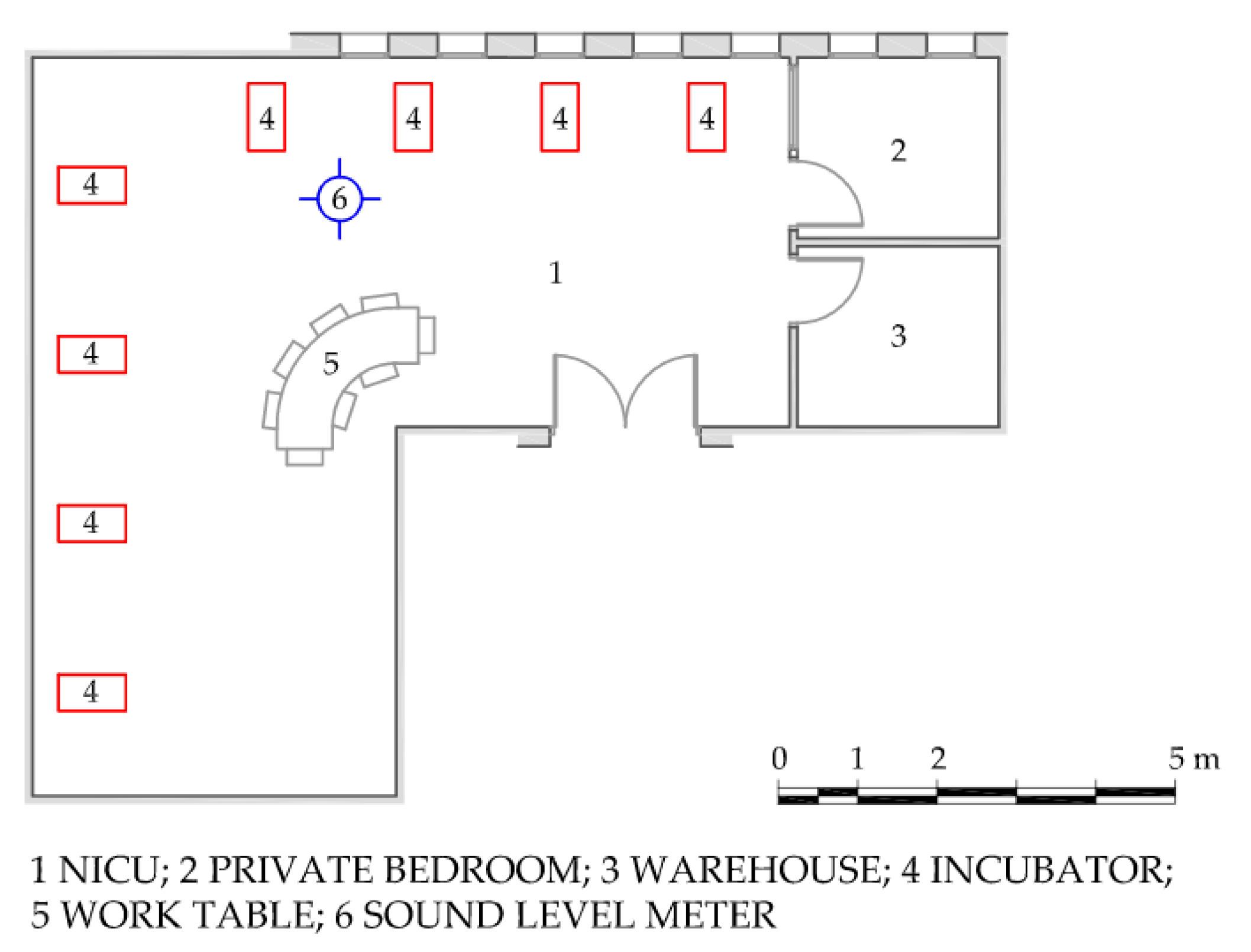
2.2. Features of NICUs

The neonatal rooms analysed in this study exhibit remarkably similar characteristics that significantly influence their acoustic properties. In both cases, the vertical walls are constructed with brick walls rendered with a layer of gypsum paste with an approximate thickness of 1.5 cm. Similarly, the ceilings are made of plaster plates of the same thickness suspended from the structure, leaving an air gap without acoustic insulation, where the installations are located. The flooring is made of terrazzo, making all these surfaces highly reflective in acoustic terms.
Additionally, both rooms feature various glass surfaces, which further amplifies the reflection of sounds in the surrounding environment. The deliberate absence of materials with acoustic absorption capacity is based on the imperative need to maintain strict standards in terms of safety and hygiene, which prevents the use of porous or irregular surfaces for this purpose.
This structural configuration, characterised by a notable sound reverberation, contributes significantly to the increase in noise levels present in these facilities, giving rise to a challenging acoustic environment for the neonates housed in them.

2.2.1. Puerta del Mar University Hospital (HUPM)

The main room of the NICU of this hospital centre has a rectangular distribution, as shown in Figure 1, which covers an area of 156 m2, with a height of 2.66 m, resulting in a total volume of 415 m3. Additionally, this main area is connected to other rooms that are essential for its operation, such as the entrance hall, medicine and other materials stores, and staff changing rooms. Likewise, independent rooms are included specifically for the isolation of neonates in critical condition or with contagious infections. Considering the inclusion of all these annex spaces, the total surface area of the NICU reaches approximately 230 m2.
Up to 10 neonates housed in incubators can be cared for in the main NICU room, with an additional capacity for 3 or 4 more neonates in the adjoining private rooms. Each of these care units is equipped with the necessary monitoring and conditioning devices, as well as the corresponding electromedical equipment, adapted to the individual needs of each neonate, depending on their specific health status.
This research focuses exclusively on the noise levels of the NICU main room, identified in Figure 1 with the number 2.
Regarding the position of the sound-level metre, in both measurements conducted, the device was placed in the same position to ensure comparability. In this case, the sound-level metre was positioned between incubators 3 and 4 (as assigned by the NICU).

2.2.2. Juan Ramón Jiménez University Hospital (HJRJ)

The configuration of the NICU in this hospital centre is an L-shaped configuration, as illustrated in Figure 2. Its main room covers an area of 65 m2, with an approximate height of 2.7 m, resulting in a total volume of 175 m3. In addition to this main room, the NICU has two related spaces, namely a warehouse and an independent room intended for the presence of the mother with the baby in specific cases. Considering all these additional spaces, the total surface area of the NICU is nearly 78 m2.
This research focuses exclusively on the noise levels of the NICU main room, identified in Figure 2 with the number 2. This room, with the capacity to care for 8 neonates housed in incubators, is equipped with the necessary monitoring devices, conditioning and electromedical equipment and adapted to the individual needs of each neonate according to their health status.
As in the previous case, in both measurements conducted, the sound-level metre was placed in the same position to ensure comparability. In this instance, the sound-level metre was positioned between incubators 3 and 4 (as assigned by the NICU).
The graphic documentation of this care unit has been provided by Moisés Fumero Rodríguez, Acoustic Engineering Laboratory staff, and was published as part of his final Master’s thesis in acoustic engineering in 2010 [79].

2.3. Data Analysis

A descriptive and statistical analysis of the data was conducted to assess the noise levels in both NICUs at different times and events over a 24 h period and measured on two non-consecutive days.
For this study, the following hypotheses were formulated:
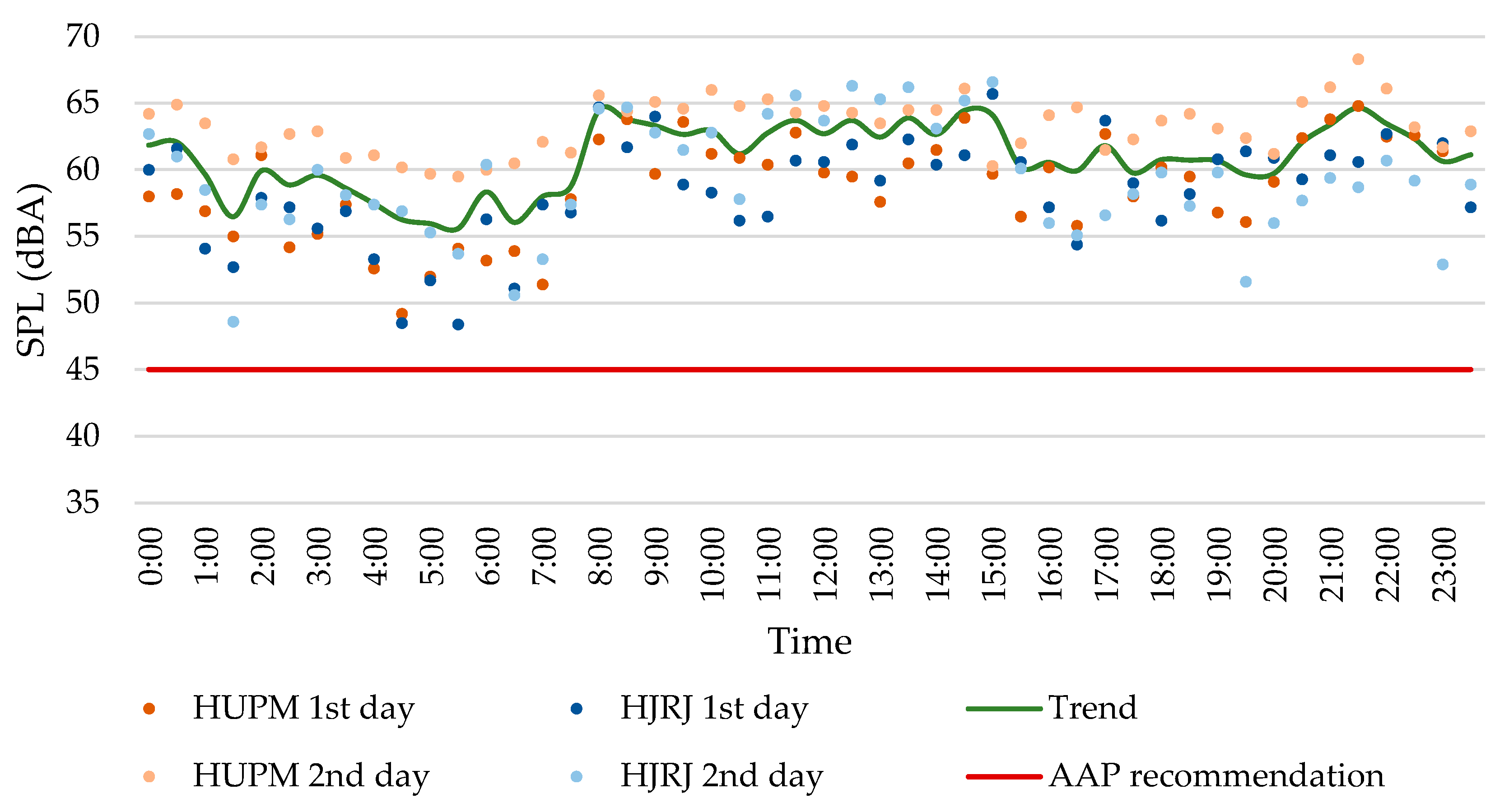
  • H1. There are no differences between the sound pressure levels measured during the day period (8:00 a.m.–10:00 p.m., excluding feeding periods and shift changes) in the NICU of the HJRJ and the HUPM;
  • H2. There are no differences between the sound pressure levels measured during the night period (10:00 p.m.–8:00 a.m.), excluding feeding periods and shift changes) in the NICUs of the HJRJ and the HUPM;
  • H3. There are no differences between the sound pressure levels of the daytime feeding periods (10:00 p.m.–8:00 a.m.) in the NICUs of the HJRJ and the HUPM;
  • H4. There are no differences between the sound pressure levels of the nocturnal feeding periods (8:00 a.m. to 10:00 p.m.) in the NICUs of the HJRJ and the HUPM;
  • H5. There are no differences between the sound pressure levels of the shift changes of healthcare personnel (8:00 a.m. to 10:00 p.m.) in the NICUs of the HJRJ and the HUPM.
After carrying out an initial analysis of the data, we proceeded to verify whether there are significant differences in the noise levels in the NICUs of Huelva and Cadiz. To do this, we first evaluate which are the most appropriate statistical tests for the analysis, depending on the nature of the data. Given the sample size, the normal distribution of the data was evaluated using the Shapiro–Wilk test. Levene’s test for homogeneity of variances was calculated to determine whether the distribution of the data between both samples was similar. The variables analysed were LAeq (dBA), LAIeq (dBA), LAFmáx (dBA), LAFmín (dBA), LCeq (dBC), LA10 (dBA), LA50 (dBA), LA90 (dBA), and the non-weighted frequency range between 40 Hz and 20 kHz (LZeq, in dBA).
According to the results of the previous analyses, it was decided to perform a parametric test, the Mann–Whitney U test, which allows for determining whether there are significant differences between two independent samples. The main objective was to compare the acoustic behaviour of the NICU rooms in Huelva and Cadiz and verify if we can rigorously speak of differences between them in order to answer the research questions. The value of 0.05 was used as a threshold to determine the statistical significance of the results of the tests, and, therefore, to decide whether to accept or reject the null hypothesis.
It will be evaluated whether the aforementioned variables present significant differences during the day (8:00 a.m.–10:00 p.m.) and night (10:00 p.m.–8:00 a.m.) periods, excluding feeding and shift change times; the night and day feeding periods; and the changes shift, which always occur during the day.
Effect size typically indicates the significance of differences between two groups, with a larger effect size meaning a more substantial difference [80]. It is a valuable metric for assessing the effectiveness of results [81]. In this study, the effect size was determined using Rosenthal’s formula for parametric tests [82] by dividing the absolute standardised test statistic z by the square root of the sample size. The interpretation of the results was conducted using Bartz criterion [83] applied to the r absolute values: 0.00 < r < 0.20, “Very low”, 0.20 ≤ r < 0.40, “Low”, 0.40 ≤ r < 0.60, “Moderate”, 0.60 ≤ r < 0.80, “Strong”, 0.80 ≤ r < 1, “Very Strong”.

3. Results

3.1. Descriptive Analysis Result

In the descriptive analysis, to make a detailed comparison with the noise levels recommended by various international entities, such as the AAP, the results will first focus on the equivalent continuous sound level (LAeq) measured over the 24 h period. These data provide a comprehensive view of noise exposure throughout the day and allow for a thorough assessment of the acoustic conditions in the NICUs.
Subsequently, a clear distinction will be made between daytime and nighttime noise levels. This differentiation is essential, as activities and the presence of staff vary significantly between day and night, affecting noise levels differently. Additionally, attention will be given to the noise levels during feeding times and staff shift changes. By comparing these levels, a more precise understanding can be gained of how noise levels fluctuate at different times of the day and how these variations may impact the well-being of neonates.
Next, the L10 levels will be analysed, which represent the noise level exceeded during 10% of the measurement period. This metric is particularly useful for identifying frequent noise peaks that can be disruptive, even if they are not sustained. The analysis of L10 helps in understanding the prevalence of high noise levels and their potential impact on the NICU environment.
Finally, the maximum noise level (Lmax) recorded during the measurement period will be examined. Lmax indicates the highest level of noise exposure and is a critical metric for identifying extreme noise peaks that may occur in the clinical environment. Evaluating Lmax is essential to identify isolated instances of extreme noise that could have a significant impact on neonate health. It is important to note that, while this parameter has been chosen to determine noise peaks, in specific studies analysing the impact of noise on sleep disorders or other biomedical parameters, it would be advisable to use Lmax alongside other acoustic parameters, such as the intermittency index [84].
As a result of the acoustic measurements conducted in the NICUs of HUPM and HJRJ, significant differences in sound pressure levels between the two facilities were observed. On the first day of measurement at HUPM, an equivalent continuous sound level (LAeq) of 60 dBA was recorded over the 24 h period. On the second day, this value increased considerably to 63.8 dBA. In contrast, at HJRJ, the LAeq on the first day was 60.1 dBA and 60.9 dBA on the second day. This indicates greater variability in noise levels at HUPM compared to HJRJ.
Several factors may explain this pronounced difference between the two days at HUPM. One possible explanation is the need to incorporate new equipment for the intensive care of certain neonates. Often, these devices and machines have motors, which can significantly increase the ambient noise levels in the room. Additionally, a substantial increase in communication among healthcare staff to coordinate and manage this new equipment may contribute to higher noise levels. The need to ensure that instructions and information are clearly understood can lead staff to speak more loudly, especially in an already noisy environment. This becomes even more critical during emergencies or when specialised and constant attention is required, thereby increasing the volume of conversations and the overall noise level in the unit.
Another factor contributing to these elevated noise levels is the physical structure of the room. Units with vertical walls and ceilings that lack acoustic absorption tend to reflect sound more, thereby increasing the ambient noise level.
By analysing the nighttime period, from 10:00 p.m. to 8:00 a.m., a slight decrease in noise levels was observed in both neonatal units. At HUPM, the LAeq values were 58.7 dBA and 62.3 dBA on the two measurement days, respectively. At HJRJ, the levels were 58.2 dBA and 58.1 dBA.
This trend of reduced noise during the nighttime period is notable and suggests a decrease in noisy activities and less interaction between healthcare staff and medical equipment at night. However, it is important to highlight the exception observed on the second measurement day at HUPM, where the noise levels increased to 62.3 dBA, contrasting with the general trend of reduction observed.
This nighttime reduction is crucial for the rest and recovery of neonates, although the levels still exceed the thresholds recommended by the international guidelines for intensive care environments. Regarding feeding periods, the average noise levels at HUPM were 60.5 dBA and 64.1 dBA. On the first day of measurement, these values ranged from 64.3 dBA at 3:00 p.m., coinciding with shift changes, to 53.2 dBA at 6:00 a.m. On the second day, the levels were 66.2 dBA at 9:00 p.m. and 60 dBA at 6:00 a.m. At HJRJ, the values on the first day ranged from 65.7 dBA at 3:00 p.m. to 55.6 dBA at 3:00 a.m., while on the second day, they were 66.6 dBA at 3:00 p.m. and 59.4 dBA at 9:00 p.m.
Regarding the shift changes of healthcare personnel, noise levels increase significantly. At HUPM, the LAeq values during these periods reached 63.8 dBA and 65.9 dBA, whereas at HJRJ, they were 64.3 dBA and 63.9 dBA. Specifically, during the 8:00 a.m. shift change, the noise levels at HUPM were 62.3 dBA and 65.6 dBA, compared to 64.8 dBA and 66.1 dBA during the 10:00 p.m. shift change, while the 3:00 p.m. shift change presented an intermediate value of 63.9 dBA. At HJRJ, the LAeq values during the 8:00 a.m. shift changes were 64.7 dBA and 64.6 dBA. At 3:00 p.m., they were 65.1 dBA on both days, and at 10:00 p.m., they were 62.7 dBA and 60.7 dBA.
The aforementioned data, along with Figure 3 depicting the sound pressure levels (SPL) throughout the day in both neonatal intensive care units over the two days of measurement, corroborate a clear trend in noise levels. There is a progressive increase from morning until approximately 3:00 a.m., followed by a decrease towards the afternoon. This pattern suggests diurnal fluctuations in noise levels, possibly influenced by activities and operational changes within the facilities during peak hours and nighttime operations.
As observed in Figure 3, the noise levels recorded in these two units exceed the recommendations established by various international entities, such as the American Academy of Pediatrics, the World Health Organization, and the Spanish Pediatric Association.
Regarding the LA10 levels recorded during the measurements, values of 63.6 dBA and 66.6 dBA were obtained at HUPM on the first and second day, respectively. Conversely, at HJRJ, values of 63.8 dBA and 64.7 dBA were recorded on the same days.
Finally, the maximum level (LAmax) recorded at HUPM was 87.3 dBA and 90.3 dBA on the first and second day, respectively, while at HJRJ, values of 89.2 dBA and 88.4 dBA were recorded on the same days.

3.2. Statistical Analysis Result

The results of the Shapiro–Wilk test show a normal distribution for all the variables analysed. However, Levene’s test shows that there is no equality of variances between all pairs of variables. Therefore, we opted to use a non-parametric test. As the groups of variables are independent, the Mann–Whitney U test was applied.
Hypothesis H1 was formulated to assess whether there were differences in the sound pressure levels measured during the day period in the NICUs of HUPM and HJRJ.
Table 1 shows the results of the Mann–Whitney U test during the day period among the NICUs studied for the values of the variables LAeq (dBA), LAIeq (dBA), LAFmax (dBA), LAFmin (dBA), LCeq (dBC), LA10 (dBA), LA50 (dBA), and LA90 (dBA), and the non-weighted range frequency LZ (dB) between 40 Hz and 20 kHz. There are significant differences between LAeq and LAIeq, LAFmin, LCeq, LA10, LA50, and LA90, and the unweighted ranges between from 40 Hz to 100 Hz, between 250 Hz and 2 kHz, and at 4 kHz. The mean ranks show that all A-weighted sound pressure levels, as well as the frequency ranges between 250 Hz and 2 kHz, and at 4 kHz are greater in the Cadiz NICU than in Huelva. In the C-weighted values, as well as in the low frequencies, it is the Huelva NICU that generates the most noise. This is explained by how the weightings are carried out. C-weighting barely corrects low-frequency values, so it is normal that, if differences in sound pressure levels occur at low frequencies, this difference is also reflected in the LCeq. In this type of room, the engines of the healthcare equipment are usually the elements that generate more noise at low frequencies. Since most of the engine noise comes from the incubators, this indicates that the incubators in Huelva are noisier than those in Cadiz. Furthermore, the size effect is very strong, so this significant difference between rooms is not due to chance.
A-weighting, however, attempts to fit a hypothetical auditory human response, not heavily penalising low frequencies, neither high nor very high frequencies, in which human noise is less sensitive. In the NICUs, the perceived frequencies are usually associated with noises produced by telephones, monitoring-system alarms, healthcare staff (both operational noises and conversations), or visitors. In this case, it is just the opposite. It is the NICU in Cadiz that is noisier than that in Huelva. While the sound pressure levels exceeded 10% (LA10) or 50% (LA50) of the time, the size of the effect is low or moderate. For background noise, which is usually measured with the L90 [52,85], the effect size is very strong, which indicates that the differences in background noise cannot be between both NICUs and cannot be due to chance. Alarms from medical equipment were especially important owing to their persistence in time and redundancy. They also imply that the healthcare staff carries out activities after hearing the alarms of the monitoring equipment. According to several studies [39,86], the alarms’ noise has a stronger impact on NICU perceived noise levels due to their high-frequency components. Since A-weighting is related to how humans perceive sounds, and alarm noises sound quite frequently, they may correspond primarily to the percentile LA50, or even to LA90. Since the NICU of HUPM has more incubators (10) than that of HJRJ (8), and consequently, more monitoring systems, it was expected that the sound pressure levels were also higher. The volume also influences the reverberation time of the rooms. That is the persistence of sound in the room. Without the materials of the room being similar, the higher volume of the NICU of the HUPM can be contributing to these differences between rooms.
Hypothesis H2 deals with assessing whether there were differences in the sound pressure levels measured during the night period in the NICUs of HUPM and HJRJ.
Table 2 shows the results of the Mann-Whitney U test during the day period among the NICUs studied. For the sake of simplicity, only the variables that show statistically significant differences between both NICUs were shown. There are significant differences between LAeq, LAFmin, LCeq, LA50, and LA90, and the unweighted ranges between 40 Hz and 315 Hz, and between 800 Hz and 1600 kHz. Just like the earlier hypothesis, the mean ranks show that all of the A-weighted values are higher in HUPM than in HJRJ. In C-weighted values, as well as in low frequencies up to 125 Hz, it is the HJRJ NICU that generates the higher sound pressure levels. Given that during the night the overall noise levels should be lower than during the day for the rest of the newborns, we assume that conversations will be in a low voice. Recuero [87] placed the spectral ranges of the human voice between approximately 100 Hz and 4000 Hz. Edgan [88], however, situated them approximately between 160 Hz and 5000 Hz. Therefore, the more spread range of existing differences at lower frequencies can be because, during the day, the voices in the NICU of Cadiz may be masking some low frequencies of healthcare sanitary equipment to make both NICUs not so different at 125 Hz (but still different at lower frequencies and at C-weighted noise levels). However, during day and night periods, there are statistically significant differences above 250 Hz between both NICUs, with the one of Cadiz being noisier. The reasons why there may be a difference between rooms above 250 Hz, as well as in the A-weighted variables, are equally attributed to the same noise sources as during the day period, including phones, voices and operational noise from the staff, and alarms from monitoring healthcare systems.
Hypothesis H3 was formulated to assess whether there were differences in the sound pressure levels measured during the day feeding period in the NICUs of HUPM and HJRJ.
Table 3 shows the results of the Mann-Whitney U test for the feeding periods during the day between Cadiz and Huelva. Here, the background noise and the LAFmin weighting (dBA) appear as the A-weighted levels with the greatest differences, with the Cadiz NICU again being the one with the highest levels.
Similar to hypothesis H1, in the daytime feeding period, there are statistically significant differences in the global C weighting and the low-frequency range. Again, the HJRJ NICU is the noisiest. During feeding periods, the infusion pumps are turned on. These pumps either do not affect the low frequencies or they do increase the differences generated by the engine noise of the incubators in such a way that the levels continue to be higher in the NICU of the HJRJ. Sound pressure levels of the alarms of different incubator brands were measured in the study by Hernández-Molina et al. [39]. In this study, the highest sound pressure level was in the octave band of 1000 Hz, which includes the one-third octave band of 800 Hz. Therefore, the statistically significant difference found in our study at 800 Hz may be due to the noise levels produced by alarms in both NICUs, as monitoring-system equipment emits intermittent sounds after the feeding pump stops. These sounds seem to be louder in the HUPM NICU.
Hypothesis H4 was formulated to assess whether there were differences in the sound pressure levels measured during the night feeding period in the NICUs of HUPM and HJRJ.
Table 4 shows the results of the Mann-Whitney U test for the overnight feeding periods. Here, the background noise and the LAFmin weighting (dBA) appear again as the weighted levels with the greatest differences, with the Cadiz NICU again being the one with the highest levels.
On the other hand, in the comparison of night feeding periods, the same trend occurs in terms of overall C weighting and unweighted low frequencies as in the comparison of night periods. The range of frequencies in which there are statistically significant differences increases up to 125 Hz compared to the day period, possibly because, under these NICU conditions, the voice cannot mask this frequency.
Hypothesis H5 was formulated to understand whether there were differences in the sound pressure levels measured during the staff shift changes in the NICUs of HUPM and HJRJ.
Table 5 shows the results of the Mann-Whitney U test for the periods of staff shift changes during the day. The same trends appear as in other daily periods evaluated with respect to LCeq and low frequencies up to 100 Hz, accentuating the idea that they are due to differences in the noise levels produced by the incubator engine.

4. Discussion

Neonatal intensive care units are environments that require strict control of sound and acoustic conditions due to the sensitivity of neonates. Within this context, the reverberation time (RT) significantly influences how sound behaves within the room, affecting speech clarity and the perception of ambient sounds [89]. A moderate RT is desirable to maintain a suitable acoustic environment for medical care and the development of premature or sick infants.
Due to the sensitivity of these types of rooms, RT has not been measured in any of the NICUs under study [36,90,91]. RT is a crucial parameter for assessing noise levels within any environment and is significantly influenced by the volume of the space, as well as other factors such as geometry and construction materials [89].
Considering Sabine’s general formulation of reverberation time, where volume plays a predominant role [92], and assuming both neonatal environments employ similar construction materials, it is anticipated that the NICU at HUPM will exhibit a longer reverberation time compared to the other healthcare unit. This is because this NICU, with its larger volume, provides more space for sound propagation and reflection, thus impacting the persistence and level of reverberation in the acoustic environment [89].
It is noteworthy that the NICU at HJRJ is distinguished by its L-shaped configuration, a feature introducing angles and corners that can impact sound dispersion and absorption differently compared to a more regular room design. This complex arrangement may influence internal acoustic behaviour, potentially affecting the measured reverberation time in this specific environment.
RT = 0 . 161 V A
where
  • V represents the volume of the analysed room in m3.
  • A denotes the equivalent acoustic absorption area in m2.
Performing an approximate calculation of the reverberation time in both rooms, considering their volumes, the absorption coefficients of the materials, and the surfaces they occupy, an RT of 2.65 s has been obtained for HUPM, while in HJRJ, it is 2.50 s. These reverberation times are estimated, although based on experience and the existing literature, it can be said that the reverberation time in these spaces is quite high [93]. Despite the significant difference in volume, the RT values are very similar, indicating comparatively equivalent acoustic behaviours in terms of reflection and absorption. Furniture has not been taken into account, as it is sparse and has a low absorption level. And there are also no curtains that could help absorb sound.
Currently, in Spain, there is no specific regulation that sets limits for RT in these types of spaces, and we are not aware of any international recommendations that establish RT limits for these environments.
The RT in the room where the incubator is located can have a considerable impact, especially due to the limited acoustic isolation of the incubator dome [36,90,91]. This emphasises the need to implement effective acoustic conditioning measures to minimise any adverse effects on neonates. An environment with a controlled TR can significantly contribute to improving auditory conditions within the neonatal unit, thereby promoting the development and well-being of premature or ill infants.
Based on the tests conducted, it has been determined that the NICUs studied exhibit a diffuse field and that the routine of the healthcare staff is similar throughout all days of the week. Therefore, it is considered that the obtained results are applicable to any day of the week under a usual routine in both NICUs. Comparing the obtained data with levels reported in other research, it can be concluded that the levels recorded in the NICUs are consistent with multiple previous studies, significantly exceeding the levels recommended by international organizations such as the APA and WHO for sensitive environments like neonatal units [52,94,95,96].
Based on the results obtained in both rooms, it is evident that the typical sound environment in these spaces is considerably noisy. However, the occasional introduction of new equipment necessary for the survival of neonates can increase noise levels by more than 3 dBA, as observed on the second day of measurement in the HUPM NICU. It should be noted that a 3 dBA increase in sound pressure levels represents a noticeable increase in sound intensity. In practical terms, this increase is perceived as approximately doubling the sound intensity compared to a reference level.
In critical contexts such as NICUs, where a controlled acoustic environment is required, a 3 dBA increase could have negative impacts on the rest and development of premature or sick neonates, exacerbating the effects of noise on their health and well-being [93]. The noise levels observed in NICUs are often equated to being near moderate traffic or even to the noise generated by a running vacuum cleaner. Specific studies on NICU patients have identified noise as the primary cause of sleep disruptions, highlighting the critical importance of addressing this issue to improve the neonatal care environment [17].
In the statistical analysis, Table 1 presents all the results obtained to assess whether there are significant differences during the daytime between the NICUs of the HJRJ and the HUPM. Table 2, Table 3, Table 4 and Table 5 display only the results that have been statistically significant, meaning those with a 95% probability of not being due to chance. The results discussed correspond to those with an effect size classified as strong or very strong, indicating that the differences between the groups are notable and further reducing the likelihood that the null hypothesis is true. Both statistical significance and effect size support the reliability of the results, which can be extrapolated to other days when the usual routines of both NICUs are in place.
The main differences observed focus on LA90, which primarily reflects the background noise in the rooms, LAeq, LCeq, and the unweighted ranges LZ between 40 Hz and 125 Hz. Discrepancies in the LA90 and LAeq variables may be explained by the occasional introduction of equipment that is not commonly used but is vital at specific times for the care of neonates in these environments.
On the other hand, as mentioned earlier, C-weighting only corrects low-frequency values, so any differences in sound pressure levels at these low frequencies are also expected to be reflected in LCeq. It is important to note that most of the noise comes from the incubators, suggesting that the incubators in HJRJ are noisier compared to those in HUPM.
Based on studies suggesting that individual or shared rooms housing two incubators tend to have lower noise levels compared to open-ward setups, one might initially assume that the NICU at HJRJ, with its L-shaped layout theoretically reducing reverberation time compared to unobstructed rectangular rooms, would have lower noise levels. However, this is not observed in practice, as the noise levels are very similar compared to those at HUPM.
This similarity could be attributed to the fact that, relative to the total volume, the NICU at HJRJ, with a surface area of 65 m², accommodates a higher number of incubators (8), while HUPM, with 156 m² of surface area, has 10 incubators. This higher concentration of incubators in a smaller space at HJRJ could be contributing to comparable or even higher noise levels, despite the theoretically advantageous L-shaped layout that might favour lower reverberation.
This phenomenon highlights the complexity of managing noise in environments like NICUs, where factors such as spatial distribution and equipment quantity can significantly influence perceived noise levels and the overall acoustic environment.
Several studies have explored various strategies to address noise issues in NICUs. One approach has been the retraining of healthcare staff working in these units, as a significant portion of the noise in these environments originates from this source [39]. Initially, this has shown a reduction in noise levels. However, over time, staff tend to readjust to previous noise levels, resulting in a return to the usual routine of elevated noise levels [17].
On the other hand, the architectural configuration of the space plays a crucial role in noise management. Careful space design, appropriate material selection, and organization of sound stimuli are fundamental aspects of controlling the acoustic environment in NICUs. Certain research efforts have focused on improving the materials used in NICU rooms, yielding promising results in reducing ambient noise. Despite these advancements, current noise levels still do not meet the recommendations established by the AAP [93].

5. Conclusions

The appropriate design of the acoustic environment in neonatal care units is essential to mitigate the adverse effects of noise on the development and well-being of premature infants. It has been demonstrated that, in noisy environments, these infants are more likely to experience physical instability, poorer sleep quality, slower growth, and potential long-term health issues.
Despite analysing two NICUs with markedly different architectural layouts and surface areas, no notable differences in noise levels have been observed between them. This lack of variation in acoustic levels could be due to the similar materials used in both NICUs, which consist mainly of highly reflective surfaces that tend to amplify sound rather than attenuate it. Additionally, HJRJ has an incubator-to-surface-area ratio that is almost double that of HUPM. This means that, relatively speaking, the number of sound sources per unit of surface area is significantly higher at HJRJ. At HJRJ, the high density of incubators and medical equipment in a confined space likely contributes to a higher noise level, as the proximity to sound sources intensifies the accumulation of noise. On the other hand, at HUPM, although the distribution of incubators is less dense, the reflective materials and architectural design have not allowed for a significant reduction in noise levels.
Additionally, it is important to consider that, in both cases, medical equipment and ventilatory support devices contribute to a heavily loaded acoustic environment, and the configurations of the spaces, despite being different, have not allowed for an effective reduction of reverberation and ambient noise. This finding suggests that achieving significant improvements in noise levels requires a more in-depth intervention in acoustic design, including the incorporation of sound-absorbing materials and architectural solutions tailored to the specific needs of NICUs. Experience shows that superficial modifications in the arrangement of equipment and furniture, while helpful, are not sufficient on their own to resolve complex acoustic issues in these critical environments.
In a high-activity hospital environment such as the NICU, noise generated by people is inevitable, given the constant flow of staff and visitors throughout the day and night. For this reason, considering acoustic factors from the planning and construction phases of hospitals is essential. Proper attention to acoustics not only helps ensure compliance with current regulations but also contributes to reducing the environmental stress faced by caregivers. Additionally, it creates more favourable conditions for the rest and recovery of patients, which is crucial for their long-term well-being and health.
Despite the numerous guidelines and strategies published to control and reduce noise in NICUs, effectively lowering constant sound levels remains a significant challenge. Healthcare staff continually strive to adjust noise levels in the immediate environment of the patients, but these efforts are often constrained by the physical structure of the unit. The problem of excessive sound reverberation persists, primarily due to the lack of materials that adequately absorb noise in the current design of many NICUs. To achieve substantial and lasting noise reduction, it is essential to integrate acoustically efficient materials into the unit’s architecture. This would not only improve the auditory comfort of the neonates but could also contribute to a calmer and more conducive recovery environment, benefiting both the patients and healthcare staff. Therefore, creating spaces that minimise noise through the use of sound-absorbing materials on ceilings and walls, and implementing strategies such as private rooms, are crucial.
Efforts to improve the acoustic environment in NICUs have faced significant challenges. Although educational initiatives and technologies such as warning lights based on noise levels in the neonatal room have been implemented, these methods have had a limited impact on effectively reducing noise. The trend toward designing single-family rooms in NICUs, while showing some improvements in outcomes for premature infants, has not achieved a significant reduction in noise levels.
In addition to the use of sound-absorbing materials on ceilings and walls, other effective strategies to improve the acoustic environment in NICUs include isolating noise sources and implementing visual alarm monitoring systems. These interventions can be crucial in reducing the impact of noise and enhancing the well-being of neonates.
Isolating noise sources involves placing acoustic barriers around noisy equipment to minimise sound propagation in the environment. Additionally, technologies that reduce the noise generated by these devices, such as quieter equipment or operational adjustments that lower noise when not critical, can be considered.
Another key strategy is implementing visual alarm systems instead of constant auditory alarms. This approach replaces acoustic alarms, which are often intrusive and contribute to acoustic stress, with visual signals that indicate the severity of a situation. Visual alarms can be colour-coded or varied in intensity to represent different levels of urgency. Only in critical situations, where a patient’s life is at immediate risk, would auditory alarms be activated to ensure a rapid response from healthcare staff.
Monitoring these visual alarms should take place in a room adjacent to the NICU that is specifically designed for overseeing critical events. In this room, alerts could be reviewed and communicated to medical staff without generating additional noise in the NICU. This system not only reduces noise levels in the unit but also allows for the more efficient management of alerts, improving response capacity while reducing environmental stress for both neonates and healthcare staff.
In addition to architectural design, the actions of healthcare staff play a fundamental role in noise reduction. Simple interventions, such as establishing calm periods, controlling the alarm volume, and educating staff about the detrimental effects of noise, can significantly improve the environment of NICUs. Awareness of the impact of noise and the adoption of practices that promote a quieter environment are necessary measures to optimise the neurological and physical development of neonates.
Another critical aspect is the review and adjustment of alarm parameters. Reducing the frequency of false alarms is essential to prevent unnecessary disturbances that can contribute to overall noise levels and increase stress for both neonates and staff. Adjusting alarms so they are triggered only in truly critical situations and using visual alarm systems instead of acoustic ones whenever possible can reduce the acoustic load in the NICU.
An integrated approach that combines favourable architectural design with conscious, well-trained care practices can provide an optimal environment for premature neonates. This approach not only facilitates more effective recovery but also maximises the chances for long-term health and optimal development. The combination of architectural, technological, and behavioural strategies is essential to creating a NICU environment that supports the well-being and healthy development of the most vulnerable patients.
Reflecting on the regulations applied in neonatal care units [73,74,75], adhering to these limits presents a significant challenge when addressing operational noise generated within the rooms themselves. This internal noise often severely constrains the ability to meet specific recommendations issued by organizations such as the AAP.
This issue underscores the complexity of maintaining a quiet and conducive environment for the development of premature neonates. While external parameters may comply with regulations, internal noise stemming from medical equipment, staff conversations, and other activities within the NICU can exceed recommended limits, negatively impacting the most vulnerable patients.
In any case, it is crucial to design NICUs with a meticulous architectural focus and carefully select their location within the hospital to minimise noise exposure. The importance of this design lies in the fact that, much like the foetal environment, the acoustic environment of a NICU has a significant impact on the auditory and overall development of neonates.
Therefore, it is crucial to consider additional strategies to mitigate internal noise, ranging from technical adjustments in medical devices to behavioural protocols for staff. These measures can significantly contribute to creating a quieter and safer environment for neonates, thereby optimising their developmental and well-being conditions during their stay in the NICU.

Author Contributions

The conceptualization of the article was undertaken by V.M.R.-M., who also handled the methodology. Data analysis and writing were carried out by V.M.R.-M. and V.P.-R. under the supervision of R.H.-M. and J.L.B.-J. Data recording was conducted by V.M.R.-M., R.H.-M. and J.L.B.-J. All authors have read and agreed to the published version of the manuscript.

Funding

This research received no external funding.

Data Availability Statement

The original contributions presented in the study are included in the article. Further inquiries can be directed to the corresponding authors.

Acknowledgments

The authors wish to express their gratitude to the Puerta del Mar University Hospital (Cadiz, Spain) and the Juan Ramón Jiménez University Hospital (Huelva, Spain) for their collaboration in allowing the necessary measurements to be carried out for this article.

Conflicts of Interest

The authors declare no conflicts of interest.

Appendix A

Acoustic acronyms used in the study:
  • LAeq: A-weighted equivalent continuous sound level;
  • LCeq: C-weighted equivalent continuous sound level, LCeq;
  • LAIeq; A-weighted Impulsive equivalent continuous sound level;
  • LZeq: linear equivalent continuous sound level;
  • LAFmin: minimum A-weighted equivalent continuous sound level, measured in an integration period of 0.125 ms (fast);
  • LAFmin: maximum A-weighted equivalent continuous sound level, measured in an integration period of 0.125 ms (fast);
  • LA10: A-weighted sound pressure level exceeded for 10% of the time;
  • LA50: A-weighted sound pressure level exceeded for 50% of the time;
  • LA90: A-weighted sound pressure level exceeded for 90% of the time.

References

  1. Graven, S.N.; Browne, J.V. Auditory Development in the Fetus and Infant. Newborn Infant. Nurs. Rev. 2008, 8, 187–193. [Google Scholar] [CrossRef]
  2. Duffy, N.; Hickey, L.; Treyvaud, K.; Delany, C. The lived experiences of critically ill infants hospitalised in neonatal intensive care: A scoping review. Early Hum. Dev. 2020, 151, 105244. [Google Scholar] [CrossRef]
  3. Philpott-Robinson, K.; Lane, S.J.; Korostenski, L.; Lane, A.E. The impact of the Neonatal Intensive Care Unit on sensory and developmental outcomes in infants born preterm: A scoping review. Br. J. Occup. Ther. 2017, 80, 459–469. [Google Scholar] [CrossRef]
  4. Sehgal, A.; Stack, J. Developmentally Supportive Care and NIDCAP. Indian J. Pediatr. 2006, 73, 1007–1010. [Google Scholar] [CrossRef] [PubMed]
  5. Als, H.; Lester, B.M.; Tronick, E.Z.; Brazelton, T.B. Toward a research instrument for the assessment of preterm infants’ behavior (APIB). In Theory and Research in Behavioral Pediatrics; Fitzgerald, H.E., Lester, B.M., Yogman, M.W., Eds.; Springer: Boston, MA, USA, 1982; pp. 35–63. [Google Scholar] [CrossRef]
  6. VanderBerg, K. Individualized developmental care for high risk newborns in the NICU: A practice guideline. Early Hum. Dev. 2007, 83, 433–442. [Google Scholar] [CrossRef]
  7. Cannizzaro, C.M.; Paladino, M.A. Fisiología y fisiopatología de la adaptación neonatal. Anest. Analg. Reanim. 2011, 24, 59–74. [Google Scholar]
  8. Bhutta, A.T.; Anand, J.S. Vulnerability of the developing brain. Neuronal mechanisms. Clin. Perinatol. 2002, 29, 357–372. [Google Scholar] [CrossRef]
  9. Orsi, K.C.; Avena, M.J.; Lurdes de Cacia Pradella-Hallinan, M.; da Luz Gonçalves Pedreira, M.; Tsunemi, M.H.; Machado Avelar, A.F.; Pinheiro, E.M. Effects of Handling and Environment on Preterm Newborns Sleeping in Incubators. J. Obs. Gynecol. Neonatal Nurs. 2017, 46, 238–247. [Google Scholar] [CrossRef]
  10. Smith, S.W.; Ortmann, A.J.; Clark, W.W. Noise in the neontal intensive care unit: A new approach to examining acoustic events. Noise Health 2018, 20, 121–130. [Google Scholar] [CrossRef]
  11. Cardoso, S.M.S.; Kozlowski, L.C.; de Lacerda, A.B.M.; Marques, J.M.; Ribas, A. Newborn physiological responses to noise in the neonatal unit. Braz. J. Otorhinolaryngol. 2015, 81, 583–588. [Google Scholar] [CrossRef] [PubMed]
  12. Haslbeck, F.B. Music therapy for premature infants and their parents: An integrative review. Nord. J. Music Ther. 2012, 21, 203–226. [Google Scholar] [CrossRef]
  13. Peng, N.; Bachman, J.; Jenkins, R.; Chen, C.; Chang, Y. Relationships between environmental stressors and stress biobehavioral responses of preterm infants in NICU. Adv. Neonatal Care 2013, 13, S2–S10. [Google Scholar] [CrossRef]
  14. Graven, S.N. Early neurosensory visual development of the fetus and newborn. Clin. Perinatol. 2004, 31, 199–216. [Google Scholar] [CrossRef] [PubMed]
  15. Hooks, B.M.; Chen, C. Critical periods in the visual system: Changing views for a model of experience-dependent plasticity. Neuron 2007, 56, 312–326. [Google Scholar] [CrossRef] [PubMed]
  16. Espinosa, J.S.; Stryker, M.P. Development and plasticity of the primary visual cortex. Neuron 2012, 75, 230–249. [Google Scholar] [CrossRef] [PubMed]
  17. Beira Jiménez, J.L. Afección Acústica de Pacientes en Incubadoras. Caso de Estudio: Neonatos Prematuros en el Hospital Universitario Puerta del Mar de Cadiz. Ph.D. Thesis, Universidad de Cádiz, Cadiz, Spain, 2021. [Google Scholar]
  18. Gabor, J.; Cooper, A.; Hanly, P. Sleep disruption in the intensive care unit. Curr. Opin. Crit. Care 2001, 7, 21–27. [Google Scholar] [CrossRef] [PubMed]
  19. Reinke, L.; Haveman, M.; Horsten, S.; Falck, T.; Heide, E.M.; Pastoor, S.; Hoeven, J.H.; Absalom, A.R.; Tulleken, J.E. The importance of the intensive care unit environment in sleep—A study with healthy participants. J. Sleep Res. 2020, 29, e12959. [Google Scholar] [CrossRef]
  20. Aaron, J.N.; Carlisle, C.C.; Carskadon, M.A.; Meyer, J.; Hill, N.S.; Millman, R.P. Environmental noise as a cause of sleep disruption in an intermediate respiratory care unit. Sleep 1996, 19, 707–710. [Google Scholar] [CrossRef]
  21. Almadhoob, A.; Ohlsson, A. Sound reduction management in the neonatal intensive care unit for preterm or very low birth weight infants. Cochrane Database Syst. Rev. 2020, 1, CD010333. [Google Scholar] [CrossRef]
  22. Morris, B.H.; Philbin, M.K.; Bose, C. Physiological effects of sound on the newborn. J. Perinatol. 2000, 20, S55–S60. [Google Scholar] [CrossRef]
  23. Wachman, E.M.; Lahav, A. The effects of noise on preterm infants in the NICU. Arch. Dis. Child. Fetal Neonatal Ed. 2011, 96, F305–F309. [Google Scholar] [CrossRef] [PubMed]
  24. Rodríguez-Montaño, V. Análisis del Acondicionamiento Acústico del Habitáculo de una Incubadora Neonatal: Propuestas de Mejora. Master’s Thesis, Universidad de Cádiz, Cadiz, Spain, 2018. [Google Scholar]
  25. Muñoz Illescas, M.L.; Sevilla Salgado, S.; Pérez Lafuente, E. Tecnología y mínima manipulación en prematuros. Enfermería Integral Rev. Científica Col. Of. Enfermería Valencia 2017, 116, 41–45. [Google Scholar]
  26. Guerra Rodríguez, A.E. Alteraciones del Sueño en los Pacientes Pediátricos Hospitalizado. Ph.D. Thesis, Universidad Autónoma de Nuevo León, San Nicolás de los Garza, Mexico, 2023. [Google Scholar]
  27. Sibrecht, G.; Wróblewska-Seniuk, K.; Bruschettini, M. Noise or sound management in the neonatal intensive care unit for preterm or very low birth weight infants. Cochrane Database Syst. Rev. 2024, 5, CD010333. [Google Scholar] [CrossRef] [PubMed]
  28. Wroblewska-Seniuk, K.; Greczka, G.; Dabrowski, P.; Szyfter-Harris, J.; Mazela, J. Hearing impairment in premature newborns—Analysis based on the national hearing screening database in Poland. PLoS ONE 2017, 12, e0184359. [Google Scholar] [CrossRef]
  29. Hille, E.T.; van Straaten, H.I.; Verkerk, P.H.; Dutch NICU Neonatal Hearing Screening Working Group. Prevalence and independent risk factors for hearing loss in NICU infants. Acta Paediatr. 2007, 96, 1155–1158. [Google Scholar] [CrossRef]
  30. Nair, V.; Janakiraman, S.; Whittaker, S.; Quail, J.; Foster, T.; Loganathan, P.K. Permanent childhood hearing impairment in infants admitted to the neonatal intensive care unit: Nested case-control study. Eur. J. Pediatr. 2021, 180, 2083–2089. [Google Scholar] [CrossRef]
  31. Joint Committee on Infant Hearing. Year 2019 position statement: Principles and guidelines for early hearing detection and intervention programs. J. Early Hear. Detect. Interv. 2019, 4, 1–44. [Google Scholar] [CrossRef]
  32. American Academy of Pediatrics. Noise: A hazard for the fetus and newborn. Pediatrics 1997, 100, 724–727. [Google Scholar] [CrossRef]
  33. White, R.D. Recommended standards for newborn ICU design. J. Perinatol. 2007, 27, S4–S19. [Google Scholar] [CrossRef]
  34. Sánchez Gómez, S. Effects of acoustic pollution on health. Rev. Salud Ambient. 2007, 7, 175–180. [Google Scholar]
  35. Altimier, L.; Barton, S.A.; Bender, J.; Browne, J.; Harris, D.; Jaeger, C.B.; Johnson, B.H.; Kenner, C.; Kolberg, K.J.S.; Loder, A.; et al. Recommended standards for newborn ICU design. J. Perinatol. 2023, 43 (Suppl. 1), 2–16. [Google Scholar] [CrossRef] [PubMed]
  36. Puyana-Romero, V.; Núñez-Solano, D.; Hernández-Molina, R.; Jara-Muñoz, E. Influence of the NICU on the Acoustic Isolation of a Neonatal Incubator. Front. Pediatr. 2020, 8, 588. [Google Scholar] [CrossRef]
  37. Hutchinson, G.; Du, L.; Ahmad, K. Incubator-based Sound Attenuation: Active Noise Control In A Simulated Clinical Environment. PLoS ONE 2020, 15, e0235287. [Google Scholar] [CrossRef] [PubMed]
  38. Hernández-Salazar, A.D.; Gallegos-Martínez, J.; Reyes-Hernández, J. Level and Noise Sources in the Neonatal Intensive Care Unit of a Reference Hospital. Investig. Educ. Enferm. 2020, 38, e13. [Google Scholar] [CrossRef]
  39. Hernández-Molina, R.; Beira-Jiménez, J.L.; Rodríguez-Montaño, V.; Fernández Zacarías, F.; Bienvenido-Huertas, D.; Lubián López, S.; Cueto-Ancela, J.L. Characterizing the acoustic environment in a Neonatal Intensive Care Unit. Appl. Acoust. 2020, 165, 107301. [Google Scholar] [CrossRef]
  40. Williams, A.L.; van Drongelen, W.; Lasky, R.E. Noise in contemporary neonatal intensive care. J. Acoust. Soc. Am. 2007, 121, 2681–2690. [Google Scholar] [CrossRef] [PubMed]
  41. Lasky, R.E.; Williams, A.L. Noise and light exposures for extremely low birth weight newborns during their stay in the neonatal intensive care unit. Pediatrics 2009, 123, 540–546. [Google Scholar] [CrossRef]
  42. Casey, L.; Fucile, S.; Flavin, M.; Dow, K. A two-pronged approach to reduce noise levels in the neonatal intensive care unit. Early Hum. Dev. 2020, 146, 105073. [Google Scholar] [CrossRef]
  43. Pineda, R.; Durant, P.; Mathur, A.; Inder, T.; Wallendorf, M.; Schlaggar, B.L. Auditory Exposure in the Neonatal Intensive Care Unit: Room Type and Other Predictors. J. Pediatr. 2017, 183, 56–66.e3. [Google Scholar] [CrossRef]
  44. Liu, W. Comparing sound measurements in the single-family room with open unit design neonatal intensive care unit: The impact of equipment noise. J. Perinatol. 2012, 32, 368–373. [Google Scholar] [CrossRef]
  45. Pineda, R.; Neil, J.; Dierker, D.; Smyser, C.D.; Wallendorf, M.; Kidokoro, H.; Reynolds, L.C.; Walker, S.; Rogers, C.; Mathur, A.M.; et al. Alterations in brain structure and neurodevelopmental outcome in preterm infants hospitalized in different neonatal intensive care unit environments. J. Pediatr. 2014, 164, 52–60.e2. [Google Scholar] [CrossRef] [PubMed]
  46. Pineda, R.; Stransky, K.; Rogers, C.; Duncan, M.H.; Smith, G.C.; Neil, J.; Inder, T. The single-patient room in the NICU: Maternal and family effects. J. Perinatol. 2012, 32, 545–551. [Google Scholar] [CrossRef]
  47. Domanico, R.; Davis, D.K.; Coleman, F.; Davis, B.O. Documenting the NICU design dilemma: Comparative patient progress in open-ward and single family room units. J. Perinatol. 2011, 31, 281–288. [Google Scholar] [CrossRef] [PubMed]
  48. Lester, B.M.; Hawes, K.; Abar, B.; Sullivan, M.; Miller, R.; Bigsby, R.; Laptook, A.; Salisbury, A.; Taub, M.; Lagasse, L.L.; et al. Single-Family Room Care and Neurobehavioral and Medical Outcomes in Preterm Infants. Pediatrics 2014, 134, 754–760. [Google Scholar] [CrossRef] [PubMed]
  49. Monson, B.; Rock, J.; Cull, M.; Soloveychilk, V. Neonatal intensive care unit incubators reduce language and noise levels more than the womb. J. Perinatol. 2020, 40, 600–606. [Google Scholar] [CrossRef]
  50. Hernández-Molina, R.; Fernádez Zacarías, F.; Puyana-Romero, V.; Rodríguez-Montaño, V.M.; Beira-Jiménez, J.L.; Cueto-Ancela, J.L.; Lubián López, S. Análisis del ambiente sonoro en una unidad de cuidados intensivos de neonatología. In Proceedings of the FIA 2018 XI Congreso Iberoamericano de Acústica-Tecniacústica: 49º Congreso Español de Acústica y X Congreso Ibérico de Acústica, Cadiz, Spain, 24–26 October 2018. [Google Scholar]
  51. Konkani, A.; Oakley, B. Noise in hospital intensive care units—A critical review of a critical topic. J. Crit. Care 2012, 27, 522.e1–522.e9. [Google Scholar] [CrossRef]
  52. Fortes-Garrido, J.C.; Velez-Pereira, A.M.; Gázquez, M.; Hidalgo-Hidalgo, M.; Bolívar, J.P. The characterization of noise levels in a neonatal intensive care unit and the implications for noise management. J. Environ. Health Sci. Eng. 2014, 12, 104. [Google Scholar] [CrossRef]
  53. Neille, J.; George, K.; Khoza-Shangase, K. A study investigating sound sources and noise levels in neonatal intensive care units. S. Afr. J. Child. Health 2014, 8, 6–10. [Google Scholar] [CrossRef]
  54. Parra, J.; de Suremain, A.; Berne Audeoud, F.; Ego, A.; Debillon, T. Sound levels in a neonatal intensive care unit significantly exceeded recommendations, especially inside incubators. Acta Paediatr. 2017, 106, 1909–1914. [Google Scholar] [CrossRef]
  55. Li, T.; Matsushima, M.; Timpson, W.; Young, S.; Miedema, D.; Gupta, M.; Heldt, T. Epidemiology of patient monitoring alarms in the neonatal intensive care unit. J. Perinatol. 2018, 38, 1030–1038. [Google Scholar] [CrossRef]
  56. Santos, J.; Carvalhais, C.; Xavier, A.; Silva, M. Assessment and characterization of sound pressure levels in portuguese Neonatal Intensive Care Units. Arch. Environ. Occup. Health 2017, 73, 121–127. [Google Scholar] [CrossRef] [PubMed]
  57. Gray, L.; Philbin, M.K. Measuring Sound in Hospital Nurseries. J. Perinatol. 2001, 20, S100–S104. [Google Scholar] [CrossRef]
  58. Rodríguez-Montaño, V.; Beira-Jiménez, J.L.; Fernández-Zacarías, F.; Cueto-Ancela, J.L.; Puyana-Romero, V.; Hernández-Molina, R. The frequency spectrum of the acoustic environment in a Neonatal Intensive Care Unit. J. King Saud Univ.-Sci. 2023, 35, 102621. [Google Scholar] [CrossRef]
  59. American Academy of Pediatrics. Guidelines for Perinatal Care, 8th ed.; American Academy of Pediatrics: Itasca, IL, USA, 2017. [Google Scholar]
  60. Quiroga, A.; Chattas, G.; Castañeda, A.G.; Julcarima, M.R.; Bueno, T.M.; Diz, A.I.; Ordaz, J.P.; López, I.; Carrera, B. Guía de Práctica Clínica de Termorregulación en el Recién Nacido; Sociedad Iberoamericana de Neonatologia: Buenos Aires, Argentina, 2010. [Google Scholar]
  61. Restin, T.; Gaspar, M.; Bassler, D.; Kurtcuoglu, V.; Scholkmann, F.; Haslbeck, F.B. Newborn incubators do not protect from high noise levels in the Neonatal Intensive Care Unit and are relevant noise sources by themselves. Children 2021, 8, 704. [Google Scholar] [CrossRef]
  62. Fernández Zacarías, F.; Beira Jiménez, J.L.; Bustillo Velázquez-Gaztelu, P.J.; Hernández Molina, R.; Lubián López, S. Noise level in neonatal incubators: A comparative study of three models. Int. J. Pediatr. Otorhinolaryngol. 2018, 107, 150–154. [Google Scholar] [CrossRef]
  63. Valdés-de la Torre, G.E.; Martina Luna, M.; Braverman Bronstein, A.; Iglesias Leboreiro, J.; Bernárdez Zapata, I. Comparative measurement of noise intensity inside and outside closed incubators. Perinatol. Reprod. Humana 2018, 32, 65–69. [Google Scholar] [CrossRef]
  64. Rodríguez-Montaño, V.; Beira-Jiménez, J.L.; Puyana-Romero, V.; Cueto-Ancela, J.L.; Hernández-Molina, R.; Fernández-Zacarías, F. Acoustic conditioning of the neonatal incubator compartment: Improvement proposal. Front. Pediatr. 2022, 10, 955553. [Google Scholar] [CrossRef] [PubMed]
  65. Hernández-Molina, R.; Puyana-Romero, V.; Beira-Jiménez, J.L.; Morgado-Estévez, A.; Bienvenido-Bárcena, R.; Fernández-Zacarías, F. Silent Neonatal Incubators, Prototype NICA+. Acoustics 2024, 6, 638–650. [Google Scholar] [CrossRef]
  66. Webb, A.R.; Heller, H.T.; Benson, C.B.; Lahav, A. Mother’s voice and heartbeat sounds elicit auditory plasticity in the human brain before full gestation. Proc. Natl. Acad. Sci. USA 2015, 112, 3152–3157. [Google Scholar] [CrossRef]
  67. Caskey, M.; Stephens, B.; Tucker, R.; Vohr, B. Importance of Parent Talk on the Development of Preterm Infant Vocalizations. Pediatrics 2011, 128, 910–916. [Google Scholar] [CrossRef]
  68. Balsan, M.J.; Burns, J.; Kimock, F.; Hirsch, E.; Unger, A.; Telesco, R.; Bloch-Salisbury, E. A pilot study to assess the safety, efficacy and ease of use of a novel hearing protection device for hospitalized neonates. Early Hum. Dev. 2021, 156, 105365. [Google Scholar] [CrossRef] [PubMed]
  69. Abujarir, R.; Salama, H.; Greer, W.; Al Thani, M.; Visda, F. The impact of earmuffs on vital signs in the neonatal intensive care unit. J. Neonatal Perinat. Med. 2012, 5, 249–259. [Google Scholar] [CrossRef]
  70. Milette, I. Decreasing Noise Level in Our NICU: The Impact of a Noise Awareness Educational Program. Adv. Neonatal Care 2010, 10, 343–351. [Google Scholar] [CrossRef] [PubMed]
  71. Liu, W. The impact of a noise reduction quality improvement project upon sound levels in the open-unit-design neonatal intensive care unit. J. Perinatol. 2010, 30, 489–496. [Google Scholar] [CrossRef]
  72. Chow, V.Y.; Shellhaas, R.A. Acoustic environment profile of the neonatal intensive care unit: High ambient noise and limited language exposure. J. Neonatal Nurs. 2016, 22, 159–162. [Google Scholar] [CrossRef]
  73. Ley 37/2003, de 17 de noviembre, del Ruido. Boletín Of. del Estado, num. 276, 2003. Available online: https://www.boe.es/eli/es/l/2003/11/17/37/con (accessed on 15 May 2024).
  74. Real Decreto 1367/2007, de 19 de octubre, por el que se desarrolla la Ley 37/2003, de 17 de noviembre, del Ruido, en lo referente a zonificación acústica, objetivos de calidad y emisiones acústicas. Boletín Of. del Estado, num. 254, 2007. Available online: https://www.boe.es/eli/es/rd/2007/10/19/1367/con (accessed on 15 May 2024).
  75. Código Técnico de la Edificación (CTE) Documento Básico de Protección frente al Ruido (DB-HR). Ministerio de Transportes, Movilidad y Agenda Urbana, 2019. Available online: https://www.codigotecnico.org/DocumentosCTE/ProteccionRuido.html (accessed on 15 May 2024).
  76. Ley 14/2007, de 3 de julio, de Investigación biomédica. Boletín Of. del Estado, num. 159, 2007, 28826–28848. Available online: https://www.boe.es/eli/es/l/2007/07/03/14 (accessed on 15 May 2024).
  77. UNE-ISO 1996-2:2020; Acoustics. Description, Measurement and Assessment of Environmental Noise. Part 2: Determination of Sound Pressure Levels. ISO: Geneva, Switzerland, 2020.
  78. EN-IEC 61672:2013; Electroacoustics. Sound Level Meters. International Electrotechnical Commission: Geneva, Switzerland, 2013.
  79. Fumero Rodríguez, M. Estudio de Afección Acústica a Neonatos en la UCI Pediátrica del Hospital General Juan Ramón Jiménez de Huelva. Master’s Thesis, Universidad de Cádiz, Cadiz, Spain, 2010. [Google Scholar]
  80. Sullivan, G.; Feinn, R. Using Effect Size—Or why the P Value Is Not Enough. J. Grad. Med. Educ. 2012, 4, 279–282. [Google Scholar] [CrossRef]
  81. Coe, R. It’s the effect size, stupid: What effect size is and why it is important. In Proceedings of the Conference of the British Educational Research Association, Exeter, UK, 14 September 2002. [Google Scholar]
  82. Rosenthal, R. Parametric measures of effect size. In The Handbook of Research Synthesis; Russell Sage Foundation: New York, NY, USA, 1994; pp. 231–244. [Google Scholar]
  83. Bartz, A.E. Basic Statistical Concepts, 4th ed.; Merrill: Upper Saddle River, NJ, USA, 1999. [Google Scholar]
  84. Mangini, C.; Zarantonello, L.; Formentin, C.; Giusti, G.; Domenie, E.D.; Ruggerini, D.; Costa, R.; Skene, D.J.; Basso, D.; Battagliarin, L.; et al. Managing circadian disruption due to hospitalization: A pilot randomized controlled trial of the circadian care inpatient management system. J. Biol. Rhythm. 2024, 39, 183–199. [Google Scholar] [CrossRef]
  85. Bergmans, D.H.T.; Veerbeek, H.W.; Jabben, J.; Schreurs, E. A Method to Calculate Ambient Aircraft Background Noise. In Proceedings of the 8th European Conference on Noise Control 2009: EURONOISE 2009, Edinburgh, UK, 26–28 October 2009. [Google Scholar]
  86. Moreira Pinheiro, E.; Guinsburg, R.; de Araujo Nabuco, M.A.; Kakehashi, T.Y. Ruido en la Unidad de Terapia Intensiva Neonatal y en el interior de la incubadora. Rev. Lat.-Am. Enferm. 2011, 19, 1214–1221. [Google Scholar]
  87. Recuero, M. Contaminación Acústica; Universidad Politécnica de Madrid: Madrid, Spain, 2002. [Google Scholar]
  88. Egan, M.D. Architectural Acoustics; McGraw-Hill: New York, NY, USA, 1998. [Google Scholar]
  89. Carrión, A. Diseño Acústico de Espacios Arquitectónicos; Edicions UPC: Barcelona, Spain, 1998. [Google Scholar]
  90. Puyana-Romero, V.; Núñez-Solano, D.; Hernández-Molina, R.; Fernández-Zacarías, F.; Beira-Jiménez, J.L.; Garzón, C.; Jara-Muñoz, E. Reverberation time measurements of a neonatal incubator. Appl. Acoust. 2020, 167, 107374. [Google Scholar] [CrossRef]
  91. Puyana-Romero, V.; Núñez-Solano, D.; Fernández-Zacarías, F.; Jara-Muñoz, E.; Hernández-Molina, R. The importance of reverberation for the design of neonatal incubators. Front. Pediatr. 2021, 9, 584736. [Google Scholar] [CrossRef]
  92. Sabine, W.C. Collected Papers on Acoustics; Harvard University Press: Cambridge, UK, 1922. [Google Scholar]
  93. Negreira, J. Reducción de niveles de ruido en unidades de neonates. Integración 2020, 96, 10–13. Available online: https://integration.ecophon.com/globalassets/media/pdf-and-documents/es/integracion96_acustica_neonatos.pdf/ (accessed on 15 May 2024).
  94. Da Silva, L.; Soares, L.; Rocha, R.; Edson, J. Measurement of acoustic noise levels in a neonatal intensive care unit. REME 2015, 19, 27–31. [Google Scholar] [CrossRef]
  95. Kuhn, P.; Zores, C.; Langlet, C.; Escande, B.; Astruc, D.; Dufour, A. Moderate acoustic chances can disrupt the sleep of very preterm infants in their incubators. Acta Paediatr. 2013, 102, 949–954. [Google Scholar] [CrossRef] [PubMed]
  96. Joshi, G.; Tada, N. Analysis of noise level in neonatal intensive care unit and post natal ward of a tertiary care hospital in an urban city. Int. J. Contemp. Pediatr. 2016, 3, 1358–1361. [Google Scholar] [CrossRef]
Figure 1. Floor plan of the NICU of the Puerta del Mar University Hospital (Cadiz, Spain).
Buildings 14 02778 g001
Figure 2. Floor plan of the NICU of the Juan Ramón Jiménez University Hospital (Huelva, Spain).
Buildings 14 02778 g002
Figure 3. Comparison of A-weighted noise levels measured at the HUPM and the HJRJ throughout 24 h.
Buildings 14 02778 g003
Table 1. Mann-Whitney U test (p-value, z-score, and mean ranks) and effect size results to evaluate the differences during the day period between the NICUs from the Juan Ramón Jiménez University Hospital (HJRJ) and the Puerta del Mar University Hospital (HUPM) from Huelva and Cadiz, respectively.
Day Period (without Feeding and Staff Shift Changes), Huelva and Cadiz
MetricsMean RankZp-Valuer, Absolute ValueEffect Size
HJRJHUPM
LAeq [dBA]43.1665.24−3.690.000.36Low
LAIeq [dBA]46.3860.84−2.410.020.24Low
LAFmáx [dBA]55.8048.00−1.300.190.13
LAFmín [dBA]30.5082.50−8.690.000.85Very Strong
LCeq [dBC]72.2323.37−8.110.000.80Very Strong
LA10 [dBA]44.7563.07−3.060.000.30Low
LA50 [dBA]37.4573.02−5.940.000.58Moderate
LA90 [dBA]32.4979.78−7.900.000.77Very Strong
LZeq 40 Hz [dB]74.5022.50−8.690.000.85Very Strong
LZeq 50 Hz [dB]74.5022.50−8.710.000.85Very Strong
LZeq 63 Hz [dB]74.5022.50−8.690.000.85Very Strong
LZeq 80 Hz [dB]73.8823.35−8.440.000.83Very Strong
LZeq 100 Hz [dB]72.7924.83−8.010.000.79Very Strong
LZeq 125 Hz [dB]55.4648.47−1.170.240.11
LZeq 160 Hz [dB]48.8057.55−1.460.140.14
LZeq 200 Hz [dB]49.7156.31−1.100.270.11
LZeq 250 Hz [dB]41.7967.10−4.230.000.41Moderate
LZeq 315 Hz [dB]40.0269.52−4.930.000.48Moderate
LZeq 400 Hz [dB]45.2962.33−2.850.000.28Low
LZeq 500 Hz [dB]46.6060.55−2.330.020.23Low
LZeq 630 Hz [dB]45.2562.39−2.860.000.28Low
LZeq 800 Hz [dB]39.4870.25−5.140.000.50Moderate
LZeq 1 kHz [dB]43.1665.24−3.690.000.36Low
LZeq 1.25 kHz [dB]39.6270.07−5.090.000.50Moderate
LZeq 1.6 kHz [dB]41.4967.51−4.350.000.43Moderate
LZeq 2 kHz [dB]47.1659.78−2.110.030.21Low
LZeq 2.5 kHz [dB]48.4458.03−1.600.110.16
LZeq 3.15 kHz [dB]51.9353.27−0.220.820.02
LZeq 4 kHz [dB]45.5961.92−2.730.010.27Low
LZeq 5 kHz [dB]51.1354.36−0.540.590.05
LZeq 6.3 kHz [dB]52.4252.61−0.030.970.00
LZeq 8 kHz [dB]51.7553.52−0.300.770.03
LZeq 10 kHz [dB]51.3354.10−0.460.640.05
LZeq 12.5 kHz [dB]51.1454.35−0.540.590.05
LZeq 16 kHz [dB]50.2155.63−0.900.370.09
LZeq 20 kHz [dB]48.1958.38−1.700.090.17
Table 2. Mann–Whitney U test (p-value, z-score and mean ranks) and effect size results to evaluate the differences during the night period between the NICUs from the Juan Ramón Jiménez University Hospital (HJRJ) and the Puerta del Mar University Hospital (HUPM) from Huelva and Cadiz, respectively.
Night Period (without Feeding and Staff Shift Changes), Huelva and Cadiz
MetricsMean RankZp-Valuer, Absolute ValueEffect Size
HJRJHUPM
LAeq [dBA]33.9250.38−3.100.000.35Low
LAFmín [dBA]24.5064.50−7.550.000.84Very Strong
LCeq [dBC]56.5016.50−7.560.000.84Very Strong
LA50 [dBA]32.3452.73−3.850.000.43Moderate
LA90 [dBA]25.2563.38−7.190.000.80Very Strong
LZeq 40 Hz [dB]56.5016.50−7.560.000.84Very Strong
LZeq 50 Hz [dB]56.5016.50−7.580.000.85Very Strong
LZeq 63 Hz [dB]55.1918.47−7.560.000.84Very Strong
LZeq 80 Hz [dB]52.3622.70−6.930.000.77Strong
LZeq 100 Hz [dB]46.2231.92−5.590.000.63Very Strong
LZeq 125 Hz [dB]46.2231.92−2.700.010.30Low
LZeq 250 Hz [dB]34.8149.03−2.680.010.30Low
LZeq 315 Hz [dB]35.7447.64−2.240.020.25Low
LZeq 800 Hz [dB]29.3057.30−5.280.000.59Moderate
LZeq 1 kHz [dB]36.2146.94−2.020.040.23Low
LZeq 1.25 kHz [dB]31.7453.64−4.130.000.46Moderate
LZeq 1.6 kHz [dB]33.2051.45−3.440.000.38Low
Table 3. Mann-Whitney U test (p-value, z-score and mean ranks) and effect size results to evaluate the differences during the feeding day period between the NICUs from the Juan Ramón Jiménez University Hospital (HJRJ) and the Puerta del Mar University Hospital (HUPM) from Huelva and Cadiz, respectively.
Feeding, Day Period, Huelva and Cadiz
MetricMean RankZp-Valuer, Absolute ValueEffect Size
HJRJHUPM
LAFmín [dBA]6.5016.00−3.560.000.82Very Strong
LCeq [dBC]14.334.75−3.180.000.73Strong
LA90 [dBA]7.0815.00−2.960.000.68Very Strong
LZeq 40 Hz [dB]13.504.00−3.560.000.82Very Strong
LZeq 50 Hz [dB]13.504.00−3.570.000.82Very Strong
LZeq 63 Hz [dB]13.504.00−3.570.000.82Very Strong
LZeq 80 Hz [dB]13.174.57−3.210.000.74Strong
LZeq 100 Hz [dB]13.504.00−3.550.000.82Very Strong
LZeq 800 Hz [dB]7.9613.50−2.070.040.48Moderate
Table 4. Mann-Whitney U test (p-value, z-score and mean ranks) and effect size results to evaluate the differences during the feeding night period between the NICUs from the Juan Ramón Jiménez University Hospital (HJRJ) and the Puerta del Mar University Hospital (HUPM) from Huelva and Cadiz, respectively.
Feeding, Night Period, Huelva and Cadiz
MetricMean RankZp-Valuer, Absolute ValueEffect Size
HJRJHUPM
LAFmín [dBA]5.0012.50−3.180.000.82Very Strong
LCeq [dBC]11.003.50−3.180.000.82Very Strong
LA90 [dBA]5.8911.17−2.250.020.58Moderate
LZeq 40 Hz [dB]11.003.50−3.190.000.82Very Strong
LZeq 50 Hz [dB]11.003.50−3.210.000.83Very Strong
LZeq 63 Hz [dB]11.003.50−3.200.000.83Very Strong
LZeq 80 Hz [dB]11.003.50−3.190.000.82Very Strong
LZeq 100 Hz [dB]11.003.50−3.190.000.82Very Strong
LZeq 125 Hz [dB]10.833.75−3.020.000.78Strong
Table 5. Mann-Whitney U test (p-value, z-score and mean ranks) and effect size results to evaluate the differences during the staff shift changes between the NICUs from the Juan Ramón Jiménez University Hospital (HJRJ) and the Puerta del Mar University Hospital (HUPM) from Huelva and Cadiz, respectively.
Staff Shift Changes, Day Period, Huelva and Cadiz
MetricMean RankZp-Valuer, Absolute ValueEffect Size
HJRJHUPM
LAFmín [dB]5.0012.00−3.010.000.80Very Strong
LCeq [dB]10.003.00−3.500.000.93Very Strong
LA90 [dB]5.6710.80−2.200.030.59Moderate
LZeq 40 Hz [dB]10.003.00−3.010.000.80Very Strong
LZeq 50 Hz [dB]10.003.00−3.010.000.80Very Strong
LZeq 63 Hz [dB]10.003.00−3.020.000.81Very Strong
LZeq 80 Hz [dB]10.003.00−3.000.000.80Very Strong
LZeq 100 Hz [dB]9.563.80−2.470.010.66Strong
Disclaimer/Publisher’s Note: The statements, opinions and data contained in all publications are solely those of the individual author(s) and contributor(s) and not of MDPI and/or the editor(s). MDPI and/or the editor(s) disclaim responsibility for any injury to people or property resulting from any ideas, methods, instructions or products referred to in the content.

