
Mycobacterium bovis Transmission between Cattle and a Farmer in Central Poland

 , , , ,
  ,
,
Abstract
:1. Introduction
2. Materials and Methods
2.1. Human
2.2. Animals
2.3. Genetic Analysis
3. Results
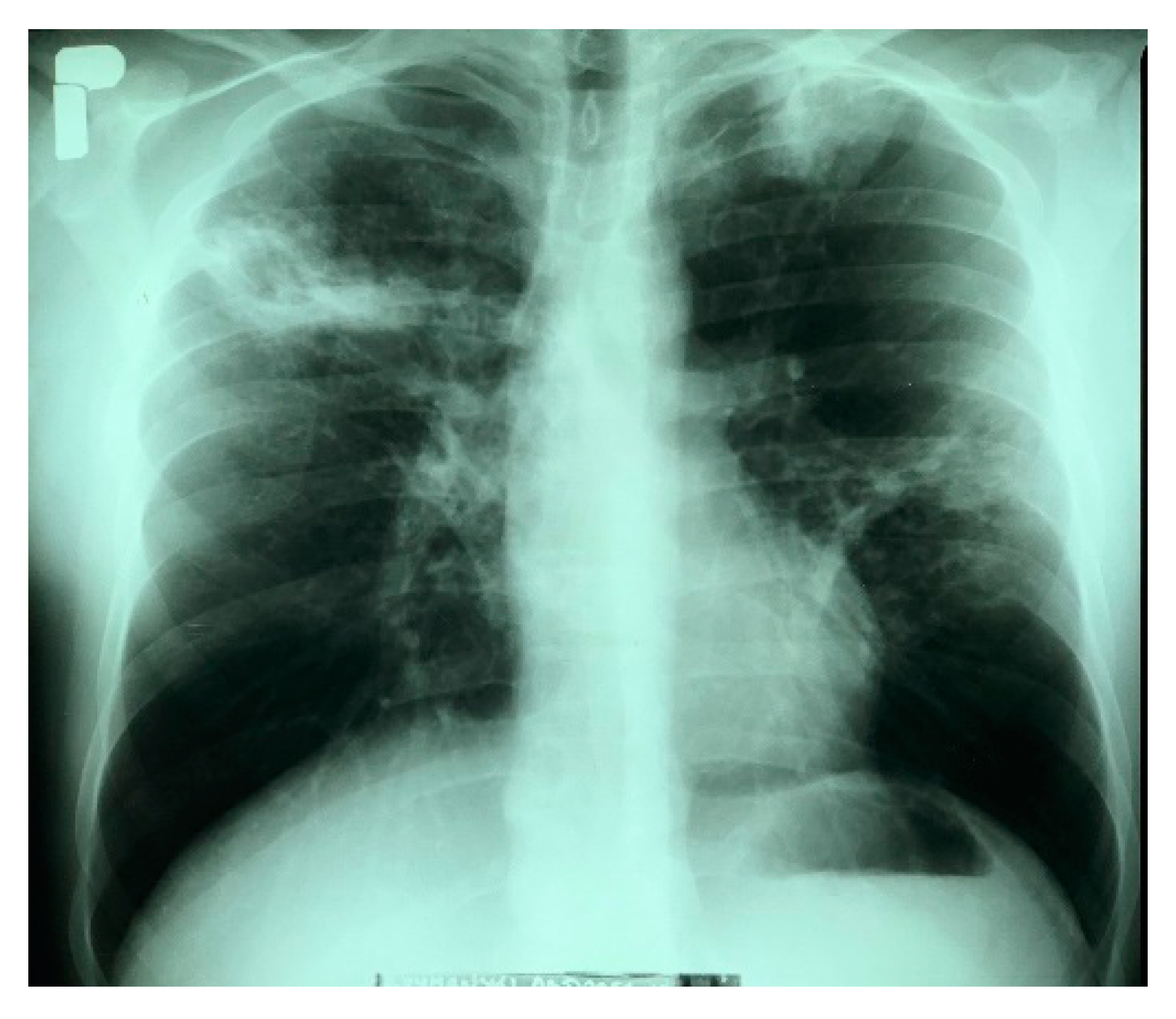
3.1. Human Case
3.2. Herd History

| No. | Cattle Numbers as Marked in the Laboratory | Macroscopic Findings | |||||
|---|---|---|---|---|---|---|---|
| Mandibular | Retropharyngeal | Bronchial | Mediastinal | Mesenteric | Supramammary | ||
| 1. | T-135, T-145, T-140, T-150, T-151, T-154, T-153 | enlarged and haemorrhagic | Firm, white nodules | caseous tubercles | caseous tubercles | Haemorrhagic and/or enlarged | enlarged and haemorrhagic |
| 2. | T-136 | clear | enlarged and haemorrhagic | caseous tubercles | caseous tubercles | haemorrhagic | enlarged and haemorrhagic |
| 3. | T-137 | whitish lumps | enlarged and haemorrhagic with fibrous, pale-yellow lesions (Figure 3) | caseous tubercles | caseous tubercles | haemorrhagic | enlarged and haemorrhagic |
| 4. | T-138 | enlarged and haemorrhagic | enlarged and haemorrhagic | caseous tubercles (Figure 4) | caseous tubercles | haemorrhagic | enlarged and haemorrhagic |
| 5. | T-139 | clear | clear | clear | clear | clear | clear |
| 6. | T-141, T-143, T-152 | enlarged and haemorrhagic | clear | caseous tubercles | caseous tubercles | haemorrhagic and/or enlarged | enlarged and haemorrhagic |
| 7. | T-142, T-146 | clear | clear | caseous tubercles | caseous tubercles | clear | enlarged and haemorrhagic |
| 8. | T-144 | whitish lumps | clear | caseous tubercles | caseous tubercles | clear | enlarged and haemorrhagic |
| 9. | T-147, T-148 | clear | firm, white nodules | caseous tubercles | caseous tubercles | clear | enlarged and haemorrhagic |
| 10. | T-149 | clear | clear | enlarged and haemorrhagic | clear | clear | clear |
| 11. | T-155 | enlarged and haemorrhagic | clear | caseous tubercles | caseous tubercles | clear | enlarged and haemorrhagic |
3.3. Genetic Analysis
4. Discussion
5. Conclusions
Author Contributions
Funding
Institutional Review Board Statement
Informed Consent Statement
Data Availability Statement
Acknowledgments
Conflicts of Interest
References
- Morse, S.S.; Mazet, J.A.; Woolhouse, M.; Parrish, C.R.; Carroll, D.; Karesh, W.B.; Zambrana-Torrelio, C.; Lipkin, W.; Daszak, P. Predicting and preventing the next zoonotic pandemic. Lancet 2012, 380, 1956–1965. [Google Scholar] [CrossRef]
- Karesh, W.B.; Dobson, A.; Lloyd-Smith, J.O.; Lubroth, J.; Dixon, M.A.; Bennett, M.; Aldrich, S.; Harrington, T.; Formenty, P.; Loh, E.H.; et al. The ecology of zoonotic diseases: Natural and unnatural stories. Lancet 2012, 380, 1936–1945. [Google Scholar] [CrossRef]
- Huard, R.C.; Fabre, M.; de Haas, P.; Lazzarini, L.C.; van Soolingen, D.; Cousins, D.; Ho, J.L. New genetic polymorphisms that further define the phylogeny of the Mycobacterium tuberculosis complex. J. Bacteriol. 2006, 188, 4271–4287. [Google Scholar] [CrossRef] [PubMed] [Green Version]
- Eldholm, V.; Rønning, J.O.; Mengshoel, A.T.; Arnesen, T. Import and transmission of Mycobacterium orygis and Mycobacterium africanum, Norway. BMC Infect. Dis. 2021, 21, 562. [Google Scholar] [CrossRef] [PubMed]
- Prodinger, W.M.; Indra, A.; Koksalan, O.K.; Kilicaslan, Z.; Richter, E. Mycobacterium caprae infection in humans. Expert Rev. Anti-Infect. Ther. 2014, 12, 1501–1513. [Google Scholar] [CrossRef] [PubMed]
- World Health Organization. WHO Report 2020: Global Tuberculosis Report; World Health Organization: Geneva, Switzerland, 2020; pp. 1–207. [Google Scholar]
- O’Reilly, L.M.; Daborn, C.J. Epidemiology of Mycobacterium bovis infections in animals and humans: A Review. Tuberc. Lung Dis. 1995, 76, 1–46. [Google Scholar] [CrossRef]
- Garnier, T.; Eiglmeier, K.; Camus, J.C.; Medina, N.; Mansoor, H.; Pryor, M.; Duthoy, S.; Grondin, S.; Lacroix, C.; Monsempe, C.; et al. The complete genome sequence of Mycobacterium bovis. Proc. Natl. Acad. Sci. USA 2003, 100, 7877–7882. [Google Scholar] [CrossRef] [Green Version]
- Thoen, C.; Lobue, P.; De Kantor, I. Significance of Mycobacterium bovis as a zoonotic disease. Vet. Microbiol. 2006, 112, 339–345. [Google Scholar] [CrossRef]
- Cadmus, S.; Oluwatoyin Akinseye, V.; van Soolingen, D. Mycobacterium bovis in humans and M. tuberculosis in animals in Nigeria: An overview from 1975 to 2014. Int. J. Tuberc. Lung Dis. 2019, 23, 1162–1170. [Google Scholar] [CrossRef]
- Amdekar, Y.K. Tuberculosis—A persistent threat to human health. Indian. J. Pediatr. 2005, 72, 333–338. [Google Scholar]
- Evans, J.T.; Smith, E.G.; Banerjee, A.; Smith, R.M.; Dale, J.; Innes, J.A.; Hunt, D.; Tweddell, A.; Wood, A.; Anderson, C.; et al. Cluster of human tuberculosis caused by Mycobacterium bovis: Evidence for person-to-person transmission in the UK. Lancet 2007, 369, 1270–1276. [Google Scholar] [CrossRef]
- Food Safety Authority of Ireland (FSAI). Zoonotic Tuberculosis and Food Safety, 2nd ed.; Food Safety Authority of Ireland: Dublin, Ireland, 2008; pp. 1–32. [Google Scholar]
- Verdugo Escárcega, D.A.; Perea Razo, C.A.; González Ruíz, S.; Sosa Gallegos, S.L.; Suazo, F.M.; Cantó Alarcón, G.J. Analysis of Bovine Tuberculosis Transmission in Jalisco, Mexico through Whole-genome Sequencing. J. Vet. Res. 2020, 64, 51–61. [Google Scholar] [CrossRef] [PubMed]
- Lipiec, M.; Radulski, Ł.; Szulowski, K. A case of bovine tuberculosis in pigs in Poland—A country free from the disease. Ann. Agric. Environ. Med. 2019, 26, 29–32. [Google Scholar] [CrossRef] [PubMed]
- Malone, K.M.; Gordon, S.V. Mycobacterium tuberculosis Complex Members Adapted to Wild and Domestic Animals. Adv. Exp. Med. Biol. 2017, 1019, 135–154. [Google Scholar] [CrossRef]
- Aranaz, A.; Liébana, E.; Gmóez-Mampaso, E.; Galán, J.C.; Cousins, D.; Ortega, J.; Blázquez, J.; Baquero, F.; Mateos, A.; Suarez, G.; et al. Mycobacterium tuberculosis subsp. caprae subsp. nov.: A taxonomic study of a new member of the Mycobacterium tuberculosis complex isolated from goats in Spain. Int. J. Syst. Bacteriol. 1999, 49, 1263–1273. [Google Scholar] [CrossRef]
- Niemann, S.; Richter, E.; Rusch-Gerdes, S. Biochemical and genetic evidence for the transfer of Mycobacterium tuberculosis subsp. caprae Aranaz et al. 1999 to the species Mycobacterium bovis Karlson and Lessel 1970 (Approved Lists 1980) as Mycobacterium bovis subsp. caprae comb. nov. Int. J. Syst. Evol. Microbiol. 2002, 52, 433–436. [Google Scholar] [CrossRef] [Green Version]
- Aranaz, A.; Cousins, D.; Mateos, A.; Domínguez, L. Elevation of Mycobacterium tuberculosis subsp. caprae Aranaz et al. 1999 to species rank as Mycobacterium caprae comb. nov. sp. nov. Int. J. Syst. Evol. Microbiol. 2003, 53, 1785–1789. [Google Scholar] [CrossRef] [Green Version]
- Amato, L.; Mulatti, P.; Pacciarini, M.; Schiavon, E.; Zanoni, M.; Bonfanti, L. Mycobacterium caprae in a dairy farm in Northeast Italy. Vet. Ital. 2019, 55, 375–379. [Google Scholar] [CrossRef]
- Valcheva, V.; Savova-Lalkovska, T.; Vyazovaya, A.; Dimitrova, A.; Bonovska, M.; Najdenski, H. First insight into phylogeography of Mycobacterium bovis and M. caprae from cattle in Bulgaria. Inf. Gen. Evol. 2020, 81, 104240. [Google Scholar] [CrossRef]
- Krajewska, M.; Kozińska, M.; Zwolska, Z.; Lipiec, M.; Augustynowicz—Kopeć, E.; Szulowski, K. Human as a source of tuberculosis for cattle. First evidence of transmission in Poland. Vet. Microbiol. 2012, 159, 269–271. [Google Scholar] [CrossRef]
- Sweetline, A.; Ronald, S.; Kumar, T.M.A.; Palaniyandi, K.; Thangavelu, A. Molecular identification of Mycobacterium tuberculosis in cattle. Vet. Microbiol. 2017, 198, 81–87. [Google Scholar] [CrossRef] [PubMed]
- Palaniyandi, K.; Kumar, N.; Veerasamy, M.; Refaya, A.K.; Dolla, C.; Balaji, S.; Baskaran, D.; Thiruvengadam, K.; Rajendran, A.; Narayanan, S.; et al. Isolation and comparative genomics of Mycobacterium tuberculosis isolates from cattle and their attendants in South India. Sci. Rep. 2019, 9, 17892. [Google Scholar] [CrossRef] [PubMed] [Green Version]
- Instruction of the Chief Veterinary Officer No. GIWpr-02010-8/2016 of 8 February 2016 on the Matter of Procedure for Suspicion, Confirmation, and Eradication of Bovine Tuberculosis in a Herd of Cattle and Procedure for Conducting Relevant Monitoring Tests. 8 February 2016. Available online: http://www.ostrowmaz.piwet.net/instrukcje/instrukcja_gruzlica.pdf (accessed on 21 April 2021). (In Polish).
- European Union (EU). Commission Delegated Regulation (EU) 2020/689 of 17 December 2019 Supplementing Regulation (EU) 2016/429 of the European Parliament and of the Council as Regards Rules for Surveillance, Eradication Programmes, and Disease-Free Status for Certain Listed and Emerging Diseases. EU OJ L 174, 3.6.2020; p. 211–340. Available online: https://eur-lex.europa.eu/legal-content/EN/ALL/?uri=CELEX%3A32020R0689 (accessed on 21 April 2021).
- European Centre for Disease Prevention and Control (ECDC). Handbook on Tuberculosis Laboratory Diagnostic Methods in the European Union; European Centre for Disease Prevention and Control (ECDC): Stockholm, Sweden, 2018; pp. 1–119. Available online: https://www.ecdc.europa.eu/sites/default/files/documents/TB-handbook-2018-final.pdf (accessed on 21 April 2021).
- Kamerbeek, J.; Schouls, L.; Kolk, A.; van Agterveld, M.; van Soolingen, D.; Kuijper, S.; Bunschoten, A.; Molhuizen, H.; Shaw, R.; Goyal, M.; et al. Simultaneous detection and strain differentiation of Mycobacterium tuberculosis for diagnosis and epidemiology. J. Clin. Microbiol. 1997, 35, 907–914. [Google Scholar] [CrossRef] [PubMed] [Green Version]
- Brudey, K.; Driscoll, J.R.; Rigouts, L.; Prodinger, W.M.; Gori, A.; Al-Hajoj, S.A.; Allix, C.; Aristimuño, L.; Arora, J.; Baumanis, V.; et al. Mycobacterium tuberculosis complex genetic diversity: Mining the fourth international spoligotyping database (SpolDB4) for classification, population genetics and epidemiology. BMC Microbiol. 2006, 6, 1–17. [Google Scholar] [CrossRef] [PubMed] [Green Version]
- Smith, N.H.; Upton, P. Naming spoligotype patterns for the RD9-deleted lineage of the Mycobacterium tuberculosis complex. Inf. Gen. Evol. 2012, 12, 873–876. [Google Scholar] [CrossRef] [PubMed]
- Alonso-Rodríguez, N.; Martínez Lirola, M.; Herránz, M.; Sanchez Benitez, M.; Barroso, P.; Bouza, E.; García de Viedma, D. Evaluation of the new advanced 15-loci MIRU-VNTR genotyping tool in Mycobacterium tuberculosis molecular epidemiology studies. BMC Microbiol. 2008, 8, 1–9. [Google Scholar] [CrossRef] [Green Version]
- World Organisation for Animal Health (WOAH, founded as OIE 2022, July 18). Bovine Tuberculosis. Available online: https://www.woah.org/fileadmin/Home/eng/Media_Center/docs/pdf/Disease_cards/BOVINE-TB-EN.pdf (accessed on 18 July 2022).
- Menin, Á.; Fleith, R.; Reck, C.; Marlow, M.; Fernandes, P.; Pilati, C.; Báfica, A. Asymptomatic cattle naturally infected with Mycobacterium bovis present exacerbated tissue pathology and bacterial dissemination. PLoS ONE 2013, 8, e53884. [Google Scholar] [CrossRef]
- Centers for Disease Control and Prevention (CDC 2022, July 18). Mycobacterium Bovis (Bovine Tuberculosis) in Humans. Available online: https://www.cdc.gov/tb/publications/factsheets/general/mbovis.htm (accessed on 18 July 2022).
- Lombard, J.E.; Patton, E.A.; Gibbons-Burgener, S.N.; Klos, R.F.; Tans-Kersten, J.L.; Carlson, B.W.; Keller, S.J.; Pritschet, D.J.; Rollo, S.; Dutcher, T.V.; et al. Human-to-Cattle Mycobacterium tuberculosis Complex Transmission in the United States. Front. Vet. Sci. 2021, 8, 1–11. [Google Scholar] [CrossRef]
- Urbanowski, M.E.; Ordonez, A.A.; Ruiz-Bedoya, C.A.; Sanjay, K.J.; Bishai, W.R. Cavitary tuberculosis: The gateway of disease transmission. Lancet Infect. Dis. 2020, 20, e117–e128. [Google Scholar] [CrossRef]
- de Jong, B.C.; Onipede, A.; Pym, A.S.; Gagneux, S.; Aga, R.S.; DeRiemer, K.; Small, P.M. Does resistance to pyrazinamide accurately indicate the presence of Mycobacterium bovis? J. Clin. Microbiol. 2005, 43, 3530–3532. [Google Scholar] [CrossRef] [Green Version]
- Shi, J.; Zheng, D.; Zhu, Y.; Ma, X.; Wang, S.; Li, H. Role of MIRU-VNTR and spoligotyping in assessing the ge-netic diversity of Mycobacterium tuberculosis in Henan Province, China. BMC Inf. Dis. 2018, 18, 447. [Google Scholar] [CrossRef]
- Branger, M.; Loux, V.; Cochard, T.; Boschiroli, M.L.; Biet, F.; Michelet, L. The complete genome sequence of Mycobacterium bovis Mb3601, a SB0120 spoligotype strain representative of a new clonal group. Inf. Gen. Evol. 2020, 82, 104309. [Google Scholar] [CrossRef] [PubMed]
- Ghavidel, M.; Mansury, D.; Nourian, K.; Ghazvini, K. The most common spoligotype of Mycobacterium bovis isolated in the world and the recommended loci for VNTR typing. A systematic review. Microb. Pathog. 2018, 118, 310–315. [Google Scholar] [CrossRef] [PubMed]
- Duarte, E.L.; Domingos, M.; Amado, A.; Botelho, A. Spoligotype diversity of and animal isolates. Vet. Micrbiol. 2008, 130, 415–421. [Google Scholar] [CrossRef] [PubMed]
- Hauer, A.; De Cruz, K.; Cochard, T.; Godreuil, S.; Karoui, C.; Henault, S.; Bulach, T.; Banuls, A.L.; Biet, F.; Boschiroli, M.L. Genetic evolution of Mycobacterium bovis causing tuberculosis in livestock and wildlife in France since 1978. PLoS ONE 2015, 10, e0117103. [Google Scholar] [CrossRef] [PubMed]
- Hauer, A.; Michelet, L.; Cochard, T.; Branger, M.; Nunez, J.; Boschiroli, M.L.; Biet, F. Accurate phylogenetic relationships among Mycobacterium bovis strains circulating in France based on whole genome sequencing and single nucleotide polymorphism analysis. Front. Microbiol. 2019, 10, 955. [Google Scholar] [CrossRef] [Green Version]
- Rodriguez-Campos, S.; Smith, N.H.; Boniotti, M.B.; Aranaz, A. Overview and phylogeny of Mycobacterium tuberculosis complex organisms: Implication for diagnostics and legislation of bovine tuberculosis. Res. Vet. Sci. 2014, 97, 5–19. [Google Scholar] [CrossRef]


| No. | Strain Number | Spoligotyping | MIRU Pattern (Number of Strains Possessing This MIRU Pattern) | |
|---|---|---|---|---|
| Assigned by www.Mbovis.org 1 Assigned by SITIVIT 2 | ||||
| 1 | Strain isolated from the farmer | SB0112 1 | Bov_1 4822 | 422432155421434 (1) |
| 2 | bovine strains T-135-155 | SB0112 1 | Bov_1 4822 | 422432155421434 (20) |
Publisher’s Note: MDPI stays neutral with regard to jurisdictional claims in published maps and institutional affiliations. |
© 2022 by the authors. Licensee MDPI, Basel, Switzerland. This article is an open access article distributed under the terms and conditions of the Creative Commons Attribution (CC BY) license (https://creativecommons.org/licenses/by/4.0/).
Share and Cite
Krajewska-Wędzina, M.; Radulski, Ł.; Waters, W.R.; Didkowska, A.; Zabost, A.; Augustynowicz-Kopeć, E.; Brzezińska, S.; Weiner, M. Mycobacterium bovis Transmission between Cattle and a Farmer in Central Poland. Pathogens 2022, 11, 1170. https://doi.org/10.3390/pathogens11101170
Krajewska-Wędzina M, Radulski Ł, Waters WR, Didkowska A, Zabost A, Augustynowicz-Kopeć E, Brzezińska S, Weiner M. Mycobacterium bovis Transmission between Cattle and a Farmer in Central Poland. Pathogens. 2022; 11(10):1170. https://doi.org/10.3390/pathogens11101170
Chicago/Turabian StyleKrajewska-Wędzina, Monika, Łukasz Radulski, W. Ray Waters, Anna Didkowska, Anna Zabost, Ewa Augustynowicz-Kopeć, Sylwia Brzezińska, and Marcin Weiner. 2022. "Mycobacterium bovis Transmission between Cattle and a Farmer in Central Poland" Pathogens 11, no. 10: 1170. https://doi.org/10.3390/pathogens11101170