Article

The Fascinating Cross-Paths of Pathogenic Bacteria, Human and Animal Faecal Sources in Water-Stressed Communities of Vhembe District, South Africa

by
Mulalo Mudau
1,*,
Renay Ngobeni-Nyambi
2 and
Maggy Ndombo Benteke Momba
1,*
1
Department of Environmental, Water and Earth Sciences, Tshwane University of Technology, Arcadia Campus, Private Bag X680, Pretoria 0001, South Africa
2
Department of Medicine, Division of Infectious Diseases, University of North Carolina School of Medicine, Chapel Hill, NC 27599, USA
*
Authors to whom correspondence should be addressed.
Pathogens 2023, 12(9), 1085; https://doi.org/10.3390/pathogens12091085
Submission received: 30 June 2023 / Revised: 16 August 2023 / Accepted: 24 August 2023 / Published: 26 August 2023
(This article belongs to the Special Issue Water-Borne Pathogens)

Abstract

:
Access to clean and safe drinking water still remains a major challenge in the developing world, causing public health risks in terms of waterborne infections, especially in rural areas of sub-Saharan Africa. This study aimed to track and detect enteric pathogens (Salmonella enterica subsp. enterica serovar Typhimurium str. LT2, Shigella flexneri, and Campylobacter jejuni subsp. jejuni) in rural water sources. It also sought to establish a correlation between these pathogens and the sources of faecal pollution. Multiplex qPCR and specific primers and probes were used for detection and tracking. The study successfully correlated the occurrence of target pathogens with sources of human and animal faecal contamination using host-specific genetic markers (BacHum and HF183 for humans, BacCow for cows, Pig-2-Bac for pigs, Cytb for chickens, and BacCan for dogs). The study revealed that enteric pathogens were found in 47.69% and 32.80% of samples during the wet and dry seasons, respectively. These pathogens were associated with human or animal faecal contamination. Correlations between pathogens and contamination sources were significant (p ≤ 0.05), with varying strengths during the wet and dry seasons. The findings emphasize the importance of identifying faecal contamination sources to protect rural communities from waterborne infections.

1. Introduction

Waterborne infections pose significant public health and regulatory concerns worldwide, affecting various water sources such as drinking water, wastewater, rivers, and lakes [1]. With microorganisms present in food, water, soil, and human and animal bodies, access to safe drinking water remains a challenge for many, leading to waterborne diseases and related deaths. Globally, about 525,000 children die from diarrhoeal diseases each year, and 2.5 billion people worldwide lack access to safely managed sanitation [2]. Estimates state that contaminated drinking water, poor sanitation, and inadequate hygiene contribute to 829,000 deaths in middle- and low-income countries annually [3]. Waterborne diseases have also been linked to more than 2.2 million annual deaths, making them a significant global health issue [4]. An estimated 66% of people worldwide contract waterborne and enteric diseases each year, including acute gastroenteritis, cholera, dysentery, hepatitis-A, and typhoid [5]. Consequently, 2.11 million South Africans are living in rural areas without safe water infrastructure, exposing them to waterborne illnesses and their complications [6]. Campylobacteriosis, shigellosis, salmonellosis, cholera, yersiniosis, and a number of other bacterial, fungal, viral, and parasitic infections are among the water-related diseases affecting people in South African rural communities [7].
Waterborne infections have severe effects on rural communities, exacerbating already burdensome healthcare needs and infrastructure challenges [5]. Salmonellosis, a major concern, causes millions of morbidity cases and over a million fatalities worldwide annually, characterized by fever, diarrhoea, abdominal pain, nausea, and vomiting [5]. In some regions, typhoidal Salmonella isolates lead to gastrointestinal illnesses with approximately 21.6 million positive cases and 200,000 fatalities per year [5]. Shigella, commonly found in human and primate intestinal tracts, is heavily discharged in faeces, persisting in water for extended periods and facilitating waterborne transmission [8]. There are an estimated 164.7 million Shigella episodes worldwide each year, with 163.2 million cases in underdeveloped countries causing 1.1 million fatalities, predominantly in children under five [8]. Symptoms like fever, anorexia, fatigue, and malaise are common with Shigella spp. [9]. Campylobacter is a prevalent bacterial cause of human gastroenteritis globally [10]. In 2008, nearly 200,000 confirmed cases of campylobacteriosis were reported, with Campylobacter jejuni being the primary cause of zoonotic disease in humans [5]. In many rural areas, dependence on private water wells and natural sources like rivers is necessary due to the lack of public water supplies and sewerage networks. Understanding contaminant sources and attenuation processes is essential for treating and disposing of domestic wastewater, ensuring safe drinking water in spatially constrained rural households [11]. The lack of access to safe water supply hinders efforts to promote good health, income, living conditions, equitable resource use, and a better quality of life in South Africa and other developing countries.
Like the majority of rural communities in developing countries, Vhembe District Municipality is facing challenges to supply clean water and improved sanitation to all; currently, those who lack improved water sources have to rely on highly contaminated sources. The Luvuvhu River serves as the primary source of water for the majority of the villages in the Vhembe District Municipality, either directly or through water treatment facilities. Previous investigators have reported that the river water of the Vhembe district has poor microbiological quality and is unfit for human consumption [7]. Due to its dense network of tributaries and extensive route through numerous villages, the Luvuvhu River is more vulnerable to contamination from various sources and it is extremely susceptible to waterborne pathogens. Therefore, the aim of the current study was to detect and track enteric pathogens (Shigella flexneri, Salmonella enterica subsp. enterica serovar Typhimurium str. LT2, and Campylobacter jejuni subsp. jejuni) from different water sources, which might contribute to outbreaks of water-related diseases in water-stressed rural communities. To achieve the set aim, multiplex real-time PCR using specific primers and probes was employed for the detection and tracking of enteric pathogens in different water sources of participating households. A correlation was then established between the occurrence of the target pathogens and the sources of human and animal faecal contamination in water sources.

2. Materials and Methods

2.1. Study Site Description, Water Supply, and Storage

The Vhembe District Municipality is situated in the northern part of the Limpopo Province of South Africa. It shares borders with the eastern district of Mopani and the western district of Capricorn. In 2016, a Stats SA [12] community survey revealed that the population of the Vhembe District was approximately 1,393,948, occupying a land area of 27,969,148 km2. This district is comprised of the local municipalities of Collins Chabane (CC), Thulamela (TM), Makhado (MK), and Musina (MS), with populations of 347.974, 497.237, 416.728, and 132.009, respectively. For this particular study, three local municipalities (Thulamela, Makhado, and Collins Chabane) were selected, and out of these, five villages experiencing water scarcity were randomly selected. These five villages were identified as Lambani, Tshifudi, Njhakanjhaka (Watervaal), Makuleke, and Gandlanani [13].
Within the Collins Chabane (CC) local municipality, the study focused on two villages, identified as Gandlanani and Makuleke. Gandlanani village receives its water supply from the Gandlanani Water Treatment Works, situated in the village, which draws its raw water from the Luvuvhu River, flowing through the village. On the other hand, Makuleke village relies on two treatment plants for its water supply, Xikundu Water Treatment Works and the Mhinga Water Treatment Works, depending on their operational status. The Xikundu Water Treatment Works, located in Xikundu village, draws its raw water from the Luvuvhu River passing through that area, while the Mhinga Water Treatment Works, situated in Mhinga village, also draws water from the Luvuvhu River.
In the Thulamela (TM) Local Municipality, the study included two villages, Tshifudi and Lambani, both of which receive their water supply from the Xikundu Water Treatment Works located in Xikundu village. Lastly, in the Makhado (MK) Local Municipality, one village, Njhakanjhaka, was selected, whose primary water supply is obtained from a treatment plant, Albasini Water Works, that draws water from the Albasini Dam. However, during the study period, the participating households in this village did not receive their water supply from this plant due to water shortages and blockage in the pipeline, leading them to purchase water from households with private boreholes in their vicinity. Figure 1 illustrates the selected villages, the water treatment plants providing water to these villages, and the rivers from which the treatment plants draw their water.

2.2. Ethical Clearance

Ethical clearance approval was granted by the Faculty of Science Research Ethics Committee at the Tshwane University of Technology (TUT). Permission was obtained from the Vhembe District Municipality and the community leaders to conduct the study in selected villages of this district after consultation and briefing them on the aim and objectives of the study. Informed consent was sought from community participants and plant managers after clearly explaining the motives behind the study and their involvement. Before taking part in the study, participants were asked to sign a consent form.

2.3. Sample Collection at Different Points

Water samples were collected sequentially from the main source all the way to the point of use: the storage containers in the households of the selected villages. Although the majority of households had standpipes in their yards, most of the villages did not have running water in their taps during the sampling periods. Thus, the sampling regime also included container-stored water in households. In villages where running water was available, water samples were collected from both the tap and the storage container. Water samples were collected eight (8) times at each sampling location. During the dry season, water samples were obtained four times at each sampling location, and similarly during the wet season. In total, 1.128 water samples were collected from the five communities as shown in Table 1. Seasonal water sampling was carried out at several water sources using known techniques and APHA 2001 plate count methods [14]. Water samples were transported on ice in cooler boxes to the water research laboratory at Tshwane University of Technology. Analysis was performed within 24 h after collection.

2.4. Sample Concentration of Bacteria and DNA Extraction

Within 24 h of collection, all water samples (each measuring 500 mL) underwent filtration using a vacuum manifold. Specific filters were used for this process: mixed cellulose ester filters with a diameter of 47 mm and a pore size of 0.22 μm (obtained from Merck Millipore in Billerica, MA, USA and Merck SA, Singapore). However, in the case of river water samples, a larger filter with a diameter of 90 mm and a pore size of 0.45 μm (Merck Millipore) was used, following the method described by Haramoto and Osado [15].
To ensure that the samples were not contaminated during processing, sterile deionised water was filtered alongside them. The filter papers were then rolled and placed inside 5 mL PBS tubes. Each PBS tube received a drop of Tween® 20 and was vortexed vigorously to dislodge any materials retained on the filter paper. Subsequently, the water samples underwent centrifugation at a high speed of 15,000 rpm for 10 min. After centrifugation, DNA extraction was performed on the resulting pellet using the Zymo Research DNA isolation kit (Quick-DNA Fecal/Soil Microbe Miniprep Kit, Sigma-Aldrich, St. Louis, MO, USA) following the instructions provided by the manufacturer. To account for potential contamination during DNA extraction, negative controls, known as blanks, were included in each extraction batch. The extracted genomic DNA was then stored in a freezer at −20 °C until further use.

2.5. Multiplex Real-Time PCR for the Detection of Enteric Pathogens

The DNA extracted from the water samples was subjected to multiplex real-time PCR for the detection of Campylobacter jejuni subsp. jejuni, Shigella flexneri, and Salmonella enterica subsp. enterica serovar Typhimurium str. LT2. The three different primer/probe sets used for the purpose of this study were previously developed by Wiemer et al. [16] to target the tetrathionate reductase subunit C (ttrC) gene for Salmonella enterica subsp. enterica serovar Typhimurium str. LT2, the DNA gyrase subunit A gene (GyrA) for Campylobacter jejuni subsp. jejuni and Campylobacter jejuni subsp. doylei, and finally the invasion plasmid antigen H (ipaH) gene for Shigella flexneri. Following the method used by Wiemer et al. [16] with slight modifications, the multiplex quantitative PCR (qPCR) assay was performed using CFX96 Touch Real-Time PCR Detection System combined with the C1000™ Thermal Cycler (Bio-Rad Laboratories, Hercules, CA, USA) and assays for detecting the above-mentioned pathogens. Table 2 lists the names and sequences of primers, probes, and cycling conditions used for the assay.
The multiplex qPCR total reaction volume of 25 μL contained 12.5 μL of the GoTaq® probe qPCR master mix, 7.5 μL of the primer/probe mix, 3 μL of nuclease-free water, and 2 μL of genomic DNA. Pure extracts of reference strains of the target pathogens were used (Shigella flexneri (serotype 2b) ATCC 12022, Campylobacter jejuni subsp. jejuni ATCC 33291, Salmonella Typhimurium ATCC 13311) as positive controls and nuclease-free water was used as a negative control together with DNA extraction blanks.
For a standard curve, 10-fold serial dilution (10×) of plasmid DNA containing the target region to be amplified was used to prepare standards. The lowest cycle threshold (Ct) value obtained from the serially diluted samples was regarded as the limit of detection and a cut-off value for each assay was determined by the highest Ct value obtained during the optimisation run. Microbial source tracking markers were used to identify the possible sources of faecal contamination in water. As described in a study by Mudau [13], host-specific marker assays were used in real-time PCR for the identification of the possible sources of contamination.

2.6. The Occurrence of Sources of Faecal Contamination in Different Water Sources

In our previous study [13], microbial source tracking markers were used to identify the possible sources of faecal contamination in different water sources. Host-specific assays were used to attribute the faecal contamination to its original source. The data obtained were then used in the current study to establish the correlation between the occurrence of the target enteric pathogens and human or animal (pigs, chickens, cows, and dogs) sources of faecal contamination. Table 3 shows the data obtained from the above-mentioned study.

2.7. Statistical Analysis

The paired t-test was used to compare concentrations across assays targeting the same host, whilst the Chi-square test was used to examine the difference in positive ratios between assays targeting the same host and other hosts in the case of faecal source samples and water samples, respectively. The correlation between enteric pathogens and the different sources of contamination in water samples was analysed using a paired t-test and Pearson correlation coefficient ®. The results were reported to be statistically significant if the p-value was less than 0.05.

3. Results

3.1. Parameters of qPCR Assay in Detection and Quantification of Enteric Pathogens

Using the standard curves, the efficiency of the PCR and limit of detection (LOD) for each assay were determined. Further, the lower limit of quantification (LLOQ) and cut-off values for each assay were also determined. The efficiency of the different PCR assays was found to be 81% for Campylobacter jejuni subsp. jejuni, 83% for Shigella flexneri, and 93% for Salmonella enterica subsp. enterica serovar Typhimurium str. LT2. The LLOQ was determined by calculating the average Ct values from the standard curve (Figure 2) for each assay and they ranged from 27.5% to 28.58%, as shown in Table 4. The y-intercept was used as the cut-off value for each assay; any sample that amplified below the LLOQ/LOD and above the cut-off value was regarded as negative. Table 4 and Figure 2 below shows the parameters of the qPCR assays used for the detection of the target enteric pathogens.

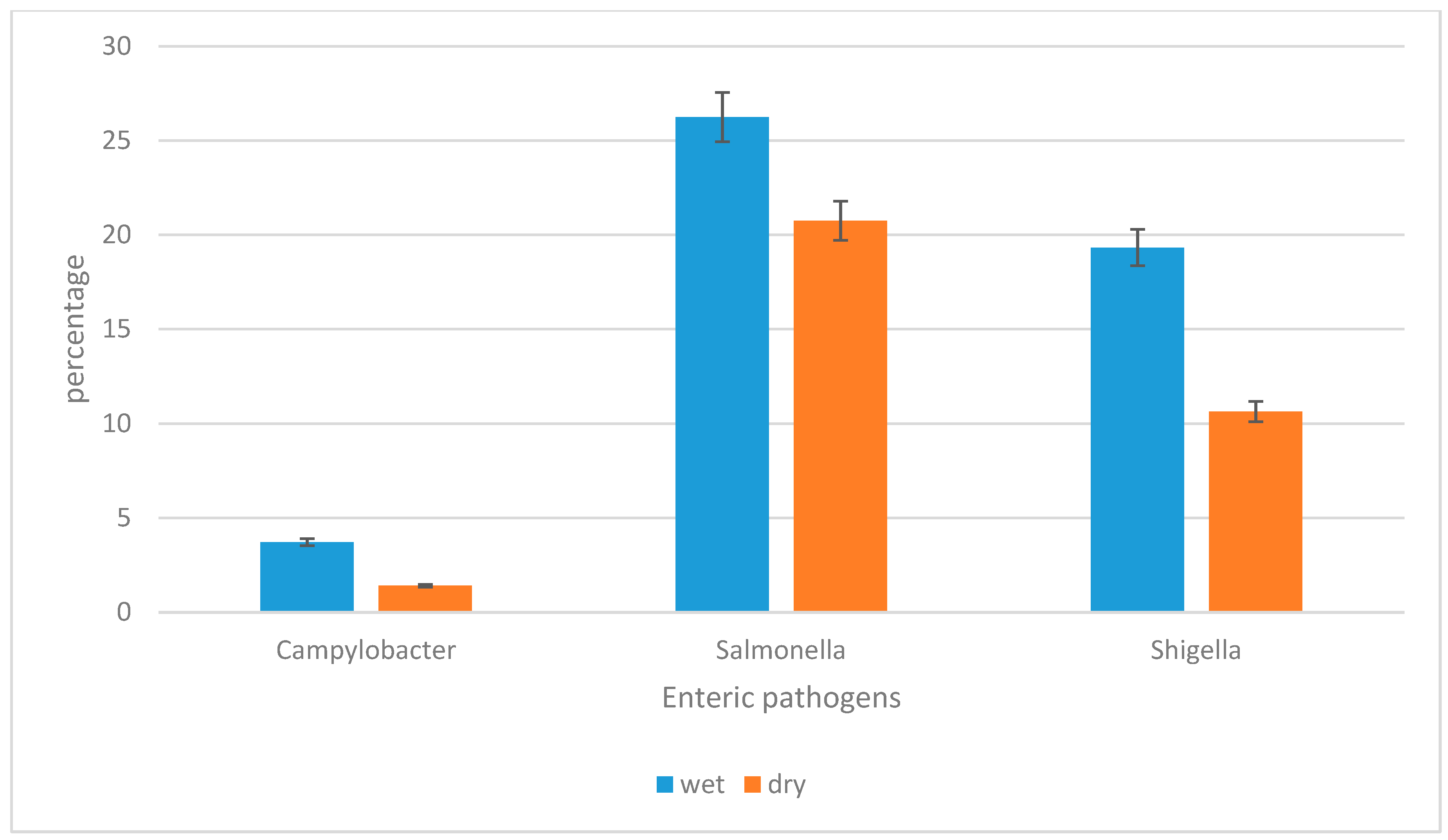
3.2. The Prevalence of Campylobacter jejuni subsp. jejuni, Shigella flexneri, and Salmonella enterica subsp. enterica Serovar Typhimurium str. LT2 in Water Sources per Season

A multiplex real-time PCR assay was employed to track and detect different enteric pathogens in water sources. Salmonella enterica subsp. enterica serovar Typhimurium str. LT2 was the most prevalent enteric pathogen detected during dry and wet seasons, followed by Shigella flexneri, and lastly by Campylobacter jejuni subsp. jejuni. In the wet season, about 49.29% (n = 278/564) of the samples were found to be positive for either one of the target microorganisms, of which 26.24% (n = 148/564) were positive for Salmonella enterica subsp. enterica serovar Typhimurium str. LT2, 19.32% (n = 109/564) for Shigella flexneri, and 3.72% (n = 21/564) for Campylobacter jejuni subsp. jejuni. In the dry season, a total of 32.80% (n = 185/564) of the samples were found to be positive, of which 20.74% (n = 117/564) were positive for Salmonella enterica subsp. enterica serovar Typhimurium str. LT2, 10.63% (n = 60/564) for Shigella flexneri, and 1.42% (n = 8/564) for Campylobacter jejuni subsp. jejuni. The above results indicate that Salmonella enterica subsp. enterica serovar Typhimurium str. LT2 was the most prevalent enteric pathogen during both seasons and Campylobacter jejuni subsp. jejuni was the least detected pathogen during both seasons. With the exception of treatment plant final water that showed zero correlation, positive strong correlations were established between the occurrence of enteric pathogens across seasons and river water, household drinking water and treatment plant raw water (r = 0.75, r = 0.97 and r = 1, respectively). Furthermore, with the exception of household-container-stored water and treatment plant final water, the seasonal variation in the prevalence of the target enteric pathogens was statistically significant in river water (p = 0.05) and in treatment plant raw water (p = 0.01) between wet and dry seasons. Figure 3 depicts the prevalence of the different target enteric pathogens in water sources per season.

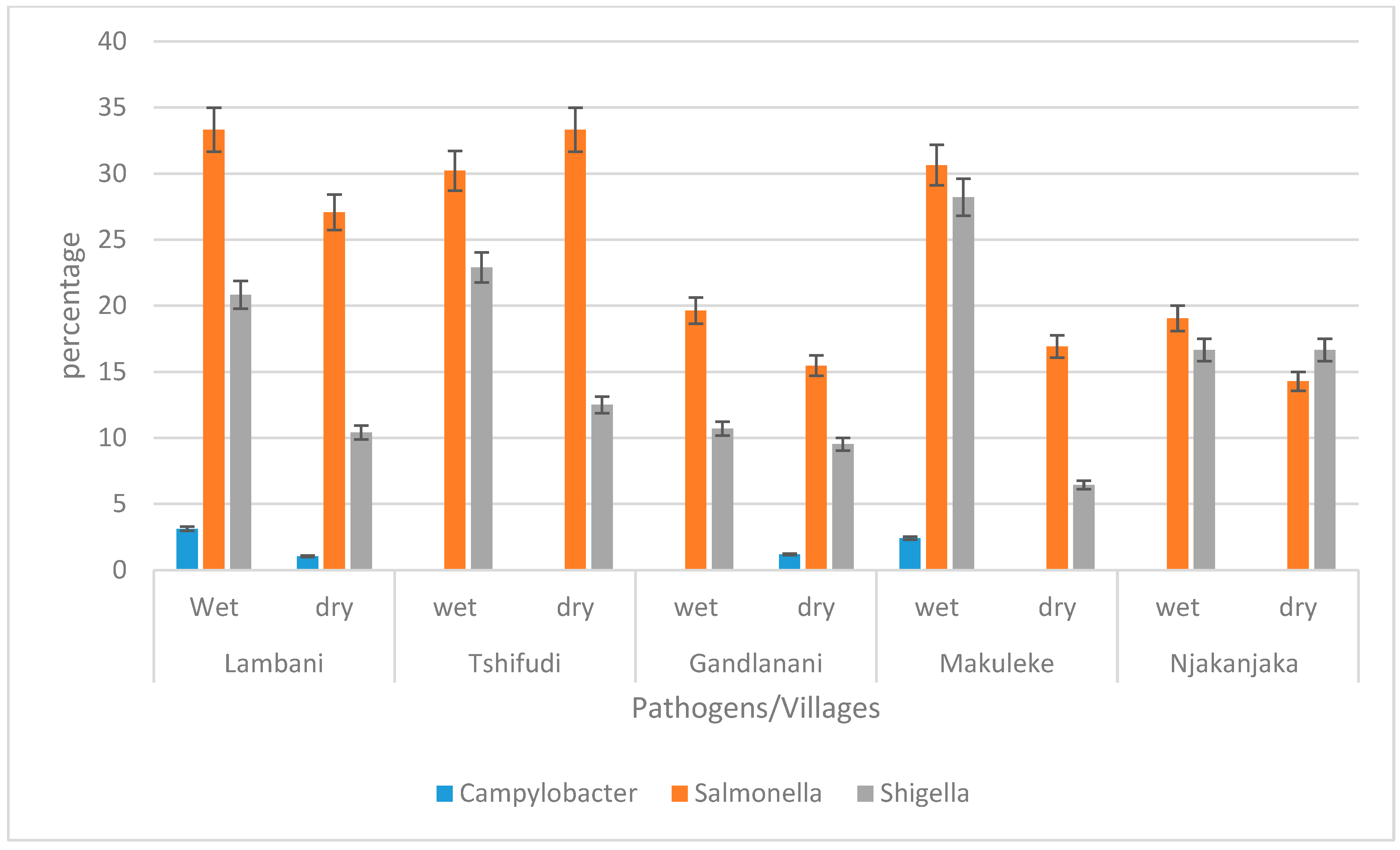
3.3. The Prevalence of Campylobacter jejuni subsp. jejuni, Shigella flexneri, and Salmonella enterica subsp. enterica Serovar Typhimurium str. LT2 in Water Sources per Village during Wet and Dry Seasons

The occurrence of enteric pathogens was further evaluated in all the water sources used in the selected villages for both seasons. As shown in Figure 4, in Lambani village, the most frequently identified pathogen was Salmonella enterica subsp. enterica serovar Typhimurium str. LT2 (33.33%), followed by Shigella flexneri (20.83%), and Campylobacter jejuni subsp. jejuni (3.13%). Furthermore, it is essential to note that the prevalence of Campylobacter jejuni subsp. jejuni was the highest in Lambani village as compared to its occurrence in other villages. In Tshifudi village, only Salmonella enterica subsp. enterica serovar Typhimurium str. LT2 and Shigella flexneri were detected in both seasons and the prevalence of Salmonella enterica subsp. enterica serovar Typhimurium str. LT2 was 33.33% in the dry season and 30.21% in the wet season, while for Shigella flexneri, it was higher (22.91%) during the wet season than during the dry season (12.5%). In Gandlanani village, during the wet season, the presence of only two pathogens was detected with Salmonella enterica subsp. enterica serovar Typhimurium str. LT2 being the most prevalent at 19.64%, followed by Shigella flexneri at a prevalence of 10.71%. However, during the dry season, all three target pathogens were detected, with the prevalence of Salmonella enterica subsp. enterica serovar Typhimurium str. LT2 being 15.47%, and that of Shigella flexneri and Campylobacter jejuni subsp. jejuni being 9.52% and 1.19%, respectively.

3.4. The Occurrence of Campylobacter jejuni subsp. Jejuni, Shigella flexneri, and Salmonella enterica subsp. enterica Serovar Typhimurium str. LT2 in Different Water Sources during Wet and Dry Seasons

The occurrence of enteric pathogens was found to vary with the source of water. The prevalence of enteric pathogens was found to be the highest in river water; of the 28 samples collected in the rivers during the wet season, 71.43% (n = 20/28) tested positive for Shigella flexneri, 60.81% (n = 17/28) for Salmonella enterica subsp. enterica serovar Typhimurium str. LT2, and 42.86% (n = 12/28) for Campylobacter jejuni subsp. jejuni. During the dry season, similar observations were noted, wherein the prevalence of target enteric pathogenic bacteria was found to be the highest in the river water as compared to other sources. However, during the dry season, Salmonella enterica subsp. enterica serovar Typhimurium str. LT2 [46.43% (n = 13/28)] was the most frequently detected pathogen, followed by Shigella flexneri [35.71% (n = 10/28)], while the lowest prevalence was recorded for Campylobacter jejuni subsp. jejuni [14.29% (n = 4/28)]. The difference in the occurrence of these enteric pathogens in the river water during wet and dry seasons was found to be statistically significant (p = 0.05), with r = 0.75, indicating a strong correlation between the two seasons (Table 5).
The presence of the selected pathogens was also detected at the point of treatment. In total, 32 samples were collected at water treatment plants prior to treatment and after treatment during the wet and dry seasons. During the wet season, 16 samples were collected in the raw water prior to treatment, and a similar prevalence of Campylobacter jejuni subsp. jejuni and Shigella flexneri was observed. A total of 7 (48.75%) samples were positive for both Campylobacter jejuni subsp. jejuni and Shigella flexneri, and 14 (87.50%) were positive for Salmonella enterica subsp. enterica serovar Typhimurium str. LT2. Furthermore, during the wet season, all 16 samples collected after chlorination (final water) tested negative for both Campylobacter jejuni subsp. jejuni and Salmonella enterica subsp. enterica serovar Typhimurium str. LT2, but two samples [2/16 (12.5%)] tested positive for Shigella flexneri. It is essential to note that the highest prevalence of enteric pathogens in water treatment plant samples was detected in the raw water entering the treatment plant (water before chlorination). During the dry season, the results were found to be fairly similar for the raw water samples, with Salmonella enterica subsp. enterica serovar Typhimurium str. LT2 being the most prevalent at 62.50%, and both Campylobacter jejuni subsp. jejuni and Shigella flexneri having a similar prevalence of 25%. However, in the final water (post-chlorination), all the samples tested negative for the target enteric pathogen during the dry season. Additionally, the seasonal variation in the occurrence of enteric pathogens in the raw water entering treatment plants was statistically significant (p = 0.01), showing a very strong positive correlation (r = 1). However, the seasonal variation in the occurrence of pathogens in the final water was insignificant (p = 0.45) and no correlation was established between the occurrence of enteric pathogens in final water and the dry and wet seasons.
At the household level, 508 water samples were tested, and the results revealed that Salmonella enterica subsp. enterica serovar Typhimurium str. LT2 (23.03%, n = 117/508)) was the most prevalent pathogen during the wet season, while Shigella flexneri was detected in 16.14% (82/508), and Campylobacter jejuni subsp. jejuni was the least detected at 0.39% (n = 2/508). During the dry season, the prevalence of Salmonella enterica subsp. enterica serovar Typhimurium str. LT2 and Shigella flexneri in household samples was 18.50% and 9.06%, respectively. Campylobacter jejuni subsp. jejuni was not detected in any household samples during the dry season. The seasonal variation in the prevalence of enteric pathogens in the household water was statistically insignificant (p = 0.17); however, a strong positive association (r = 0.97) was observed. Table 5 indicates the occurrence of target enteric pathogens in different water sources.

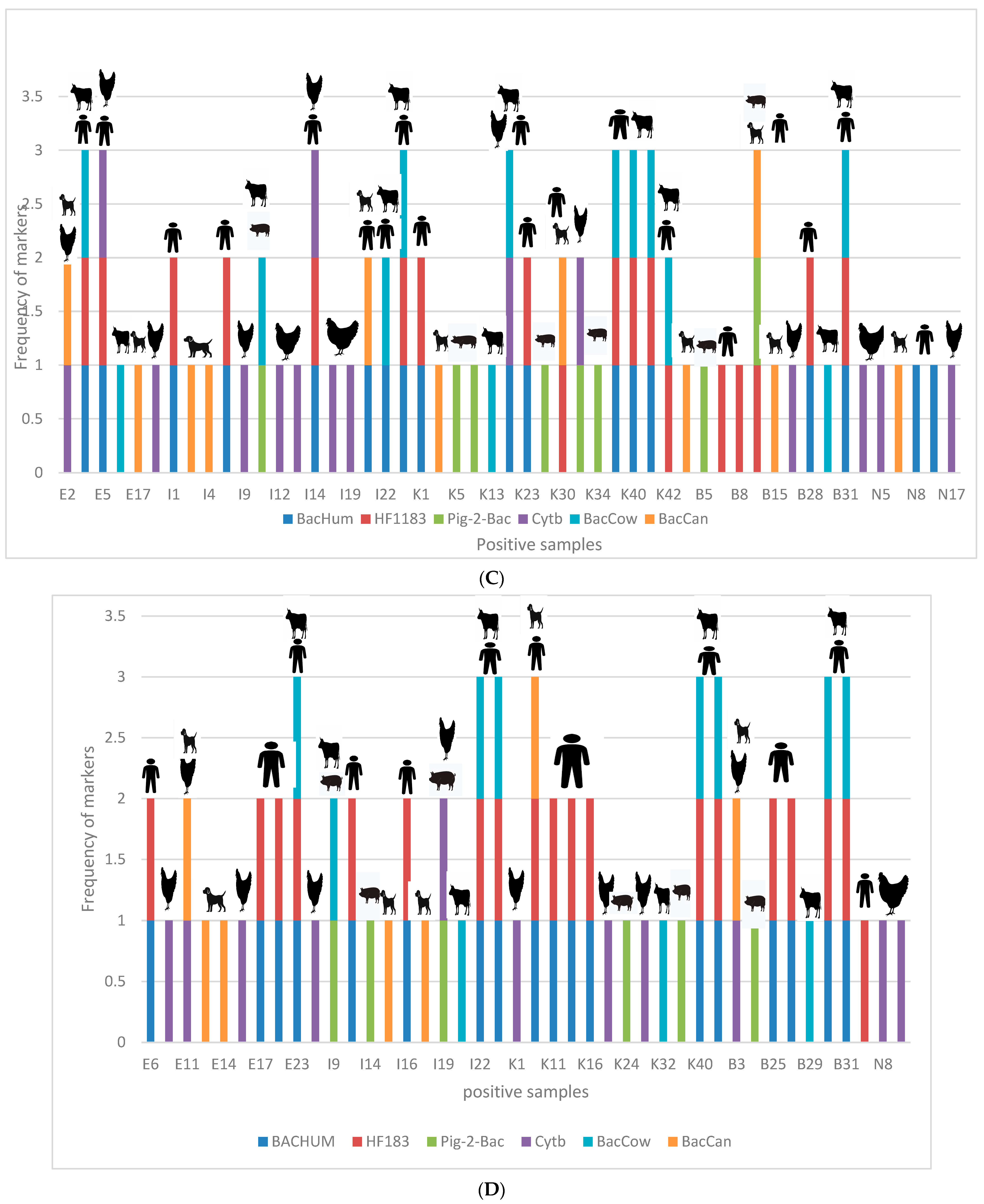
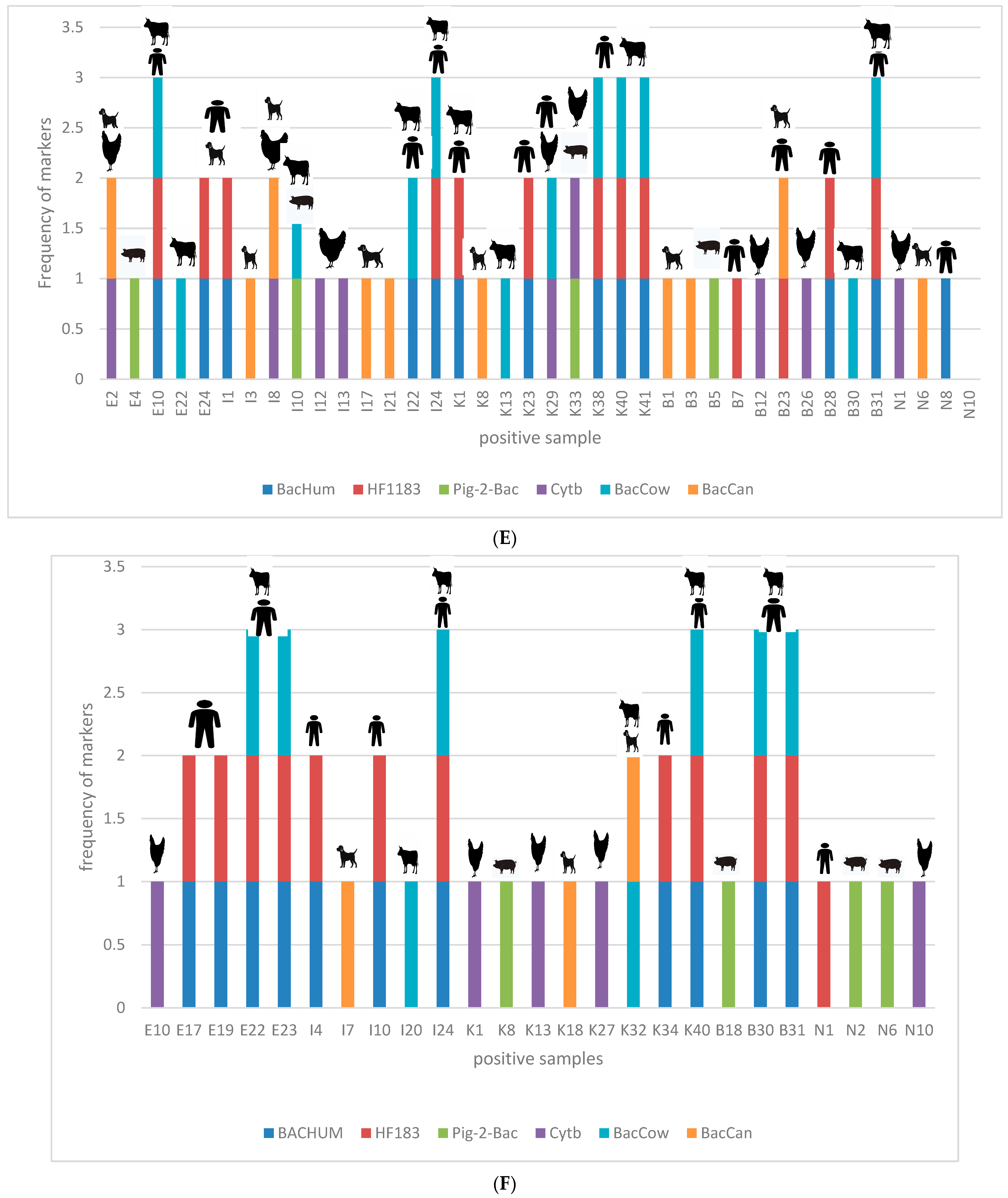
3.5. Correlation between Occurrence of Enteric Pathogens and Different Sources of Contamination Using Host-Specific Markers

During the study period, various activities around water sources were observed, potentially contributing to faecal contamination in water and, consequently, the occurrence of different enteric pathogens in water. Correlations between faecal contamination of water sources and host-specific Bacteroidales marker genes from our concurrent study were used to establish the relationships between pathogens and their sources [13]. Table 6 presents correlation coefficients (r) and p-values for various enteric pathogens during both wet and dry seasons. During the wet season, a weak negative correlation was observed between Campylobacter jejuni subsp. jejuni and all sources of contamination (ranging from −0.19 to −0.29); generally, some of these correlations were not statistically significant (p > 0.05), with others being significant (p ≤ 0.05). Positive correlations were established between Salmonella enterica subsp. enterica serovar Typhimurium str. LT2 and all the sources of contamination (ranging from 0.36 to 0.74); some of the correlations were weakly positive, such as r = 0.36, and some moderate (r = 0.55–0.62), while others showed strong correlations (r = 0.72–0.74), and these correlations were all statistically significant (p < 0.01). Weak positive correlations were observed between Shigella flexneri and some sources of contamination (ranging from 0.04 to 0.24); these correlations were statistically significant (p ≤ 0.05). Furthermore, in the dry season, a positive correlation was observed between Campylobacter jejuni subsp. jejuni and all sources of contamination (ranging from 0.25 to 0.84). However, most of the correlations were weak or moderate, with only one source of contamination showing a strong correlation (r = 0.84). The correlations were statistically significant (p < 0.03). Moderate and strong positive correlations were observed between Salmonella enterica subsp. enterica serovar Typhimurium str. LT2 and all sources of contamination (ranging from 0.45 to 0.92), and these correlations were all statistically significant (p < 0.01). Moreover, mixed correlations were established between Shigella flexneri and the sources of contamination, with some positive and some negative correlations; however, the correlations were not consistently statistically significant. In general, weaker and less significant correlations were observed during the wet season than during the dry season. In addition, a consistent positive correlation was established between Salmonella enterica subsp. enterica serovar Typhimurium str. LT2 and most sources of contamination during both the seasons. Weaker and negligible correlations between Campylobacter jejuni subsp. jejuni and contamination sources were observed during the wet season, and positive correlations in the dry season, while the correlations between Shigella flexneri and contamination sources appeared to be inconsistent and generally statistically insignificant.
Furthermore, the association between the different enteric pathogens and host-specific markers is indicated in Figure 5A–F. As can be seen in Figure 5A, during the wet season, an association was established between the occurrence of Campylobacter jejuni subsp. jejuni in household-container-stored water and humans. Sample E1 is a sample collected in household-stored water. Dog-specific markers were also detected in the same sample, making dogs a possible source of contamination. In sample B28s, also a household stored water sample, the detected host-specific markers were human markers, suggesting that human faecal materials could be a source of contamination. Furthermore, in samples B30 and B31, both collected in the Luvuvhu River, human- and cow-specific markers were detected, which indicated that either humans or cows could be the source of faecal contamination in the river, further suggesting that the possible source of the Campylobacter jejuni subsp. jejuni detected in those samples may either be from humans or cows. During the dry season, as indicated by Figure 5B, in all the samples in which Campylobacter jejuni subsp. jejuni was detected, humans and cows were also identified as the possible sources of contamination.
Figure 5C indicates the possible sources of Salmonella enterica subsp. enterica serovar Typhimurium str. LT2 in the different water samples during the wet season. The different host-specific markers for chickens, dogs, pigs, humans, and cows were detected in different water samples. Interestingly, in all the river water samples and treatment plant raw water samples (water before treatment), the host-specific Bacteroidales markers suggest that the source of Salmonella enterica subsp. enterica serovar Typhimurium str. LT2 in the river could either be humans or cows. The host-specific Bacteroidales markers shown in Figure 5C indicate that most of the Salmonella enterica subsp. enterica serovar Typhimurium str. LT2 in water was contributed by humans and cows, followed by chickens, dogs, and finally pigs.
Additionally, Figure 5D shows the possible sources of contamination during the dry season. The host-specific genetic markers observed during the wet season (Figure 5C) indicated that the occurrence of Salmonella enterica subsp. enterica serovar Typhimurium str. LT2 in the river and in raw water at the point of abstraction of the water treatment plant is from humans (BacHum and HF183) and cows (BacCow). As indicated in Figure 5D, different markers were detected in households during the dry season. Additionally, in other water samples, multiple sources of contamination were detected in the same sample, while in other samples multiple pathogens were detected in the same sample.
Furthermore, the occurrence of Shigella flexneri was also evaluated against the possible sources in both wet and dry seasons as indicated in Figure 5E,F. The results shown for the rainy season in Figure 5E suggest that the assessed sources of contamination might be the possible source of Shigella flexneri in the tested water samples. Interestingly, in one of the water treatment plants, in the final water (water after chlorination), Shigella flexneri was detected together with both the human-specific genetic markers (BacHum and HF183). These results suggest that humans are the possible source of the Shigella flexneri detected in the water. Since Shigella flexneri was detected in the treatment plant final water (post-chlorination), it was to be expected that the same pathogen would be detected in household water supplied by the treatment plant; however, in most of the households, different enteric pathogens and sources of contamination were detected other than humans. Similar results are shown in Figure 5F, which represents the findings for the dry season; different sources of contamination were identified in the same samples in which Shigella flexneri was detected.

4. Discussion

More than 500 microorganisms can be classified as waterborne pathogens and they are responsible for waterborne diseases [17]. The burden of disease from poor water quality, sanitation, and hygiene has been established and associated with up to 4% of all deaths worldwide [18,19]. Diarrhoeal disease, for example, is known as one of the main causes of morbidity and mortality of immunocompromised individuals, especially in elderly people and children under the age of five years in developing countries. Bacterial diarrhoea in children under the age of five years has been linked to pathogens such as Campylobacter, Salmonella, and Shigella [20]. To protect and promote public health, strategies have been developed for managing water quality. Among these strategies, microbial source tracking (MST) plays a major role in discriminating between human and animal sources of faecal pollution to improve water quality management [21]. The main focus of this study was, therefore, tracking and detecting enteric pathogens such as Salmonella enterica subsp. enterica serovar Typhimurium str. LT2, Shigella flexneri, and Campylobacter jejuni subsp. jejuni. The study further established the correlation between these pathogens and the sources of faecal pollution with reference to five selected water-stressed villages of the Vhembe District Municipality.
In this study, qPCR assays were evaluated using standard curves as shown in Figure 2, with efficiencies ranging from 81% to 93%. The acceptable efficiency for a successful qPCR assay is between 80% and 110% [22]. The presence of target enteric pathogens in water sources of rural communities was confirmed, raising concerns about potential waterborne diseases. Salmonella enterica subsp. enterica serovar Typhimurium str. LT2 was the most prevalent pathogen, except in river water during the wet season, where Shigella flexneri predominated (Table 5 and Figure 3). Campylobacter jejuni subsp. jejuni was the least detected in river and treated water, while it could not be detected in household-container-stored water during the dry season. Positive strong correlations were observed between pathogen occurrence across seasons in river, household drinking, and treatment plant raw water (Table 5). Seasonal variation in pathogen prevalence was statistically significant in river and treatment plant raw water. These results align with previous studies showing Salmonella spp. as the most frequently detected pathogen in water sources, often linked to faecal contamination and discharge of untreated waste [5,23,24]. Anthropogenic activities such as swimming, bathing, and washing were observed along the river, likely contributing to faecal pollution and the presence of enteric pathogens [25]. Shigella, among other harmful bacteria, was frequently detected in water samples. The diversity of enteric pathogens found in drinking water may vary by region and be influenced by seasonal fluctuations and anthropogenic activities near water sources [23,26,27,28]. In the current study, the prevalence of the target pathogens was higher during the wet season as compared to the dry season (Figure 3), and the results are consistent with the findings of prior research [26,27,29]. Heavy rainfall events during the wet season can cause surface runoff containing various contaminants to enter the water sources, resulting in an expanded microbial population in water. Cooler temperatures may also enhance the growth of many enteric pathogens, increasing the concentration of bacteria in water. However, during the dry season, bacterial cells are susceptible to desiccation and may survive but cannot grow and divide, and thus the concentration of bacteria may be lower. Because of the aforementioned factors, the high incidence of enteric infections during the wet season relative to the dry season is not surprising.
Furthermore, the occurrence of these enteric pathogens was evaluated at different sampling points (water treatment plants, rivers, and households). At the water treatment plants, the target pathogens were frequently detected in the raw water (before chlorination); however, Shigella flexneri was detected in two samples of the treatment plant final water (Table 5). Chlorination is the disinfection method commonly used worldwide, especially in developing countries. However, previous studies have demonstrated that chlorination as a cost-effective water treatment process cannot be a guarantee to safeguard against bacteria in treated water [30,31]. This clearly explains the detection of some enteric pathogens in the final water produced by the water treatment plant, in spite of a decrease in their concentration (Table 5). The detection of pathogens at the point of treatment implies that a pathogen detected at the treatment plant level has a high probability of being detected at the household level. Water contamination in households may also occur as a result of poor hygiene, failure to cover storage containers, ways of dispensing water, and animals roaming freely in the house. The findings of this investigation are in agreement with those of a study conducted in 2022 by Khabo-Mmekoa and colleagues [32], who found that household water samples were mainly contaminated by enteric pathogens such as Salmonella and Shigella. Muringani et al. [33] also reported similar findings, showing that 87.5% of all water samples analysed from both treated and untreated sources in their study were contaminated with pathogenic bacteria. The findings of this study will assist water service authorities in rural areas to develop strategies aimed at reducing the exposure of humans to pathogens that cause diseases, and to create an awareness that household factors such as hygiene practices might be responsible for the recontamination of treated water.
Environmental contamination exposes water sources to a constant risk and threat; contamination from either point or non-point sources has made water sources susceptible to pathogenic bacteria and viruses that threaten the health of humans. Consequently, it is crucial to establish the association between the sources of faecal contamination and the prevalence of target pathogens in water. As mentioned above, the current study was conducted simultaneously with one that focused on the identification of predominant faecal contamination sources in similar water sources using host-specific genetic markers [13]. Considering the results obtained, a correlation was established between pathogens detected in water sources and faecal contamination sources. The results showed that human or animal faecal matter present in water is contributing to the presence of enteric pathogens in water. Using microbial source tracking markers, the different enteric pathogens isolated in each of the water samples were correlated to their possible source. As reported in Table 6, mixed correlations between Shigella flexneri and the sources of contamination were observed, with some negligible positive and some negative correlations. During the wet season, weak negative and weak correlations (r = −0.2 to r = 0.24) were found, while the dry season showed varying correlations (negligible negative to moderate positive) without consistent statistical significance. Salmonella enterica consistently correlated positively with contamination sources in both seasons (r = 0.34 to r = 0.92). Campylobacter had weaker correlations in the wet season and positive correlations in the dry season, except for specific cases (p ≤ 0.05). Furthermore, the target enteric pathogens were associated with the different sources of contamination in water. Campylobacter jejuni subsp. jejuni was frequently isolated from river water, and the presence of dog markers (BacCan) suggested that dog faeces might be a possible source of contamination. Dogs can transmit a variety of bacterial infections to people and serve as a key reservoir for zoonotic diseases [34,35,36,37]. Further, Campylobacter jejuni subsp. jejuni was found in river water, alongside human- and cow-specific Bacteroidales genetic markers, suggesting humans and cows as possible sources. The human-associated genetic marker BacHum-UCD has also been linked to Campylobacter [38,39]. While poultry is commonly associated with Campylobacter, Wilon et al. [40] revealed that ruminants are the second highest source of C. jejuni in humans.
Additionally, in the same water samples collected during the study period, Salmonella enterica subsp. enterica serovar Typhimurium str. LT2 and Shigella flexneri were detected and their possible sources of contamination were identified (Figure 5A–F). The possible sources of all the pathogens detected in the river water were either humans or cows. Savichtcheva et al. [41] reported that total and human-specific Bacteroides 16S rRNA genetic markers showed a significant correlation with Salmonella and Shigella. These markers owe their presence in the water samples to the faecal matter and various anthropogenic activities observed in the vicinity of the river.
In household water samples, varying host-specific markers believed to be the possible sources of enteric pathogens were detected. Salmonella enterica subsp. enterica serovar Typhimurium str. LT2 contamination in domestic water samples has been linked to poultry, dogs, pigs, cows, and even humans. Human-specific markers were shown to be the most common source of Salmonella enterica subsp. enterica serovar Typhimurium str. LT2, followed by the chicken-associated marker Cytb (Figure 5C,D). According to Mughini-Gras et al. [42], pigs and cattle were the most likely reservoirs of salmonellosis in children, while hens were linked to salmonellosis in adults. Salmonella is naturally found in both wild and domestic animals; however, it is most typically detected in the intestines of chickens [43]. Another study found that dogs are important asymptomatic reservoirs of potentially dangerous Salmonella strains [44]. In 2020, Askari and co-workers [45] reported the possible transfer of antibiotic-resistant Salmonella from dogs to humans. As can be seen from the earlier research, there is strong evidence that the faeces of these animals may likely be associated with the occurrence of Salmonella.
Similarly, several sources of contamination were identified in the water samples in which Shigella flexneri was detected (Figure 5E,F). Shigella flexneri was detected in samples of the final water of the water treatment plant, at the same time as human-specific Bacteroidales genetic markers. It was to be expected that the same pathogen would be detected in household water supplied by the treatment plant; however, in most of the households, different enteric pathogens and sources of contamination were detected other than humans, suggesting that the supplied water is also being polluted at the household level. According to the findings of the current investigation, human-specific genetic markers were commonly detected as a likely source of Shigella flexneri in water. As stated by Shi et al. [44], humans and other primates are natural hosts of Shigella. However, new hosts, such as calves and piglets, have emerged over time. In addition, these authors have pointed out that chickens are also Shigella hosts, and a probable cross-infection between poultry and humans has been observed [46]. It is important to highlight that other sources of contamination were observed in other water samples, making it difficult to adequately link the contamination to its exact source. The current study only focused on the detection of three enteric pathogens; therefore, it is not surprising that sources of contamination were detected in water samples that tested negative for either of the enteric pathogens. If enteric pathogens were detected in a sample without a possible source, a source other than the ones tested for could be a possible source of contamination in the water sample.

5. Conclusions

The data acquired in this study clearly show that activities occurring in and around water sources significantly contribute to faecal pollution of water sources. The presence of enteric pathogens in water demonstrates the importance of raising awareness about water treatment. The enteric pathogens were also detected in household drinking water, implying that people living in rural areas are at a greater risk of waterborne illnesses. The findings of this study indicate the need for additional intervention in water purification at both the household and water treatment plant levels. Knowledge of the source of contamination will also aid in the prevention of future contamination concerns by implementing strategies that can adequately counteract the sources of faecal contamination with the aim of improving water quality management. Good quality water will not only reduce the frequency of waterborne infections, but will also help to improve people’s lives, particularly in rural communities. Future studies should be conducted to gain a better understanding of the transmission of enteric pathogens from the primary source of water to the point of use. Further research into the relationship between enteric pathogens and microbial source tracking markers cannot be underestimated.

6. Study Limitations

This study focused on detecting three specific enteric pathogens (Campylobacter jejuni subsp. jejuni, Salmonella enterica subsp. enterica serovar Typhimurium str. LT2, and Shigella flexneri). Other potential pathogens or sources of contamination may not have been examined, limiting the comprehensive understanding of water quality. While the study attempted to identify potential contamination sources through host-specific genetic markers, accurately attributing pathogens to specific sources can be challenging due to multiple potential sources in the environment. To successfully correlate the occurrence of enteric pathogens with a source of contamination, the pathogens need to be tracked back to one original source. The sensitivity of the detection methods used in the study may influence pathogen identification. Some pathogens may exist in low concentrations, leading to potential underestimation of their presence.

Author Contributions

The study concept and design were developed by M.N.B.M. The data were collected by M.M. and R.N.-N. All authors conceived, designed and performed the experiments. The data were analysed and interpreted by M.M. All the authors were involved in the drafting and revising of the article. M.N.B.M. approved the last version of the article for publication. All authors have read and agreed to the published version of the manuscript.

Funding

This work was supported by the South African Research Chairs Initiatives (SARChI) in Water Quality and Wastewater Management, funded by the Department of Science and Technology, as administrated by the National Research Foundation (UID87310). Additional funding was received from Tshwane University of Technology.

Institutional Review Board Statement

The study was conducted in accordance with the Declaration of Helsinki, and approved by the Tshwane University of Technology Research Ethics committee (FCRE 2019/08/003 (FCPS 03) (SCI) and 20 March 2020).

Informed Consent Statement

Written informed consent was obtained from all subjects involved in the study.

Data Availability Statement

All the relevant data are included in the article.

Acknowledgments

We thank Ingrid G. Buchan (Pr.Sci.Nat) for language editing and proof-reading. We also thank Nemaungani Amazement for proof-reading and editing the work. Sample collection was carried out with the assistance of Arinao Murei, Barbara Mogane, Dikeledi Prudence Mothiba, Opelo Tlotlo Wryl Mochware, Jeridah Matlhokha Sekgobela, and Ndamulelo Musumuvhi. The study site map was generated by Ndamulelo Musumuvhi.

Conflicts of Interest

The authors declare no conflict of interest. The opinions expressed and conclusions drawn are those of the authors.

References

  1. Saylan, Y.; Erdem, Ö.; Cihangir, N.; Denizli, A. Detecting fingerprints of waterborne bacteria on a sensor. Chemosensors 2019, 7, 33. [Google Scholar] [CrossRef]
  2. Mosisa, D.; Aboma, M.; Girma, T.; Shibru, A. Determinants of diarrheal diseases among under five children in Jimma Geneti District, Oromia region, Ethiopia, 2020: A case-control study. BMC Pediatr. 2021, 21, 532. [Google Scholar] [CrossRef]
  3. World Health Organization. Water, Sanitation, Hygiene and Health: A Primer for Health Professionals; (No. WHO/CED/PHE/WSH/19.149); World Health Organization: Geneva, Switzerland, 2019; Available online: https://apps.who.int/iris/bitstream/handle/10665/330100/WHO-CED-PHE-WSH-19.149-eng.pdf (accessed on 12 December 2019).
  4. Ramírez-Castillo, F.Y.; Loera-Muro, A.; Jacques, M.; Garneau, P.; Avelar-González, F.J.; Harel, J.; Guerrero-Barrera, A.L. Waterborne pathogens: Detection methods and challenges. Pathogens 2015, 4, 307–334. [Google Scholar] [CrossRef] [PubMed]
  5. Mahagamage, M.G.Y.L.; Pathirage, M.V.S.C.; Manage, P.M. Contamination status of Salmonella spp., Shigella spp.; Campylobacter spp. in surface and groundwater of the Kelani River Basin, Sri Lanka. Water 2020, 12, 2187. [Google Scholar] [CrossRef]
  6. Edokpayi, J.N.; Rogawski, E.T.; Kahler, D.M.; Hill, C.L.; Reynolds, C.; Nyathi, E.; Smith, J.A.; Odiyo, J.O.; Samie, A.; Bessong, P.; et al. Challenges to sustainable safe drinking water: A case study of water quality and use across seasons in rural communities in Limpopo Province, South Africa. Water 2018, 10, 159. [Google Scholar] [CrossRef] [PubMed]
  7. Obi, C.L.; Potgieter, N.; Bessong, P.O.; Matsaung, G. Assessment of the microbial quality of river water sources in rural Venda communities in South Africa. Water 2002, 28, 287–292. [Google Scholar] [CrossRef]
  8. Chouhan, S. Recovery of Salmonella and Shigella isolates from drinking water. Eur. J. Exp. Biol. 2015, 5, 49–61. [Google Scholar]
  9. Bardhan, P.; Faruque, A.S.G.; Naheed, A.; Sack, D.A. Decreasing shigellosis-related deaths without Shigella spp.–specific interventions, Asia. Emerg. Infect. Dis. 2010, 16, 1718. [Google Scholar] [CrossRef]
  10. World Health Organization (WHO). Campylobacter; Fact sheet; World Health Organization: Geneva, Switzerland, 2020; Available online: https://www.who.int/news-room/fact-sheets/detail/campylobacter (accessed on 1 May 2020).
  11. Fennell, J.; Geris, J.; Wilkinson, M.E.; Daalmans, R.; Soulsby, C. Lessons from the 2018 drought for management of local water supplies in upland areas: A tracer-based assessment. Hydrol. Process. 2020, 34, 4190–4210. [Google Scholar] [CrossRef]
  12. Stats, S.A. The State of Basic Service Delivery in South Africa: In-Depth Analysis of the Community Survey 2016 Data, Report 03-01-22; Statistics South Africa: Pretoria, South Africa, 2016.
  13. Mudau, M. Application of Microbial Source Tracking Markers for the Identification of Predominant Faecal Pollution Sources in Water-Stressed Rural, Communities of South Africa. Master’s Dissertation, Tshwane University of Technology, Pretoria, South Africa, 2023. [Google Scholar]
  14. Silva, N.; Taniwaki, M.; Junqueira, V.; Silveira, N.; Nascimento, M.; Gomes, R. Plate Count Method APHA 2001 for Total Coliforms in Foods: Microbiological Examination Methods of Food and Water a Laboratory Manual; Institute of Food Technology: Campinas, Brazil, 2016. [Google Scholar]
  15. Haramoto, E.; Osada, R. Assessment and application of host-specific Bacteroidales genetic markers for microbial source tracking of river water in Japan. PLoS ONE 2018, 13, e0207727. [Google Scholar] [CrossRef]
  16. Wiemer, D.; Loderstaedt, U.; Von Wulffen, H.; Priesnitz, S.; Fischer, M.; Tannich, E.; Hagen, R.M. Real-time multiplex PCR for simultaneous detection of Campylobacter jejuni, Salmonella, Shigella and Yersinia species in faecal samples. Int. J. Med. Microbiol. 2011, 301, 577–584. [Google Scholar] [CrossRef] [PubMed]
  17. Ashbolt, N.J. Microbial contamination of drinking water and human health from community water systems. Curr. Environ. Health Rep. 2015, 2, 95–106. [Google Scholar] [CrossRef]
  18. Prüss, A.; Kay, D.; Fewtrell, L.; Bartram, J. Estimating the burden of disease from water, sanitation, and hygiene at a global level. Environ. Health Perspect. 2002, 110, 537–542. [Google Scholar] [PubMed]
  19. Troeger, C.; Forouzanfar, M.; Rao, P.C.; Khalil, I.; Brown, A.; Reiner, R.C.; Fullman, N.; Thompson, R.L.; Abajobir, A.; Ahmed, M.; et al. Estimates of global, regional, and national morbidity, mortality, and aetiologies of diarrhoeal diseases: A systematic analysis for the Global Burden of Disease Study 2015. Lancet Infect. Dis. 2017, 17, 909–948. [Google Scholar] [CrossRef]
  20. Mulatu, G.; Beyene, G.; Zeynudin, A. Prevalence of Shigella, Salmonella and Cmpylobacter species and their susceptibility patters among under five children with diarrhea in Hawassa Town, South Ethiopia. Ethiop. J. Health Sci. 2014, 24, 101. [Google Scholar] [PubMed]
  21. Hagedorn, C.; Benham, B.L.; Zeckoski, S.C. Microbial Source Tracking and The TMDL (Total Maximum Daily Loads) Process. Virginia Cooperative Extension. 2009. Available online: http://hdl.handle.net/10919/47483 (accessed on 1 May 2009).
  22. Nybo, K. qPCR efficiency calculations. Biotechniques 2011, 51, 401–402. [Google Scholar] [CrossRef] [PubMed]
  23. Potgieter, N.; Karambwe, S.; Mudau, L.S.; Barnard, T.; Traore, A. Human enteric pathogens in eight rivers used as rural household drinking water sources in the northern region of South Africa. Int. J. Environ. Res. Public Health 2020, 17, 2079. [Google Scholar] [CrossRef]
  24. Bessong, P.O.; Odiyo, J.O.; Musekene, J.N.; Tessema, A. Spatial distribution of diarrhoea and microbial quality of domestic water during an outbreak of diarrhoea in the Tshikuwi community in Venda, South Africa. J. Health Popul. Nutr. 2009, 27, 652. [Google Scholar] [CrossRef]
  25. Samie, A.; Mashao, M.B.; Bessong, P.O.; NKgau, T.F.; Momba, M.N.B.; Obi, C.L. Diversity and antibiograms of bacterial organisms isolated from samples of household drinking-water consumed by HIV-positive individuals in rural settings, South Africa. J. Health Popul. Nutr. 2012, 30, 241. [Google Scholar] [CrossRef]
  26. Lipp, E.K.; Kurz, R.; Vincent, R.; Rodriguez-Palacios, C.; Farrah, S.R.; Rose, J.B. The effects of seasonal variability and weather on microbial faecal pollution and enteric pathogens in a subtropical estuary. Estuaries 2001, 24, 266–276. [Google Scholar] [CrossRef]
  27. Sadik, N.J.; Uprety, S.; Nalweyiso, A.; Kiggundu, N.; Banadda, N.E.; Shisler, J.L.; Nguyen, T.H. Quantification of multiple waterborne pathogens in drinking water, drainage channels, and surface water in Kampala, Uganda, during seasonal variation. GeoHealth 2017, 1, 258–269. [Google Scholar] [CrossRef]
  28. Shah, M.; Kathiiko, C.; Wada, A.; Odoyo, E.; Bundi, M.; Miringu, G.; Guyo, S.; Karama, M.; Ichinose, Y. Prevalence, seasonal variation, and antibiotic resistance pattern of enteric bacterial pathogens among hospitalized diarrheic children in suburban regions of central Kenya. Trop. Med. Health 2016, 44, 39. [Google Scholar] [CrossRef] [PubMed]
  29. Egberongbe, H.O.; Bankole, M.O.; Popoola, T.O.S.; Olowofeso, O. Seasonal variation of enteric bacteria population in surface water sources among rural communities of Ijebu North, Ogun State, Nigeria. Agro-Science 2021, 20, 81–85. [Google Scholar] [CrossRef]
  30. Momba, M.N.B.; Binda, M.A. Combining chlorination and chloramination processes for the inhibition of biofilm formation in drinking surface water system models. J. Appl. Microbiol. 2002, 92, 641–648. [Google Scholar] [CrossRef] [PubMed]
  31. Momba, M.N.; Notshe, T.L. The microbiological quality of groundwater-derived drinking water after long storage in household containers in a rural community of South Africa. J. Water Supply: Res. Technol.—AQUA 2003, 52, 67–77. [Google Scholar] [CrossRef]
  32. Khabo-Mmekoa, C.M.; Genthe, B.; Momba, M.N.B. Enteric pathogens risk factors associated with household drinking water: A case study in Ugu District Kwa-Zulu Natal Province, South Africa. Int. J. Environ. Res. Public Health 2022, 19, 4431. [Google Scholar] [CrossRef]
  33. Muringani, B.N.; Obi, C.L.; Apalata, T.; Vasaikar, S.D. Prevalence of potential enteric pathogens in treated and untreated water sources around Eastern Cape region. J. Bacteriol. Mycol. 2015, 1, 14–19. [Google Scholar] [CrossRef]
  34. Ghasemzadeh, I.; Namazi, S.H. Review of bacterial and viral zoonotic infections transmitted by dogs. J. Med. Life 2015, 8, 1. [Google Scholar] [PubMed]
  35. Boyle, S.F.; Corrigan, V.K.; Buechner-Maxwell, V.; Pierce, B.J. Evaluation of risk of zoonotic pathogen transmission in a university-based animal assisted intervention (AAI) program. Front. Vet. Sci. 2019, 6, 167. [Google Scholar] [CrossRef]
  36. Santaniello, A.; Varriale, L.; Dipineto, L.; Borrelli, L.; Pace, A.; Fioretti, A.; Menna, L.F. Presence of Campylobacter jejuni and C. coli in dogs under training for animal-assisted therapies. Int. J. Environ. Res. Public Health 2021, 18, 3717. [Google Scholar] [CrossRef]
  37. Lemos, M.L.; Nunes, A.; Ancora, M.; Cammà, C.; Costa, P.M.D.; Oleastro, M. Campylobacter jejuni in different canine populations: Characteristics and zoonotic potential. Microorganisms 2021, 9, 2231. [Google Scholar] [CrossRef]
  38. Boehm, A.B.; Soller, J.A.; Shanks, O.C. Human-associated faecal quantitative polymerase chain reaction measurements and simulated risk of gastrointestinal illness in recreational waters contaminated with raw sewage. Environ. Sci. Technol. Lett. 2015, 2, 270–275. [Google Scholar] [CrossRef]
  39. Viau, E.J.; Lee, D.; Boehm, A.B. Swimmer risk of gastrointestinal illness from exposure to tropical coastal waters impacted by terrestrial dry-weather runoff. Environ. Sci. Technol. 2011, 45, 7158–7165. [Google Scholar] [CrossRef] [PubMed]
  40. Wilson, D.J.; Gabriel, E.; Leatherbarrow, A.J.; Cheesbrough, J.; Gee, S.; Bolton, E.; Fox, A.; Fearnhead, P.; Hart, C.A.; Diggle, P.J. Tracing the source of campylobacteriosis. PLoS Genet. 2008, 4, e1000203. [Google Scholar] [CrossRef] [PubMed]
  41. Savichtcheva, O.; Okayama, N.; Okabe, S. Relationships between Bacteroides 16S rRNA genetic markers and presence of bacterial enteric pathogens and conventional faecal indicators. Water Res. 2007, 41, 3615–3628. [Google Scholar] [CrossRef] [PubMed]
  42. Mughini-Gras, L.; Enserink, R.; Friesema, I.; Heck, M.; van Duynhoven, Y.; van Pelt, W. Risk factors for human salmonellosis originating from pigs, cattle, broiler chickens and egg laying hens: A combined case-control and source attribution analysis. PLoS ONE 2014, 9, e87933. [Google Scholar] [CrossRef] [PubMed]
  43. Heredia, N.; García, S. Animals as sources of food-borne pathogens: A review. Anim. Nutr. 2018, 4, 250–255. [Google Scholar] [CrossRef]
  44. Bataller, E.; García-Romero, E.; Llobat, L.; Lizana, V.; Jiménez-Trigos, E. Dogs as a source of Salmonella spp. in apparently healthy dogs in the Valencia Region. Could it be related with intestinal lactic acid bacteria? BMC Vet. Res. 2020, 16, 268. [Google Scholar] [CrossRef]
  45. Askari, N.; Rafiee, M.; Amini, K. A case control study of Salmonella spp. infection in stray dogs in Tehran shelters and the correlation between paraclinical tests results and clinical findings. Arch. Razi Inst. 2020, 75, 93. [Google Scholar]
  46. Shi, R.; Yang, X.; Chen, L.; Chang, H.T.; Liu, H.Y.; Zhao, J.; Wang, X.W.; Wang, C.Q. Pathogenicity of Shigella in chickens. PLoS ONE 2014, 9, e100264. [Google Scholar] [CrossRef]
Figure 1. Map showing sampled villages, water treatment plants, and rivers.
Pathogens 12 01085 g001
Figure 2. Standard curves for enteric pathogens.
Pathogens 12 01085 g002
Figure 3. The prevalence of Campylobacter jejuni subsp. jejuni, Shigella flexneri, and Salmonella enterica subsp. enterica serovar Typhimurium str. LT2 during wet and dry seasons.
Pathogens 12 01085 g003
Figure 4. The prevalence of Campylobacter jejuni subsp. jejuni, Shigella flexneri, and Salmonella enterica subsp. enterica serovar Typhimurium str. LT2 in water sources used in selected villages.
Pathogens 12 01085 g004
Figure 5. (A): Number of markers associated with the presence of Campylobacter jejuni subsp. jejuni in water sources during the rainy season. E1: HHS (Lambani household storage); B28: XRAW (Xikundu Water Treatment Works raw water, Xikundu); B30: ALD (Luvuvhu River downstream of Xikundu Water Treatment Works); B31: ALU (Luvuvhu River upstream of Xikundu Water Treatment Works). (B): Number of markers associated with the presence of Campylobacter jejuni subsp. jejuni in the water sources during the dry season. E23: XRAW (Xikundu Water Treatment Works raw water, Lambani samples); K40: KLD (Luvuvhu River downstream, Gandlanani Village); K41: KLU (Luvuvhu River upstream, Gandlanani Village). (C): Number of markers associated with the presence of Salmonella enterica subsp. enterica serovar Typhimurium str. LT2 in water sources during the rainy season. E2–E17: Lambani HHS; I1–I19: Tshifudi HHS; I22: XRAW: Xikundu Water Treatment Works raw water; K1–K34: Gandlanani HHS; K40–K42: Gandlanani Luvuvhu River; B5–B15: Makuleke HSS; B28: XRAW, B31: Xikundu Luvuvhu River; N5–N17: Njhakanjhaka stored borehole water. (D): Number of markers associated with the presence of Salmonella enterica subsp. enterica serovar Typhimurium str. LT2 in water sources during the dry season. E6–E17: Lambani HHS; E23: XRAW: Xikundu Water Treatment Works raw water; I9–I19: Tshifudi HHS, I22: XRAW; K1–K32: Gandlanani HHS; K40–K42: Gandlanani Luvuvhu River; B3–B25: Makuleke HSS, B29: XRAW; B31: Xikundu Luvuvhu River; N8: Njhakanjhaka stored borehole water. (E): Number of markers associated with the presence of Shigella flexneri in water sources during the rainy season. E2–E10: Lambani HHS; * E24: XFINAL (Xikundu Water Treatment Works final water); I3–I21: Tshifudi HHS; I24: Xikundu Luvuvhu River Extraction point; K8–K33: Gandlanani HHS; * K40: Gandlanani Luvuvhu River; B1–B26: Makuleke HSS; B30: ALD Xikundu Luvuvhu River downstream; N1–N8: Njhakanjhaka stored borehole water. (F): Number of markers associated with the presence of Shigella flexneri in possible water sources during the dry season. E10–E19: Lambani HHS; E22: Xikundu Luvuvhu River Extraction; E23: XRAW (Xikundu Water Treatment Works Raw water); I4–I20: Tshifudi HHS; I24: Xikundu Luvuvhu River; K1–K34: Gandlanani HHS; K40: Gandlanani Luvuvhu River; B18: Makuleke HSS; B30: ALD (Xikundu Luvuvhu River downstream, Makuleke Village), B31: ALU (Xikundu Luvuvhu River upstream, Makuleke village); N1–N10: Njhakanjhaka stored borehole water.
Pathogens 12 01085 g005aPathogens 12 01085 g005bPathogens 12 01085 g005c
Table 1. Total number of water samples collected from the main sources to the point of use during both dry and wet seasons.
Sources of WaterVillages
LambaniTshifudiGandlananiMakulekeNjhakanjhakaTotal
WPPB0000112112
River16161616064
Treatment plant16161616064
Household storage2481527248304
Household/communal tap1361601441440584
Total1922003282481601128
WPPB: water purchased from private boreholes.
Table 2. Primers and probes used for the detection of enteric pathogens.
PathogenTarget GenePrimer/ProbeSequence 5–3ConditionsReference
Campylobacter jejuni subsp. jejunigyrA366FCTA TAA CAA CTG CAC CTA CTA ATPathogens 12 01085 i001Wiemer et al. (2011) [16]
614RATG AAA TTT TTG CCA GTG GTG
409PFam-CTT AAT AGC CGT CAC CCC AC-Tam.
Shigella flexneriipaH1635FCAG AAG AGC AGA AGT ATG AG
1804RCAG TAC CTC GTC AGT CAG
1747PROX-ACA GGT GAT GCG TGA GAC TG-BHQ2
Salmonella enterica subsp. enterica serovar Typhimurium str. LT2ttrC4136FAAT TAG CCA TGT TGT AAT CTC
4315RATT GTT GAT TCA GGT ACA AAC
4163PJOE-CAA GTT CAA CGC GCA ATT TA-BHQ1a
Table 3. The prevalence of sources of faecal contamination in water sources.
Sources of ContaminationWater Source Categories
Wet Season
HouseholdRiverTreatment PlantTotal
FrequencyPercentFrequencyPercentFrequencyPercentFrequencyPercent
BacHum2010.86318.75535.712813.08
HF1832312.5743.75535.713516.35
Cytb4423.9100004420.56
BacCow2614.13531.25428.573516.35
BacCan4122.2800004119.15
Pig-2-Bac3016.316.25003114.48
Total1841001610014100214100
Dry Season
BacHum3423.94321.43318.754023.26
HF1833121.83321.43212.52620.93
Cytb2316.217.14212.52615.12
BacCow139.15535.71531.252313.37
BacCan2114.79214.29212.52514.53
Pig-2-Bac2014.0800212.52212.79
Total1421001410016100172100
BacHum: Human; HF183: Human; Cytb: Chickens; BacCow: Cows; BacCan: Dogs; Pig-2-Bac: pigs.
Table 4. Standard curve parameters for enteric pathogens.
Standard Curve Parameters for Enteric Pathogens
TargetSlopey-InterceptEfficiencyLLOQ (Ct)Gene Copies
per μL
Log10 Gene
Copies per ng
Campylobacter jejuni subsp. jejuni−3.88239.2128127.54.00 × 103737.60
Salmonella enterica subsp. enterica serovar Typhimurium str. LT2−3.49338.8829328.393.32 × 103939.52
Shigella flexneri−3.79939.6048328.581.01 × 103939.00
Table 5. The occurrence of enteric pathogens in different water sources.
Enteric Pathogens
Sources of WaterCampylobacter jejuni subsp. jejuniSalmonella enterica subsp. enterica serovar Typhimurium str. LT2Shigella flexneri p-ValuePearson Correlation (r)
River n = 28
per season
WET1242.861760.712071.430.050.75
DRY414.291346.431035.71
Treatment plant (RW)
n = 16
per season
WET743.751487.5743.750.011
DRY4251062.5425
Treatment plant (FW)
n = 16
per season
WET0000212.50.420
DRY00.000000.00
Household (drinking water)
n = 508
per season
WET20.3911723.038216.140.170.97
DRY009418.50469.06
n: total number of samples tested. RW: raw water (water before treatment). FW: final water (water after treatment).
Table 6. Correlation between target enteric pathogens and the different sources of contamination.
Enteric Pathogens (Correlation Coefficient (r))
Sources of ContaminationWET SEASON
Campylobacter jejuni subsp. jejuniSalmonella enterica subsp. enterica Serovar Typhimurium str. LT2Shigella flexneri
rprprp
BacHum−0.190.050.60.010.050.01
HF183−0.250.080.550.010.160.01
Cytb−0.080.010.720.010.240.02
Pig-2-Bac−0.010.010.620.010.040.01
BacCow−0.290.20.360.01−0.20.03
BacCan−0.020.020.740.010.20.02
DRY SEASON
Campylobacter jejuni subsp. jejuniSalmonella enterica subsp. enterica serovar Typhimurium str. LT2Shigella flexneri
rprprp
BacHum0.340.030.9200.080.3
HF1830.460.020.9200.250.13
Cytb0.650.020.450.010.540
Pig-2-Bac0.250.020.780.010.250.01
BacCow−0.10.090.780.01−0.050.1
BacCan0.840.020.660.010.50.01
Disclaimer/Publisher’s Note: The statements, opinions and data contained in all publications are solely those of the individual author(s) and contributor(s) and not of MDPI and/or the editor(s). MDPI and/or the editor(s) disclaim responsibility for any injury to people or property resulting from any ideas, methods, instructions or products referred to in the content.

