Article

Macrolides from Streptomyces sp. SN5452 and Their Antifungal Activity against Pyricularia oryzae

1
College of Plant Protection, Shenyang Agricultural University, Shenyang 110866, China
2
Engineering & Technological Research Center of Biopesticide for Liaoning Province, Shenyang 110866, China
*
Author to whom correspondence should be addressed.
Microorganisms 2022, 10(8), 1612; https://doi.org/10.3390/microorganisms10081612
Submission received: 26 July 2022 / Revised: 7 August 2022 / Accepted: 8 August 2022 / Published: 9 August 2022
(This article belongs to the Section Antimicrobial Agents and Resistance)

Abstract

:
Pyricularia oryzae causes rice blast, the major destructive disease in nearly all rice fields. In order to obtain highly active compounds against P. oryzae, four new 20-membered macrolides named venturicidins G–J (1–4) were isolated from the culture broth of Streptomyces sp. SN5452 along with two known ones, venturicidins A (5) and B (6). Their structures were determined by the cumulative analyses of nuclear magnetic resonance (NMR) spectroscopy and high-resolution electrospray ionization mass spectrometry (HRESIMS) data. All isolated compounds were evaluated for their antifungal activity against P. oryzae. Interestingly, these compounds exhibited obvious inhibition to mycelial growth and conidial germination of P. oryzae. Remarkably, the EC50 values of venturicidins A (5), B (6), and I (3) against mycelial growth were 0.11, 0.15 and 0.35 µg/mL, and their EC50 values of conidial germination were 0.27, 0.39 and 1.14 µg/mL, respectively. The analysis of structure-activity relationships (SARs) revealed that the methylated positions might be involved in the antifungal activity of venturicidins. These results indicate that the venturicidins are prospective candidates for novel fungicides that can be applied in controlling rice blast.

Graphical Abstract

1. Introduction

The need for food quality and quantity is urgent due to the increase in population and the improvement of people’s living standards [1]. Rice is the staple food for most peoples in different countries around the world [2], including China, Bangladesh, and Malaysia. However, the yield of rice is threatened by a variety of plant diseases every year, among which rice blast can cause up to 30% yield loss in some regions [3,4]. Rice blast is caused by Pyricularia oryzae [5], a haploid filamentous ascomycete [6,7], which can infect any stage of plant growth, causing leaf blast, node blast and panicle blast [8,9,10,11].
At present, the control of rice blast mainly depends on applying synthetic chemical agents [12,13]. These have been repeatedly applied for decades, which not only develops resistance of P. oryzae to them [14,15], but also destroys the balance of ecosystem [16]. Moreover, after several years of planting disease-resistant varieties [17], the pathogens may develop new pathogenic races, resulting in the collapse of resistance [11,18]. In order to solve these difficulties, scientists expect to seek for environmentally friendly and safe microbial biocontrol agents.
Actinomycetes, renowned for their ability to produce various novel bioactive compounds, were applied in the agricultural field as microbial biocontrol agents [19,20,21]. Streptomyces is the largest genus of the phylum Actinobacteria, which produces abundant secondary metabolites such as macrolides, terpenoids, alkaloids, flavones, and polyketides [22,23]. In previous studies, these secondary metabolites were considered as crop protection agents, due to their antimicrobial, insecticidal and herbicidal activities [24,25,26,27].
In our ongoing efforts to seek for new and bioactive secondary metabolites produced by actinomycetes [28,29], the crude extract from the fermentation culture of Streptomyces sp. SN5452 at 50 μg/mL could completely inhibit the mycelial growth of P. oryzae (Figure S1). In this study, we isolated and purified the active compounds from the crude extract. The structures of these active compounds were determined based on the analysis of mass spectrometry (MS) and nuclear magnetic resonance (NMR) data. Furthermore, their antifungal activity against P. oryzae was also evaluated with mycelial growth inhibition and conidial germination assays.

2. Materials and Methods

2.1. General Experimental Procedures

Optical rotations were obtained at the sodium D line with a polarimeter (Atago, Tokyo, Japan), maintained at room temperature. NMR spectra experiments were operated on an Avance-600 NMR spectrometer (Bruker, Karlsruhe, Germany). Chemical shifts were calibrated by carbon signals and the residual proton signals of DMSO-d6 (δC 39.5 and δH 2.50). High-resolution mass spectra (HRESIMS) were recorded on an Agilent 1260/6520 Q-TOF mass spectrometer. The crude extract was chromatographed on silica column with silica gel of 100–200 and 200–300 mesh (Qingdao Ocean Chemical Co., Ltd., Qingdao, China) and Sephadex LH-20 (GE Healthcare, Uppsala, Sweden). High-performance liquid chromatography (HPLC) analysis was performed using the C18 column (Agilent ZORBAX Eclipse XDB, 4.60 × 250 mm, 5 μm) on an Agilent 1260 series system (Agilent, Santa Clara, CA, USA). Active compounds were collected using semi-preparative HPLC with a C18 column (Agilent ZORBAX Eclipse XDB, 9.4 × 250 mm, 5 μm). The germination number of conidia was observed by microscope (Nikon, Tokyo, Japan). All chemical agents were purchased from Sinopharm Chemical Reagent company (Shanghai, China).

2.2. Actinomycete Material

The stain was isolated from the gut of millipede (Kronopolites svenhedind Verhoeff) which was obtained from campus of Shenyang Agricultural University. The processing of the Diplopoda gut sample and the isolation of the strain were conducted according to Heo et al.’s method [30]. For taxonomic identification, the strain 16S rRNA sequence was compared and analyzed by EzTaxon database. A phylogenetic tree was constructed based on 16S rRNA sequence using Molecular Evolutionary Genetics (MEGA v 7.0) [31,32]. Colonies were deposited in 20% (v/v) glycerol solution at −80 °C.

2.3. Fermentation and Extraction

Streptomyces sp. SN5452 was cultured on Gause’s synthetic agar no. 1 (GS) [33] plates at 28 °C in the dark for 10 days for subsequent fermentation. The mycelia were inoculated into test tubes with 5 mL liquid ISP 2 medium [34] and shaken (180 rpm) at 28 °C in the dark for 2 days, and then were transferred to 250 mL Erlenmeyer flask with 50 mL liquid ISP 2 medium with shaking for 2 days to prepare the seed culture. Finally, the seed culture was shifted into 2 L Erlenmeyer flasks, which contained 400 mL of the GS liquid medium and 16 g of Amberlite XAD-16 resin. A total of 48 L fermentation culture was obtained after cultivating for 7 days under identical conditions.
The resin was collected from the fermentation broth by repeated deionized water washing. After it was dried in an oven at 30 °C to remove moisture, the resin was extracted four times with CH3OH. The CH3OH fractions were pooled and concentrated to obtain the CH3OH extract, and then it was redissolved in 50% CH3OH in H2O (0.6 L). The solution was extracted four times with the same volume of CH2Cl2. Collecting CH2Cl2 fractions were concentrated to produce 11 g of crude extract.

2.4. Isolation and Purification

The purification of concentrated crude extract was operated by normal phase silica gel chromatography (6 cm × 40 cm), using a gradient of CH3OH-CH2Cl2 solvent (100:0, 50:1, 25:1, 25:2, 25:4, 1:1 and 0:100, 2 L each) as the mobile phase, which produced three fractions, A–C. Fraction B was further fractionated via silica gel chromatography with petroleum ether-ethyl acetate (7:3, 6.5:3.5, 6:4, 5.5:4.5 and 1:1, 1 L each) as the mobile phase to obtain B1 and B2 fractions. Fraction B1 was purified using Sephadex LH-20 column with petroleum ether-dichloromethane-methanol (2:1:1) as eluent to remove impurities. Then, it was isolated by reverse-phase semipreparative HPLC, and eluted with 80% CH3OH in H2O to yield compounds 1 (3.2 mg, tR = 18.04 min), 6 (71.5 mg, tR = 19.86 min), and 2 (8.1 mg, tR = 31.62 min). Fraction B2 was also separated applying reverse-phase semipreparative HPLC with the same chromatographic separation conditions as fraction B1, yielding compounds 5 (101.6 mg), 3 (4.5 mg) and 4 (10.3 mg) at 17.57, 20.57 and 26.36 min, respectively.
Venturicidin G, 1. White power; α D 24 + 33.33 c 0.3, CH3OH); HRESIMS m/z 715.4400 [M + Na]+ (calcd for C39H64O10Na, 715.4397).
Venturicidin H, 2. White power; α D 24 + 50.00 (c 0.40, CH3OH); HRESIMS m/z 743.4695 [M + Na]+ (calcd for C41H68O10Na, 743.4710).
Venturicidin I, 3. White power; α D 24 + 48.78 (c 0.41, CH3OH); HRESIMS m/z 753.4900 [M + NH4]+ (calcd for C40H69N2O11, 753.4943).
Venturicidin J, 4. White power; α D 24 + 50.00 (c 0.40, CH3OH); HRESIMS m/z 781.5243 [M + NH4]+ (calcd for C42H73N2O11, 781.5256).
Venturicidin G–J (1–4), 1H and 13C NMR data (d6-DMSO), see Table 1 and Table 2.

2.5. Effect of Compounds on the Mycelial Growth of P. oryzae

Mycelial growth inhibition assay was performed as described by Li et al. [35] to measure the inhibitory effect of compounds 1–6 on P. oryzae. The tested compounds were dissolved in sterile water containing 0.5% DMSO and 0.25% Tween-80 to produce a desired agent concentration of 0.078125–8 µg/mL. The agents were blended with 40–45 °C potato dextrose agar, in which 0 µg/mL represented the negative control. The carbendazim was considered as the positive control. The 5 mm plugs of P. oryzae were placed in the center of treated plates (d = 90 mm). After 15 days of dark culture at 25 °C, the colony diameter of each test group was measured. Experiments were repeated in three replicates. The antifungal activities were calculated using the following formula:
inhibition (%) = (DC − DT)/(DC − 5) × 100
among which, DC is colony diameter in the negative control plate and DT is colony diameter in the plate containing tested compounds.

2.6. Effect of Compounds on the Conidia Germination of P. oryzae

P. oryzae was incubated on oatmeal tomato agar medium as described by Miao et al. [36]. When P. oryzae reached the edge of the plate, the aerial growth was scraped off with sterilized cotton swabs. They were then placed in an incubator with black light lamp at 25 °C. After 7 days, the conidia were harvested from conidial colonies. The concentrations of conidia in suspension were adjusted to 1 × 10⁵ conidia per mL by a hemocytometer [37] and 30 μL of various concentrations (0.078125–100 µg/mL) of tested compounds were dropped onto the glass slides with 30 μL conidial suspension before incubating both of them for 6 h. The sterile water containing 0.5% DMSO and 0.25% Tween-80 was considered as the negative control. The carbendazim was considered as the positive control. The conidia were considered to have germinated when the length of the germ tube was greater than the short radius of the conidia. When the negative control conidial germination rate was greater than 80%, the number of conidial germinated at various concentration was observed and assessed by under the microscope. Experiments were repeated in three replicates.

3. Results

3.1. Identification of Strain SN5452

The 16S rRNA gene sequence analysis and comparisons showed that strain SN5452 was affiliated with the genus Streptomyces and shared the greatest gene sequence similarity to Streptomyces setonii (99.79%). The phylogenetic analysis of the 16S rRNA gene sequences indicated the stain forms a cluster with Streptomyces clavifer NRRL B-2557T, Streptomyces mutomycini NRRL B-65393T, Streptomyces atroolivaceus NRRL ISP-5137T and Streptomyces finlayi NRRL B-12114T (Figure S2). Therefore, the strain SN5452 belongs to the genus Streptomyces and was named Streptomyces sp. SN5452 (Genbank accession no. ON358333).

3.2. Extraction, Separation and Purifcation of Extract

The CH2Cl2 extract from the fermentation culture of Streptomyces sp. SN5452 was chromatographed on silica gel column followed by further purification on Sephadex LH-20 columns and reversed-phase HPLC, to afford compounds 1–6. Analysis of ESI-MS data and 1H and 13C NMR spectra suggested 5 and 6 to be venturicidins A and B, respectively, whose identities were unambiguously confirmed by extensive 2D NMR (COSY, HSQC and gHMBC) spectroscopic analyses as well as comparison to previously reported spectroscopic data (Figure 1) [38].

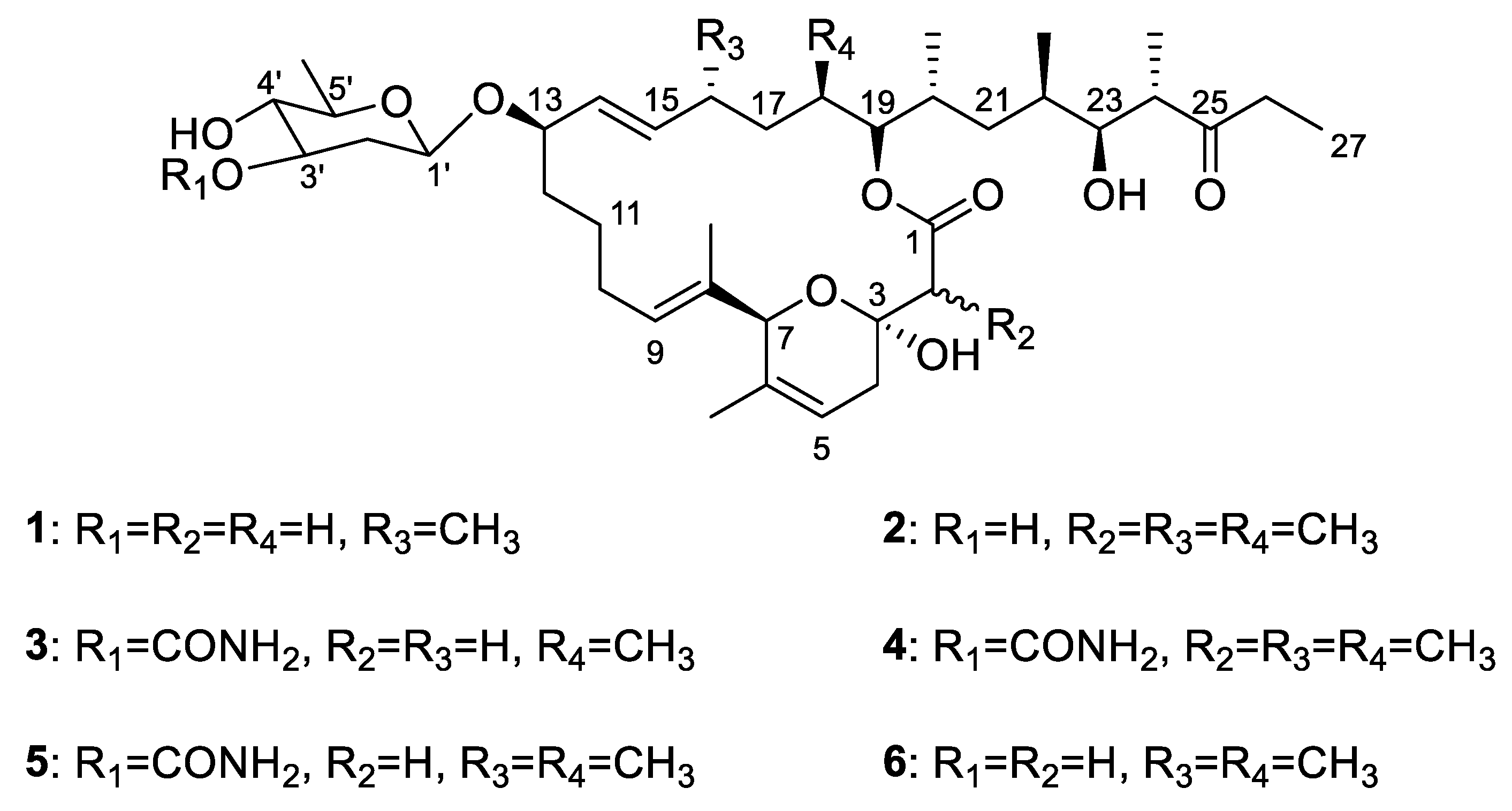
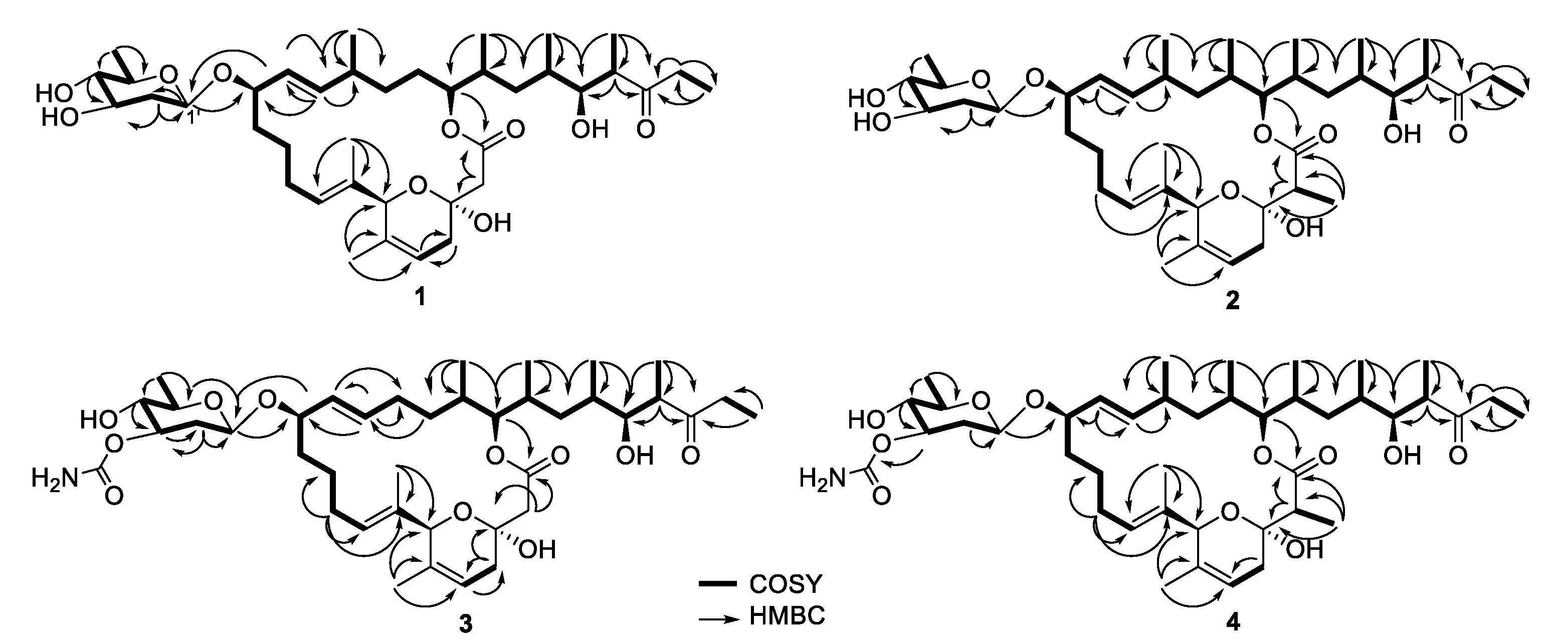
3.3. Structure Elucidation of Compounds

Compound 1 was obtained as a white powder. Its molecular formula, C39H64O10, was established on the basis of HRESIMS data, indicating eight degrees of unsaturation (Figure S8). The 1H NMR spectrum of 1 showed eight methyl proton signals at δH 0.71–1.41 and four olefinic proton signals [δH 5.15 (1H, dd), 5.30 (1H, dd), 5.38 (1H, m), 5.45 (1H, m)] (Table 1 and Figure S3). The 13C NMR and HSQC spectra of 1 showed eight methyl groups, ten methylene groups, sixteen methine groups, and five quaternary carbons (Figures S4 and S5). The spectroscopic data also revealed two olefinic, one ketone, one ketal and one lactone carbon signals. These results indicated that 1 possesses the same venturicidin scaffold as that of 6 (Figure 2). Analyses of the 1H and 13C NMR spectra of 1 and 6 showed that one methyl group (δH 0.78; δC 12.9) and one methine group (δH 1.80; δC 34.6) found in 6 were not present in 1 (Table S1). Instead, 1 contains an additional methylene group (δH 1.29, 1.47; δC 29.0). The HMBC cross peaks of H2-18 (δH 1.29, 1.47) with C-17 (δC 32.8) and C-19 (δC 78.3) indicated the methylene is located at C-18 (Figure 2 and Figure S6). Therefore, the structure of 1 differs from that of 6 in only one aspect: the methyl group at C-18 in 6 is replaced by a proton in 1. This was also confirmed by the HRESIMS data and molecular formulas of 1 and 6 which indicated 6 has one more carbon and two more hydrogens than 1. The structure of compound 1 was thus elucidated, and compound 1 was named as venturicidin G.
Compound 2 was purified as a white powder. The molecular formula of 2 was determined as C41H68O10 based on HRESIMS data (Figure S15). Similarly, 2 was identified to have the same scaffold as 6 based on their spectroscopic data (Figures S9–S14). However, the 1H and 13C NMR data revealed that the signals of methylene (δH 2.73, 2.54; δC 43.7) in 6 were substituted by signals of a methyl group (δH 1.12; δC 13.0) and a methine group (δH 2.57; δC 47.6) in 2, respectively (Table 1, Table 2 and Table S1). The HMBC correlations of H3-2CH3 (δH 1.12) with C-1 (δC 176.0), C-2 (δC 47.6) and C-3 (δC 95.7) and COSY correlation between H3-2CH3 (δH 1.12) and H-2 (δH 2.57) illustrated the position of the methyl at C-2 (Figure 2). From these results, the structure of compound 2 was elucidated, and compound 2 was named as venturicidin H.
Compound 3 was obtained as white powder. Its molecular formula, C40H65NO11, was inferred by HRESIMS data (Figure S21). The NMR data exhibited structure of 3 was highly similar to that of 5 (Figure S16–S20). The analysis of 1H NMR data of 5 exhibited a methyl (H3-16CH3, δH 0.89) that was absent in 3, while 3 had a methylene at H2-16 (δH 2.04, 1.97) rather than a methine at H-16 (δH 2.06) in 5 (Table 1 and Table S1). The HMBC correlations of H2-16 with C-15 (δC 137.4) and C-17 (δC 34.3) also revealed the methyl (δH 0.89) in 5 was missing in 3 (Figure 2), corresponding to a 14 Da mass decrease. Consequently, 3 was elucidated and named as venturicidin I.
Compound 4 was isolated as white powder, and revealed spectroscopic data remarkable similar with those of 5 (Figure S22–S27). The molecular formula of 4 was determined as C42H69NO11 compatibly with its HRESIMS data (Figure S28), which was 14 amu higher than that of 5, suggesting the presence of an additional methyl group (δH 1.13, δC 13.0) in 4 (Table 1 and Table 2). The HMBC correlations of H3-2CH3 (δH 1.13) with C-1 (δC 176.0), C-2 (δC 47.6) and C-3 (δC 95.7) and COSY correlation between H3-2CH3 (δH 1.13) and H-2 (δH 2.57) confirmed that the additional methyl was located at C-2 of 4 (Figure 2). Therefore, the structure of 4 was determined as illustrated in Figure 1, and compound 4 was named as venturicidin J.

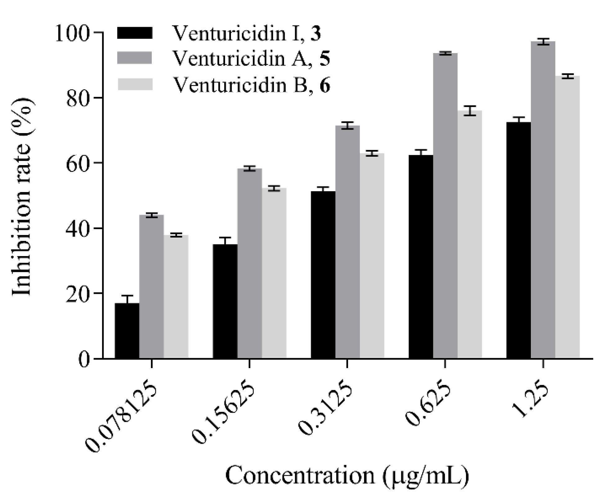
3.4. Antifungal Activity Assay

Compounds 1–6 were evaluated for their antifungal activities against P. oryzae in mycelial growth inhibition and conidial germination assays. The results showed EC50 values of the compounds 1–6 against mycelial growth were approximately 1.78, 1.43, 0.35, 1.40, 0.11, 0.15 µg/mL, respectively (Table 3). Notably, the compounds 3, 5 and 6 remarkably limit the mycelial radial elongation of P. oryzae (Figure 3), which showed comparable antifungal activity to the positive control carbendazim (EC50 = 0.30 µg/mL). Compounds 1, 2 and 4 showed moderate antifungal activities.
In addition to inhibiting mycelial growth, compounds 1–6 also inhibited the conidial germination of P. oryzae in different levels, with the EC50 values estimated to be 24.95, 5.55, 1.14, 4.49, 0.27 and 0.39 µg/mL, respectively (Table 3). These results indicated that compounds 5 and 6 exert higher antimicrobial activity against P. oryzae than the positive control carbendazim (EC50 = 3.99 µg/mL). Compounds 2, 3 and 4 showed comparable antifungal activity to the positive control, and compound 1 exhibited weaker inhibitory activity than carbendazim.

3.5. Structure Activity Relationship Analysis

The relationship between the structures and antifungal activity was elucidated based on the compounds’ molecular structures, and the discussion upon their antimicrobial activity results against different developmental stages of P. oryzae. Both compounds 5 and 6 showed similar antifungal activity against P. oryzae, indicating the presence of acylamino group at the C-3 position does not cause a prominent effect on its activity. Comparing the structures of 1 with 6, a methyl group at the C-18 (R4) was replaced by a hydrogen atom in 1, which impairs the antifungal activity against P. oryzae, so they have large activity differences in conidial germination inhibition. In contrast, between 3 and 5 the replacement in 3 happened on C-16 (R3), which indicates that they have no differences in antifungal activity. Thus, the methyl group in position R4 seems to increase the activity. Both compounds 2 and 4 introduce a methyl group at the C-2 position, lower the antimicrobial activity in comparison with that of 5 and 6. These results suggested that the antifungal activities of these compounds are influenced by the methylated positions in venturicidins. The SARs of these compounds provided a scientific basis for discovery of potent fungicides.

4. Discussion

With the increasing devastating rice blast disease, P. oryzae has provoked severe loss in the world [39]. The control of rice blast remains a long-standing agricultural issue associated with highly variable nature of P. oryzae [37,40]. In addition, P. oryzae has developed resistance to most of commercially available fungicides [16]. Thus, novel fungicides are a constant and critical need. Recently, microbial pesticides are widely applied in the control of plant diseases due to friendliness to the environment [41]. Unfortunately, there are few reports on the microbial pesticides against P. oryzae. This study indicated that secondary metabolites of Streptomyces sp. SN5452 may be promising fungicides in the control of rice blast, because they strongly inhibit the mycelial growth and the conidial germination of P. oryzae. The isolation and screening of Streptomyces are the prerequisites for obtaining bioactive natural products [42]. In our study, Streptomyces sp. SN5452 was isolated and purified from the gut of a millipede. By the analysis of 16S rRNA gene sequence, the strain belonged to Streptomyces genus, and the crude extract made from the fermentation culture of the strain showed prominent inhibitory activity against P. oryzae.
Streptomyces produce a number of secondary metabolites used in fields. The commercial kasugamycin produced by Streptomyces kasugaensis showed potential preventive effects against rice blast [43]. The insecticide avermectin has been used extensively for controlling Plutella xylostella and Pieris rapae in fields, which was produced by Streptomyces avermitilis [44,45,46]. Furthermore, Streptomyces fermentation products are considered as a valuable resource for the development of novel pesticides. The dimethyl sulfide and trimethyl sulfide produced by Streptomyces sp. AN090126, showed broad-spectrum antimicrobial activity against various plant-pathogenic bacteria and fungi, including Ralstonia solanacearum, Xanthomonas euvesicatoria, Sclerotinia homoeocarpa [47]. The LC50 value of endostemonine J, an ionophore antibiotic produced by Streptomyces sp. BS-1, against Aphis gossypii was 3.55 μg/mL at 72 h via leaf dipping assay [48]. Compound cinnoline-4-carboxylic acid was isolated from the fermentation culture of Streptomyces sp. KRA17-580 by Kim’s group, and completely inhibited the growth Digitaria ciliaris at a concentration of 50 μg/mL [49]. In this study, venturicidins A, B and venturicidins G-J were isolated from the fermentation culture of Streptomyces sp. SN5452. These compounds inhibited mycelial growth and conidial germination of P. oryzae, with EC50 values ranging from 0.11 to 1.78 μg/mL and from 0.27 to 24.95 μg/mL, respectively.
As we all know, structures of compounds were determined by the cumulative analyses of NMR, MS and X-ray data. The current study showed that the absolute configuration of compounds 1, 3, 5 and 6 were elucidated based on NMR and MS analyses and previously reported X-ray data [46]. However, the absolute configuration of compounds 2 and 4 were uncertain by spectroscopic data. We also tried to cultivate single-crystals of the compounds 2 and 4, but unfortunately it was not successful due to their structural specificity. Named as venturicidins G-J, compounds 1–4 enriched the structural diversity of 20-membered macrolide compounds [50,51,52,53,54,55], and compounds 5–6 were identified as venturicidins A–B. Further, venturicidins A, B and I exhibited good inhibition to mycelial growth and conidial germination of P. oryzae, which was comparable or superior to the positive control carbendazim. However, the actual field efficacy of these compounds should be further studied.

5. Conclusions

In conclusion, the crude extract made from the fermentation culture of Streptomyces sp. SN5452 showed inhibitory activity against P. oryzae. Compounds 1, 2, 3 and 4 were isolated and identified for the first time from the fermentation culture of Streptomyces sp. SN5452. Compounds 3, 5 and 6 exhibited good antimicrobial activity against P. oryzae. Given that microbial secondary metabolites exhibit excellent fungicide activity, they have the potential to become lead molecules for agricultural fungicides.

Supplementary Materials

The following supporting information can be downloaded at: https://www.mdpi.com/article/10.3390/microorganisms10081612/s1, Table S1: 1H (600 MHz) and 13C (150 MHz) NMR Data of Compounds 5 and 6 in d6-DMSO; Figure S1: Effects of crude extract of Streptomyces sp. SN5452 on the mycelial growth of P. oryzae; Figure S2: Neighbour-joining phylogenetic tree based on the 16S rRNA gene sequences with members of the genus Streptomyces; Figures S3–S28: HRESI-MS and NMR spectra of compounds 1–4.

Author Contributions

Conceptualization, Y.W. and D.Y.; methodology, Y.W.; software, Y.W. and Y.B.; validation, D.Y. and Y.B.; formal analysis, Y.W. and D.Y.; investigation, Y.B.; resources, D.Y.; writing—original draft preparation, Y.W.; writing—review and editing, Z.Y.; supervision, Y.B. and Z.Y. All authors have read and agreed to the published version of the manuscript.

Funding

This research was funded by the Innovation & Cultivation Project for Postgraduate of Shenyang Agricultural University in 2021 (grant number 2021YCXB17).

Institutional Review Board Statement

Not applicable.

Informed Consent Statement

Not applicable.

Data Availability Statement

Not applicable.

Acknowledgments

The authors thank Yongming Yan of Shenzhen University for technical assistance with NMR and MS spectra.

Conflicts of Interest

The authors declare no conflict of interest.

References

  1. Godfray, H.C.J.; Beddington, J.R.; Crute, I.R.; Haddad, L.; Lawrence, D.; Muir, J.F.; Pretty, J.; Robinson, S.; Thomas, S.M.; Toulmin, C. Food security: The challenge of feeding 9 billion people. Science 2010, 327, 812–818. [Google Scholar] [CrossRef] [PubMed]
  2. Muthayya, S.; Sugimoto, J.D.; Montgomery, S.; Maberly, G.F. An overview of global rice production, supply, trade, and consumption. Ann. N. Y. Acad. Sci. 2014, 1324, 7–14. [Google Scholar] [CrossRef] [PubMed]
  3. Sakulkoo, W.; Osés-Ruiz, M.; Garcia, O.E.; Soanes, D.M.; Littlejohn, G.R.; Hacker, C.; Correia, A.; Valent, B.; Talbot, N.J. A single fungal MAP kinase controls plant cell-to-cell invasion by the rice blast fungus. Science 2018, 359, 1399–1403. [Google Scholar] [CrossRef] [PubMed]
  4. Talbot, N.J. On the trail of a cereal killer: Exploring the biology of Magnaporthe grisea. Annu. Rev. Microbiol. 2003, 57, 177–202. [Google Scholar] [CrossRef]
  5. Lam, V.B.; Meyer, T.; Arias, A.A.; Ongena, M.; Oni, F.E.; Höfte, M. Bacillus cyclic lipopeptides Iturin and Fengycin control rice blast caused by Pyricularia oryzae in potting and acid sulfate soils by direct antagonism and induced systemic resistance. Microorganisms 2021, 9, 1441. [Google Scholar] [CrossRef]
  6. Odjo, T.; Diagne, D.; Adreit, H.; Milazzo, J.; Raveloson, H.; Andriantsimialona, D.; Kassankogno, A.I.; Ravel, S.; Gumedzoé, Y.M.D.; Ouedraogo, I.; et al. Structure of African populations of Pyricularia oryzae from rice. Phytopathology 2021, 111, 1428–1437. [Google Scholar] [CrossRef]
  7. Dang, Y.J.; Wei, Y.; Wang, Y.Y.; Liu, S.S.; Julia, C.; Zhang, S.H. Cleavage of PrePL by Lon promotes growth and pathogenesis in Magnaporthe oryzae. Environ. Microbiol. 2021, 23, 4881–4895. [Google Scholar] [CrossRef]
  8. Manandhar, H.K.; Jorgensen, H.J.L.; Smedegaard-Petersen, V.; Mathur, S.B. Seedborne infection of rice by Pyricularia oryzae and its transmission to seedlings. Plant Dis. 1998, 82, 1093–1099. [Google Scholar] [CrossRef]
  9. Hayashi, K.; Yoshida, T.; Hayano-Saito, Y. Detection of white head symptoms of panicle blast caused by Pyricularia oryzae using cut-flower dye. Plant Methods 2019, 15, 159. [Google Scholar] [CrossRef]
  10. Langner, T.; Białas, A.; Kamoun, S. The blast fungus decoded: Genomes in flux. mBio 2018, 9, e00571-18. [Google Scholar] [CrossRef]
  11. Law, J.W.; Ser, H.L.; Khan, T.M.; Chuah, L.H.; Pusparajah, P.; Chan, K.G.; Goh, B.H.; Lee, L.H. The potential of Streptomyces as biocontrol agents against the rice blast fungus, Magnaporthe oryzae (Pyricularia oryzae). Front. Microbiol. 2017, 8, 3. [Google Scholar] [CrossRef]
  12. Kunova, A.; Pizzatti, C.; Cortesi, P. Impact of tricyclazole and azoxystrobin on growth, sporulation and secondary infection of the rice blast fungus, Magnaporthe oryzae. Pest Manag. Sci. 2013, 69, 278–284. [Google Scholar] [CrossRef]
  13. Asibi, A.E.; Chai, Q.; Coulter, J.A. Rice blast: A disease with implications for global food security. Agronomy 2019, 9, 451. [Google Scholar] [CrossRef]
  14. Peng, Q.; Zhao, H.; Zhao, G.; Gao, X.; Miao, J.; Liu, X. Resistance assessment of pyraoxystrobin in Magnaporthe oryzae and the detection of a point mutation in cyt b that confers resistance. Pestic. Biochem. Physiol. 2022, 180, 105006. [Google Scholar] [CrossRef]
  15. Li, C.; Wang, K.; Zhang, H.; Yang, D.; Deng, Y.; Wang, Y.; Qi, Z. Development of a LAMP method for detecting F129L mutant in azoxystrobin-resistant Pyricularia oryzae. Fungal Biol. 2022, 126, 47–53. [Google Scholar] [CrossRef]
  16. Zheng, F.; Li, Y.C.; Zhang, Z.X.; Jia, J.L.; Hu, P.T.; Zhang, C.Q.; Xu, H.H. Novel strategy with an eco-friendly polyurethane system to improve rainfastness of tea saponin for highly efficient rice blast control. J. Clean. Prod. 2020, 264, 121685. [Google Scholar] [CrossRef]
  17. Pooja, K.; Katoch, A. Past, present and future of rice blast management. Plant Sci. Today 2014, 1, 165–173. [Google Scholar] [CrossRef]
  18. Yoon, M.Y.; Cha, B.; Kim, J.C. Recent trends in studies on botanical fungicides in agriculture. Plant Pathol. J. 2013, 29, 1–9. [Google Scholar] [CrossRef]
  19. Palaniyandi, S.A.; Yang, S.H.; Zhang, L.X.; Suh, J.W. Effects of actinobacteria on plant disease suppression and growth promotion. Appl. Microbiol. Biotechnol. 2013, 97, 9621–9636. [Google Scholar] [CrossRef]
  20. Chaiharn, M.; Theantana, T.; Pathom-Aree, W. Evaluation of biocontrol activities of Streptomyces spp. against rice blast disease fungi. Pathogens 2020, 9, 126. [Google Scholar] [CrossRef]
  21. Zhao, J.W.; Han, L.Y.; Yu, M.Y.; Cao, P.; Li, D.M.; Guo, X.W.; Liu, Y.Q.; Wang, X.J.; Xiang, W.J. Characterization of Streptomyces sporangiiformans sp. nov., a novel soil actinomycete with antibacterial activity against Ralstonia solanacearum. Microorganisms 2019, 7, 360. [Google Scholar] [CrossRef]
  22. Yadav, A.N.; Verma, P.; Kumar, S.; Kumar, V.; Kumar, M.; Chellammal, T.; Sugitha, K.; Singh, B.P.; Saxena, A.K.; Dhaliwal, H.S. Actinobacteria from Rhizosphere: Molecular Diversity, Distributions, and Potential Biotechnological Applications. In New and Future Developments in Microbial Biotechnology and Bioengineering; Elsevier: Amsterdam, The Netherlands, 2018; pp. 13–41. [Google Scholar]
  23. Hwang, K.S.; Kim, H.U.; Charusanti, P.; Palsson, B.Ø.; Lee, S.Y. Systems biology and biotechnology of Streptomyces species for the production of secondary metabolites. Biotechnol. Adv. 2014, 32, 255–268. [Google Scholar] [CrossRef]
  24. Copping, L.G.; Duke, S.O. Natural products that have been used commercially as crop protection agents. Pest Manag. Sci. 2007, 63, 524–554. [Google Scholar] [CrossRef]
  25. Han, C.Y.; Yu, Z.Y.; Zhang, Y.T.; Wang, Z.Y.; Zhao, J.W.; Huang, S.X.; Ma, Z.H.; Wen, Z.Y.; Liu, C.X.; Xiang, W.S. Discovery of Frenolicin B as potential agrochemical fungicide for controlling Fusarium head blight on wheat. J. Agric. Food Chem. 2021, 69, 2108–2117. [Google Scholar] [CrossRef]
  26. Shi, L.Q.; Wu, Z.Y.; Zhang, Y.N.; Zhang, Z.G.; Fang, W.; Wang, Y.Y.; Wan, Z.Y.; Wang, K.M.; Ke, S.Y. Herbicidal secondary metabolites from actinomycetes: Structure diversity, modes of action, and their roles in the development of herbicides. J. Agric. Food Chem. 2020, 68, 17–32. [Google Scholar] [CrossRef]
  27. Kaur, T.; Vasudev, A.; Sohal, S.K.; Manhas, R.K. Insecticidal and growth inhibitory potential of Streptomyces hydrogenans DH16 on major pest of India, Spodoptera litura (Fab.) (Lepidoptera: Noctuidae). BMC Microbiol. 2014, 14, 227. [Google Scholar] [CrossRef]
  28. Bi, Y.H.; Yu, Z.G. Diterpenoids from Streptomyces sp. SN194 and their antifungal activity against Botrytis cinerea. J. Agric. Food Chem. 2016, 64, 8525–8529. [Google Scholar] [CrossRef]
  29. Tian, H.; Shafi, J.; Ji, M.S.; Bi, Y.H.; Yu, Z.G. Antimicrobial metabolites from Streptomyces sp. SN0280. J. Nat. Prod. 2017, 80, 1015–1019. [Google Scholar] [CrossRef]
  30. Heo, J.; Hamada, M.; Cho, H.; Weon, H.Y.; Kim, J.S.; Hong, S.B.; Kim, S.J.; Kwon, S.W. Weissella cryptocerci sp. nov., isolated from gut of the insect Cryptocercus kyebangensis. Int. J. Syst. Evol. Microbiol. 2019, 69, 2801–2806. [Google Scholar] [CrossRef]
  31. Saitou, N.; Nei, M. The neighbor-joining method: A new method for reconstructing phylogenetic trees. Mol. Biol. Evol. 1987, 4, 406–425. [Google Scholar]
  32. Kumar, S.; Stecher, G.; Tamura, K. Mega7: Molecular evolutionary genetics analysis version 7.0 for bigger datasets. Mol. Biol. Evol. 2016, 33, 1870–1874. [Google Scholar] [CrossRef] [PubMed]
  33. Atlas, R.M. The Handbook of Microbiological Media for the Examination of Food, 2nd ed.; CRC Press: Boca Raton, FL, USA, 2006. [Google Scholar] [CrossRef]
  34. Shirling, E.B.; Gottlieb, D. Methods for characterization of Streptomyces species. Int. J. Syst. Bacteriol. 1966, 16, 313–340. [Google Scholar] [CrossRef]
  35. Li, R.Y.; Wu, X.M.; Yin, X.H.; Long, Y.H.; Li, M. Naturally produced citral can significantly inhibit normal physiology and induce cytotoxicity on Magnaporthe grisea. Pestic. Biochem. Physiol. 2015, 118, 19–25. [Google Scholar] [CrossRef] [PubMed]
  36. Miao, J.Q.; Zhao, G.S.; Wang, B.; Du, Y.X.; Li, Z.W.; Gao, X.H.; Zhang, C.; Liu, X.L. Three point-mutations in cytochrome b confer resistance to trifloxystrobin in Magnaporthe oryzae. Pest Manag. Sci. 2020, 76, 4258–4267. [Google Scholar] [CrossRef]
  37. Maciel, J.L.; Ceresini, P.C.; Castroagudin, V.L.; Zala, M.; Kema, G.H.; McDonald, B.A. Population structure and pathotype diversity of the wheat blast pathogen Magnaporthe oryzae 25 years after its emergence in Brazil. Phytopathology 2014, 104, 95–110. [Google Scholar] [CrossRef]
  38. Shaaban, K.A.; Singh, S.; Elshahawi, S.I.; Wang, X.C.; Ponomareva, L.V.; Sunkara, M.; Copley, G.C.; Hower, J.C.; Morris, A.J.; Kharel, M.K.; et al. Venturicidin C, a new 20-membered macrolide produced by Streptomyces sp. TS-2-2. J. Antibiot. 2014, 67, 223–230. [Google Scholar] [CrossRef]
  39. Chadha, S. Molecular detection of Magnaporthe oryzae from rice seeds. Methods Mol. Biol. 2021, 2356, 187–197. [Google Scholar]
  40. Park, S.Y.; Milgroom, M.G.; Han, S.S.; Kang, S.; Lee, Y.H. Genetic differentiation of Magnaporthe oryzae populations from scouting plots and commercial rice fields in Korea. Phytopathology 2008, 98, 436–442. [Google Scholar] [CrossRef]
  41. Ling, L.; Han, X.Y.; Li, X.; Zhang, X.; Wang, H.; Zhang, L.D.; Cao, P.; Wu, Y.T.; Wang, X.J.; Zhao, J.W.; et al. A Streptomyces sp. NEAU-HV9: Isolation, identification, and potential as a biocontrol agent against Ralstonia Solanacearum of tomato plants. Microorganisms 2020, 8, 351. [Google Scholar] [CrossRef]
  42. Montesinos, E. Development, registration and commercialization of microbial pesticides for plant protection. Int. Microbiol. 2003, 6, 245–252. [Google Scholar] [CrossRef]
  43. Umezawa, H.; Hamada, M.; Suhara, Y.; Hashimoto, T.; Ikekawa, T. Kasugamycin, a new antibiotic. J. Antibiot. 1965, 18, 101–104. [Google Scholar]
  44. Kaziem, A.E.; Gao, Y.H.; Zhang, Y.; Qin, X.Y.; Xiao, Y.N.; Zhang, Y.H.; You, H.; Li, J.H.; He, S. α-Amylase triggered carriers based on cyclodextrin anchored hollow mesoporous silica for enhancing insecticidal activity of avermectin against Plutella xylostella. J. Hazard Mater. 2018, 359, 213–221. [Google Scholar] [CrossRef]
  45. Wang, P.; Lu, Y.; Dong, J.; Jing, L.; Yuan, Z.Q.; Yang, J.G.; Qiao, Y. Control effect of 13 pesticides on Pieris rapae in the cauliflower field. Agrochemicals 2017, 56, 300–302. [Google Scholar]
  46. Burg, R.W.; Miller, B.M.; Baker, E.E.; Birnbaum, J.; Currie, S.A.; Hartman, R.; Kong, Y.L.; Monaghan, R.L.; Olson, G.; Putter, I.; et al. Avermectins, new family of potent anthelmintic agents: Producing organismand fermentation. Antimicrob. Agents Chemother. 1979, 15, 361–367. [Google Scholar] [CrossRef]
  47. Le, K.D.; Yu, N.H.; Park, A.R.; Park, D.J.; Kim, C.J.; Kim, J.C. Streptomyces sp. AN090126 as a biocontrol agent against bacterial and fungal plant diseases. Microorganisms 2022, 10, 791. [Google Scholar] [CrossRef]
  48. Zhao, H.M.; Yang, A.P.; Zhang, N.; Li, S.Y.; Yuan, T.J.; Ding, N.; Zhang, S.W.; Bao, S.; Wang, C.; Zhang, Y.N.; et al. Insecticidal endostemonines A-J produced by endophytic Streptomyces from Stemona sessilifolia. J. Agric. Food Chem. 2020, 68, 1588–1595. [Google Scholar] [CrossRef]
  49. Kim, H.J.; Bo, A.B.; Kim, J.D.; Kim, Y.S.; Khaitov, B.; Ko, Y.K.; Cho, K.M.; Jang, K.S.; Park, K.W.; Choi, J.S. Herbicidal characteristics and structural identification of the potential active compounds from Streptomyces sp. KRA17-580. J. Agric. Food Chem. 2020, 68, 15373–15380. [Google Scholar] [CrossRef]
  50. Brufani, M.; Cerrini, S.; Fedeli, W.; Musu, C.; Cellai, L.; Keller-Schierlein, W. Structures of the venturicidins A and B. Experientia 1971, 27, 604–606. [Google Scholar] [CrossRef]
  51. Omura, S.; Tanaka, Y.; Nakagawa, A.; Iwai, Y.; Inoue, M.; Tanaka, H. Irumamycin, a new antibiotic active against phytopathogenic fungi. J. Antibiot. 1982, 35, 256–257. [Google Scholar] [CrossRef]
  52. Omura, S.; Nakagawa, A.; Imamura, N.; Kushida, K.; Liu, C.M.; Sello, L.H.; Westley, J.W. Structure of a new macrolide antibiotic, X-14952B. J. Antibiot. 1985, 38, 674–676. [Google Scholar] [CrossRef]
  53. Ohta, S.; Uy, M.M.; Yanai, M.; Ohta, E.; Hirata, T.; Ikegami, S. Exiguolide, a new macrolide from the marine sponge Geodia exigua. Tetrahedron Lett. 2006, 47, 1957–1960. [Google Scholar] [CrossRef]
  54. Peng, F.; Wang, C.X.; Xie, Y.; Jing, H.L.; Che, L.J.; Uribe, P.; Bull, A.T.; Goodfellow, M.; Jiang, H.; Lian, Y.Y. A new 20-membered macrolide produced by a marine-derived Micromonospora strain. Nat. Prod. Res. 2013, 27, 1366–1371. [Google Scholar]
  55. Li, H.H.; Zhang, M.X.; Li, H.J.; Yu, H.; Chen, S.; Wu, W.H.; Sun, P. Discovery of venturicidin congeners and identification of the biosynthetic gene cluster from Streptomyces sp. NRRL S-4. J. Nat. Prod. 2021, 84, 110–119. [Google Scholar] [CrossRef]
Figure 1. Structures of compounds 1–6.
Microorganisms 10 01612 g001
Figure 2. 2D NMR correlations of compounds 14. 2D NMR: two-dimensional nuclear magnetic resonance.
Microorganisms 10 01612 g002
Figure 3. Effect of venturicidin I, 3, venturicidin A, 5, and venturicidin B, 6 on mycelial growth of P. oryzae.
Microorganisms 10 01612 g003
Table 1. 1H (600 MHz) NMR Data of Compounds 1–4 in d6-DMSO a.
PositionδH, Mult (J in Hz)
1234
2 α2.74, d (15.6)2.57, q (7.8)2.76, d (16.2)2.57, q (7.8)
2 β2.51, overlap 2.56, d (16.2)
2-CH3 1.12, overlap 1.13, d (7.2)
3-OH5.49, s4.93, s5.50, overlap4.93, s
4 α2.16, d (17.4)2.08, m2.18, m2.07, m
4 β1.92, m2.00, m1.97, m2.03, m
55.45, m5.46, m5.45, m5.46, m
6-CH31.41, s1.41, s1.41, s1.41, s
74.33, brs4.30, brs4.35, brs4.30, brs
8-CH31.37, s1.36, s1.37, s1.36, s
95.38, m5.24, t (7.2)5.50, overlap5.41, dd (10.2, 4.2)
10 α2.01, m2.08, m2.11, m2.07, m
10 β1.92, m1.79, m1.79, m1.79, m
11 α1.56, m1.27, m1.31, m1.46, m
11 β1.23, m1.12, overlap1.23, m1.22, m
12 α1.47, m1.56, m1.53, m1.56, m
12 β1.23, m1.27, m1.44, m1.32, m
133.78, m3.89, dd (12.6, 7.2)4.02, m3.92, m
145.30, dd (15.6, 8.4)5.40, dd (10.2, 4.2)5.28 m5.25, m
155.15, dd (15.6, 9.0)5.24, t (7.2)5.40, m5.25, m
16 α1.92, m2.00, m2.04, m2.07, m
16 β 1.97, m
16-CH30.93, d (6.6)0.90, overlap 0.91, d (6.6)
17 α1.29, m1.27, m1.31, m1.22, m
17 β0.90, overlap0.90, overlap0.89, overlap0.89, overlap
18 α1.47, m1.69, m1.74, m1.70, m
18 β1.29, m
18-CH3 0.77, d (6.6)0.82, d (7.2)0.77, d (6.6)
194.72, m4.54, dd (6.6, 4.8)4.64, m4.54, m
201.71, m1.79, m1.69, m1.79, m
20-CH30.80, d (6.6)0.79, d (6.6)0.81, d (7.2)0.79, d (6.6)
21 α1.29, m1.27, m1.23, m1.32, m
21 β0.98, m0.95, m0.84, d (7.2)0.95, m
221.56, m1.56, m1.53, m1.56, m
22-CH30.71, d (6.6)0.72, d (6.6)0.69, d (6.6)0.72, d (6.6)
233.42, m3.40, m3.41, m3.40, m
23-OH4.88, brs 5.05, brs5.04, brs
242.62, m2.62, m2.61, m2.62, m
24-CH30.85, d (6.6)0.85, d (7.2)0.84, d (7.2)0.85, d (7.2)
26 α2.51, overlap2.53, m2.49, m2.53, m
26 β2.01, m2.00, m1.97, m2.03, m
270.90, overlap0.90, overlap0.89, overlap0.89, overlap
1′4.41, dd (9.6, 1.8)4.47, dd (9.6, 1.8)4.58, m4.54, m
2′ α1.92, m1.89, m2.04, m2.03, m
2′ β1.29, m1.27, m1.31, m1.22, m
3′4.47, m3.40, m4.49, m4.47, m
3′-OH4.88, brs
3′-CONH2 6.44, brs6.48, brs
4′2.68, m2.68, m2.91, m2.91, m
4′-OH 4.56, brs
5′3.01, m3.03, m3.19, m3.14, m
5′-CH31.10, d (6.0)1.12, overlap1.15, d (6.0)1.15, d (6.0)
a Assignments were based on COSY, HSQC and gHMBC experiments. NMR: nuclear magnetic resonance); DMSO: dimethyl sulfoxide; COSY: correlation spectroscopy; HSQC. heteronuclear singular quantum correlation); gHMBC: gradient heteronuclear multiple bond correlation.
Table 2. 13C (150 MHz) NMR Data of Compounds 14 in d6-DMSO.
PositionδC, Type
1234
1171.7, s176.0, s171.9, s176.0, s
244.0, t47.6, d43.6, t47.6, d
2-CH3 13.0, q 13.0, q
393.5, s95.7, s93.4, s95.7, s
434.4, t32.7, t34.6, t32.7, t
5117.5, d117.5, d117.5, d117.4, d
6131.7, s131.9, s131.8, s131.9, s
6-CH318.8, q18.9, q18.9, q18.9, q
779.2, d79.0, d79.1, d79.0, d
8134.3, s133.5, s134.0, s133.6, s
8-CH311.1, q10.7, q10.8, q10.7, q
9129.3, d129.7, d129.1, d129.0, d
1026.7, t27.2, t26.7, t27.1, t
1125.3, t25.7, t24.7, t25.7, t
1229.0, t33.9, t32.9, t33.8, t
1381.3, d80.0, d78.4, d80.2, d
14131.8, d129.0, d131.1, d129.6, d
15135.7, d138.3, d137.4, d138.5, d
1637.2, d36.4, d30.4, t36.4, d
16-CH321.1, q21.6, q 21.6, q
1732.8, t40.4, t34.3, t40.4, t
1829.0, t32.7, d33.9, d32.7, d
18-CH3 16.5, q12.6, q16.5, q
1978.3, d82.2, d80.9, d82.2, d
2031.9, d31.2, d31.5, d31.2, d
20-CH316.0, q16.2, q15.5, q16.2, q
2134.6, t35.0, t36.8, t35.0, t
2231.2, d31.3, d31.3, d31.3, d
22-CH311.1, q11.0, q10.9, q10.9, q
2376.0, d76.3, d76.3, d76.3, d
2449.0, d49.1, d48.9, d49.1, d
24-CH313.4, q13.4, q13.4, q13.4, q
25214.4, s214.4, s214.5, s214.4, s
2635.2, t35.2, t35.3, t35.2, t
277.4, q7.4, q7.4, q7.4, q
1′98.7, d97.5, d96.7, d97.0, d
2′40.0, t40.0, t37.5, t37.5, t
3′70.5, d70.5, d72.6, d72.7, d
3′-CONH2 156.4, s156.4, s
4′76.9, d76.9, d73.5, d73.5, d
5′71.5, d71.5, d71.7, d71.7, d
5′-CH318.1, q18.1, q18.0, q18.0, q
Table 3. Half maximal effective concentration (EC50) values of Compounds 1–6 on P. oryzae a.
CompoundMycelial Growth Inhibition EC50, µg/mL (±SD)Conidial Germination Inhibition EC50, µg/mL (±SD)
venturicidin G, 11.78 ± 0.0924.95 ± 1.63
venturicidin H, 21.43 ± 0.025.55 ± 0.12
venturicidin I, 30.35 ± 0.031.14 ± 0.03
venturicidin J, 41.40 ± 0.094.49 ± 0.28
venturicidin A, 50.11 ± 0.000.27 ± 0.02
venturicidin B, 60.15 ± 0.010.39 ± 0.01
Carbendazim b0.30 ± 0.013.99 ± 0.08
a Data shown are the mean of three independent experiments and presented as mean ± standard deviation (SD). b Positive control.
Publisher’s Note: MDPI stays neutral with regard to jurisdictional claims in published maps and institutional affiliations.

