Communication

Investigation of Delafloxacin Resistance in Multidrug-Resistant Escherichia coli Strains and the Detection of E. coli ST43 International High-Risk Clone

1
Institute of Medical Microbiology, Semmelweis University, 1089 Budapest, Hungary
2
Central Microbiology Laboratory, National Institute of Hematology and Infectious Disease, Central Hospital of Southern-Pest, 1097 Budapest, Hungary
3
Human Microbiota Study Group, Semmelweis University-Eötvös Lóránd Research Network, 1089 Budapest, Hungary
*
Author to whom correspondence should be addressed.
Microorganisms 2023, 11(6), 1602; https://doi.org/10.3390/microorganisms11061602
Submission received: 26 May 2023 / Revised: 13 June 2023 / Accepted: 15 June 2023 / Published: 16 June 2023

Abstract

:
Delafloxacin is a novel fluoroquinolone agent that is approved for clinical application. In this study, we analyzed the antibacterial efficacy of delafloxacin in a collection of 47 Escherichia coli strains. Antimicrobial susceptibility testing was performed by the broth microdilution method and minimum inhibitory concentration (MIC) values were determined for delafloxacin, ciprofloxacin, levofloxacin, moxifloxacin, ceftazidime, cefotaxime, and imipenem. Two multidrug-resistant E. coli strains, which exhibited delafloxacin and ciprofloxacin resistance as well as extended-spectrum beta-lactamase (ESBL) phenotype, were selected for whole-genome sequencing (WGS). In our study, delafloxacin and ciprofloxacin resistance rates were 47% (22/47) and 51% (24/47), respectively. In the strain collection, 46 E. coli were associated with ESBL production. The MIC50 value for delafloxacin was 0.125 mg/L, while all other fluoroquinolones had an MIC50 value of 0.25 mg/L in our collection. Delafloxacin susceptibility was detected in 20 ESBL positive and ciprofloxacin resistant E. coli strains; by contrast, E. coli strains that exhibited a ciprofloxacin MIC value above 1 mg/L were delafloxacin-resistant. WGS analysis on the two selected E. coli strains (920/1 and 951/2) demonstrated that delafloxacin resistance is mediated by multiple chromosomal mutations, namely, five mutations in E. coli 920/1 (gyrA S83L, D87N, parC S80I, E84V, and parE I529L) and four mutations in E. coli 951/2 (gyrA S83L, D87N, parC S80I, and E84V). Both strains carried an ESBL gene, blaCTX-M-1 in E. coli 920/1 and blaCTX-M-15 in E. coli 951/2. Based on multilocus sequence typing, both strains belong to the E. coli sequence type 43 (ST43). In this paper, we report a remarkable high rate (47%) of delafloxacin resistance among multidrug-resistant E. coli as well as the E. coli ST43 international high-risk clone in Hungary.

1. Introduction

The spread of multidrug-resistant bacteria is a considerable challenge worldwide. In the wake of COVID-19, the circulation of antibiotic-resistant bacteria was enhanced, turning into a pandemic that threatens public health globally [1,2,3]. Antibiotic-resistant bacteria are the leading causative agents of severe infections, including bacteriaemia, pneumonia, and urinary tract infection. Usually, antibiotic-resistant bacterial infections occur in hospitalized patients, and these infections are associated with higher mortality rates. A recent study described that antibiotic-resistant bacterial infections correspond to 1.27 million patients’ deaths in a single year worldwide [4,5,6]. The major pathogens of antibiotic-resistant bacteria belong to the ESKAPE group, (Enterococcus spp., Staphylococcus aureus, Klebsiella pneumoniae, Acinetobacter baumannii, Pseudomonas aeruginosa, and Enterobacter spp.), which are responsible for the high number of difficult-to-treat infections, because a limited number of effective antibiotics are available for antibacterial therapy [7,8]. World Health Organization (WHO) released a priority list of antibiotic-resistant bacteria, where discovery and research are needed for effective antibiotics. On the list of the “Priority 1: critical” group, certain bacteria are present, namely, A. baumannii, P. aeruginosa, and Enterobacteriaceae, which are resistant to carbapenems and to third-generation cephalosporins [9].
In recent years, several novel antimicrobial agents were approved for clinical use that are effective against bacteria that are already resistant to commonly used antibiotics. Among these novel agents, we can find plazomicin, cefiderocol as well as beta-lactam and beta-lactamase-inhibitor combinations [10,11,12,13].
New fluoroquinolone agents, such as delafloxacin, finafloxacin, and zabofloxacin, with enhanced antibacterial efficacy were also approved for clinical application and were marketed in several countries [14,15,16,17]. Fluoroquinolones are nucleic-acid-synthesis-inhibitor antibacterial agents, targeting both bacterial gyrase and topoisomerase IV enzymes, which makes them suitable as products with broad-spectrum antibacterial efficacy [18,19,20,21]. Due to widespread fluoroquinolone resistance, the clinical indication of earlier fluoroquinolone agents (e.g., ciprofloxacin and levofloxacin) has been restricted [22,23].
Delafloxacin (whose earlier names were ABT-492, RX-3341, and WQ-3034) is a new fluoroquinolone agent with an anionic, non-zwitterionic structure. Delafloxacin demonstrates a potent bactericidal effect against both Gram-positive and Gram-negative bacteria because it inhibits both bacterial DNA gyrase and topoisomerase IV enzymes. The chemical structure of delafloxacin is 1-(6-amino-3,5-difluoro-2-pyridinyl)-8-chloro-6-fluoro-7-(3-hydroxy-1-azetidinyl)-4-oxo-1,4-dihydro-3-quinolinecarboxylate [14,18]. Based on its chemical structure, it is a weak acid, and it remains uncharged in acidic environment, which enables its transmembrane transfer into the bacterial cell, where it accumulates. In a neutral pH (e.g., in intracellular space), it stays in its anionic, deprotonated form and maintains its concentration-dependent antibacterial activity. The anionic feature of delafloxacin makes it suitable as a product with an enhanced antibacterial efficacy in acidic environments, for example, in inflammations of human tissues, in phagolysosomes, in infections of skin and soft tissue, and in abscesses [18,23]. Furthermore, delafloxacin has other features in its chemical structure. A heteroaromatic substitution is located on delafloxacin, which provides a larger molecular surface that enhances antibacterial activity against strains that are resistant to earlier fluoroquinolones, because delafloxacin can bind to more sites on the target molecules. A chlorine atom is also located on delafloxacin, which provides a strong polarity and enhances its efficacy against anaerobic bacteria [14,18]. Delafloxacin can be administered through the oral or intravenous way. In an oral administration of delafloxacin, a 450 mg dose is necessary to achieve a corresponding concentration–time profile, compared to that of the intravenous 300 mg dose. In certain cases, 58.8% of the oral bioavailability of delafloxacin can be decreased through pharmacological interactions (e.g., by chelation effects) with other medications that contain multivalent metal cations, such as Al3+, Mg2+, Fe2+, or Zn2+ [14,18,22,23]. Delafloxacin does not inhibit cytochrome P450 isoenzymes; therefore, it does not have clinically relevant drug–drug interactions with the most frequently used medicines. Interestingly, no potential synergistic or antagonistic effects were detected between delafloxacin and other commonly used antibiotics [14,18]. Approximately 84% of delafloxacin binds to plasma proteins. A higher dose of delafloxacin was administered to patients with kidney failure, but hepatic impairment did not significantly affect the antibacterial activity of delafloxacin [14]. The side effects of earlier fluoroquinolone agents are well known. Among the most frequent side effects of fluoroquinolones, we find tendinitis, tendon rupture, photosensitivity, neurological symptoms, exacerbations of myasthenia gravis, muscle weakness, and QT interval prolongation. The side effects of delafloxacin have also been described, namely, peripheral neuropathy, hypersensitivity, and Clostridioides (Clostridium) difficile-associated diarrhea as possible, but less severe, compared to earlier fluoroquinolones; it is noteworthy to mention that these adverse effects were detected in a dose-dependent manner. The most frequently detected treatment-emergent adverse effects during a delafloxacin therapy were diarrhea, vomiting, and extravasation on the infusion site. Among the uncommon treatment-related complications, we find hyperglycemic episodes (in a single patient out of ten examined) and an elevation of transaminase enzymes (in a single patient out of twenty-two examined). Additionally, a lack of photosensitivity and cardiotoxicity were also demonstrated. According to the side effect profile, delafloxacin is a well-tolerated antibiotic [14,18,23]. Delafloxacin is currently approved for therapy in adults’ acute bacterial skin and skin-structure infections (ABSSSI), as well as for community-acquired bacterial pneumonia (CABP) [23,24,25,26,27]. However, additional potential indications for delafloxacin therapy can be bloodstream infection and intra-abdominal infection [23].
Resistance to fluoroquinolones in Enterobacterales is explained by the accumulation of mutations in gyrase- (gyrA, gyrB) and topoisomaers IV (parC, parE)-coding genes. These mutations develop in the specific sequences of gyrA, gyrB, parC, and parE genes, which are called quinolone-resistance-determining regions (QRDRs) [28,29]. Additionally, plasmid-mediated quinolone resistance (PMQR) determinants were described, which enhance the development of fluoroquinolone resistance. PMQR includes Qnr determinants, Aminoglycoside-acetyltransferase-(6′)-Ib-cr variant, and QepA and OqxAB efflux pumps [30,31,32,33].
The aim of this study is the investigation of the antibacterial efficacy of delafloxacin in Escherichia coli strains.

2. Materials and Methods

2.1. Strains

A total of 47 non-repetitive E. coli strains were collected between September and December 2022 at South-Pest Central Hospital, National Institute of Hematology and Infectious Diseases, from various clinical samples, including hemoculture and urine. All isolates were identified by matrix-assisted laser desorption ionization time-of-flight mass spectrometry (MALDI Biotyper, Bruker, Bremen, Germany). The inclusion criteria of E. coli strains in this study were resistance to ciprofloxacin and/or resistance to third-generation cephalosporins or confirmed extended-spectrum β-lactamase (ESBL) positivity. ESBL positivity was determined by a double-disk synergy test.

2.2. Determination of the Minimum Inhibitory Concentration (MIC)

Antibiotic susceptibility testing was performed for delafloxacin, ciprofloxacin, levofloxacin, moxifloxacin, ceftazidime, cefotaxime, and imipenem. MIC values were determined by the broth microdilution method in Muller–Hinton broth in 96-well microplates. The MIC results were interpreted according to the latest EUCAST protocol (www.eucast.org), accessed on 10 January 2023. E. coli ATCC 25922 was the control strain.

2.3. Whole-Genome Sequencing (WGS)

WGS analysis was performed on two selected E. coli strains (ECO-SEOMI-LKH 920/1 and 951/2). The selection criteria were E. coli strains exhibiting ciprofloxacin and delafloxacin resistance as well as ESBL phenotype. WGS was performed by the Illumina MiSeq system in Eurofins BIOMI Kft (Gödöllő, Hungary). Briefly, genomic DNA was extracted by the NucleoSpin Microbial DNA Mini kit (Macherey-Nagel, Düren, Germany). The amount of isolated DNA was measured by Qubit fluorometer, and the quality of DNA was tested by microcapillary electrophoresis (Tape Station 4150, Agilent, Waldbronn, Germany). Libraries were prepared by az Illumina DNA Prep kit, according to the manufacturer’s instruction. Sequencing was performed on an Illumina Miseq system using MiSeq Reagent Kit v2 generating 250 bp paired-end reads. Genome assembly was performed with the SPAdes Genome assembler algorithm v3.15.3. Antibiotic-resistance genes were detected in the assembled genomes by Bionumerics v8.1 software.
The assembled genomes of E. coli strains (920/1 and 951/2) were submitted to NCBI Genbank at Bioproject PRJNA971108; sequence read archive (SRA) identifiers: SAMN35019574 (ECO-SEOMI-LKH 920/1 strain) and SAMN35019575 (ECO-SEOMI-LKH 951/2 strain).

3. Results

The investigated 47 E. coli strains represented a wide range of fluoroquinolone MIC distribution. Altogether, 20 of them were susceptible to all tested fluoroquinolones, while on the other hand, 18 were resistant to all tested fluoroquinolones. In our collection, ceftazidime MIC values were in the range of 0.5–128 mg/L and cefotaxime MIC values were in the range of 0.125–128 mg/L. Altogether, 46 E. coli strains were confirmed as ESBL producers. Among the investigated strains, 43 out of 47 E. coli exhibited imipenem MIC values between 1 and 4 µg/mL (Figure 1 and Figure 2).
Altogether, 25 out of 47 E. coli strains showed sensitivity to delafloxacin, and a single strain was sensitive only to delafloxacin but exhibited resistance to all other fluoroquinolones. The delafloxacin resistance rate was 47% (22/47), ciprofloxacin resistance was 51% (24/47), moxifloxacin resistance was 51% (24/47), and levofloxacin resistance was 38% (18/47). MIC50 and MIC90 values were determined in our study. These values indicate the MIC value of 50% and 90% of the tested strains, respectively. In our collection, the MIC50 value was 0.125 mg/L for delafloxacin and all other fluoroquinolones had 0.25 mg/L. The MIC90 values for delafloxacin, ciprofloxacin, moxifloxacin, and levofloxacin were 64 mg/L, 64 mg/L, 32 mg/L, and 16 mg/L, respectively. In our collection, 20 E. coli strains were delafloxacin-susceptible but exhibited an ESBL phenotype and ciprofloxacin resistance. The E. coli strains exhibiting ciprofloxacin MIC value above 1 mg/L were resistant to delafloxacin. The E. coli strains exhibiting 4 and 8 mg/L delafloxacin MIC values were moxifloxacin-resistant.
In our study, unusual phenotypes were also detected. The levofloxacin MIC values did not strongly correlate with other fluoroquinolones. Two E. coli strains were levofloxacin-susceptible but resistant to all other tested fluoroquinolones. Two E. coli strains were susceptible to levofloxacin and delafloxacin, but resistant to other fluoroquinolones. A single E. coli strain was levofloxacin- and moxifloxacin-susceptible, but resistant to other fluoroquinolones. Another strain was ciprofloxacin- and levofloxacin-susceptible but resistant to other fluoroquinolones. Furthermore, two strains were resistant only to ciprofloxacin, but were susceptible to all other fluoroquinolones. The data are summarized in Figure 3.
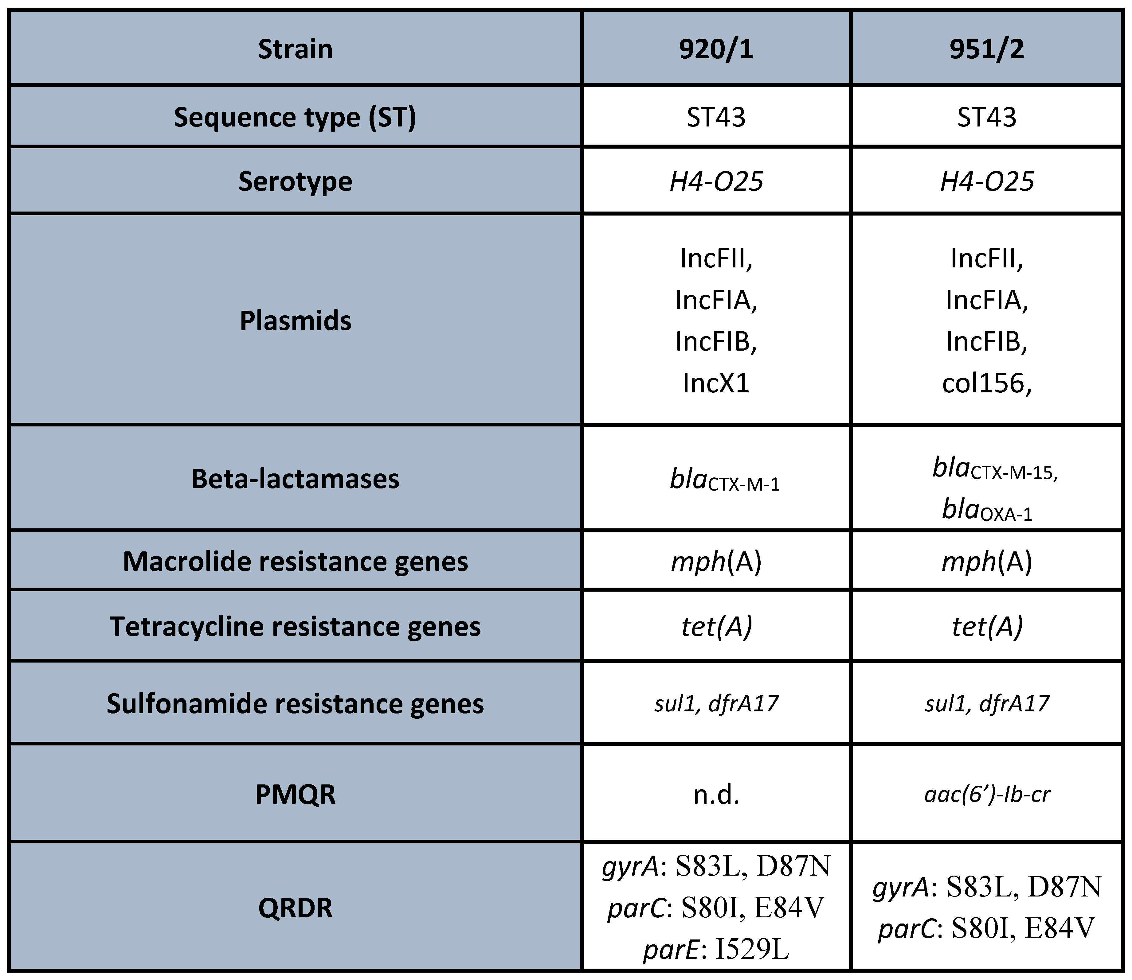
Based on the results of the susceptibility testing, two multidrug-resistant (MDR) strains, namely, ECO-SEOMI-LKH 920/1 and 951/2, were selected for WGS analysis. The MIC values are presented in Figure 4. Both strains belong to E. coli ST43 international high-risk clone and both carried diverse resistance genes, including ESBLs, namely, blaCTX-M-1, blaCTX-M-15. In the case of E. coli 920/1, five amino acid alterations in multiple positions of QRDR, namely, gyrA S83L, D87N, parC S80I, E84V, and parE I529L, were detected. E. coli 951/2 was marked by similar multiple mutations of QRDR; namely, gyrA S83L, D87N and parC S80I, and E84V were identified. Among the PMQR determinants, the bifunctional Aminoglycoside-acetyltransferase(6′)-Ib-cr was detected in this strain. Further resistance mechanisms were also detected, which are summarized in Figure 5.

4. Discussion

In our study, we investigated the delafloxacin-resistance rate in 47 E. coli strains that were isolated from clinical samples (e.g., hemoculture and urine). Delafloxacin is a novel fluoroquinolone agent, which was approved for clinical application in recent years. Our results demonstrate that 47% (22/47) of the E. coli strains exhibited resistance to delafloxacin compared to ciprofloxacin resistance (51% (24/47)), moxifloxacin resistance (51% (24/47)), and levofloxacin resistance (38% (18/47)). The MIC50 value of delafloxacin was 0.125 mg/L, while on the other hand, all other fluoroquinolone agents had a 0.25 mg/L MIC50 value. Despite of the fact that delafloxacin is not currently available in our country for clinical use, it can be clearly seen that E. coli can be selected from its resistant subtypes against this new fluoroquinolone agent. Furthermore, we also detected that ciprofloxacin and moxifloxacin resistance are frequently associated (20/47) to delafloxacin resistance. Notably, E. coli strains exhibiting ciprofloxacin MIC above 1 mg/L were already resistant to delafloxacin. This phenotype can be used as a marker to indicate delafloxacin resistance.
Our results also show indications for the clinical application of delafloxacin, as 20 E. coli strains were delafloxacin-susceptible but exhibited an ESBL phenotype and ciprofloxacin resistance. On the other hand, we also found 22 delafloxacin-resistant E. coli strains that exhibited an ESBL phenotype.
Interestingly, levofloxacin MIC values did not strongly correlate to other fluoroquinolones in this study. We detected two E. coli strains with delafloxacin resistance, but one strain was sensitive to ciprofloxacin and levofloxacin, while the other was sensitive to levofloxacin and moxifloxacin (Figure 3).
In our study, we selected two MDR E. coli strains for WGS analysis. Both strains belong to the ST43 international high-risk clone of E. coli based on Pasteur’s multilocus sequence typing (MLST) database. Interestingly, the E. coli ST43 in Pasteur’s MLST database corresponds to ST131 in the Achtman MLST scheme [34,35,36]. The high-risk clones of E. coli are disseminated worldwide, and these carry diverse resistance mechanisms, including ESBLs (e.g., blaCTX-M, blaSHV, blaTEM), carbapenemases (e.g., blaKPC, blaNDM, blaVIM, blaIMP, blaOXA-48), and colistin-resistance determinants (e.g., mcr). Furthermore, these high-risk clones are causative agents of a high number of hospital-acquired infections [37,38].
The predominant E. coli high-risk clone is ST131; however, additional high-risk clones are also known, namely, ST10, ST69, ST73, ST405, ST410, ST457, and ST1193 [37,38,39].
E. coli ST131 is a multi-resistant high-risk clone and it is responsible for the spreading of resistance determinants. The two major serotypes of E. coli ST131 are O16:H5 and O25:H4. Additionally, E. coli ST131 has three fimH alleles, namely, 41, 22, and 30. According to antibiotic-resistance patterns, ST131 is also classified into subclones H30R and H30Rx [37,38]. Originally, the E. coli ST131 clone was reported as O25b:H4 serotype, and CTX-M-type ESBL production was usually detected. The majority of strains in the ST131 clone are usually resistant to several antibiotics, including cephalosporins, carbapenems, aminoglycosides, fluoroquinolones, sulfonamides, nitrofurantoin, and tetracycline [37].
The ST131 clone is the main member of the phylogroup B2 of E. coli, according to the phylogenetic analysis of whole-genome data. E. coli ST131 is also recognized as the origin of different sequence types, namely, ST1680, ST1982, ST1461, and ST1193.
High number and diverse virulence factors are usually detected in E. coli strains belonging to phylogroup B2 [37]. Unlike other B2 E. coli strains, ESBL production and fluoroquinolone resistance are commonly detected in ST131. The phylogeny of ST131 has been clustered into three major clades based on resistance traits and population genetics. The three clades are A, B, and C. Among the clades, the A/H41 clade and B/H22 clade are smaller subgroups. However, ST131 strains in clade A have been reported in many community-acquired infections worldwide, and these strains have also been reported in stool samples of healthy children in China [37,38]. Clade A E. coli ST131 has been reported in environmental samples, namely, in water from the Jurong river reservoir in Singapore. Interestingly, the strains of clade B have been described to colonize poultry, contaminate meat, and carry colistin-resistance genes (e.g., mcr-1 and mcr-3); therefore, these strains are considered as foodborne pathogens. The strains of clade B have been isolated from several human infections, notably, from clinical samples of urine, blood, and peritoneal fluid. However, at present, the most challenging clade is related to the strains of clade C. This clade originates from clade B, and it can be divided into two major subclades, namely, C1/H30-R and C2/H30-Rx. E. coli strains of clade C usually carry blaTEM; additionally, subclade C1 usually carry blaCTX-M-14 or blaCTX-M-27 ESBL genes, and by contrast, subclade C2 is mainly associated with blaCTX-M-15 [37,38].
To date, E. coli ST43 was described from clinical isolates in some countries. In Panama, Central America, it was detected as blaCTX-M-15-positive and ciprofloxacin-resistant [34]. In Italy, E. coli ST43 was detected as KPC-3-positive and fluoroquinolone-resistant [40]. E. coli ST43 in USA was reported as fluoroquinolone-resistant having multiple QRDR mutations, but it was susceptible to third-generation cefalosporins and carbapenems [41].
In our study, the two strains of E. coli ST43 were ESBL-positive, harboring blaCTX-M-1 and blaCTX-M-15, and both strains were resistant to all tested fluoroquinolone agents, including delafloxacin (Figure 5). The genetic background of fluoroquinolone resistance in these two strains of E. coli ST43 demonstrated the accumulation of multiple QRDR mutations. In the case of E. coli 920/1, five mutations were present: gyrA S83L, D87N, parC S80I, E84V, and parE I529L, while in E. coli 951/2, four mutations, gyrA S83L, D87N and parC S80I, E84V plus aac(6′)-Ib-cr PMQR determinant, were detected (Figure 5).
Fluoroquinolone resistance in Enterobacterales is frequently detected, especially in MDR international high-risk clones, such as in E. coli ST131, ST1193, ST69, and CC10 strains [38,42,43,44], as well as in K. pneumoniae ST11, ST15, ST101, ST147, and ST307 [45,46,47,48,49,50].
It has been demonstrated that diverse fitness cost is associated with fluoroquinolone resistance in different clones of K. pneumoniae and E. coli. High-risk clones of K. pneumoniae and E. coli suffer a lower fitness cost and retain a favorable fitness after the development of fluoroquinolone resistance, which enable them to survive and disseminate [51,52]. These features are usually seen in high-risk clones because they can persist and spread for a longer period of time in hospital environments and cause different infections. Additionally, high-risk clones can acquire further resistance determinants during dissemination in hospital settings [37,47]. Double serin mutations in the QRDR sequences of the gyrA and parC genes are linked to high-risk clones, which are associated with a favorable fitness [53]. Our results correlate well to these earlier data because we detected fluoroquinolone resistance in the E. coli ST43 international high-risk clone, which is associated with double serin mutations in the QRDR sequences of the gyrA and parC genes.
To date, delafloxacin resistance has been scarcely reported among bacterial isolates. According to the available data, delafloxacin resistance was reported in S. aureus, Neisseria gonorrhoeae, and non-tuberculous Mycobacteria [54,55,56].
In conclusion, in our study, we analyzed the efficacy of delafloxacin in a collection of 47 E. coli strains. A limitation of this study is that we investigated only 47 strains; however, these early results are useful to gain insights into the possible clinical use of delafloxacin. We detected a remarkably high (47%) delafloxacin resistance among MDR E. coli strains. This report can be considered as a baseline result, because delafloxacin is not yet available for clinical application in Hungary. We also reported the detection of the E. coli ST43 high-risk international clone in Hungary. This clone has been reported in several countries with different resistance determinants, and in our study, both strains of E. coli ST43 exhibited ciprofloxacin and delafloxacin resistance together with CTX-M-1- and CTX-M-15-type ESBL production.

Author Contributions

D.G. performed the antibiotic susceptibility testing and drafted the manuscript; K.K. collected the strains; D.S. analyzed the WGS data; and B.K. revised the manuscript and designed the study. All authors have read and agreed to the published version of the manuscript.

Funding

This study was supported by the European Union’s Horizon 2020 research and innovation program (952491-AmReSu). B.K. was supported by the János Bolyai Research Scholarship (BO/00286/22/5) of the Hungarian Academy of Sciences. This work was supported by Semmelweis University-Eötvös Lóránd Research Network, Human Microbiota Study Group No. 0272.

Data Availability Statement

The assembled genomes of E. coli strains (920/1 and 951/2) were submitted to NCBI Genbank at Bioproject PRJNA971108; sequence read archive (SRA) identifiers: SAMN35019574 (ECO-SEOMI-LKH 920/1 strain) and SAMN35019575 (ECO-SEOMI-LKH 951/2 strain).

Acknowledgments

We would like to thank Eurofins BIOMI Kft (Gödöllő) for the whole-genome sequencing.

Conflicts of Interest

The authors declare no conflict of interest.

Ethics Approval

This is a retrospective study, not directly associated with patients, and it was consistent with principles of the Declaration of Helsinki.

References

  1. Mendelson, M.; Sharland, M.; Mpundu, M. Antibiotic resistance: Calling time on the ‘silent pandemic’. JAC Antimicrob. Resist. 2022, 4, dlac016. [Google Scholar] [CrossRef] [PubMed]
  2. Rehman, S. A parallel and silent emerging pandemic: Antimicrobial resistance (AMR) amid COVID-19 pandemic. J. Infect. Public. Health 2023, 16, 611–617. [Google Scholar] [CrossRef] [PubMed]
  3. Livermore, D.M. Antibiotic resistance during and beyond COVID-19. JAC Antimicrob. Resist. 2021, 3 (Suppl. S1), i5–i16. [Google Scholar] [CrossRef] [PubMed]
  4. Antimicrobial Resistance Collaborators. Global burden of bacterial antimicrobial resistance in 2019: A systematic analysis. Lancet 2022, 399, 629–655. [Google Scholar] [CrossRef]
  5. Thompson, T. The staggering death toll of drug-resistant bacteria. Nature, 2022; epub ahead of print. [Google Scholar] [CrossRef]
  6. O’Neill, J. Review on Antimicrobial Resistance. Antimicrobial Resistance: Tackling a Crisis for the Health and Wealth of Nations; Wellcome Trust: London, UK, 2014; p. 20. Available online: https://amr-review.org/Publications.html (accessed on 10 February 2020).
  7. Zhen, X.; Lundborg, C.S.; Sun, X.; Hu, X.; Dong, H. Economic burden of antibiotic resistance in ESKAPE organisms: A systematic review. Antimicrob. Resist. Infect. Control 2019, 8, 137. [Google Scholar] [CrossRef] [Green Version]
  8. Orosz, L.; Lengyel, G.; Ánosi, N.; Lakatos, L.; Burián, K. Changes in resistance pattern of ESKAPE pathogens between 2010 and 2020 in the clinical center of University of Szeged, Hungary. Acta Microbiol. Immunol. Hung. 2022, 69, 27–34. [Google Scholar] [CrossRef]
  9. Tacconelli, E.; Carrara, E.; Savoldi, A.; Harbarth, S.; Mendelson, M.; Monnet, D.L.; Pulcini, C.; Kahlmeter, G.; Kluytmans, J.; Carmeli, Y.; et al. Discovery, research, and development of new antibiotics: The WHO priority list of antibiotic-resistant bacteria and tuberculosis. Lancet Infect. Dis. 2018, 18, 318–327. [Google Scholar] [CrossRef]
  10. Butler, M.S.; Paterson, D.L. Antibiotics in the clinical pipeline in October 2019. J. Antibiot. 2020, 73, 329–364. [Google Scholar] [CrossRef]
  11. Syed, Y.Y. Cefiderocol: A Review in Serious Gram-Negative Bacterial Infections. Drugs 2021, 81, 1559–1571. [Google Scholar] [CrossRef]
  12. Saravolatz, L.D.; Stein, G.E. Plazomicin: A new aminoglycoside. Clin. Infect. Dis. 2020, 70, 704–709. [Google Scholar] [CrossRef]
  13. Bush, K.; Bradford, P.A. Interplay between beta-lactamases and new beta-lactamase inhibitors. Nat. Rev. Microbiol. 2019, 17, 295–306. [Google Scholar] [CrossRef]
  14. Kocsis, B.; Gulyás, D.; Szabó, D. Delafloxacin, Finafloxacin, and Zabofloxacin: Novel Fluoroquinolones in the Antibiotic Pipeline. Antibiotics 2021, 10, 1506. [Google Scholar] [CrossRef]
  15. Dong Wha Pharm’s Quinolone Antibacterial Agent, “Zabolante”, Wins at the 19th KNDA (Press Release 28 February 2018). Available online: https://www.dong-wha.co.kr/english/customer/dnews/content.asp?t_idx=1139 (accessed on 20 August 2021).
  16. U.S. Food and Drug Administration. BAXDELA (Delafloxacin) Prescribing Information and Medication Guide. Available online: https://www.accessdata.fda.gov/drugsatfda_docs/label/2017/208610s000,208611s000lbl.pdf (accessed on 8 December 2018).
  17. U.S. Food and Drug Administration. XTORO Prescribing Information and Medication Guide 2014. Available online: https://www.accessdata.fda.gov/drugsatfda_docs/label/2014/206307s000lbl.pdf (accessed on 20 August 2021).
  18. Kocsis, B.; Domokos, J.; Szabo, D. Chemical structure and pharmacokinetics of novel quinolone agents represented by avarofloxacin, delafloxacin, finafloxacin, zabofloxacin and nemonoxacin. Ann. Clin. Microbiol. Antimicrob. 2016, 15, 34. [Google Scholar] [CrossRef] [Green Version]
  19. Rusu, A.; Lungu, I.A.; Moldovan, O.L.; Tanase, C.; Hancu, G. Structural Characterization of the Millennial Antibacterial (Fluoro)Quinolones-Shaping the Fifth Generation. Pharmaceutics 2021, 13, 1289. [Google Scholar] [CrossRef]
  20. Lungu, I.A.; Moldovan, O.L.; Biriș, V.; Rusu, A. Fluoroquinolones Hybrid Molecules as Promising Antibacterial Agents in the Fight against Antibacterial Resistance. Pharmaceutics 2022, 14, 1749. [Google Scholar] [CrossRef]
  21. Rusu, A.; Munteanu, A.C.; Arbănași, E.M.; Uivarosi, V. Overview of Side-Effects of Antibacterial Fluoroquinolones: New Drugs versus Old Drugs, a Step Forward in the Safety Profile? Pharmaceutics 2023, 15, 804. [Google Scholar] [CrossRef]
  22. European Medicines Agency (EMA). Quinolone- and Fluoroquinolone-Containing Medicinal Products: Disabling and Potentially Permanent Side Effects Lead to Suspension or Restrictions of Quinolone and Fluoroquinolone Antibiotics. Available online: https://www.ema.europa.eu/en/medicines/human/referrals/quinolone-fluoroquinolone-containing-medicinal-products (accessed on 8 December 2018).
  23. Mogle, B.T.; Steele, J.M.; Thomas, S.J.; Bohan, K.B.H.; Kufel, W.D. Clinical review of delafloxacin: A novel anionic fluoroquinolone. J. Antimicrob. Chemother. 2018, 73, 1439–1451. [Google Scholar] [CrossRef] [Green Version]
  24. Sharma, R.; Sandrock, C.E.; Meehan, J.; Theriault, N. Community-Acquired Bacterial Pneumonia-Changing Epidemiology, Resistance Patterns, and Newer Antibiotics: Spotlight on Delafloxacin. Clin. Drug Investig. 2020, 40, 947–960. [Google Scholar] [CrossRef]
  25. European Medicines Agency. Quofenix (Delafloxacin): Summary of Product Characteristics. 2019. Available online: https://www.ema.europa.eu/ (accessed on 16 March 2020).
  26. Scott, L.J. Delafloxacin: A review in acute bacterial skin and skin structure infections. Drugs 2020, 80, 1247–1258. [Google Scholar] [CrossRef]
  27. Melinta Therapeutics. Baxdela (Delafloxacin) Tablets, for Oral Use; Baxdela (Delafloxacin) for Injection, for Intravenous Use: U.S. Prescribing Information. 2019. Available online: https://baxdela.com/docs/baxdela-prescribing-information.pdf (accessed on 16 March 2020).
  28. Hooper, D.C.; Jacoby, G.A. Mechanisms of drug resistance: Quinolone resistance. Ann. N. Y. Acad. Sci. 2015, 1354, 12–31. [Google Scholar] [CrossRef] [PubMed] [Green Version]
  29. Vila, J.; Ruiz, J.; Marco, F.; Barcelo, A.; Goñi, P.; Giralt, E.; Jimenez de Anta, T. Association between double mutation in gyrA gene of ciprofloxacin-resistant clinical isolates of Escherichia coli and MICs. Antimicrob. Agents Chemother. 1994, 38, 2477–2479. [Google Scholar] [CrossRef] [PubMed] [Green Version]
  30. Rodríguez-Martínez, J.M.; Machuca, J.; Cano, M.E.; Calvo, J.; Martínez-Martínez, L.; Pascual, A. Plasmid-mediated quinolone resistance: Two decades on. Drug Resist. Update 2016, 29, 13–29. [Google Scholar] [CrossRef] [PubMed]
  31. Kocsis, B.; Szmolka, A.; Szabo, O.; Gulyas, D.; Kristóf, K.; Göcző, I.; Szabo, D. Ciprofloxacin Promoted qnrD Expression and Phylogenetic Analysis of qnrD Harboring Plasmids. Microb. Drug Resist. 2019, 25, 501–508. [Google Scholar] [CrossRef] [PubMed]
  32. Albornoz, E.; Tijet, N.; De Belder, D.; Gomez, S.; Martino, F.; Corso, A.; Melano, R.G.; Petroni, A. qnrE1, a Member of a New Family of Plasmid-Located Quinolone Resistance Genes, Originated from the Chromosome of Enterobacter Species. Antimicrob. Agents Chemother. 2017, 61, e02555-16. [Google Scholar] [CrossRef] [Green Version]
  33. Wang, C.; Yin, M.; Zhang, X.; Guo, Q.; Wang, M. Identification of qnrE3 and qnrE4, New Transferable Quinolone Resistance qnrE Family Genes Originating from Enterobacter mori and Enterobacter asburiae, Respectively. Antimicrob. Agents Chemother. 2021, 65, e0045621. [Google Scholar] [CrossRef]
  34. Núñez-Samudio, V.; Pecchio, M.; Pimentel-Peralta, G.; Quintero, Y.; Herrera, M.; Landires, I. Molecular Epidemiology of Escherichia coli Clinical Isolates from Central Panama. Antibiotics 2021, 10, 899. [Google Scholar] [CrossRef]
  35. Clermont, O.; Gordon, D.; Denamur, E. Guide to the various phylogenetic classification schemes for Escherichia coli and the correspondence among schemes. Microbiology 2015, 161, 980–988. [Google Scholar] [CrossRef]
  36. Johnson, J.R.; Clermont, O.; Johnston, B.; Clabots, C.; Tchesnokova, V.; Sokurenko, E.; Junka, A.F.; Maczynska, B.; Denamur, E. Rapid and specific detection, molecular epidemiology, and experimental virulence of the O16 subgroup within Escherichia coli sequence type 131. J. Clin. Microbiol. 2014, 52, 1358–1365. [Google Scholar] [CrossRef] [Green Version]
  37. Kocsis, B.; Gulyás, D.; Szabó, D. Emergence and Dissemination of Extraintestinal Pathogenic High-Risk International Clones of Escherichia coli. Life 2022, 12, 2077. [Google Scholar] [CrossRef]
  38. Pitout, J.D.D.; DeVinney, R. Escherichia coli ST131: A multidrug-resistant clone primed for global domination. F1000Res 2017, 6, 1–7. [Google Scholar] [CrossRef] [Green Version]
  39. Asgharzadeh, S.; Golmoradi Zadeh, R.; Taati Moghadam, M.; Farahani Eraghiye, H.; Sadeghi Kalani, B.; Masjedian Jazi, F.; Mirkalantari, S. Distribution and expression of virulence genes (hlyA, sat) and genotyping of Escherichia coli O25b/ST131 by multi-locus variable number tandem repeat analysis in Tehran, Iran. Acta Microbiol. Immunol. Hung. 2022, 69, 314–322. [Google Scholar] [CrossRef] [PubMed]
  40. Kocsis, E.; Lo Cascio, G.; Piccoli, M.; Cornaglia, G.; Mazzariol, A. KPC-3 carbapenemase harbored in FIIk plasmid from Klebsiella pneumoniae ST512 and Escherichia coli ST43 in the same patient. Microb. Drug Resist. 2014, 20, 377–382. [Google Scholar] [CrossRef] [PubMed]
  41. Suwantarat, N.; Rudin, S.D.; Marshall, S.H.; Hujer, A.M.; Perez, F.; Hujer, K.M.; Domitrovic, T.N.; Dumford, D.M., 3rd; Donskey, C.J.; Bonomo, R.A. Infections caused by fluoroquinolone-resistant Escherichia coli following transrectal ultrasound-guided biopsy of the prostate. J. Glob. Antimicrob. Resist. 2014, 2, 71–76. [Google Scholar] [CrossRef] [PubMed] [Green Version]
  42. Pajand, O.; Rahimi, H.; Darabi, N.; Roudi, S.; Ghassemi, K.; Aarestrup, F.M.; Leekitcharoenphon, P. Arrangements of Mobile Genetic Elements among Virotype E Subpopulation of Escherichia coli Sequence Type 131 Strains with High Antimicrobial Resistance and Virulence Gene Content. mSphere 2021, 6, e0055021. [Google Scholar] [CrossRef]
  43. Massella, E.; Giacometti, F.; Bonilauri, P.; Reid, C.; Djordjevic, S.; Merialdi, G.; Bacci, C.; Fiorentini, L.; Massi, P.; Bardasi, L.; et al. Antimicrobial Resistance Profile and ExPEC Virulence Potential in Commensal Escherichia coli of Multiple Sources. Antibiotics 2021, 10, 351. [Google Scholar] [CrossRef]
  44. Tóth, K.; Tóth, Á.; Kamotsay, K.; Németh, V.; Szabó, D. Population snapshot of the extended-spectrum β-lactamase-producing Escherichia coli invasive strains isolated from a Hungarian hospital. Ann. Clin. Microbiol. Antimicrob. 2022, 21, 3. [Google Scholar] [CrossRef]
  45. Melegh, S.; Schneider, G.; Horváth, M.; Jakab, F.; Emődy, L.; Tigyi, Z. Identification and characterization of CTX-M-15 producing Klebsiella pneumoniae clone ST101 in a Hungarian university teaching hospital. Acta Microbiol. Immunol. Hung. 2015, 62, 233–245. [Google Scholar] [CrossRef] [Green Version]
  46. Kocsis, B.; Kocsis, E.; Fontana, R.; Cornaglia, G.; Mazzariol, A. Identification of blaLAP-2 and qnrS1 genes in the internationally successful Klebsiella pneumoniae ST147 clone. J. Med. Microbiol. 2013, 62, 269–273. [Google Scholar] [CrossRef] [Green Version]
  47. Domokos, J.; Damjanova, I.; Kristof, K.; Ligeti, B.; Kocsis, B.; Szabo, D. Multiple Benefits of Plasmid-Mediated Quinolone Resistance Determinants in Klebsiella pneumoniae ST11 High-Risk Clone and Recently Emerging ST307 Clone. Front. Microbiol. 2019, 10, 157. [Google Scholar] [CrossRef] [Green Version]
  48. Horváth, M.; Kovács, T.; Kun, J.; Gyenesei, A.; Damjanova, I.; Tigyi, Z.; Schneider, G. Virulence Characteristics and Molecular Typing of Carbapenem-Resistant ST15 Klebsiella pneumoniae Clinical Isolates, Possessing the K24 Capsular Type. Antibiotics 2023, 12, 479. [Google Scholar] [CrossRef]
  49. Muraya, A.; Kyany’a, C.; Kiyaga, S.; Smith, H.J.; Kibet, C.; Martin, M.J.; Kimani, J.; Musila, L. Antimicrobial Resistance and Virulence Characteristics of Klebsiella pneumoniae Isolates in Kenya by Whole-Genome Sequencing. Pathogens. 2022, 11, 545. [Google Scholar] [CrossRef]
  50. Navon-Venezia, S.; Kondratyeva, K.; Carattoli, A. Klebsiella pneumoniae: A major worldwide source and shuttle for antibiotic resistance. FEMS Microbiol. Rev. 2017, 41, 252–275. [Google Scholar] [CrossRef]
  51. Toth, A.; Kocsis, B.; Damjanova, I.; Kristof, K.; Janvari, L.; Paszti, J.; Csercsik, R.; Topf, J.; Szabo, D.; Hamar, P.; et al. Fitness cost associated with resistance to fluoroquinolones is diverse across clones of Klebsiella pneumoniae and may select for CTX-M-15 type extended-spectrum beta-lactamase. Eur. J. Clin. Microbiol. Infect. Dis. 2014, 33, 837–843. [Google Scholar] [CrossRef]
  52. Machuca, J.; Briales, A.; Labrador, G.; Díaz-de-Alba, P.; López-Rojas, R.; Docobo-Pérez, F.; Martínez-Martínez, L.; Rodríguez-Baño, J.; Pachón, M.E.; Pascual, A.; et al. Interplay between plasmid-mediated and chromosomal-mediated fluoroquinolone resistance and bacterial fitness in Escherichia coli. J. Antimicrob. Chemother. 2014, 69, 3203–3215. [Google Scholar] [CrossRef] [Green Version]
  53. Fuzi, M.; Szabo, D.; Csercsik, R. Double-Serine Fluoroquinolone Resistance Mutations Advance Major International Clones and Lineages of Various Multi-Drug Resistant Bacteria. Front. Microbiol. 2017, 8, 2261. [Google Scholar] [CrossRef]
  54. Soge, O.O.; Salipante, S.J.; No, D.; Duffy, E.; Roberts, M.C. In Vitro Activity of Delafloxacin against Clinical Neisseria gonorrhoeae Isolates and Selection of Gonococcal Delafloxacin Resistance. Antimicrob. Agents Chemother. 2016, 60, 3106–3111. [Google Scholar] [CrossRef] [Green Version]
  55. De la Rosa, J.M.O.; Fernández, M.A.; Rodríguez-Villodres, Á.; Casimiro-Soriguer, C.S.; Cisneros, J.M.; Lepe, J.A. High-level delafloxacin resistance through the combination of two different mechanisms in Staphylococcus aureus. Int. J. Antimicrob. Agents 2023, 61, 106795. [Google Scholar] [CrossRef]
  56. Brown Elliott, B.A.; Wallace, R.J., Jr. Comparison of In Vitro Susceptibility of Delafloxacin with Ciprofloxacin, Moxifloxacin, and Other Comparator Antimicrobials against Isolates of Nontuberculous Mycobacteria. Antimicrob. Agents Chemother. 2021, 65, e0007921. [Google Scholar] [CrossRef]
Figure 1. Distribution of fluoroquinolone MIC values of the 47 E. coli strains in this study. Arrows indicate EUCAST breakpoint for each fluoroquinolone, namely, D: delafloxacin, C: ciprofloxacin, M: moxifloxacin, and L: levofloxacin.
Microorganisms 11 01602 g001
Figure 2. Distribution of the beta-lactam MIC values of the 47 E. coli strains in this study. Cefotaxime, ceftazidime, and imipenem MIC values are shown.
Microorganisms 11 01602 g002
Figure 3. Fluoroquinolone resistance patterns of the 47 E. coli strains. R: resistant, S: susceptible.
Microorganisms 11 01602 g003
Figure 4. MIC values of the two E. coli strains selected for whole-genome sequencing.
Microorganisms 11 01602 g004
Figure 5. Results of the whole-genome sequencing of the two E. coli strains (920/1 and 951/2). PMQR: plasmid-mediated quinolone resistance, QRDR: quinolone-resistance-determining region, n.d.: not detected.
Microorganisms 11 01602 g005
Disclaimer/Publisher’s Note: The statements, opinions and data contained in all publications are solely those of the individual author(s) and contributor(s) and not of MDPI and/or the editor(s). MDPI and/or the editor(s) disclaim responsibility for any injury to people or property resulting from any ideas, methods, instructions or products referred to in the content.