Article

Resistance and Biofilm Production Profile of Potential Isolated from Kpètè-Kpètè Used to Produce Traditional Fermented Beer

by
Christine N’Tcha
1,
Haziz Sina
1,
Dyana Ndiade Bourobou
2,
S. M. Ismaël Hoteyi
1,
Bawa Boya
1,
Raoul Agnimonhan
1,
Jacques François Mavoungou
2,
Adolphe Adjanohoun
3,
Olubukola Oluranti Babalola
4,* and
Lamine Baba-Moussa
1,*
1
Laboratory of Biology and Molecular Typing in Microbiology, Department of Biochemistry and Cell Biology, University of Abomey-Calavi, Abomey-Calavi 05 BP 1604, Benin
2
Institut de Recherches Agronomiques et Forestières (IRAF), BP.12978 Gros-Bouquet, Libreville B.P. 16 182, Gabon
3
National Agronomic Research Institute of Benin, Cotonou 01 BP 884, Benin
4
Food Security and Safety Focus Area, Faculty of Natural and Agricultural Sciences, North-West University, Private Bag X2046, Mmabatho 2735, South Africa
*
Authors to whom correspondence should be addressed.
Microorganisms 2023, 11(8), 1939; https://doi.org/10.3390/microorganisms11081939
Submission received: 25 May 2023 / Revised: 16 July 2023 / Accepted: 26 July 2023 / Published: 29 July 2023

Abstract

:
This study aimed to characterize the pathogenicity of bacteria isolated from the starter of two traditional beers produced and consumed in Benin. After standard microbial identification, species were identified by specific biochemical tests such as catalase, coagulase, and API 20 E. Antibiotic sensitivity was tested according to the French Society of Microbiology Antibiogram Committee. The crystal violet microplate technique evaluated the biofilm production and conventional PCR was used to identify genes encoding virulence and macrolide resistance. According to our data, the traditional starter known as kpètè-kpètè that is used to produce beer is contaminated by Enterobacteriaceae and staphylococci species. Thus, 28.43% of the isolated bacteria were coagulase-negative staphylococci (CNS), and 10.93% coagulase-positive staphylococci (CPS). Six species such as Klebsiella terrigena (1.38%), Enterobacter aerogens (4.14%), Providencia rettgeri (5.51%), Chryseomonas luteola (6.89%), Serratia rubidae (15.16%), and Enterobacter cloacae (27.56%) were identified among Enterobacteriaceae. Those bacterial strains are multi-resistant to conventional antibiotics. The hight capability of produced biofilms was recorded with Enterobacter aerogens, Klebsiella terrigena (100%), Providencia rettgeri (75%), and Staphylococcus spp (60%). Enterobacter cloacae (4%) and coagulase-negative Staphylococcus (5.55%) harbor the macrolide resistance gene. For other strains, these genes were not detected. Foods contaminated with bacteria resistant to antibiotics and carrying a virulence gene could constitute a potential public health problem. There is a need to increase awareness campaigns on hygiene rules in preparing and selling these traditional beers.

1. Introduction

Food poisoning is a serious issue that can cause more than 200 different illnesses, ranging from diarrhea to cancer, and results in nearly 420,000 deaths each year worldwide [1]. Unfortunately, street food is often a culprit when it comes to incidents of food poisoning [2]. This is particularly true in Benin, where Tchoukoutou and Tchapalo, two traditional beers made from red or brown sorghum (Sorghum bicolor) and maize [3,4], are sold on the street. These beers are an important part of social gatherings and traditional ceremonies [5] and are a significant income source, particularly for women [3,6]. However, there have been reports of a lack of hygiene during the cooking and sale of these beers, which can lead to contamination with pathogenic microorganisms that cause food poisoning.
The production of Tchoukoutou and Tchapalo involves fermentation with microorganisms that create various microbial communities. These communities significantly impact the final products’ sensory quality, nutrient availability, and safety [7,8,9]. Bacteria, yeasts, and molds can be found in the raw materials, utensils, or even the producers themselves [10,11]. Although lactic acid bacteria can reduce the levels of pathogenic bacteria present in the drinks [7,8,12,13], some bacteria such as Enterobacter sakazaki, Klebsiella pneumonia, Escherichia coli, and Staphylococcus aureus remain resistant to the acidic environment and cooking process of the fermented foods [14]. Enterobacteriaceae in Tchoukoutou, sold in northern Benin, has been reported [15]. It is concerning that the bacteria found in beer can lead to food spoilage [16,17] and negatively impact individuals with health conditions or habitat disorders, such as sepsis, meningitis, endocarditis, peritonitis, or heart disease [18].
In West African nations, the prevalence of bacterial infections has risen alongside the increase in antibiotic consumption [19,20]. Improper and unregulated use of synthetic products has made it difficult to control bacterial and fungal infections, as many conventional antibiotics and antifungals have become ineffective due to the emergence of resistant bacteria and fungi [21,22]. This resistance to multiple drugs may be caused by chromosomal mutations in these strains or by acquiring resistance genes [23]. However, the Enterobacteriaceae species and many pathogenic bacteria found in traditional beers have been overlooked in the literature, despite being identified through biochemical and molecular characterization. Thus, the present study aims to characterize the pathogenic bacteria isolated from two traditional beers produced and consumed in Benin.

2. Materials and Methods

2.1. Sampling

Thirty-seven (37) isolates of the enterobacteria and staphylococci used were isolated from samples of kpètè-kpètè of two traditional beers (Tchoukoutou and Tchapalo) collected by N’tcha et al. [15] in areas with a high production of conventional beers in Benin such as Tanguiéta, Natitingou, Parakou, Boukoumbé, Tchaourou, N’Dali, Bantè, Dassa Zoumè, and Glazoué.

2.2. Microbiological Analyses

2.2.1. Identification of Bacterial Strains

In the laboratory, 5 mL of Mueller Hinton broth was added to each sample. The cases were incubated at 37 °C for 24 h. Following incubation, areas with cloudy points indicating bacterial growth were selected for germ testing. Isolation and purification of strains were carried out using selective media. Mac Conkey, EMB, and SS Agar media were used to isolate Enterobacteriaceae, and Baird Parker medium supplied with egg yolk + potassium tellurite + 0.2% sulfamethazine and a coagulase test were used for the staphylococci. To ensure the purity of the sample, the isolated colony was subcultured for isolation. The pure strains were obtained after three successive subcultures were used for biochemical and molecular identification. We utilized the API 20E commercial kit from BioMerieux (Marcy l’Etoile, France) to identify isolated Enterobacteriaceae. The identification process was done through plating.

2.2.2. Biofilm Formation

The ability of the isolates to generate a biofilm was assessed through a process described in reference [24]. To begin, 10 µL of 18-h-old isolate suspension was diluted with 150 µL of Brain Heath Infusion, and the mixture was incubated for 24 h at 37 °C. Once incubated, wells were washed three times with approximately 0.2 mL of sterile physiological water containing 0.9% NaCl. To observe the biofilms created by the attachment of stationary organisms to the microplate in each well, crystal violet (0.1%) was used to stain them for 10 min. The violet staining in a well indicated a positive test. The plates were washed thoroughly to remove excess dye and left to dry at room temperature [25].

2.2.3. Susceptibility of Bacterial Isolates to Tested Antibiotic

To assess the antibiotic susceptibility of the isolated strains, we utilized the disc diffusion method, per the guidelines set forth by the Antibiogram Committee of the French Society of Microbiology [26]. For the Enterobacteria, we utilized a range of antibiotics from Oxoid®, including ampicillin (A, 10 µg), cefoxitin (FOX 30 µg), ceftriaxone (Cl, 30 µg), cefotaxime (CTX 30 µg), chloramphenicol (C 30), gentamicin (G 10 µg), doxycycline (DO 30 µg), nalidixic acid (NA 30 µg), and trimethoprim-sulfamethoxazole (STX 25 µg). The antibiotics agents (Oxoid®) used for staphylococci were penicillin G (1 µg), ceftriaxone (Cl 30 µg), cefotaxime (CTX 30 µg), cephalothin (KF 30 µg), vancomycin (VA 5µg), chloramphenicol (C 30), gentamicin (G 10 µg), streptomycin (S 10 µg), erythromycin (E 15 µg), trimethoprim-sulfamethoxazole (STX 25 µg), and nalidixic acid (NA 30 µg).

2.3. Molecular Characterization of Isolates

2.3.1. DNA Extraction

DNA extraction was performed according to an adaptation of the previously described method by Rasmussen et Morrissey [27]. To prepare for DNA extraction, 3–4 colonies from a fresh bacterial culture (18 h old) were used for preculture in 1 mL of brain heart infusion and incubated at 37 °C for 18 h. The resulting mixture was centrifuged at 12,000 rpm for 5 min, and the supernatant was discarded. The bacterial pellet was mixed with 500 µL of TBE 1x and heated at 95 °C for 15 min in a dry bath. After centrifuging the mixture at 12,000 rpm for 5 min, the supernatant was transferred to a sterile tube and mixed with 500 µL of ethanol. The DNA pellets were then suspended in 50 µL of distilled water and stored at +4 °C for immediate use or at −20 °C for long-term storage.

2.3.2. Search for Alleles of Genes Encoding Virulence

For Enterobacteriaceae, genes encoding for the synthesis of adhesins (fimA: fim AF: 5′-CGGCTC TGTCCCTSAGT-3′ and fim AR: 5′-GTCGCATCCGCATTAGC-3′) [28,29] and the one encoding for the synthesis of the cytotoxic necrosis factor (cnf1: cnf1F: 5′-GGGGGAAGTACAG AAGAATTA-3′ and cnf1R: 5′-TTGCCGTCCACTCTCACCAGT-3′) [30,31] were searched for. The reaction mix included 20 µL of 10x GoTaq (PROMEGA, Madison, WI, USA) master mix, 1 µL of 10 µM primer F, 1 µL of 10 µM primer R, and 3 µL of DNA extract. The PCR reactions were performed using the thermal cycler using the following program: a cycle of initial denaturation (94 °C/3 min), followed by 30 cycles of denaturation (94 °C/1 min), hybridization (52 °C/1 min), and elongation (72 °C/1 min). To end, a final extension (72 °C/10 min) was performed.
Concerning the Staphylococcus spp isolates, gene alleles encoding macrolide resistance (ermB ermB1: 5′-FGAAAAGGTACTCAACCAAATA-3′ and ermB2: 5′-AGTAACGGTAC TTAAATTGTTTAC-3′) [32] and gene alleles encoding erythromycin resistance (mefA: mef(A/E)1: 5′-AGTATCATTAATCACTAGTGC-3′ and mef(A/E)2: 5′-TTCTTCTGGTAC TAAAAGTGG-3′) [32] were investigated. The reaction mix included 20 µL of 10x GoTaq (PROMEGA, Madison, WI, USA) master mix, 1 µL of 10 µM primer F, 1 µL of 10 µM primer R, and 3 µL of DNA extract. The amplification process involved an initial denaturation at 95 °C for 3 min. This was followed by 35 cycles of denaturation, taking 1 min at 95 °C, hybridization for 1 min at 57 °C, and elongation for 1 min at 72 °C. Finally, a final extension was carried out for 10 min at 72 °C.

2.4. Data Analysis and Processing

The results of all experiments carried out in duplicate were plotted on a bench sheet and then entered into the Excel 2016 spreadsheet. The Excel API 20 E spreadsheet was used to identify the biochemical profiles of the strains. The data were subjected to an analysis of variance (ANOVA) using the GraphPad Prism 8 software. Tukey’s multiple comparisons test was used to compare the difference in means. A statistically significant value was determined to be when p < 0.05.

3. Results

3.1. Identification of Pathogenic Bacteria Isolated

Ninety-four bacteria strains containing Enterobacteriaceae (60.64%) and staphylococci (39.36%) were isolated. Of the isolated bacteria, 28.43% were coagulase-negative staphylococci (CNS) and 10.93% were coagulase-positive staphylococci (CPS). Concerning the Enterobacteriaceae, six different species such as Klebsiella terrigena (1.38%), Enterobacter aerogens (4.14%), Providencia rettgeri (5.51%), Chryseomonas luteola (6.89%), Serratia rubidae (15.16%), and Enterobacter cloacae (27.56%) were isolated. However, the most predominant species remained CNS and E. cloacae (Figure 1).

3.2. Biofilm Production

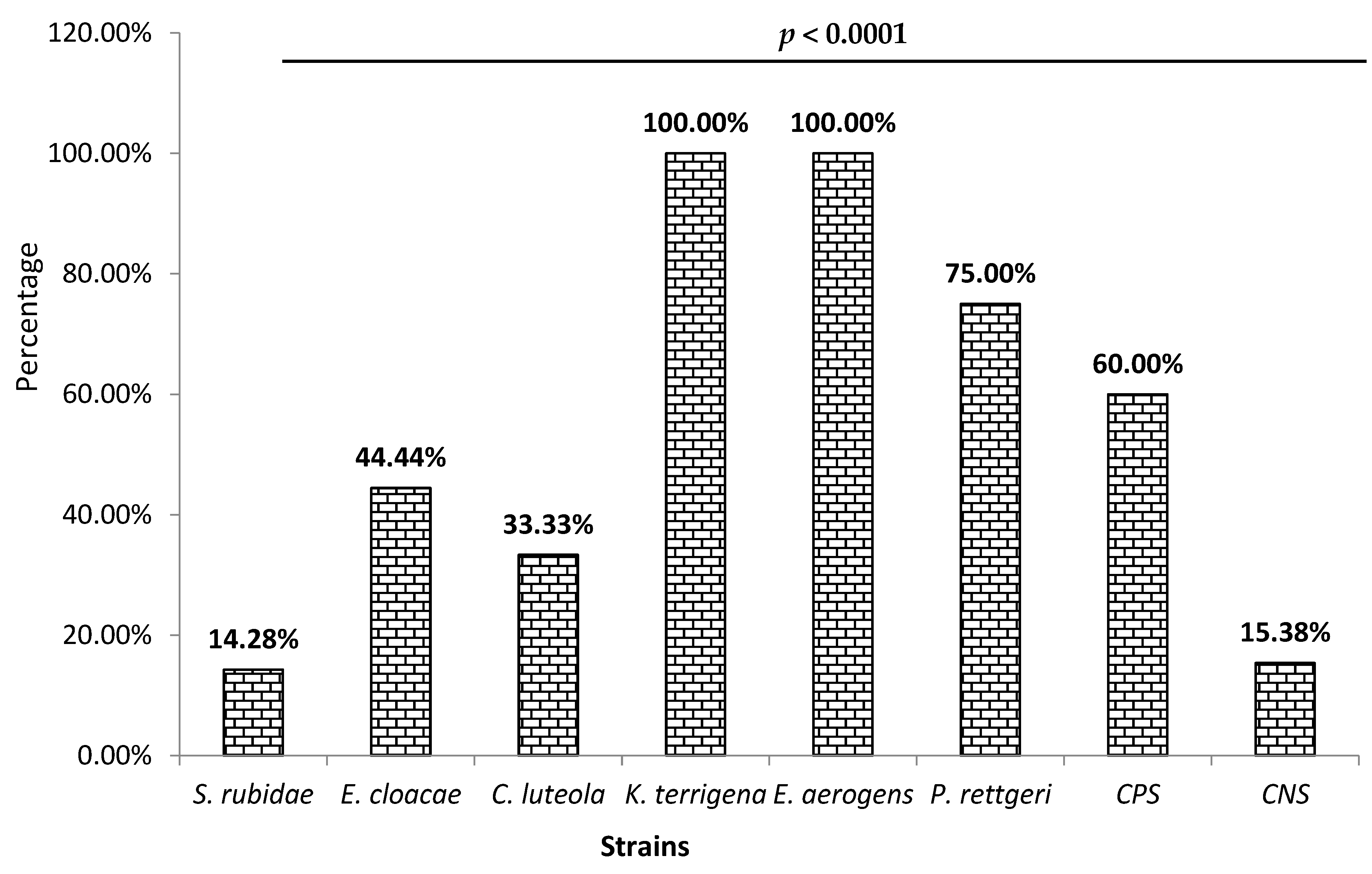
Among the isolated Enterobacteriaceae, 44% were biofilm producers. The biofilm production rate significantly varied (p < 0.0001) according to the species (Figure 2). Thus, Figure 2 shows that 100% of Enterobacter aerogens and Klebsiella terrigena were biofilm producers, following Providencia rettgeri (75%). The lowest production rate was observed with Serratia rubidaea (14.28%). Concerning the staphylococci, 28% were biofilm producers. As shown in Figure 2, this production level was high among coagulase-positive staphylococci (60%) compared to coagulase-negative staphylococci (15.38%).

3.3. Resistance Profile of Enterobacteriaceae to Antibiotics by Isolated Species

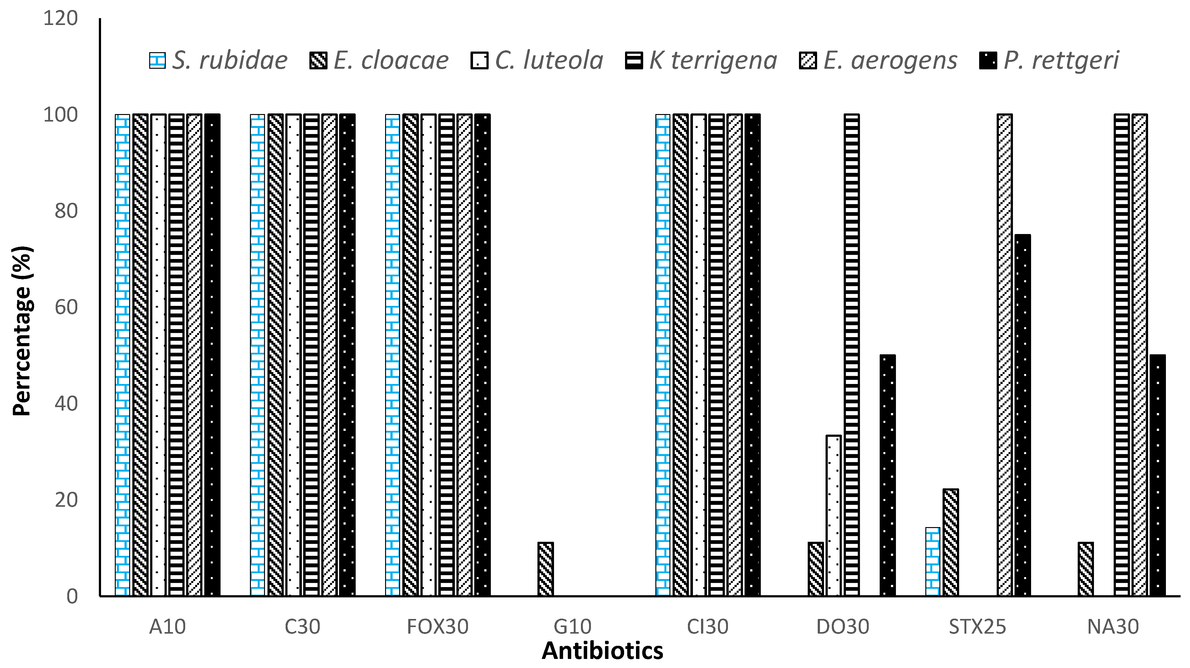
The isolated Enterobacteriaceae were divided into 6 species to evaluate their antibiotic susceptibility. Thus, Table 1 shows that Serratia rubidae, Enterobacter cloacae, Enterobacter aerogens, Providencia rettgeri, Chryseomonas luteola, and Klebsiella terrigena were highly resistant (100%) to ampicillin, chloramphenicol, cefoxitin, and ceftriaxone. These strains showed sensitivity to gentamicin (1.85%), doxycycline (32.4%), trimethoprim/sulfamethoxazole (32.87%), and nalidixic acid (43.51%). Nevertheless, Klebsiella terrigena and Enterobacter aerogens were 100% resistant to quinolones such as nalidixic acid and developed, respectively, high immunity to cyclones such as doxycycline and sulfonamides such as trimethoprim-sulfamethoxazole. The statistical analysis of Table 1 shows a significant difference between the effect of using antibiotics (p < 0.001).
Gentamicin completely inhibited all strains of Enterobacteriaceae without Enterobacter cloacae, but only 11.11% of Enterobacter cloacae showed resistance to this antibiotic. Figure 3 shows that only Enterobacter cloacae developed resistance to all antibiotics, followed by Providencia rettgeri, while Chryseomonas luteola remained the most sensitive strain.

3.4. Resistance Profile of Staphylococci to Antibiotics by Species

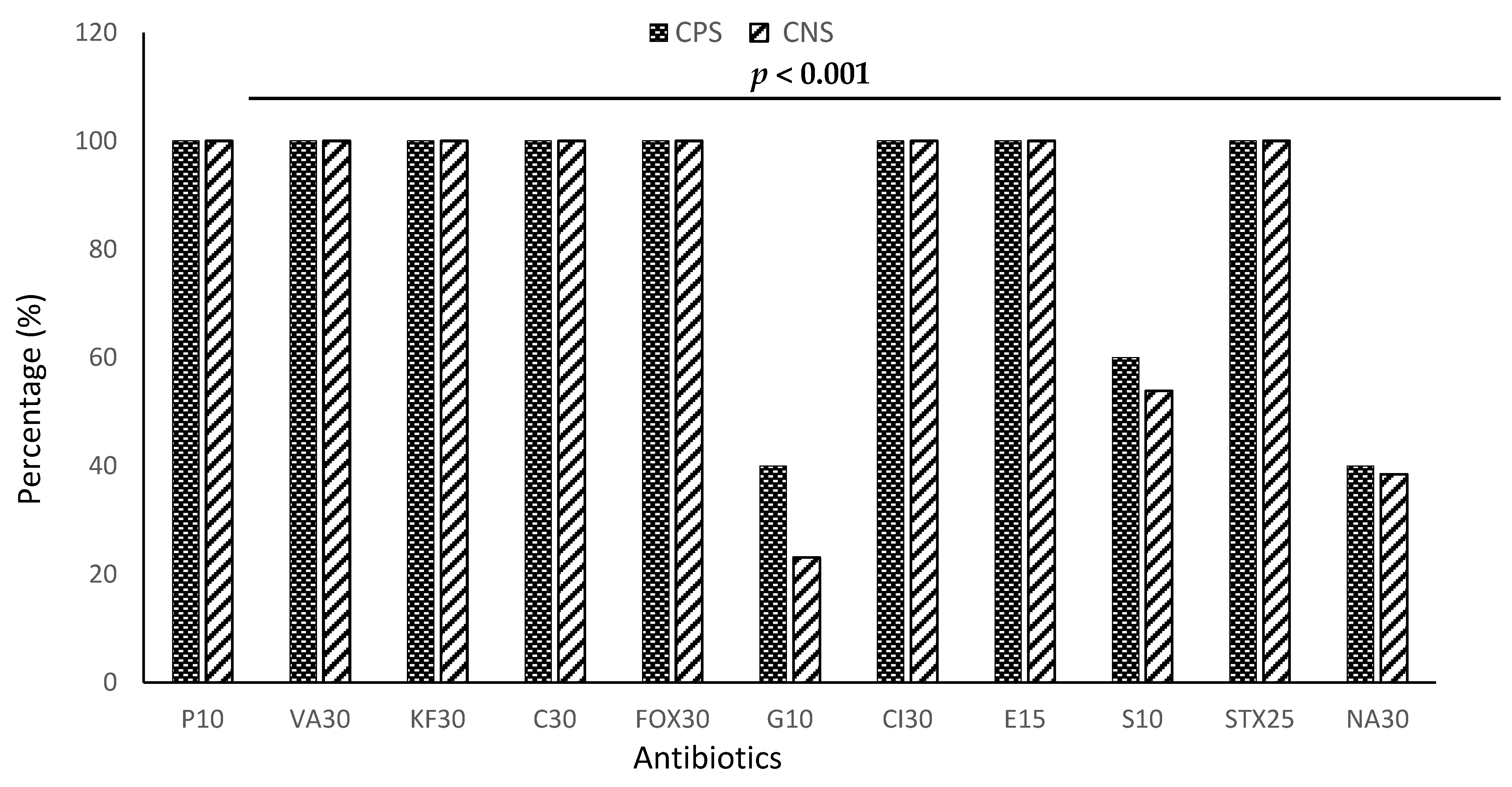
Staphylococci were divided into two categories for assessing susceptibility to antibiotics: Staphylococcus aureus (CPS) and Staphylococcus spp (CNS). Figure 4 shows the results of the antibiotic resistance profile of staphylococci.
Based on the statistical analysis of Table 2, it is evident that there existed a significant disparity in the usage of antibiotics (p < 0.001). Table 2 shows that all strains of staphylococci were 100% resistant to β-lactams, particularly penicillin G, cefoxitin, cephalothin, chloramphenicol, and ceftriaxone, sulfonamides such as trimethoprim-sulfamethoxazole, glycopeptides such as vancomycin, and macrolides such as erythromycin. However, their resistance was low to gentamicin (31.54%), nalidixic acid (39.23%), and streptomycin (59.92%). Gentamicin showed a low efficacy against staphylococci, particularly Staphylococcus aureus.

3.5. Virulence Genes and Resistance Gene Macrolides

In this study, 4% of Enterobacteriaceae strains harbored the fimA gene, and 5.55% of staphylococci strains contained the ermB gene encoding macrolide resistance (Table 3). However, there is no cnf1 and mefA gene for Enterobacteriaceae exhibiting enterotoxin produced by E. coli and staphylococci showing erythromycin resistance.

4. Discussion

The primary source of human infections is ingesting contaminated food [33]. Indeed, pathogenic bacteria such as staphylococci and Enterobacteriaceae have been identified in African fermented cereal foods [34] and traditional fermented drinks [15]. Our study showed that the percentages of samples contaminated with these microorganisms were 60.64% of Enterobacteriaceae and 39.36% of staphylococci. This rate of Enterobacteriaceae contamination is much higher than the 14.4% obtained by Cason et al. [35] in Sesotho, a traditional beer from South Africa produced from corn or sorghum. The low rate of Enterobacteriaceae and the absence of staphylococci in Sesotho can be explained by the fact that Sesotho is obtained from two fermentations with the addition of three different ferments (Tomoso, Mmela, and Yomoso) and by the fact that the two studies were not carried out under the same conditions. However, observing the rules of Good Hygiene Practices and Good Preparation Practices can reduce contamination rates in this situation.
The biochemical characterization of the enterobacteria and staphylococci isolated strains gave us more precision as to the species in these traditional beers. We noted the presence of Klebsiella terrigena (2.28%), Enterobacter aerogens (6.82%), Providencia rettgeri (9.09%), Chryseomonas luteola (11.36%), Serratia rubidae (25%), and Enterobacter cloacae (45.45%) for Enterobacteriaceae. As for the Staphylococcus genus, we noted the presence of 13.51% of coagulase-positive staphylococci (probably Staphylococcus aureus) and 86.48% of coagulase-negative staphylococci (Staphylococcus spp). Certain pathogenic microorganisms and alterations of traditional beers observed in Sesotho, namely, the genus’ Chryseobacterium, Enterobacter, and Klebsiella [35], were also present in our samples. These strains have also been isolated in other traditional beers prepared from raw materials, such as rice for Chicha, a Brazilian fermented drink [36], and cactus juice for pulque, a Mexican alcoholic beverage [37]. The high proportion of Enterobacter cloacae (45.45%) observed in these traditional beers could be due to their habitat, which remains the environment. The results of our study corroborate those of several studies [35,36,37] by highlighting the presence of unusual species such as Serratia rubidae, Providencia rettgeri, and Staphylococcus aureus in our samples. Indeed, Serratia rubidae and Providencia rettgeri have been found in stool [38] and flies [39], respectively. Staphylococci are ubiquitous natural bacteria, i.e., they are found in air, water, soil, food, furniture, and equipment [18,40]. The presence of these different species can be explained by their contaminating of the beer production process, raw materials, the sales environment, lack of supervision, or non-compliance with the hygiene rules.
During bacterial infections, the way of life developed by bacteria is biofilm production [41]. The production of biofilm is one of the defense mechanisms of bacteria. Biofilm formation is a default lifestyle for staphylococci and Enterobacteriaceae [42,43]. The high rate of biofilm production observed in Enterobacter aerogens and Klebsiella terrigena during our research can be explained by the fact that they are Gram-negative thermotolerant bacilli of food origin. Regarding staphylococci, the low rate observed in coagulase-negative Staphylococci (15.35%) was also found in the work of Ahouandjinou et al. [44], with a respective proportion of biofilm production of 28% for Staphylococcus lugdunensis and 20% for S. warneri on food coagulase-negative Staphylococcus (CNS) strains. Indeed, some CNSs, such as Staphylococcus epidermidis, adhere more quickly to the stainless steels of food industry equipment [45]. Therefore, these CNSs can easily comply with the kitchen utensils involved in producing these traditional beers. Therefore, properly cleaning these equipment and kitchen utensils could justify the low rate observed in the CNS. As for S. aureus (60%), the high rate observed can be explained by the fact that they adhere and quickly develop biofilm in contact with the food surface [46]. This lifestyle facilitates the adhesion of bacteria to food surfaces creating a public health problem [47]. In short, in these beers, the high rate of contamination observed with Providencia rettgeri, Enterobacter aerogens, Klebsiella terrigena, and Staphylococcus aureus can be explained by the fact that they can produce a biofilm, which provides specific adhesion characteristics allowing for their persistence and their adaptation to bad conditions. The production of biofilms is potentially a risk of poisoning for the consumer.
Gentamicin and nalidixic acid had shown efficacy in almost all strains isolated except for Enterobacter cloacae. Our results corroborate those of Hama et al. [48] and Anago et al. [49], who qualify gentamicin as having excellent efficacy against staphylococci and ESBL-producing strains of enterobacteria, respectively. This effectiveness of gentamicin, accompanied by nalidixic acid, can be explained by the fact that aminoglycosides (gentamicin) act on the bacterial ribosome and induce the synthesis of erroneous proteins [50]. Regarding quinolones, such as nalidixic acid, their mechanism of action involves targeting DNA gyrase and topoisomerase IV during DNA replication. These enzymes regulate the DNA topology necessary for replication [51]. The inhibition of DNA gyrase leads to the suppression of the positive supertowers of DNA.
In contrast, that of topoisomerase IV leads to the accumulation of daughter replicons, thus disrupting this replication. In addition to gentamicin and nalidixic acid, we note that some antibiotics, such as doxycycline and trimethoprim-sulfamethoxazole, remain active on enterobacteria, as does streptomycin on staphylococci. A study conducted by Kadja et al. [52] on combining tetracycline and trimethoprim sulfonylurea on strains from dairy farming corroborated our results with an efficacy of 62% and 67%, respectively. As for β-lactams and others, the Enterobacteriaceae were all resistant (100%). Previous work corroborates our results on the sensitivity of Enterobacteriaceae strains isolated to the antibiotics tested [53,54]. In addition, some work has shown that vancomycin, penicillin G, and macrolides are effective against food-borne S. aureus strains [52,55]. Globally, the resistance of Enterobacteriaceae and staphylococci isolates to β-lactams can be explained by the production of β-lactamase, which hydrolyzes these antibiotics or the structure of the pores is reduced by the antibiotics that pass [56]. The development of resistance to other antibiotics can be explained by the fact that these strains have already been in contact with different families of antibiotics. Particularly for staphylococci, resistance to ceftriaxone implies resistance to almost all β-lactams currently available [57] and the development of resistance to many antibiotics widely used to control infections such as food poisoning [58,59]. The multidrug resistance of the isolates could be explained by self-medication and the excessive and uncontrolled use of antibiotics accompanied by a selection of resistant bacteria [60,61]. This selection could be due to acquiring resistance genes such as erm, msr, mef, and mph [56], or virulence genes such as fimA and cnf1 [62].
Our study only revealed a shallow presence of fimA and ermB genes in Enterobacteriaceae (Enterobacter cloacae) and staphylococci (CNS). The work of Le Trong [63] showed the fimA gene’s presence in many Enterobacteriaceae species. In 2020, Soria-Bustos et al. [64] isolated Enterobacter cloacae with virulence properties in the appearance of extra-intestinal infections. Thus, the biofilm production capacity of E. cloacae could be explained by the fimA gene, which codes for the synthesis of adhesin, an essential protein in biofilm formation. As for the cnf1 gene, its absence can be explained by the fact that it is mainly produced by E. coli [65]. Regarding staphylococci, a study on the transferability of antibiotic resistance genes between bacteria carried out during the food fermentation of milk showed that Staphylococcus aureus and potentially pathogenic coagulase-negative staphylococci are carriers of macrolide resistance genes [66,67].
However, the low proportion of ermB genes with a complete absence of mefA genes, even though staphylococci were phenotypically resistant to erythromycin in our study, can be explained by the fact that these staphylococci could develop other resistance mechanisms through the acquisition of other genes such as msr, mph, and ere, encoding for macrolides resistance. Apart from the addition of genes, the multidrug resistance of strains can be explained by forming a biofilm that protects bacteria from antibacterial molecules [68]. There is a correlation between the ability to form biofilms and antibiotic resistance. This antibiotic resistance can be explained by the diffusion of drugs in the biofilm [69]. After exposure to these agents, a small surviving population can repopulate the surface immediately and become more resistant to antimicrobial treatment. Food contaminated with bacteria resistant to antibiotics and carrying genes for resistance or virulence are dangerous germs that could threaten public health.
We must acknowledge the limitations of our study, as we solely analyzed samples from the northern region of the country. It would be beneficial to expand our sampling to encompass other areas. Additionally, we could broaden our investigation to include other pathogenic strains and examine further pathological factors.

5. Conclusions

From our research, it was found that the kpètè-kpètè used to produce traditional fermented beers (Tchoukoutou and Tchapalo) are contaminated with pathogenic bacteria such as Serratia rubidae, Enterobacter cloacae, Enterobacter aerogens, Providencia rettgeri, Chryseomonas luteola, Klebsiella terrigena, and Staphylococcus aureus. These bacteria exhibit multidrug resistance to antibiotics with the ability to form biofilms. The presence of these strains in traditional drinks is a reality and remains worrying for the consumer’s health. It would be better to extend this study to other types of fermented foods, to deepen this study on the search for bacterial toxins and genes for antibiotic resistance given the phenotypic character resistant to macrolides in general and erythromycin that we observed in this study.

Author Contributions

Conceptualization, C.N., H.S. and L.B.-M.; methodology, B.B., R.A. and D.N.B.; software, D.N.B. and R.A.; validation, C.N., H.S., A.A., S.M.I.H. and L.B.-M.; formal analysis, D.N.B., S.M.I.H. and H.S.; investigation, C.N. and B.B.; resources, O.O.B. and L.B.-M.; data curation, C.N., B.B. and H.S.; writing—original draft preparation, C.N. and D.N.B.; writing—review and editing, J.F.M., O.O.B. and L.B.-M.; visualization, H.S.; supervision, J.F.M., O.O.B. and L.B.-M.; project administration, A.A., H.S., and L.B.-M.; funding acquisition, O.O.B., A.A. and H.S. All authors have read and agreed to the published version of the manuscript.

Funding

This research received no external funding.

Data Availability Statement

The data used to support the findings of this work are available from the corresponding author upon request.

Acknowledgments

The authors would like to express their appreciation for the traditional fermented beers of Benin, particularly those of Natitingou, Boukoumbé, Tanguiéta, Parakou, Tchaourou, N’Dali, Bantè, Dassa Zoumè, and Glazoué.

Conflicts of Interest

The authors declare no conflict of interest.

References

  1. WHO. Rapport OMS: 420 000 Personnes Tuées par l’intoxication Alimentaire; WHO: Geneva, Switzerland, 2018. [Google Scholar]
  2. Osseyi, E.G.; Tagba, P.; Karou, S.D. Stabilization of the Traditional Sorghum Beer “Tchoukoutou” using Rustic Wine- Making Method. Adv. J. Food Sci. Technol. 2011, 3, 254–258. [Google Scholar]
  3. Kayodé, A.P.P.; Linmemann, A.R.; Nout, M.J.R.; Hounhouigan, D.J.; Stomph, T.J.; Smulders, M.J.M. Diversity and food quality properties of farmers’ varieties of sorghum from Benin. J. Sci. Food Agric. 2006, 86, 1032–1039. [Google Scholar] [CrossRef]
  4. Missihoun, A.A.; Agbangla, C.; Sagbadja, H.A.; Ahanhanzo, C.; Vodouhe, R. Gestion traditionnelle et statut des ressources génétiques du sorgho (Sorghum bicolor L. Moench) au Nord-Ouest du Bénin. Int. J. Biol. Chem. Sci. 2012, 6, 1003–1018. [Google Scholar] [CrossRef] [Green Version]
  5. Aholou, C.C. Construction identitaire et urbaine autour du cabaret à Lomé (Togo). Hommes Migr. 2010, 1283, 32–41. [Google Scholar] [CrossRef] [Green Version]
  6. Baba-Moussa, L.; Bokossa, Y.I.; Baba-Moussa, F.; Ahisou, H.; Adeoti, Z.; Yehouenou, B.; Mamadou, A.; Toukourou, F.; Sanni, A. Etude des possibilités de contamination des aliments de rues au Bénin: Cas de la ville de Cotonou. J. Rech. Sci. L’université Lomé (Togo) Série A 2006, 8, 149–156. [Google Scholar]
  7. Adesetan, T.O.; Efuntoye, M.O.; Babalola, O.O. Profiling of Bacillus Cereus Enterotoxigenic Genes from Retailed Foods and Detection of the Nhe and Hbl Toxins with Immunological Assay. J. Appl. Nat. Sci. 2022, 14, 254–267. [Google Scholar] [CrossRef]
  8. Imade, E.E.; Omonigho, S.E.; Babalola, O.O.; Enagbonma, B.J. Lactic acid bacterial bacteriocins and their bioactive properties against food-associated antibiotic-resistant bacteria. Ann. Microbiol. 2021, 71, 44. [Google Scholar] [CrossRef]
  9. Chuah, L.O.; Shamila-Syuhada, A.K.; Liong, M.T.; Rosma, A.; Thong, K.L.; Rusul, G. Physio-chemical, microbiological properties of tempoyak and molecular characterisation of lactic acid bacteria isolated from tempoyak. Food Microbiol. 2016, 58, 95–104. [Google Scholar] [CrossRef]
  10. Egbuta, M.A.; Mwanza, M.; Babalola, O.O. Health Risks Associated with Exposure to Filamentous Fungi. Int. J. Environ. Res. Public Health 2017, 14, 719. [Google Scholar] [CrossRef] [Green Version]
  11. Achi, O.K.; Ukwuru, M. Cereal-based fermented foods of Africa as functional foods. Int. J. Microbiol. Appl. 2015, 2, 71–83. [Google Scholar]
  12. Adesetan, T.O.; Efuntoye, M.O.; Babalola, O.O. Biochemical Characterization and Antimicrobial Susceptibility of Bacillus Cereus Isolates from Some Retailed Foods in Ogun State, Nigeria. J. Microbiol. Biotechnol. Food Sci. 2019, 9, 616–621. [Google Scholar] [CrossRef]
  13. Yu, Z.; Su, Y.; Zhang, Y.; Zhu, P.; Mei, Z.; Zhou, X.; Yu, H. Potential use of ultrasound to promote fermentation, maturation, and properties of fermented foods: A review. Food Chem. 2021, 357, 129805. [Google Scholar] [CrossRef]
  14. Akaki, K.D.; Aw, S.; Loiseau, G.; Guyot, J.P. Etude du comportement des souches de Bacillus cereus ATCC 9139 et d’Escherichia coli ATCC 25922 par la méthode des challenge-tests lors de la confection de bouillies à base de pâte de mil fermentée en provenance de Ouagadougou (Burkina-Faso). Rev. Ivoirienne Des Sci. Et Technol. 2008, 11, 103–117. [Google Scholar]
  15. N’tcha, C.; Adéyèmi, A.D.; Kayodé, A.P.P.; Vieira-Dalodé, G.; Agbobatinkpo, B.P.; Codjia, J.C.; Baba-Moussa, L. Indigenous knowledge associated with the production of starters culture used to produce Beninese opaque sorghum beers. J. Appl. Biosci. 2015, 88, 8223–8234. [Google Scholar] [CrossRef] [Green Version]
  16. Ekonomou, S.I.; Parlapani, F.F.; Kyritsi, M.; Hadjichristodoulou, C.; Boziaris, I.S. Preservation status and microbial communities of vacuum-packed hot smoked rainbow trout fillets. Food Microbiol. 2022, 103, 103959. [Google Scholar] [CrossRef]
  17. Sundaramoorthy, N.; Yogesh, P.; Dhandapani, R. Production of prodigiosin from Serratia marcescens isolated from soil. Indian J. Sci. Technol. 2009, 2, 32–34. [Google Scholar] [CrossRef]
  18. Rodríguez-Saavedra, M.; González de Llano, D.; Moreno-Arribas, M.V. Beer spoilage lactic acid bacteria from craft brewery microbiota: Microbiological quality and food safety. Food Res. Int. 2020, 138 Pt A, 109762. [Google Scholar] [CrossRef] [PubMed]
  19. Hounsa, A.; Kouadio, L.; De Mol, P. Self-medication with antibiotics obtained from private pharmacies in Abidjan, Ivory Coast. Médecine Mal. Infect. 2009, 40, 333–340. [Google Scholar] [CrossRef]
  20. Alsan, M.; Morden, N.; Gottlieb, J.D.; Weiping, Z.; Skinner, J. Antibiotic use in cold and flu season and prescribing quality: A retrospective cohort study. Med. Care 2015, 53, 1066. [Google Scholar] [CrossRef] [PubMed] [Green Version]
  21. Singh, A.; Gautam, P.K.; Verma, A.; Singh, V.; Shivapriya, P.M.; Shivalkar, S.; Sahoo, A.K.; Samanta, S.K. Green synthesis of metallic nanoparticles as effective alternatives to treat antibiotics resistant bacterial infections: A review. Biotechnol. Rep. 2020, 25, e00427. [Google Scholar] [CrossRef] [PubMed]
  22. Akindolire, M.A.; Babalola, O.O.; Ateba, C.N. Detection of Antibiotic Resistant Staphylococcus aureus from Milk: A Public Health Implication. Int. J. Environ. Res. Public Health 2015, 12, 10254–10275. [Google Scholar] [CrossRef] [Green Version]
  23. Muylaert, A.; Mainil, J.G. Résistances aux fluoroquinolones: La situation actuelle. Ann. Med. Vet. 2013, 157, 15–26. [Google Scholar]
  24. Christensen, G.D.; Simpson, W.A.; Bisno, A.L.; Beachy, E.H. Adherence of biofilm producing strains of Staphylococci epidermidis to smooth surfaces. Infect. Immun. 1985, 37, 318–326. [Google Scholar] [CrossRef]
  25. Stepanović, S.; Vuković, D.; Dakić, I.; Savić, B.; Švabić-Vlahović, M. A modified microtiter-plate test for quantification of staphylococcal biofilm formation. J. Microbiol. Methods 2000, 40, 175–179. [Google Scholar] [CrossRef] [PubMed]
  26. CASFM. Comité de l’antibiogramme de la Société Française de Microbiologie: Recommandations 2020 V.1.1. 181 p 2020. Available online: https://www.sfm-microbiologie.org/wp-content/uploads/2020/04/CASFM2020_Avril2020_V1.1.pdf (accessed on 1 September 2022).
  27. Rasmussen, R.S.; Morrissey, M.T. DNA-based methods for the identification of commercial fish and seafood species. Compr. Rev. Food Sci. Food Saf. 2008, 7, 280–295. [Google Scholar] [CrossRef] [PubMed]
  28. Lamy, M.C.; Dramsi, S.; Billoët, A.; Réglier-Poupet, H.; Tazi, A.; Raymond, J.; Guérin, F.; Couvé, E.; Kunst, F.; Glaser, P. Rapid detection of the highly virulent group B Streptococcus ST-17 clone. Microb. Infect. 2006, 8, 1714–1722. [Google Scholar] [CrossRef]
  29. Kaczmarek, A.; Budzyńska, A.; Gospodarek, E. Prevalence of genes encoding virulence factors among Escherichia coli wiyh K1 antigen and-K1 E. coli strains. J. Med. Microbiol. 2012, 61, 1360–1365. [Google Scholar] [CrossRef] [PubMed] [Green Version]
  30. Moulin-Schouleur, M.; Schouleur, C.; Tailliez, P.; Kao, M.R.; Brée, A.; Germon, P.; Oswald, E.; Mainil, J.; Blanco, M.; Blanco, J. Common Virulence factors and genetic relationships between O18:K1:H7 Escherichia coli isolates of human and avian origin. J. Clin. Microbiol. 2006, 44, 3484–3492. [Google Scholar] [CrossRef] [Green Version]
  31. Germon, P.; Chen, Y.H.; He, L.; Blanco, J.E.; Bree, A.; Schouler, C.; Huang, S.H.; Moulin-Schouleur, M. IbeA, a virulence factors of avian pathogenic Escherichia coli. Microbiology 2005, 151, 1179–1186. [Google Scholar] [CrossRef] [PubMed] [Green Version]
  32. Desjardins, M.; Delgaty, K.L.; Ramotar, K.; Toye, B. Prevalence and mechanisms of erythromycin resistance in group A and group B Streptococcus: Implications for reporting susceptibility results. J. Clin. Microbiol. 2004, 42, 5620–5623. [Google Scholar] [CrossRef] [Green Version]
  33. Waqas, S.; Bilad, M.R.; Man, Z.B.; Klaysom, C.; Jaafar, J.; Khan, A.L. An integrated rotating biological contactor and membrane separation process for domestic wastewater treatment. Alex. Eng. J. 2020, 59, 4257–4265. [Google Scholar] [CrossRef]
  34. Tankoano, A.; Diop, M.B.; Sawadogo-Lingani, H.; Kaoré, D.; Savadogo, A. Les aspects technologiques, microbiologiques et nutritionnels des aliments fermentés à base de lait de mil en Afrique de l’ouest. Int. J. Adv. Res. 2017, 5, 1509–1526. [Google Scholar] [CrossRef] [Green Version]
  35. Cason, D.E.; Mahlomaholo, J.B.; Taole, M.M.; Abong, O.G.; Vermeulen, J.G.; de Smidt, O.; Vermeulen, M.; Steyn, L.; Valverde, A.; Vijoen, B. Bacterial and Fungal Dynamics during the Fermentation Process of Sesotho, a Traditional Beer of Southern Africa. Front. Microbiol. 2020, 11, 1451. [Google Scholar] [CrossRef] [PubMed]
  36. Escalante, A.; Maria, E.R.; Martinez, A.; Munguia, A.L. Characterization of bacterial diversity in Pulque, a traditional Mexican alcoholic fermented beverage, as determined by 16S rDNA analysis. FEMS Microbiol. Lett. 2004, 235, 273. [Google Scholar] [CrossRef] [Green Version]
  37. Bokulich, N.A.; Bamforth, C.W.; Mills, D.A. Brewhouse-resident microbiota is responsible for multi-stage fermentation of American coolshipale. PLoS ONE 2012, 7, e35507. [Google Scholar] [CrossRef] [PubMed]
  38. Tavares-Carreon, F.; De Anda-Mora, K.; Rojas-Barrera, I.C.; Andrade, A. Serratia marcescens antibiotic resistance mechanisms of an opportunistic pathogen: A literature review. Peer J. 2023, 5, e14399. [Google Scholar] [CrossRef]
  39. Zhang, J.; Wang, J.; Chen, L.; Yassin, A.K.; Kelly, P.; Butaye, P.; Li, J.; Gong, J.; Cattley, R.; Qi, K.; et al. Housefly (Musca domestica) and Blow Fly (Protophormia terraenovae) as Vectors of Bacteria Carrying Colistin Resistance Genes. Appl. Environ. Microbiol. 2017, 84, e01736-17. [Google Scholar] [CrossRef] [PubMed] [Green Version]
  40. Bensakhria, A. Biologie: Staphylococcus aureus (staphylocoques). 2017. Available online: www.magazinescience.com (accessed on 5 May 2022).
  41. Kostaki, M.; Chorianopoulos, N.; Braxou, E.; Nychas, G.J.; Efstathios, G. Differential biofilm formation and chemical disinfection resistance of sessile cells of Listeria monocytogenes strains under monospecies and dual-species (with Salmonella enterica) conditions. Appl. Environ. Microbiol. 2012, 8, 2586–2595. [Google Scholar] [CrossRef] [Green Version]
  42. Verstraeten, N.; Braeken, K.; Debkumari, B.; Fauvart, M.; Fransaer, J.; Vermant, J.; Michiels, J. Living on a surface: Swarming and biofilm formation. Trends Microbiol. 2008, 16, 496–506. [Google Scholar] [CrossRef]
  43. Balestrino, D.; Ghigo, J.M.; Charbonnel, N.; Haagensen, J.A.; Forestier, C. The characterization of functions involved in the establishment and maturation of Klebsiella pneumoniae in vitro biofilm reveals dual roles for surface exopolysaccharides. Environ. Microbiol. 2008, 10, 685–701. [Google Scholar] [CrossRef]
  44. Ahouandjinou, H. Qualité Microbiologique, Antibiorésistance des Souches de Staphylococcus spp et d’Escherichia coli Isolées des Carcasses Bovines au Bénin. Ph.D. Thesis, University of Abomey, Calavi, Benin, 2016. [Google Scholar]
  45. Lerebour, G.; Cupferman, S.; Bellon-Fontaine, M.N. Adhesion of Staphylococcus aureus and Staphylococcus epidermidis to the Episkin reconstructed epidermis model and to an inert 304 stainless steel substrate. J. Appl. Microbiol. 2004, 97, 7–16. [Google Scholar] [CrossRef] [PubMed]
  46. Marques, S.C.; Silva-Rezende, J.G.; de Freitas-Alves, L.A. Formation of biofilms by Staphylococcus aureus on stainless steel and glass surfaces and its resistance to some selected chemical sanitiziers. Braz. J. Microbiol. 2007, 38, 538–543. [Google Scholar] [CrossRef] [Green Version]
  47. Mah, T.F. Biofilm-specific antibiotic resistance. Future Microbiol. 2012, 7, 1061–1072. [Google Scholar] [CrossRef] [Green Version]
  48. Hama, H. Recherche de Bactéries Associées aux Mammites Subcliniques Dans le Lait de Chèvre en Mauritanie et au Togo et Détermination de leur Antibiosensibilité. Ph.D. Thesis, Veterinary Medicine, Dakar, Senegal, 2004. [Google Scholar]
  49. Anago, E.; Ayi-Fanou, L.; Akpovi, C.D.; Hounkpe, W.B.; Tchibozo, M.; Bankole, H.S. Antibiotic resistance and genotype of beta-lactamase producing Escherichia coli in nosocomial infections in Cotonou, Benin. Ann. Clin. Microbiol. Antimicrob. 2015, 14, 5. [Google Scholar] [CrossRef] [Green Version]
  50. Touati, A.; Brasme, L.; Benallaoua, S.; Gharout, A.; Madoux, J.; De Champs, C. First report of qnrB-producing Enterobacter cloacae and qnrA-producing Acinetobacter baumannii recovered from Algerian hospitals. Diagn. Microbiol. Infect. Dis. 2008, 60, 287–290. [Google Scholar] [CrossRef]
  51. Soussy, C.J. Quinolones et bactéries à Gram négatif. In Patrice Courvalin, Roland Leclercq, 2nd ed.; Bingen, E., Ed.; ESKA: Paris, France, 2006; 277p. [Google Scholar]
  52. Kadja, M.C.; Kane, Y.; Viban, B.V.; Kaboret, Y.; Alambedji, B. Sensibilité aux antibiotiques des Bactéries associées aux mammites Clinique des petits ruminants dans la région de Dakar. Ann. Sci. Agron. 2013, 17, 205–216. [Google Scholar]
  53. Davin-Regli, A.; Bolla, J.M.; James, C.E.; Lavigne, J.P.; Chevalier, J.; Garnotel, E. Membrane permeability and regulation of drug “influx and efflux” in enterobacterial pathogens. Curr. Drug Targets 2008, 9, 750–759. [Google Scholar] [CrossRef] [PubMed]
  54. Wilson, K.; Shafiee, M.; Hasso, M.; Sharif, U. Providencia rettgeri: An unexpected case of Gram-negative cellulitis. Case Rep. Wounds Int. 2015, 6, 30–32. [Google Scholar]
  55. Sina, H.; Attien, P.; Wélé, M.; Socohou, A.; Boukary-Chabi, A.; Dougnon, V.T.; Baba-Moussa, F.; Adjanohoun, A.; Baba-Moussa, L. Sanitary Risk Factors and Microbial Contamination of Grilled Meats Sold in Cotonou, Benin. J. Food Secur. 2019, 7, 175–182. [Google Scholar]
  56. Schwarz, S.; Chaslus-Dancla, E. Use of antimicrobials in veterinary medicine and mechanisms of resistance. Vet. Res. 2001, 32, 201–225. [Google Scholar] [CrossRef] [Green Version]
  57. Chiquet, C.; Max, M.; Altayrac, J.; Aptel, F.; Boisset, S.; Vandenesch, F.; Carricajo, A. Correlation between clinical data and antibiotic resistance in coagulase negative Staphylococcus species isolated from 68 patients with acute post-cataract endophthalmitis. Clin. Microbiol. Infect. 2015, 21, 592.e1–592.e8. [Google Scholar] [CrossRef] [PubMed] [Green Version]
  58. Bonnel, A.S.; Quinque, K.; Le Roux, P.; Le Luyer, B. Aspergillose nécrosante semi invasive chez un enfant de 14 ans atteint de mucoviscidose. Rev. Mal. Respir. 2006, 23, 343–347. [Google Scholar] [CrossRef]
  59. Rodrigues, M.X.; Silva, C.C.N.; Trevillin, H.J.; Cruzado, B.M.; Mui, S.T.; Sanches Duarte, R.F.; Castillo, C.C.; Canniatti-Brazaca, G.S.; Porto, E. Antibiotic resistance and molecular characterization of Staphylococcus species from mastitic milk. Afr. J. Microbiol. Res. 2017, 11, 84–91. [Google Scholar]
  60. Lesch, C.; Itokazu, G.; Danziger, L.; Weinstein, R. Multi-hospital analysis of antimicrobial usage and resistance trends. Diagn. Microbiol. Infect. Dis. 2001, 41, 149–154. [Google Scholar] [CrossRef]
  61. Trivedi, K.; Cupakova, S.; Karpiskova, R. Virulence factors and antibiotic resistance in enterococci isolated foodstuffs. Vet. Med. 2011, 56, 352–357. [Google Scholar] [CrossRef] [Green Version]
  62. Clegg, S.; Hughes, K.T. FimZ is a molecular link between sticking and swimming in Salmonella enterica serovar Typhimurium. J. Bacteriol. 2002, 184, 1209–1213. [Google Scholar] [CrossRef] [Green Version]
  63. Le Trong, I. Donor strand exchange and conformational changes during E. coli fimbrial formation. J. Struct. Biol. 2008, 172, 380–388. [Google Scholar] [CrossRef] [Green Version]
  64. Soria-Bustos, J.; Ares, M.A.; Gómez-Aldapa, C.A.; González-y-Merchand, J.A.; Girón, J.A.; De la Cruz, M.A. Two Type VI Secretion Systems of Enterobacter cloacae Are Required for Bacterial Competition, Cell Adherence, and Intestinal Colonization. Front. Microbiol. 2020, 11, 560488. [Google Scholar] [CrossRef]
  65. Lockman, H.A. Yersinia pseudotuberculosis produces a cytotoxic necrotizing factor. Infect. Immun. 2002, 70, 2708–2714. [Google Scholar] [CrossRef] [PubMed] [Green Version]
  66. Yi, X.; Wang, M.; Zhou, Z. The potential impact of naturally produced antibiotics, environmental factors, and anthropogenic pressure on the occurrence of erm genes in urban soils. Environ. Pollut. 2019, 245, 282–289. [Google Scholar] [CrossRef]
  67. Luthje, P.; Schwarz, S. Antimicrobial resistance of coagulase-negative staphylococci from bovine subclinical mastitis with particular reference to macrolide lincosamide resistance phenotypes and genotypes. J. Antimicrob. Chemother. 2006, 57, 966–969. [Google Scholar] [CrossRef] [PubMed] [Green Version]
  68. Donlan, R.M.; Costerton, J.W. Biofilms: Survival mechanisms of clinically relevant microorganisms. Clin. Microbiol. Rev. 2002, 15, 167–193. [Google Scholar] [CrossRef] [PubMed] [Green Version]
  69. Zhang, L.; Mah, T.F. Involvement of a novel efflux system in biofilm-specific resistance to antibiotics. J. Bacteriol. 2008, 190, 4447–4452. [Google Scholar] [CrossRef] [PubMed] [Green Version]
Figure 1. Distribution of Enterobacteriaceae and staphylococci strains isolated from kpètè-kpètè samples.
Microorganisms 11 01939 g001
Figure 2. Distribution of biofilm production capacity according to identified species.
Microorganisms 11 01939 g002
Figure 3. Resistance profile of isolated Enterobacteriaceae to tested antibiotics. A10: Ampicillin (10 μg), C30: Chloramphenicol (30 µg), FOX30: Cefoxitin (30 µg), G10: Gentamicin (10 µg), Cl30: ceftriaxone (30 µg), DO30: Doxycycline (30 µg), SXT25: trimethoprim -sulfamethoxazole (25 µg), NA30: nalidixic acid (30 µg).
Microorganisms 11 01939 g003
Figure 4. Resistance profile of staphylococci to tested antibiotics. CPS: Staphylococcus aureus, CNS: Staphylococcus spp, P 10: Penicillin (10 μg), VA30: Vancomycin (30 μg), KF 30: Cephalothin (30 μg), C30: Chloramphenicol (30 µg), FOX: Cefoxitin (30 µg), G10: Gentamicin (10 µg), Cl30: ceftriaxone (30 µg), E15: Erythromycin(15 μg), S10: Streptomycin (10 μg), SXT2: trimethoprim-sulfamethoxazole (25 µg), NA30: Nalidixic acid (30 µg).
Microorganisms 11 01939 g004
Table 1. Distribution of Enterobacteriaceae resistant phenotypes to antibiotics.
AntibioticsPercentage of Resistancep Value
Ampicillin (A10)100.00%<0.001
Ceftriaxone (Cl30)100.00%
Cefoxitin (FOX30)100.00%
Chloramphenicol (C30)100.00%
Gentamicin (G10)1.85%
Doxycycline (DO30)32.40%
Trimethoprim-sulfamethoxazole (STX25)32.85%
Nalidixic acid (NA30)43.51%
Table 2. Distribution of antibiotics-resistant phenotypes of staphylococci.
AntibioticsPercentage of Resistancep Value
Penicillin G (P10)100.00%<0.0001
Ceftriaxone (Cl30)100.00%
Cefoxitin (FOX30)100.00%
Chloramphenicol (C30)100.00%
Cephalothin (KF30)100.00%
Vancomycin (VA30)100.00%
Erythromycin (E15)100.00%
Trimethoprim-sulfamethoxazole (STX25)100.00%
Gentamicin (G10)31.54%
Nalidixic Acid (NA30)39.23%
Streptomycin (S10)59.92%
Table 3. Distribution of virulence and resistance genes.
fimAcnf1ermBmefA
Klebsiella terrigena(0%)(0%)NANA
Enterobacter aerogens(0%)(0%)NANA
Providencia rettgeri(0%)(0%)NANA
Chryseomonas luteola(0%)(0%)NANA
Serratia rubidae(0%)(0%)NANA
Enterobacter cloacae(4%)(0%)NANA
S. aureusNANA(0%)(0%)
SCNNANA(5.55%)(0%)
NA: Not applicable.
Disclaimer/Publisher’s Note: The statements, opinions and data contained in all publications are solely those of the individual author(s) and contributor(s) and not of MDPI and/or the editor(s). MDPI and/or the editor(s) disclaim responsibility for any injury to people or property resulting from any ideas, methods, instructions or products referred to in the content.