Article

Physiological Functions of the Cello-Oligosaccharides Binding CebE in the Pathogenic Streptomyces sp. AMCC400023

1
College of Life Sciences, Shandong Agricultural University, Tai’an 271018, China
2
State Key Laboratory of Microbial Technology, Institute of Microbial Technology, Shandong University, Qingdao 266237, China
3
Research Center of Agricultural Biotechnology, Ningxia Academy of Agricultural and Forestry Sciences, Yinchuan 750002, China
*
Authors to whom correspondence should be addressed.
These authors contributed equally to this work.
Microorganisms 2024, 12(3), 499; https://doi.org/10.3390/microorganisms12030499
Submission received: 6 February 2024 / Revised: 25 February 2024 / Accepted: 27 February 2024 / Published: 29 February 2024
(This article belongs to the Special Issue Molecular Analysis of Plant Pathogenic Bacteria, 2nd Edition)

Abstract

:
Potato common scab, an economically important disease worldwide, is caused by pathogenic Streptomyces strains mainly through the effects of thaxtomin. The cello-oligosaccharides binding protein CebE is proposed as a gateway to the pathogenic development of Streptomyces scabiei. In this study, two functional CebE encoding genes, GEO5601 and GEO7671, were identified in pathogenic Streptomyces sp. AMCC400023. With a higher binding affinity towards signal molecules, the deletion of GEO5601 severely impaired thaxtomin-producing capacity and reduced the strain’s pathogenicity. Transcriptional analysis confirmed that CebE5601 is also responsible for the import and provision of carbon sources for cell growth. With lower binding affinity, the pathogenicity island (PAI)-localized CebE7671 may assume a new function of mediating the biological process of sporulation, given the significantly impaired formation of ΔGEO7671 spores. The mechanisms of action of CebE proteins unraveled in Streptomyces sp. AMCC400023 will help pave the way for more effective prevention of the potato common scab disease.

1. Introduction

Streptomyces are well-known soil saprophytes that actively participate in the recycling of nutrients in the environment, produce a great variety of secondary metabolites, and induce beneficial activities in plants, such as growth promotion and disease resistance [1]. However, in the last decades, a particular group of Streptomyces species was found to harbor the ability to infect important commercial root and tuber crops, including potatoes, beets, radishes, sweet potatoes, and peanuts [2]. It has been confirmed that pathogenic Streptomyces, including Streptomyces scabiei, S. acidiscabies, and S. turgidiscabies, can cause raised, pitted, or superficial scab lesions on the potato surface [3,4]. Such potato common scab significantly decreases crop quality and leads to severe economic losses [5].
Thaxtomin is a family of phytotoxins produced by pathogenic Streptomyces and is known as the major causative agent of potato common scab [6,7]. These compounds can induce plant cell hypertrophy, root growth retardation, and tissue necrosis at a nanomolar concentration by inhibiting the cellulose synthase complex [8,9,10]. Notably, almost all pathogenic Streptomyces have a pathogenicity island (PAI) composed of two regions. The toxin region (TR) harbors the thaxtomin biosynthetic gene cluster, and the colonization region (CR) contains two genes of nec1 and tomA, which encode secreted proteins known or suspected to act as virulence factors [11,12,13]. A previous study has experimentally confirmed the transferability of the PAI among Streptomyces species, which might account for the emergence of new pathogenic Streptomyces and the spread of potato common scab [14].
Cello-oligosaccharide is the main product of cellulose degradation and is abundant in nature. Some saprophytic Streptomyces species specifically utilize cellulose as their carbon source, and employ extracellular cellulolytic enzymes and an inducible ATP-dependent uptake system specific to cello-oligosaccharide [15]. Interestingly, cellobiose and cellotriose were found to possess a function other than providing energy in S. scabiei strains. As a major molecule released in the expansion period of plant tissues, cellotriose is proposed as the best inducer and trigger of the synthesis of virulome, including thaxtomin [16,17]. It has been authenticated that the cello-oligosaccharide transport system CebEFG-MsiK is essential for the induction of thaxtomin in pathogenic Streptomyces strains [4,18,19]. As the core component of this transporter system, the solute-binding protein CebE is responsible for recognizing and binding the cello-oligosaccharide substrates [20,21]. In contrast to the non-pathogenic Streptomyces strains, the transcriptional repressor CebR not only regulates cello-oligosaccharides uptake by binding with cellobiose or cellotriose, but is also involved in unlocking thaxtomin synthesis when binding with the upstream sites within the thaxtomin biosynthetic cluster. CebR has two binding sites (cbs) within the thaxtomin gene cluster; one located upstream of txtR, the cluster-situated regulator, and another within txtB, the peptide synthetase gene (thaxtomin synthetase). As the ligands of CebR, the binding of cellobiose and cellotriose can inhibit its DNA-binding activity, thereby allowing the transcription of txtR and inducing thaxtomin biosynthesis [19,22,23,24].
The special regulation mode of thaxtomin biosynthesis confirms the significant contributions of cello-oligosaccharides in Streptomyces pathogenicity; CebE was named the “doorway” in pathogenic Streptomyces due to its extracellular binding of cello-oligosaccharides and transmission of the signal to the cells, and consequently triggering the synthesis of arsenal thaxtomins [25]. The investigation of CebE can enhance our understanding of the pathogenic mechanism and elucidate how pathogenic Streptomyces establishes a connection between sensing extracellular plant material and initiating its virulent behavior [25], thereby establishing a solid foundation for future biological control. In accordance with some pathogenic Streptomyces, the common scab representative species S. scabiei 87–22 possesses three homologs of CebE, among which SCAB57751 was initially identified as the major contributor to potato scab, exhibiting a nanomolar range KD value towards cellotriose and cellobiose [25]. Recently, the physiological functions of the other two CebE proteins, SCAB2421 and SCAB77271, were also investigated. The deletion of SCAB2421 caused a moderately attenuated virulence phenotype, while the loss of the PAI-located CebE (SCAB77271) had no significant influence on strain growth or virulence capacity [26].
To date, many pathogenic Streptomyces species have been isolated, and diverse sets of CebEFG-Msik ABC transporters have been annotated in different pathogenic Streptomyces strains. Whether these CebEFG-MsiK ABC transporters play a role in virulence is of great interest and remains to be elucidated. However, the primary research focus on CebE primarily lies within strain S. scabiei 87–22, with a scarcity of related studies concerning other strains. Therefore, it is interesting to explore whether other mechanisms exist in other pathogenic Streptomyces strains. The pathogenic Streptomyces sp. AMCC400023 was previously isolated from potato common scab in Hebei province, China [27]. By possessing the well-known PAI component in its genome, all evidence from radish seedling assay, potted back experiment, and thaxtomin production confirmed Streptomyces sp. AMCC400023 is a virulent pathogen causing potato scab. As revealed by genome-based phylogenetic analysis, average nucleotide identity (ANI value is 88%), and in silico DNA-DNA hybridization (isDDH), strain AMCC400023 has a relatively close relationship with S. scabiei at the species level. However, comparative genomic analysis in the Virulence Factors of Pathogenic Bacteria Database identified 60 unique virulence-associated genes in the genome of Streptomyces sp. AMCC400023 when compared to S. scabiei 87–22. Also, unlike the three functional clusters for cello-oligosaccharides uptake in S. scabiei 87–22, two CebEFG-MsiK clusters were identified in the genome of AMCC400023. In the present study, we found that these two CebEs perform diverse physiological roles in strain growth and pathogenesis by comparing their binding affinities with cello-oligosaccharide substrates and pathogenesis of CebE-deficient strains, as well as transcriptomic responses in the presence of cellobiose.

2. Materials and Methods

2.1. Bacterial Strains, Plasmids, and Growth Conditions

Bacterial strains and plasmids used in this study are described in Table 1. Streptomyces strains were grown in Gause’s No. 1 medium [28], oat bran broth (OBB) medium [29], thaxtomin-defined medium (TDM) that was supplemented with 7 g L−1 cellobiose (TDMc) medium [16], or mannitol soya flour (MS) medium [30] at 28 °C. E. coli strains were grown in Luria-Bertani (LB) medium at 37 °C. When required, the medium was supplemented with ampicillin (100 μg mL−1), kanamycin (50 μg mL−1), chloramphenicol (30 μg mL−1), nalidixic acid (25 μg mL−1), or thiostrepton (20 μg mL−1). Cellobiose and cellotriose were biotech grade and purchased from MACKLIN.

2.2. Bioinformatic Analysis

MAFFT was used for multiple sequence alignment and conserved sites were selected to construct the NJ phylogenetic tree using the JTT substitution model, with a bootstrap value of 1000. The CebR binding sites were predicted using the PRODORIC tool.

2.3. Protein Expression and Purification

Genomic DNA was isolated by the Tiangen DNA extraction kit (Tiangen Biotech Co., Ltd., Beijing, China). The hydrophobic region of the sequence was analyzed using SignalIP 6.0 and DeepTMHMM v0.0.10. The open reading frame encoding GEO1108/GEO5601/GEO7671 without the hydrophobic region was amplified by PCR using the genomic DNA of Streptomyces sp. AMCC400023 as the template and primers listed in Table S3. The amplified fragment was ligated with pEASY-Blunt and transformed into E. coli DH5α for DNA sequencing. Then, the gene fragment was ligated with pET28a and transformed into E. coli BL21(C43) competent cells. For protein expression, the E. coli transformants were cultured in the LB medium at 37 °C containing 50 μg mL−1 of kanamycin until the growth reached an absorbance at 600 nm (OD600nm) of 0.6. Then, the His-tagged CebE was induced overnight (12 h) at 16 °C by a final concentration of 0.5 mM isopropyl-β-D-thiogalactopyranoside (IPTG). Cells were collected by centrifugation, resuspended in the lysis buffer (25 mM Tris-HCl, 150 mM NaCl, pH 8.0), and lysed using a high-pressure cell crusher. After centrifugation, the soluble proteins were loaded onto the pre-equilibrated Ni-NTA column and eluted with 250 mM imidazole. The cell lysis and purified proteins were verified by SDS-PAGE.

2.4. MST Assays

Microscale thermophoresis (MST) assays measurements were performed in triplicate at 25 °C, with 90% excitation power and medium MST power, on the Monolith NT.115 system. The protein was labeled with a red fluorescent dye and applied at a final concentration of 800 nM. The cellobiose and cellotriose solutions were two-fold diluted 16 times, with an initial concentration of 100 μΜ for CebE5601 and 270 mM for GebE7671. The labeled protein was then added to a dilution solution of the cello-oligosaccharides. Samples were filled into standard-treated capillaries for measurement. All the data were analyzed with MO.Affinity Analysis v2.3 software provided by the manufacturer.

2.5. Construction of CebE-Deficient Mutants

The in-frame deletion Streptomyces mutants were constructed by homologous recombination technology. DNA fragments about 1 kb upstream and downstream of GEO5601 or GEO7671 were amplified, using primers listed in Table S3. The PCR products were purified and ligated into the shuttle plasmid pJTU1278 to generate the pJTU1278-5601 and pJTU1278-7671 plasmids. Then, the constructed plasmids were introduced into E. coli ET12567 by transformation. After sequence verification, E. coli ET12567 strains carrying pJTU1278 plasmids (E. coli ET12567/pJTU1278-5601 or E. coli ET12567/pJTU1278-7671) were cultured in LB medium containing antibiotics (100 μg mL−1 Ampicillin, 50 μg mL−1 Kanamycin, 30 μg mL−1 Chloramphenicol) until OD600nm reached 0.4–0.6. The cells were collected by centrifuging at 8000× g and washed with fresh LB medium, then collected and suspended in 500 μL of sterile water. For the preparation of Streptomyces competent cells, the spores of Streptomyces sp. AMCC400023 were collected from the OBB plates and placed in 5 mL TES buffer (10 mM Tris HCl, 1 mM EDTA, 0.1 mM SDS). After heat shock and ice bath, 5 mL YT medium was added and cultured at 37 °C for 2.5 h. Then, the spores were collected by centrifugation and resuspended with sterile water, mixed with the cultures of E. coli ET12567/pJTU1278-5601 or E. coli ET12567/pJTU1278-7671, and spread on the MS plates. After culturing at 30 °C for 2–3 d, 1 mL of antibiotic solution (0.25 mg mL−1 thiostrepton and 0.5 mg mL−1 nalidixic acid) was added and then incubated for 16 h. The colonies were transferred to MS medium supplemented with 0.25 mg mL−1 tryptophan and 0.5 mg mL−1 nalidixic acid, then cultured at 30 °C until confirmed by PCR amplification and sequencing, using primers listed in Table S3.

2.6. Plant Virulence Assays

To assess the virulence phenotype of Streptomyces sp. AMCC400023 and mutants, an in vitro radish seedling assay was performed. Radish seeds were surface sterilized with 75% ethanol for 8–10 min and rinsed 3–4 times with sterile distilled water. The seeds were allowed to germinate for about 24–48 h in the dark, in a petri dish containing a moistened filter paper. Homogenously germinated seeds with a similar shoot length were placed into a glass tube of 1% agar water, with 3 seeds per tube. An appropriate amount of spore suspension with an OD value of 1.0 was inoculated in 100 mL of OBB for 5 d, then 200 µL of the culture was inoculated with newly sprouted radish seedlings, and the same amount of OBB medium was used as blank control. The tubes were incubated at 24 °C for 6 d, with intermittent light (16 h light and 8 h dark). Each experiment was performed in triplicate. To perform statistical analysis, we employed the Kruskal-Wallis test and Mann-Whitney test. Additionally, we applied the Benjamini-Hochberg procedure to correct the false discovery rate (FDR).
The potato pot trial was conducted from August to December 2020 at Shandong Agricultural University. Fifteen healthy potato tubers of variety Favorita were selected, surface sterilized with 1% NaClO for 5 min, and rinsed 3 times with sterile distilled water. After germination, potato seedlings with a similar shoot length were planted in a 35 × 30 (cm) plastic pot with 10 kg of soil. Streptomyces sp. AMCC400023 and the mutant derivatives were cultured on Gause’s No. 1 medium at 28 °C for 10 d. Spores were collected with sterile distilled water and inoculated in OBB media at 28 °C for 7 d. The cultures were adjusted to the same OD value by fresh OBB, then inoculated during the potato expansion period and the final concentration of the Streptomyces strains was 106 CFU/cm3. Each treatment was repeated in fifteen pots. Samples treated with OBB medium and sterile distilled water worked as a blank control. After 90 d of cultivation, the disease index of potatoes was calculated.
Disease index (percentage) = Σ(incidence level × number of corresponding grades)/(the highest incidence level × total number in this survey) × 100.

2.7. Thaxtomin Quantification

With similar spore inoculation, Streptomyces sp. AMCC400023 and its mutants were inoculated into OBB medium that was prepared in the same batch, and cultured at 28 °C and 180 rpm for 6–7 d. The cultures were filtered, the supernatant was extracted 3 times with an equal volume of ethyl acetate for 12 h, and then an appropriate amount of anhydrous sodium sulfate was added to remove the water. After rotary evaporation, the residue was dissolved in 2 mL of acetonitrile and filtered with a 0.22 μm membrane to get the thaxtomin solution. Liquid chromatography with tandem mass spectrometry (4000 QTRAP; AB SCIEX, Framingham, MA, USA) was used to quantify the thaxtomin content in solution with 0.3 mL min−1 flow rate of an isocratic mobile phase of 40:60 acetonitrile/water, both of them containing 2 mM ammonium acetate and 0.1% acetic acid. Thaxtomin was detected according to the method described previously [27]. The mycelia of the culture strains were dried and weighed. After dividing the total thaxtomin production by the dry weight of strains, the average thaxtomin production of culture strains was obtained. All experiments were repeated three times with different biological samples of Streptomyces strains.

2.8. Transcriptomic and RT-qPCR Analyses

Streptomyces sp. AMCC400023 was cultured in TDMc medium that contained 7 g L−1 of cellobiose. Based on the cell growth and thaxtomin accumulation curves, we collected samples from 36 h, 72 h, and 120 h, with three biological replicates in each group. Total RNA was isolated by using TRIzol RNA extraction kit (Thermo Fisher Scientific, New York, NY, USA) according to the manufacturer’s protocol. Assessment of the RNA integrity was performed on the Agilent 4200 Tape Station (Agilent, Santa Clara, CA, USA); meanwhile, RNA concentration and purity were measured on the Thermo NanoDrop One (Thermo Fisher Scientific). Epicentre Ribo-Zero rRNA Removal Kit was used to remove the ribosomal RNA, and the cDNA libraries were constructed according to the protocol of Illumina’s NEBNextő Ultra II Directional RNA Library Prep Kit and sequenced using Illumina HiSeq 2500 (Illumina, San Diego, CA, USA).
The quality control process used Fastp v0.22.0 software, and clean reads were then mapped to the reference genome using Bowtie2 v2.4.4. FPKM values were calculated for each gene using cufflinks and transformed to TPM values. The DESeq R package was used to identify DEGs with a threshold of P < 0.05 and foldChange > 2 or foldChange < 0.5. Heatmap analyses were performed using the complexHeatmap v2.18.0 R package.
The expression levels of GEO5601 and GEO7671 were further verified by RT-qPCR, with primers listed in Table S3. For PCR amplification, a total volume of 10 μL containing 5 μL of 2× PerfectStartTM Green qPCR SuperMix, 1 μL cDNA, 0.2 μL gene-specific primer (Table S3), and 3.6 μL ddH2O, was used. The program was 30 s at 94 °C, followed by 45 cycles with 5 s at 94 °C, and 30 s at 60 °C. To normalize the gene expression, 16S rDNA was used as an endogenous control.

3. Results

3.1. Bioinformatics Analysis of the Cello-Oligosaccharide Transporters

The complete genome of Streptomyces sp. AMCC400023 was sequenced, annotated, and deposited in the NCBI database under the accession number CP024989. According to the GhostKOALA and eggNOG-mapper annotation results, there are three annotated cello-oligosaccharide binding protein-encoding genes (cebE) in the genome, named GEO1108, GEO5601, and GEO7671, respectively. These three cebE genes are widely dispersed in the genome (Figure 1); GEO5601 and GEO1108 are far away from the TR and CR region, while GEO7671 is in the CR region of the PAI.
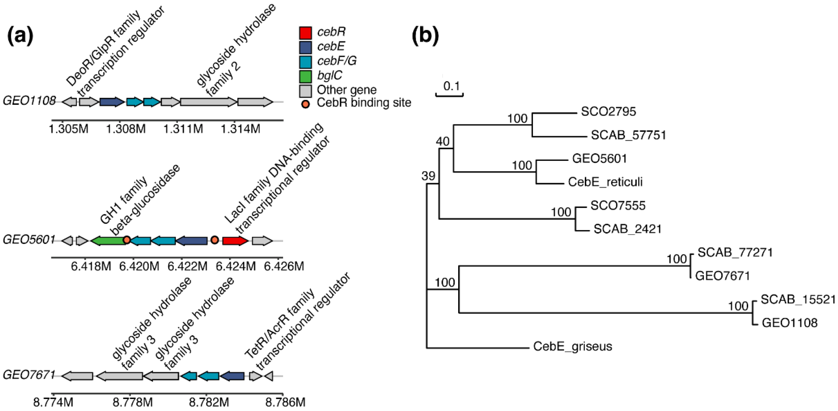
The gene structure of all three annotated transporters follows the typical mode in Actinobacteria strains, with CebE, CebF, and CebG, as well as the adjacent glycoside hydrolase (BglC) and a transcription regulator from different families (CebR) included (Figure 2a). All three putative cebEs were respectively clustered with one or two glycoside hydrolases, indicating their potential involvement in sugar transport. The glycoside hydrolase gene clustered with GEO1108 was annotated as beta-galactosidase, while glycoside hydrolases in GEO5601 and GEO7671 clusters were both annotated as beta-glucosidases, implying that GEO1108 may not function as a cello-oligosaccharide binding protein in Streptomyces sp. AMCC400023. When searching for the binding sites of CebR regulator, two binding sites were identified in the GEO5601 gene cluster (TGGGAGCGCTCCCA and TGGAAGCGCTCCCA), while no such cis-acting element was found within the GEO7671 cluster, as observed in its homolog protein SCAB77271 of strain 87–22 [26]. As for the GEO1108 gene cluster, no cis-acting element was observed. The gene cluster for thaxtomin synthesis contains two CebR binding sites, which is consistent with the presence of cbstxtB (GGGGAGCGCTCCCA) and cbstxtR-A (CGGGAGCGCTCCCA) in strain S. scabiei 87–22 [24].
Based on the phylogenetic analysis, these three putative CebE sequences are separately distributed into different branches, in which CebE5601 is most closely related to CebEreti (79.05% amino acid sequence identity) that has been previously recognized for its cellobiose-binding ability (Figure 2b). Also, with 46.06% and 46.93% sequence identity, it is closely related to the CebE homologs SCAB2421 and SCAB57751 in strain S. scabiei 87–22 (Table S1). The PAI-located CebE7671 is clustered with the third CebE sequence (SCAB77271) of S. scabiei 87–22, both of which are located in another subtree, with high sequence identity up to 98.8%.

3.2. Cellobiose/Cellotriose Affinities of Three CebE Proteins

To assess the binding affinity to the cello-oligosaccharide substrates, GEO1108, GEO5601, and GEO7671 were heterologously expressed and purified (Figure S1) for MST assays. After titration experiments using varying cellobiose concentrations, we obtained KD values of 977.19 nM for CebE5601 and a value of 1.28 mM for CebE7671, which is three orders of magnitude higher (Figure 3a). The micromolar level KD value of CebE5601 is consistent with its close homolog CebEreti (1.5 µM) but remarkably higher than that of SCAB57751 (14.2 nM) as measured by the equilibrium dialysis assay or tryptophan intrinsic fluorescence assays [20,25]. However, as different measurement methods were used, the binding differences among these homologs remain to be further evaluated under identical conditions. When switched to cellotriose as the ligand, similar binding affinities were observed as those of cellobiose, with KD values of 1.61 μM for CebE5601 and 2.72 mM for CebE7671 (Figure 3b). In agreement with the clustered beta-galactosidase encoding gene of GEO1108, no binding activity was detected between ligands cellobiose/cellotriose and CebE1108. Therefore, CebE1108 should be assigned as a binding protein for other sugars than cellobiose/cellotriose, and was not considered for further study. Of note, possibly due to strain differences, the indiscriminate binding affinity of cellobiose/cellotriose in the strain we investigated is different from those CebE results of strain 87–22, in which SCAB57751 has a stronger binding affinity with cellotriose than cellobiose [25].

3.3. Pathogenicity Assay and Thaxtomin Production of CebE Mutants

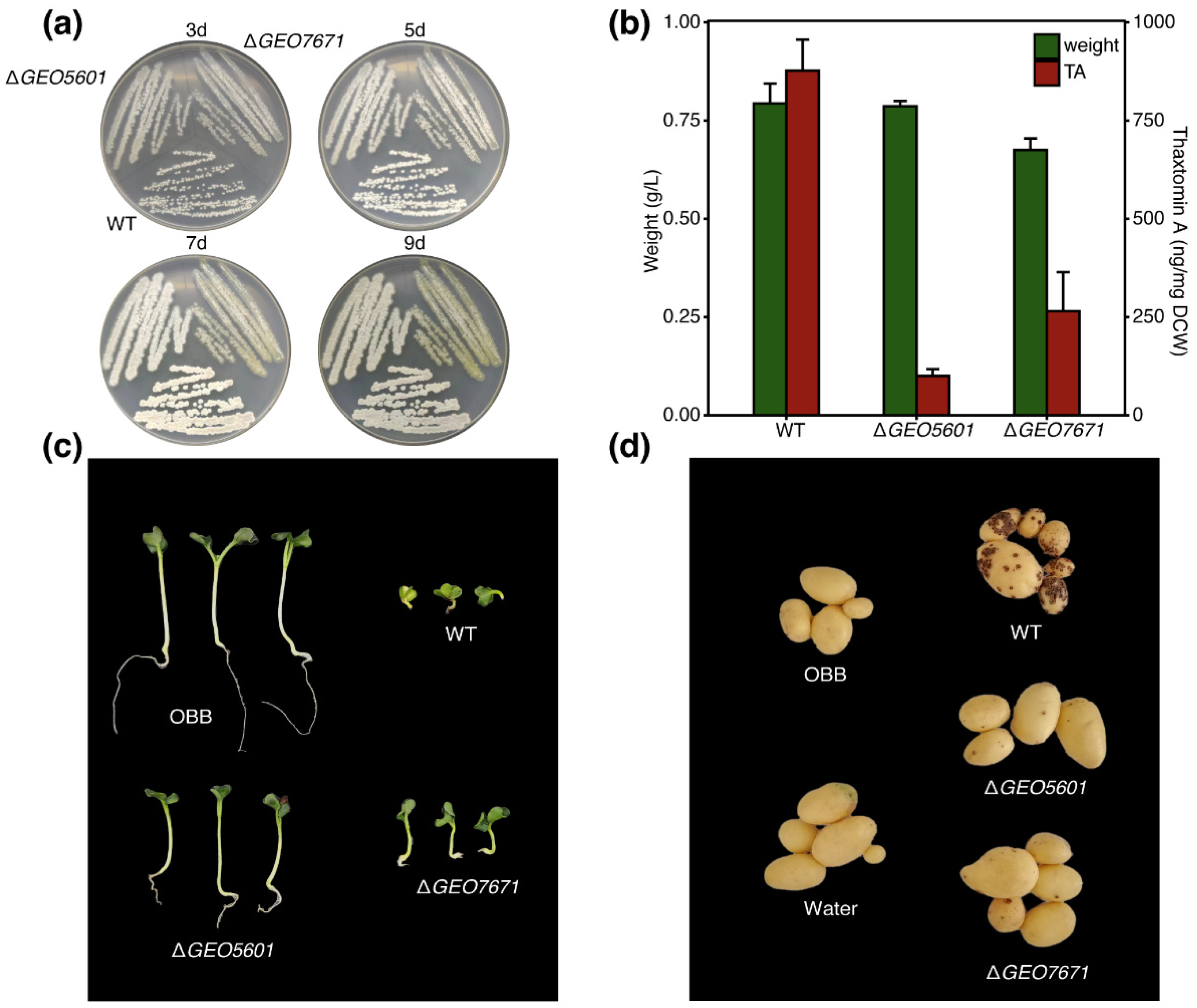
To access the contributions of CebE5601 and CebE7671 to the pathogenic phenotype and thaxtomin production, we successfully constructed the in-frame deletion mutants of ΔGEO5601 and ΔGEO7671 for the pathogenicity analysis, with 688 bp and 1063 bp fragments deleted, respectively (Figure S2). To investigate the influence of CebE’s absence on cell growth, the wild-type strain and two mutants were cultured on TDMc medium [16]. After 3-d incubation, the wild-type strain developed intact and regular colonies and quickly produced many spores during 9-d incubations. However, very few spores appeared in ΔGEO7671 (Figure 4a), even when we prolonged the incubation time to 9 d, implying that CebE7671 may assume special functions of mediating spore formation in strain Streptomyces sp. AMCC400023. Furthermore, we compared the cell biomass of three strains after culturing in TDMc liquid medium and observed a decreased biomass in strain ΔGEO7671 (16.4% less than that of the wild-type strain). As for ΔGEO5601, growth in solid and liquid medium was relatively stable and only a 5% reduction of the biomass was observed in liquid medium (Figure 4b). Thaxtomin production by two mutants was further compared with the wild-type strain in OBB medium under the same conditions. As a result, abundant thaxtomin was accumulated in strain Streptomyces sp. AMCC400023, with the highest detected level of production of 877.09 µg g−1. In contrast, the absence of GEO5601 resulted in a remarkable decrease in thaxtomin synthesis, with only 85.01 µg g−1 detected in mutant ΔGEO5601. Mutant ΔGEO7671 showed a modest reduction in thaxtomin synthesis and produced 312.63 µg g−1 of it in cultures of 7-d incubation (Figure 4b). We suspect that such reduction may be attributed to the impaired growth of this mutant, which thereby influenced TA synthesis.
The radish seedling bioassay was further performed to compare the pathogenicity of the wild-type Streptomyces sp. AMCC400023 and two CebE mutants. As shown in Figure 4c, compared to the strong and well-developed seedlings inoculated with the OBB medium, infection of radish seedlings by wild-type Streptomyces sp. AMCC400023 induced typical pathogenic symptoms, including tissue swelling and necrosis, as well as severe stunting of the roots and shoots (Figure 4c). Meanwhile, the infection symptoms of ΔGEO7671 were also serious, but with somewhat alleviated virulence in the radish seedlings. By contrast, seedlings infected with ΔGEO5601 exhibited significant differences from the wild-type strain, with obviously reduced tissue necrosis and root and shoot stunting (Figure S3). In agreement with the radish seedling bioassay, the treatment of wild-type Streptomyces sp. AMCC400023 also caused severe potato common scab disease, with disease incidence and disease grade up to 97.06% and 63.53%, respectively (Table 2). In contrast to the high pathogenicity of Streptomyces sp. AMCC400023, the control treatments of OBB medium and sterile distilled water did not show any scab symptoms (Figure 4d). Compared with the wild-type AMCC400023, the CebE-deficient strains, especially ΔGEO5601, obtained remarkably reduced disease indexes. The disease incidence and disease grade caused by ΔGEO5601 reduced to 37.50% and 11.25%, and a modest disease incidence (70.37%) and disease grade (16.30%) were observed in ΔGEO7671. These results matched well with the radish seeding bioassay, suggesting that both CebE5601 and CebE7671 contribute to the plant-pathogenic phenotype, with CebE5601 being the more significant.

3.4. Transcriptional Variations of CebE Genes in the Presence of Cellobiose

To further evaluate the contributions of CebE5601 and CebE7671 to the nutrient consumption and virulence of Streptomyces sp. AMCC400023, the strain was cultured in TDMc and subjected to transcriptomic analysis. As shown in Figure 5a, the growth of strain AMCC400023 exhibited a typical S-curve, with exponential growth after 24 h incubation and entering the stationary phase after 96 h. However, thaxtomin accumulation exhibited a growth-uncoupled mode, with minor thaxtomin detected during the growth phase, while rapid accumulation of thaxtomin occurred after 96h cultivation. Therefore, we sampled the cultures at 36, 72, and 120 h for transcriptomic analysis, with three replicates per treatment. As a result, a total of 56,484,073 clean reads from 9 samples were obtained after quality assessment and data filtering, with an average mapping rate of 96.16% to the reference genome Streptomyces sp. AMCC400023 (CP024989) (Table S2). As shown in Figure 5b, the principal component analysis (PCA) shows that samples from three groups were distinguished, with the 36 h group being more dispersed from the 72 h and 120 h groups. Such distribution is reasonable, as the 36 h samples were collected during the early stage of the exponential phase of cell growth, while samples from 72 h were in the latter stage of the exponential phase, and 120 h samples were from the stationary phase. Due to the special phase for thaxtomin synthesis, there were more distinctly expressed genes in the 120 h samples than in the two other groups. Pairwise comparisons of 36 h vs. 72 h, 36 h vs. 120 h, and 72 h vs. 120 h showed their differentially expressed genes (DEGs) were 2592 (1338 up-regulated and 1254 down-regulated), 3446 (1807 up-regulated and 1639 down-regulated), and 2102 (1070 up-regulated and 1032 down-regulated), respectively (Figure 5c).
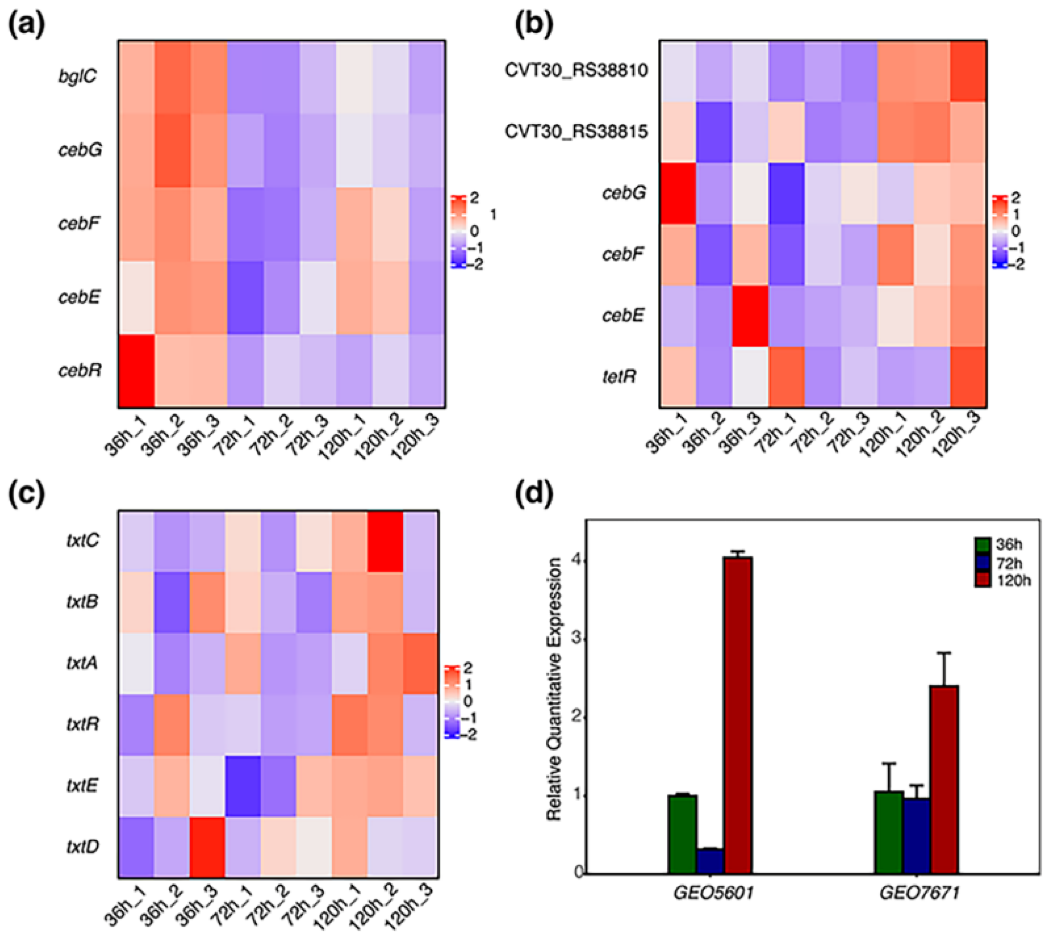
The expression heatmap related to GEO5601 and GEO7671 gene clusters, as well as the thaxtomin biosynthetic gene cluster, was calculated using the Transcripts Per Million (TPM) values. As the operon, these three clusters are all co-transcribed in all samples and showed regular transcriptional variations during the 120 h cultivation. When comparing the overall expression of two CebE clusters, the GEO5601 gene cluster displayed relatively high expression in all nine samples. Interestingly, except for the LacI transcriptional regulator that merely upregulated in the 36 h samples, the expression of the other four genes in this cluster all exhibited a trend of ascending after descending (Figure 6a), with the high expressions at 36 h and 120 h and low at 72 h. Compared with the high expressions of GEO5601 cluster genes, six genes in the GEO7671 cluster all exhibited relatively low expression during three sampling periods. However, obvious upregulations of all these genes were observed after incubation for 120 h, a period in which thaxtomin accumulated rapidly (Figure 6b). Being resident in the PAI region, the overall expression of the thaxtomin biosynthetic genes was similar to those of GEO7671 cluster genes, with low expression but upregulated in 120 h samples (Figure 6c). The expression levels of GEO5601 and GEO7671 were further verified by RT-qPCR and obtained similar expression profiles (Figure 6d). Well consistent with the transcriptomic data, GEO5601 also displayed a significantly higher expression level than GEO7671. The expression value of 36 h samples was set to 1. The expression level of GEO5601 showed around 3-fold downregulation in the 72 h samples, then significantly upregulated in 120 h samples, which reached a 4-fold increase. In contrast, the expressions of GEO7671 remained lower and constant at 72 h, while 2.4-fold upregulation was observed in the 120 h samples.

4. Discussion

Pathogenic Streptomyces strains generally have two or three sets of cebR-cebEFG-bglC gene clusters for cello-oligosaccharides transport. As the signal receptor, the solute-binding protein CebE captures the external signal and functions as the doorway to trigger the regulation network of pathogen virulence [25]. In the well-studied strain S. scabiei 87–22, three putative CebEs were identified and their functions for the transporting of cellobiose/cellotriose were verified. Apart from some non-pathogenic Streptomyces species, the second CebE (SCAB2421) seems only present in the pathogenic S. scabiei [26]. In this pathogenic strain, two CebE homologs of SCAB57751 and SCAB2421 both serve as the signal receptors, with SCAB57751 being the most important one. Based on the transcriptional analysis and mutant virulence assay, the PAI-localized SCAB77271 did not show significant physiological function.
Unlike strain S. scabiei 87–22, Streptomyces sp. AMCC400023 employs two sets of transporters for uptake of cello-oligosaccharides. Being most closely related to the functional important proteins SCAB57751 and CebEreti, we suspected that CebE5601 is more likely to fulfill the major biological functions of cellobiose/cellotriose recognition and binding in strain Streptomyces sp. AMCC400023. Experimentally, several lines of proof confirmed our speculation for the central status of CebE5601 in binding cello-oligosaccharides, not only for nutrient consumption but also as a signal in triggering thaxtomin synthesis and pathogenic virulence.
Firstly, CebE5601 showed much higher binding affinities toward cellobiose/cellotriose in the MST assay, whereas CebE7671 had more than 1000-fold higher KD value. Maybe due to the assay bias of different methods or protein difference, when comparing the binding affinity of SCAB57751 that was measured by the tryptophan intrinsic fluorescence, a difference of 2–3 magnitudes existed between SCAB57751 and CebE5601 [25]. Also, in S. scabiei 87–22, SCAB57751 showed an apparent binding preference towards cellotriose but not cellobiose (2.1 nM and 14.2 nM, respectively). It was supposed that the higher affinity to cellotriose could be a key adaptation for this bacterium to perceive cellotriose as the major signal for triggering thaxtomin synthesis instead of a nutrient uptake response [25]. By contrast, and worth noticing, no apparent difference in binding values between cellobiose and cellotriose was observed for the CebE5601 protein of Streptomyces sp. AMCC400023. Species differences would be an applicable explanation for such affinity and need to be further addressed.
Secondly, as proven by transcriptomic and RT-qPCR analyses, the cebE clusters of GEO5601 are significantly more highly expressed throughout the cultivation process in TDMc liquid medium. Also, compared to the later exponential period (72 h), GEO5601 was highly upregulated during the midst of the exponential phase when the strain grew vigorously (36 h). In combination with the constant and low expressions of the GEO7671 gene cluster, we propose that the GEO5601 transport system is functional in cellobiose uptake and substantially contributes to the nutrient consumption of the cells. After the later exponential period, the restored expression of GEO5601 in the 120 h samples further confirmed its functions in thaxtomin synthesis. This result is partly consistent with the findings in strains S. scabiei 87–22 and S. scabiei EF-35. When grown in the minimal starch medium supplement with 5 g L−1 of cellobiose, the expressions of SCAB2421, especially SCAB57751, were both upregulated [31,32].
Thirdly, in-frame deletions of two cebE genes resulted in different responses in plant infection. Through radish seedling assay, we found that the growth of the seedlings infected by two CebE mutants was better than that of the wild-type strain, and both showed alleviated pathogenic symptoms, which preliminarily indicated that the knocking-out of the cebE did have an impact on pathogenicity. Remarkably, the seedling growth of ∆GEO5601 was better than that of ∆GEO7671, implying that CebE5601 might be more important for its pathogenicity. As radish seedling assay can only partly reflect the pathogenicity of Streptomyces and the whole infection process is more complicated, we further performed the potato pot assay. Consistent with these results, the thaxtomin production of ∆GEO7671 did not change significantly compared with that of the wild-type strain, but the thaxtomin production of ∆GEO5601 showed a significant decrease, with only 11% of the wild-type yield retained.
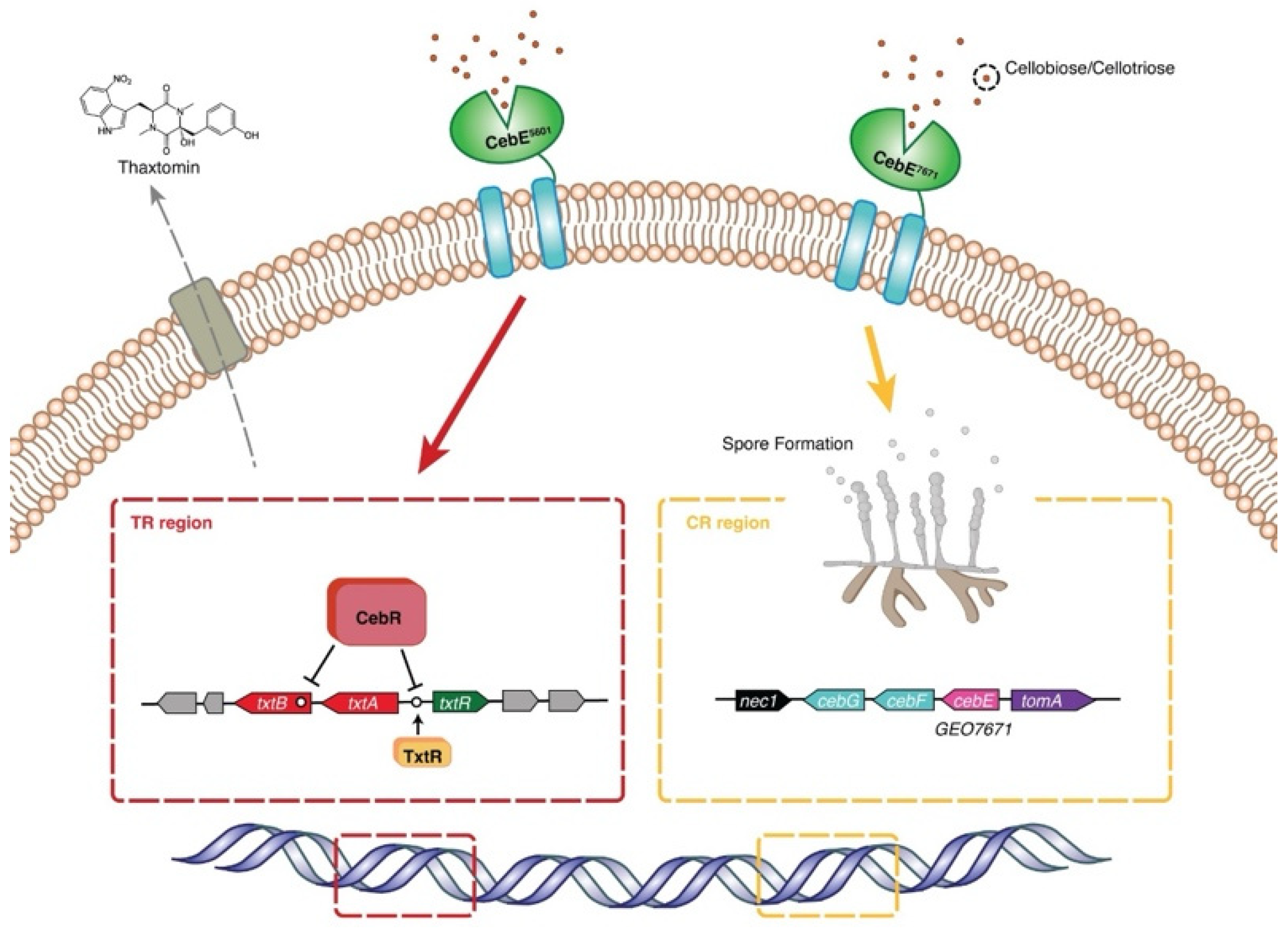
In addition to the central status of CebE5601 in cello-oligosaccharides transport and strain virulence, the specific location (PAI) of GEO7671 makes it an attractive target to explore its physiological functions in pathogenic Streptomyces strains. With weak binding affinities to cellobiose/cellotriose, the seriously impaired spore formation of ΔGEO7671 implies that it might be involved in the spore development process. Meanwhile, the absence of CebE7671 modestly impaired cell growth and consequently alleviated the pathogenicity of Streptomyces sp. AMCC400023. In combination with the 4.1-fold upregulation of GEO7671 in the late growth stage (120 h), we propose that this PAI-located CebE transporter may also be involved in strain virulence, maybe not for signal reception, but is functional in the sporulation and thus makes it conducive to occupying a specific ecological niche. In the identified GEO7671 gene cluster (Figure 2a), a regulator of the TexR/AcrR family is adjacent to the GEO7671 gene. Referring to the finding that a TexR/AcrR regulator in S. coelicolor mediates the development of aerial hyphae and spores, we suspect that this gene regulation would be an explanation for the impaired sporulation of ΔGEO7671 and needs to be further elucidated [33,34]. Integrating the physiological roles of CebE5601 and CebE7671, we propose that the pathogenic Streptomyces sp. AMCC400023 relies on two sets of CebEFG-BglC transporters for growth and pathogenicity, with distinct contributions to strain colonization, growth dynamics, and virulence (Figure 7). Specifically, under cellobiose/cellotriose signals, CebE5601 plays a pivotal role in sugar uptake, nutrient consumption, thaxtomin synthesis, and infection symptom manifestation. Meanwhile, CebE7671 assumes a novel biophysical function of spore formation and potentially contributes to the colonization of expanding plant tissue.

5. Conclusions

Investigating the physiological function of CebE is of utmost significance due to its pivotal role as the recipient of cello-oligosaccharides, not only in sugar transport but also as a signal for strain virulence. In this study, we have discovered a novel mechanism of two CebE systems in the pathogenic Streptomyces sp. AMCC400023. The unique presence of PAI-localized CebE7671 for sporulation suggests that diverse mechanisms may exist in various pathogenic Streptomyces strains, and thereby provide additional targets for drug development against potato common scab.

Supplementary Materials

The following supporting information can be downloaded at: https://www.mdpi.com/article/10.3390/microorganisms12030499/s1, Figure S1: SDS-PAGE spectrums of the purification process of three putative CebE protein; Figure S2: PCR verification of the in-frame deletion Streptomyces mutants; Figure S3: The significance analysis of radish seedling bioassay; Table S1: The amino acid sequence identity of protein used in the phylogenetic tree; Table S2: Summary of sequencing and assembly results for the RNA samples; Table S3: Primers used in this study.

Author Contributions

Conceptualization, C.Y. and B.Z.; Methodology, C.Y. and Z.G.; Software, Q.L. and J.C.; Validation, C.Y., B.Z. and Z.G.; Formal Analysis, Q.L. and J.C.; Investigation, P.L., J.L. and Y.D.; Resources, C.Y., B.Z. and Z.G.; Data Curation, C.Y., B.Z. and Z.G.; Writing–Original Draft Preparation, Q.L. and J.C.; Writing–Review & Editing, C.Y., B.Z. and Z.G.; Visualization, D.T., F.G., G.S. and F.N.; Supervision, C.Y., B.Z. and Z.G.; Project Administration, C.Y., B.Z. and Z.G.; Funding Acquisition, C.Y., B.Z. and Z.G. All authors have read and agreed to the published version of the manuscript.

Funding

This research was funded by the Shandong Provincial Natural Science Foundation (ZR2022MC109), the National Natural Science Foundations of China (42077214, 42077027, and 42377309), the State Key Natural Science Foundations of China Laboratory of Microbial Technology Open Projects Fund (M2023-07), and the Key R&D project of Ningxia Hui Autonomous Region (2023BCF01015).

Data Availability Statement

The genomic sequencing data of Streptomyces sp. AMCC400023 was deposited in the NCBI database with accession number CP024989. Transcriptomic sequencing data of samples were deposited in the NCBI Short Read Archive (SRA) database under Bioproject accession number PRJNA419158, with accession numbers SRR26158258-26158294.

Acknowledgments

We thank Zhifeng Li, Jing Zhu, and Jinyao Qu for their experimental advice and technical assistance in MST analysis. The scientific calculations in this paper were performed on the HPC Cloud Platform of Shandong University.

Conflicts of Interest

The authors declare no conflicts of interest.

References

  1. Viaene, T.; Langendries, S.; Beirinckx, S.; Maes, M.; Goormachtig, S. Streptomyces as a plant’s best friend. FEMS Microbiol. Ecol. 2016, 92, fiw119. [Google Scholar] [CrossRef]
  2. Braun, S.; Gevens, A.; Charkowski, A.; Allen, C.; Jansky, S. Potato common scab: A review of the causal pathogens, management practices, varietal resistance screening methods, and host resistance. Am. J. Potato Res. 2017, 94, 283–296. [Google Scholar] [CrossRef]
  3. Kreuze, J.F.; Suomalainen, S.; Paulin, L.; Valkonen, J.P. Phylogenetic analysis of 16S rRNA genes and PCR analysis of the nec1 gene from Streptomyces spp. causing common scab, pitted scab, and netted scab in Finland. Phytopathology 1999, 89, 462–469. [Google Scholar] [CrossRef]
  4. Li, Y.; Liu, J.; Díaz-Cruz, G.; Cheng, Z.; Bignell, D.R.D. Virulence mechanisms of plant-pathogenic Streptomyces species: An updated review. Microbiology 2019, 165, 1025–1040. [Google Scholar] [CrossRef] [PubMed]
  5. Hill, J.; Lazarovits, G. A mail survey of growers to estimate potato common scab prevalence and economic loss in Canada. Can. J. Plant Pathol. 2005, 27, 46–52. [Google Scholar] [CrossRef]
  6. King, R.R.; Calhoun, L.A. The thaxtomin phytotoxins: Sources, synthesis, biosynthesis, biotransformation and biological activity. Phytochemistry 2009, 70, 833–841. [Google Scholar] [CrossRef]
  7. Wang, L.; Wang, M.; Fu, Y.; Huang, P.; Kong, D.; Niu, G. Engineered biosynthesis of thaxtomin phytotoxins. Crit. Rev. Biotechnol. 2020, 40, 1163–1171. [Google Scholar] [CrossRef] [PubMed]
  8. Fry, B.A.; Loria, R. Thaxtomin A: Evidence for a plant cell wall target. Physiol. Mol. Plant Pathol. 2002, 60, 1–8. [Google Scholar] [CrossRef]
  9. Bischoff, V.; Cookson, S.J.; Wu, S.; Scheible, W.R. Thaxtomin A affects CESA-complex density, expression of cell wall genes, cell wall composition, and causes ectopic lignification in Arabidopsis thaliana seedlings. J. Exp. Bot. 2009, 60, 955–965. [Google Scholar] [CrossRef] [PubMed]
  10. Awwad, F.; Bertrand, G.; Grandbois, M.; Beaudoin, N. Auxin protects Arabidopsis thaliana cell suspension cultures from programmed cell death induced by the cellulose biosynthesis inhibitors thaxtomin A and isoxaben. BMC Plant Biol. 2019, 19, 512. [Google Scholar] [CrossRef]
  11. Joshi, M.; Rong, X.; Moll, S.; Kers, J.; Franco, C.; Loria, R. Streptomyces turgidiscabies secretes a novel virulence protein, Nec1, which facilitates infection. Mol. Plant-Microbe Interact. 2007, 20, 599–608. [Google Scholar] [CrossRef]
  12. Seipke, R.F.; Loria, R. Streptomyces scabies 87-22 possesses a functional tomatinase. J. Bacteriol. 2008, 190, 7684–7692. [Google Scholar] [CrossRef]
  13. Kers, J.A.; Cameron, K.D.; Joshi, M.V.; Bukhalid, R.A.; Morello, J.E.; Wach, M.J.; Gibson, D.M.; Loria, R. A large, mobile pathogenicity island confers plant pathogenicity on Streptomyces species. Mol. Microbiol. 2005, 55, 1025–1033. [Google Scholar] [CrossRef] [PubMed]
  14. Zhang, Y.; Jiang, G.; Ding, Y.; Loria, R. Genetic background affects pathogenicity island function and pathogen emergence in Streptomyces. Mol. Plant Pathol. 2018, 19, 1733–1741. [Google Scholar] [CrossRef]
  15. Book, A.J.; Lewin, G.R.; McDonald, B.R.; Takasuka, T.E.; Wendt-Pienkowski, E.; Doering, D.T.; Suh, S.; Raffa, K.F.; Fox, B.G.; Currie, C.R. Evolution of high cellulolytic activity in symbiotic Streptomyces through selection of expanded gene content and coordinated gene expression. PLoS Biol. 2016, 14, e1002475. [Google Scholar] [CrossRef] [PubMed]
  16. Johnson, E.G.; Joshi, M.V.; Gibson, D.M.; Loria, R. Cello-oligosaccharides released from host plants induce pathogenicity in scab-causing streptomyces species. Physiol. Mol. Plant Pathol. 2007, 71, 18–25. [Google Scholar] [CrossRef]
  17. Benoit, D.; Nudzejma, S.; Sören, P.; Sinaeda, A.; Bonometti, B.; Karim, L.; Coppieters, W.; Devreese, B.; Rigali, S. The virulome of Streptomyces scabiei in response to cello-oligosaccharide elicitors. Microb. Genom. 2022, 8, 000760. [Google Scholar]
  18. Schlösser, A.; Jantos, J.; Hackmann, K.; Schrempf, H. Characterization of the binding protein-dependent cellobiose and cellotriose transport system of the cellulose degrader Streptomyces reticuli. Appl. Environ. Microbiol. 1999, 65, 2636–2643. [Google Scholar] [CrossRef] [PubMed]
  19. Marushima, K.; Ohnishi, Y.; Horinouchi, S. CebR as a master regulator for cellulose/cellooligosaccharide catabolism affects morphological development in Streptomyces griseus. J. Bacteriol. 2009, 191, 5930–5940. [Google Scholar] [CrossRef]
  20. Schlösser, A.; Schrempf, H. A lipid-anchored binding protein is a component of an ATP-dependent cellobiose/cellotriose-transport system from the cellulose degrader Streptomyces reticuli. Eur. J. Biochem. 1996, 242, 332–338. [Google Scholar] [CrossRef]
  21. Eitinger, T.; Rodionov, D.A.; Grote, M.; Schneider, E. Canonical and ECF-type ATP-binding cassette importers in prokaryotes: Diversity in modular organization and cellular functions. FEMS Microbiol. Rev. 2011, 35, 3–67. [Google Scholar] [CrossRef]
  22. Joshi, M.V.; Bignell, D.R.; Johnson, E.G.; Sparks, J.P.; Gibson, D.M.; Loria, R. The AraC/XylS regulator TxtR modulates thaxtomin biosynthesis and virulence in Streptomyces scabies. Mol. Microbiol. 2007, 66, 633–642. [Google Scholar] [CrossRef]
  23. Sören, P.; Jourdan, S.; Francis, I.M.; Benoit, D.; Rigali, S.; Devreese, B. Proteomic response to thaxtomin phytotoxin elicitor cellobiose and to deletion of cellulose utilization regulator CebR in Streptomyces scabies. J. Proteome Res. 2018, 17, 3837–3852. [Google Scholar]
  24. Francis, I.M.; Jourdan, S.; Fanara, S.; Loria, R.; Rigali, S. The cellobiose sensor CebR is the gatekeeper of Streptomyces scabies pathogenicity. mBio 2015, 6, e02018. [Google Scholar] [CrossRef] [PubMed]
  25. Jourdan, S.; Francis, I.M.; Kim, M.J.; Salazar, J.J.; Planckaert, S.; Frère, J.M.; Matagne, A.; Kerff, F.; Devreese, B.; Loria, R.; et al. The CebE/MsiK Transporter is a doorway to the cello-oligosaccharide-mediated induction of Streptomyces scabies pathogenicity. Sci. Rep. 2016, 6, 27144. [Google Scholar] [CrossRef] [PubMed]
  26. Francis, I.M.; Bergin, D.; Deflandre, B.; Gupta, S.; Salazar, C.; Villagrana, R.; Stulanovic, N.; Monteiro, S.R.; Kerff, F.; Loria, R.; et al. Role of alternative elicitor transporters in the onset of plant host colonization by Streptomyces scabiei 87-22. Biology 2023, 12, 234. [Google Scholar] [CrossRef] [PubMed]
  27. Liang, F.; Lin, R.; Yao, Y.; Xiao, Y.; Zhang, M.; Shi, C.; He, X.; Zhou, B.; Wang, B. Systematic identification of pathogenic Streptomyces sp. AMCC400023 that causes common scab and genomic analysis of its pathogenicity island. Phytopathology 2019, 109, 1115–1128. [Google Scholar] [CrossRef] [PubMed]
  28. Zhou, Y.; Sun, Y.; He, H.; Feng, J.; Zhang, X.; Han, L. Optimization of medium compositions to improve a novel glycoprotein production by Streptomyces kanasenisi ZX01. AMB Express 2017, 7, 6. [Google Scholar] [CrossRef] [PubMed]
  29. Zhao, X.; Wei, W.; Zong, Y.; Bai, C.; Guo, X.; Zhu, H.; Lou, C. Novel switchable ECF sigma factor transcription system for improving thaxtomin A production in Streptomyces. Synth. Syst. Biotechnol. 2022, 7, 972–981. [Google Scholar] [CrossRef]
  30. Ahmed, Y.; Rebets, Y.; Tokovenko, B.; Brötz, E.; Luzhetskyy, A. Identification of butenolide regulatory system controlling secondary metabolism in Streptomyces albus J1074. Sci. Rep. 2017, 7, 9784. [Google Scholar] [CrossRef]
  31. Padilla-Reynaud, R.; Simao-Beaunoir, A.M.; Lerat, S.; Bernards, M.A.; Beaulieu, C. Suberin regulates the production of cellulolytic enzymes in Streptomyces scabiei, the causal agent of potato common scab. Microbes Environ. 2015, 30, 245–253. [Google Scholar] [CrossRef] [PubMed]
  32. Lerat, S.; Simao-Beaunoir, A.M.; Wu, R.; Beaudoin, N.; Beaulieu, C. Involvement of the plant polymer suberin and the disaccharide cellobiose in triggering thaxtomin A biosynthesis, a phytotoxin produced by the pathogenic agent Streptomyces scabies. Phytopathology 2010, 100, 91–96. [Google Scholar] [CrossRef] [PubMed]
  33. Hillerich, B.; Westpheling, J. A New TetR family transcriptional regulator required for morphogenesis in Streptomyces coelicolor. J. Bacteriol. 2008, 190, 61–67. [Google Scholar] [CrossRef] [PubMed]
  34. Xu, D.; Seghezzi, N.; Esnault, C.; Virolle, M.J. Repression of antibiotic production and sporulation in Streptomyces coelicolor by overexpression of a TetR family transcriptional regulator. Appl. Environ. Microbio. 2010, 76, 7741–7753. [Google Scholar] [CrossRef]
Figure 1. Chromosome position and gene structure maps of three putative CebE sugar transporters and pathogenicity island. The positions and directions of the open reading frames are shown by the arrows. The pink arrows represent the cebE genes and the light green arrows represent the cebF and cebG genes. The red arrows represent the thaxtomin biosynthetic genes, the green arrows represent the integrase and recombination directional factor (RDF), the orange arrow represents the antibiotic synthesis genes, the blue arrow represents the conjugated integration genes, the brown arrow represents the aviX1 gene, the light blue arrows represent bacA genes, the black arrow represents the nec1 gene, the gray arrows represent the sugar metabolism genes, and the purple arrow represents the tomA gene. The att sites used to distinguish toxin region (TR; TR1 and TR2) are also shown. CR stands for colonization region.
Microorganisms 12 00499 g001
Figure 2. Gene structure of three sugar transporters that anchor the putative cello-oligosaccharide binding gene cebE and phylogenetic analysis of CebE orthologues. (a) Gene organization of the three cello-oligosaccharide ABC-type transporters. (b) NJ tree was constructed with CebE orthologues protein from pathogenic strains: S. scabies 87–22 (SCAB_2421: CBG67458.1, SCAB_15521: CBG68694.1, SCAB_57751: CBG72800.1, and SCAB_77271: CBG74696.1), Streptomyces sp. AMCC400023 (GEO1108, GEO5601, and GEO7671), nonpathogenic strains: S. reticuli (CAB46342), S. griseus (WP_012379731.1), and S. coelicolor A3 (SCO2795: CAC10104.1, SCO7555: CAC16435.1). Bootstrap values (percentages) are indicated at branching points.
Microorganisms 12 00499 g002
Figure 3. Binding curves of three CebE proteins with cellobiose (a) and cellotriose (b) by MST assay. The final concentration of CebE was 800 nM. The cellobiose and cellotriose solutions were two-fold diluted 16 times, with an initial concentration of 100 μΜ for CebE5601 and 270 mM for GebE7671. The KD value was determined by MO.Affinity Analysis v2.3 software. Experiments were conducted in triplicate.
Microorganisms 12 00499 g003
Figure 4. Pathogenicity assays, biomass and thaxtomin production, and morphology of wild-type strain and two CebE mutants. (a) The morphology of wild-type strain and mutants on TDMc solid medium. (b) Biomass and thaxtomin production of the wild-type strain and mutants. (c) Radish seedling bioassay. (d) Pot experiment.
Microorganisms 12 00499 g004
Figure 5. Transcriptomic analysis of Streptomyces sp. AMCC400023. (a) Growth curve and thaxtomin production of wild-type Streptomyces sp. AMCC400023. (b) PCA analysis of transcriptome data between treatments. (c) A bar plot displaying the numbers of DEGs from the transcriptome data.
Microorganisms 12 00499 g005
Figure 6. Transcriptional responses of two CebE transporters and thaxtomin biosynthetic clusters in the presence of cellobiose. (a) Heatmap of GEO5601 ABC transporter genes. (b) Heatmap of GEO7671 ABC transporter genes. (c) Heatmap of thaxtomin biosynthetic genes. (d) Relative expression levels of GEO5601 and GEO7671 in RT-qPCR.
Microorganisms 12 00499 g006
Figure 7. Schematic of the collaborative mode of two CebEs in Streptomyces sp. AMCC400023. CebE5601 and CebE7671 are anchored and separately distributed on the lipid membrane. CebE5601 serves as the protagonist and invokes the synthesis of arsenal thaxtomin by repressively binding with CebR to activate the transcriptional activator TxtR. CebE7671 not only participates in the signal transfer for thaxtomin synthesis, but also assumes a novel and regulative function of sporulation that may be significant to the host colonization of the pathogen.
Microorganisms 12 00499 g007
Table 1. Bacterial strains and plasmids used in this study.
Descriptions
Plasmids
pJTU1278Shuttle plasmid for DNA transfer (Km, Amp, Cml)
pEASY-BluntCloning plasmid (Amp)
pET28aExpression plasmid used to produce N-terminal His-tagged protein (Km)
Strains
E. coli DH5αGeneral cloning host
E. coli BL21(C43)Host for protein expression
E. coli ET12567Host for transfer of DNA into Streptomyces spp.
Streptomyces sp. AMCC400023Wild type strain
ΔGEO5601AMCC400023 derivative with GEO5601 deficient
ΔGEO7671AMCC400023 derivative with GEO7671 deficient
Table 2. Morbidity of pot assay.
TreatmentIncidence Rate (%)Disease Index (%)
CK1-Water--
CK2-OBB--
T1-WT97.0663.53
T2-ΔGEO560137.5011.25
T2-ΔGEO767170.3716.30
Disclaimer/Publisher’s Note: The statements, opinions and data contained in all publications are solely those of the individual author(s) and contributor(s) and not of MDPI and/or the editor(s). MDPI and/or the editor(s) disclaim responsibility for any injury to people or property resulting from any ideas, methods, instructions or products referred to in the content.