Article

Molecular and Phylogenomic Analysis of a Vancomycin Intermediate Resistance USA300LV Strain in Chile

by
Daniela Núñez
1,
Pablo Jiménez
2,
Marcelo Cortez-San Martín
1,
Carolina Cortés
1,
Matías Cárdenas
1,
Sofia Michelson
1,
Tamara Garay
3,
Maggie Vecchiola
3,
Alejandra Céspedes
3,
Jonathan E. Maldonado
2,4,* and
Yesseny Vásquez-Martínez
1,3,*
1
Molecular Virology and Pathogen Control Laboratory, Departamento de Biología, Facultad de Química y Biología, Universidad de Santiago de Chile (USACH), Santiago 9170022, Chile
2
Laboratorio de Multiómica Vegetal y Bioinformática, Departamento de Biología, Facultad de Química y Biología, Universidad de Santiago de Chile (USACH), Santiago 9170022, Chile
3
Escuela de Medicina, Facultad de Ciencias Médicas, Universidad de Santiago de Chile (USACH), Santiago 9170022, Chile
4
Millennium Institute for Integrative Biology (iBio), Departamento de Genética Molecular y Microbiología, Facultad de Ciencias Biológicas, Pontificia Universidad Católica de Chile, Santiago 8380000, Chile
*
Authors to whom correspondence should be addressed.
Microorganisms 2024, 12(7), 1284; https://doi.org/10.3390/microorganisms12071284
Submission received: 13 May 2024 / Revised: 13 June 2024 / Accepted: 18 June 2024 / Published: 25 June 2024
(This article belongs to the Section Medical Microbiology)

Abstract

:
Antimicrobial resistance is a major global health problem, and, among Gram-positive bacteria, methicillin-resistant Staphylococcus aureus (MRSA) represents a serious threat. MRSA causes a wide range of infections, including bacteremia, which, due to the limited use of β-lactams, is difficult to treat. This study aimed to analyze 51 MRSA isolates collected in 2018 from samples of patients with bacteremia from two hospitals of the Metropolitan Health Service of Santiago, Chile, both in their resistance profile and in the identification of virulence factors. In addition, genomic characterization was carried out by the WGS of an isolate that was shown to be the one of greatest concern (N°. 42) due to its intermediate resistance to vancomycin, multiple virulence factors and being classified as ST8 PVL-positive. In our study, most of the isolates turned out to be multidrug-resistant, but there are still therapeutic options, such as tetracycline, rifampicin, chloramphenicol and vancomycin, which are currently used for MRSA infections; however, 18% were PVL positive, which suggests greater virulence of these isolates. It was determined that isolate N°42 is grouped within the USA300-LV strains (ST8, PVL+, COMER+); however, it has been suggested that, in Chile, a complete displacement of the PVL-negative ST5 clone has not occurred.

1. Introduction

Antimicrobial resistance is a major global health concern, and among the Gram-positive bacteria, drug-resistant Staphylococcus aureus (S. aureus) is a severe threat. S. aureus causes a wide range of infections commonly involving the skin, soft tissues, bones, and infections associated with indwelling catheters or prosthetic devices [1]. In addition, S. aureus is a leading reason of bacteremia, causing metastatic infections such as infective endocarditis, septic arthritis, and osteomyelitis; moreover, it can result in complications such as septic shock [2]. These issues make S. aureus bacteremia particularly challenging to treat, especially if they are methicillin-resistant S. aureus (MRSA) due to the limiting use of β-lactams. Methicillin resistance is mediated by the mecA gene and acquired by the horizontal transfer of a mobile genetic element designated as staphylococcal cassette chromosome mec (SCCmec), which confers high variability. The mecA gene encodes penicillin-binding protein 2a (PBP2a), an enzyme responsible for crosslinking the peptidoglycans in the bacterial cell wall [3]. PBP2a has a low affinity for β-lactams, resulting in resistance to the entire class of these antibiotics [4].
MRSA has been associated with infections in the community (CA-MRSA) and healthcare settings (HA-MRSA). CA-MRSA bacteremia, including healthcare-associated community-onset, has replaced HA-MRSA bacteremia globally [5]. It has been described that these strains can be distinguished genetically using typing methods due to the presence of different mobile genetic elements (MGE), like SCCmec [6]. SCCmec are highly variable genomic elements whose sizes range from 20 to 60 kb. However, they all share a three-component structure: (1) a mec complex related to methicillin resistance, (2) a ccr complex that allows for the excision and integration of the cassette into the chromosome of methicillin-susceptible S. aureus (MSSA) strains and (3) three joining regions described as hypervariable regions with non-essential genes such as resistance to other pharmacological families of antimicrobials and resistance to heavy metals [7]. The differences among the mec and ccr complexes allow for SCCmec typing, while the characterization of joining regions gives different subtypes [4].
HA-MRSA strains often contain SCCmec type II; in contrast, SCCmec type IV is more prevalent in CA-MRSA strains [8]. Other molecular features that distinguish HA-MRSA from CA-MRSA include the presence of the virulence factors acquired by mobile genetic elements (MGEs), such as Panton–Valentine leucocidin (PVL) [9]. The pore-forming exotoxin PVL, encoded by the lukSF-PV gene, can facilitate the destruction of leukocytes by forming pores in their membranes, allowing for the influx of ions and small molecules and leading to cell death [10]. The presence of the gene that codes for PVL is a marker of CA-MRSA and is associated with increased disease severity [11].
PVL is not the only toxin produced by MRSA. Different strains produce a range of virulence factors, including toxic shock syndrome toxin-1 (TSST1), exfoliative toxin A (eta), and adhesins like collagen adhesin (Cna), SrdC, and SrdD, that have been found most significantly in MRSA than those among MSSA isolates [12,13]. Those adhesins play an essential role in virulence by establishing a foothold in host tissues and evading the host’s immune system. Adhesins are also crucial in forming biofilms that contribute to persistent infections [14]. Overall, the expression of virulence factors is higher in CA- than in HA-MRSA strains; therefore, CA-MRSA strains tend to be more virulent, as has been described for USA300 strains [15]. In the United States (US), HA-MRSA infections are generally caused by the USA100 or USA200 strains, whereas CA-MRSA infections are commonly associated with the USA300 strain [16].
The USA300 strain (ST8 SCCmec IV PVL+) is a very successful clone in its propagation, displacing the previously predominant clone, USA400, in the US [17]. This clone has spread rapidly in countries such as Canada, Switzerland, Belgium, Denmark, Germany, England, Italy, South Korea, Japan, China, and Australia, which has high epidemic potential [18]. Meanwhile, a genetic lineage clone closely related to USA300 was found in Latin America, designated as USA300-LV [19], which is prevalent in Colombia, Venezuela, and Ecuador [20]. Molecular analyses suggest that a common ancestor between USA300 and USA300LV may have emerged in the mid-1970s and that the geographical segregation of the clades occurred in the late 1980s, which coincided with the independent MGE for each clone: the arginine catabolic mobile element (ACME) for USA300 variants and the copper and mercury resistance mobile element (COMER) for USA300-LV variants [21]. Phylogenomic analyses based on WGS among Latin American MRSA strains showed three main clades, a majority multiresistance clade A harboring strains of allelic profile ST5, ST105, and ST1011, a less resistance clade B with ST72, ST88, ST97 and ST8 strains harboring USA300 and USA300-LV, and a minor clade C mainly composed of ST30 strains [21].
In Chile, the PVL-negative Chilean/Cordobes ST5 SCCmec I clone has shown high prevalence [22]. However, the emergence of the USA300 clone, detected for the first time in 2008, marked a change in the dynamics of MRSA clones worldwide [23]. The Institute of Public Health of Chile (ISP), responsible for monitoring MRSA in Chile, reported that within the samples collected during the years 2012 and 2013, MRSA PVL+ ST8 isolates were found, corresponding to 11% of the total samples [24]. There has been no update on this clone’s progress in Chile since 2016, highlighting the need for constant and specific surveillance to monitor the circulation of these strains.
Given the lack of use of specific techniques for lineage monitoring, such as WGS, phylogenomic analyses, or SCCmec typing with MRSA samples collected in more recent years, a gap is created in the ability to monitor the spread and behavior (phenotypic and molecular characterization) of different MRSA lineages in the country. This study aimed to analyze 51 isolates collected in 2018 from samples of patients with bacteremia from two hospitals in the Metropolitan Health Service, Santiago, Chile, analyzing their antibiotic resistance profile and identifying some virulence factors. Furthermore, given the lack of bioinformatics tools applied in the country, genomic characterization by the WGS of an isolate of concern (ST8 PVL-positive) due to its resistance profile (vancomycin-intermediate resistance) and multiple virulence factors was carried out.

2. Materials and Methods

2.1. Bacterial Strain (Sampling)

In 2018, 51 positive MRSA isolates were collected from blood culture samples from adult patients with bacteremia at two hospitals in the northern and western areas of the metropolitan region of Santiago, Chile. As a prospective observational study, we included patients older than 18 years who had an episode of S. aureus bacteremia. Inclusion and exclusion criteria were as follows. Inclusion criteria: Male and female adult hospitalized patients (>18 years old), S. aureus in the bloodstream. Exclusion criteria: Polymicrobial bacteremia; relapsed bacteremia, meaning the resolution of clinical features during treatment and recurrence with phenotypically similar isolates as the original episode. A previous episode during current hospitalization was considered a relapse. Blood cultures were incubated in an automated BacT/ALERT® system for seven days; in case of a positivity alert, they were transferred to solid culture in blood and chocolate agar and incubated at 35 °C for 72 h [25]. Colonies were identified by MALDI-TOF using a VITEK®MS and Vitek® 2 system. Quality control was performed with S. aureus strains ATCC 25923 and ATCC 43300 [26].
This study was approved by the Ethics Committee of the University of Santiago, Chile (approval number 267/2019), and the Ethics Committee of each hospital (approval number 3917).

2.2. Antimicrobial Susceptibility Testing

The Kirby–Bauer disk diffusion method was used to estimate the susceptibility of isolates against eight antimicrobial agents: cefoxitin (30 μg), chloramphenicol (30 μg), ciprofloxacin (5 μg), levofloxacin (5 μg), norfloxacin (10 μg), tetracycline (30 μg), gentamicin (120 μg), and rifampicin (5 mg) according to CLSI [25]. Susceptibility to oxacillin and vancomycin was tested by MIC determination and interpreted according to the CLSI [25] using vancomycin hydrochloride and oxacillin sodium salt. Susceptibility testing was performed according to the clinical and laboratory standard institute CLSI 2020 and CLSI guidelines (M100 Performance standards for antimicrobial susceptibility testing 12 th editions), which entailed choosing different antibiotic families according to Medina et al. 2013 [24]. Quality control was performed with S. aureus strains ATCC 25923 and ATCC 43300.

2.3. Molecular Characterization

2.3.1. Characterization of Virulence Factors

Isolates were cultured overnight in Müller Hinton broth (MHB), and the cells were harvested by centrifugation at 8000× g for 1 min. Bacterial genomic DNA was then extracted using a DNeasy Ultra Clean Microbial Kit. Genomic DNA was used as the template for PCR-based screening assays. All isolates underwent an initial molecular characterization to detect staphylococcal virulence genes by PCR assays. The detection of 7 genes encoding the virulence factor was performed using seven specific primer sets: PVL encoding gene (lukSF-PV) and TSST-1 gene (tst) according to Víquez-Molina et al. [17]; gamma–hemolysin gene (hlg) according to Truong-Bolduc et al. [18]; and collagen adhesin (cna), exfoliative toxin A (eta), and adhesins (sdrC, sdrD) according to Carpenter et al. [27]. PCR reactions were set to a final volume of 25 µL, containing 0.5 µL of 10 mM of each primer, 2.5 µL of 10 × 5000 DNA polymerase buffer, 0.5 µL of 5 U/µL Paq 5000TM DNA polymerase, 0.2 µL of 10 mM dNTPs, 18.8 µL of DEPC treated water, and 2 µL of extracted DNA according to manufacture instruction. The amplified PCR products were analyzed by 1% agarose gel electrophoresis. Positive and negative controls used in all experiments belonged to the strain collection of San Juan de Dios Hospital’s Clinical Laboratory.

2.3.2. Multilocus Sequence Typing (MLST)

PVL-positive MRSA isolates were further characterized by MLST sequencing using an internal fragment of 7 housekeeping genes to identify the following allelic profiles: triosephosphate isomerase (tpi), phosphate acetyltransferase (pta), shikimate dehydrogenase (aroE), acetyl-coenzyme A acetyltransferase (yqiL), carbamate kinase (arc), guanylate kinase (gmk), and glycerol kinase (glp) according to Enright et al. [28]. After PCR, the products were purified by Wizard® SV Gel and PCR Clean-Up System (Promega). Purified products were subjected to DNA sequence analysis by Macrogen Inc., Seoul, Republic of Korea.
MLST was supported by the public PubMLST database and the Ridom SpaServer to assign isolates to clonal complexes [29]. Each isolate was grouped according to the corresponding clonal complex according to the sequence type.

2.4. WGS and Genome Assembly

Genomic DNA was obtained as previously described in Section 2.3.1. Genomic DNA libraries were prepped using the NexteraXT DNA Sample Preparation kit and sequenced in a configuration of 150 bp paired-end reads using the Nextseq 500 System (Illumina Inc., San Diego, CA, USA).
The raw reads were trimmed using Trimmomatic v0.39 [30] at its ends if their quality was below a Phred score of 30, and de novo assemblies were performed using Velvet v1.2.10 [31]. Quality improvement was represented with the BoxPlotR tool [32]. According to statistical metrics N50 and the number of contigs, the most contiguous assembled genome was selected to perform genome and functional annotation with Prodigal v2.6.3 [33], Prokka v1.13 [34] and RAST v2.0 [35,36]. The completeness of the assembled genome was evaluated with BUSCO v5.4.3 [37]. Genome representation was obtained with GenoVi v0.2.16 [38].
The genome sequence of the isolate n°42 was deposited in the NCBI database under the Bioproject ID PRJNA1073500.
For SCCmec typification, the tool SCCmecFinder v1.2.1 [39], a 90% threshold for ID, 60% minimum length and both the referenced and extended database were evaluated. Mobile elements COMER and ACME were identified following the methodology described by Arias et al. (2017) [21]. Through BlastN [40,41], positive hits were selected if they had an e-value lower than 1 × 10−100, at least 90% of identity and a coverage higher than the 80% of the CDSs in the reference sequences for COMER (CP007672.1 region: 53520–77705) and ACME (Accession: CP000255.1 region: 63100–88681). Representation of genomic elements found in the assembled genome was visualized with UGENE v46.0 [42].
For detecting more antibiotic resistance determinants and virulence factors in the assembled genome, the tool VirulenceFinder v2.0 [43,44] was used. Parameters were set at a 90% threshold for ID, 60% of minimum length, and S. aureus was selected as a species. Annotated genes found with Prokka and Rast were also considered.

2.5. Phylogenomic Analysis

Sequenced MRSA genomes and non-assembled MRSA reads of Chilean human hosts were collected from the NCBI database for the phylogenomic reconstruction (see Supplementary Table S4). Additionally, thirty-four USA300-LV ST8 Latin American genomes available from the NCBI database were included in the reconstruction, NCBI accession codes are listed in Supplementary Table S5. Reference S. aureus strains NCTC8325, HA-MRSA N315 and CA-MRSA CA12 were considered, and the reference strain Macrococcus caseolyticus FDAARGOS 868 was included as an outgroup.
Non-sequenced genomes found in the NCBI database were processed as described in the previous section. Assembled genomes with completeness below 90% were not considered for the phylogenomic reconstruction. MLST analysis was carried out with the public PubMLST database for all the assembled genomes.
A core genome was calculated with Roary v3.13.0 [45]. Sequences of the core genome were aligned with MAFFT v7.511 [46] and the alignment was processed in MEGA v10.2.6 [47] to reconstruct the phylogeny with a maximum likelihood method and the Tamura–Nei substitution model with a 200 bootstrap resampling for the first phylogeny reconstructed setting a site coverage cutoff of 99% due to the high amount of informative sites. and a 1000 bootstrap resampling for the second phylogeny.

3. Results

3.1. Antimicrobial Resistance

All MRSA isolates (n = 51) underwent a minimum inhibitory concentration test (MIC) against nine antibiotics to test antimicrobial resistance. It was found that 94.1% (48/51) were resistant to oxacillin, while three were susceptible, considered as oxacillin-susceptible MRSA (OS-MRSA). Therefore, following the Clinical and Laboratory Standards Institute (CLSI) protocol, these isolates were subjected to a complementary test for susceptibility to cefoxitin (30 g) by the Kirby-Bauer method, which confirmed the same results as with oxacillin. However, interestingly, all 51 MRSA isolates were mecA positive. Concerning vancomycin, most of the isolates evaluated showed sensitivity (98%), and just one (isolated n°42) showed intermediate sensitivity.
Furthermore, the isolates were susceptible to tetracycline, chloramphenicol, and rifampin; half of the isolates were susceptible to gentamicin. The isolates mainly showed resistance to norfloxacin, levofloxacin, and ciprofloxacin (Table 1). The results show that 23 isolates are resistant to one or two pharmacological families of antimicrobials, and 27 are multiresistant (resistance to at least one antimicrobial in three or more families). Just one isolate was sensitive to all antibiotics tested.

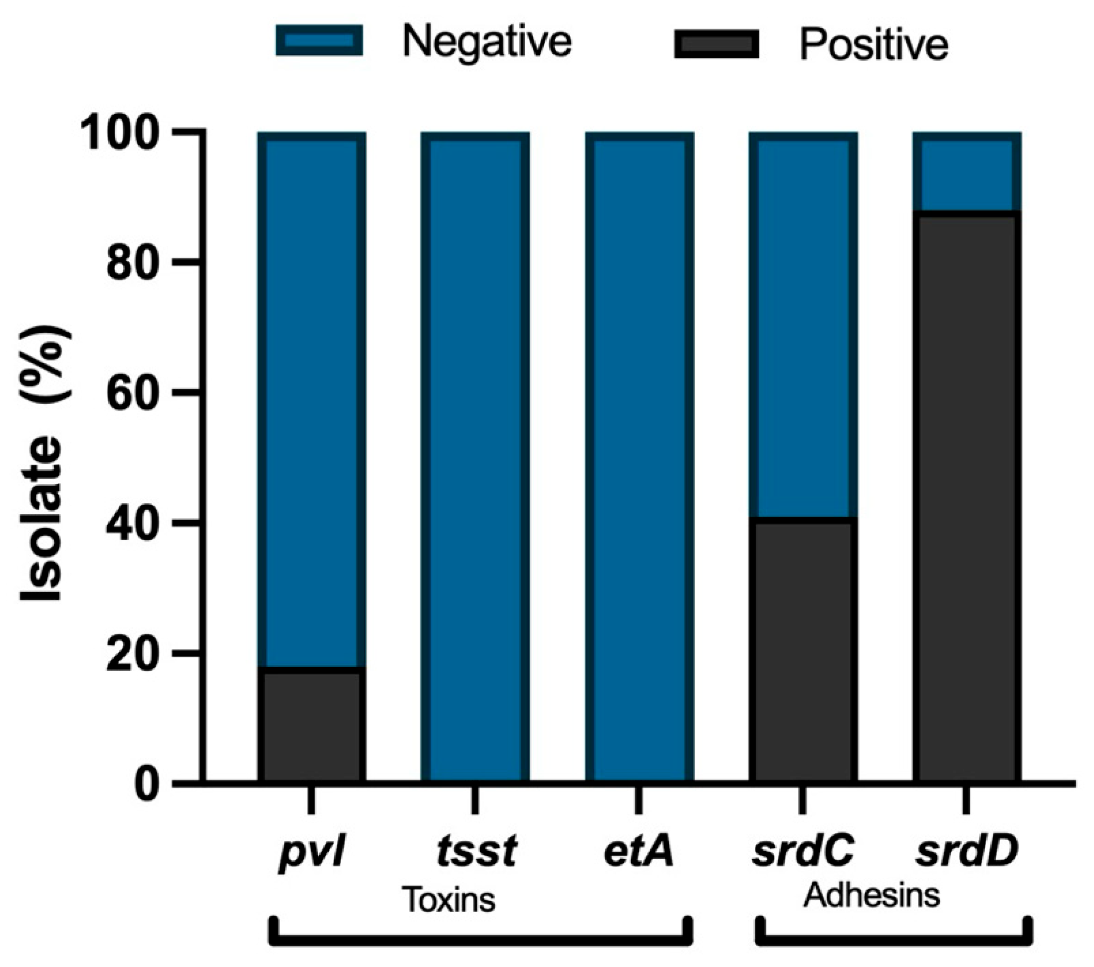
3.2. Characterization of Virulence Factors in MRSA Isolates

Molecular characterization of some virulence factors in the MRSA strains was carried out using the primers shown in Supplement Table S1. It was observed that the predominant virulence factors in the isolates were the genes codified for adhesins: sdrD gene and sdrC gene. The pvl gene was found in 18% of the isolates. On the other hand, only 4% of the isolates have the cna gene, and no positive strains were found to have tsst and etA genes (Figure 1).

3.3. Multilocus Sequence Typing (MLST) of PVL-Positive Isolates

The allelic profile for the PVL-positive isolates was obtained, and the sequence types (STs) and clonal complexes (CCs) were determined using PubMLST (https://pubmlst.org/saureus/, accessed on 10 May 2023). In the nine PVL-positive isolates, it was found that the predominant STs were ST5 and ST8. Two clonal complexes were obtained: CC5 and CC8. Table 2 summarizes the antimicrobial resistance profile and the virulence factors in the nine PVL-positive isolates.
Among the nine PVL positive isolates, isolate n°42 was considered a sample of interest because it was the only vancomycin-intermediate isolate with an MLST profile ST8 CC8, including the multiresistance and virulence factors all isolates also presented. Interestingly, the above data suggest that isolate n°42 is related to USA300-LV.

3.4. Sequencing and Genome Assembly of Isolate n°42

Due to the importance of understanding and to update the genetic characteristics of MRSA PVL-positive isolates, such as CA-MRSA or USA300/USA300-LV in Chile, we performed the genomic characterization and established their phylogenomic relationship with the USA300-LV clone and others by whole genome sequencing the isolate n°42.
Table 3 shows the quality improvement after raw sequence trimming. Filtered reads improved by 1.2 and 1.4 points in the Phred 30 scale for forward and reverse reads, respectively. Both forward and reverse sequences showed a decrease of 22.6% in the total amount of reads, corresponding to 1.8 million reads discarded and leaving 6.3 million reads available for assembly. A graphic representation of quality improvement is shown in Supplementary Figure S1.
Using the software Velvet v1.2.10 and the filtered reads, an assembled genome of 2.8 Mb with an average coverage of 290X was obtained. The genome features of this new genome are summarized in Table 4. A range of k-mers sizes was tested, and a size of 111 bp gave the maximum N50 (453,788 bp) and a genome with the minimum number of contigs (49) and L50 (3). The completeness of the genome was evaluated for the first four taxa classes, and the genome showed the completeness of over 99% for the BUSCO groups searched. Through the functional annotation of 2574 protein-coding genes, 12 rRNA and 62 tRNA were found. The genome GC content was 32%, like the other S. aureus genomes reported. Figure 2 shows the circular representation of the assembled genome, adding functional annotations from the Cluster of Orthologues Groups (COGs). COG categories are listed in Supplementary Table S2.

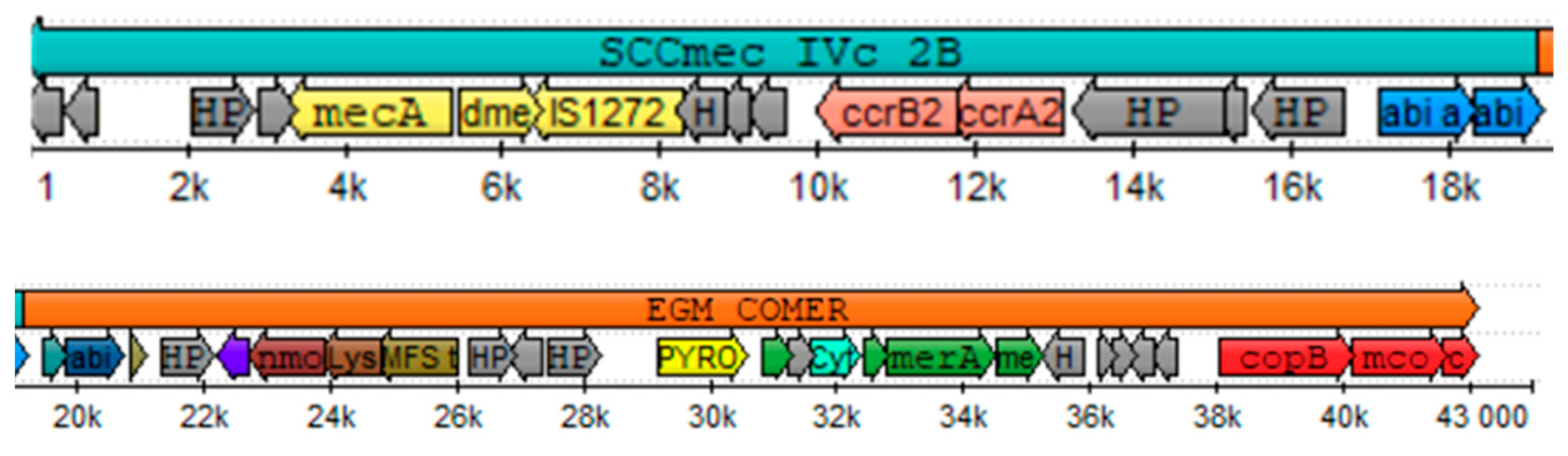
3.5. SCCmec Typification, Virulence Factors and Antimicrobial Resistance Profile of Isolate n°42

By the complete cassette prediction method over the isolate n°42, a region was found within the first 19 kb with 93.7% identity to SCCmec IVc 2B from S. aureus strain 2314 (GenBank #AY271717.1). By component search method, all the searched components have an identity above 99%. A type 2 ccr complex was identified with the allotypes ccrA2 and ccrB2, and a class B mec complex was described according to the mecA gene, including the truncated mecR1 gene and the IS1272 insertion sequence. The typification of the complexes mentioned above placed the SCCmec as type IV while, as the gene for an abi-c abortive phage resistance protein was found, we can place this SCCmec cassette as a subtype c.
The search for the ACME/COMER components showed that the isolate n°42 possesses the COMER element downstream of the SCCmec between 19 and 43 kb, as shown in Figure 3. Each component described in the reference strain S. aureus CA12 was found in isolate n°42 with an identity of over 90%. The genes for copper metabolism (copB, mco and copL) and mercury metabolism (merA, merB and merR) were found in the 3′ region, while various proteins and hypothetical unidentified proteins were found in the 5′ region (see Supplementary Table S3 for alignment results). The search for the components of the ACME element showed that no gene was found in the isolate n°42 except for the copB and copL genes shared between ACME and COMER.
On the isolate n°42, genes coding for virulence factors such as gamma hemolysin, leucocidins, staphylococcal enterotoxins and exoenzymes such as serine protease and aureolysin (Table 5) were also found. All these elements were identified with an identity percentage greater than 99%. The functional annotation carried out by the RAST platform identified 22 genes which were classified within the adhesin subsystem, coding mainly for surface proteins of staphylococci, serine–aspartate repeat proteins for the binding of extracellular matrix proteins and proteins associated with coagulation such as fibronectin- and fibrinogen-binding proteins and clumping factor A. Of the seven virulence factors evaluated by PCR, within the genome of isolate n°42, only the genes corresponding to the PVL toxin and the adhesins SdrC and SdrD were found.
The search for genes coding for antimicrobial resistance besides methicillin showed the presence of response genes to several antibiotics, as seen in Table 6, including bacitracin, quinolones, fosfomycin, tetracycline and vancomycin resistance (vraR and vraS). Some genes associated with response to multiple drugs, such as mdtG, mdtH, emrY, emrK, sepA and marA, were associated with resistance through efflux pump complexes. Response genes to antiseptics such as acraflavin (acr) were found.

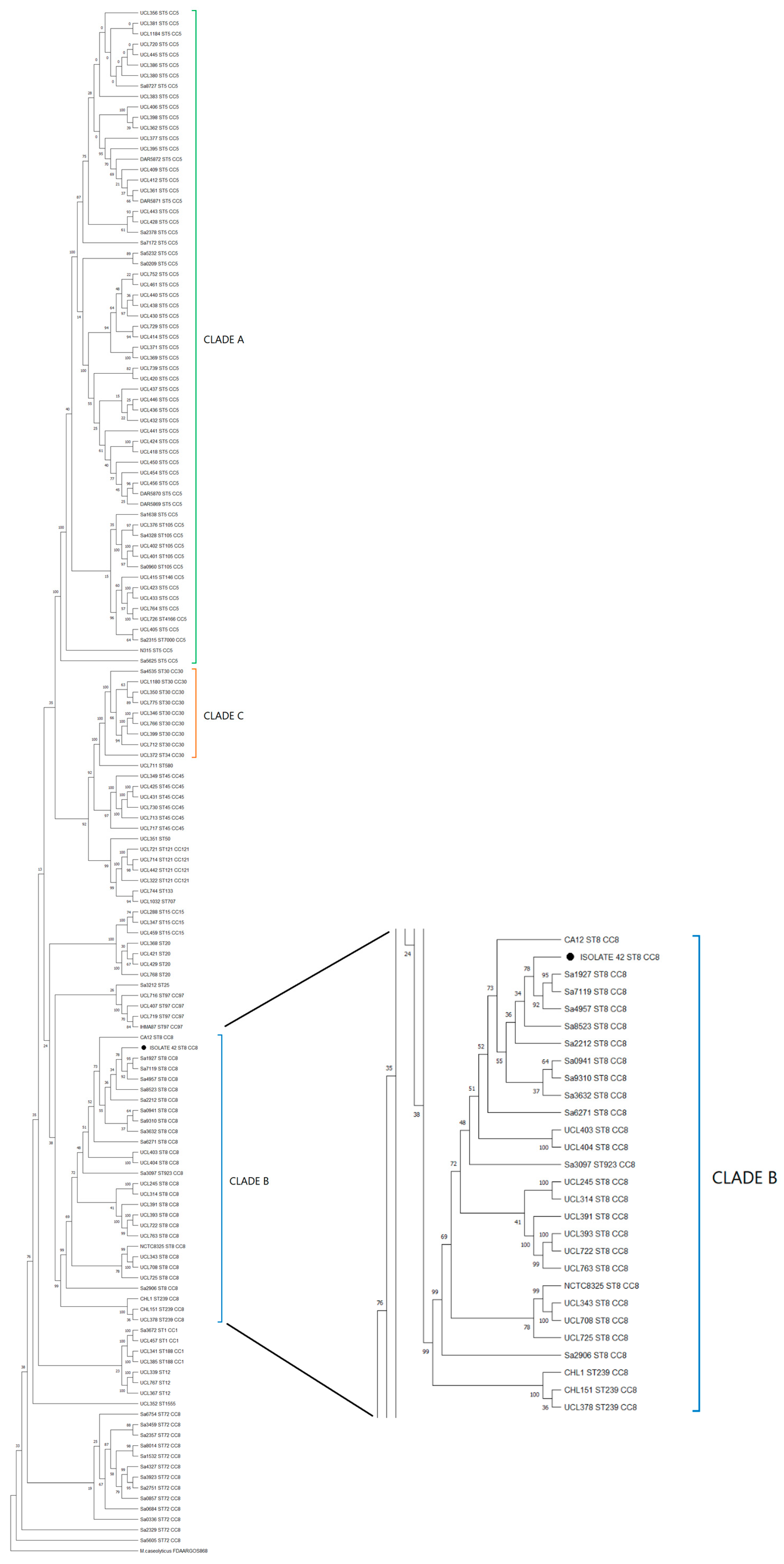
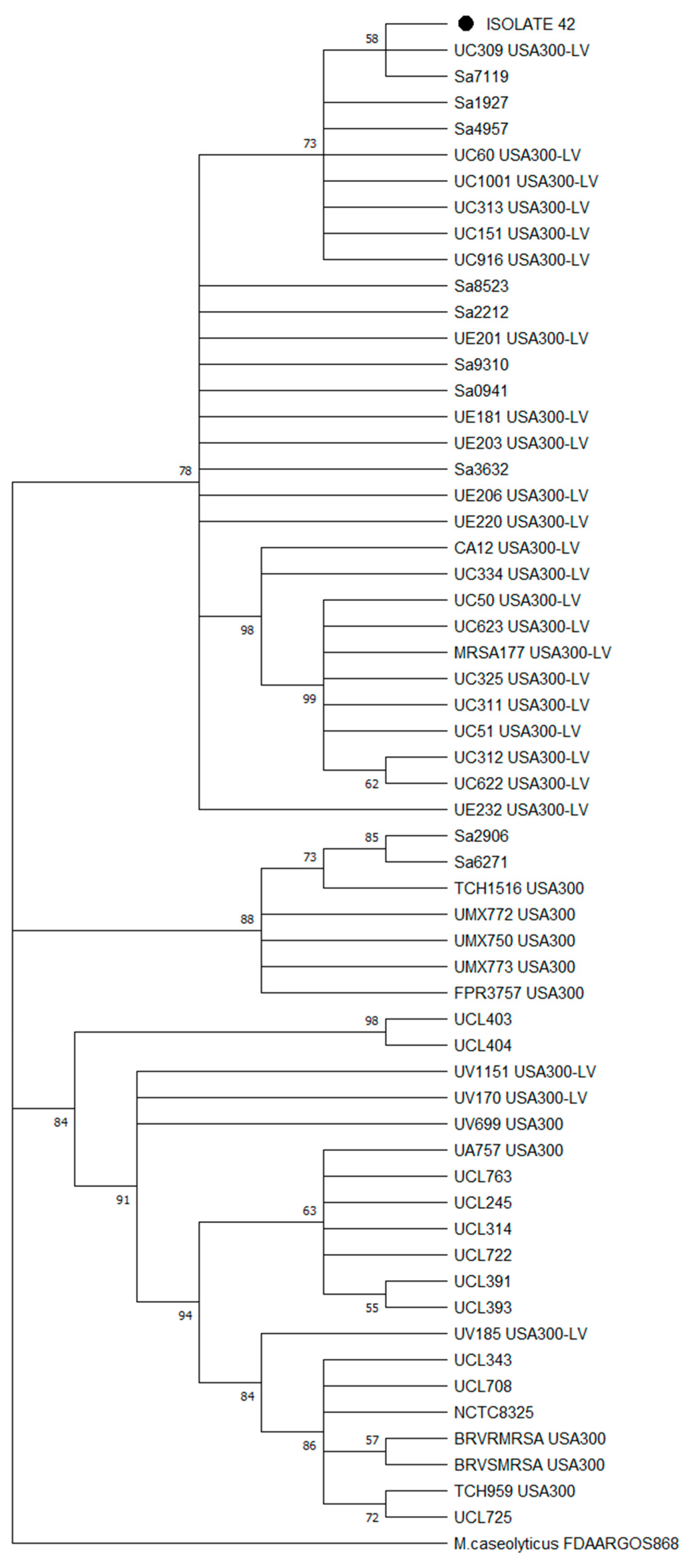
3.6. Phylogomics of Isolate n°42

To discover the genetic diversity of Chilean genome MRSA strains available from NCBI and establish the relationship between the isolate n°42 and the Chilean strains, a phylogenomic tree that included 143 S. aureus genomes was constructed (see Supplementary Table S4). The first selection criterion considered only MRSA genomes, which would have left only 83 genomes for the phylogenomic reconstruction. A second criterion was the addition of another 59 S. aureus genomes from the NCBI database to dissect the population structure of all regional genetic lineages (see Methods).
The phylogenomic analysis revealed that the core genomes were associated according to their MLST profiles, and, as reported by Arias et al. [21], three major clades, A, B, and C, were found (Figure 4). Clade A, which included S. aureus N315 (an HA-MRSA reference strain), was the clade that grouped the largest number of isolates, corresponding mainly to ST5 and ST105 profiles. Clade B, which included isolate n°42 and S. aureus NCTC8325 ST8 CC8, harbored ST8, ST239 and ST923 profiles. Finally, clade C grouped a low amount of ST30 profiles. Outside of the three major clades, genomes grouped into minor clades corresponding to clonal complexes CC1, CC5, CC8, CC15, CC30, CC45, CC97, and CC121 indicated genotypic diversity.
Since Arias [21] reported that the USA300-LV lineage was part of the ST8 CC8 clade B, we performed a second phylogenomic reconstruction among the 22 Chilean genomes found in clade B, with 34 Latin American ST8 genomes (recovered from the Arias study, see Supplementary Table S5) reported as South American USA300-LV and North American USA300 strains. The phylogenomic tree (Figure 5) showed that isolate n°42 was mainly grouped among the USA300-LV Ecuadorian and Colombian strains rather than other USA-300 strains like FPR3757 (a USA300 reference strain).

4. Discussion

Bacteremia is arguably the most important infection caused by S. aureus, associated with a mortality rate of 14–45%, which increases to 60% when the pathogen is resistant to methicillin [48]. The prevalence of bloodstream infection by MRSA in Latin America appears to be heterogeneous, ranging from 6% in Central America to 80% in some South American countries [49]. The rising prevalence of methicillin resistance and the higher associated mortality compel us to understand the characteristics of MRSA isolates better. Currently, MRSA strains are classified as hospital-acquired and community acquisition [50], presenting molecular characteristics that allow them to be differentiated. Therefore, in this study, the phenotypic, molecular, and phylogenic characterization of MRSA strains was carried out.
The results of 51 Chilean MRSA isolates dated in 2018 show that 94.1% of the isolates (48/51) were resistant to oxacillin, and three were susceptible; however, 100% were mecA-positive, confirming the initial identification of MRSA in the laboratories of the respective hospitals. Positive mecA strains sensitive to oxacillin (OS-MRSA) have been previously described worldwide [51] and are sensitive to cefoxitin, as in our study [52]. Because OS-MRSA shows low-level β-lactam resistance, patients infected with this MRSA can be treated with β-lactam antibiotics, which could cause the emergence of high-level β-lactam-resistant MRSA. As the clinical diagnosis is almost always performed by phenotypic techniques and not by amplifying the mecA gene, it could be erroneously identified as methicillin-sensitive S. aureus (MSSA). Therefore, since MSSA strains are handled differently from MRSA strains, using as, principal antibiotics, β-lactams, it is crucial to investigate and report the occurrence of OS-MRSA to allow for the correct decision-making in treatment, as incorrect treatment could contribute to the development of greater antimicrobial resistance. To our knowledge, this is the first report of an isolated OS-MRSA in Chile.
Despite their resistance to oxacillin and possibly to all currently available β-lactam antimicrobial agents, it is notable that among the Chilean isolates, high levels of susceptibility to chloramphenicol, rifampin, and vancomycin were found. However, interestingly, tetracycline shows 100% antimicrobial efficacy, suggesting that these commonly used antibiotics are an option for treating infections. On the other hand, the isolates showed high levels of resistance to fluoroquinolones, including norfloxacin, levofloxacin, and ciprofloxacin, like a report by Li et al. in 2019 [53], in which MRSA isolates showed significantly higher resistance to levofloxacin than MSSA.
Regarding vancomycin susceptibility, almost all isolates showed sensitivity, suggesting that it remains a therapeutic option to treat MRSA infection. However, one isolate (n°42) showed intermediate susceptibility, which interests us. The prevalence of vancomycin-resistant S. aureus (VRSA) in Europe is 1.1% among 179 isolates, and in America, it is 3.6% among 140 isolates, and the prevalence of vancomycin-intermediate S. aureus (VISA) is 1.8% and 1% in Europe and America, respectively [54]. According to the last Public Health Institute report, sensitivity to vancomycin in Chile has been maintained, which showed that 1526 isolates (100%) collected during 2014–2016 were sensitive to this antibiotic [55], suggesting appropriate treatment management. However, this VISA isolate n°42 strongly suggests that the surveillance of vancomycin resistance needs to be maintained.
Concerningly, 27 isolates were considered multidrug-resistant (MDR), defined as non-susceptibility to at least one agent in three or more antimicrobial categories [56], and 23 isolates were resistant to at least one antibiotic. CA-MRSA strains are often reported as MDR and can present resistance to fluoroquinolones, macrolides, aminoglycosides, tetracyclines, and rifampin [57]. This can make treatment more difficult and underscores the need for appropriate antibiotic stewardship practices to preserve the effectiveness of available drugs. Overall, these results highlight the importance of maintaining the ongoing surveillance of antimicrobial resistance patterns to guide appropriate treatment and antibiotic stewardship.
The multi-resistance of the isolates is aggravated by the presence of virulence factors that benefit the infection process. Virulence factors associated with adherence and tissue invasion, such as adhesins, also promote MRSA infection. This study found that the primary virulence factor genes correspond to factors such as adhesins (88% sdrD, 41% sdrC). These factors have also been reported to be the most common among MRSA isolates in studies of Korean and Mexican strains, with values of 76.2 and 94.5%% for sdrD and 92.3 and 89%% for sdrC, respectively [58,59].
On the other hand, toxins or proteases secreted by S. aureus, like PVL, are essential components in the ability to cause disease. PVL causes leukocyte destruction and tissue necrosis and is present in less than 5% of strains [60]. In our study, 18% were PVL-positive, which suggests greater virulence of these isolates. Although the prevalence of PVL in bacteremia isolates has been reported to be lower than that in soft tissue and skin infections, the presence of PVL has been linked to an increased risk of complications, including infective endocarditis, osteomyelitis, and necrotizing pneumonia [11]. It can be suggested that due to the presence of these virulence factors and the previously mentioned antibiotic resistance such as oxacillin, levofloxacin, norfloxacin, ciprofloxacin, these isolates can cause severe infection, such as bacteremia; however, host factors and the immune response also play a role. In Chile, there are few previous reports on the molecular characterization of virulence genes in MRSA isolates, so the results obtained in this study contribute to the molecular characterization of MRSA virulence factors in this country.
The emergence of PVL-positive MRSA isolates has been reported worldwide. Previous studies revealed a strong association between the presence of PVL genes and CA-MRSA strains, and HA-MRSA strains that carry PVL genes have been reported in various regions in Europe and Asia [61,62]. According to MLST, we report that PVL-positive MRSA isolates correspond to sequence type 8 (ST8) and ST5. USA300 is a ST8-PVL clone characterized by present staphylococcal chromosomal cassette mec (SCCmec) type IV, ACME, and is a predominant strain in North America [58,63]. In 2005, it was reported that a variant of USA300 lacking ACME was identified in northern South America with clinical and epidemiological characteristics similar to those of the North American clone, suggesting the exportation of USA300 to South America caused epidemics [20]. Between 2006 and 2008, it was reported that this USA300 variant, USA300LV, could ultimately replace previously prevalent hospital-associated clones in Colombia and Ecuador and became the most predominant [20]. Otherwise, isolate ST5 belongs to the Chilean/Cordobes clone that typically carries SCCmec I. This clone has been reported widely in several countries in Latin America, as well as Argentina, Brazil, Colombia, Chile, Peru, and Venezuela [59]. USA300 was described in Chile for the first time in 2008 [64] and again in 2017 [65]; however, this clone has not been tracked until this study. The presence of both clones in our isolates suggests the need for a discussion about the ability of ST8 (USA300) to replace ST5 (Chilean/Cordobes), as happened in Colombia and Ecuador [20]. The background suggested the importance of performing a more in-depth molecular characterization, so isolate n°42 was chosen for WGS characterization and to analyze if there are differences from those reported in other South American countries.
Whole genome sequencing has become one of the most recent methods to assist in MRSA’s global and local epidemiological monitoring. The preservation and availability of genomes are crucial in monitoring retrospective and prospective studies. Through WGS and phylogenomic reconstruction, it was possible to compare clinical isolates grouped in clades among USA300-LV strains and identify the isolates responsible for the CA-MRSA outbreak in France 2007 [66]. Using the above-mentioned methodologies, it was determined that isolate n°42 is grouped among USA300-LV strains.
Latin American MRSA strains are grouped into three main clades (A ST5, B ST8 and C ST30) [21]. The similar distribution obtained with the Chilean strains corroborates the prevalence of the most frequent lineages already described within the South American continent, these being the pediatric clone (ST5 SCCmec IV), the Chilean/Cordoban clone (ST5 SCCmec I), the New York/Japanese clone (ST5 SCCmec II), the Brazilian clone (ST239 CC8 SCCmec III) and the Oceania-Southwest Pacific clone (ST30 CC30 SCCmec IV) [67]. The main abundance of strains for clade A shows the prevalence of the predominant lineages in Chile. However, the abundance of isolates grouped within clade B, where isolate n°42 was found along with isolates collected from 2009–2014, shows the advance of ST8 strains where USA300 and USA300-LV are found [24]. The predominance of these strains would be associated with advantages that SCCmec IV provides, such as easier transmission and faster replication due to a smaller SCCmec, a lower number of antibiotic resistance genes that represents an advantage for the cost of biological effectiveness and a higher growth rate that allows for more successful colonization against other bacteria [67].
The Chilean ISP CA-MRSA surveillance program considers only the PCR detection of the mecA and pvl genes, Spa protein typing and MLST, and does not consider SCCmec typing or WGS-based analyses [68]. Due to the high plasticity of SCCmec components, typing requires WGS and bioinformatic tools since typing based exclusively on PCR can increase the number of non-typeable elements [39]. The rapid expansion of CA-MRSA strains has led to SCCmec IVc 2B being the most frequently identified SCCmec, and the exclusivity of a class B mec complex and a type 2 ccr complex allows SCCmec typing to identify CA-MRSA strains [4].
The lack of deep sequencing techniques and the exclusive search for the ACME element during the early 2000s led to a late identification of USA300-LV [66]. In Chile, MRSA isolates corresponding to USA300-LV were characterized as ACME-negative strains [24]. The COMER element found downstream of SCCmec in isolate n°42 has been described to be present only in USA300-LV, unlike the ACME element, which is widely distributed among MRSA ST8 isolates [69]. Copper is an essential element since it acts as a cofactor within catabolic reactions and facilitates the transfer of electrons for many bacteria. However, its excess can increase the formation of radicals, increasing oxidative damage and potentially causing the death of the bacteria [69]. To prevent that, all S. aureus harbor the copAZ operon that allows for the export of excess copper; however, the copBL and copBmco operons found in the COMER element of isolate n°42 have been reported only in some invasive strains such as MRSA252 and ATCC12600 [69]. The merABR operon, which mediates the resistance of S. aureus to toxic mercury compounds, was found in isolate n°42; the operon allows for resistance to inorganic mercury through the enzyme mercury reductase encoded by the merA gene and resistance to organic mercury compounds through the enzyme alkylmercury lyase encoded by the merB gene, respectively [69]. Despite the advantages in resistance provided by the COMER element, the absence of the ACME element could imply disadvantages associated with pathogenicity. To date, no studies indicate a direct relationship between the formation of biofilm in USA300-LV associated with the COMER element. Unlike USA300, it has been described that the speG gene, present in the ACME element at high concentrations of polyamines, positively regulates genes required for biofilm formation, and comparative studies indicate that strains with the ACME element show more capacity for biofilm formation [69]. Further studies are needed to establish a relation among COMER elements and biofilm formation in CA-MRSA.
According to the virulence factors described in isolate n°42, adhesins and toxins were found, as was previously reported; among them were those identified by PCR, pvl genes, and adhesins as sdrC, sdrD as expected. Related to resistance genes in isolate n°42, four bacitracin resistance genes were found. Bacitracin resistance is prevalent within CA-MRSA strains [70]. For USA300, it has been reported that resistance to bacitracin is accompanied by resistance to neomycin [71]; for isolate n°42, no neo gene was found within the genome, only the bceAB and bceRS genes for the efflux pumps of bacitracin [72].
The norB gene, found in isolate n°42, encoding multidrug efflux pumps, is distributed within various strains of S. aureus, and for those that present it, they show resistance to a variety of antibiotics, including norfloxacin, and ciprofloxacin [73], which corresponds to the high percentage of resistance found for these antibiotics in the phenotypic characterization.
For MRSA strains, the tetKM genotype has been reported as one of the most commonly found and the one that presents the highest levels of resistance through associated protection proteins for ribosomes against antibiotics such as tetracycline, doxycycline or minocycline; however, isolate n°42 only has a tetAR gene encoding efflux pumps [74]. Moreover, the RAST platform classification indicates that the genome of this isolate is sensitive to tetracycline, which agrees with the observed phenotypic profile.
The fosB gene found in isolate n°42 is considered predominant over others, such as fosA and fosC, within MRSA strains and other pathogens for resistance to fosfomycin [73].
As mentioned before, vancomycin is one of the antibiotics of choice for treating MRSA infections; however, resistance to this antimicrobial was discovered in enterococci in the 1980s and was associated with the vanA operon and in the early 2000s, strains of S. aureus resistant to vancomycin VRSA were first reported [55]. From reports of strains with intermediate resistance VISA, heterogeneous h/VISA subsets were described with low resistance to vancomycin and a phenotype that considers increased thickening of the cell wall that would prevent the passage of the antimicrobial. These strains present a resistance mechanism not associated with van genes and are not fully described [74,75]. To date, no cases of VRSA have been reported in Chile. In 2015, the first case of a h/VISA strain in the country was reported [55].
For isolate n°42, no vanA or vanB genes were found in the genome, indicating a resistance phenotype not associated with those genes; instead, the vraR and vraS genes found in isolate n°42 are described as part of a proposed mechanism for the development of resistance to vancomycin, in which, when faced with vancomycin, the vraRS genes induce the expression of vraCP, which promotes the expression of genes related to cell wall synthesis such as glyS, sgtB, ddl and alr2 and hydrolysis genes such as sceD, lytM and isaA [75]. VISA and h/VISA strains are associated with different genetic lineages, including CC5, CC8, CC30 and CC45; both are widely adapted to hospital environments and have a much higher prevalence than VRSA strains. Considering that the things associated with VRSA are pretty scarce, the VISA and h/VISA strains must be addressed as a significant problem, considering also that the h/VISA strains present a rapid reversibility to vancomycin-susceptible strains, VSSA, if the selective pressures caused are reduced by the antibiotic [76].

5. Conclusions

In conclusion, despite the high resistance and the fact that most of the isolates are considered MDR, there are still some therapeutic options, such as tetracycline, rifampin, chloramphenicol and vancomycin, which are currently used for MRSA infections. However, these results highlight the importance of maintaining ongoing surveillance of both antimicrobial resistance and virulence factors in MRSA isolates to guide appropriate treatment strategies and prevent the emergence and spread of antibiotic-resistant and virulent strains. Moreover, as could be observed with isolate n°42, the presence of USA300-LV clones (ST8, PVL+, COMER+) can be suggested in Chile as in the other Latin America countries; however, a complete displacement of the PVL-negative ST5 clone has not occurred. Many factors may be contributing to this, such as environmental and genetic factors and the selective pressure caused using antibiotics, so the important thing is to continue monitoring the molecular characteristics and antimicrobial profile of S. aureus. The results presented here allow us to demonstrate the phenotypic and genetic characteristics of MRSA strains circulating during 2018 in our town, being relevant to strengthening the epidemiological surveillance systems of our country and region. The study will guide the choice of treatments and design preventive control strategies against the spread of MRSA infection. An important limitation of our study was the participation of only two hospitals in Santiago, so the sample size was small and not representative of the entire country. However, the hospitals participating in this study serve a significant portion of the city’s population. On the other hand, we did not have the support to perform WGS on the entire collection, which could have provided a deeper knowledge of the characteristics of the circulating MRSA strains; nonetheless, relevant information was obtained from the strain studied through WGS that gives support to the conclusions of this study.

Supplementary Materials

The following supporting information can be downloaded at: https://www.mdpi.com/article/10.3390/microorganisms12071284/s1, Figure S1: Graphic representation of isolate n°42 reads quality improvement after quality control; Table S1: Primers used for the detection of virulence factors; Table S2: COGs annotation categories; Table S3: Alignment of element COMER components between S. aureus CA12 strain and isolate n°42; Table S4: Chilean isolates included in the phylogenetic analysis; Table S5: Latin American USA300/USA300LV isolates included in the phylogenetic analysis.

Author Contributions

Y.V.-M. and J.E.M. conceived the work and interpreted the data. Y.V.-M., P.J. and D.N. wrote the manuscript; T.G., M.V. and A.C. performed sampling, isolation, and identification of MRSA; C.C. performed antimicrobial susceptibility testing; S.M. and Y.V.-M. carried out the molecular characterization of virulence factors; C.C. and D.N., performed multiloccus sequence typing; M.C. and M.C.-S.M. performed alignment of PVL sequences; J.E.M. and P.J. performed the phylogenomic analysis. All the authors contributed to the revision of the manuscript. All authors have read and agreed to the published version of the manuscript.

Funding

This research was funded by DICYT-VRIDEI of the Universidad de Santiago de Chile, grant number 021901VM_MED and Fondo Conjunto de Cooperación Chile-México 190, product of the Strategic Association Agreement signed between the Republic of Chile and the United Mexican States in 2006. In addition, this research was partially supported by the supercomputing infrastructure of the NLHPC (ECM-02) (Powered@NLHPC).

Institutional Review Board Statement

The study was conducted in accordance with the Declaration of Helsinki and approved by the Ethics Committee of the Universidad de Santiago de Chile (approval number 267/2019, 10 June 2019) and the Ethics Committee of Hospital San Juan de Dios and Hospital San José (approval number 3917).

Data Availability Statement

The original contributions presented in the study are included in the article/Supplementary Materials, further inquiries can be directed to the corresponding authors.

Conflicts of Interest

The authors declare no conflicts of interest.

References

  1. Molecular Pathogenesis of Staphylococcus aureus Infection—PubMed. Available online: https://pubmed.ncbi.nlm.nih.gov/19190527/ (accessed on 19 February 2024).
  2. Cosgrove, S.E.; Sakoulas, G.; Perencevich, E.N.; Schwaber, M.J.; Karchmer, A.W.; Carmeli, Y. Comparison of mortality associated with methicillin-resistant and methicillin-susceptible Staphylococcus aureus bacteremia: A meta-analysis. Clin. Infect. Dis. 2003, 36, 53–59. [Google Scholar] [CrossRef] [PubMed]
  3. Ito, T.; Kuwahara-Arai, K.; Katayama, Y.; Uehara, Y.; Han, X.; Kondo, Y.; Hiramatsu, K. Staphylococcal Cassette Chromosome mec (SCCmec) Analysis of MRSA. In Methicillin-Resistant Staphylococcus Aureus (MRSA) Protocols; Ji, Y., Ed.; Methods in Molecular Biology; Humana Press: Totowa, NJ, USA, 2014; pp. 131–148. ISBN 978-1-62703-664-1. [Google Scholar]
  4. Lakhundi, S.; Zhang, K. Methicillin-Resistant Staphylococcus aureus: Molecular Characterization, Evolution, and Epidemiology. Clin. Microbiol. Rev. 2018, 31, e00020-18. [Google Scholar] [CrossRef] [PubMed]
  5. Hassoun, A.; Linden, P.K.; Friedman, B. Incidence, prevalence, and management of MRSA bacteremia across patient populations—A review of recent developments in MRSA management and treatment. Crit. Care 2017, 21, 211. [Google Scholar] [CrossRef] [PubMed]
  6. Lindsay, J.A.; Knight, G.M.; Budd, E.L.; McCarthy, A.J. Shuffling of mobile genetic elements (MGEs) in successful healthcare-associated MRSA (HA-MRSA). Mob. Genet. Elem. 2012, 2, 239–243. [Google Scholar] [CrossRef] [PubMed]
  7. Classification of Staphylococcal Cassette Chromosome mec (SCCmec): Guidelines for Reporting Novel SCCmec Elements|Antimicrobial Agents and Chemotherapy. Available online: https://journals.asm.org/doi/10.1128/aac.00579-09 (accessed on 19 February 2024).
  8. Naimi, T.S.; LeDell, K.H.; Como-Sabetti, K.; Borchardt, S.M.; Boxrud, D.J.; Etienne, J.; Johnson, S.K.; Vandenesch, F.; Fridkin, S.; O’Boyle, C.; et al. Comparison of community- and health care-associated methicillin-resistant Staphylococcus aureus infection. JAMA 2003, 290, 2976–2984. [Google Scholar] [CrossRef]
  9. Meyer, F.; Girardot, R.; Piémont, Y.; Prévost, G.; Colin, D.A. Analysis of the specificity of Panton-Valentine leucocidin and gamma-hemolysin F component binding. Infect. Immun. 2009, 77, 266–273. [Google Scholar] [CrossRef] [PubMed]
  10. Leukotoxin and Pyrogenic Toxin Superantigen Gene Backgrounds in Bloodstream and Wound Staphylococcus aureus Isolates from Eastern Region of China—PubMed. Available online: https://pubmed.ncbi.nlm.nih.gov/30103694/ (accessed on 19 February 2024).
  11. Lo, W.-T.; Wang, C.-C. Panton-Valentine leukocidin in the pathogenesis of community-associated methicillin-resistant Staphylococcus aureus infection. Pediatr. Neonatol. 2011, 52, 59–65. [Google Scholar] [CrossRef] [PubMed]
  12. Sabat, A.; Melles, D.C.; Martirosian, G.; Grundmann, H.; Van Belkum, A.; Hryniewicz, W. Distribution of the Serine-Aspartate Repeat Protein-Encoding sdr Genes among Nasal-Carriage and Invasive Staphylococcus aureus Strains. J. Clin. Microbiol. 2006, 44, 1135–1138. [Google Scholar] [CrossRef] [PubMed]
  13. Bukowski, M.; Wladyka, B.; Dubin, G. Exfoliative Toxins of Staphylococcus aureus. Toxins 2010, 2, 1148–1165. [Google Scholar] [CrossRef] [PubMed]
  14. Foster, T.J.; Geoghegan, J.A.; Ganesh, V.K.; Höök, M. Adhesion, invasion and evasion: The many functions of the surface proteins of Staphylococcus aureus. Nat. Rev. Microbiol. 2014, 12, 49–62. [Google Scholar] [CrossRef] [PubMed]
  15. Montgomery, C.P.; Boyle-Vavra, S.; Adem, P.V.; Lee, J.C.; Husain, A.N.; Clasen, J.; Daum, R.S. Comparison of virulence in community-associated methicillin-resistant Staphylococcus aureus pulsotypes USA300 and USA400 in a rat model of pneumonia. J. Infect. Dis. 2008, 198, 561–570. [Google Scholar] [CrossRef] [PubMed]
  16. Weber, J.T. Community-associated methicillin-resistant Staphylococcus aureus. Clin. Infect. Dis. 2005, 41 (Suppl. S4), S269–S272. [Google Scholar] [CrossRef] [PubMed]
  17. Kempker, R.R.; Farley, M.M.; Ladson, J.L.; Satola, S.; Ray, S.M. Association of methicillin-resistant Staphylococcus aureus (MRSA) USA300 genotype with mortality in MRSA bacteremia. J. Infect. 2010, 61, 372–381. [Google Scholar] [CrossRef] [PubMed]
  18. Como-Sabetti, K.; Harriman, K.H.; Buck, J.M.; Glennen, A.; Boxrud, D.J.; Lynfield, R. Community-associated methicillin-resistant Staphylococcus aureus: Trends in case and isolate characteristics from six years of prospective surveillance. Public. Health Rep. 2009, 124, 427–435. [Google Scholar] [CrossRef] [PubMed]
  19. Planet, P.J.; Diaz, L.; Kolokotronis, S.-O.; Narechania, A.; Reyes, J.; Xing, G.; Rincon, S.; Smith, H.; Panesso, D.; Ryan, C.; et al. Parallel Epidemics of Community-Associated Methicillin-Resistant Staphylococcus aureus USA300 Infection in North and South America. J. Infect. Dis. 2015, 212, 1874–1882. [Google Scholar] [CrossRef]
  20. Arias, C.A.; Rincon, S.; Chowdhury, S.; Martínez, E.; Coronell, W.; Reyes, J.; Nallapareddy, S.R.; Murray, B.E. MRSA USA300 clone and VREF—A U.S.-Colombian connection? N. Engl. J. Med. 2008, 359, 2177–2179. [Google Scholar] [CrossRef] [PubMed]
  21. Arias, C.A.; Reyes, J.; Carvajal, L.P.; Rincon, S.; Diaz, L.; Panesso, D.; Ibarra, G.; Rios, R.; Munita, J.M.; Salles, M.J.; et al. A Prospective Cohort Multicenter Study of Molecular Epidemiology and Phylogenomics of Staphylococcus aureus Bacteremia in Nine Latin American Countries. Antimicrob. Agents Chemother. 2017, 61, e00816-17. [Google Scholar] [CrossRef] [PubMed]
  22. Martínez, J.R.W.; Planet, P.J.; Spencer-Sandino, M.; Rivas, L.; Díaz, L.; Moustafa, A.M.; Quesille-Villalobos, A.; Riquelme-Neira, R.; Alcalde-Rico, M.; Hanson, B.; et al. Dynamics of the MRSA Population in a Chilean Hospital: A Phylogenomic Analysis (2000–2016). Microbiol. Spectr. 2023, 11, e0535122. [Google Scholar] [CrossRef] [PubMed]
  23. Bianco, C.M.; Moustafa, A.M.; O’Brien, K.; Martin, M.A.; Read, T.D.; Kreiswirth, B.N.; Planet, P.J. Pre-epidemic evolution of the MRSA USA300 clade and a molecular key for classification. Front. Cell. Infect. Microbiol. 2023, 13, 1081070. [Google Scholar] [CrossRef] [PubMed]
  24. Medina, G.; Egea, A.L.; Otth, C.; Otth, L.; Fernández, H.; Bocco, J.L.; Wilson, M.; Sola, C. Molecular epidemiology of hospital-onset methicillin-resistant Staphylococcus aureus infections in Southern Chile. Eur. J. Clin. Microbiol. Infect. Dis. 2013, 32, 1533–1540. [Google Scholar] [CrossRef] [PubMed]
  25. CLSI-2020.pdf. Available online: https://www.nih.org.pk/wp-content/uploads/2021/02/CLSI-2020.pdf (accessed on 6 May 2024).
  26. Bhattacharya, S. Evaluation of Multidrug Resistant Staphylococcus aureus and their Association with Biofilm Production in a Tertiary Care Hospital, Tripura, Northeast India. JCDR 2015, 9, DC01. [Google Scholar] [CrossRef] [PubMed]
  27. Carpenter, S.L.; Goldman, J.; Sherman, A.K.; Jeremiah Bell, J.; Selveraju, S.; Newland, J.G.; Jarka, D.E.; Chastain, K.; Selvarangan, R. Clinical variables and Staphylococcus aureus virulence factors associated with venous thromboembolism in children. Thromb. Res. 2016, 138, 69–73. [Google Scholar] [CrossRef] [PubMed]
  28. Enright, M.C.; Day, N.P.J.; Davies, C.E.; Peacock, S.J.; Spratt, B.G. Multilocus Sequence Typing for Characterization of Methicillin-Resistant and Methicillin-Susceptible Clones of Staphylococcus aureus. J. Clin. Microbiol. 2000, 38, 1008–1015. [Google Scholar] [CrossRef] [PubMed]
  29. Jolley, K.A.; Bray, J.E.; Maiden, M.C.J. Open-access bacterial population genomics: BIGSdb software, the PubMLST.org website and their applications. Wellcome Open Res. 2018, 3, 124. [Google Scholar] [CrossRef] [PubMed]
  30. Bolger, A.M.; Lohse, M.; Usadel, B. Trimmomatic: A flexible trimmer for Illumina sequence data. Bioinformatics 2014, 30, 2114–2120. [Google Scholar] [CrossRef] [PubMed]
  31. Zerbino, D.R.; Birney, E. Velvet: Algorithms for de novo short read assembly using de Bruijn graphs. Genome Res. 2008, 18, 821–829. [Google Scholar] [CrossRef] [PubMed]
  32. Spitzer, M.; Wildenhain, J.; Rappsilber, J.; Tyers, M. BoxPlotR: A web tool for generation of box plots. Nat. Methods 2014, 11, 121–122. [Google Scholar] [CrossRef] [PubMed]
  33. Hyatt, D.; Chen, G.-L.; LoCascio, P.F.; Land, M.L.; Larimer, F.W.; Hauser, L.J. Prodigal: Prokaryotic gene recognition and translation initiation site identification. BMC Bioinform. 2010, 11, 119. [Google Scholar] [CrossRef] [PubMed]
  34. Seemann, T. Prokka: Rapid prokaryotic genome annotation. Bioinformatics 2014, 30, 2068–2069. [Google Scholar] [CrossRef] [PubMed]
  35. Aziz, R.K.; Bartels, D.; Best, A.A.; DeJongh, M.; Disz, T.; Edwards, R.A.; Formsma, K.; Gerdes, S.; Glass, E.M.; Kubal, M.; et al. The RAST Server: Rapid Annotations using Subsystems Technology. BMC Genom. 2008, 9, 75. [Google Scholar] [CrossRef] [PubMed]
  36. Overbeek, R.; Olson, R.; Pusch, G.D.; Olsen, G.J.; Davis, J.J.; Disz, T.; Edwards, R.A.; Gerdes, S.; Parrello, B.; Shukla, M.; et al. The SEED and the Rapid Annotation of microbial genomes using Subsystems Technology (RAST). Nucl. Acids Res. 2014, 42, D206–D214. [Google Scholar] [CrossRef] [PubMed]
  37. Simão, F.A.; Waterhouse, R.M.; Ioannidis, P.; Kriventseva, E.V.; Zdobnov, E.M. BUSCO: Assessing genome assembly and annotation completeness with single-copy orthologs. Bioinformatics 2015, 31, 3210–3212. [Google Scholar] [CrossRef] [PubMed]
  38. Cumsille, A.; Durán, R.E.; Rodríguez-Delherbe, A.; Saona-Urmeneta, V.; Cámara, B.; Seeger, M.; Araya, M.; Jara, N.; Buil-Aranda, C. GenoVi, an open-source automated circular genome visualizer for bacteria and archaea. PLoS Comput. Biol. 2023, 19, e1010998. [Google Scholar] [CrossRef]
  39. Kaya, H.; Hasman, H.; Larsen, J.; Stegger, M.; Johannesen, T.B.; Allesøe, R.L.; Lemvigh, C.K.; Aarestrup, F.M.; Lund, O.; Larsen, A.R. SCC mec Finder, a Web-Based Tool for Typing of Staphylococcal Cassette Chromosome mec in Staphylococcus aureus Using Whole-Genome Sequence Data. mSphere 2018, 3, e00612-17. [Google Scholar] [CrossRef] [PubMed]
  40. Altschul, S.F.; Gish, W.; Miller, W.; Myers, E.W.; Lipman, D.J. Basic local alignment search tool. J. Mol. Biol. 1990, 215, 403–410. [Google Scholar] [CrossRef]
  41. Camacho, C.; Coulouris, G.; Avagyan, V.; Ma, N.; Papadopoulos, J.; Bealer, K.; Madden, T.L. BLAST+: Architecture and applications. BMC Bioinform. 2009, 10, 421. [Google Scholar] [CrossRef]
  42. Okonechnikov, K.; Golosova, O.; Fursov, M. The UGENE team Unipro UGENE: A unified bioinformatics toolkit. Bioinformatics 2012, 28, 1166–1167. [Google Scholar] [CrossRef] [PubMed]
  43. Joensen, K.G.; Scheutz, F.; Lund, O.; Hasman, H.; Kaas, R.S.; Nielsen, E.M.; Aarestrup, F.M. Real-Time Whole-Genome Sequencing for Routine Typing, Surveillance, and Outbreak Detection of Verotoxigenic Escherichia coli. J. Clin. Microbiol. 2014, 52, 1501–1510. [Google Scholar] [CrossRef]
  44. Malberg Tetzschner, A.M.; Johnson, J.R.; Johnston, B.D.; Lund, O.; Scheutz, F. In Silico Genotyping of Escherichia coli Isolates for Extraintestinal Virulence Genes by Use of Whole-Genome Sequencing Data. J. Clin. Microbiol. 2020, 58, e01269-20. [Google Scholar] [CrossRef] [PubMed]
  45. Page, A.J.; Cummins, C.A.; Hunt, M.; Wong, V.K.; Reuter, S.; Holden, M.T.G.; Fookes, M.; Falush, D.; Keane, J.A.; Parkhill, J. Roary: Rapid large-scale prokaryote pan genome analysis. Bioinformatics 2015, 31, 3691–3693. [Google Scholar] [CrossRef]
  46. Katoh, K.; Rozewicki, J.; Yamada, K.D. MAFFT online service: Multiple sequence alignment, interactive sequence choice and visualization. Brief. Bioinform. 2019, 20, 1160–1166. [Google Scholar] [CrossRef] [PubMed]
  47. Kumar, S.; Stecher, G.; Li, M.; Knyaz, C.; Tamura, K. MEGA X: Molecular Evolutionary Genetics Analysis across Computing Platforms. Mol. Biol. Evol. 2018, 35, 1547–1549. [Google Scholar] [CrossRef] [PubMed]
  48. Kumar, J.D.; Negi, Y.K.; Gaur, A.; Khanna, D. Detection of virulence genes in Staphylococcus aureus isolated from paper currency. Int. J. Infect. Dis. 2009, 13, e450–e455. [Google Scholar] [CrossRef] [PubMed]
  49. Seas, C.; Garcia, C.; Salles, M.J.; Labarca, J.; Luna, C.; Alvarez-Moreno, C.; Mejía-Villatoro, C.; Zurita, J.; Guzmán-Blanco, M.; Rodríguez-Noriega, E.; et al. Staphylococcus aureus bloodstream infections in Latin America: Results of a multinational prospective cohort study. J. Antimicrob. Chemother. 2018, 73, 212–222. [Google Scholar] [CrossRef] [PubMed]
  50. Sutton, J. Statistical Brief #212Hospital-, Health Care-, and Community-Acquired MRSA: Estimates from California Hospitals. 2013. Available online: https://www.ncbi.nlm.nih.gov/books/NBK396238/ (accessed on 7 March 2023).
  51. Saeed, K.; Ahmad, N.; Dryden, M.; Cortes, N.; Marsh, P.; Sitjar, A.; Wyllie, S.; Bourne, S.; Hemming, J.; Jeppesen, C.; et al. Oxacillin-susceptible methicillin-resistant Staphylococcus aureus (OS-MRSA), a hidden resistant mechanism among clinically significant isolates in the Wessex region/UK. Infection 2014, 42, 843–847. [Google Scholar] [CrossRef] [PubMed]
  52. Cuirolo, A.; Canigia, L.F.; Gardella, N.; Fernández, S.; Gutkind, G.; Rosato, A.; Mollerach, M. Oxacillin- and cefoxitin-susceptible meticillin-resistant Staphylococcus aureus (MRSA). Int. J. Antimicrob. Agents 2011, 37, 178–179. [Google Scholar] [CrossRef]
  53. Li, X.; Huang, T.; Xu, K.; Li, C.; Li, Y. Molecular characteristics and virulence gene profiles of Staphylococcus aureus isolates in Hainan, China. BMC Infect. Dis. 2019, 19, 873. [Google Scholar] [CrossRef] [PubMed]
  54. Shariati, A.; Dadashi, M.; Moghadam, M.T.; van Belkum, A.; Yaslianifard, S.; Darban-Sarokhalil, D. Global prevalence and distribution of vancomycin resistant, vancomycin intermediate and heterogeneously vancomycin intermediate Staphylococcus aureus clinical isolates: A systematic review and meta-analysis. Sci. Rep. 2020, 10, 12689. [Google Scholar] [CrossRef]
  55. ISP. Vigilancia de Resistencia a Vancomicina en Bacterias que Pueden Producir Infecciones Asociadas a la Atención en Salud (IAAS); ISP: Santiago, Chile, 2019; Volume 9. [Google Scholar]
  56. Magiorakos, A.-P.; Srinivasan, A.; Carey, R.B.; Carmeli, Y.; Falagas, M.E.; Giske, C.G.; Harbarth, S.; Hindler, J.F.; Kahlmeter, G.; Olsson-Liljequist, B.; et al. Multidrug-resistant, extensively drug-resistant and pandrug-resistant bacteria: An international expert proposal for interim standard definitions for acquired resistance. Clin. Microbiol. Infect. 2012, 18, 268–281. [Google Scholar] [CrossRef] [PubMed]
  57. Preeja, P.P.; Kumar, S.H.; Shetty, V. Prevalence and Characterization of Methicillin-Resistant Staphylococcus aureus from Community- and Hospital-Associated Infections: A Tertiary Care Center Study. Antibiotics 2021, 10, 197. [Google Scholar] [CrossRef] [PubMed]
  58. Chong, Y.P.; Park, S.-J.; Kim, H.S.; Kim, E.S.; Kim, M.-N.; Park, K.-H.; Kim, S.-H.; Lee, S.-O.; Choi, S.-H.; Jeong, J.-Y.; et al. Persistent Staphylococcus aureus Bacteremia: A Prospective Analysis of Risk Factors, Outcomes, and Microbiologic and Genotypic Characteristics of Isolates. Medicine 2013, 92, 98–108. [Google Scholar] [CrossRef] [PubMed]
  59. Paniagua-Contreras, G.; Sáinz-Espuñes, T.; Monroy-Pérez, E.; Raymundo Rodríguez-Moctezuma, J.; Arenas-Aranda, D.; Negrete-Abascal, E.; Vaca, S. Virulence Markers in Staphylococcus aureus Strains Isolated from Hemodialysis Catheters of Mexican Patients. AiM 2012, 2, 476–487. [Google Scholar] [CrossRef]
  60. Perbet, S.; Soummer, A.; Vinsonneau, C.; Vandebrouck, A.; Rackelboom, T.; Etienne, J.; Cariou, A.; Chiche, J.-D.; Mira, J.-P.; Charpentier, J. Multifocal community-acquired necrotizing fasciitis caused by a Panton-Valentine leukocidin-producing methicillin-sensitive Staphylococcus aureus. Infection 2010, 38, 223–225. [Google Scholar] [CrossRef] [PubMed]
  61. Martínez, J.R.W.; Diaz, L.; Rojas, M.; Rios, R.; Hanson, B.; Rivas, L.M.; Spencer, M.; Moustafa, A.M.; Araos Bralic, R.; Peters, A.; et al. Phylogenomic Epidemiology of Methicillin-Resistant Staphylococcus aureus (MRSA) Chilean-Cordobes Clone in Latin America. Open Forum Infect. Dis. 2019, 6, S263–S264. [Google Scholar] [CrossRef]
  62. Noriega, L.M.; González, P.; Hormazábal, J.C.; Pinto, C.; Canals, M.; Munita, J.M.; Thompson, L.; Marcotti, A.; Pérez, J.; Ibáñez, D.; et al. Staphylococcus aureus comunitario resistente a cloxacilina: Comunicación de los primeros cinco casos descritos en Chile. Rev. Méd. Chile 2008, 136, 885–891. [Google Scholar] [CrossRef] [PubMed]
  63. Brown, M.L.; O’Hara, F.P.; Close, N.M.; Mera, R.M.; Miller, L.A.; Suaya, J.A.; Amrine-Madsen, H. Prevalence and Sequence Variation of Panton-Valentine Leukocidin in Methicillin-Resistant and Methicillin-Susceptible Staphylococcus aureus Strains in the United States. J. Clin. Microbiol. 2012, 50, 86–90. [Google Scholar] [CrossRef] [PubMed]
  64. Prévost, G.; Cribier, B.; Couppié, P.; Petiau, P.; Supersac, G.; Finck-Barbançon, V.; Monteil, H.; Piemont, Y. Panton-Valentine leucocidin and gamma-hemolysin from Staphylococcus aureus ATCC 49775 are encoded by distinct genetic loci and have different biological activities. Infect Immun 1995, 63, 4121–4129. [Google Scholar] [CrossRef]
  65. Albrecht, N.; Jatzwauk, L.; Slickers, P.; Ehricht, R.; Monecke, S. Clonal Replacement of Epidemic Methicillin-Resistant Staphylococcus aureus Strains in a German University Hospital over a Period of Eleven Years. PLoS ONE 2011, 6, e28189. [Google Scholar] [CrossRef] [PubMed]
  66. Sassi, M.; Felden, B.; Revest, M.; Tattevin, P.; Augagneur, Y.; Donnio, P.-Y. An outbreak in intravenous drug users due to USA300 Latin-American variant community-acquired methicillin-resistant Staphylococcus aureus in France as early as 2007. Eur. J. Clin. Microbiol. Infect. Dis. 2017, 36, 2495–2501. [Google Scholar] [CrossRef] [PubMed]
  67. Ocampo, A.M.; Vélez, L.A.; Robledo, J.; Jiménez, J.N. Cambios a lo largo del tiempo en la distribución de los complejos de clones dominantes de Staphylococcus aureus resistente a la meticilina en Medellín, Colombia. BioMedica 2013, 34, 34. [Google Scholar] [CrossRef]
  68. Aguayo-Reyes, A.; Quezada-Aguiluz, M.; Mella, S.; Riedel, G.; Opazo-Capurro, A.; Bello-Toledo, H.; Domínguez, M.; González-Rocha, G. Bases moleculares de la resistencia a meticilina en Staphylococcus aureus. Rev. Chil. Infectol. 2018, 35, 7–14. [Google Scholar] [CrossRef] [PubMed]
  69. Shokrollahi, P.; Hasani, A.; Aghazadeh, M.; Memar, M.Y.; Hasani, A.; Zaree, M.; Rezaee, M.A.; Sadeghi, J. Contribution of Arginine Catabolic Mobile Element and Copper and Mercury Resistance Element in Methicillin-Resistant Staphylococcus aureus: A Vantage Point. Can. J. Infect. Dis. Med. Microbiol. 2022, 2022, 9916255. [Google Scholar] [CrossRef] [PubMed]
  70. Kim, J.-C.; Jeon, B. Novel adjuvant strategy to potentiate bacitracin against MDR MRSA. J. Antimicrob. Chemother. 2016, 71, 1260–1263. [Google Scholar] [CrossRef] [PubMed]
  71. Suzuki, M.; Yamada, K.; Nagao, M.; Aoki, E.; Matsumoto, M.; Hirayama, T.; Yamamoto, H.; Hiramatsu, R.; Ichiyama, S.; Iinuma, Y. Antimicrobial Ointments and Methicillin-Resistant Staphylococcus aureus USA300. Emerg. Infect. Dis. 2011, 17, 1917–1920. [Google Scholar] [CrossRef] [PubMed]
  72. Ma, J.; Liu, J.; Zhang, Y.; Wang, D.; Liu, R.; Liu, G.; Yao, H.; Pan, Z. Bacitracin resistance and enhanced virulence of Streptococcus suis via a novel efflux pump. BMC Vet. Res. 2019, 15, 377. [Google Scholar] [CrossRef] [PubMed]
  73. Otarigho, B.; Falade, M.O. Analysis of antibiotics resistant genes in different strains of Staphylococcus aureus. Bioinformation 2018, 14, 113–122. [Google Scholar] [CrossRef] [PubMed]
  74. Velhner, M.; Milanov, D. Resistance to tetracycline in Escherichia coli and Staphylococcus aureus: Brief overview on mechanisms of resistance and epidemiology. AVM 2016, 8, 27–36. [Google Scholar] [CrossRef]
  75. Wang, W.; Sun, B. VraCP regulates cell wall metabolism and antibiotic resistance in vancomycin-intermediate Staphylococcus aureus strain Mu50. J. Antimicrob. Chemother. 2021, 76, 1712–1723. [Google Scholar] [CrossRef] [PubMed]
  76. McGuinness, W.A.; Malachowa, N.; DeLeo, F.R. Vancomycin Resistance in Staphylococcus aureus. Yale J. Biol. Med. 2017, 90, 269–281. [Google Scholar] [PubMed]
Figure 1. Virulence factor genes in MRSA isolates (n = 51): presence (black) and absence (light gray).
Microorganisms 12 01284 g001
Figure 2. Circular map of the assembled genome of isolate n°42. From outer to inside. Positive COGs, positive CDS (cyan), RNA genes (tRNAs, yellow; rRNA, pink), Negative CDS (blue), Negative COGs, GC content (green) and GC skew. Genome scaffolds were artificially sorted against the Staphylococcus aureus CA12 strain reference genome for this visualization to evaluate a correct GC skew distribution over the sequences.
Microorganisms 12 01284 g002
Figure 3. Representation of the Staphylococcal cassette chromosome SCCmec IVc 2B and copper and mercury resistance (COMER) mobile element typified in isolate n°42. Color assignment for CDS SCCmec components: yellow = complex mec genes, pink = complex ccr genes, blue = subtype determinants in region J1. Color assignment for element CDS COMER components: red = copper metabolism, green mercury metabolism. Gray = hypothetical proteins.
Microorganisms 12 01284 g003
Figure 4. Maximum likelihood phylogenomic tree with bootstrap values (% of 200 replicates) of 145 Chilean MRSA strains. The three main clades (A, B, and C) highlighted in green, blue, and orange, respectively; isolate n°42 (● Tag) was grouped in Clade B. The tree was constructed from core gene alignment of 99% genes present in the genomes with 576,605 informative sites. Bootstrap consensus tree is shown.
Microorganisms 12 01284 g004
Figure 5. Maximum likelihood phylogenomic tree with bootstrap values (% of 1000 replicates) of 57 MRSA isolates, including isolate n°42 (● Tag), Chilean ST8 CC8 strains and Latin American USA300/USA300-LV strains identified by PFGE. The tree was constructed from a core gene alignment of 98% of genes present in the genomes with 1602 informative sites. Bootstrap consensus tree is shown.
Microorganisms 12 01284 g005
Table 1. Antimicrobial susceptibility of MRSA isolates.
AntibioticMRSA n = 51 (%)
RIS
Oxacillin48 (94.1)-3 (5.9)
Vancomycin-1(2.0)50 (98.0)
Ciprofloxacin43 (84.3)2 (3.9)6 (11.8)
Chloramphenicol2 (3.9)1 (2.0)48 (94.1)
Gentamicin25 (49.0)-26 (51.0)
Levofloxacin44 (86.3)1 (2.0)6 (11.8)
Norfloxacin45 (88.2)1 (2.0)5 (9.8)
Rifampin6 (11.8)-45 (88.2)
Tetracycline--51 (100)
MIC and zone diameter breakpoint according to CLSI (30th ed.) for S. aureus: Oxacillin: S ≤ 2 µg/mL, R ≥ 4 µg/mL; Vancomycin: S ≤ 2 µg/mL, I 4–8 µg/mL, R ≥ 16 µg/mL; Ciprofloxacin: S ≥ 21 mm, I 16–20 mm, R ≤ 15 mm; Chloramphenicol: S ≥ 18 mm, I 13–17 mm, R ≤ 12 mm; Gentamicin: S ≥ 15 mm, I 13–14 mm, R ≤ 12 mm; Levofloxacin: S ≥ 19 mm, I 16–18 mm, R ≤ 15 mm; Norfloxacin: S ≥ 17 mm, I 13–16 mm, R ≤ 12 mm; Rifampicin: S ≥ 21 mm, I 16–20 mm, R ≤ 15 mm; Tetracycline: S ≥ 19 mm, I 15–18 mm, R ≤ 14 mm.
Table 2. Resistance profiles, virulence factors, and MLST of PVL-positive MRSA isolates.
Isolate No.ST (CC)Antibiotic Resistance PhenotypeVirulence Factors
115 (5)OXA, GEN, CIP, LEV, NORsdrC, sdrD, pvl
12923 (8)OXAsdrC, sdrD, pvl
135 (5)OXA, GEN, CIP, NORsdrC, sdrD, pvl
1472 (8)OXA, CIP, LEV, NOR, RIFsdrD, pvl
388 (8)OXA, CHLO, CIP, LEV, NORsdrD, pvl
428 (8)OXA, Vancomycin intermediate (VISA)sdrC, sdrD, pvl
438 (8)OXAsdrD, pvl
445 (5)OXA, GEN, CIP, LEV, NOR, RIFsdrD, pvl
455 (5)OXA, GEN, CIP, LEV, NOR, RIFsdrD, pvl
CIP, ciprofloxacin; LEV, levofloxacin; NOR, norfloxacin; GEN, gentamicin; OXA, oxacillin; RIF, rifampin; CHLO, chloramphenicol; MLST, multilocus sequence typing; ST, sequence type; CC, clonal complex.
Table 3. Results of isolate n°42 sequence quality control.
ID
Sequence
Phred 30
Average Quality
Number of Reads
(Millions)
Reads Length
(bp)
42_S34_R1_00133.58.1149
42_S34_R2_00133.28.1148
42_S34_trimmed_1P34.76.3129
42_S34_trimmed_2P34.66.3123
Table 4. Genome features of the assembled genome of isolate n°42.
Genome FeatureResult
Average coverage290X
k-mer size used by Velvet assembler111 bp
N50453,788 bp
L503
Number of contigs49
Genome size2,800,637 bp
Number of protein-coding genes2574
GC content32%
Complete and single copy BUSCOs—Domain100%
Complete and single copy BUSCOs—Phylum99.5%
Complete and single copy BUSCOs—Class99.7%
Complete and single copy BUSCOs—Order100
23S rRNA genes2
16S rRNA genes3
5S rRNA genes7
tRNA genes62
Table 5. Virulence factor genes found in the assembled genome of isolate n°42.
GeneEncoded Protein
hlgAGamma-hemolysin chain II precursor
hlgBPrecursor of component B gamma-hemolysin
hlgCGamma-hemolysin component C
lukDLeukocidin D component
lukELeukocidin E component
lukF-PVPanton Valentine Leukocidin F Component
lukS-PVPanton Valentine Leukocidin S Component
sekEnterotoxin K
seqEnterotoxin Q
aurAureolysin
splASerine protease splA
splBSerine protease splB
splESerine protease splE
spaStaphylococcal protein A
sdrCSerine-aspartate repeat proteins
sdrDSerine-aspartate repeat proteins
sdrESerine-aspartate repeat proteins
sasAStaphylococcus aureus surface protein A
sasCStaphylococcus aureus surface protein C
sasDStaphylococcus aureus surface protein D
sasFStaphylococcus aureus surface protein F
sasGStaphylococcus aureus surface protein G
sasHStaphylococcus aureus surface protein H
fnbAFibronectin binding protein A
fnbBFibronectin binding protein B
efbExtracellular fibrinogen-binding protein
clfAClumping factor A
Table 6. Antimicrobial resistance genes found in the assembled genome of isolate n°42.
AntimicrobialGene
BacitracinbceA
bceB
bceR
bceS
QuinolonesgyrA
gyrB
norB
TetracyclinetetR
tetA
VancomycinvraR
vraS
FosfomycinfosB
Bicozamycinbcr
β-lactammecA
Disclaimer/Publisher’s Note: The statements, opinions and data contained in all publications are solely those of the individual author(s) and contributor(s) and not of MDPI and/or the editor(s). MDPI and/or the editor(s) disclaim responsibility for any injury to people or property resulting from any ideas, methods, instructions or products referred to in the content.

