Article

First Description of Mycoplasma agalactiae Anatomical Localization in Naturally Infected Hard Ticks (Rhipicephalus bursa)

1
Istituto Zooprofilattico Sperimentale della Sicilia, Via Gino Marinuzzi 3, 90129 Palermo, Italy
2
Department of Biological, Chemical and Pharmaceutical Science and Technology (STEBICEF), University of Palermo, Viale delle Scienze, 90128 Palermo, Italy
3
Department of Veterinary Medicine, University of Perugia, 06126 Perugia, Italy
4
Department of Bacteriology, OIE Reference Centre for Contagious Agalactia, Animal and Plant Health Agency, Addlestone KT15 3NB, UK
*
Author to whom correspondence should be addressed.
These authors contributed equally to this work.
Microorganisms 2024, 12(7), 1390; https://doi.org/10.3390/microorganisms12071390
Submission received: 3 June 2024 / Revised: 21 June 2024 / Accepted: 6 July 2024 / Published: 9 July 2024
(This article belongs to the Special Issue Detection, Diagnosis, and Host Interactions of Animal Mycoplasmas)

Abstract

:
Mycoplasma agalactiae (Ma) is considered the primary causative agent of contagious agalactia (CA) in sheep and goats, which causes severe losses to the small ruminant dairy industry. As early as 1816, it was thought that environmental factors played a role in pathogen maintenance in endemic areas. Specifically, recent studies hypothesized a vector role for arthropods in the epidemiology of disease. The aim of this study was to investigate the presence and anatomical localization of Ma in naturally infected Riphicephalus bursa ticks to better evaluate tick–pathogen interactions. Salivary glands and ovaries of confirmed Ma-positive R. bursa were analyzed to look for the Ma antigen using immunohistochemistry (IHC). IHC showed strong positivity to Ma in the cytoplasm of salivary cells as well as in cells from the ovary. Our work demonstrated for the first time the crossing of the tick midgut barrier by Ma and the subsequent infection of organs capable of spreading the infection, and this result represents an absolute novelty in disease-related knowledge. Our preliminary results provide conclusive evidence of the potential vector role represented by hard ticks in the epidemiology of CA. Further field and laboratory investigations are necessary to confirm the tick role in the transmission of clinical CA.

1. Introduction

Mycoplasma agalactiae (Ma) is considered the primary causative agent of contagious agalactia (CA) in small ruminants [1]. CA is a multiorgan syndrome, characterized by monolateral or bilateral mastitis and less frequently keratoconjunctivitis, arthritis, and abortion both in sheep and goats [2]. The disease spreads in farms using traditional husbandry for milk and dairy products throughout many regions of the world; it is associated with severe losses to the small ruminant dairy industries, primarily due to high morbidity in sheep and goat populations and consequent drastic fall or complete loss in milk production [3,4].
CA has been known of for more than 200 years, as it was first reported in 1816 in Italy, where it was recognized by experienced shepherds and veterinarians as “mal del sito” (“disease of the place”), owing to its ability to persist in an environment and contaminate newly introduced herds grazing the same pasture [4]. The potential link to ecological and environmental conditions associated with the survival of Ma outside the primary host and transmission in the absence of close contact with infected animals remains an important risk factor to be clarified. Ticks, mites, or other hematophagous insects may have a role in the transmission of CA, but this has not been demonstrated experimentally. Further studies are needed to confirm whether ticks and insects are capable of infecting the small ruminant host rather than just carrying the mycoplasmas (www.discontools.eu, accessed on 2 March 2024).
It is well known that ticks are efficient vectors of diseases, and a lot of pathogens can be transmitted by them, including several diseases of major zoonotic importance [5]. The biological persistence of the pathogens in the vector allows the passage of pathogens through the different stages of the tick life cycle (transstadial), vertically in the ovaries (transovarial), and sometimes within the same tick stage following interrupted feeding (intrastadial) [6].
Ticks are obligatory hematophagous ectoparasites, and around 900 species have been described [6]. Their survival is strongly influenced by environmental conditions as well as by their capacity to find hosts to perpetuate their life cycle [7]. In particular, hard ticks (Ixodidae) parasitize their hosts for the blood meal only; among these, the genera Amblyomma, Dermacentor, Haemophysalis, Hyalomma, and Rhipicephalus frequently parasitize small ruminants [8]. In the Mediterranean area, the presence of hard ticks in small ruminants can be observed throughout the year but more commonly in spring and autumn.
Hematophagous arthropods have been proposed as reservoirs or vectors for several members of the order of Mycoplasmatales [9]; this role was first confirmed in flea and mite species [10]. Several studies have reported that hematophagous ectoparasites may represent the natural vehicle for transmission of uncultivable blood-borne hemoplasma among cats [9,11,12,13] and dogs [14,15]. In ruminants, mosquitoes and hematophagous flies have been linked to transmission of the hemoplasmas Mycoplasma wenyonii and “Candidatus Mycoplasma haemobos” [16], the latter associated with a drop in milk production, lower calf birth weight, fever, anorexia, depression, and hematuria [17,18,19,20,21].
In small ruminants, a minor role of flies, lice, and mosquitoes in the transmission of hemotropic Mycoplasma ovis [22] and Mycoplasma mycoides subsp. capri (formerly large colony-type), another causative agent of CA [10], has been proposed. Finally, a recent study reports the presence of live and viable Ma in Riphicephalus bursa, suggesting a role for this species in maintaining and spreading CA among herds and in the maintenance of endemism in the environment [23].
To date, transmission pathways of animal mycoplasmas as well as many other epidemiological and pathological aspects remain unclear; therefore, a multidisciplinary approach is needed to fill these gaps. The aim of this preliminary study was the use of immunohistochemistry (IHC) to detect the presence and the anatomical localization of the Ma antigen in naturally infected R. bursa ticks to investigate their role in the epidemiology of CA. For this purpose, ticks were collected from sheep in a flock in an endemic area of Sicily (Southern Italy) in which CA caused by Ma had been previously reported.

2. Materials and Methods

2.1. Sampling and Tick Identification

Adult ticks at various stages of engorgement were randomly collected during the spring season using fine surgical forceps from lactating sheep with a massive presence of attached parasites. Sheep were raised in a farm historically infected by Ma in the province of Palermo (Northwest Sicily—Italy), with approximately 200 sheep reared in traditional semi-extensive system, characterized by pastures, sometimes shared with other flocks. The last CA outbreak was resolved in 2020, and at the time of sampling, the farm was considered free of CA and vaccination protocols were in place. Contextually, to assert the negativity to Mycoplasma spp. infection, a sample of milk for each sampled sheep was also collected.
Sampled ticks were placed in a portable cool box and immediately transferred to the Istituto Zooprofilattico Sperimentale della Sicilia “A. Mirri” (WOAH Reference laboratory for Contagious Agalactia) for identification and Mycoplasma spp. detection. Before laboratory analyses, ticks were kept alive for a week at room temperature, in order to cleanse themselves of any ingested blood. Species, sex, and status identification of ticks were performed by standard morphological observations [24]. Once morphologically identified, ticks were grouped according to species, stage, and animal of origin.

2.2. Tick Dissection

Adult male and female ticks were placed in separate sampling tubes. Before dissection, ticks were anesthetized by thermal shock at +4 °C for one day and washed twice in distilled water and once in 70% ethanol for 5 min and then allowed to dry on a clean paper towel [25] to remove surface debris including host blood and hair or soil particles. The ticks were at first immobilized using a drop of balsam on a Petri dish in plastic and dissected lengthwise in two parts using sterile fine-tipped forceps with the tick’s dorsal surface facing up. Initial incision was made using sterile surgical blade, starting from the basis capitulum, mouth parts, and continued along soft tissue between the dorsal and ventral surfaces and slicing up the tick’s dorsum to expose the organs. PBS solution was used to move debris and visualize organs of interest. One half of the sample was screened by cultural and molecular methods and homogenized in 500 μL of Mycoplasma broth before sub-culturing for pathogen isolation and molecular tests. In the second half, internal organs were isolated. After removal, the salivary glands (grape-like clusters) and ovaries were gently washed with PBS to remove minimal midgut contamination. Finally, the isolated organs were placed in a special cytoinclusion box (Cytomatrix®, UCS Diagnostic S.r.l. & Università Campus Bio-Medico di Roma, Rome, Italy) and fixed in 10% neutral formalin. Stereomicroscope (SMZ800N, Nikon, Tokyo, Japan) was used to visualize the ticks throughout the dissection process.

2.3. Microbiological Analysis and Molecular Analysis

Viable Mycoplasma spp. was cultured starting from 300 μL of milk sample or 300 μL of tick homogenate, transferred to a sterile bijou containing 2.7 mL of Mycoplasma broth medium [26] and incubated at 37 °C in 5% CO2 atmosphere. A reference strain of M. agalactiae NCTC10123 was used as a positive control and the culture broth as a negative control. After 24 h, to exclude the growth of the potential contaminating microorganisms, the broth culture was passed through a 0.45 µm filter. Following an incubation time between 48 and 72 h, 10 µL of broth was sub-cultured on Mycoplasma agar media and then monitored every 24 h for up to 7 days for the presence of typical Mycoplasma colonies (“fried egg”) [26].
All of the Mycoplasma-like colonies had their DNA extracted using the Quick-DNA Miniprep Plus Kit (Zymo Research, Irvine, CA, USA), following the manufacturer’s instructions. Culture broth, with and without M. agalactiae NCTC10123, was subjected to DNA extraction in order to obtain positive and negative controls, respectively, which were useful as controls in the real-time PCR. To confirm the presence of Ma DNA, a commercial TaqMan real-time PCR was carried out. To determine the presence of other agents responsible for CA in goats (mycoplasmas of the M. mycoides group), the VetMAXTM M. agalactiae and M. mycoides Kit (Thermo Fisher Scientific, Waltham, MA, USA) was used. This kit contains additional checks useful for understanding the goodness of the result obtained: the EPC CAS, positive control for M. mycoides and for Ma, already extracted positive control to be amplified during real-time PCR, and the IPC CAS, an internal extraction positive control to be added to each sample in the lysis step of the nucleic acid extraction. A positive IPC result validates the extraction of samples, eliminating false negatives and verifying the effect of the potential inhibitors. Real-time PCR was performed using the CFX96 Touch Real-Time PCR Detection System (Bio-Rad, Hercules, CA, USA).

2.4. Histology

Ticks and dissected organ adsorbed in the cytomatrix were fixed in 10% neutral formalin. The samples were subsequently washed in tap water, dehydrated through an ascending series of alcohols (70%, 80%, 90%, 96%, and 100%), and cleared in xylene by an automatic processor (ASP300, Leica, Wetzlar, Germany) with a brief protocol for small biopsies. The half ticks and the dissected organ absorbed in the cytomatrix were embedded into paraffin blocks. The blocks were sectioned into 5 µm thick sections using microtome (RM2255, Leica, Wetzlar, Germany) and put onto standard glass slide for standard evaluation with Hematoxylin and Eosin (H&E) and in polysine adhesion for IHC staining.

2.5. Hematoxylin and Eosin Staining

The sections of both ticks and isolated organs were stained with H&E by an automatic colorimeter (Shandon Varistain Gemini, Thermofisher Scientific, Waltham, MA, USA) and then examined for standard morphologic evaluation under a microscope (Leica DM LB) with a camera attachment (Nikon Optiphot-2, Nikon Coolpix P7100, Tokyo, Japan) and photographed.

2.6. IHC for Mycoplasma agalactiae

The sections were first dried in an oven at 56 °C for 15 min and then deparaffinized in xylol and descending series of alcohols (100%–96%–80%–70%) to distilled water. For immunohistochemical staining, slides underwent antigen retrieval with citrate buffer (pH 6) at 95 °C for 15 min, washing in distilled water, and blockage of non-specific sites by blocking buffer; IHC staining was performed using a primary monoclonal antibody 5G12 (VSD, Stormont, Belfast) (kindly donated by H. J. Ball) at a dilution of 1:2000 in PBS for 18 h at 4 °C [25]. Afterwards, the slides were incubated with a secondary antibody for 1 h at room temperature and then developed with DAB and counterstained with Carazzi’s Emallume, dehydrated in an increasing series of alcohols, clarified in xylol, and mounted with a coverslip using a Eukitt mount. For blocking buffer, a universal secondary antibody was used with the MACH 1 Universal HRP-Polymer Detection kit (Bio-optica, Milan, Italy). The negative control was performed by omitting the primary antibody and incubating with PBS.

3. Results

3.1. Ethical Statement

This study did not involve controls under EU Directive 2010 (2010/63/EU); tick removal from infested animals was below the threshold of the directive and also improved animal health. For the purpose of this study, permissions from the farmer were sought in advance for the collection of ticks from sheep.

3.2. Ticks Collection and Identification

A total of 13 females (6 engorged) and 7 males were collected from the perineum of five lactating sheep. All ticks collected were morphologically identified as R. bursa (Table 1).

3.3. Isolation and Molecular Identification of Mycoplasma agalactiae from Ticks

Microbiological investigations performed on milk samples confirmed the negativity of Mycoplasma spp. In contrast, typical Mycoplasma-like colonies (“fried eggs”) were cultured from two samples (10%) of each half tick from engorged female
Specific real-time PCR performed on the recovered isolates from Mycoplasma-like colonies confirmed Ma in each sample. In addition, the real-time PCR analysis using the VetMAXTM M. agalactiae and M. mycoides kit also confirmed Ma in all isolated strains (Table 1).

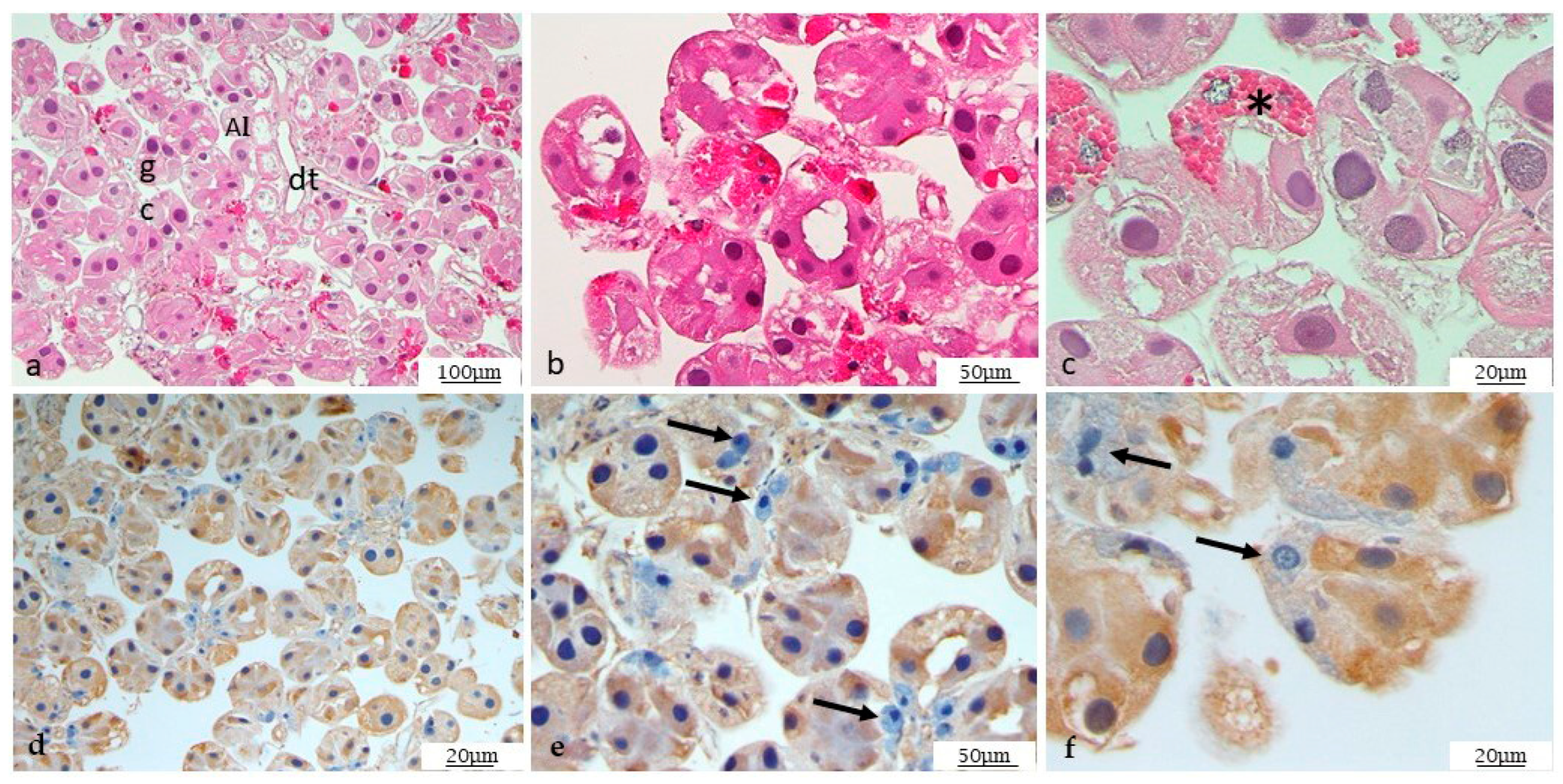
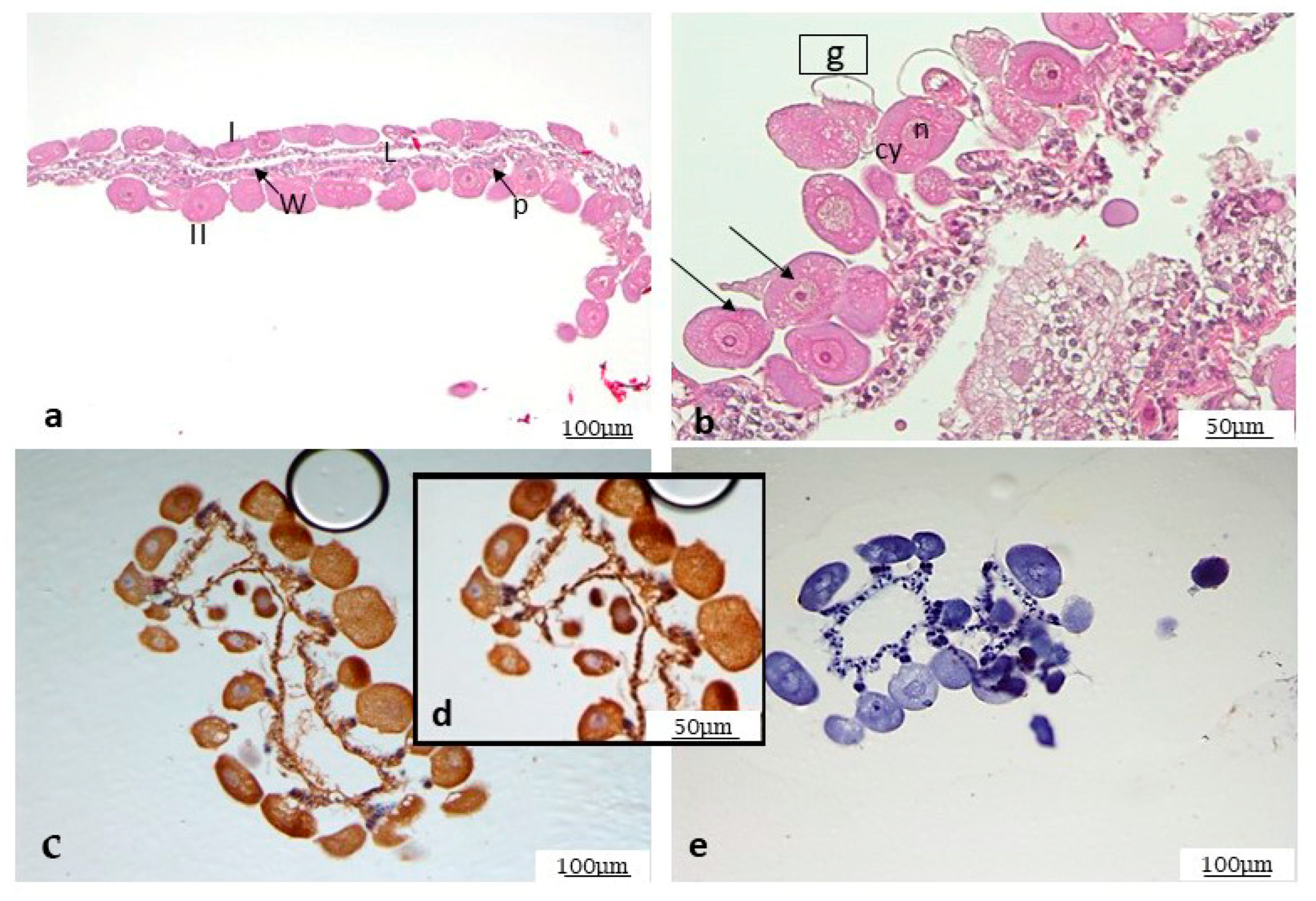
3.4. Histological Analysis

For the morphological analysis of samples, H&E-stained slides of both half ticks and dissected salivary glands and ovaries were observed.
Different anatomical parts of the tick are easily distinguishable, as described in Figure 1. By morphological analysis of the salivary glands, following standard staining with H&E, it was possible to observe the ducts and different gland cells, mostly represented by type I and type II acini (Figure 2a–c). In addition, some cells are still half engorged (Figure 2c), in which red blood cells are clearly distinguishable. The same procedure was also followed for the ovary. The histological analysis revealed oocytes at different development stages, as shown in Figure 3a,b, and the particular oocyte cell compartment.

3.5. Immunohistochemistry on Mycoplasma agalactiae Positive Ticks

Specific immunohistochemical investigations with anti-Ma antibody showed positivity (Table 1) at the cytoplasmic level of the salivary glands, although not all cells are positive, as shown in Figure 2e.
In ovary, most cells showed a strong positivity for Ma detected by IHC, which again proved to be purely cytoplasmic, as shown in Figure 3c,d. The specificity of the signal is demonstrated by the negative control, as shown in Figure 3e.

4. Discussion

The present study used IHC to detect and localize, for the first time, Ma in positive R. bursa ticks confirmed by isolation and PCR. Ticks were collected from sheep belonging to a farm declared free from CA at the time of this study but with confirmed historical outbreaks by Ma until 2020. Ma was detected in the salivary glands and in the ovaries of two adult females out of twenty collected ticks, demonstrating the passage through the wall of the midgut and the infection of various tick organs capable of propagating the infection to progeny and reference hosts. These results show absolute novelty in disease-related knowledge. A recent study from our team performed in three different endemic areas for CA in Sicily provided evidence of the potential involvement of ticks in the spread and maintenance of CA [23]. The data obtained in the aforementioned study showed an overall prevalence of 8.9% of viable Ma in R. bursa ticks collected from sheep and goats in three different herds, in which Ma infection had been previously confirmed and notified.
Sicily represents a typical Mediterranean ecosystem characterized by low or moderate rainfall and long dry seasons, in which R. bursa is considered the major ectoparasite of small ruminants, which spreads in hilly, marginal areas, grassy slopes or semi-desert environments [27,28]. R. bursa is a dual-host tick: immature stages commonly infest one host while adults infest a different one [29].
Several studies have recognized the vector role of R. bursa in the transmission of several pathogens, namely Theileria spp., Anaplasma marginale, Anaplasma ovis, Ehrlichia canis [30,31,32,33], Babesia ovis [24], and Coxiella burnetii [34], in ruminants. Additionally, the presence of unconventional tick-borne pathogens has been described in R. bursa [23,35].
The ability of the vector to establish a symbiotic relationship with a pathogen facilitating their multiplication and transmission is essential as a determinant of tick-borne disease [36]. In particular, the competence of ticks as a vector of a specific pathogen is determined by the capacity of the pathogen to survive in the tick midgut environment [37] and to reach the salivary glands where the saliva acts as the major transmission medium [38].
On the one hand, migration of the pathogen to the salivary glands would lead to survival of the pathogen after ingestion during an earlier phase of the tick’s life cycle and transmission to the next host during the next blood meal. On the other hand, the colonization of the ovary would represent the perpetuation of the infection to subsequent generations of the tick. In addition, it is well demonstrated that the pathogen transmission can occur between infected and uninfected ticks co-feeding in time or space in the absence of the host systemic infection [39]. These strategies appear similar to several tick-borne pathogens.
The detection of Ma in the salivary glands reported in this study suggests a high affinity of Ma for tick salivary glands with a consequent possibility of pathogen transmission to the host. However, further studies are needed to determine what is the minimum dose of Ma in the tick salivary glands to cause infection in the parasitized host. Nevertheless, it can be speculated that a sheep with massive and repeated infection by Ma-positive ticks may result in contracting Ma infection.
In this study, we also reported the presence of Ma in tick ovaries. The evidence of Ma in female reproductive organs such as the ovaries could lead to a subsequent transovarial passage which could guarantee the maintenance of the pathogen in subsequent generations.
The demonstration of R. bursa in the salivary glands and ovary confirmed that Ma is able to cross the wall of the midgut and infect various tick organs; this may indicate the potential for biological development and transmission of Ma in ticks. For this purpose, it would be intriguing to investigate the presence of Ma in other anatomical sites of infected ticks, such as hemocytes, synganglia, testes, fat bodies, and midgut.
The immunohistochemical technique employed in the present study is certainly less sensitive than that of real-time PCR in the detection of Ma. However, IHC has the advantage of not being compromised by cross-contamination, which is very likely during tick dissection and the isolation of organs. Furthermore, IHC is a method that allows for the identification of a specific antigen in specific cellular and subcellular compartments utilizing a specific antibody. In addition, for the inclusion of the isolated organs from dissected ticks, an innovative synthetic matrix, called Cytomatrix®, was used successfully in this study. The Cytomatrix® matrix is characterized by a high adsorption capacity that helps to maintain the tissue architecture and three-dimensional structure of the sample being loaded onto it, reducing handling and therefore the loss of sample. This system proved to be particularly suitable for our purpose.
Since 1816, it has been believed that environmental factors play an indirect, additional role in pathogen maintenance on endemic farms for CA [4]. However, mycoplasmas are obligate parasites of cells and are poorly resistant to environmental conditions for extended periods of time [40]. Despite this, CA is endemic in Sicily and other Mediterranean countries, where unfavorable climatic conditions suggest that other factors play a role in the perpetuation of CA endemism. The distribution of CA as a climate or vector-related disease has not been generally recognized; however, keeping in mind the geographic distribution and the potential for insect or tick bites, this may be a possibility. Relatedly, climate change and the resulting colonization of new geographic areas by competent vectors could be of concern for the spread of the disease.
Our study supports this hypothesis, recognizing in the hard ticks a potential reservoir or vectors for CA agents which have not been proven to date. However, further work, including examination of other isolated anatomical parts, is required to strengthen our evidence. In addition, the reproducibility of our field results should be verified in vitro, using artificial tick feeding/infection systems. In this way, it would be possible to test the competence of several species of ticks, of different sex and stages of life (transstadial persistence), as possible vectors in the transmission and maintenance of Ma in areas endemic for CA. This approach could also be extended to study additional relevant diseases caused by other species of the Mycoplasma genus.
Our findings clearly highlight an additional risk factor in the transmission and maintenance of CA in endemic areas. In addition to the available vaccination protocols and the biosecurity measures to be applied to control the disease, it would be appropriate to think about ticks’ management in terms of both surveillance and prevention, depending on the prediction based on the presence of ticks (in which quantity and at what time of year) and climatic conditions (seasonal temperatures and precipitation). Acaricidal treatment is considered essential for the prevention of most tick-borne diseases and may be an additional control method for CA. However, its well known that the widespread use of insecticides in agriculture is a risk for promoting resistance in vectors carrying zoonotic agents and would need to be carefully assessed.

5. Conclusions

Our results provide further evidence of the vector competence of hard ticks, in particular R. bursa, in the maintenance and transmission of Ma in areas endemic for CA, demonstrating for the first time the crossing of the tick midgut barrier by Ma and the subsequent infection of other organs capable of spreading the infection.
However, the evidence of these additional environmental sources for Ma infection and their role in transmission and clinical disease warrant further investigation. In particular, additional field and laboratory investigations and in vitro feeding/infection studies to better evaluate tick–pathogen interactions are required.

Author Contributions

Conceptualization, A.R., G.R.L. and R.P.; methodology, L.C., P.G. and L.G.; formal analysis, S.M., L.C. and R.P.; investigation, L.C., P.G., L.G. and A.C.; data curation, S.M. and L.C.; writing—original draft preparation, S.M. and L.C.; writing—review and editing, S.M., L.C., E.L., A.R., G.R.L. and R.P.; supervision, A.R., G.R.L. and R.P.; project administration, A.R. and G.R.L.; funding acquisition, S.M., A.R. and G.R.L. All authors have read and agreed to the published version of the manuscript.

Funding

This research was supported by the GB Animal Exotic Disease research budget, project code RDSC2003, held and administered centrally by the UK Government’s Department for Environment, Food, and Rural Affairs (Defra) on behalf of England, Scotland and Wales, and the Italian Ministry of Health, grant number RC IZSSi 01/2023.

Data Availability Statement

The original contributions presented in the study are included in the article, further inquiries can be directed to the corresponding author.

Acknowledgments

The authors thank Paolo Russo for the support provided in image editing.

Conflicts of Interest

The authors declare no conflicts of interest.

References

  1. Migliore, S.; Puleio, R.; Nicholas, R.A.J.; Loria, G.R. Mycoplasma agalactiae: The Sole Cause of Classical Contagious Agalactia? Animals 2021, 11, 1782. [Google Scholar] [CrossRef] [PubMed]
  2. Bergonier, D.; Berthelot, X.; Poumarat, F. Contagious agalactia of small ruminants: Current knowledge concerning epidemiology, diagnosis and control. OIE Rev. Scienific Techique 1997, 16, 848–873. [Google Scholar] [CrossRef] [PubMed]
  3. Loria, G.R.; Nicholas, R.A. Contagious agalactia: The shepherd’s nightmare. Vet. J. 2013, 198, 5–6. [Google Scholar] [CrossRef] [PubMed]
  4. Jay, M.; Tardy, F. Contagious agalactia in sheep and goats: Current perspectives. Vet. Med. Res. Rep. 2019, 10, 229–247. [Google Scholar]
  5. Jongejan, F.; Uilenberg, G. The global importance of ticks. Parasitology 2004, 129, S3–S14. [Google Scholar] [CrossRef]
  6. Sonenshine, D.E. Biology of Ticks; Oxford University Press: Oxford, UK, 1991; p. 447. [Google Scholar]
  7. McCoy, K.D.; Léger, E.; Dietrich, M. Host specialization in ticks and transmission of tick-borne diseases: A review. Front. Cell. Infect. Microbiol. 2013, 4, 57. [Google Scholar] [CrossRef] [PubMed]
  8. Estrada-Peña, A.; Bouattour, A.; Camicas, J.L.; Walker, A.R. Ticks of Domestic Animals in the Mediterranean Region: A Guide to Identification of Species; University of Zaragoza: Zaragoza, Spain, 2004. [Google Scholar]
  9. Neimark, H.; Johansson, K.E.; Rikihisa, Y.; Tully, J.G. Proposal to transfer some members of the genera Haemobartonella and Eperythrozoon to the genus Mycoplasma with descriptions of ‘Candidatus Mycoplasma haemofelis’, ‘Candidatus Mycoplasma haemomuris’, ‘Candidatus Mycoplasma haemosuis’ and ‘Candidatus Mycoplasma wenyonii’. Int. J. Syst. Evol. Microbiol. 2001, 51, 891–899. [Google Scholar] [PubMed]
  10. Nayak, N.C.; Blownik, M.K. Goat flea (order Siphonaptera) as a possible vector for the transmission of caprine mycoplasmal polyarthritis with septicaemia. Prev. Vet. Med. 1990, 9, 259–266. [Google Scholar] [CrossRef]
  11. Foley, J.E.; Pedersen, N.C. Candidatus Mycoplasma haemominutum, a low-virulence erpierythrocytic parasite of cats. Int. J. Syst. Evol. Microbiol. 2001, 51, 815–817. [Google Scholar] [CrossRef]
  12. Willi, B.; Boretti, F.S.; Cattori, V.; Tasker, S.; Meli, M.L.; Reusch, C.; Lutz, H.; Hofmann-Lehmann, R. Identification, molecular characterization, and experimental transmission of a new hemoplasma isolate from a cat with hemolytic anemia in Switzerland. J. Clin. Microbiol. 2005, 43, 2581–2585. [Google Scholar] [CrossRef]
  13. Willi, B.; Boretti, F.S.; Baumgartner, C.; Cattori, V.; Meli, M.L.; Doherr, M.G.; Reusch, C.E.; Hofmann-Lehmann, R. Feline hemoplasmas in Switzerland: Identification of a novel species, diagnosis, prevalence, and clinical importance. Schweiz. Arch. Tierheilk. 2006, 148, 139–144. [Google Scholar] [CrossRef] [PubMed]
  14. Seneviratna, P.; Weerasinghe, N.; Ariyadasa, S. Transmission of Haemobartonella canis by the dog tick, Rhipicephalus sanguineus. Res. Vet. Sci. 1973, 14, 112–114. [Google Scholar] [CrossRef] [PubMed]
  15. Novacco, M.; Meli, M.L.; Gentilini, F.; Marsilio, F.; Ceci, C.; Pennisi, M.G.; Lombardo, G.; Lloret, A.; Santos, L.; Carrapiço, T.; et al. Prevalence and geographical distribution of canine hemotropic mycoplasma infections in Mediterranean countries and analysis of risk factors for infection. Vet. Microbiol. 2010, 142, 276–284. [Google Scholar] [CrossRef]
  16. Hornok, S.; Micsutka, A.; Meli, M.L.; Lutz, H.; Hofmann-Lehmann, R. Molecular investigation of transplacental and vector-borne transmission of bovine haemoplasmas. Vet. Microbiol. 2011, 152, 411–414. [Google Scholar] [CrossRef] [PubMed]
  17. Ayling, R.D.; Bisgaard-Frantzen, S.; Adler, A.; Blowey, R.W.; Barlow, A.M.; Millar, M.F.; Van Der Burgt, G.M. Detection of ‘Candidatus Mycoplasma haemobos’, Mycoplasma wenyonii and Anaplasma phagocytophilum from cattle in England. Vet. Rec. 2012, 170, 543. [Google Scholar] [CrossRef] [PubMed]
  18. Girotto, A.; Zangirólamo, A.F.; Bogado, A.L.; Souza, A.S.; Silva, G.C.; Garcia, J.L.; Vilas Boas, L.A.; Biondo, A.W.; Vidotto, O. Molecular detection and occurrence of ‘Candidatus Mycoplasma haemobos’ in dairy cattle of Southern Brazil. Rev. Bras. Parasitol. Vet. 2012, 21, 342–344. [Google Scholar] [CrossRef]
  19. Hornok, S.; Sugár, L.; Fernández de Mera, I.G.; De La Fuente, J.; Horváth, G.; Kovács, T.; Micsutka, A.; Gönczi, E.; Flaisz, B.; Takács, N.; et al. Tick-and fly-borne bacteria in ungulates: The prevalence of Anaplasma phagocytophilum, haemoplasmas and rickettsiae in water buffalo and deer species in Central Europe, Hungary. BMC Vet. Res. 2018, 14, 98. [Google Scholar] [CrossRef] [PubMed]
  20. Santos, N.J.; Brito, D.R.; Abate, H.L.; Paixão, S.F.; Soares, E.D.; Vieira, T.S.; Garcia, J.L.; Vieira, R.F.; Vidotto, O. Hemotropic mycoplasmas infection in water buffaloes (Bubalus bubalis) from northeastern Brazil. Comp. Immunol. Microbiol. Infect. Dis. 2018, 56, 27–29. [Google Scholar] [CrossRef] [PubMed]
  21. Shi, H.; Hu, Y.; Leng, C.; Shi, H.; Jiao, Z.; Chen, X.; Peng, Y.; Yang, H.; Kan, Y.; Yao, L. Molecular investigation of “Candidatus Mycoplasma haemobos” in goats and sheep in central China. Transbound. Emerg. Dis. 2019, 66, 22–27. [Google Scholar] [CrossRef]
  22. Neimark, H.; Hoff, B.; Ganter, M. Mycoplasma ovis comb. nov. (formerly Eperythrozoon ovis), an eperythrocytic agent of haemolytic anaemia in sheep and goats. Int. J. Syst. Evol. Microbiol. 2004, 54, 365–371. [Google Scholar] [CrossRef] [PubMed]
  23. Galluzzo, P.; Migliore, S.; Puleio, R.; Galuppo, L.; La Russa, F.; Blanda, V.; Tumino, S.; Torina, A.; Ridley, A.; Loria, G.R. Detection of Mycoplasma agalactiae in Ticks (Rhipicephalus bursa) Collected by Sheep and Goats in Sicily (South-Italy), Endemic Area for Contagious Agalactia. Microorganisms 2021, 9, 2312. [Google Scholar] [CrossRef] [PubMed]
  24. Walker, J.B.; Keirans, J.E.; Horak, I.G. The Genus Rhipicephalus (Acari, Ixodidae): A Guide to the Brown Ticks of the World; Cambridge University Press: Cambridge, UK, 2000; p. 643. ISBN 9780521019774. [Google Scholar]
  25. Rodríguez, F.; Ramírez, G.A.; Ramírez, A.S.; Ball, H.J.; Espinosa de los Monteros, A.; Fernandez, A. Immunohistochemical detection of Mycoplasma agalactiae in formalin-fixed, paraffin-embedded tissues from naturally and experimentally infected goats. J. Vet. Med. B Infect. Dis. Vet. Public Health 2002, 49, 226–229. [Google Scholar] [CrossRef] [PubMed]
  26. Friis, N.F. Some recommendations concerning primary isolation of Mycoplasma suipneumoniae and Mycoplasma flocculare a survey. Nord. Vet. Med. 1975, 27, 337–339. [Google Scholar] [PubMed]
  27. Torina, A.; Khoury, C.; Caracappa, S.; Maroli, M. Ticks infesting livestock on farms in Western Sicily, Italy. Exp. Appl Acarol. 2006, 38, 75–86. [Google Scholar] [CrossRef] [PubMed]
  28. Raele, D.A.; Galante, D.; Pugliese, N.; De Simone, E.; Cafiero, M.A. Coxiella-like endosymbiont associated to the Anatolian brown tick Rhipicephalus bursa in Southern Italy. Microbes Infect. 2015, 17, 799–805. [Google Scholar] [CrossRef]
  29. Matei, I.A.; Ionică, A.M.; Corduneanu, A.; Domșa, C.; Sándor, A.D. The Presence of Ehrlichia Canis in Rhipicephalus bursa Ticks Collected from Ungulates in Continental Eastern Europe. J. Vet. Res. 2021, 65, 271–275. [Google Scholar] [CrossRef] [PubMed]
  30. Ferrolho, J.; Antunes, S.; Santos, A.S.; Velez, R.; Padre, L.; Cabezas-Cruz, A.; Santos-Silva, M.M.; Domingos, A. Detection and phylogenetic characterization of Theileria spp. and Anaplasma marginale in Rhipicephalus bursa in Portugal. Ticks Tick Borne Dis. 2016, 7, 443–448. [Google Scholar] [CrossRef]
  31. Renneker, S.; Abdo, J.; Salih, D.E.A.; Karagenc, T.; Bilgic, H.; Torina, A.; Oliva, A.G.; Campos, J.; Kullmann, B.; Ahmed, J.; et al. Can Anaplasma ovis in small ruminants be neglected any longer? Transbound. Emerg. Dis. 2013, 60 (Suppl. S2), 105–112. [Google Scholar] [CrossRef]
  32. Masala, G.; Chisu, V.; Foxi, C.; Socolovschi, C.; Raoult, D.; Parola, P. First detection of Ehrlichia canis in Rhipicephalus bursa ticks in Sardinia Italy. Ticks Tick Borne Dis. 2012, 3, 396–397. [Google Scholar] [CrossRef]
  33. Agnone, A.; La Manna, M.; Sireci, G.; Puleio, R.; Usticano, A.; Ozdemir, U.; Nicholas, R.A.J.; Chiaracane, V.; Dieli, F.; Di Marco, V.; et al. A comparison of the efficacy of commercial and experimental vaccines for contagious agalactia in sheep. Small Rum. Res. 2013, 112, 230–234. [Google Scholar]
  34. Enayati, A.A.; Asgarian, F.; Sharif, M.; Boujhmehrani, H.; Amouei, A.; Vahedi, N.; Boudaghi, B.; Piazak, N.; Hemingway, J. Propetamphos resistance in Rhipicephalus bursa (Acari, Ixodidae). Vet. Parasitol. 2009, 162, 135–141. [Google Scholar] [CrossRef] [PubMed]
  35. Cappelli, A.; Capone, A.; Valzano, M.; Bozic, J.; Preziuso, S.; Mensah, P.; Varotto Boccazzi, I.; Rinaldi, L.; Favia, G.; Ricci, I. Denaturing Gradient Gel Electrophoresis Analysis of Bacteria in Italian Ticks and First Detection of Streptococcus equi in Rhipicephalus bursa from the Lazio Region. Vector Borne Zoonotic Dis. 2019, 19, 328–332. [Google Scholar] [CrossRef] [PubMed]
  36. de la Fuente, J.; Antunes, S.; Bonnet, S.; Cabezas-Cruz, A.; Domingos, A.G.; Estrada-Peña, A.; Johnson, N.; Kocan, K.M.; Mansfield, K.L.; Nijhof, A.M.; et al. Tick-pathogen interactions and vector competence: Identification of molecular drivers for tick-borne diseases. Front. Cell. Infect. Microbiol. 2017, 7, 114. [Google Scholar] [CrossRef] [PubMed]
  37. Jones, L.D.; Davies, C.R.; Steel, G.M.; Nuttall, P.A. Vector capacity of Rhipicephalus appendiculatus and Amblyomma variegatum for Thogoto and Dhori viruses. Med. Vet. Entomol. 1989, 3, 195–202. [Google Scholar] [CrossRef] [PubMed]
  38. Randolph, S.E. Tick-borne disease systems emerge from the shadows: The beauty lies in molecular detail, the message in epidemiology. Parasitology 2009, 136, 1403–1413. [Google Scholar] [CrossRef] [PubMed]
  39. Randolph, S.E.; Gern, L.; Nuttall, P.A. Co-feeding ticks: Epidemiological significance for tick-borne pathogen transmission. Parasitol Today. 1996, 12, 472–479. [Google Scholar] [CrossRef] [PubMed]
  40. Dudek, K.; Sevimli, U.; Migliore, S.; Jafarizadeh, A.; Loria, G.R.; Nicholas, R.A.J. Vaccines for Mycoplasma Diseases of Small Ruminants: A Neglected Area of Research. Pathogens 2022, 11, 75. [Google Scholar] [CrossRef] [PubMed]
Figure 1. H&E-stained sagittal section of Rhipicephalus Bursa tick. Some structures are clearly distinguishable: (a) R = rostro; Mg = midgut; CAE = caecum; SG = salivary gland (2.5× magnification); (b) close-up of salivary gland (5× magnification); (c) higher magnification of the enclosed region in the box in (b) 10×.
Microorganisms 12 01390 g001
Figure 2. H&E-stained view of salivary glands. (ac) In the picture, ducts and different gland cells are visible, mostly represented by type I and type II acini. dt = duct; gc = gland cells; AI = acini I; AII = acini II. (c) Close-up of engorged cells (*). IHC for Ma (df). Immunohistochemistry showed a high positivity, purely at the cytoplasmic level, in salivary glands, both in cells and ducts. Some cells, such as those indicated by arrows, are negative, including engorged cells.
Microorganisms 12 01390 g002
Figure 3. H&E-stained ovary (a,b). Two types of oocytes at stage I and II are well recognizable; L = Lumen; the arrows point to ovarian wall (W) and to pedicel (P); (b) close-up at a higher magnification in which it is possible to distinguish oocyte cellular compartments, arrows point indicate two oocytes in different stage. g = germinal vesicle; n = nucleolus, cy = cytoplasm (ce). IHC for Ma. (ce) A strong positivity in oocytes, again purely cytoplasmic; R. bursa oocyte close-up of picture in (d); (e) negative control.
Microorganisms 12 01390 g003
Table 1. Summary table of the results.
SheepMycoplasma spp. Isolation from MilkNo. of Collected TicksTicks IdentificationMycoplasma spp. Isolation from Half Ticks (No. of Positive/Total)Ma Detection from Positive Samples by VETMAXTM Real-Time PCRMa Antigen Detection from Positive Samples by IHC
#1-3R. bursa0/2--
#2-5R. bursa2/22/22/2
#3-3R. bursa0/3--
#4-4R. bursa0/1--
#5-5R. bursa0/3--
20 2/20 (10%)
Disclaimer/Publisher’s Note: The statements, opinions and data contained in all publications are solely those of the individual author(s) and contributor(s) and not of MDPI and/or the editor(s). MDPI and/or the editor(s) disclaim responsibility for any injury to people or property resulting from any ideas, methods, instructions or products referred to in the content.

