Article

Laundry Isolate Delftia sp. UBM14 Capable of Biodegrading Industrially Relevant Aminophosphonates

1
Chair of Biotechnology of Water Treatment Brandenburg, Institute of Environmental Science and Environmental Technology, BTU Cottbus-Senftenberg, 03046 Cottbus, Germany
2
German Environment Agency (UBA), Section II 3.3, 14195 Berlin, Germany
3
Department of Environmental Microbiomics, University of Technology Berlin, 10587 Berlin, Germany
4
Applied Biosciences and Process Engineering, Anhalt University of Applied Sciences, 06366 Köthen, Germany
5
Chair of Bioprocess Engineering, Otto von Guericke University, 39106 Magdeburg, Germany
6
Bioprocess Engineering, Max Planck Institute for Dynamics of Complex Technical Systems, 39106 Magdeburg, Germany
*
Author to whom correspondence should be addressed.
Microorganisms 2024, 12(8), 1664; https://doi.org/10.3390/microorganisms12081664
Submission received: 19 July 2024 / Revised: 8 August 2024 / Accepted: 12 August 2024 / Published: 13 August 2024
(This article belongs to the Section Environmental Microbiology)

Abstract

:
Phosphonates such as ethylenediaminetetra (methylenephosphonic acid) (EDTMP) and aminotris (methylenephosphonic acid) (ATMP) are used every day in water treatment processes or in household products. Their consumption is still increasing, regardless of the debates on their environmental impact. Here, the microbial characterisation and determination of the biodegradation potential of selected industrially relevant phosphonates for the isolate Delftia sp. UMB14 is reported. The opportunistic strain was isolated from a biofilm that was derived from a conventional washing machine using conventional detergents containing phosphonates. In antimicrobial susceptibility testing, the strain was only susceptible to sulfonamide, tetracycline, and chloramphenicol. Physiological and biochemical characteristics were determined using the BIOLOG EcoPlate assay. Most importantly, the strain was shown to convert D-malic acid and D-mannitol, as confirmed for strains of Delftia lacustris, and thus the new isolate could be closely related. Biodegradation tests with different phosphonates showed that the strain preferentially degrades ATMP and EDTMP but does not degrade glyphosate (GS) and amino (methylphosphonic acid) (AMPA). A specific gene amplification confirmed the presence of phnX (phosphonoacetaldehyde hydrolase) and the absence of PhnJ (the gene for the core component of C–P lyase). The presence of PhnCDE is strongly suggested for the strain, as it is common in Delftia lacustris species.

1. Introduction

Phosphonates, a large group of soluble, unreactive phosphorus compounds, have attracted much attention in recent years. There is no doubt that they have made an imperceptible contribution to our modern lifestyle. The most famous aminophosphonate, glyphosate (GS), has emerged as a global player as a systemic herbicide. However, the negative effects of GS and its transformation product amino (methylphosphonic acid) (AMPA) on human health and the environment have been the subject of intense debates for several years [1,2,3].
Independent of GS, phosphonates are used on a daily basis in many areas of modern life. This is due to their excellent complexing properties with alkaline earth and transition metals. Therefore, aminophosphonates such as ethylenediaminetetra (methylenephosphonic acid) (EDTMP) and diethylenetriamine penta (methylenephosphonic acid) (DTPMP) are now important anti-scaling and bleach-stabilising additives in detergents. Aminotris (methylenephosphonic acid) (ATMP) and hydroxyethelidene (diphosphonic acid) (HEDP) are commonly used in industrial detergents and cleaners. In addition, in the paper and textile industries, phosphonates are often used as dispersants to stabilise peroxide bleaching baths [4]. They are important components of flame-retardant compounds [5] and essential in the oilfield industry [5,6]. For the latter, complexing of barium with DTPMP is an important method to minimise or avoid the formation of incrustations on the drilling elements. Apart from technical applications, phosphonates such as HEDP and EDTMP are also successfully used in the medical treatment of bone diseases [7,8]. As a result, the use of EDTMP has become the standard treatment for bone cancer and osteoporosis [9]. The absence of phosphonates would therefore result in a dramatic loss of cleaning performance and quality, as well as medical advances in treatment.
Unfortunately, the intensive use of phosphonates in everyday life has also led to their accumulation in the aquatic environment, as they are not completely removed during wastewater treatment. This has already been reported independently by Armbruster et al. [10], Rott et al. [11,12], and Wang et al. [13]. For more than three decades, it was assumed that industrially and commercially available phosphonates such as HEDP, ATMP, EDTMP, and, especially, DTPMP were not readily biodegradable [14,15]. As recently reported, this assumption was further supported by the outcomes of the OECD test series 301 A–F for the determination of the ready biodegradability of organic compounds [16]. Synthetic phosphonates very often do not promote microbial growth due to their chemical imbalance of carbon to phosphorus. Until today, only one exception, the surfactant cocoamine bis (methylene phosphonate), meets the requirements of the test design and the conditions of the OECD test series 301 A–F. For all other phosphonates, it is not surprising that the OECD test series 301 A–F results are generally negative, as the chemical imbalance of phosphonates does not favour their biodegradation. Therefore, a new standardised biodegradation batch test has been developed to overcome the drawbacks of the OECD test series 301 A–F. Most importantly, the test can also be used to isolate novel strains from different aquatic environments, such as activated sludge and/or biofilms.
The successful application of the novel biodegradation batch test was recently reported for the novel strain Ochrobactrum sp. BTU1, isolated from a DTPMP standard solution [17]. The strain is able to degrade EDTMP and GS as well as common metabolites, i.e., iminodi (methylenephosphonic) (IDMP) and ethylaminobis (methylenephosphonic) acid (EABMP). Genomic and proteomic analyses confirmed that the strain degraded the synthetic phosphonates via the widespread C–P lyase pathway. The mechanism of the C–P lyase pathway was first discovered in Escherichia coli using the metabolic pathway from ribosyl 1,5-biphosphate to generate 5-phospho-d-ribosyl-α-1-diphosphate (PRPP) [18]. It was found that E. coli cannot utilise GS as a direct P source. Therefore, the C–P lyase pathway is designated as a tolerance mechanism in E. coli [19,20]. As recently described, the C–P lyase enzyme complex is encoded by the phnCDEFGHIJKLMNOP operon, within which, the PhnGHIJKLM is essential for cleavage activity. In brief, the initial catalytic step is carried out by the purine ribonucleoside triphosphate phosphonylase component PhnI, which activates the phosphonate for further catabolism. However, PhnI requires the multi-subunit protein complex (PhnGHIJK) to yield the 5′-triphosphoribosyl-1′-phosphonate [19,21]. The final C–P cleavage is then mediated by PhnJ, leading to the formation of PRPP and either sarcosine, glyoxylate, or methane for GS, AMPA, or methylphosphonate (MP), respectively.
It is now known that C–P lyase catabolism has a broad substrate specificity that allows bacteria to degrade phosphonates other than GS. Other more substrate-specific catabolisms, such as the hydrolytic pathway for the degradation of phosphonates, are also widespread in the environment and are mostly found in marine ecosystems [22]. Several hydrolases have been discovered that mediate the hydrolytic pathway and are responsible for cleaving the carbon–phosphorus (C–P) bond, such as phosphonopyruvate hydrolase (PalH) [23], phosphonoacetate hydrolase (PhnA) [24], phosphonoacetaldehyde hydrolase (PhnX) [25,26], and phosphohydrolase (PhnZ) [27,28]. According to Lockwood et al. [22], the latter three target the most abundant and ubiquitous marine biogenic phosphonate, 2-aminoethylphosphonate (2-AEP). Several bacterial strains have been reported to break down 2-AEP via PhnX [29,30,31,32]. Similar to the substrate activation in the C–P lyase pathway, the hydrolytic pathway by PhnX also requires an initial enzymatic step. The final C–P cleavage of 2-AEP by PhnX requires transamination by the 2-AEP pyruvate aminotransferase PhnW [33]. As recently stated by Zangelmi [34], the transamination reaction of PhnW is reversible, i.e., this enzyme is involved in both the biosynthesis and catabolism of phosphonates. However, PhnX does not always require PhnW as an initial enzymatic step. Recently, Zangelmi et al. [30] demonstrated that also accessory enzymes such as phosphonates breakdown factor B, C and D (PbfB, PbfB and PbfBD) can convey into the PhnWX pathway. They suggested that some of these enzymes can carry out an oxidative deamination of 2-AEP. Thus, the degradation of 2-AEP via the PhnWX pathway is not exclusively limited by PhnW, even if both PhnW and PhnX are closely associated. Further detailed information on the various enzymatic pathways, in addition to the C–P pathway, and the distinctive and unconventional characteristics of the PhnWX pathway can be found in reference [35]. Nevertheless, up to now, the biodegradation of industrially relevant phosphonates, as mentioned above, that are mediated by the PhnWX pathway has not been reported. Its narrow substrate spectrum does not seem to be predestined for phosphonates other than 2-AEP.
Since phosphonates are daily and widely used in various technical and industrial processes, especially in detergents and cleaning agents to reduce water hardness, we hypothesized that bacteria capable of degrading phosphonates can be isolated from the biofilms of washing machines. The aim of this research was to isolate and characterise a biofilm isolate for its ability to grow on industrial phosphonates that are commonly used in detergent powders, such as, ATMP, EDTMP, DTPMP, and HEDP. Additionally, GS, AMPA, and IDMP were investigated, as GS is one of the most important phosphonates, and AMPA and IDMP are major transformation products of several industrially relevant aminophosphonates. The characterisation of the strain was carried out by physiological, biochemical, and molecular analyses. The mass balance of the tested phosphonate and the minimum phosphorous requirements for bacterial growth were determined.

2. Materials and Methods

2.1. Chemicals and Reagents

HEDP, ATMP, EDTMP, and DTPMP were provided by Zschimmer & Schwarz Mohsdorf (Burgstädt, Germany). IDMP, AMPA, and GS were purchased from Sigma Aldrich (Steinheim, Germany). The chemical structures of all phosphonates are presented below (Figure 1). NH4COOCH3 was purchased from Roth (Neu-Ulm Karlsruhe, Germany). FeSO4 × 7H2O, ZnSO4 × 7H2O, H3BO3, CuCl2 × 2 H2O, NiCl2 × 6 H2O, NaMoO4 × 2 H2O, EDTA, KHCO3, and MgSO4 × 7H2O were purchased from Merck (Darmstadt, Germany) and KH2PO4 was purchased from Riedel-de Haën (Seelze, Germany). Nutrient agar was purchased from Roth (Karlsruhe, Germany). Nutrient broth II (NB II) was purchased from Sifin (Berlin, Germany). The local tap water (TW) of Cottbus was used for all degradation tests and provided 90 mg L−1 Ca2+, 20 mg L−1 Mg2+, 15 mg L−1 Na+, 2.5 mg L−1 K, 32 mg L−1 Cl, ≤0.05 mgPO43− L−1, and 115 mg L−1 SO42−. The local TW of Berlin was used for the enrichment procedure and provided 94.6 mg L−1 Ca2+, 8.75 mg L−1 Mg2+, 20 mg L−1 Na+, 5.5 mg L−1 K+, 47.5 mg L−1 Cl, 0.04 mg TP L−1, and 77 mg L−1 SO42−.

2.2. Enrichment Procedure and the Purification of Phosphonate Degrading Isolates

For the enrichment of phosphonate degrading isolates, a total of 24 biofilm samples were taken from different locations within standard washing machines. Operating of the appliance was performed with standard household laundry and commercially available detergents.
The biofilm was scraped off the detergent drawer using a sterile spatula. The removed biofilm was then suspended in a liquid medium consisting of 125 mL TW, 6 g L−1 ammonium acetate, 100 mg L−1 MgSO4, 125 µL SL-8 solution [36], and the investigated sole phosphorus source, with final concentrations of 50 mg L−1 HEDP, 100 mg L−1 EDTMP, or 100 mg L−1 DTPMP. The flasks were incubated at room temperature and shaken at 120 rpm. When the optical density (OD) at 660 nm of the culture reached >0.5, 1 mL of the culture was inoculated into fresh medium. A portion of the solution from passage 5 was applied on nutrient agar (BD DIFCO, Franklin Lakes, NJ, USA). The agar plates were incubated at 25 °C, and colonies were subsequently picked and isolated from the plates. Pure cultures were obtained by passaging the cultures on these plates and verified by microscopic examinations using a light microscope (Zeiss Axioskop, Oberkochen, Germany).

2.3. Nucleic Acid Extraction and Sequencing of the 16S rRNA Gene

DNA was extracted from all 27 isolates using a GeneMATRIX Soil DNA Purification Kit (Roboklon, Berlin, Germany) according to the manufacturer’s instructions. PCR was performed with the personal cycler (Biometra, Göttingen, Germany). Amplification of the DNA was performed in a final volume of 50 µL, containing 25 µL 2× Color Opti Taq-PCR master mix (Roboklon, Berlin, Germany), 1 µL primer (20 pmol/µL, each), 1 µL DNA, and 22 µL PCR water. The primer pair 63f and 1387r [37] was chosen for the amplification of the 16S rRNA genes. PCR conditions were as follows: initial denaturation at 95 °C for 120 s, followed by 35 cycles, each consisting of 95 °C for 20 s, 55 °C for 30 s, and 72 °C for 120 s, terminated by a final extension step at 72 °C for 10 min. PCR amplicons were analysed under UV light after electrophoresis in 1.5% (w/v) agarose gel stained with ROTI®GelStain (Carl Roth, Karlsruhe, Germany). Afterwards, PCR products were purified using the GeneMATRIX Basic DNA Purification Kit (Roboklon, Berlin, Germany), following the manual. The automatic sequencing was carried out at LGC Genomics GmbH (Berlin, Germany). The quality of the obtained sequences from both primers was assessed with Chromas version 2.6.6 (https://chromas.software.informer.com/download/ accessed on 6 May 2024). Areas of particularly low quality at the beginning and end of the sequences were trimmed, exported to FASTA format, and further processed in MEGA 11 (https://www.megasoftware.net/ accessed on 6 May 2024). Sequences were aligned using MUSCLE, and the overlapping region was examined for inconsistencies. The phylogenic affiliation of the obtained 16S rDNA sequence was checked using the NCBI blastn algorithm and EzBioCloud databases.

2.4. Antimicrobial Susceptibility Testing

For the antibiotic susceptibility tests, the strains were grown in 8 mL of nutrient broth, followed by one passage, and then grown to the McFarland standard of 0.5. Antibiotics were chosen in accordance with [17]. Briefly, 150 µL of the culture was spread on NB agar plates. Cotton disks containing gentamycin 10 µg; ampicillin 10 µg; doxycycline 30 µg; linomycin 15 µg; sulfonamide 300 µg; polymyxin B 300 U; kanamycin 5 µg; or chloramphenicol 30 µg were placed on agar. Incubation took place at room temperature for 24 and 48 h. For evaluation of antibiotic susceptibility, the diameter of the inhibition zones was measured using calipers.

2.5. Physiological and Biochemical Characteristics

The BIOLOG EcoPlatesTM assay was originally created to obtain the metabolic patterns of bacterial communities. Here, BIOLOG EcoPlatesTM (Thermo Fisher Scientific (Oxoid, Hampshired, UK)) were used to evaluate the metabolic profile of a pure culture to distinguish between two closely related strains. For the BIOLOG assays with the bacterial isolate, a cell suspension was prepared as follows: an overnight suspension of the strain was washed twice in 50 mL sterile PBS, mixed, and centrifuged for 5 min at 4000× g. Following the cell washes, the pelleted cells were resuspended in 20 mL of sterile PBS and 150 µL aliquots corresponding to 5 × 10⁸ CFU per well (enumerated by plate counts, ref. [38] were dispensed into each of the 96 wells of the BIOLOG EcoPlate (Oxoid, Hampshire, UK)). The plates were sealed with breathe-easy sealing membrane, incubated at 28 °C, and manually scored after 8 days to determine the number of substrates utilized after 8 days. For each reading, a well was scored as positive based on a visual inspection of colour change.

2.6. Amplification of PhnJ and PhnX Genes

Specific primers were used to identify the C–P lyase pathway or C–P hydrolase pathways of the bacterial isolate. PCRs for each primer set were performed in a final volume of 50 µL, containing 25 µL 2× Taq-PCR master mix (Roboklon, Berlin, Germany), 2 µL primer (10 pmol/µL, each), 1 µL DNA template, and 20 µL PCR water. For the amplification of the C–P lyase key gene phnJ, the primer pairs phnJ F1/phnJ R1 and phnJ F1/phnJ R2 [39] were used with the following PCR conditions: initial denaturation at 94 °C for 120 s, followed by 35 cycles, each consisting of 94 °C for 30 s, 60 °C for 30 s, and 72 °C for 30 s, terminated by a final extension step at 72 °C for 5 min. The molecular detection of the phosphonoacetaldehyde hydrolase phnX [40] was done by using the primer pair phnX-FW and phnX-RW [41]. The PCR reaction conditions were: initial denaturation at 95 °C for 60 s, followed by 35 cycles each consisting of 95 °C for 60 s, 60 °C for 60 s, and 72 °C for 45 s, terminated by a final extension step at 72 °C for 7 min. The successful amplification of phnJ and phnX genes was analysed under UV light after electrophoresis in 1.5% (w/v) agarose gel stained with ROTI®GelStain (Carl Roth, Karlsruhe, Germany).

2.7. Biodegradation Batch Test with Delftia sp. UMB14

A single colony of pure strain Delftia sp. UMB14 was picked from a nutrient agar plate and incubated in 5 mL NB II medium on a rotary shaker at 180 rpm at room temperature (RT) overnight. Subsequently, 250 µL of well-suspended bacteria were transferred in 250 mL of fresh NB II medium and further incubated (180 rpm, RT, overnight). After 24 h, the bacteria were harvested by centrifuging the complete medium (5000× g, 10 min, RT). The bacteria were washed with sterile 0.9 % NaCl and resuspended in the degradation medium (free of phosphorus). The degradation medium was composed of sterile, filtered local tap water. The medium contained 4 g L−1 ammonium acetate and 2 mL L−1 SL-8 [35]. The SL-8 trace metal stock solution was prepared with EDTA. The OD at 660 nm of the degradation medium with washed bacteria was adjusted to an absorbance of 0.05. The liquid to headspace volume was 1:3 (vL/vG). The bacteria suspension was then continuously shaken on an orbital shaker at 180 rpm and RT (23 °C). After five days of phosphorus starvation, the bacterial suspension was diluted with a fresh degradation medium to achieve again the starting OD660 of 0.05. For the degradation test, the resulting phosphonate or KH2PO4 concentration was adjusted to 20 mgP L−1. The negative control did not contain any phosphorus source. All flasks were covered with aluminum foil before incubation at 180 rpm at RT. Samples were frequently taken for analyses of OD660, ortho-phosphate (o-PO43−), total phosphorus (TP), ammonium (NH4+), total organic carbon (TOC), and total inorganic carbon (TIC). All batch tests were performed as triplicates. The development of the complete degradation test for phosphonates was described elsewhere [16].

2.8. Biodegradation Batch Test with Different Phosphorus Concentrations

The degradation test was prepared as described in Section 2.7, i.e., the strain was first phosphorus-starved and then diluted in fresh a degradation medium with a resulting OD660 of 0.05. Subsequently, either 1 mgP L−1, 5 mg L−1, 10 mgP L−1, 15 mg L−1, or 20 mg L−1 of phosphonate (HEDP or ATMP) or KH2PO4 as positive control was added by sterile filtration using a cellulose nitrate filter with a pore size of 0.2 µm (Sartorius, Göttingen, Germany). All flasks were incubated at 180 rpm and at RT. Samples were frequently taken for analyses of OD660. All batch tests were performed as triplicates.

2.9. Adaptation Test with ATMP or EDTMP Preconditioned Bacteria

The adaptation tests were conducted using preconditioned bacteria that had previously grown on either ATMP or EDTMP for at least 20 days. Prior to the adaptation test, the bacteria were harvested from the exponential growth phase by centrifuging the biomass (10 min, 17,000× g, 4 °C). The supernatant was discarded. The bacterial pellet was resuspended in fresh degradation medium without a phosphorus source and divided into eight bottles of 125 mL each. Subsequently, 20 mg P L−1 phosphonate was added to each bottle. The respective phosphorus sources, namely AMPA, IDMP, HEDP, GS, ATMP, EDTMP, and DTPMP, were used as the source of phosphorus. The bottles were incubated on an orbital shaker at 180 rpm and 23 °C. Samples were frequently taken for analyses of OD660.

2.10. Analytical Methods

OD660 was measured out using a Beckman DU 600 spectrophotometer (Fullerton, CA, USA). For TOC, o-PO43−, TP, and NH4+ measurements, well-suspended bacterial samples were taken from flasks and centrifuged at 17,000× g for 10 min at 4 °C. The supernatants were collected and submitted to further analyses. Bacterial samples without centrifugation were also taken for TP analyses.
For the determination of TP, chemical digestion was carried out by adding 200 mg of “Oxisolv” (Merck, Darmstadt, Germany) into 5 mL sample volume. The sample digestion was performed by using the microwave digestion unit MARS 5 (CEM, Kamp-Lintfort, Germany). The digestion was started by linearly heating up the sample to 170 °C within 3 min and holding for another 3 min. Subsequently the samples were cooled down to RT and o-PO43− was measured as described above. o-PO43− and NH4+ were measured with a Shimadzu UV-2450 spectrophotometer (Tokyo, Japan), according to the European standard procedures DIN 38406-5:1983-10 [42] and EN ISO 6878:2004 [43]. The TOC was analysed using the TOC analyser DIMATOC-100 (Diamtech, Essen, Germany) according to DIN EN 12260 [44].

3. Results

3.1. Identification and Characterization

A total of 27 pure cultures were isolated from the phosphonate enrichment cultures. The data showed that phosphonate-degrading bacteria were obtained from the HEDP (16 isolates) and EDTMP (11 isolates) enrichments. At the family level, the isolates belonged to the families Rhiizobiaceae (9 isolates), Brucellaaceae (7 isolates), Boseaceae (1 isolate), Nitrobacteraceae (1 isolate), Paracoccaceae (4 isolates), Comamonadaceae (1 isolate), and Caulobacteraceae (4 isolates). All isolates, 27, belonged to the group of Gram (-) bacteria.
One isolate that was obtained from the HEDP enrichment was selected for further study and designated as strain UMB 14.

3.2. Substrate Utilization Pattern

After eight days of incubation, the formation of formazan in several wells indicated the positive metabolism of the following compounds by isolate UMB 14: L-threonine, Tween 40, D-mannitol, Tween 80, D-xylose, D-galactonic acid, D-malic acid, L-asparagine, L-phenylalanine, 4-hydroxy-benzoic acid, β-hydroxy-glycyl-L-glutamic acid, and β-keto-butyric acid. Only weak formazan formation was recorded for itaconic acid and glucose-1 phosphate.

3.3. Sequencing and Phylogenetic Analysis

The amplification and alignment of the 16S rDNA gene sequence of isolate UMB 14 resulted in an almost complete 16S rRNA gene, comprising 1260 nucleotides. An NCBI blast search revealed that sequence UMB 14 originated from the genus Delftia, and its nearest cultured neighbours are Delftia tsuruhatensis strain D9 (GenBank accession number MT374262.1) and Delftia lacustris strain NC46 (GenBank accession number MT269010.1), with a sequence identity of 100%. The 16S rRNA gene sequence of this bacterial strain was deposited in the GenBank database under accession number PP980724.

3.4. Antimicrobial Susceptibility Testing

For better biochemical and physiological strain characterization, we carried out antimicrobial susceptibility tests. The disk diffusion test was used to determine the susceptibility of strain Delftia sp. UMB 14. Antibiotics acting through the inhibition of protein synthesis, inhibition of dihydropteroate synthetase (DHPS) and alteration of membrane permeability were selected, and the inhibition zones around discs were measured. Antimicrobial susceptibility testing revealed a multidrug resistance of strain Delftia sp. UMB 14, with resistance to aminoglycosides (gentamycin and kanamycin), β-lactams (ampicillin), a polypeptide (polymyxin B), and lincosamide (linomycin). Susceptibility was only observed for sulphonamide (inhibition zone 2.6 ± 0.2 cm), tetracycline (doxycycline, inhibition zone 3.35 ± 0.1 cm), and chloramphenicol (inhibition zone 0.93 ± 0.2 cm).

3.5. Detection of phnJ and phnX Genes

Universal primers for phnJ and phnX gene amplification were used for the detection of either the C–P lyase core gene phnJ or the phosphonoacetaldehyde hydrolase gene phnX. Only with primer pair phnX-FW and phnX-RW, a PCR fragment of the expected amplicon length of 147 bp was obtained. Neither the primer pair phnJ F1/phnJ R1 nor phnJ F1/phnJ R2 revealed a PCR product of the expected length of 322 bp or 198 bp, respectively.

3.6. Biodegradation Test with Selected Synthetic Phosphonates

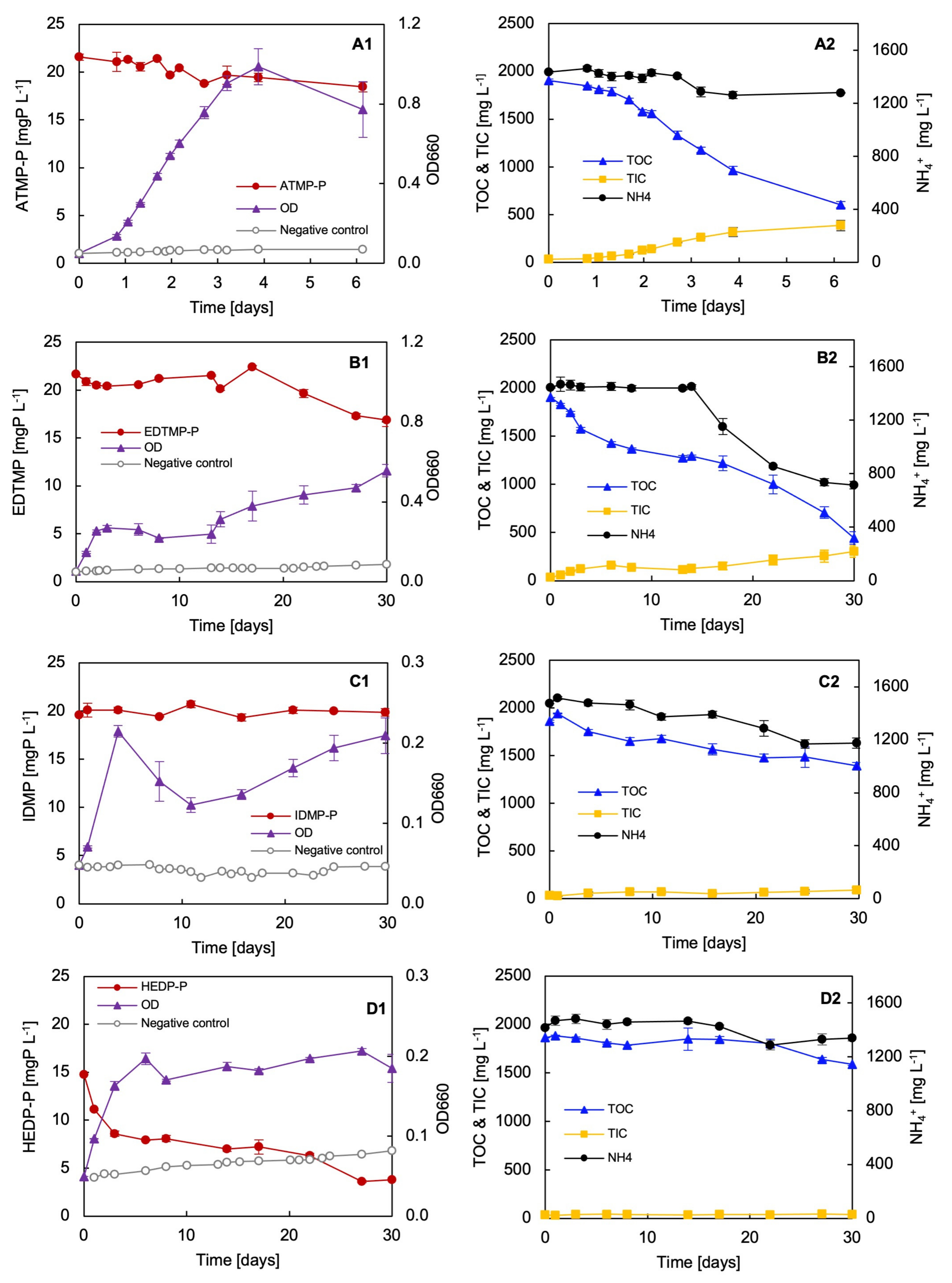
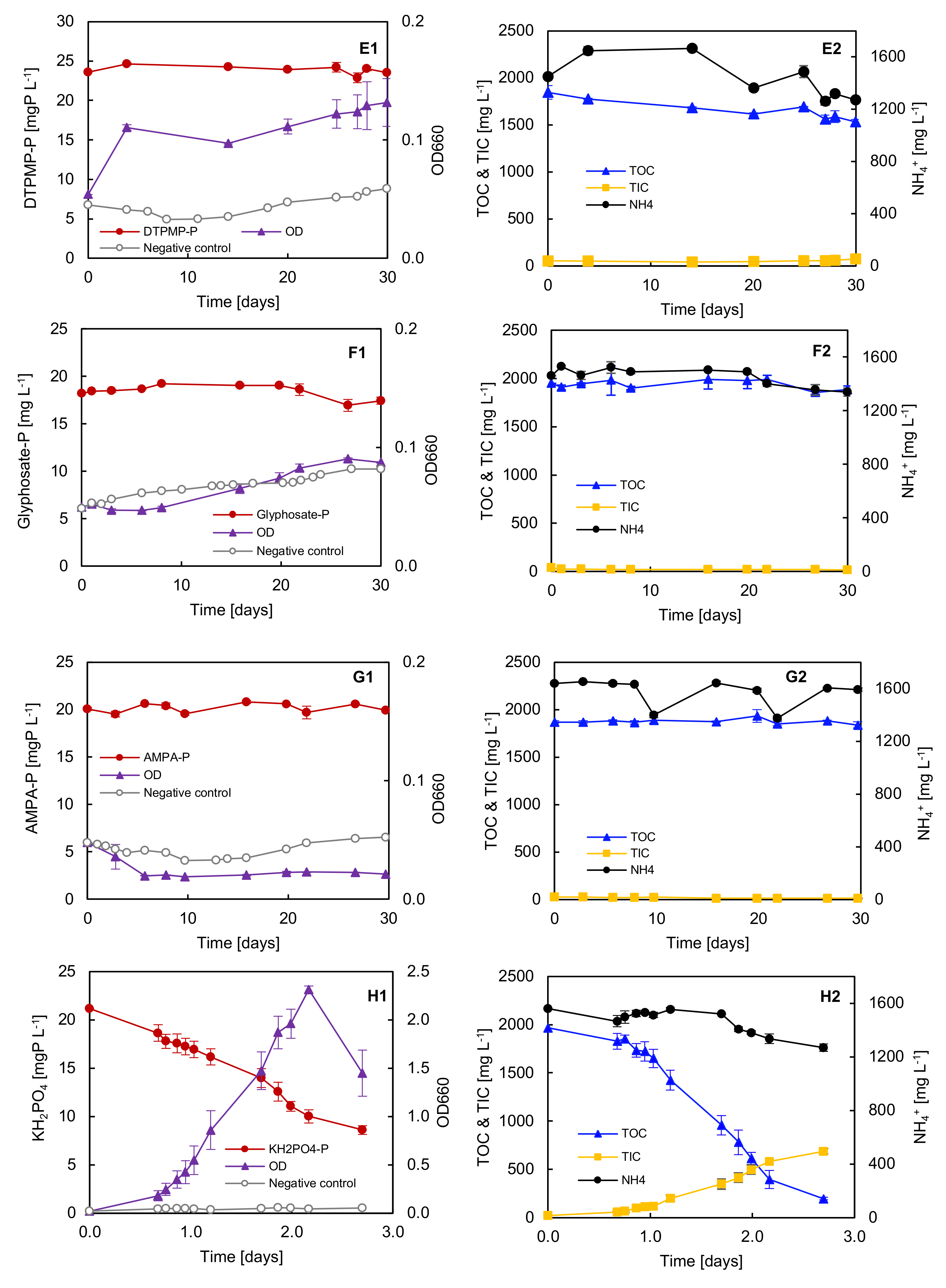
Biodegradation tests with seven different phosphonates as the sole P source were performed with the bacterial strain Delftia sp. UMB14. The strain grew differently on the provided phosphonates. In particular, no significant growth was determinable when GS and/or AMPA were the sole P source (Figure 2). The strain exhibited the most optimal growth on the phosphonates ATMP and EDTMP. On IDMP, HEDP, and DTPMP, the strain demonstrated rapid growth, particularly during the initial two days. Subsequently, the rate of growth either slowed down or kept stable, indicating no further bacterial growth. The comprehensive mass balance of the consumption of organic carbon (Corg), nitrogen (N), and P provided a more detailed insight into the degradation test of the different phosphonates (Table 1A,B).
As above mentioned, best growth was observed for the biodegradation test of ATMP (Figure 2(A1,A2)). Also, the growth rate for ATMP was found to be the highest of all the phosphonates tested. Consequently, the shortest doubling time was determined to be 12.8 h for ATMP. The strain exhibited consumption rates of over 1300 mgCorg L−1, 120 mgN L−1, and nearly 4 mgP L−1 during the test period. In total, over 75% of the organic carbon was consumed.
The most intriguing growth occurred when EDTMP was the sole P source (Figure 2(B1,B2)). The strain exhibited growth during the initial five days, followed by a 10-day period of stagnation, which was similar to the growth pattern observed in both IDMP and GS. Following an incubation period of approximately 15 days, the strain continued to grow exponentially again. The strain achieved the second-highest final OD. The results of the mass balance also confirmed that the strain obviously utilised high amounts of Corg and N, i.e., 1450 mgC L−1 and 570 mgN L−1, respectively. Consequently, a total of 77% of the Corg was consumed through the biodegradation of EDTMP by Delftia sp. UMB14.
In contrast, the strain exhibited minimal growth on IDMP as a P source (Figure 2(C1,C2)). The mass balance also revealed that the strain consumed carbon, nitrogen, and phosphorus, which resulted in bacterial growth, but to a lower extent as compared with ATMP and/or EDTMP. The growth on HEDP as a P source occurred comparably with IDMP (Figure 2(D1,D2)). However, the P analysis implied rapid P reduction within the initial three days and then kept constant. In fact, the rapid P reduction was due partly to HEDP precipitation, which was visible at the bottom of the flasks. Consequently, the results of the mass balance were corrected, taking the loss of HEDP in the medium into account. An average P consumption was calculated of 2.4 mgP L−1. However, this result may still be incorrect. This may be further supported by the low carbon footprint compared to ATMP and EDTMP.
For DTPMP, the bacterial growth was consistently low throughout the testing period (Figure 2(E1,E2)). A slight increase in the OD was observed at the beginning and towards the end of the experiment. The data from the mass balance indicated that the strain consumed a total of approximately 250 mgCorg L−1 and 120 mgN L−1. Nevertheless, this represents only a low fraction of the total amount consumed by the strain in comparison to ATMP and/or EDTMP during the same period. On GS, the strain exhibited growth initially, but seemed subsequently inhibited (Figure 2(F1,F2)). After ten days of incubation, a slight growth on a very low level was observable. The results of the mass balance indicated that the strain exhibited low consumption of Corg, N, and P. This minimal consumption indirectly corroborates the low final OD.
The results obtained for the AMPA degradation test demonstrated that indeed no phosphorus of AMPA (AMPA-P) was consumed over the measured period of 30 days (Figure 2(G1,G2)). Similar results were observed for Corg and N. The determined data from the mass balance thus confirmed the OD measurement. The strain was unable to grow on AMPA as a P source.
Finally, the strain was grown on KH2PO4 as a positive control (Figure 2(H1,H2)). The results clearly showed that the strain was able to grow exponentially within a few days. The bacteria consumed almost all of the Corg, while there was still sufficient N and P available. Thus, the limited amount of available Corg was ultimately the limiting growth factor.
The ratio (P/S) was calculated between td of the positive control (P) and the investigated substrate (S), i.e., the individual aminophosphonates investigated. The P/S gavve a better comparison of our data and final interpretation of the determined growth rates. The results of the P/S ratio clearly indicated that the lowest growth and biodegradation in terms of C–P cleavage occurred in HEDP, DTPMP, glyphosate, and AMPA.

3.7. Phosphorus Requirements for Growth with Different P Sole Sources

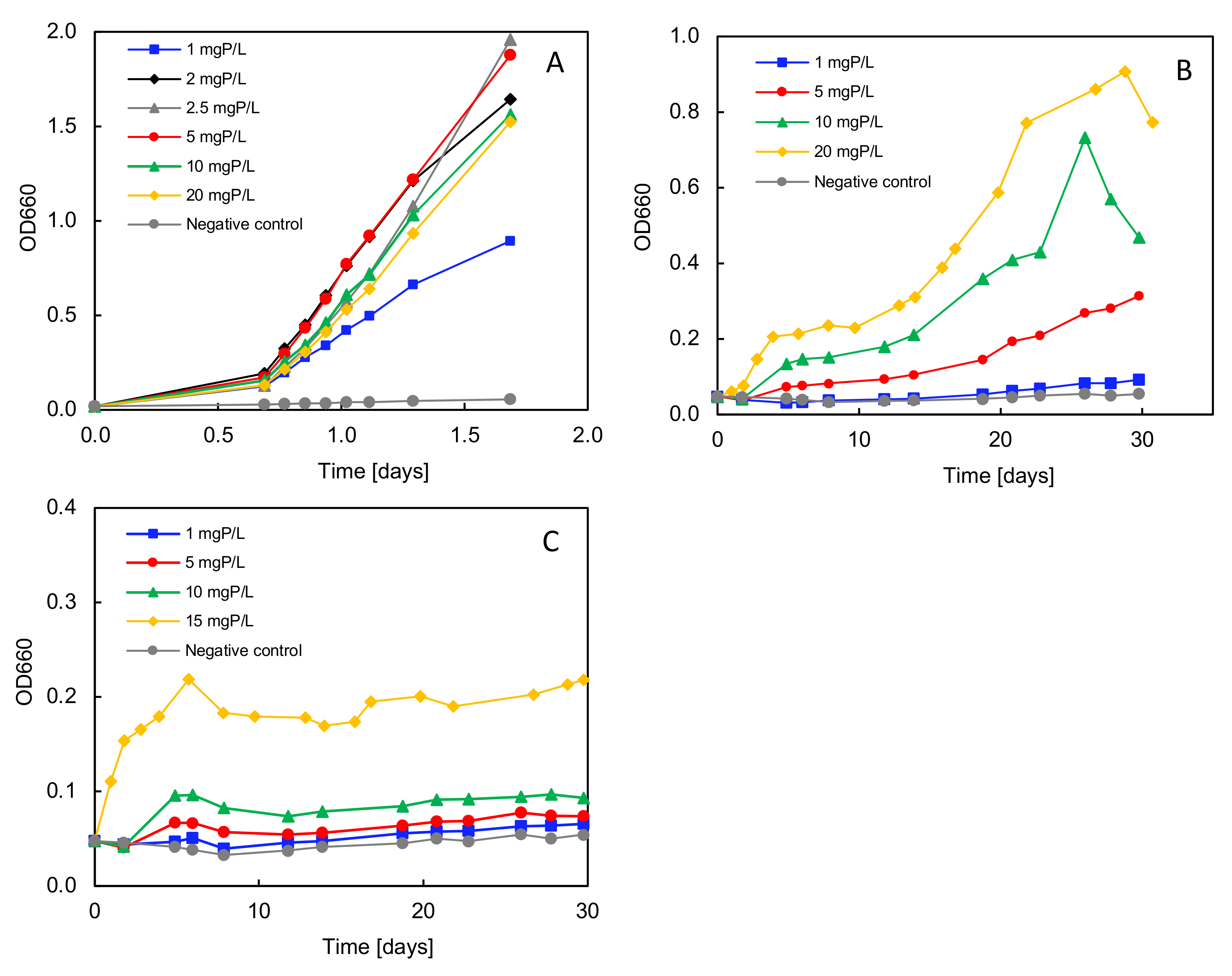
The outcomes obtained from the previous biodegradation tests with various phosphonates clearly indicated that strain Delftia sp. UMB14 exhibited its best growth on ATMP as the sole P source. Nevertheless, the full analysis independently validated that only minimal amounts of P was consumed for bacterial growth. Consequently, the minimum P concentration required for unrestricted bacterial growth was determined for the aminophosphonate AMTP and compared with the organophosphonate HEDP and with the positive control KH2PO4 (Figure 3). The results demonstrate that the minimum phosphorus concentration necessary is dependent on the P-substrate provided. In the reference test, in which KH2PO4 was the only sole P source, only the lowest concentration of 1 mgP L−1 resulted in an early stagnation of bacterial growth due to substrate consumption (Figure 3A). The growth rates (µmax) and doubling times (td) exhibited no significant influence with regard to the tested P concentrations for the positive control, as these were determined for the exponential growth phase (Table 2).
In contrast, a significant influence of the substrate concentration on the bacterial growth was determined for both ATMP and HEDP (Figure 3B,C). In both cases, the strain exhibited the fastest and most optimal growth at the highest concentration. Also, the final OD value was observed to be the highest for the highest substrate concentration provided. The lowest substrate concentration for both ATMP and HEDP resulted in an average doubling time of 243.5 h and 222.6 h, respectively. At the highest substrate concentration, the respective doubling times for ATMP and HEDP were determined to be 24.7 h and 35.1 h (Table 2). For both substrates, there appears to be a linear relationship between the substrate concentration and the resulting bacterial growth rate. For HEDP, the growth rate could not be determined above 15 mg L−1, as the phosphonate tends to precipitate in the test medium above this critical concentration.

3.8. Adaptation Test with ATMP and EDTMP

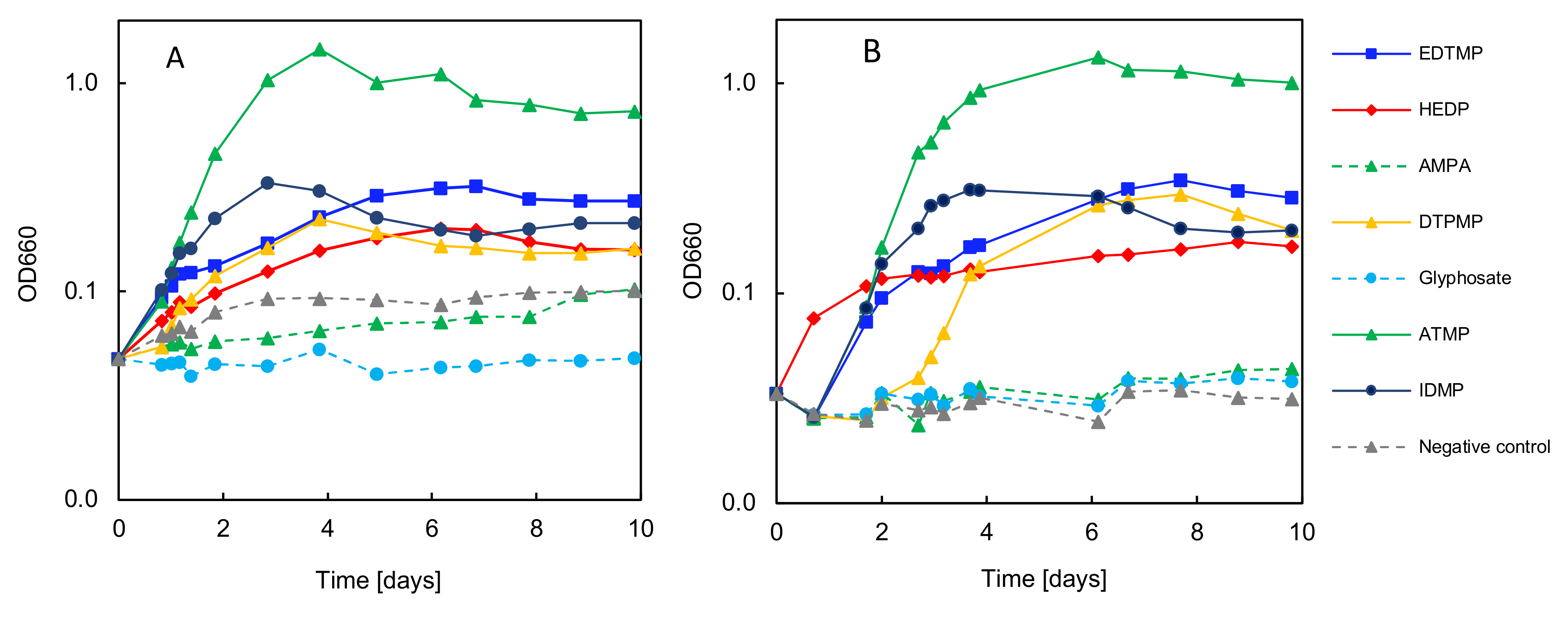
A third series of experiments was carried out to determine to what extent preconditioned bacteria that were previously grown on ATMP or EDTMP were able to grow on the other phosphonates used for biodegradation described in Section 3.6. Emphasis was placed on GS and AMPA, as these always resulted negatively in the previous degradation tests (Figure 4).
The results clearly show that bacterial growth on different phosphonates is possible with both preconditioning substrates, i.e., ATMP or EDTMP. In particular, the strongest bacterial growth was observed on ATMP (Figure 4A,B). However, the preconditioned bacteria also grew well on EDTMP and IDMP. Overall, the same trends were observed for the biodegradation of the phosphonates, regardless of the respective preconditioning. The EDTMP-preconditioned bacteria only seemed to show a lag phase of two days, which was not observed for the ATMP-preconditioned bacteria.
Most interestingly, no significant growth on GS and/or AMPA was detected in either test. An inhibited enzyme induction for their potential degradation is therefore ruled out, as the enzymes for phosphonate degradation were already present in the adapted bacteria due to preconditioning.

4. Discussion

4.1. Biodegradation of Phosphonates by Delftia sp. UMB14

In this study, we surveyed two different washing machines that used conventional phosphonate-based detergents. They were, therefore, suitable sources for the isolation of phosphonate-degrading bacteria. Overall, various bacteria were found in the washing machines, including in the forming biofilms. From both machines, in total, twenty-seven pure strains were isolated after specific enrichments to different phosphorous sources, such as HEDP and EDTMP. It is important to note that the capacity for phosphonate biodegradation has not been confirmed for all 27 isolates, and further investigation is required. The isolated organisms represented seven families. However, no isolates were obtained from the DTPMP enrichments. A phylogenetic analysis of isolate UMB 14 revealed 100% affiliation on an 16S rDNA gene basis to Delftia tsuruhatensis and Delftia lacustris, by NCBI Genebank searches. The latter is known as hazardous bacteria with the potential to cause ocular infection [45,46]. We cannot exclude that our isolate is also potentially capable of causing such infections. However, as we have stated above, our strain identification is currently based on the combined information of the results of the physiological and biochemical test and 16S rRNA gene sequencing. A more detailed genomic analysis is urgently required to ultimately confirm whether our isolate Delftia sp. UMB14 belongs to Delftia lacustris and has a high infection potential.
Delftia tsuruhatensis and Delftia lacustris are closely related and undistinguishable by 16S rRNA gene sequencing. For Delftia tsuruhatensis, antibiotic susceptibility has been observed for chloramphenicol [47], but resistance has been observed to gentamycin, ampicillin [48], and sulphonamide [47]. Also, Delfita lacustis was revealed to be resistant to aminoglycosides (gentamycin) [46,49]. However, Jørgensen et al. [50] were able to differentiate Delfita lacustris from its closest relative Delftia tsuruhatensis by physiological and biochemical characteristics, such as D-malic acid and D-mannitol utilisation. In accordance with Jørgensen et al. [48], strain Delftia sp. UMB 14 could therefore be assigned to Delftia lacustris, due to its utilisation of D-mannitol or D-malic acid. However, the utilisation pattern of D-malic acid and D-mannitol alike does not allow the adequate differentiation of Deltfia lacustris from its closest relative Delftia tsuruhatensis. Therefore, further genomic approaches are required to definitively identify the isolate (in progress).
In 1999, the genus Delftia was classified and proposed by Wen et al. [51], based on a re-evaluation of its phylogenetic and phenotypic characteristics. The two species, Delftia tsuruhatensis and Delftia lacustris, have only recently been isolated and identified, by Shigematsu et al. [52] for Delftia tsuruhatensis in 2003 and by Jørgensen et al. [50] for Delftia lacustris in 2009. Thus, only little is known on the metabolic and catabolic potential, for example, towards persistent substances. The opportunist Delftia lacustris has been reported to have a high potential to biodegrade organic compounds such as erythromycin [53] and pentachlorophenol [54], or to remove graffiti [55]. In addition, the potential to combat heavy metals stress through the application of metallophores [56] and the reduction of selenate and selenite has been described [57].
It is, therefore, perhaps not surprising that this opportunistic strain has been found to be able to degrade industrially relevant aminophosphonates that are commonly used in water treatments or commercial laundry detergents. The results of the present study showed that the new isolate, Delftia sp. UMB14, preferentially grew on ATMP and EDTMP as the sole P source. However, neither GS nor AMPA were degraded by strain Delftia sp. UMB14, as shown clearly by the degradation test results. Also, the result of the adaptation experiments doubtlessly confirmed that the strain could not growth on GS or AMPA as the sole P source. This observation was also supported by the results of the mass balance for Corg, N, and P. The consumption of Corg, for example, was a good indicator of the active metabolism of the bacteria in the batch test. The positive control showed a 97% consumption of Corg, of 2 gCorg L−1, and 50% for P of the 20 mgP L−1 provided. With the supplied C, N, and P as substrates, the bacteria were able to achieve an OD660 value of more than 2.0 in the degradation test of the positive control. The substrate consumption determined that the strain grew unrestrictedly as long as sufficient Corg was available. In fact, in the case of the positive control, it was evident that Corg represented the limiting factor for further growth.
In comparison to the positive control, it was observed that the quantity of Corg consumed by the bacteria for growth on the two aminophosphonates ATMP and EDTMP was less. It is important to note, however, that significantly lower final OD values were obtained for ATMP and EDTMP, namely 1.0 and 0.5, respectively. Thus, for bacterial growth and further metabolism, more Corg was actually consumed when compared to the positive control. Regardless of the increased C consumption, the P turnover was reduced threefold for ATMP and fourfold for EDTMP. This indicated that the degradation of ATMP and EDTMP represented a significantly higher metabolic challenge for the strain Delftia sp. UMB14 compared to KH2PO4, an easily accessible P substrate.
The remaining results for IDMP, HEDP, and DTPMP similarly indicated that the strain was capable of accessing phosphonates as a phosphorus source. However, the results of the mass balance also demonstrated that a significantly lower quantity of Corg was consumed in comparison to ATMP and EDTMP. This evidence suggested that these structures are either more challenging for the strain to degrade or that metabolites are formed that cannot be further metabolised. As previously stated, no growth was observed for GS or AMPA. The results of the mass balance analysis unequivocally corroborated this conclusion. The fact that the strain obviously could not grow on GS or AMPA was also confirmed by the results of the genetic analysis, especially by the results of the PCR. More importantly, the determined growth rates in combination with the results of the mass balances were further evaluated and suggested the following trend from the best towards the lowest or no biodegradability of the tested phosphonates as follows: ATMP > EDTMP > IDMP > HEDP > DTPMP > GS/AMPA. This ranking contradicts the previous results of Riedel et al. [15,16] where for the pure strain Ochrobactrum sp. BTU1, as well as for activated sludge and TW inocula, a correlation depending on the molecule size from small to large (i.e., very good to poorly biodegradable) was determined. In these cases, however, the classical C–P lyase pathway was found to be responsible for the biodegradation of the phosphonates investigated, whose core gene pnhJ was not detected in strain Delftia sp. UMB 14. Further analyses, for example LC/MS, may help to identify potential degradation products and to better understand the degradation mechanism of the new isolate (in progress).

4.2. Potential Enzymatic Pathway Responsible for the Biodegrading of Phosphonates

The results showed that strain Delftia sp. UMB 14 possesses the phnX gene product phosphonoacetaldehyde hydrolase, which is consistent with the data from other strains of Delftia lacustris, e.g., Delftia lacustris strain FDAARGOS_890 [58]. Interestingly, the annotation data of these strains also suggested that Delftia lacustris possesses, on the one hand, genes associated with the phosphonate ATP-binding cassette transporter (phnCDE) of the C–P lyase complex and, on the other hand, does not possess the 2-AEP pyruvate aminotransferase encoded by phnW, as often described to be closely associated with phnX [32,59,60,61].
At first glance, this result seemed somewhat contradictory. However, it can also indirectly confirm the lack of degradation and growth on GS or AMPA. Both substrates are commonly known to be degradable via the C–P lyase pathway. The observation of the phnCDE genes of C–P lyase does not lead to confirmation of the presence of the complete C–P lyase operon. As Villarreal-Chiu et al. [60] stated, the seven gene products (PhnGHIJKLMN) constitute the core component of the C–P lyase reaction pathway. The transporter of C–P lyase, PhnCDE, with high affinity towards phosphonates, may therefore be also present for different degradation pathways, as recently demonstrated by Lee et al. [61].
Villarreal-Chiu et al. [60] evaluated in silico the distribution of the genes of the phosphonate metabolism by bacteria in the marine environment. In their analyses, they showed that nearly 50% of the sequenced genomes possessed one or more of the known phosphonate metabolic pathways. It is remarkable that four times as many strains were analysed that have genes associated to catabolic phosphonate metabolism. The C–P lyase pathway and the phosphonoacetaldehyde hydrolase pathway were both found to be most abundant among bacteria. This finding suggests that both catabolic pathways may have a very long evolutionary history. Villarreal-Chui et al. [60] showed that the distribution of the gene-encoding PhnD (periplasmic binding protein) was very broad across bacteria and even extends the distribution of the core C–P lyase genes. The authors concluded that the distribution of phnCDE and a number of homologues of phnD, especially in marine environments lacking phosphonate catabolism, suggests an extensive evolutionary rearrangement, as described by Martinez et al. [27], for the PhnY*/PhnZ pathway.
As mentioned above, the abundance of the PhnWX pathway has been suggested to be currently one of the most abundant pathways in the marine environment. However, this suggestion was based on the in-silico analyses of 1384 bacterial gene sequences and may require further confirmation. The importance of phosphonoacetaldehyde hydrolase is undoubtedly directly related to the natural product 2-AEP. As the PhnX operon is encoded as part of a genomic island, horizontal gene transfer is highly suggested. Villarreal-Chui et al. [60] also found an extensive rearrangement of the phnCDE operon components and highlighted that they are almost completely absent in pathways with a shorter evolutionary history, such as the genes of phnA-encoding phosphonacetate hydrolase. Thus, it may be concluded, with regard to the Delftia lacustris, that the presence of the genes of phnCDE and phnX are justified via horizontal gene transfer, which may also be applicable to the strain Delftia sp. UMB14. The absence of phnW for Delftia sp. UMB14 remains to be verified by more comprehensive genomic analyses. Currently, the draft genome analysis is in progress.
It also has to be verified how the phnX pathway is accessible for phosphonates different from 2-AEP, especially for industrially relevant ones, as investigated in the present study. As already stated, PhnX has a very specific substrate spectrum and requires the formation of phosphonoacetaldehyde (PnAA), which is thought to be exclusively released by PhnW and/or the three putative FAD-dependent oxidoreductases PbfB, PbfC, and PbfD [32]. The current genomic data of strain Delftia sp. UMB14 strongly indicate the absence of phnW, which is not necessarily contradictory. Zangelmi et al. showed that the absence of phnW is more often found in the taxonomically diverse organisms associated with phnZ and phnX, and in such cases, pbfD is suggested to replace phnW [34]. Thus, the absence of phnW and the presence of phnCDE in the genes of strains of Delftia lacustris may therefore be justified, but deserves more detailed genomic analyses.

4.3. Factors Influencing the Degradation Efficiency during Phosphonate Degradation

With regard to the biodegradation of selected industrial phosphonates by strain Delftia sp. UMB14, it is currently assumed that the PhnX pathway was responsible for their degradation. However, not all the phosphonates tested were degradable. The reason for this apparent specificity has not yet been identified and will be addressed by future research.
It can further be assumed that their chemical structure has an enormous influence on their biodegradation, as recently proposed by Riedel et al. [16,17]. A first indication that the chemical structure of selected phosphonates might affect their degradation efficiency in general was demonstrated for PhnD cloned from E. coli XL1-Blue by Rizk et al. [62]. They showed that the efficiency of different small aminophosphonates such as methylphosphonate (MP), 2-aminoethylphosphonate (2-AEP), AMPA, and GS to bind to the PhnD polypeptide varied over five orders of magnitude. Rizk et al. [62] determined the dissociation constants (Kd) for all four phosphonates, with GS having the highest value. Thus, GS had the poorest binding efficiency and the authors concluded that the compound may not be fully transported for further catabolism in the C–P lyase pathway. However, Rizk et al. [62] demonstrated this for the C–P lyase pathway, and their finding may not be fully transferable to the finding in the present study as it may be assumed that the biodegradation of ATMP and EDMPT might be even enzymatically more challenging as compared with GS.
Despite the enzymes responsible for mediating C–P cleavage, the relatively low consumption of available P due to cleavage by strain Delftia sp. UMB14 still raised questions and uncertainties. More specifically, the mass balance results suggested that only small amounts of P were consumed by the biodegradation of phosphonates. The highest consumption was found for ATMP, with an average of 4 mgP L−1. As mentioned above, the strain consumed up to 12.5 mgP L−1 when KH2PO4 was supplied. This discrepancy led to the general question of how much P is required for the unrestricted growth of the strain Delftia sp. UMB14. Interestingly, it was found that KH2PO4 and phosphonates appeared to require different threshold P concentrations. For KH2PO4, the lowest tested P concentration of 1 mgP L−1 resulted still in exponential growth but limited the biomass yield. In comparison, the phosphonates tested, ATMP and HEDP, showed a different relationship as they appeared to result in increased growth and biomass yield with increasing P concentration supplied. One explanation may be that both ATMP and HEDP are strong chelating agents with a high potential for interaction with alkaline earth metals. As recently shown for EDTMP, lower Ca2+ and Mg2+ exposure result in suppressed bacterial growth compared to KH2PO4 [15]. A similar phenomenon cannot be excluded for the biodegradation of ATMP and HEDP tested in this study. In addition to this finding, it has to be taken into account that the hydrolytic pathway proposed here to be responsible for the degradation of ATMP and EDTMP is highly Mg2+ dependent [34]. Therefore, the higher input concentration of phosphonates tested in this study may be justified. However, the reason for their higher required input concentration also remains to be verified and should be addressed in future research.

5. Conclusions

In this study, the inaugural description was made of the extensive diversity of bacteria (27 strains) that are capable of producing biofilms in washing machines. These biofilms have the potential to degrade detergents, including phosphonates, by colonising the surfaces of the washing machines. In addition, this is the first description of the potentially opportunistic Delftia sp. UMB14, isolated from a washing machine with demonstrated resistance to some antibiotics.
Sequencing and phylogenetic analyses showed that the isolate belonged to the genus Delftia, with 100% affiliation to Delftia tsuruhatensis and Delftia lacustris on the basis of 16S rDNA. Antimicrobial susceptibility testing showed that Delftia lacustris UMB14 was only susceptible to sulfonamide, tetracycline, and phenicol (chloramphenicol). The substrate utilisation pattern confirmed that strain Delftia sp. UMB14 utilised D-mannitol and D-malic acid, as recently reported to be characteristic of the Delftia lacustris. However, this utilisation pattern alone does not justify the ultimate identification of the strain. Therefore, further genomic approaches are required to definitively identify the isolate (in progress). The strain showed preferential biodegradation of the industrial aminophosphonates ATMP and EDTMP, but lacks GS and AMPA. Specific gene amplification confirmed the presence of phnX but not phnJ. Thus, the biodegradation of the studied phosphonates was not mediated by the C–P lyase pathway but is related to the phosphonoacetaldehyde hydrolase pathway. The annotation of published Delftia lacustris genes suggested that Delftia sp. UMB14 also possesses the phnCDE operon of the C–P lyase complex, but not the 2-AEP pyruvate aminotransferase encoded by phnW. However, an extensive evolutionary rearrangement by horizontal gene transfer has recently been reported and may be a reasonable explanation for the possible presence of the phnCDE and phnX genes of the isolated strain Delftia sp. UMB14. Similarly, the absence of phnW may initially appear contradictory, but has recently been frequently found in taxonomically diverse organisms in association with phnZ and phnX. In such cases, pbfD has been proposed to replace phnW, which remains to be verified for strain Delftia sp. UMB14.
Thus, it may be expected that the strain is also capable of degrading the industrially relevant aminophosphonates ATMP and EDTMP in the aquatic environment if the conditions for their biodegradation are similar to those in our biodegradation test.

Author Contributions

Conceptualization, R.R., K.M. and B.B; methodology, R.R., K.M. and B.B.; software, K.M. and B.B.; validation, R.R., K.M., A.K. and B.B.; formal analysis, R.R., K.M., A.K. and B.B.; investigation, R.R., K.M., A.K. and B.B.; data curation, R.R. and B.B.; writing—original draft preparation, R.R., K.M., B.B. and D.B.; writing—review and editing, R.R., K.M., B.B., D.B. and M.M.; visualization, R.R.; supervision, R.R. and B.B.; project administration, R.R. and B.B.; All authors have read and agreed to the published version of the manuscript.

Funding

This research received no external funding.

Data Availability Statement

The raw data supporting the conclusions of this article will be made available by the authors on request.

Acknowledgments

The authors are grateful to Kathrin Krahl and Kai Buder for their technical support. The skillful technical assistance of Stefanie Arndt, Tatjana Fritscher-Klöppel, and Konstantinos Xouveroudis is also highly appreciated. The authors thank “Zschimmer & Schwarz” for providing several phosphonate compounds.

Conflicts of Interest

The authors declare no conflicts of interest.

References

  1. Mazuryk, J.; Klepacka, K.; Kutner, W.; Sharma, P.S. Glyphosate: Impact on the microbiota-gut-brain axis and the immune-nervous system, and clinical cases of multiorgan toxicity. Ecotoxicol. Environ. Saf. 2024, 271, 115965. [Google Scholar] [CrossRef]
  2. López, A.; Ruiz, P.; Fuentes, E.; Yusà, V.; Dualde, P.; Miralles, P.; Coscollà, C. Simultaneous direct determination of Glyphosate and AMPA in the ambient air and inhalation risk assessment in a Mediterranean Region (Spain). Atmos. Environ. 2024, 317, 120204. [Google Scholar] [CrossRef]
  3. Costas-Ferreira, C.; Durán, R.; Faro, L.R.F. Toxic Effects of Glyphosate on the Nervous System: A Systematic Review. Int. J. Mol. Sci. 2022, 23, 4605. [Google Scholar] [CrossRef] [PubMed]
  4. Nowack, B. Environmental chemistry of phosphonates. Water Res. 2003, 37, 2533–2546. [Google Scholar] [CrossRef] [PubMed]
  5. Yawen, H.; Yu, Y.; Jiajun, M.; Junxiao, Y. Preparation of ferric phosphonate/phosphonate and their special action on flame retardancy of epoxy resin. J. Appl. Polym. Sci. 2017, 135, 46206–46216. [Google Scholar] [CrossRef]
  6. Khormali, A.; Sharifov, A.R.; Torba, D.I. Increasing efficiency of calcium sulphate prevention using a new mixture of phosphonate scale inhibitors during waterflooding. J. Petrol. Sci. Eng. 2018, 164, 245–258. [Google Scholar] [CrossRef]
  7. Kong, G.; Kashyap, R.; Hicks, R.J. Adverse effects of radionuclide therapy. In Radiotheranostics—A Primer for Medical Physicists IN: Physics, Chemistry, Biology and Clinical Application, 1st ed.; Borrás, C., Stabin, M.G., Eds.; CRC Press: Boca Raton, FL, USA, 2024; Volume 1. [Google Scholar]
  8. Lange, R.; Heine, R.; Knapp, R.; de Klerk, J.M.H.; Bloemendal, H.J.; Hendrikse, H. Pharmaceuticial and clinical development of phosphonate-based radiopharmaceuticals for the targeted treatment of bone metastases. Bone 2016, 91, 159–179. [Google Scholar] [CrossRef] [PubMed]
  9. Taheri, M.; Azizmohammadi, Z.; Ansari, M.; Dadkhah, P.; Dehghan, K.; Valizadeh, R.; Assadi, M. 153Sm-EDTMP and 177Lu-EDTMP are equally safe and effective in pain palliation from skeletal metastases. Nuklearmedizin 2018, 57, 174–180. [Google Scholar] [CrossRef] [PubMed]
  10. Armbruster, D.; Rott, E.; Minke, R.; Happel, O. Trace-level determination of phosphonates in liquid and solid phase of wastewater and environmental samples by IC-ESI MS/MS. Anal. Bioanal. Chem. 2020, 412, 4807–4825. [Google Scholar] [CrossRef]
  11. Rott, E.; Happel, O.; Armbruster, D.; Minke, R. Influence of wastewater discharge on the occurrence of PBTC, HEDP, and aminophosphonates in sediment, suspended matter, and the aqueous phase of rivers. Water 2020, 12, 803. [Google Scholar] [CrossRef]
  12. Rott, E.; Happel, O.; Armbruster, D.; Minke, R. Behavior of PBTC, HEDP, and aminophosphonates in the process of wastewater treatment. Water 2020, 12, 53. [Google Scholar] [CrossRef]
  13. Wang, S.; Zhang, B.; Shan, C.; Yan, X.; Chen, H.; Pan, B. Occurrence and transformation of phosphonates in textile dyeing wastewater along full-scale combined treatment process. Water Res. 2020, 184, 116173. [Google Scholar] [CrossRef] [PubMed]
  14. Schowanek, D.; Verstraete, W. Phosphonate utilization by bacterial cultures and enrichments from environmental samples. Appl. Environ. Microbiol. 1990, 56, 895–903. [Google Scholar] [CrossRef] [PubMed]
  15. Rott, E.; Steinmetz, H.; Metzger, J.W. Organophosphonates: A review on environmental relevance, biodegradability and removal in wastewater treatment plants. Sci. Total Environ. 2018, 615, 1176–1191. [Google Scholar] [CrossRef] [PubMed]
  16. Riedel, R.; Krahl, K.; Buder, K.; Böllmann, J.; Braun, B.; Martienssen, M. Novel standard biodegradation test for synthetic phosphonates. J. Microbiol. Method. 2023, 212, 106793. [Google Scholar] [CrossRef] [PubMed]
  17. Riedel, R.; Commichau, F.M.; Benndorf, D.; Hertel, R.; Holzer, K.; Hoelzle, L.E.; Mardoukhi, M.S.Y.; Noack, L.E.; Martienssen, M. Biodegradation of selected aminophosphonates by the novel bacterial isolate Ochrobactrum sp. BTU1. Microbiol. Res. 2024, 280, 127800. [Google Scholar] [CrossRef]
  18. Chen, C.M.; Ye, Q.Z.; Zhu, Z.M.; Wanner, B.L.; Walsh, C.T. Molecular biology of carbon-phosphorus bond cleavage. Cloning and sequencing of the phn (psiD) genes involved in alkylphosphonate uptake and C-P lyase activity in Escherichia coli K-12. J. Biol. Chem. 1990, 265, 4461–4471. [Google Scholar] [CrossRef] [PubMed]
  19. Hove-Jensen, B.; Zechel, D.; Jochimsen, B. Utilization of glyphosate as phosphate source: Biochemistry and genetics of bacterial carbon-phosphorus lyase. Microbiol. Mol. Biol. Rev. 2014, 78, 176–197. [Google Scholar] [CrossRef]
  20. Hertel, R.; Gibhardt, J.; Martienssen, M.; Kuhn, R.; Commichau, F.M. Molecular mechanisms underlying glyphosate resistance in bacteria. Environ. Microbiol. 2012, 23, 2891–2905. [Google Scholar] [CrossRef]
  21. Kamat, S.S.; Wiliams, H.J.; Rauschel, F.M. Intermediates in the transformation of phosphonates to phosphate by bacteria. Nature 2011, 480, 570–573. [Google Scholar] [CrossRef]
  22. Lockwood, S.; Greening, C.; Baltar, F.; Morales, S.E. Global and seasonal variation of marine phosphonate metabolism. ISME 2022, 16, 2198–2212. [Google Scholar] [CrossRef]
  23. Ternan, N.G.; Quinn, J.P. In vitro cleavage of the carbon-phosphorus bons of phosphonopyruvate by cell extracts of an environmental Burkholdria cepacian isolate. Biochem. Biophys. Res. Commun. 1998, 248, 378–381. [Google Scholar] [CrossRef] [PubMed]
  24. McMullan, G.; Harrington, F.; Quinn, J.P. Metabolism of phosphonoacetate as the sole carbon and phosphorus source by an environmental bacterial isolate. Appl. Environ. Microbiol. 1992, 58, 1364–1366. [Google Scholar] [CrossRef] [PubMed]
  25. LaNauze, J.M.; Coggins, J.R.; Dixon, H.B.F. Aldolase-like imine formation in mechanism of action of phosphonoacetaldehyde hydrolase. Biochem. J. 1977, 165, 409–411. [Google Scholar] [CrossRef] [PubMed]
  26. Dumora, C.; Lacoste, A.M.; Cassaigne, A. Phosphonoacetaldehyde hydrolase from Pseudomonas aeruginosa—Purification, properties and comparison with Bacillus cereus enzyme. Biochem. Biophys. Acta 1989, 997, 193–198. [Google Scholar] [CrossRef] [PubMed]
  27. Martinez, A.; Tyson, G.W.; DeLong, E.F. Widespread known and novel phosphonate utilization pathways in marine bacteria revealed by functional screening and metagenomic analyses. Environ. Microbiol. 2010, 12, 222–238. [Google Scholar] [CrossRef] [PubMed]
  28. McScorley, A.; Wyatt, P.B.; Martinez, A.; DeLong, E.F.; Hove-Jeansen, B.; Zechel, D.L. PhnY and PhnZ comprise a new oxidative pathway for enzymatic cleavage of a carbon-phosphorus bond. J. Am. Chem. Soc. 2012, 134, 8364–8367. [Google Scholar] [CrossRef] [PubMed]
  29. Epiktetov, D.O.; Sviridov, A.V.; Tarlachkov, S.V.; Shushkova, T.V.; Leontievsky, A.A. Phosphonatase operons of organophosphonates-degrading soil bacteria of the genus Achromobacter. Microbiology 2023, 92, 45–49. [Google Scholar] [CrossRef]
  30. Zangelmi, E.; Ruffolo, F.; Dinhof, T.; Secchi, A.; Pallitsch, K.; Peracchi, A. Deciphering the role of recurrent FAD-dependent enzyme in bacterial phosphonate catabolism. Iscience 2023, 26, 108108. [Google Scholar] [CrossRef]
  31. Quinn, J.P.; Kulakova, A.N.; Cooley, N.A.; McGrath, J.W. New ways to break an old bond: The bacterial carbon-phosphorus hydrolases and their role in biogeochemical phosphorus cycling. Environ. Microbiol. 2007, 9, 2392–2400. [Google Scholar] [CrossRef]
  32. Zangelmi, E.; Stankovic, T.; Malatesta, M.; Acquotti, D.; Pallitsch, K.; Peracchi, A. Discovery of a new, recent enzyme in bacterial phosphonate degradation: (R)-1-hydroxy-2-aminoethylphosphonate ammonia-lyase. Biochemistry 2021, 60, 1214–1225. [Google Scholar] [CrossRef]
  33. Kim, A.D.; Baker, S.; Dunaway-Mariano, D.; Metcalf, W.W.; Wanner, B.L.; Martin, B.M. The 2-aminoethylphosphonate-specific transaminase of the 2-aminoethylphosphonate degradation pathway. J. Bacteriol. 2002, 184, 4134–4140. [Google Scholar] [CrossRef] [PubMed]
  34. Zangelmi, E. Identification and Characterization of New Enzymes Involved in Bacterial Phosphonate Catabolism. Ph.D. Thesis, University of Parma, Parma, Italy, 2021. [Google Scholar]
  35. Epiktetov, D.O.; Sviridov, A.V.; Tarlachkov, S.V.; Shushkova, T.V.; Toropygin, I.Y.; Leontievsky, A.A. Glyphosate-induced phosphonatase operons in soil bacteria of the genus Achromobacter. Int. J. Mol. Sci. 2024, 25, 6409. [Google Scholar] [CrossRef] [PubMed]
  36. Pfennig, N.; Trüper, H.G. The Prokaryotes, A Handbook on the Biology of Bacteria, Vol. 5 Proteobacteria: Alpha and Beta Subclasses, 3rd ed.Starr, M.P., Stolp, H., Trüper, H.G., Balows, A., Schegel, H.G., Eds.; Springer: Berlin, Germany, 1981; Volume 5, pp. 279–289. [Google Scholar] [CrossRef]
  37. Marchesi, J.R.; Sato, T.; Weightman, A.J.; Martin, T.A.; Fry, J.C.; Hiom, S.J.; Wade, W.G. Design and evaluation of useful bacterium-specific PCR primers that amplify genes coding for bacterial 16S rRNA. Appl. Environ. Microbiol. 1998, 64, 795–799. [Google Scholar] [CrossRef]
  38. Engelen, B.; Meinken, K.; von Wintzingerode, F.; Heuer, H.; Malkomes, H.P.; Backhaus, H. Monitoring impact of a pesticide treatment on bacterial soil communities by metabolic and genetic fingerprinting in addition to conventional testing procedures. Appl. Environ. Microbiol. 1998, 64, 2814–2821. [Google Scholar] [CrossRef] [PubMed]
  39. Morales, M.E.; Allegrini, M.; Basualdo, J.; Villamil, M.B.; Zabaloy, M.C. Primer design to assess bacterial degradation of glyphosate and other phosphonates. J. Microbiol. Methods. 2020, 169, 105814. [Google Scholar] [CrossRef] [PubMed]
  40. Hsieh, Y.J.; Wanner, B.L. Global regulation by the seven-component Pi signaling system. Curr. Opin. Microbiol. 2010, 13, 198–203. [Google Scholar] [CrossRef] [PubMed]
  41. Bergkemper, F.; Kublik, S.; Lang, F.; Krüger, J.; Vestergaard, G.; Schloter, M.; Schulz, S. Novel oligonucleotide primers reveal a high diversity of microbes which drive phosphorous turnover in soil. J. Microbiol. Methods 2016, 125, 91–97. [Google Scholar] [CrossRef]
  42. DIN 38406-5:1983-10; German Standard Methods for the Examination of Water, Waste Water and Sludge; Cations (Group E); Determination of Ammonia-Nitrogen (E 5). DIN Media: Berlin, Germany, 1983. [CrossRef]
  43. DIN EN ISO 6878:2004-09; Water Quality-Determination of Phosphorus—Ammonium Molybdate Spectrometric Method (ISO 6878:2004); German Version EN ISO 6878:2004. DIN Media: Berlin, Germany, 2004. [CrossRef]
  44. DIN EN 12260:2003-12; Water Quality—Determination of Nitrogen—Determination of Bound Nitrogen (TN), Following Oxidation to Nitrogen Oxides. German Institute for Standardisation (Deutsches Institut für Normung): Berlin, Germany, 2003.
  45. Shin, S.Y.; Choi, J.Y.; Ko, K.S. Four cases of possible human infections with Delftia lacustris. Infection 2012, 40, 709–712. [Google Scholar] [CrossRef]
  46. Sohn, K.M.; Baek, J.Y.; Cheon, S.; Kim, Y.S.; Koo, S.H. Ocular infection associated with Delftia lacustris: First report. Braz. J. Infect. Dis. 2015, 19, 449–450. [Google Scholar] [CrossRef]
  47. Cheng, C.; Zhou, W.; Dong, X.; Zhang, P.; Zhou, K.; Zhou, D.; Qian, C.; Lin, X.; Li, P.; Li, K.; et al. Genomic analysis of Delftia tsuruhatensis strain TR1180 isolated from a patient from China with in4-like integron-associated antimicrobial resistance. Front. Cell Infect. Microbiol. 2021, 11, 663933. [Google Scholar] [CrossRef] [PubMed]
  48. Preiswerk, B.; Ullrich, S.; Speich, R.; Bloemberg, G.V.; Hombach, M. Human Infection with Delftia tsuruhatensis isolated from a central venous catheter. J. Med. Microbiol. 2011, 60, 246–248. [Google Scholar] [CrossRef] [PubMed]
  49. Zhang, L.; Zhang, X.; Bai, H.; Li, T.; Zhang, Z.; Zong, X.; Shang, X.; Lui, Z.; Fan, L. Characterization and genome analysis of the Delftia lacustris strain LzhVag01 isolated from vaginal discharge. Curr. Microbiol. 2024, 81, 232. [Google Scholar] [CrossRef] [PubMed]
  50. Jørgensen, N.O.; Brandt, K.K.; Nybroe, O.; Hansen, M. Delftia lacustris sp. nov., a peptidoglycan-degrading bacterium from fresh water, and emended description of Delftia tsuruhatensis as a peptidoglycan-degrading bacterium. Int. J. Syst. Evol. Microbiol. 2009, 59, 2195–2199. [Google Scholar] [CrossRef] [PubMed]
  51. Wen, A.; Fegan, M.; Hayward, C.; Chakraborty, S.; Sly, L.I. Phylogenetic Relationships Among Members of the Comamonadaceae, and Description of Delftia Acidovorans (Den Dooren De Jong 1926 and Tamaoka Et Al. 1987) Gen. Nov., Comb. Nov. Int. J. Syst. Bacteriol. 1999, 49, 567–576. [Google Scholar] [CrossRef] [PubMed]
  52. Shigematsu, T.; Yumihara, K.; Ueda, Y.; Numaguchi, M.; Morimura, S.; Kida, K. Delftia tsuruhatensis sp. nov., a terephthalate-assimilating bacterium isolated from activated sludge. Inter. J. Syst. Evol. Microbiol. 2003, 53, 1479–1483. [Google Scholar] [CrossRef] [PubMed]
  53. Ren, J.; Wang, Z.; Deng, L.; Niu, D.; Fan, B.; Huhe, T.; Li, Z.; Zhang, J.; Li, C. Biodegradation of erythromycin by Delftia lacustris RJJ-61 and characterization of its erythromycin esterase. J. Basic. Microbiol. 2021, 61, 55–62. [Google Scholar] [CrossRef] [PubMed]
  54. Cai, X.; Li, J.; Guan, F.; Luo, F.; Yu, Z.; Yuan, Y. Complete pentachlorophenol biodegradation in a dual-working electrode bioelectrochemical system: Performance and functional microorganism identification. Water Res. 2023, 230, 119529. [Google Scholar] [CrossRef] [PubMed]
  55. Cattò, C.; Sanmartín, P.; Gulotta, D.; Troiano, F.; Cappitelli, F. Bioremoval of graffiti using novel commercial strains of bacteria. Sci. Total Environ. 2021, 756, 144075. [Google Scholar] [CrossRef]
  56. Ahmed, M.M.A.; Boudreau, P.D. LCMS-Metabolomic profiling and genome mining of Delftia lacustris DSM 21246 revealing lipophilic delftibactin metallophores. J. Nat. Prod. 2024, 87, 1384–1393. [Google Scholar] [CrossRef]
  57. Wadgaonkar, S.L.; Nancharaiah, Y.V.; Jacob, C.; Esposito, G.; Lens, P.N.L. Microbial transformation of Se oxyanions in cultures of Delftia lacustris grown under aerobic conditions. J. Microbiol. 2019, 57, 362–371. [Google Scholar] [CrossRef] [PubMed]
  58. US Food and Drug Administration. Delftia Lacustris Genome Assembly ASM1602781v1 [WWW Document]. NCBI. 2020. Available online: https://www.ncbi.nlm.nih.gov/data-hub/assembly/GCF_016027815.1/ (accessed on 7 July 2024).
  59. Jiang, W.; Metcalf, W.W.; Lee, K.S.; Wanner, B.L. Molecular cloning, mapping, and regulation of Pho regulon genes for phosphonate breakdown by the phosphonatase pathway of Salmonella typhimurium LT2. J. Bacteriol. 1995, 177, 6411–6421. [Google Scholar] [CrossRef] [PubMed]
  60. Villarreal-Chiu, J.F. The genes and enzymes of phosphonate metabolism by bacteria, and their distribution in the marine environment. Front. Microbiol. 2012, 3, 19. [Google Scholar] [CrossRef] [PubMed]
  61. Lee, K.S.; Metcalf, W.W.; Wanner, B.L. Evidence for two phosphonate degradative pathways in Enterobacter aerogenes. J. Bacteriol. 1992, 174, 2501–2510. [Google Scholar] [CrossRef]
  62. Rizk, S.S.; Cuneo, M.J.; Hellinga, H.W. Identification of cognate ligands for the Escherichia coli phnD protein product and engineering of a reagentless fluorescent biosensor for phosphonates. Protein Sci. 2006, 15, 1745–1751. [Google Scholar] [CrossRef]
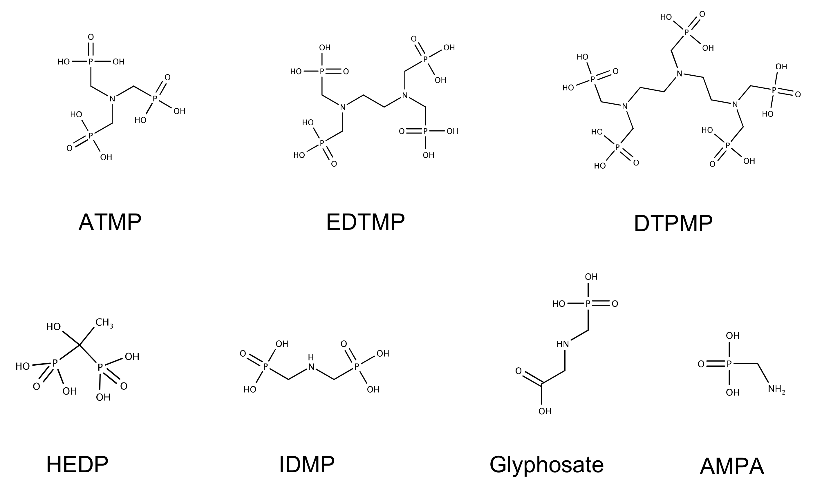
Figure 1. Chemical structures of the phosphonates used in the degradation tests.
Microorganisms 12 01664 g001
Figure 2. Biodegradation test of selected phosphonates with strain Delftia sp. UMB14. The phosphonates were dosed at 20 mgP L−1 to ensure comparability, except of HEDP, to prevent the precipitation of the latter. (A1,A2) ATMP. (B1,B2) EDTMP. (C1,C2) IDMP. (D1,D2) HEDP. (E1,E2) DTPMP. (F1,F2) GS. (G1,G2) AMPA. (H1,H2) Positive control KH2PO4. The optical density was measured at 660 nm.
Microorganisms 12 01664 g002aMicroorganisms 12 01664 g002b
Figure 3. Growth test with strain Delftia sp. UMB14 and different sole phosphorus sources. (A) KH2PO4 as sole P source. (B) ATMP as sole P source. (C) HEDP as sole P source. Test was running in triplicates for each test condition. The optical density was measured at 660 nm.
Microorganisms 12 01664 g003
Figure 4. Adaptation test with inocula with preconditioned bacteria either grown on ATMP or EDTMP. (A) Growth test on different phosphonates with pre-inoculated bacteria on ATMP as P source. (B) Growth test on different phosphonates with pre-inoculated bacteria on EDTMP as P source. The optical density was measured at 660 nm.
Microorganisms 12 01664 g004
Table 1. (A) Growth rates and substrate consumption by strain Delftia sp. UMB14 for ATMP, EDTMP, and IDMP. (B) Growth rates and substrate consumption by strain Delftia sp. UMB14 for HEDP, DTPMP, GS, and AMPA.
(A)
ParameterUnitKH2PO4ATMPEDTMPIDMP
Growthµ [days−1]3.22 ± 0.21.30 ± 0.10.80 ± 0.00.44 ± 0.0
td [h]5.2 ± 0.412.8 ± 0.720.8 ± 0.338.1 ± 0.6
P/S [-]-0.41 ± 0.00.25 ± 0.00.14 ± 0.0
ConsumptionC [mgCorg L−1]1770.1 ± 13.81301.5 ± 47.81459.9 ± 63.4469.1 ± 49.4
N [mgN L−1]227.0 ± 21.7122.6 ± 9.6567.8 ± 22.0232.3 ± 31.3
P [mgP L−1]12.5 ± 2.13.9 ± 0.22.9 ± 0.61.7 ± 0.2
EfficiencyC [%]96.6 ± 3.872.7 ± 2.876.8 ± 3.426.8 ± 2.9
N [%]18.8 ± 2.111.0 ± 0.850.2 ± 0.218.5 ± 2.6
P [%]56.5 ± 9.418.2 ± 0.813.6 ± 2.87.8 ± 0.9
(B)
ParameterUnitHEDPDTPMPGSAMPA
Growthµ [days−1]0.50 ± 0.00.18 ± 0.00.06 ± 0.0-
td [h]33.4 ± 1.290.6 ± 2.6301.5 ± 35.5-
P/S [-]0.16 ± 0.00.06 ± 0.00.02 ± 0.0-
ConsumptionC [mgCorg L−1]276.5 ± 8.9310.3 ± 76.3130.9 ± 18.932.3 ± 26.3
N [mgN L−1]58.8 ± 17.0156.1 ± 31.095.1 ± 22.636.0 ± 9.0
P [mgP L−1]2.4 ± 0.50.7 ± 0.10.1 ± 0.30.2 ± 0.1
EfficiencyC [%]15.1 ± 0.517.8 ± 4.46.8 ± 1.01.7 ± 1.4
N [%]5.8 ± 1.013.8 ± 2.57.2 ± 2.02.1 ± 1.0
P [%]16.1 ± 3.32.9 ± 0.35.0 ± 2.01.2 ± 0.7
Table 2. Growth rates and doubling times of Delftia sp. UMB14 on different P sources.
P SourceConc.
[mg L−1]
µmax
[days−1]
td
[h]
KH2PO414.82 ± 0.43.5 ± 0.3
25.15 ± 0.73.3 ± 0.4
2.55.60 ± 1.03.0 ± 0.5
55.50 ± 1.03.1 ± 0.5
104.75 ± 0.13.5 ± 0.1
205.29 ± 0.63.2 ± 0.3
ATMP10.07 ± 0.01243.5 ± 31.5
50.22 ± 0.0175.9 ± 4.1
100.38 ± 0.0044.1 ± 0.4
200.68 ± 0.0524.7 ± 2.0
HEDP10.08 ± 0.01222.6 ± 16.4
50.16 ± 0.01101.6 ± 1.9
100.27 ± 0.0062.7 ± 0.8
150.47 ± 0.1035.1 ± 1.3
Disclaimer/Publisher’s Note: The statements, opinions and data contained in all publications are solely those of the individual author(s) and contributor(s) and not of MDPI and/or the editor(s). MDPI and/or the editor(s) disclaim responsibility for any injury to people or property resulting from any ideas, methods, instructions or products referred to in the content.