Article

The Isolation and Characterization of Novel Caulobacter and Non-Caulobacter Lysogenic Bacteria from Soil and the Discovery of Broad-Host-Range Phages Infecting Multiple Genera

Department of Biological Sciences, University of South Carolina, Columbia, SC 29208, USA
*
Author to whom correspondence should be addressed.
Microorganisms 2024, 12(9), 1894; https://doi.org/10.3390/microorganisms12091894 (registering DOI)
Submission received: 19 August 2024 / Revised: 4 September 2024 / Accepted: 10 September 2024 / Published: 14 September 2024
(This article belongs to the Special Issue Rhizosphere Microbial Community, 3rd Edition)

Abstract

:
To explore how microbial interactions within the rhizosphere influence the diversity and functional roles of bacterial communities, we isolated 21 bacterial strains from soil samples collected near Rocky Branch Creek on the University of South Carolina campus. Our findings revealed that a significant proportion of the isolated bacterial strains are lysogenic. Contrary to predictions of a narrow host range, most of the bacteriophages derived from these lysogenic bacteria demonstrated the ability to infect a broad range of bacterial strains. These results suggest that the bacterial community shares a complex phage community, creating an intricate web of interactions. This study enhances our understanding of the relationships between phages and their bacterial hosts in soil ecosystems, with implications for ecological balance and agricultural practices aimed at improving plant health through microbial management strategies.

1. Introduction

A wide variety of bacteria are associated with plant roots. Many of these bacteria have the capacity to enhance plant growth or improve stress tolerance through diverse mechanisms. These mechanisms include hormone modulation, the facilitation of nutrient uptake, and the suppression of diseases [1,2,3]. Thus, plant-growth-promoting rhizobacteria (PGPR) [4] have garnered increasing attention due to their potential to offer sustainable and ecological solutions to agricultural challenges [5]. Recent reports suggest that Caulobacter species play a functional role in the plant microbiome [6,7], leading to their classification as a hub species due to their pivotal interactions with plants. This recognition highlights their importance in shaping plant health and ecosystem dynamics [8]. Caulobacter is a genus of Gram-negative, aerobic, oligotrophic bacteria that is widely distributed in freshwater and soil [9,10] and is part of the α-subdivision of Proteobacteria [11]. It is an unusual bacterium because it divides into two different cell types, swarmer cells and stalked cells, due to asymmetric cell division [9]. The mature ‘stalked cell’ is sessile and has a stalk with a terminal holdfast that is a biological adhesive that enables it to stay in one place. The immature form, known as a ‘swarmer cell’, has a single flagellum located at one pole of the cell and is motile. Upon maturation, the swarmer cell sheds its flagellum and becomes a mature cell as it grows a stalk to replace the flagellum. This mature cell can then replicate its DNA and divide [9].
Bacteriophages, also known as phages, are viruses that infect bacteria, and they are found in almost all environments, including soil, fresh, and marine water samples, and most recently, in the free atmosphere [12,13]. Bacteriophages can have one of four different life cycles (Figure 1): lytic, lysogenic, chronic, and pseudolysogenic cycles. Typically, lytic phages bind to bacterial receptors, inject their DNA or RNA into the bacteria, replicate their genome, package it into protein capsid structures, and then lyse the cell and release new phage particles. However, during the alternative of a lysogenic infection, the phage DNA genome integrates into the host chromosome and then replicates as part of the bacterial chromosome which is inherited by the resulting daughter cells. This chromosomal integration will continue until stress conditions, such as DNA damage, cause the lysogenic phage genome to excise from the bacterial chromosome and enter the lytic cycle. The newly formed phage particles are then released through host–cell lysis and can infect another host bacterium. Functionally, a lysogenic pattern is one that enhances both phage and host survival, particularly under adverse conditions since lysogeny generally protects the host–cell from infection by that type of phage [14,15]. In the chronic cycle, phages do not kill their host because new phage particles are synthesized and extruded one by one from their host cells. Chronic infection is common with filamentous phages [16]. Finally, in the pseudolysogenic cycle, the bacteriophage genome is unstable in a host bacterium such that the phages neither lyse the host nor integrate into the host chromosome, and instead, they reside within the cell in a plasmid-like form called an episome [17,18]. Under starvation conditions, there is not enough energy for the phage to initiate either the lysogenic cycle or the lytic cycle. When the host–cell’s nutrient level increases, the phage is energized to integrate into the host’s chromosome as true lysogeny or to lyse cells and release new phage particles [17].
Most well-studied model bacteriophages have a narrow host range infecting only a single species of bacteria [19,20], whereas broad-host-range phages can infect multiple species or even bacteria from different genera [21]. Although bacteriophages are abundant and widely distributed, there have been limited investigations into their diversity in natural ecosystems. Gaining knowledge about the diversity of phages present in natural environments is crucial to comprehending their interactions with bacterial communities in these ecosystems.
Both Stewart and Levin [22], and later Marsh and Wellington [23], suggested that the heterogeneous nature of soil leads to a patchy distribution of host bacteria and diversity factors that should favor the increased survival of temperate phages. To test this idea, we have isolated seven new Caulobacter strains and fourteen new non-Caulobacter strains from a soil sampling site next to Rocky Branch Creek on the campus of the University of South Carolina. Our results have shown that a high percentage of both the Caulobacter and the non-Caulobacter soil strains that we sampled are lysogenic bacteria. However, in contrast to the narrow host range prediction, we found that most of the bacteriophages purified from these lysogenic bacteria can infect both Caulobacter and non-Caulobacter strains.

2. Materials and Methods

2.1. Bacterial Strain Isolation and Growth

To isolate additional Caulobacter strains, soil and plant roots obtained from our sampling site on the bank of Rocky Branch Creek on the University of South Carolina campus were soaked for 24 h in room-temperature sterilized tap water to allow the release of bacteria. Subsequently, the suspension was passed through a 0.45 µm filter. After the filtration step, soil particles on a filter were streaked on a PYE plate [24] containing ampicillin (20 mg/L) since we have shown that most Caulobacter wild-type strains are ampicillin-resistant. The plates were incubated at 30 °C. After two nights of incubation, single colonies were chosen and streaked twice on PYE ampicillin plates to isolate individual single colonies. The resulting strains were observed under a light microscope, and colonies from different bacterial plates with characteristics of Caulobacter strains were selected and suspended in 3 mL of PYE broth [24]. After an overnight incubation at 30 °C, bacterial DNA was extracted from each culture using a Qiagen DNA isolation kit (Qiagen GmbH 40724 Hilden, Germany). Subsequently, each bacterial DNA sample underwent PCR amplification using forward and reverse primers targeting the 16S rDNA region. The resulting 16S rDNA sequences were then compared to known bacterial 16S rDNA sequences via NCBI BLASTn analysis to determine the genus of each isolate.

2.2. Lysogeny Test

First, 10 μL of an overnight culture of each bacterium was carefully spotted onto PYE plates overlaid with 100 μL of either Caulobacter vibrioides CB15 or CB13 cultures in 3.5 mL of SSM (PYE plus 0.3% agar) on PYE agar plates. Following overnight incubation at 34 °C, the plates were examined for the presence of lysis around the spotted bacterial growth.
If lysis was observed, 500 µL from each bacterial culture was transferred into a 250 mL flask containing 25 mL of PYE broth and incubated in a shaker at 30 °C overnight. The following day, the bacterial cultures were centrifuged at least twice at 7000× g for 10 min to pellet the bacterial cells. Once no pellets were observed after centrifugation, 1 mL of chloroform was added to each of the supernatants, and the mixtures were shaken thoroughly to ensure the complete removal of bacteria. Subsequently, 10 μL of each sample was carefully spotted onto bacterial lawns, using the same procedure as before. After overnight incubation at 34 °C, the plates were examined for the presence of plaques or lysis, which would suggest the presence of phages. If lysis was observed, the new phage was purified twice from a single plaque. The phage titer was determined by plating serial dilutions on lawns of the host strain in SSM. In addition, 500 μL aliquots of the lysate were frozen at −70 °C for long-term storage.

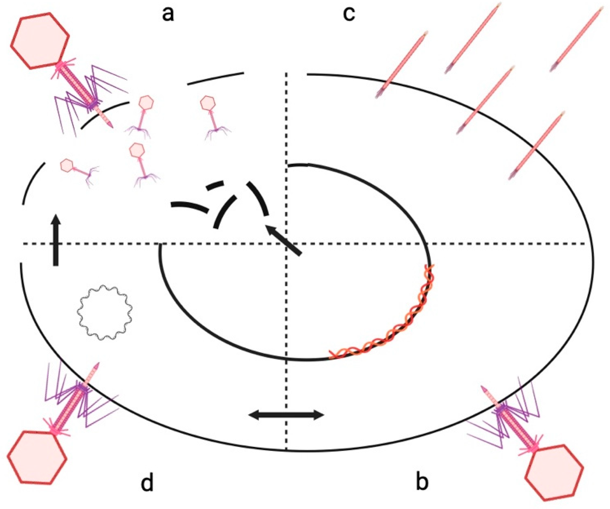
2.3. Lysogenic Phage Induction with Mitomycin C

A volume of 500 µL from each bacterial culture was added to a 250 mL flask containing 25 mL of PYE broth and incubated in a shaker at 30 °C until the cultures reached the log phase growth. To induce lysogenic phages, 24 μL of a Mitomycin C solution (12.5 mg/mL) was added to flasks containing 25 mL of log phase bacterial cultures. The cultures were then incubated overnight in a shaker at 30 °C. The following day, the bacterial cultures were centrifuged at 7000× g for 10 min to pellet the bacterial cells. Subsequently, 1 mL of chloroform was added to each of the supernatants, and the mixtures were shaken thoroughly to ensure the complete removal of bacteria. Dilutions of the lysates were then mixed with 100 μL of the CB15 or CB13 host bacteria in 3.5 mL of SSM and layered on PYE plates. After overnight incubation, isolated plaques were chosen for further purification.
Additionally, for bacterial strains such as RBW1, RBW6, RBW11, RBW18, RBW23, and RBW25, which did not exhibit lytic activity against Caulobacter strains CB15 or CB13, 10 μL of the resultant supernatant from each sample was carefully spotted onto lawns of 21 recently isolated bacterial strains. Following an overnight incubation at 34 °C, the plates were examined for the formation of plaques or signs of lysis, indicating potential lytic activity against these strains.

2.4. Host Range Determination

Host range was determined by mixing 100 μL of a potential host culture with 3.5 mL of melted PYE SSM and pouring the mixture onto the surface of a PYE plate. After the top layer cooled and solidified, a 10 μL aliquot of each phage lysate was spotted on the surface. After overnight incubation at 34 °C, each spot was examined for the ability to produce a clear zone indicating that the phage could lyse the potential host cells. The host strains tested were C. vibrioides strains CB15, CB2, CB13, RBW21, RBW22, and RBW29; C. segnis strains CBR1 and TK0059; FWC26 and ME4 that belong to a third un-named Caulobacter species; Rhizosphaerae strains RBW14, RBW23, RBW25, and RBW26; Sphingomonas strains RBW1 and RBW11; Acidovorax strains RBW12 and RBW13; Variovorax paradoxus strain RBW16; Xanthomonas strains RBW17 and RBW19; Brevundimonas strain RBW18; Pseudomonas strains RBW20 and RBW6; Bacillus cereus strain RBW27; Chromobacterium strain RBW28; Lysobacter firmicutimachus strain RBW3; and Rhizobium strain RBW7.

3. Results

3.1. Isolation and Characterization of Bacterial Strains from Soil and Plant Roots

A total of 21 bacterial strains (RBW designations) were isolated from soil and plant roots collected along Rocky Branch Creek at the University of South Carolina. The primary objective of this isolation effort was to target Caulobacter bacteria, known for their resilience to ampicillin and slow growth characteristics, requiring a minimum of 2 days to develop on PYE agar supplemented with ampicillin.
To identify the newly isolated strains, the 16S rDNA gene from each bacterial isolate was amplified using PCR. Subsequently, the nucleotide sequences obtained from the PCR products were analyzed to determine the genus of each strain. Among the 21 bacterial strains isolated, 7 were identified as members of the genus Caulobacter (Table 1). To further characterize the Caulobacter strains, PCR primers designed to amplify the CB15 dnaK gene were used to generate PCR products from each strain for Sanger sequencing. The nucleotide sequence results indicated that four strains, RBW14, RBW23, RBW25, and RBW26, were most closely related to C. rhizosphaerae, and three strains, RBW21, RBW22, and RBW29, were most closely related to C. vibrioides.
The other fourteen strains included two Sphingomonas strains (RBW1 and RBW11), two Acidovorax strains (RBW12 and RBW13), a Variovorax strain (RBW16), two Xanthomonas strains (RBW17 and RBW19), a Brevundimonas strain (RBW18), two Pseudomonas strains (RBW20 and RBW6), a Bacillus strain (RBW27), a Chromobacterium strain (RBW28), a Lysobacter strain (RBW3), and a Rhizobium strain (RBW7).

3.2. Detection of Bacteriophages Capable of Lysing Caulobacter Strains

When streaks of each of the 21 bacterial strains were replicated onto lawns of Caulobacter strains CB15 and CB13, four strains, Pseudomonas strain RBW20, Bacillus cereus strain RBW27, Chromobacterium strain RBW28, and Caulobacter vibrioides strain RBW29, lysed strain CB13. However, only RBW27 and RBW29 were able to lyse strain CB15 as well. Thus, bacteriophages associated with both Gram-negative and Gram-positive strains were able to infect our laboratory Caulobacter host strains.
To determine whether any other strains harbored complete bacteriophage genomes, cultures of each strain were grown in the presence of mitomycin C, and phages that could lyse either CB15 or CB13 were obtained from 15 strains (Table 2; phages have RBC designations). The resulting eighteen bacteriophages obtained from the lysogenic bacteria included five from Caulobacter strains, RBW14, RBW21, RBW22, RBW26, and RBW29 (Table 2). In addition, 10 prophages capable of infecting CB15 or CB13 were isolated from Acidovorax strains RBW12 and RBW13, Variovorax strain RBW16, Xanthomonas strains RBW17 and RBW19, Pseudomonas strain RBW20, Lysobacter strain RBW3, Rhizobium strain RBW7, a Bacillus cereus strain (RBW27), and Chromobacterium strain RBW28 (Table 2). These results demonstrate that most of these phages are capable of infecting multiple bacterial genera.
Since 15 of the 21 bacterial strains isolated in this study were lysogenic for phages capable of infecting the CB15 or CB13 Caulobacter strains, we tested supernatants from Mitomycin C-treated cultures of the remaining six bacterial strains against all twenty-one of the bacterial strains isolated in this study. Four of the six strains, Sphingomonas strain RBW1, Pseudomonas strain RBW6, Brevundimonas strain RBW18, and Caulobacter rhizosphaerae strain RBW23, were able to lyse other bacterial strains (Table 3). Only two of the strains, RBW11 and RBW25, showed no infectivity towards any of the tested strains, suggesting that they may be non-lysogenic bacteria. Thus, all but two of the twenty-one bacterial strains harbored a lysogenic bacteriophage genome that could form a lytic broad-host-range bacteriophage.

3.3. Characterization of Bacteriophage Host Ranges

To better characterize the broad-host-range of our newly discovered bacteriophages, we tested the phages on all the new rhizosphere bacterial strains. These phages demonstrated infectivity not only towards Caulobacter laboratory strains (CB15, CB13, CB2, CBR1, FWC26, and TK0059), but also towards recently isolated soil Caulobacter strains (RBW14, RBW23, RBW26), which were closely related to C. rhizosphaerae, and RBW22, closely related to C. vibrioides. Remarkably, their infectivity extended beyond Caulobacter strains to include non-Caulobacter strains such as Sphingomonas frigidaeris (RBW1) and Acidovorax strains (RBW12 and RBW13) (Table 4). Also, although most prophages are thought to protect their host strains from infection by closely related bacteriophages, we found that six of the nineteen prophages can infect their original lysogenic host strains (Table 3 and Table 4).

4. Discussion

The isolation and characterization of bacterial strains from soil and plant roots along Rocky Branch Creek at the University of South Carolina provided a snapshot of the role of Caulobacters thriving in this unique environment. We isolated 21 bacterial strains including seven Caulobacter strains and fourteen additional bacteria that share some properties with Caulobacters. The non-Caulobacter strains included representatives of the Sphingomonas, Acidovorax, Variovorax, Xanthomonas, Brevundimonas, Pseudomonas, Bacillus, Chromobacterium, Lysobacter, and Rhizobium genera.
One notable finding from this study was the discovery that 19 out of 21 bacterial isolates harbored bacteriophages with broad host ranges, and 6 of these strains were capable of being re-infected by the phages they harbor as lysogens. Surprisingly, these lysogenic bacteria were not protected by their resident phage genomes and remained susceptible to various additional bacteriophages, such as the new genus of Dolichocephalovirinae.
This broad host range of the phages highlights the complex interactions within the microbial ecosystem. The ability of these phages to infect a diverse array of bacterial hosts suggests a dynamic and interconnected phage–bacteria network, which likely plays a significant role in shaping microbial community structure and function. Understanding these intricate phage–host relationships is crucial for elucidating the roles of bacteriophages in microbial ecology. For example, the broad-host-range phages likely play a role in the composition of the bacterial rhizosphere community. Moreover, these insights have potential applications in biotechnology, agriculture, and environmental management, where phages could be leveraged for controlling bacterial populations and enhancing ecosystem health. These dynamics may reveal novel aspects of phage biology and their impact on ecosystem resilience and functionality.
Future research could focus on identifying the molecular mechanisms behind phage–host interactions and the genetic determinants of host specificity. Additionally, examining the genetic information of newly discovered prophages could reveal novel species, genera, or even families of viruses. A deeper understanding of these dynamics could provide valuable insights into the evolutionary processes of phages and bacteria. This knowledge has the potential to advance various applications in biotechnology, including phage therapy and biocontrol strategies.

Author Contributions

Conceptualization, T.M. and B.E.; Methodology, B.E.; Investigation, T.M.; Resources, B.E.; Writing—original draft, T.M.; Writing—review & editing, B.E.; Supervision, B.E. All authors have read and agreed to the published version of the manuscript.

Funding

This research received no external funding.

Data Availability Statement

The original contributions presented in the study are included in the article, further inquiries can be directed to the corresponding author.

Conflicts of Interest

The authors declare no conflicts of interest.

References

  1. Tsukanova, K.A.; Chebotar, V.K.; Meyer, J.J.M.; Bibikova, T.N. Effect of plant growth-promoting rhizobacteria on plant hormone homeostasis. S. Afr. J. Bot. 2017, 113, 91–102. [Google Scholar] [CrossRef]
  2. Richardson, A.E.; Barea, J.-M.; McNeill, A.M.; Prigent-Combaret, C. Acquisition of phosphorus and nitrogen in the rhizosphere and plant growth promotion by microorganisms. Plant Soil 2009, 321, 305–339. [Google Scholar] [CrossRef]
  3. Berendsen, R.L.; Vismans, G.; Yu, K.; Song, Y.; de Jonge, R.; Burgman, W.P.; Burmølle, M.; Herschend, J.; Bakker, P.A.H.M.; Pieterse, C.M.J. Disease-induced assemblage of a plant-beneficial bacterial consortium. ISME J. 2018, 12, 1496–1507. [Google Scholar] [CrossRef] [PubMed]
  4. Vacheron, J.; Desbrosses, G.; Bouffaud, M.-L.; Touraine, B.; Moënne-Loccoz, Y.; Muller, D.; Legendre, L.; Wisniewski-Dyé, F.; Prigent-Combaret, C. Plant growth-promoting rhizobacteria and root system functioning. Front. Plant. Sci 2013, 4, 356. [Google Scholar] [CrossRef] [PubMed]
  5. Gouda, S.; Kerry, R.G.; Das, G.; Paramithiotis, S.; Shin, H.-S.; Patra, J.K. Revitalization of plant growth promoting rhizobacteria for sustainable development in agriculture. Microbiol. Res. 2018, 206, 131–140. [Google Scholar] [CrossRef]
  6. Luo, D.; Langendries, S.; Mendez, S.G.; De Ryck, J.; Liu, D.; Beirinckx, S.; Willems, A.; Russinova, E.; Debode, J.; Goormachtig, S. Plant growth promotion driven by a novel Caulobacter strain. Mol. Plant-Microbe Interact 2018, 10, 1162–1174. [Google Scholar] [CrossRef]
  7. Yang, E.; Sun, L.; Ding, X.; Sun, D.; Liu, J.; Wang, W. Complete genome sequence of Caulobacter flavus RHGG3 T, a type species of the genus Caulobacter with plant growth-promoting traits and heavy metal resistance. 3 Biotech 2019, 9, 42. [Google Scholar] [CrossRef] [PubMed]
  8. Agler, M.T.; Ruhe, J.; Kroll, S.; Morhenn, C.; Kim, S.T.; Weigel, D.; Kemen, E.M. Microbial hub taxa link host and abiotic factors to plant microbiome variation. PLoS Biol. 2016, 14, e1002352. [Google Scholar] [CrossRef]
  9. Poindexter, J.S. Biological properties and classification of the Caulobacter group. Bacteriol. Rev. 1964, 28, 231–295. [Google Scholar] [CrossRef]
  10. Wilhelm, R.C. Following the terrestrial tracks of Caulobacter—Redefining the ecology of a reputed aquatic oligotroph. ISME J. 2018, 12, 3025–3037. [Google Scholar] [CrossRef]
  11. Abraham, W.-R.; Strömpl, C.; Meyer, H.; Lindholst, S.; Moore, E.R.B.; Christ, R.; Vancanneyt, M.; Tindall, B.J.; Bennasar, A.; Smit, J.; et al. Phylogeny and polyphasic taxonomy of Caulobacter species. Proposal of Maricaulis gen. nov. with Maricaulis maris (Poindexter) comb. nov. as the type species, and emended description of the genera Brevundimonas and Caulobacter. Int. J. Syst. Bacteriol. 1999, 49, 1053–1073. [Google Scholar] [CrossRef] [PubMed]
  12. Reche, I.; D’Orta, G.; Mladenov, N.; Winget, D.M.; Suttle, C.A. Deposition rates of viruses and bacteria above the atmospheric boundary layer. ISME J. 2018, 12, 1154–1162. [Google Scholar] [CrossRef]
  13. Ely, B.; Lenski, J.; Mohammadi, T. Structural and genomic diversity of bacteriophages. In: Bacteriophages. Methods and Protocols. Methods Mol. Biol. 2024, 2738, 3–16. [Google Scholar] [PubMed]
  14. Ely, B.; Berrios, L.; Thomas, Q. S2B, a Temperate Bacteriophage That Infects Caulobacter Crescentus Strain CB15. Curr. Microbiol. 2022, 12, 98. [Google Scholar] [CrossRef] [PubMed]
  15. Paul, J.H. Prophages in marine bacteria: Dangerous molecular time bombs or the key to survival in the seas? ISME J. 2008, 2, 579. [Google Scholar] [CrossRef] [PubMed]
  16. Hoffmann Berling, H.; Maze, R. Release of male-specific bacteriophages from surviving host bacteria. Virology 1964, 22, 305–313. [Google Scholar] [CrossRef]
  17. Ripp, S.; Miller, R.V. The role of pseudolysogeny in bacteriophage-host interactions in a natural freshwater environment. Microbiology 1997, 143, 2065–2070. [Google Scholar] [CrossRef]
  18. Ripp, S.; Miller, R.V. Dynamics of the pseudolysogenic response in slowly growing cells of Pseudomonas aeruginosa. Microbiology 1998, 144, 2225–2232. [Google Scholar] [CrossRef]
  19. Ackermann, H.W.; DuBow, M.S. Viruses of prokaryotes. In General Properties of Bacteriophages; CRC Press Inc.: Boca Raton, FL, USA, 1987; Volume 1, pp. 49–85. [Google Scholar]
  20. Weinbauer, M.G. Ecology of prokaryotic viruses. FEMS Microbiol. Rev. 2004, 28, 127–181. [Google Scholar] [CrossRef]
  21. Ross, A.; Ward, S.; Hyman, P. More is better: Selecting for broad host range bacteriophages. Front. Microbiol. 2016, 7, 1352. [Google Scholar] [CrossRef]
  22. Stewart, F.M.; Levin, B.R. The population biology of bacterial viruses—Why be temperate. Theor. Popul. Biol. 1984, 26, 93–117. [Google Scholar] [CrossRef] [PubMed]
  23. Marsh, P.; Wellington, E.M.H. Phage-host interactions in soil. FEMS Microbiol. Ecol. 1994, 15, 99–107. [Google Scholar] [CrossRef]
  24. Ely, B.; Johnson, R.C. Generalized transduction in Caulobacter crescentus. Genetics 1977, 87, 391–399. [Google Scholar] [CrossRef] [PubMed]
Figure 1. The life cycle of bacteriophages. (a) Lytic cycle: Bacteriophages bind to bacterial cells, inject their DNA into the bacteria, replicate the DNA and assemble it into protein capsid structures, and then lyse the cell and release new phage particles. (b) Lysogenic cycle: The phage DNA genome integrates into the host chromosome and then replicates as part of the bacterial chromosome. Under stress conditions, it is separated from the bacterial chromosome and enters the lytic cycle. (c) Chronic cycle: Bacteriophages do not kill their host because new phage particles are synthesized and extruded one by one from their host cells. Chronic infection is common with filamentous phages. (d) Pseudolysogenic cycle: The phage DNA genome exists in the bacterial cell as an episome. Depending on subsequent conditions, the phage DNA can adopt either the lytic cycle or the lysogenic cycle.
Microorganisms 12 01894 g001
Table 1. Colony characteristics of 21 bacteria isolated from soil and plant roots along Rocky Branch Creek.
Bacterial NameBacterial StrainColony SizePigmentationFormElevation Margin
RBW1Sphingomonas frigidaerisPinpointYellowCircularConvexEntire
RBW3LysobacterLargeYellowIrregularUmbonateUndulate
RBW6PseudomonousLargeWhiteIrregularFlatUndulate
RBW7RhizobiumLargeWhiteCircularPulvinateEntire
RBW11Sphingomonas sp.PinpointYellowCircularConvexEntire
RBW12Acidovorax sp.MediumWhiteCircularConvexEntire
RBW13Acidovorax sp.MediumWhiteCircularUmbonateErose
RBW14Caulobacter rhizosphaeraeLargeWhiteIrregularUmbonateUndulate
RBW16VariovoraxLargeYellowIrregularUmbonateUndulate
RBW17Xanthomonas translucensLargeYellowIrregularUmbonateUndulate
RBW18BrevundimonasLargeOrangeCircularUmbonateEntire
RBW19XanthomonasPinpointYellowCircularConvexEntire
RBW20PseudomonousLargeWhiteIrregularUmbonateUndulate
RBW21Caulobacter vibrioidesLargeWhiteIrregularUmbonateUndulate
RBW22Caulobacter vibrioidesLargeWhiteCircularUmbonateUndulate
RBW23Caulobacter rhizosphaeraeLargeYellowIrregularUmbonateUndulate
RBW25Caulobacter rhizosphaeraeMediumYellowCircularConvexEntire
RBW26Caulobacter soil strainSmallYellowCircularFlatEntire
RBW27Bacillus cereusLargeWhiteRhizoidUmbonateLobate
RBW28Chromobacterium sp.LargeWhiteIrregularUmbonateUndulate
RBW29Caulobacter vibrioidesSmallWhiteCircularConvexEntire
Table 2. Identification of lysogenic bacterial strains capable of infecting CB15 or CB13 Caulobacter strains.
Lysogenic BacteriaCB15CB13
RBW1
RBW3+
RBW6
RBW7+
RBW11
RBW12+
RBW13+
RBW14+
RBW16+
RBW17+
RBW18
RBW19+
RBW20+
RBW21++
RBW22+
RBW23
RBW25
RBW26+
RBW27+
RBW28+
RBW29+
Table 3. Identification of 4 additional lysogenic bacterial strains.
RBWs Bacteria RBW1RBW6RBW11RBW18RBW23RBW25
RBW1+
RBW3
RBW6
RBW7
RBW11
RBW12
RBW13++
RBW14
RBW16
RBW17
RBW18
RBW19++++
RBW20++
RBW21++++
RBW22++
RBW23++
RBW25
RBW26
RBW27++++
RBW28
RBW29
Table 4. Bacteriophage host range and self-sensitivity.
Bacteriophage NameOriginal HostBacterial StrainPlaque Morphology 1Positive Host RangeSelf
RBC51RBW3 LysobacterCB13TurbidCB15, CB13, CB2, RBW1, RBW14, RBW23
RBC52RBW7 RhizobiumCB13TurbidCB15, CB13, CB2, RBW1, RBW13, RBW14, RBW22
RBC53RBW12 AcidovoraxSC1004TurbidCB15, CB13, CB2, FWC26, RBW1, RBW12, RBW14, RBW22, RBW23+
RBC54RBW12 AcidovoraxSC1004ClearCB15, CB13, CB2, FWC26, RBW1, RBW12, RBW22, RBW23+
RBC55RBW13 AcidovoraxSC1004TurbidCB15, CB13, CB2, CBR1, RBW14, RBW22
RBC56RBW14 Caulobacter rhizosphaeraeCB13TurbidCB15, CB13, CB2, FWC26, CBR1, RBW14, RBW12+
RBC57RBW16 VariovoraxSC1004ClearCB15, CB13, CB2, CBR1, FWC26, TK0059, RBW12, RBW14, RBW22, RBW23, RBW26
RBC58RBW16 VariovoraxSC1004TurbidCB15, CB13, CB2, RBW12, RBW14, RBW22
RBC59RBW17 XanthomonasSC1004ClearCB15, CB13, CB2, TK0059, RBW12, RBW14, RBW22
RBC60RBW19 XanthomonasSC1004ClearCB15, CB13, CB2, CBR1, TK0059, RBW12, RBW14, RBW22, RBW23
RBC61RBW20 PseudomonasSC1004ClearCB15, CB13, CB2, RBW12, RBW14
RBC62RBW21 Caulobacter vibrioidesSC1004ClearCB15, CB13, CB2, RBW14
RBC63RBW21 Caulobacter vibrioidesCB13ClearCB15, CB13, CB2, FWC26, RBW12, RBW13, RBW14, RBW22
RBC64RBW22 Caulobacter vibrioidesSC1004ClearCB15, CB13, CB2, FWC26, RBW13, RBW14, RBW22+
RBC65RBW27 Bacillus cereusSC1004ClearCB15, CB13, CB2, RBW12, RBW13, RBW14, RBW22
RBC66RBW28 ChromobacteriumCB13TurbidCB15, CB13, CB2, FWC26, RBW1, RBW12, RBW13, RBW14, RBW22, RBW23
1 Turbid plaques are caused by incomplete lysis of the host cells.
Disclaimer/Publisher’s Note: The statements, opinions and data contained in all publications are solely those of the individual author(s) and contributor(s) and not of MDPI and/or the editor(s). MDPI and/or the editor(s) disclaim responsibility for any injury to people or property resulting from any ideas, methods, instructions or products referred to in the content.

