Article

AraC Functional Suppressors of Mutations in the C-Terminal Domain of the RpoA Subunit of the Escherichia coli RNA Polymerase

1
Department of Pathology and Immunology, Faculty of Medicine, University of Geneva, 1 Rue Michel-Servet, CH-1211 Geneva, Switzerland
2
Department of Microbiology and Molecular Medicine, Faculty of Medicine, University of Geneva, 1 Rue Michel-Servet, CH-1211 Geneva, Switzerland
*
Author to whom correspondence should be addressed.
Current address: Department of Microbiology and Molecular Medicine, Faculty of Medicine, University of Geneva, 1 Rue Michel-Servet, CH-1211 Geneva, Switzerland.
Microorganisms 2024, 12(9), 1928; https://doi.org/10.3390/microorganisms12091928
Submission received: 29 August 2024 / Revised: 18 September 2024 / Accepted: 20 September 2024 / Published: 23 September 2024
(This article belongs to the Special Issue Transcriptional Regulation in Bacteria)

Abstract

:
In E. coli, transcriptional activation is often mediated by the C-terminal domain of RpoA, the α subunit of RNA polymerase. Random mutations that prevent activation of the arabinose PBAD promoter are clustered in the RpoA C-terminal domain (α-CTD). We have isolated functional suppressors of rpoA α-CTD mutations that map to araC, the main transcriptional regulator of ara genes, or to the PBAD promoter. No mutation was found in the DNA regulatory region between araC and PBAD. Most suppressors that improve PBAD transcription are localized to the N-terminal domain of AraC. One class of araC mutations generates substitutions in the core of the N-terminal domain, suggesting that they affect its conformation. Other suppressors localize to the flexible N-terminal arm of AraC. Some, but not all, suppressors confer an arabinose constitutive phenotype. Suppression by both classes of araC mutations requires the α-CTD to stimulate expression from PBAD. Surprisingly, in rpoA+ strains lacking Crp, the cAMP receptor protein, these araC mutations largely restore arabinose gene expression and can essentially bypass Crp activation. Thus, the N-terminal domain of AraC exhibits at least three distinct activities: dimerization, arabinose binding, and transcriptional activation. Finally, one mutation maps to the AraC C-terminal domain and can synergize with AraC mutations in the N-terminal domain.

1. Introduction

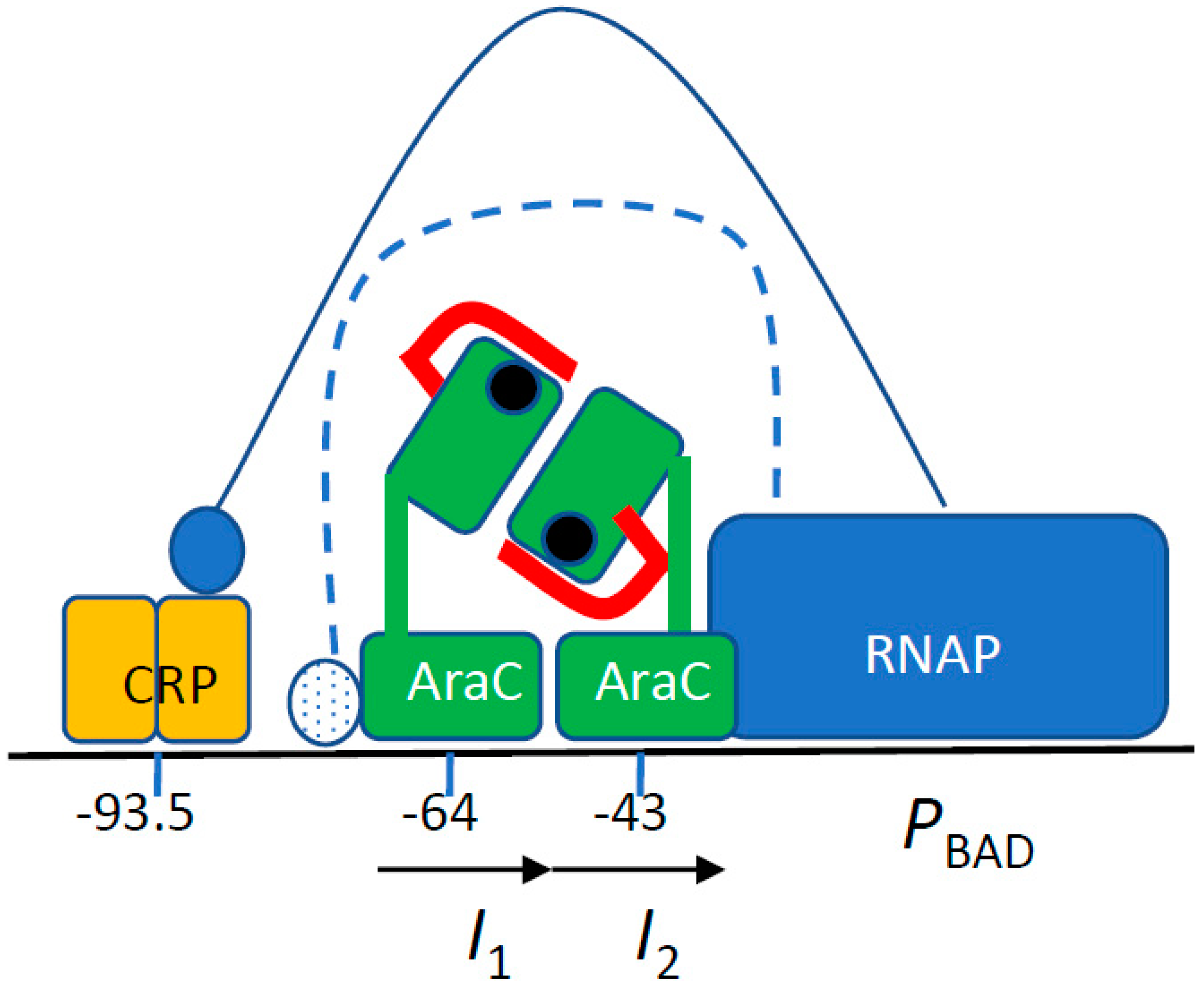
The arabinose regulon was the first system in which positive control of gene expression was genetically demonstrated [1]. AraC, the product of the activator gene, binds to arabinose and stimulates transcription of the catabolic genes araB, araA, and araD (araBAD operon), as well as those of the arabinose import systems, araE and araFGH. In the absence of arabinose, AraC represses transcription from the PBAD promoter. In both instances, one AraC monomer is bound to the I1 site (Figure 1). The transition between the repressor and activator states of AraC involves a large conformational movement, named the light switch, that displaces one subunit of the AraC dimer from the distant O2 operator site to the promoter proximal I2 activator site. During the switch, the N-terminal arm of AraC covers the arabinose-binding pocket of the N-terminal dimerization domain (AraC-NTD) [2,3,4]. The PBAD promoter is intrinsically very weak, and AraC bound to the I2 site enhances RNA polymerase binding and open complex formation [5]. In addition, Crp, the cAMP receptor protein, is required for full activity of the PBAD promoter [6,7]. Crp activates many bacterial promoters, and several mechanisms of activation have been described [8]. The properties of the arabinose system were exploited to construct a widely used set of expression plasmids [9].
The α subunit of RNA polymerase, encoded by the rpoA gene, plays at least two roles in transcription. Its N-terminal domain (α-NTD) is involved in the assembly of the enzyme [10,11]. In contrast, the C-terminal domain (α-CTD), which is attached by a flexible linker to the α-NTD, plays multiple roles in transcription activation [8,12,13,14]. It activates transcription by interacting with activators in different ways and/or by binding to UP DNA sites upstream of promoters [15,16]. Since RNA polymerase contains two α subunits, each subunit can interact independently with two activating regions [13]. In addition to α-CTD structures [17,18], several structures of α-CTD-containing complexes have been resolved, including that of α-CTD bound to Fis at the proP P2 promoter [19], that of the α-CTD–Crp–DNA complex [14], and that of the RNA polymerase holoenzyme bound to a synthetic promoter in the presence of Crp [20].
At the arabinose PBAD promoter, one α-CTD interacts with Crp [7]. The potential target of the second α-CTD has not yet been identified. However, by analogy with other Crp-dependent promoters [8], it has been proposed to bind the AraC DNA-binding domain and possibly a DNA motif localized between the Crp and AraC binding sites [3,21] (Figure 1). Several random rpoA α-CTD mutations that interfere with PBAD activity have been characterized.
We have used a genetic approach to search for functional suppressors that compensate for the detrimental effects of mutations in the RpoA α-CTD. Random mutagenesis of araC and of the intergenic regulatory region between araC and araB led to the isolation of araC suppressors. Most of these random mutations are scattered at several positions in the AraC-NTD, and their effect requires the RpoA α-CTD. We have also characterized a mutation in the DNA-binding C-terminal domain of AraC that exerts a synergistic effect with mutations in the AraC-NTD. However, we did not identify an AraC contact site with the RpoA α-CTD. Rather, all the mutations may promote or stabilize a conformation of AraC that more efficiently promotes transcription, thereby functionally compensating for the deleterious effects of the rpoA mutations.

2. Experimental Procedures

Strains. MC4100 is an F-araD139 Δ(argF-lac)U169 flhD5301 fruA25 relA1 rpsL150 rbsR22 Δ(fimB-fimE)632 deoC1 thi strain [22]. DB504 is MC4100 araΔ714 malE18 [23]. JW5702 contains the Δcrp-765::kan allele [24] and was obtained from the Coli Genetic Stock Center (CGSC strain 11596). HfrH was obtained from the Coli Genetic Stock Center (CGSC strain 5081).
The rpoA mutations were introduced into DB504 by bacteriophage P1-mediated co-transduction with a gspA::Tn10 marker (90–95% co-transduction) [25]. The presence of the rpoA mutations was verified by suppression of the toxic phenotype caused by induction of the appropriate proteins, by growth on M63 minimal media with or without cysteine and methionine, and by fermentation on McConkey-melibiose indicator plates [26]. The Δcrp-765::kan allele was introduced by P1-mediated transduction in strains carrying the rpoA alleles; the transductants were plated on streptomycin (100 µg/mL) containing plates to select for recombination between the rpsL150 marker of DB504 and crp, and retention of the gspA::Tn10 marker. Strains containing the Δcrp-765::kan allele were grown in LB containing 0.1% glucose [25]. Information on the E. coli genes and regulatory sequences was obtained from https://ecocyc.org/ (accessed on 25 August 2024).
Plasmids. Plasmids derived from pBAD24 [9] contain the araC gene, the intergenic region between araC and PBAD, the PBAD promoter, and the first 13 nt of the araBAD transcript. pBAD18-vs.1 expresses a lysozyme encoded by bacteriophage T4 [27]. pBAD72K expresses a chimeric protein containing the signal sequence of murine PAI2 fused to the mature portion of PhoA [28]. pDB2114 expresses gene 55.2 of bacteriophage T4 [29]. These three plasmids confer a toxic phenotype that prevents growth in the presence of arabinose.
Maspin (SERPINB5, P70124) is a member of the ovalbumin-related protease inhibitors family [30] whose N-terminal region is devoid of signal sequence activity [31]. psM74c expresses a Maspin::PhoA chimera: the initial Met is fused to residues 23–46 of Maspin, with a P32L substitution to allow export, followed by a synthetic ALA-A c-region cleaved by signal peptidase, and residues 23–471 of PhoA (P00634). The export of this chimeric protein is slightly more efficient than that of wild-type PhoA; hence psM74c was used to isolate and characterize suppressors of the rpoA mutations.
The rpoA+ gene was PCR amplified from chromosomal DNA with the primers RPOAUP (5′cgggatcccacctgatcgtcg) and RPOADON (5′ccggtaccttaacctgtgatccggttactcg), and cloned between the BamHI and KpnI restriction sites of pUC19 to generate pUC-rpoA. The deletion of the α-CTD was performed by PCR amplification of pUC-rpoA with the primers RPOAUP and RPOAdel (5′cggggtaccttacacttcaggctgacg). The pUC-rpoAdel plasmid expresses amino acids 1 to 242 of RpoA. The expression level of the proteins from the Plac promoter is estimated to be ~50-fold higher than that from the chromosomal rpoA gene in rpoA+ crp+ strains; the level should be ~5-fold in Δcrp strains. The pUC-rpoAdel plasmid confers a Mel phenotype in an rpoA+ strain.
The original pBAD24-derived plasmids (AraC+, AraC D7N, and AraC I71V) were digested with NsiI and XbaI. The araC-Maspin::PhoA fragments were cloned into the cognate sites of pBAD33 whose origin is compatible with that of the rpoA plasmids [9].
A complete deletion of the araC coding region was performed by removing the two MfeI-digested fragments of psM74c.
Enzyme assays. PhoA activity was qualitatively detected on LB plates supplemented with 0.2% arabinose and 40 µg/mL XP (5-bromo-4-chloro-3 indolyl phosphate). PhoA enzymatic activity was determined by measuring the rate of p-nitro-phenyl-phosphate hydrolysis [32]. Cultures were assayed after a 1 h induction with 0.2% arabinose. LacZ activity was determined by measuring the rate of o-nitro-phenyl-galactoside hydrolysis [25]. For each strain, triplicate independent cultures were assayed twice. The values were analysed with a Mann–Whitney test or an unpaired t-test.
Random mutagenesis. Hydroxylamine mutagenesis of psM74c plasmid DNA was performed as described [31]. Upon transformation in DB504, approximately 6% of the colonies carried inactivating mutations in araC, the regulatory region, or the maspin::phoA gene and formed white colonies on PhoA indicator plates containing arabinose. Plasmids allowing the formation of light blue colonies in rpoA Δcrp strains occurred at a frequency of 1–2%; in these strains, the parental plasmid formed white colonies. More than half of the mutant plasmids exhibited a higher plasmid copy number than the original plasmid. The complete araC gene was recloned in the parental plasmid. In a second experiment, a complete araC fragment (NsiI-MluI, 1130 bp long) was randomly mutagenized by PCR [28] and cloned in psM74c. Ten percent of the resulting plasmids transformed into an rpoA+ crp+ strain formed white colonies on PhoA indicator plates containing arabinose. After transformation in the rpoA A272E Δcrp strain, candidate suppressors were sequenced. Three additional AraC residues were isolated: N16S, I51T, and I71V; three mutants had different substitutions at previously identified residues (S14P, A152T, and A152P), and the A152V substitution was isolated again. The araC mutations described here have been independently isolated once (D7N, N16S, I51T, I71V, A152T, and A152P), or 4–6 times (S14L, S14P, G22D, and A152V). To isolate suppressors of rpoA N268T, the mutagenized araC PCR fragment was digested with NsiI and EcoRV and cloned in psM74c. After transformation in the rpoA N268T crp+ strain, three suppressors were identified: R61H was isolated six times, while R261C and C205Y were isolated twice.

3. Results

3.1. RpoA Mutations in the α-CTD Region That Interfere with PBAD Expression

The chimeric protein PAI2::PhoA contains the signal sequence of murine PAI-2 fused to the mature portion of PhoA. Expression of this chimeric protein from the arabinose PBAD promoter is lethal because it interferes with protein export to the periplasm [31]. Suppressors of this toxicity were selected as colony-forming mutants in the presence of arabinose. While many chromosomal suppressors map to sec genes involved in protein export to the periplasm thus preventing the toxicity of the chimera [28,33], we also isolated fifteen rpoA mutants (Table 1). To modulate the severity of the toxicity in our selective system, we used several genes of bacteriophage T4 that also confer a toxic phenotype when expressed from the arabinose PBAD promoter. The expression of one of these genes, vs.1, encoding an exported lysozyme whose toxicity is weaker than that of PAI2::PhoA [27] led to the isolation of six rpoA mutants. Finally, one rpoA mutant was isolated as a suppressor of phage T4 gene 55.2, a topoisomerase inhibitor that exhibit the strongest toxicity of the three proteins [29]. A total of twenty-two independent rpoA mutations have been isolated (Table 1).
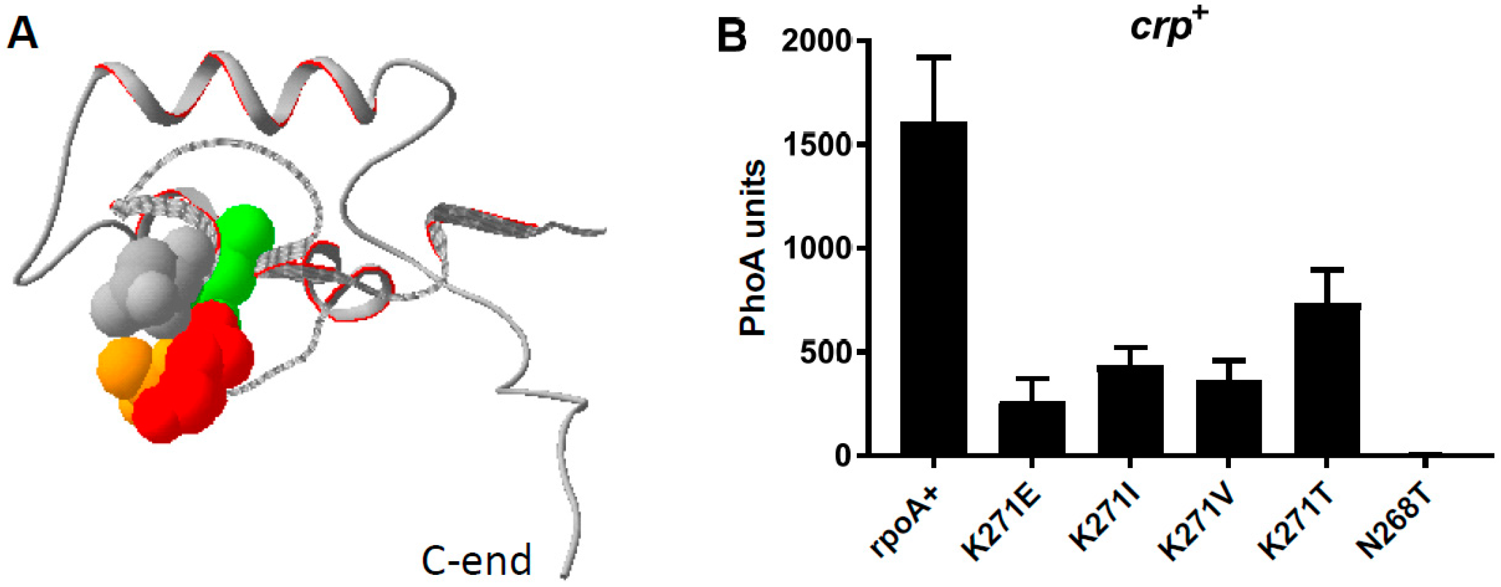
The randomly selected rpoA mutations all map to amino acids 268–272 of the RpoA C-terminal domain (α-CTD) (Figure 2A). The RpoA K271E substitution (rpoA341 [26,34]) was the most frequent, but three other new substitutions (V, T, and I) were also identified at this position (Table 1). The four substitutions of K271 exert similar effects on PBAD promoter expression, although the K271E substitution has a slightly stronger effect (Figure 2B, Table 1). These mutations affect CysB regulatory function and prevent growth on M63 minimal medium in the absence of cysteine or methionine (Cym phenotype) [26]. The L270F substitution appears to be the weakest of the rpoA alleles since it allows growth on minimal media (Table 1) and thus does not detectably interfere with activation of the cysA operon by CysB [35]. The N268T substitution is the strongest rpoA mutation observed (Figure 2B), as it prevents growth on minimal media, even in the presence of casamino acids. Thus, this mutation must affect one or more promoter(s) in addition to PBAD and PcysA.
The rpoA mutations were selected on arabinose-containing LB plates, under conditions where the expression of the indicated toxic protein prevents growth. For each substitution, the number of independent isolates is shown. The N268T allele suppresses the toxicity of all three proteins. The L270F allele suppresses the toxicity of T4 vs.1 and partially that of PAI2::PhoA. The other alleles suppress the toxicity of T4 vs.1 and PAI2::PhoA but not that of T4 55.2.
Colonies grown on McConkey-melibiose indicator plates were scored as R (red), identical to those of rpoA* strains; K (pink), reflecting an intermediate melibiose fermentation; and W (white), reflecting no detectable melibiose fermentation. On M63-minimal-glucose plates, only the weak L270F allele allows growth. Upon addition of Cys and Met, most rpoA mutants grew. The N268T allele must prevent the expression of one or more auxotrophic genes.

3.2. Suppressors of the rpoA Mutations

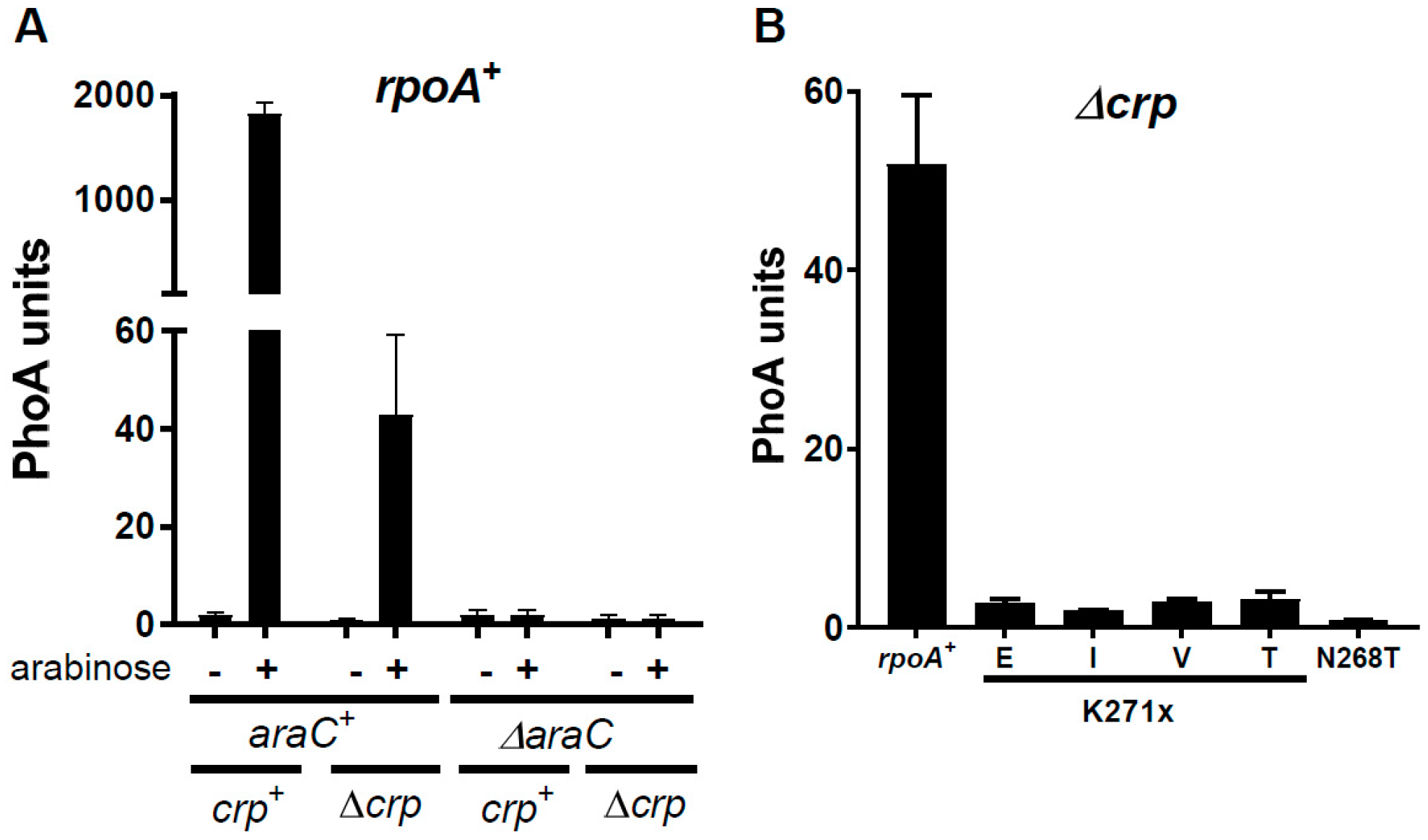
Both AraC and Crp promote transcription from the PBAD promoter in the presence of arabinose [3,4]. While AraC is absolutely required, a 20- to 40-fold reduction in expression can be detected in an rpoA+ strain in the absence of Crp (Figure 3A). Quantification of phoA expression from the PBAD promoter in strains deleted for crp and carrying five alleles of rpoA demonstrated a strong reduction of promoter activity with the four substitutions of K271, while the N268T mutation totally abolishes expression (Figure 3B). These findings imply that the α-CTD is required for PBAD transcription in the absence of Crp.
To genetically define whether the interactions defective in the RpoA α-CTD mutants can be functionally suppressed, we carried out a suppressor screen. We used Δcrp strains containing either the K271E or K271V RpoA substitution. We also employed a Maspin::PhoA fusion that contains a very efficient signal sequence. This chimeric protein provides an extremely sensitive reporter, compensating for the reduced expression from the PBAD promoter in the absence of Crp. Cells were transformed with mutagenized plasmids harboring the araC gene and the PBAD regulatory region and plated on LB plates containing arabinose and XP (a color indicator of PhoA activity). Colonies in which AraC-dependent expression of maspin::phoA is restored are blue. Plasmids isolated from such strains were sequenced.
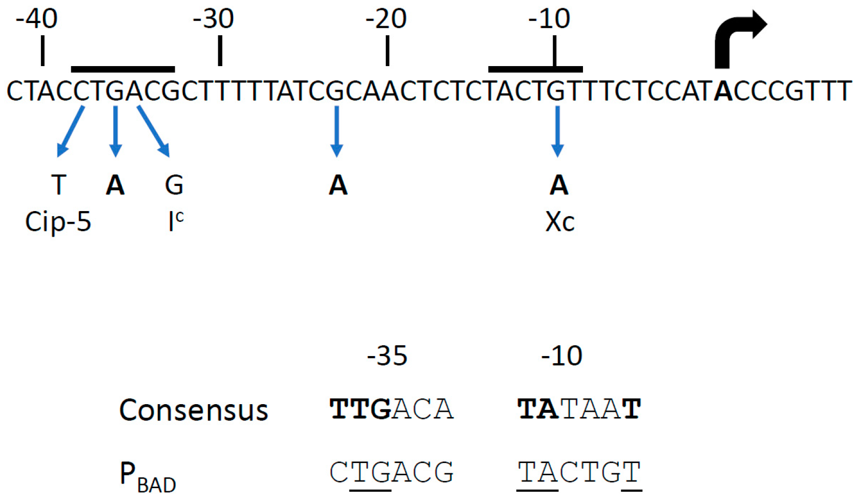
We isolated four plasmids that contain mutations in the PBAD promoter (Figure 4). One of these, at position −36, was located in the −35 hexamer, between the cip-5 site [6,36] and the Ic mutations [37,38]. The cip-5 and Ic promoters allow AraC-independent constitutive expression, and the −36 mutation could also facilitate RNA polymerase binding. The second one, at position −23, has a weaker stimulatory effect. The last two, located at position −10, are identical to the Xc mutation, which has been described as creating a strong AraC-independent promoter [36,39]; a PBAD promoter carrying the Xc mutation is not affected by the rpoA341 K271E mutation [26]. No mutation was isolated in the regulatory region between the araC coding sequence and the PBAD promoter (Figure 1).
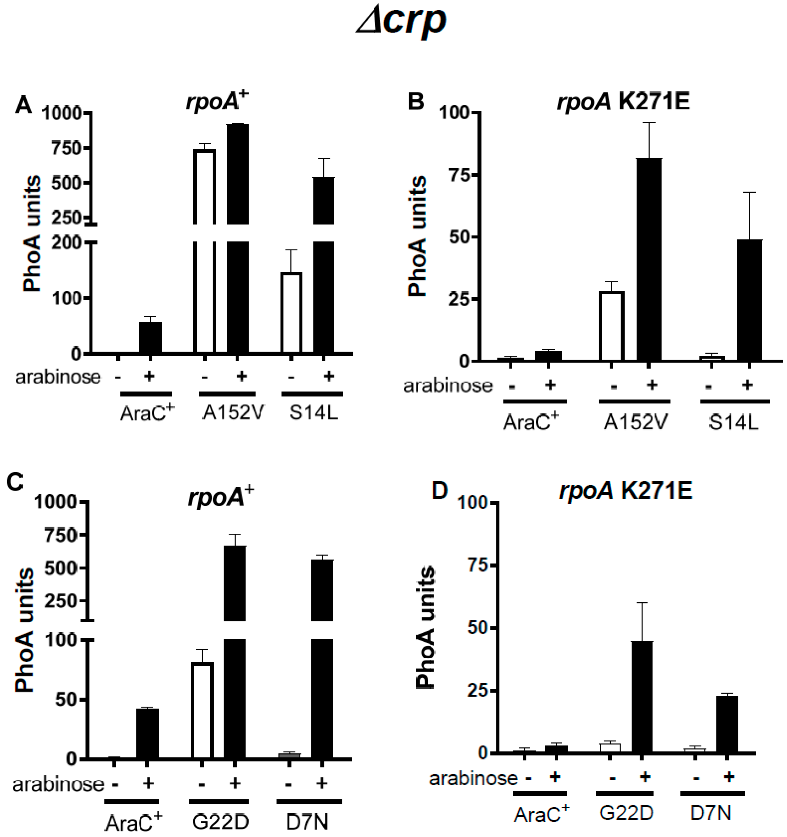
We isolated four mutations in araC. These araC suppressors all localize to the N-terminal domain of AraC (AraC-NTD). Since most araC constitutive mutations map to this region [41,42], we measured expression from the PBAD promoter in the presence and absence of arabinose with these AraC mutant proteins. In a rpoA+ Δcrp strain, the A152V substitution confers a nearly complete constitutive phenotype (Figure 5A). In contrast, G22D and S14L confer an expression level of only 12–27% in the absence of the inducer (Figure 5A,C). Constitutive expression with A152V has been observed in a crp+ strain [42]. Finally, the D7N substitution is entirely devoid of constitutive activity (Figure 5C). In the rpoA K271E strain, only A152V confers a partial constitutive phenotype (34% of the induced level) (Figure 5B,D); this suggests that the RpoA α-CTD may stabilize the activator form of the constitutive AraC proteins. However, an arabinose constitutive phenotype is not necessary for functional suppression of the rpoA mutations.
While induced expression mediated by wild-type AraC is strongly decreased in an rpoA+ Δcrp strain, the araC mutations confer robust expression in this strain, at levels of 30–50% of that measured in an araC+ rpoA+ crp+ strain. Thus, they can partially compensate for the absence of Crp in rpoA+ strains (Figure 5A,C).
These araC mutants were isolated in strains carrying either the K271E or K271V RpoA substitutions. We determined the capacity of the araC mutations to suppress the four different K271X substitutions, as well as the N268T one. The results shown in Figure 6 were obtained with AraC D7N and A152V; similar results were obtained with S14L and G22D (Supplementary Figure S1). In induced rpoA+ crp+ strains, expression from the PBAD promoter was slightly increased (1.5- to 3-fold) by the araC mutations (Figure 6A,C). In almost all cases, the expression observed with the rpoA mutants did not reach the levels observed in rpoA+ strains, indicating that the AraC suppressors do not fully compensate for the rpoA defect. However, the AraC D7N substitution restored the AraS phenotype conferred by PAI2::PhoA in an rpoA K271T crp+ strain; an effect of the other araC mutations could not be tested because of their constitutive activity. In Δcrp strains, stimulation by the araC mutations was much higher (Figure 6B,D). In rpoA+ Δcrp strains, the suppressors nearly compensated for the absence of Crp, as shown above in Figure 5. With the four rpoA K271 substitutions, the araC mutations increased expression 8- to 30-fold when compared to araC+. In no case was expression as high as that detected in rpoA+ strains, confirming that the araC mutations only partially compensate for the rpoA defects. Finally, the RpoA N268T substitution, which exerts the most drastic effect on PBAD expression, was not suppressed by any of these araC mutations, both in the presence and absence of Crp, suggesting two independent mechanisms for α-CTD activation.
To determine whether any specificity could be observed between the AraC suppressors and other α-CTD mutated residues, we compared the K271E substitution with substitutions at the two flanking residues, L270 and A272 (Figure 7). The three mutant AraC proteins exerted similar effects in all cases, although small quantitative differences were detected. Expression was highest with the rpoA L270F mutant, the weakest rpoA allele, and lowest with the rpoA A272E one. When compared to araC+, increased expression with the araC mutants was found to be stronger in Δcrp (Figure 7) than in crp+ strains (Supplementary Figure S2). In conclusion, we did not succeed in isolating a single araC mutation that selectively suppressed, in an allele-specific manner, any of the RpoA mutated residues tested.
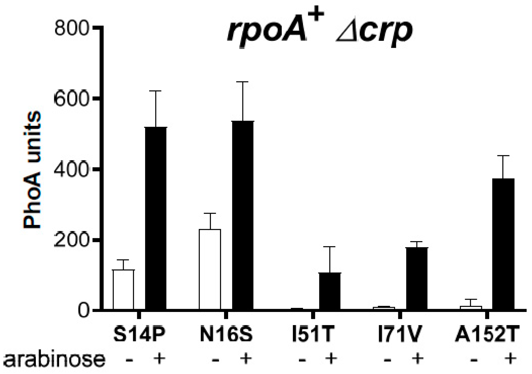
In a different approach, using a mutagenic PCR protocol on a plasmid harboring the wild-type araC gene, we identified three additional AraC residues (N16S, I51T, and I71V) whose substitutions resulted in increased PBAD expression in an rpoA A272E Δcrp strain. In addition, different substitutions were found at two previously identified residues (S14P and A152T). In the rpoA+ Δcrp strain, all compensated to a varying extent for the absence of Crp (Figure 8). The effect was maximal for the S14P mutant and minimal for the I51T mutant; the A152T mutant was less efficient than the A152V one (see Figure 6D). Furthermore, while the S14P and N16S mutants were partially constitutive, the other three mutants remained fully inducible; the A152T mutant, therefore, behaves clearly differently from the A152V mutant (Figure 8). These results confirm that a constitutive phenotype is not required for the suppression of the rpoA defects. The AraC I71V substitution restored the AraS phenotype conferred by PAI2::PhoA in the rpoA K271T crp+ strain, confirming that increased expression from the PBAD promoter can also be observed with this mutant in the presence of Crp. In Δcrp mutant strains carrying different rpoA alleles, increased expression was also detected (Supplementary Figure S3). Here again, we did not observe any allele specificity.

3.3. The Activity of the AraC Mutant Proteins Requires the α-CTD of RpoA

The observed functional suppression of RpoA α-CTD mutations and the lack of allele specificity of the AraC mutations are compatible with at least two models. Firstly, the suppressors could create or improve an interaction between the AraC-NTD and the RNA polymerase core. Alternatively, RpoA α-CTD could contact a specific site of AraC that remains to be identified. In this first scenario, functional suppression would not depend on the RpoA α-CTD. To discriminate between these two possibilities, we expressed either wild-type RpoA or an RpoA deletion that lacks the α-CTD (residues 243–329). Both proteins were expressed from the Plac promoter on a plasmid, with levels predicted to be ~5-fold higher than that of RpoA derived from the chromosomal rpoA+ gene in the absence of Crp.
In rpoA+ Δcrp strains, the activity of AraC+ decreased about 2-fold in the presence of the RpoA deletion (Figure 9A). A similar decrease was observed with the two AraC suppressors, namely the D7N mutant located in the AraC N-terminal arm and the I71V mutant located in the core of the AraC-NTD (Figure 9B,C). This result is more evident when the activity of the three AraC proteins in cells carrying the RpoA+ (Figure 9E) or the RpoA deletion plasmid (Figure 9F) are directly compared. As a control, expression from the lacZ promoter, which is activated only by an interaction of the RpoA α-CTD with Crp [8], was decreased to the same extent in the presence of the RpoA deletion (Figure 9D). When RpoA and the RpoA deletion were expressed from the strong bacteriophage T4 gene 32 promoter [43], we observed an almost complete inhibition of PBAD expression (Supplementary Figure S4). Taken together, these results demonstrate that the RpoA α-CTD is required for transcriptional activation at the PBAD promoter with both wild-type and mutant AraC in the absence of Crp, suggesting that the AraC mutants functionally compensate for defects in the RpoA α-CTD by increasing AraC activation of RNA polymerase.

3.4. A Suppressor of RpoA N268T Synergizes with Mutations in the NTD of AraC

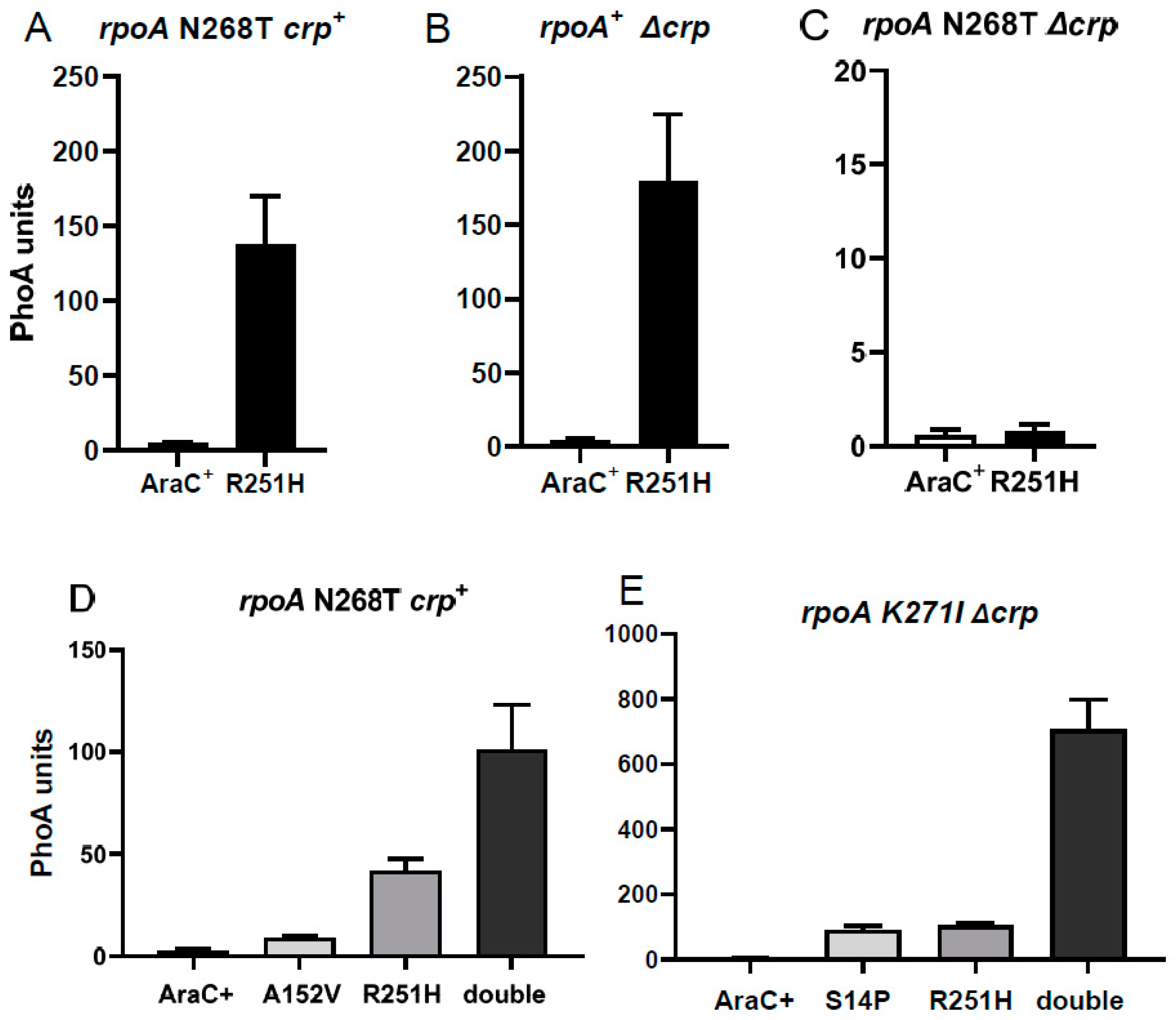
Since none of the AraC mutants described above suppressed the rpoA N268T mutant, even in the presence of Crp, we repeated the mutant screen in the rpoA N268T crp+ strain and isolated three mutants in the C-terminal DNA-binding domain of AraC. The largest effect was observed with R251H, while a 3-fold lower effect was observed with R251C and C205Y. The AraC R251H mutant compensated to the same extent for the effect of the rpoA N268T mutant in a crp+ strain (Figure 10A) or in the absence of Crp in a rpoA+ strain (Figure 10B); in rpoA K271X crp+ strains, expression was >50% of that measured in an araC+ rpoA+ crp+ strain. In contrast, this AraC mutant was totally inactive in the rpoA N268T Δcrp strain (Figure 10C).
Since R251H also strongly increased expression in the rpoA K271E crp+ strain, we investigated the effect of combining AraC mutations in the CTD (R251H) and in the NTD (S14L, in the N-terminal arm, or A152V, in the NTD core). We found that in the rpoA N268T crp+ strain, the combined effect of both AraC mutations was synergistic and not additive (Figure 10D,E). This synergistic effect was most striking in an rpoA K271I Δcrp strain, reaching about 40% of the expression observed in the araC+ rpoA+ crp+ strain. These results suggest that the R251H mutation in the DNA-binding domain of AraC exerts its effect at a different step of transcriptional activation than mutations in the AraC-NTD, underlying a regulatory mechanism not directly related to AraC-NTD activation.
In conclusion, we have characterized mutations in AraC that functionally compensate for the deleterious effect of rpoA mutations. Mutations in the AraC-NTD can almost fully compensate for the absence of Crp. Finally, combinations of mutations in the AraC-NTD and in the AraC-CTD synergistically activate transcription at the PBAD promoter.

4. Discussion

The RpoA α-CTD domain is a pleiotropic hub required for transcription activation in multiple systems [8,12]. The residues that functionally interact with each activator are often different. For instance, the E261K/G substitutions affect a subset of Crp-dependent promoters, while P322S affects OmpR [44], and D250 and R310 affect TyrR [45]. The α-CTD has also been shown to play a role in genome-wide transcription regulation in B. subtilis [46].
Considering the number of rpoA mutations detected through our genetic selections, a large fraction of the RpoA residues necessary for PBAD activation have probably now been identified. However, truncations of the α-CTD and several substitutions, including R265A and N268A, are not viable in the absence of a functional RpoA and must be studied in strains containing an rpoA+ gene on the chromosome [15,47,48]. Thus, our experimental setup does not allow the detection of some mutations, including nonsense ones that would truncate RpoA.
The RpoA K271 substitutions affect CysB and MelR activity in addition to AraC [26]. However, several RpoA C-terminal truncations allow MelR transcriptional activity without any effect on AraC or CysB [11]. More recently, K271 was shown to mediate transcription activation by Fis at the proP P2 and rrnB P1 promoters, and the Fis residues that interact with the α-CTD were identified through structure-based models [19,49]. The K271E substitution also impaired phage λ PRE activation by cII [50,51]. Finally, we observed a 3- to 5-fold decrease in MalE synthesis in strains carrying the rpoA K271E substitution; it had no effect on malPQ transcription, which is independent of Crp [26]. The A272E change described here has similar effects to the K271E substitution. In contrast, the A272T substitution decreased ompF transcription mediated by OmpR, but had no detectable effect on activation by AraC, CysB, and MelR [52]. The replacement of the wild-type A residue by a larger, charged one likely confers a more drastic effect on α-CTD-mediated transcription.
The properties of the rpoA mutants provided the basis for the isolation of suppressor mutations that could compensate for the RpoA defects. A similar approach identified mutations in the phage P2 Ogr activator gene that suppress the rpoA109 (L290H) mutation [53]. Furthermore, extensive genetic interactions between TyrR and RpoA have recently been described [45]. In our initial experiments, we used Δcrp strains, and only two elements were targets of mutagenesis: the araC coding region and the regulatory sequence between araC and PBAD. Since expression is strongly decreased in the absence of Crp (Figure 3), we used a highly sensitive reporter for screening on indicator plates. In our study, suppressor mutations in genes expressed from the chromosome, such as potential compensatory mutations in rpoA or crp, could not be identified. The collection of suppressors described here is limited for two reasons. Firstly, the number of characterized mutants is not large, although most araC mutations were isolated several times. Secondly, the suppressors were isolated after hydroxylamine treatment of plasmid DNA, using a mutagen that induces only GC to AT transitions, or after mutagenic PCR, which mostly induces both transitions.
We first isolated two types of suppressors. Mutations in the PBAD promoter are believed to improve intrinsic promoter efficiency, thus reducing the need for activation by AraC (Figure 4). This compensates indirectly for the negative effect of the rpoA mutations. The other suppressors carry a mutation in either the N-terminal arabinose-binding and dimerization domain of AraC or the C-terminal DNA-binding domain (Figure 11. Our results suggest that the AraC-NTD is a major target for functional suppression of the RpoA α-CTD mutations and contributes to transcriptional activation of the PBAD promoter, particularly in the absence of Crp.
The araC-NTD mutations largely compensate for the activation defect caused by the absence of Crp in rpoA+ strains (Figure 5, Figure 6, Figure 7 and Figure 8), thus strengthening activation by AraC. If one assumes that one α-CTD plays a crucial role by interacting with the DNA and possibly with the AraC monomer bound to I1, the second α-CTD could bind to another AraC monomer (Figure 1). In the presence of Crp, only one α-CTD would be available to interact with AraC and/or the DNA. The toxic PAI2::PhoA protein confers an AraS phenotype in an rpoA+ crp+ strain but an AraR phenotype in strains carrying an rpoA α-CTD mutation; several araC mutations restore the AraS phenotype in an rpoA crp+ strain. Thus, a functional interaction of one α-CTD with AraC could also occur in the presence of Crp.
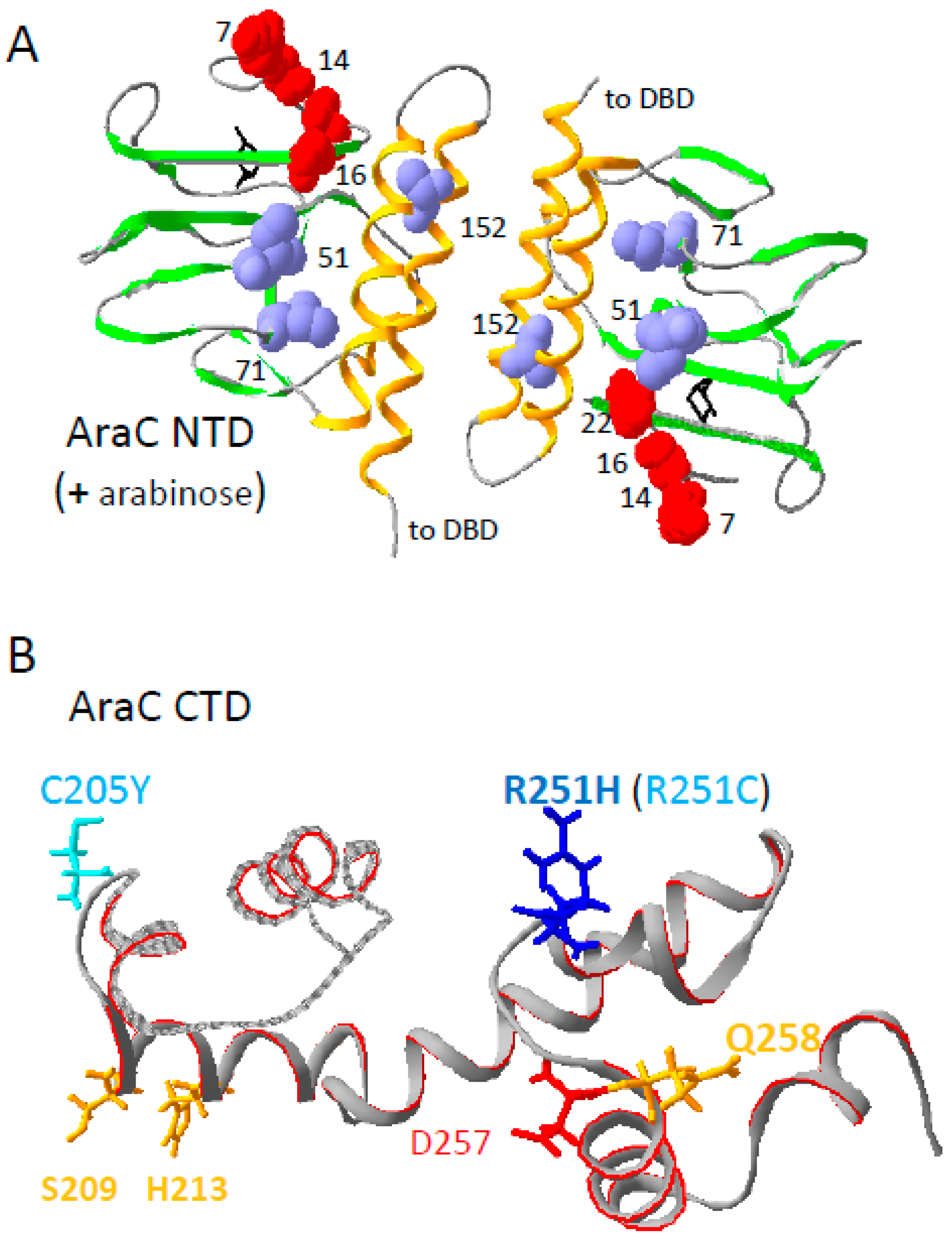
The positions of the araC mutations on the structure of the AraC N-terminal domain [54] are shown in Figure 11A. None of the affected residues are directly involved in arabinose binding. Arabinose constitutive mutations have been isolated in the N-terminal domain of araC [41,42,55]. The S14L, N16S, G22D, and A152V mutations confer a constitutive phenotype, with A152V being the most efficient. In contrast, D7N and I71V are devoid of activity in the absence of arabinose but allow efficient induction (Figure 5 and Figure 8). Thus, an arabinose constitutive phenotype is not required for rpoA suppression.
Several residues identified here must affect the conformation of the AraC-NTD. I51 and I71 are in the core of the N-terminal domain, on the side of the β-sheet that faces the two α-helices promoting dimerization, with A152 being part of one of these helices. The other residues (D7, S14, and N16) are in the arm of the AraC-NTD, and their lateral chains face the outside of the N-terminal domain. Although it is possible that these residues contact the α-CTD, the major argument against a direct interaction of the flexible arm with the α-CTD lies in the absence of allele specificity between pairs of araC and rpoA mutant alleles. Therefore, it appears more likely that the araC mutations exert an indirect effect and generate a conformation that improves the functional interaction of the α-CTD with the AraC-NTD.
Alternatively, the AraC-NTD could interact with the RNA polymerase core. If this were the case, the activity of the AraC mutant proteins would be maintained in the presence of an RpoA protein lacking its C-terminal domain. Since this deletion is not viable, we expressed the RpoA deletion protein in strains carrying the rpoA+ gene on the chromosome. Under these conditions, the activity of wild-type and mutant AraC proteins was inhibited to the same extent (Figure 9 and Supplementary Figure S4). These results indicate that a putative interaction of the AraC-NTD with the RNA polymerase core does not participate in the suppression of the RpoA defects by the mutant AraC proteins. In agreement with this notion, we did not isolate RNA polymerase mutants, outside of the α-CTD, that selectively decrease PBAD transcription mediated by mutant AraC.
The suppressors that localize to the AraC-NTD suggest a new function for this domain. In the absence of arabinose, the arm domain binds to the DNA-binding domain to ensure DNA looping and PBAD repression. In the presence of arabinose, the arm domain folds on top of the inducer binding pocket, and one of the arm residues, F15, contacts arabinose [41]. In a third function, the AraC-NTD could functionally interact with the RpoA α-CTD to promote efficient transcription at the PBAD promoter, particularly in the absence of Crp. This genetic approach may be easily extended to other activators that interact with the RpoA C-terminal domain, a hub for transcriptional activation.
The first evidence that the α-CTD domain binds DNA was observed with the UP element, upstream of the rrnB promoter [15]. R265 is a critical residue for DNA binding, along with several other residues, including N268 [14,20,48]. A crystal structure of a DNA-Crp-α-CTD complex showed that R265 and N268, together with other RpoA residues, bind to the DNA backbone and to the minor groove [14]. The RpoA N268D substitution decreased lacZ expression [56], while the N268A, which cannot complement an rpoA ts allele [48], and W substitutions abolished activation by an UP element [57]. The R265A and N268A substitutions decreased rhaBAD activation by RhaS [58]. The N268T substitution described here abolishes PBAD promoter activity, both in the presence and absence of Crp (Figure 2B and Figure 3B). Unlike the K271 and A272 substitutions, this viable mutation affects at least one more gene than cysA, as it prevented growth on minimal media in the presence of Cys and Met.
Transcription activation at PBAD has been proposed to involve the interaction of one of the α-CTD with Crp, and it has been suggested that the other α-CTD could bind to the DNA-binding domain of AraC and to a DNA sequence element between the Crp and AraC binding sites [3] (Figure 1). We did not identify any mutation in the regulatory region that would have defined a DNA site responsible for α-CTD binding, possibly because the target size is small, because a single base pair change would not compensate for the drastic effect of the RpoA N268T change or because it could interfere with AraC binding to I1. Nevertheless, the effect of this change and the role of this residue in DNA binding at other promoters indicate that an α-CTD–DNA interaction could be critical for transcription at the PBAD promoter. The isolation of mutants in the C-terminal DNA-binding domain of AraC that suppress the RpoA N268T allele provides the first genetic evidence to supports this model (Figure 11B). The activity of the R251H mutant in a rpoA+ Δcrp strain strongly suggests that the AraC-CTD could functionally interact with one α-CTD. The synergistic effect of mutations in the NTD and CTD of AraC is compatible with the notion that each domain could function independently to activate transcription.
Figure 11. Distribution of the AraC suppressors on the structures of the AraC N-terminal domain dimer (PDB 2ARC) [54] and of the C-terminal DNA-binding domain (PDB 2K9S) [59]. The images were generated with Swiss-PdbViewer v4.1.0. The domains are represented as ribbons. (A) The two large α helices that promote NTD dimerization are shown in orange and the β strands are in green. N-terminal arm D7, S14, N16, and G22 residues are shown in red and space-filling. The side chains of the internal residues I51, I71, and A152 are shown in blue and space-filling. The bound arabinose inducers are shown in black. (B) The two residues mutated in suppressors of rpoA N268T are shown in blue; R251H has the strongest phenotype and is shown in dark blue. Mutations of D257 (red) abolish DNA binding while mutations of the three other residues (orange) are defective in DNA binding.
Microorganisms 12 01928 g011

Supplementary Materials

The following supporting information can be downloaded at: https://www.mdpi.com/article/10.3390/microorganisms12091928/s1, Figure S1: PBAD activity mediated by wild type AraC+ and S14L (A,B) or G22D (C,D). Plasmids expressing AraC+ or the indicated AraC suppressor proteins were introduced in crp+ (A,C) or Δcrp (B,D) strains with the indicated rpoA alleles. Cultures were assayed after a 1h induction with arabinose; Figure S2: Effects of the AraC suppressors on the two residues that flank K271 of the RpoA α-CTD. Plasmids expressing AraC+ or the indicated AraC suppressor proteins were introduced in crp+ strains carrying the rpoA+ or the three indicated rpoA alleles. Cultures were assayed after a 1h induction with arabinose; Figure S3: PBAD activity mediated by araC suppressors of RpoA A272E. Plasmids expressing AraC+ or the indicated AraC suppressor proteins were introduced in Δcrp strains expressing either the RpoA K271E or the K271V proteins. Cultures were assayed after a 1h induction with arabinose; Figure S4: High expression of the rpoA α-CTD deletion strongly decreased expression from the araBAD promoter. RpoA and the RpoA α-CTD deletion were expressed from the bacteriophage T4 32 promoter of pRDB8 [43]. Black bars: pRDB8-rpoA+; white bars: pRDB8-rpoAdelCTD. PhoA expression was measured in a rpoA+ Δcrp strain. With AraC+, the expression of PhoA was barely detectable with both plasmids. The expression of lacZ was measured in the HfrH strain.

Author Contributions

Conceptualization, D.B. and J.C.; formal analysis, D.B.; investigations, F.S. and D.B.; writing, D.B. All authors have read and agreed to the published version of the manuscript.

Funding

The work was supported by the Swiss National Foundation, the Canton de Genève, and the CPG Research Foundation, grant number 3100-068157.02.

Data Availability Statement

The original contributions presented in the study are included in the article/Supplementary Materials, further inquiries can be directed to the corresponding author/s.

Acknowledgments

We thank Sandrine Bost, Corinne Chauffat, Giuseppe Plaia and Nessim Kaufman for the isolation of rpoA mutants, and Sylvie Trichard and Halil Bagci for performing some of the PhoA assays. We also thank Joseph Curran, Costa Georgopoulos, Gael Panis, Robert Schleif, David Shore, Michel Strubin, and Benjamin Weiss for discussions and critical, constructive reading of the manuscript. D.B. thanks Joseph Curran for his kind hospitality.

Conflicts of Interest

The authors declare no conflict of interest.

Abbreviations

α-CTDC-terminal domain of RpoA
α–NTDN-terminal domain of RpoA
AraC-NTDN-terminal domain of AraC

References

  1. Englesberg, E.; Irr, J.; Power, J.; Lee, N. Positive control of enzyme synthesis by gene C in the L-arabinose system. J. Bacteriol. 1965, 90, 946–957. [Google Scholar] [CrossRef] [PubMed]
  2. Cole, S.D.; Schleif, R. A new and unexpected domain-domain interaction in the AraC protein. Proteins 2012, 80, 1465–1475. [Google Scholar] [CrossRef] [PubMed]
  3. Schleif, R. Regulation of the L-arabinose operon of Escherichia coli. Trends Genet. 2000, 16, 559–565. [Google Scholar] [CrossRef] [PubMed]
  4. Schleif, R. AraC protein: A love-hate relationship. BioEssays News Rev. Mol. Cell. Dev. Biol. 2003, 25, 274–282. [Google Scholar] [CrossRef] [PubMed]
  5. Zhang, X.; Reeder, T.; Schleif, R. Transcription activation parameters at ara pBAD. J. Mol. Biol. 1996, 258, 14–24. [Google Scholar] [CrossRef] [PubMed]
  6. Hahn, S.; Dunn, T.; Schleif, R. Upstream repression and CRP stimulation of the Escherichia coli L-arabinose operon. J. Mol. Biol. 1984, 180, 61–72. [Google Scholar] [CrossRef] [PubMed]
  7. Zhang, X.; Schleif, R. Catabolite gene activator protein mutations affecting activity of the araBAD promoter. J. Bacteriol. 1998, 180, 195–200. [Google Scholar] [CrossRef] [PubMed]
  8. Busby, S.; Ebright, R.H. Transcription activation by catabolite activator protein (CAP). J. Mol. Biol. 1999, 293, 199–213. [Google Scholar] [CrossRef] [PubMed]
  9. Guzman, L.M.; Belin, D.; Carson, M.J.; Beckwith, J. Tight regulation, modulation, and high-level expression by vectors containing the arabinose PBAD promoter. J. Bacteriol. 1995, 177, 4121–4130. [Google Scholar] [CrossRef]
  10. Kimura, M.; Fujita, N.; Ishihama, A. Functional map of the alpha subunit of Escherichia coli RNA polymerase. Deletion analysis of the amino-terminal assembly domain. J. Mol. Biol. 1994, 242, 107–115. [Google Scholar] [CrossRef]
  11. Zou, C.; Thomas, M.S.; Keen, J.; Glass, R.E. A nested set of C-terminal deletions of the alpha subunit of Escherichia coli RNA polymerase define regions concerned with assembly, proteolysis, stabilization and transcriptional activation in vivo. Genes Cells 1997, 2, 81–94. [Google Scholar] [CrossRef] [PubMed]
  12. Russo, F.D.; Silhavy, T.J. Alpha: The Cinderella subunit of RNA polymerase. J. Biol. Chem. 1992, 267, 14515–14518. [Google Scholar] [CrossRef] [PubMed]
  13. Lloyd, G.S.; Niu, W.; Tebbutt, J.; Ebright, R.H.; Busby, S.J. Requirement for two copies of RNA polymerase alpha subunit C-terminal domain for synergistic transcription activation at complex bacterial promoters. Genes Dev. 2002, 16, 2557–2565. [Google Scholar] [CrossRef] [PubMed]
  14. Benoff, B.; Yang, H.; Lawson, C.L.; Parkinson, G.; Liu, J.; Blatter, E.; Ebright, Y.W.; Berman, H.M.; Ebright, R. Structural basis of transcription activation: The CAP-alpha CTD-DNA complex. Science 2002, 297, 1562–1566. [Google Scholar] [CrossRef] [PubMed]
  15. Ross, W.; Gosink, K.K.; Salomon, J.; Igarashi, K.; Zou, C.; Ishihama, A.; Severinov, K.; Gourse, R.L. A third recognition element in bacterial promoters: DNA binding by the alpha subunit of RNA polymerase. Science 1993, 262, 1407–1413. [Google Scholar] [CrossRef] [PubMed]
  16. Gourse, R.L.; Ross, W.; Gaal, T. UPs and downs in bacterial transcription initiation: The role of the alpha subunit of RNA polymerase in promoter recognition. Mol. Microbiol. 2000, 37, 687–695. [Google Scholar] [CrossRef] [PubMed]
  17. Jeon, Y.H.; Negishi, T.; Shirakawa, M.; Yamazaki, T.; Fujita, N.; Ishihama, A.; Kyogoku, Y. Solution structure of the activator contact domain of the RNA polymerase alpha subunit. Science 1995, 270, 1495–1497. [Google Scholar] [CrossRef] [PubMed]
  18. Lara-Gonzalez, S.; Birktoft, J.J.; Lawson, C.L. Structure of the Escherichia coli RNA polymerase alpha subunit C-terminal domain. Acta Crystallogr. D Biol. Crystallogr. 2010, 66, 806–812. [Google Scholar] [CrossRef] [PubMed]
  19. McLeod, S.M.; Aiyar, S.E.; Gourse, R.L.; Johnson, R.C. The C-terminal domains of the RNA polymerase alpha subunits: Contact site with Fis and localization during co-activation with CRP at the Escherichia coli proP P2 promoter. J. Mol. Biol. 2002, 316, 517–529. [Google Scholar] [CrossRef] [PubMed]
  20. Liu, B.; Hong, C.; Huang, R.K.; Yu, Z.; Steitz, T.A. Structural basis of bacterial transcription activation. Science 2017, 358, 947–951. [Google Scholar] [CrossRef] [PubMed]
  21. Dhiman, A.; Schleif, R. Recognition of overlapping nucleotides by AraC and the sigma subunit of RNA polymerase. J. Bacteriol. 2000, 182, 5076–5081. [Google Scholar] [CrossRef] [PubMed]
  22. Casadaban, M.J. Transposition and fusion of the lac genes to selected promoters in Escherichia coli using bacteriophage lambda and Mu. J. Mol. Biol. 1976, 104, 541–555. [Google Scholar] [CrossRef] [PubMed]
  23. Belin, D.; Plaia, G.; Boulfekhar, Y.; Silva, F. Escherichia coli SecG is required for residual export mediated by mutant signal sequences and for SecY-SecE complex stability. J. Bacteriol. 2015, 197, 542–552. [Google Scholar] [CrossRef] [PubMed]
  24. Baba, T.; Ara, T.; Hasegawa, M.; Takai, Y.; Okumura, Y.; Baba, M.; Datsenko, K.A.; Tomita, M.; Wanner, B.L.; Mori, H. Construction of Escherichia coli K-12 in-frame, single-gene knockout mutants: The Keio collection. Mol. Syst. Biol. 2006, 2, 2006.0008. [Google Scholar] [CrossRef] [PubMed]
  25. Miller, J.H. A Short Course in Bacterial Genetics; Cold Spring Harbor Laboratory Press: Cold Spring Harbor, NY, USA, 1992. [Google Scholar]
  26. Giffard, P.M.; Booth, I.R. The rpoA341 allele of Escherichia coli specifically impairs the transcription of a group of positively-regulated operons. Mol. Gen. Genet. 1988, 214, 148–152. [Google Scholar] [CrossRef] [PubMed]
  27. Plaia, G. Ruolo di SecG nel Trasporto Proteico in Escherichia coli e Studio di una Proteina Sconosciuta del Fago T4. Ph.D. Thesis, University of Geneva, Geneva, Switzerland, 2009. [Google Scholar]
  28. Bost, S.; Belin, D. A new genetic selection identifies essential residues in SecG, a component of the Escherichia coli protein export machinery. EMBO J. 1995, 14, 4412–4421. [Google Scholar] [CrossRef]
  29. Mattenberger, Y.; Silva, F.; Belin, D. 55.2, a phage T4 ORFan gene, encodes an inhibitor of Escherichia coli topoisomerase I and increases phage fitness. PLoS ONE 2015, 10, e0124309. [Google Scholar] [CrossRef]
  30. Benarafa, C.; Remold-O’Donnell, E. The ovalbumin serpins revisited: Perspective from the chicken genome of clade B serpin evolution in vertebrates. Proc. Natl. Acad. Sci. USA 2005, 102, 11367–11372. [Google Scholar] [CrossRef] [PubMed]
  31. Belin, D.; Guzman, L.M.; Bost, S.; Konakova, M.; Silva, F.; Beckwith, J. Functional activity of eukaryotic signal sequences in Escherichia coli: The ovalbumin family of serine protease inhibitors. J. Mol. Biol. 2004, 335, 437–453. [Google Scholar] [CrossRef] [PubMed]
  32. Belin, D. In vivo analysis of protein translocation to the Escherichia coli periplasm. Methods Mol. Biol. 2010, 619, 103–116. [Google Scholar] [CrossRef] [PubMed]
  33. Khatib, K.; Belin, D. A novel class of secA alleles that exert a signal-sequence-dependent effect on protein export in Escherichia coli. Genetics 2002, 162, 1031–1043. [Google Scholar] [CrossRef] [PubMed]
  34. Thomas, M.S.; Glass, R.E. Escherichia coli rpoA mutation which impairs transcription of positively regulated systems. Mol. Microbiol. 1991, 5, 2719–2725. [Google Scholar] [CrossRef] [PubMed]
  35. Lochowska, A.; Iwanicka-Nowicka, R.; Zaim, J.; Witkowska-Zimny, M.; Bolewska, K.; Hryniewicz, M.M. Identification of activating region (AR) of Escherichia coli LysR-type transcription factor CysB and CysB contact site on RNA polymerase alpha subunit at the cysP promoter. Mol. Microbiol. 2004, 53, 791–806. [Google Scholar] [CrossRef] [PubMed]
  36. Martin, K.; Huo, L.; Schleif, R.F. The DNA loop model for ara repression: AraC protein occupies the proposed loop sites in vivo and repression-negative mutations lie in these same sites. Proc. Natl. Acad. Sci. USA 1986, 83, 3654–3658. [Google Scholar] [CrossRef] [PubMed]
  37. Englesberg, E.; Sheppard, D.; Squires, C.; Meronk, F., Jr. An analysis of “revertants” of a deletion mutant in the C gene of the L-arabinose gene complex in Escherichia coli B-r: Isolation of initiator constitutive mutants (Ic). J. Mol. Biol. 1969, 43, 281–298. [Google Scholar] [CrossRef] [PubMed]
  38. Cass, L.G.; Horwitz, A.H.; Wilcox, G. The araIc mutation in Escherichia coli B/r. J. Bacteriol. 1981, 146, 1098–1105. [Google Scholar] [CrossRef] [PubMed]
  39. Colome, J.; Wilcox, G.; Englesberg, E. Constitutive mutations in the controlling site region of the araBAD operon of Escherichia coli B/r that decrease sensitivity to catabolite repression. J. Bacteriol. 1977, 129, 948–958. [Google Scholar] [CrossRef] [PubMed]
  40. Shultzaberger, R.K.; Chen, Z.; Lewis, K.A.; Schneider, T.D. Anatomy of Escherichia coli sigma70 promoters. Nucleic Acids Res. 2007, 35, 771–788. [Google Scholar] [CrossRef] [PubMed]
  41. Ross, J.J.; Gryczynski, U.; Schleif, R. Mutational analysis of residue roles in AraC function. J. Mol. Biol. 2003, 328, 85–93. [Google Scholar] [CrossRef] [PubMed]
  42. Dirla, S.; Chien, J.Y.; Schleif, R. Constitutive mutations in the Escherichia coli AraC protein. J. Bacteriol. 2009, 191, 2668–2674. [Google Scholar] [CrossRef] [PubMed]
  43. Duvoisin, R.M.; Belin, D.; Krisch, H.M. A plasmid expression vector that permits stabilization of both mRNAs and proteins encoded by the cloned genes. Gene 1986, 45, 193–201. [Google Scholar] [CrossRef] [PubMed]
  44. Slauch, J.M.; Russo, F.D.; Silhavy, T.J. Suppressor mutations in rpoA suggest that OmpR controls transcription by direct interaction with the alpha subunit of RNA polymerase. J. Bacteriol. 1991, 173, 7501–7510. [Google Scholar] [CrossRef] [PubMed]
  45. Camakaris, H.; Yang, J.; Fujii, T.; Pittard, J. Activation by TyrR in Escherichia coli K-12 by Interaction between TyrR and the alpha-Subunit of RNA Polymerase. J. Bacteriol. 2021, 203, e0025221. [Google Scholar] [CrossRef] [PubMed]
  46. Murayama, S.; Ishikawa, S.; Chumsakul, O.; Ogasawara, N.; Oshima, T. The Role of alpha-CTD in the Genome-Wide Transcriptional Regulation of the Bacillus subtilis Cells. PLoS ONE 2015, 10, e0131588. [Google Scholar] [CrossRef] [PubMed]
  47. Hayward, R.S.; Igarashi, K.; Ishihama, A. Functional specialization within the alpha-subunit of Escherichia coli RNA polymerase. J. Mol. Biol. 1991, 221, 23–29. [Google Scholar] [CrossRef] [PubMed]
  48. Gaal, T.; Ross, W.; Blatter, E.E.; Tang, H.; Jia, X.; Krishnan, V.V.; Assa-Munt, N.; Ebright, R.H.; Gourse, R.L. DNA-binding determinants of the alpha subunit of RNA polymerase: Novel DNA-binding domain architecture. Genes Dev. 1996, 10, 16–26. [Google Scholar] [CrossRef] [PubMed]
  49. Aiyar, S.E.; McLeod, S.M.; Ross, W.; Hirvonen, C.A.; Thomas, M.S.; Johnson, R.C.; Gourse, R.L. Architecture of Fis-activated transcription complexes at the Escherichia coli rrnB P1 and rrnE P1 promoters. J. Mol. Biol. 2002, 316, 501–516. [Google Scholar] [CrossRef] [PubMed]
  50. Kedzierska, B.; Lee, D.J.; Wegrzyn, G.; Busby, S.J.; Thomas, M.S. Role of the RNA polymerase alpha subunits in CII-dependent activation of the bacteriophage lambda pE promoter: Identification of important residues and positioning of the alpha C-terminal domains. Nucleic Acids Res. 2004, 32, 834–841. [Google Scholar] [CrossRef] [PubMed]
  51. Kedzierska, B.; Szambowska, A.; Herman-Antosiewicz, A.; Lee, D.J.; Busby, S.J.; Wegrzyn, G.; Thomas, M. The C-terminal domain of the Escherichia coli RNA polymerase alpha subunit plays a role in the CI-dependent activation of the bacteriophage lambda pM promoter. Nucleic Acids Res. 2007, 35, 2311–2320. [Google Scholar] [CrossRef] [PubMed]
  52. Sharif, T.R.; Igo, M.M. Mutations in the alpha subunit of RNA polymerase that affect the regulation of porin gene transcription in Escherichia coli K-12. J. Bacteriol. 1993, 175, 5460–5468. [Google Scholar] [CrossRef] [PubMed]
  53. King, R.A.; Anders, D.L.; Christie, G.E. Site-directed mutagenesis of an amino acid residue in the bacteriophage P2 ogr protein implicated in interaction with Escherichia coli RNA polymerase. Mol. Microbiol. 1992, 6, 3313–3320. [Google Scholar] [CrossRef] [PubMed]
  54. Soisson, S.M.; MacDougall-Shackleton, B.; Schleif, R.; Wolberger, C. Structural basis for ligand-regulated oligomerization of AraC. Science 1997, 276, 421–425. [Google Scholar] [CrossRef] [PubMed]
  55. Saviola, B.; Seabold, R.; Schleif, R.F. Arm-domain interactions in AraC. J. Mol. Biol. 1998, 278, 539–548. [Google Scholar] [CrossRef] [PubMed]
  56. Zou, C.; Fujita, N.; Igarashi, K.; Ishihama, A. Mapping the cAMP receptor protein contact site on the alpha subunit of Escherichia coli RNA polymerase. Mol. Microbiol. 1992, 6, 2599–2605. [Google Scholar] [CrossRef] [PubMed]
  57. Murakami, K.; Fujita, N.; Ishihama, A. Transcription factor recognition surface on the RNA polymerase alpha subunit is involved in contact with the DNA enhancer element. EMBO J. 1996, 15, 4358–4367. [Google Scholar] [CrossRef] [PubMed]
  58. Holcroft, C.C.; Egan, S.M. Roles of cyclic AMP receptor protein and the carboxyl-terminal domain of the alpha subunit in transcription activation of the Escherichia coli rhaBAD operon. J. Bacteriol. 2000, 182, 3529–3535. [Google Scholar] [CrossRef] [PubMed]
  59. Rodgers, M.E.; Schleif, R. Solution structure of the DNA binding domain of AraC protein. Proteins 2009, 77, 202–208. [Google Scholar] [CrossRef] [PubMed]
Figure 1. Transcription activation at the arabinose PBAD promoter (adapted from [3]). The sequence represented extends from −120 to +10; the araC promoter and O2 site are not included. RNA polymerase (blue) binding to the promoter is helped by the proximal AraC monomer bound to the I2 site (position −35 to −51 from the start site) that overlaps the promoter −35 site. Each AraC monomer (green) consists of an N-terminal dimerization domain that binds arabinose (black circle) attached by a linker to the C-terminal DNA-binding domain. In the presence of arabinose, the AraC N-terminal arm (red) covers the sugar-binding pocket. The Crp dimer (yellow) binds to a distal site (position −83 to −104). One α-CTD domain of RpoA contacts the Crp domain (filled blue circle, solid line). A potential contact site for the other α-CTD domain (dotted blue circle, stippled blue line) is indicated.
Microorganisms 12 01928 g001
Figure 2. (A) The structure of the RpoA C-terminal domain as determined by NMR (PDB 1C00) [17]. The image was generated with Swiss-PdbViewer v4.1.0. The domain is represented as a ribbon, and the side chains of relevant residues are shown in space filling (residues 268–272). N268 is shown in grey, L270 in green, K271 in red and A272 in orange. (B) crp+ strains with the indicated rpoA alleles contain the psM74c plasmid expressing wild-type AraC and a Maspin::PhoA chimera. Cultures were induced for 1 h with arabinose.
Microorganisms 12 01928 g002
Figure 3. Residual activity of the PBAD promoter in the absence of Crp. (A) Plasmid psM74c or its derivative with a complete deletion of araC was introduced in rpoA+ crp+ and rpoA+ Δcrp strains. Cultures were assayed with or without arabinose induction. (B) The psM74c plasmid expressing wild-type AraC was introduced in Δcrp strains carrying the indicated rpoA alleles. Cultures were induced for 1 h with arabinose.
Microorganisms 12 01928 g003
Figure 4. Nucleotide sequence of the PBAD promoter. The positions of the −35 and −10 hexamers and the transcription start site are indicated. The three G to A mutations isolated in this study are shown in bold. Previously described promoter mutations are discussed in the text. The sequence consensus of the −35 and −10 hexamers [40] is compared to that of the PBAD promoter. The most conserved residues in the consensus are in bold, and the conserved residues in the PBAD promoter are underlined.
Microorganisms 12 01928 g004
Figure 5. Constitutive PBAD activity mediated by mutant AraC proteins in the absence of Crp. Plasmids expressing AraC+ or the indicated AraC suppressor proteins were introduced in rpoA+ Δcrp (A,C) or rpoA K271E Δcrp (B,D) strains. Cultures grown in the absence of arabinose were assayed to detect the constitutive activity of each AraC protein. Cultures induced for 1 h with arabinose were used to determine the full activity of each AraC protein.
Microorganisms 12 01928 g005
Figure 6. PBAD activity mediated by wild-type AraC+ and D7N (A,B), or A152V (C,D) mutant AraC proteins. Plasmids expressing AraC+ or the indicated AraC mutant proteins were introduced in crp+ (A,C) or Δcrp (B,D) strains with the indicated rpoA alleles. Cultures were assayed after a 1 h induction with arabinose.
Microorganisms 12 01928 g006
Figure 7. Effects of the AraC suppressors on the two residues that flank K271 of the RpoA α-CTD. Plasmids expressing AraC+ or the indicated AraC mutant proteins were introduced in Δcrp strains carrying the rpoA+ or the three indicated rpoA mutant alleles.
Microorganisms 12 01928 g007
Figure 8. PBAD activity mediated by araC suppressors of RpoA A272E. Plasmids expressing AraC+ or the indicated AraC suppressor proteins were introduced in the rpoA+ Δcrp strain. Cultures grown in the absence of arabinose were assayed to detect the constitutive activity of each AraC protein. Cultures induced for 1 h with arabinose were used to determine the full activity of each AraC protein.
Microorganisms 12 01928 g008
Figure 9. A deletion of the RpoA α-CTD decreases expression from the araBAD and lac promoters. (AD) Empty bars: pUC19 vector; grey bars: pUC-rpoA+; black bars: pUC-rpoAdelCTD. The PhoA and LacZ activities were normalized to those measured with the empty vector. PhoA expression was measured in rpoA+ Δcrp strains. PhoA units with the empty vector: 4.5 ± 1 (AraC+); 390 ± 60 (D7N); 188 ± 30 (I71V). Expression of lacZ was measured in the HfrH strain: LacZ units with the empty vector: 3530 ± 340. The normalized activities of the three AraC proteins in the presence of the pUC-rpoA+ (E) or the pUC-rpoAdelCTD (F) plasmid are directly compared.
Microorganisms 12 01928 g009
Figure 10. PBAD activity mediated by AraC R251H and AraC double mutants. Plasmids expressing AraC+ or the AraC R251H mutant were introduced into the rpoA N268T crp+ (A), rpoA+ Δcrp (B), or rpoA N268T Δcrp (C) strains. Plasmids expressing AraC+, AraC A152V, AraC R251H, or the double mutant A152V-R251H were introduced into the rpoA N268T crp+ strain (D). Plasmids expressing AraC+, AraC S14P, AraC R251H or the double mutant S14P-R251H were introduced into the rpoA K271I Δcrp strain (E). Cultures were assayed after a 1 h induction with arabinose.
Microorganisms 12 01928 g010
Table 1. ropA mutations that decrease expression from the PBAD promoter.
rpoA Substitutions
Toxic ProteinN268TL270F K271EK271IK271VK271TA272E
T4 vs.1 22 2
PAI2::PhoA 73212
T4 55.21
growth phenotypes
MC-melibioseWRWKKKW
min-glucosenoyesnonononono
+cys-Metnoyesyesyesyesyesyes
Disclaimer/Publisher’s Note: The statements, opinions and data contained in all publications are solely those of the individual author(s) and contributor(s) and not of MDPI and/or the editor(s). MDPI and/or the editor(s) disclaim responsibility for any injury to people or property resulting from any ideas, methods, instructions or products referred to in the content.